Kh 2230 1702 Pdf
User Manual: kh 2230
Open the PDF directly: View PDF ![]() .
.
Page Count: 5

Singha, Kh. Bharati et al. Int. Res. J. Pharm. 2013, 4 (3)
Page 215
INTERNATIONAL RESEARCH JOURNAL OF PHARMACY
www.irjponline.com ISSN 2230 – 8407
Research Article
IN VITRO PROPAGATION OF DIPTERIS WALLICHII (R.BR.) T. MOORE:
A HOPE FOR CONSERVATION OF AN ENDANGERED PTERIDOPHYTE
Singha, Kh. Bharati1*, Dutta Choudhury, Manabendra1 and Mazumder, Pranab Behari2
1Plant Tissue Culture Laboratory, Department of Life Science and Bioinformatics, Assam University, Silchar 788011, India
2Department of Biotechnology, Assam University, Silchar 788011, India
Email: bembharati@yahoo.co.in
Article Received on: 13/01/13 Revised on: 09/02/13 Approved for publication: 11/03/13
DOI: 10.7897/2230-8407.04346
IRJP is an official publication of Moksha Publishing House. Website: www.mokshaph.com
© All rights reserved.
ABSTRACT
The pteridophytes include the fern and fern allies which jointly create the necessary environment for human life on earth with other green plants. Dipteris
wallichii (R.Br.) T. Moore is a rare and endemic pteridophyte of North East India. It is also facing severe threats due to its habitat destruction. Tribals destroy
the plant in large numbers for preparing their wine drinking pipes. An in vitro protocol for micropropagation of D.wallichii Moore was carried out using
standard Murashige and Skoog medium for spore germination and further cell proliferation. It was observed that supplementation of phytohormones mainly
auxins (Indole-3-Acetic Acid) were favourable for sporophyte development from prothallus. Further growth and differentiation of the prothallus and
sporophyte were recorded for various hormone concentrations and combinations and was observed to be best in 0.15 mg/L IAA + 0.20 mg/L KIN.
Key words: Dipteris wallichii, gametophyte, in vitro, phytohormones , sporophyte.
INTRODUCTION
In vitro propagation or micropropagation has led to two
thrust areas of biotechnological application-
micropropagation of elite plants and production of plant
metabolites. It is estimated that there is a world market
potential of 15 billion US $ per annum for tissue culture
products1. Micropropagation is of specific importance in
conservation of depletion and valuable genotypes of
medicinal plants2.
The pteridophytes are the vascular plants that produce spores
rather than seeds. Most of the ferns grow as thick ground
cover and hence provide a good means to prevent soil erosion
and they invade easily into exposed areas. Pteridophytes by
virtue of their possessing great variety and fascinating foliage
have drawn the attention and admiration of horticulturists and
plant lovers for centuries. They are represented by about 305
genera, comprising more than 10,000 species all over the
world. About 191 genera and more than 1000 species are
reported from India. Medicinal value of pteridophytes is
known to man for more than 2000 years. Again, the North
East India has a rich vegetation of pteridophytes3,4,5,6. It is
worth mentioning that the southern part of Assam (i.e. Barak
valley) also has a rich vegetation of ferns and fern-allies.
The rare and economically important pteridophyte species
should be treated as our natural heritage and properly
conserved by ex situ conservation for the endemic and rare
pteridophytes species by collecting live specimens from the
wild and grow them in pots for propagation in Fernery7.
Ferns have worldwide distribution growing in all continents
except Antarctica and most islands favouring moist temperate
and tropical regions8. Ferns have been cultured in vitro to
study their growth, development or differentiation and to
achieve their micropropagation due to their ornamental value.
Several species of ferns have successfully been propagated
by this method9. Application of in vitro culture methods
could contribute to increase sporophyte production of those
desirable species. At the present time, a high number of
individual researches are oriented towards the propagation of
pteridophytes with a view to conserve their diversity.
The culture of tissue in ferns has been utilized as research
instrument for the study of the developing potentialities of
the leaf primordia ever since the early 1960’s10. The first
successes in the field of the intensive multiplication of plants
through in vitro techniques are cited around 1970, the fern
Nephrolepis exaltata bostoniensis being the first plant
micropropagated in vitro with a commercial purpose11. 157
million plants, i.e. 74% out of the total production of
micropropagated plants, have been ornamental species12. Out
of these, approximately 40 million plants have been pot
plants. Top of the list, with 17.8 million plants, is the fern
Nephrolepis13.
Dipteris wallichii (R. Br.) T. Moore belongs to the family
Dipteridaceae. The Dipteridaceae are a rather primitive
group of ferns related to the Gleicheniaceae and distant from
the Polypodiaceae, with the fossil record beginning in the
Upper Triassic. Dipteris is the only living genus of the family
Dipteridaceae while other genera, viz., Clatopteris,
Dictyophyllum and Camptopteris are now extinct14. There are
twelve species in the genus Dipteris and only two in India-
Dipteris wallichii (R. Br. ex Hook et Grev) T. Moore15,16 and
Dipteris conjugata Reinw17. The species D.wallichii was first
reported from Assam in Luckipoor18,19. Many works on the
systematic and distribution pattern of D.wallichii was
accomplished time to time with a mention of its distribution
from Barak valley in Assam14. D.wallichii is an endemic fern
to North Eastern region of Himalayas facing severe threats
due to its habitat destruction and has been categorized under
vulnerable category of the IUCN (2000). It survives in
extremely specialized habitat near the streams in small
isolated patches and is very distinct with its large, beautiful
bi-lobed fronds. The decoction of its rhizome is taken to cure
jaundice. Tribals destroy this plant in large numbers for
preparing their wine drinking pipes.
In spite of its critical status, no attempts for in vitro
conservation of the plant have been taken till date except the
studies on the spore germination, developmental pattern and
sexuality of gametophytes20. In the present paper, taking into
consideration its sporadic occurrence in specialized habitats
and its evolutionary importance, an attempt has been made to

Singha, Kh. Bharati et al. Int. Res. J. Pharm. 2013, 4 (3)
Page 216
culture spores of D.wallichii Moore in vitro in order to save
the plant from expected extinction. Therefore, in vitro culture
and subsequent regeneration of sporophytes from the spore
culture of the ferns will be very much helpful for mass
cultivation as well as screening of phytochemicals present in
the plant.
MATERIALS AND METHODS
Source of explant and sterilization: Mature sporophylls of
D.wallichii Moore were collected from Baghbahar area of
Cachar district, Southern Assam and were authenticated by
the Department of Life Science and Bioinformatics, Assam
University, Silchar. The spores were separated from air dried
sporophylls and stored in a glass (reagent) bottle under
refrigeration. Surface sterilization of spores was done with
35% (v/v) solution of Sodium hypochlorite (4% active
chlorine) for 30 minutes before filtering through sterile
(autoclaved) filter paper and washing several times with
sterile double distilled water. The spores were then inoculated
in nutrient medium with wet condition.
Preparation of culture medium: The nutrient medium used
for inoculation of spores is Murashige and Skoog (MS)21
medium. The pH of the medium was adjusted to 5.8. After
adjusting the pH, agar powder (0.8%) was mixed with the
medium and boiled until a clear frothing solution was
obtained and then poured in culture tubes and culture bottles
respectively. Autoclave was done at 15 lbs/sq. inch of
pressure for 20 minutes to make the medium free of micro-
organisms. The culture bottles and tubes containing the
nutrient medium were then allowed to cool for 24 hours in
the culture laboratory. The surface sterilized spores were
inoculated in the culture tubes and bottles under Laminar Air
Flow Cabinet to maintain the aseptic conditions. The cultured
spores were incubated in Culture Room of the Tissue Culture
Laboratory at 250C± 10C, under 16 hour photoperiod and 8
hour darkness.
Morphological characters studied
Prothallus structure, sporophyte length, fresh weight and dry
weight of sporophyte developed from the same prothallus
were studied. Germination percentage was also calculated
from the following formula for each observation.
Germination percentage (%) =
X 100
Effect of plant growth regulators
Auxins [Indole-3-acetic acid (IAA)] and Cytokinin [Kinetin
(KIN)] were the different plant growth regulators
(phytohormones) selected for study. Proportionate amount
from stock solutions of different growth regulators were
taken and the final volume of the medium was adjusted upto
the mark in the volumetric flask. Different concentration and
combinations of the growth regulators were tried and their
effect on the growth of sporophytes studied.
Table 1: Average Germination Percentage
Media no.
Media Composition
Germination percentage
MS 1
MS + 00 mg/L Sucrose + 0.0 mg/L IAA
50
MS 2
MS + 00 mg/L Sucrose + 0.1 mg/L IAA
11
MS 3
MS + 15 mg/L Sucrose + 0.1 mg/L IAA
17
MS 4
MS + 15 mg/L Sucrose + 0.2 mg/L IAA
12
MS 5
MS + 30 mg/L Sucrose + 0.1 mg/L IAA
15
MS 6
MS + 30 mg/L Sucrose + 0.2 mg/L IAA
10
Table 2: Growth of Gametophytes (Prothalli) in fresh weight after 3 Months (N =10)
Media no.
Media composition
Weight of prothallus (mg)
MS 7 (Control)
MS + 0.00 mg/L IAA
70.25
MS 8
MS + 0.10 mg/L IAA
62.38
MS 9
MS + 0.15 mg/L IAA
80.75
MS 10
MS + 0.20 mg/L IAA
55.49
MS 11
MS + 0.25 mg/L IAA
50.82
MS 12
MS + 0.30 mg/L IAA
48.67
Table 3: Growth of Sporophytes in terms of fresh and dry weight after 6 months
Media
Number
KIN
(mg/L)
IAA
(mg/L)
Fresh weight (g)
(Mean ± SE)
Dry weight (g)
(Mean ± SE)
MS 13(C)
0.00
0.00
0.998 ± 0.20
0.325 ± 0.19
MS 14
0.05
0.15
1.045 ± 0.26
0.279 ± 0.16
MS 15
0.10
0.15
1.925 ± 0.21
0.530 ± 0.20
MS 16
0.15
0.15
2.545 ± 0.18
0.980 ± 0.27
MS 17
0.20
0.15
3.125 ± 0.11*
1.002 ± 0.15*
MS 18
0.25
0.15
3.050 ± 0.13
0.925 ± 0.24
MS 19
0.30
0.15
2.970 ± 0.14
0.875 ± 0.18
*indicates significant differences at p< 0.05. Each concentration consisted of mean value of 3 replications and each replication of 10 culture bottles or flasks.
Table 4: Length of the Sporophytes after 3, 6, 8, 10 and 12 months
Media number
Months
Length of Sporophytes (cm)
MS 17
3
2.5
MS 17
6
3.2
MS 17
8
6.5
MS 17
10
7.2
MS 17
12
7.0
Singha, Kh. Bharati et al. Int. Res. J. Pharm. 2013, 4 (3)
Page 217
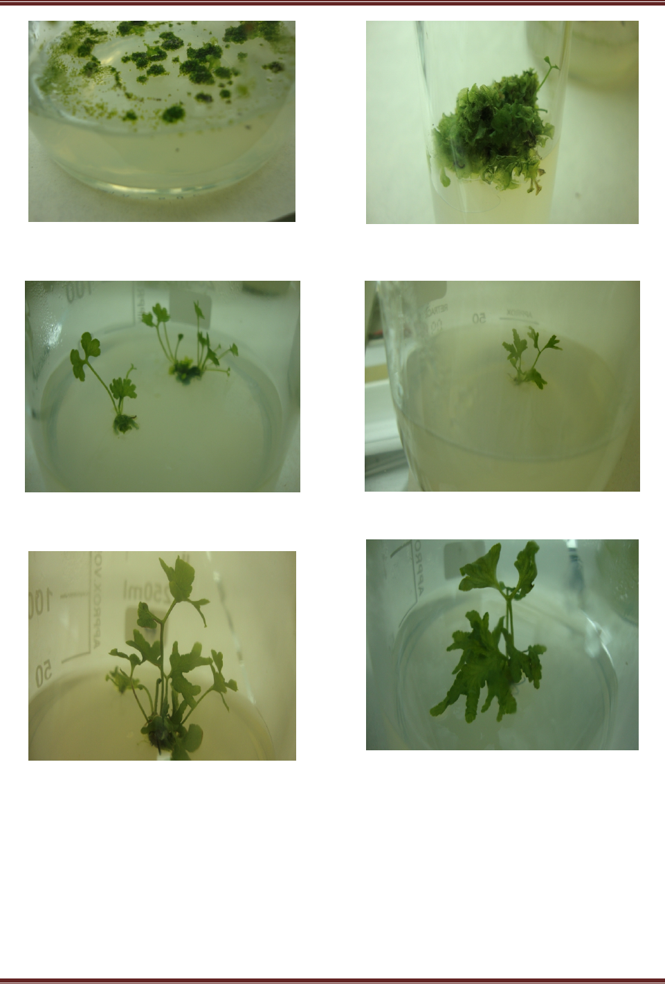
Figure1: Germinating spores and prothalli formation of D.wallichii
Moore in vitro from spores
Figure 2: Multiplication and differentiation of prothallus of D. wallichii
Moore to sporophytes in 3 months.
Figure 3: Developing sporophytes of D.wallichii Moore in MS 17 (4
months)
Figure 4: A 6 month old sporophyte of D.wallichii Moore in MS 17
Figure 5: Sporophyte of D.wallichii Moore in MS 17 (8 month)
Figure 6: A fully grown sporophyte of D.wallichii Moore in MS 17
(10 month)
RESULTS
Germination of spores: Spores of D.wallichii Moore were
found to germinate after 45 days from the day of inoculation.
The germination percentage was found to be maximum
(50%) in the medium supplemented without sucrose and
growth regulators (MS 1). Of all the germinated spores,
sporophytic bodies did not develop. This may be due to short
viability of the spores wherein delayed onset of ambient
conditions compels spore degeneration in total and the
admixed gametangia expression, where they compete for
their nutritional demands, failing which the developing
gametangia degenerate upto half of their numbers20.
Prothallus development: Spores germinate to produce
prothallus. After germination, the spores divide into
prothallial initial and rhizoidal initial. The prothallial initial
produce prothallus and rhizoidal initial produce unicellular
rhizoids. The prothallus is heart shaped structure and bears
antheridia (male sex organs) and archegonia (female sex

Singha, Kh. Bharati et al. Int. Res. J. Pharm. 2013, 4 (3)
Page 218
organs). The prothalli are long lived and become more or less
elongated with age.
Effect of growth regulators on the development of
gametophytes: Five concentrations of Indole-3-acetic acid
(IAA) viz., 0.10 mg/L, 0.15 mg/L, 0.20 mg/L, 0.25 mg/L and
0.30 mg/L were selected for culture of prothallus in MS
medium. The fresh weight (in mg) of the prothallus were
recorded after a period of 3 (three) months. It was observed
that MS 9 gave the maximum average growth of the
prothallus bearing gametophytes. Hence 0.15 mg/L of IAA
was considered as the optimum dose of IAA for the culture of
prothallus with different combination of growth regulators in
MS medium.
Development of sporophyte: Young sporophytic bodies
were found to develop from the prothallus (bearing both
gametophytic structures). The antheridia produce
spermatozoids and each archegonium produce one egg. The
spermatozoids fertilize the egg to produce diploid
sporophyte. Maximum growth of sporophytes was observed
in MS 17 followed by MS 18 and MS 19. It was observed
that increase in KIN concentration (keeping IAA
concentration constant) result in increase of fresh and dry
weight of the sporophytes22.
Development of root-like rhizoids: The young sporophytic
bodies in the culture bottles were observed to develop root-
like rhizoids. The maximum increase of plant height was
found to be 7.2 cm in MS 17. It may be due to maximum
growth of root-like rhizoids.
Hardening: The fully grown sporophytes were carefully
taken out from the culture bottles and washed gently with
sterile double distilled water to remove adhering agar
medium so that contamination does not occur. The
regenerated sporophytes were transferred to vermiculite (a
sterile inert medium) and hardened by covering them with a
thin perforated transparent polythene bag to maintain
humidity and sprayed with 1/10th strength MS salts solution
and maintained in the culture condition for acclimatization.
Finally, the sporophytes were transplanted to earthen pots
containing sterile dried moss, sand and brick bats in the ratio
of 1:1:1.
DISCUSSION
The main problem for establishment of in vitro propagation
protocols in pteridophytes is the presence of micro-organisms
in the plants, hindering the process of disinfection and
affecting their survival23. Spores have been used as the
explant source for successful high frequency regeneration of
plants. Although the regeneration of plants from spores is
quite difficult in vitro, but optimization of every step from
initiation to acclimatization makes it more feasible to produce
plants from spores in vitro24.
Sterilization of spores of D.wallichii Moore with 0.1%
Mercuric chloride for 2 to 3 minutes results in very poor
germination. But when treated with 35% (v/v) Sodium
hypochlorite solution (4% active chlorine) for half an hour,
better germination percentage was observed. Lowering the
percentage of Sodium hypochlorite and time period leads to
contamination by fungus and bacteria. So standardization of
the explants sterilant and time period by trial and error
method is essential for better germination. Harvested spores
lose their viability if stored for more than 6 months and
exhibit less germination percentage25. So it is better to use
fresh spores for germination.
Fern spores grow in sugar deficient medium containing
macro and micro nutrients and differentiate sex organs. When
supplemented with sucrose, contamination occurs and growth
regulators supplementation did not make much difference
(Table No.1). It was reported that Dyer and MS medium
supplemented with sucrose resulted in lower germination and
early gametophyte development of endangered fern
Dicksonia sellowiana25. High sucrose level in the culture
medium might result in chlorophyll degradation and reflect
on the frond development26. In vitro plant regeneration had
been successful for Cyathea lepifera in half strength MS
medium without sucrose27.
Germination percentage was found to be maximum (50%) for
spores of D.wallichii Moore in MS basal medium (MS 1). Of
all the germinated spores, sporophytic bodies did not
develop. This may be due to the fact that short viability of the
spores compels spore degeneration in total and the
developing admixed gametangia degenerate upto half of their
numbers because of nutritional demands. Also rhizomatous
propagation rather than spore germination (which is common
among most ferns) occurs in D.wallichii Moore and hence
localised in distribution.
After germination, all spores become green in colour and
were transferred to MS medium supplemented with growth
regulators. It was observed that MS medium supplemented
with 0.15 mg/L IAA (MS 9) supported best growth of
gametophytes in terms of fresh weight (80.75 mg) as shown
in Table No.2. Increasing concentration of IAA resulted in
decline of the fresh weight of the gametophytes.
Sporophytes developed from gametophytes in three months.
In order to study the combined action of IAA and KIN on the
developing sporophytes, six combinations were taken. Out of
which, 0.15 mg/L IAA + 0.20 mg/L KIN (MS 17) showed
better results in terms of fresh weight (3.125 g) of
sporophytes (Table No.3). Growth increased manifolds due to
combined action of IAA and KIN because auxins alone lead
to cell enlargement while KIN induces cell division in
presence of auxin28. Cytokinin is also helpful in lipid
metabolism to increase growth29.
Height of sporophytes of D.wallichii Moore were measured
in random. Maximum average height of the sporophytes (7.2
cm) were observed in MS 17 i.e. 0.20 mg/L KIN + 0.15 mg/L
IAA. After the complete sporophyte regeneration, hardening
was done and the sporophytes were gradually exposed to the
natural environment, where they exhibited 80% survivability.
The present study describes the requirements for in vitro
spore germination and gametophyte development of this
highly endangered species - Dipteris wallichii Moore. The
results may promote large scale cultivation to compensate its
depletion in nature.
REFERENCES
1. Govil S, Gupta SC. Plant Micropropagation Industry in India: retrospect
and prospect. J Scientific and Industrial Research 1994; 5(3):777-786.
2. Sen J, Sharma AK. Micropropagation of Withania somnifera from
germinating seeds and shoot tips. Plant Cell Tissue and Organ Cult
1991; 26:71-73. http://dx.doi.org/10.1007/BF00036108
3. Kachroo P. Ferns of Assam. J Asiatic Soc Sci 1935; 19:161-174.
4. Baishya AK, Rao RR. Fern and Fern-allies of Meghalaya State, India,
Scientific Publisher, Jodhpur; 1982.
5. Borthakur SK, Deka P, Nath RK. In: Singh B, Singh MP, Eds.
Illustrated Manual of Ferns of Assam; 1999.
6. Singh S, Panigrahi G. In: Singh B, Singh MP, Eds. Ferns and fern-allies
of Arunachal Pradesh, Dehradun; 2005.
7. Amoroso VB. Pteridophyte and gymnosperm diversity in Musuan,
Bukidnon, Philippine. J Systematic Biol 2006; 1:1-11.

Singha, Kh. Bharati et al. Int. Res. J. Pharm. 2013, 4 (3)
Page 219
8. Burkhill IH. A Dictionary of the Economic Products of Malay
Peninsular, Ministry of Agriculture Malaysia; 1935. p.1542.
9. Hartman RD, Zettler FW. Tissue culture as a plant production system
for foliage plants. In: Zimmerman RH, Griesback RJ, Hamnerschlag FA,
Lawson RH, Eds. Tissue Culture as a Plant Production System for
Horticultural Crops, Martinus Nijhof Publishers, Dordrecht; 1986. p.
293-299. http://dx.doi.org/10.1007/978-94-009-4444-2_25
10. Torres KC. Tissue Culture Techniques for Horticultural Crops. Van
Nostrand Reinhold, New York; 1988. http://dx.doi.org/10.1007/978-1-
4615-9756-8
11. Cachita-Cosma D. Metode in vitro la plantele de cultura-baze teoretice
si practice, Edit. Ceres, Bucuresti; 1988. p. 274.
12. Pierik RML. Commercial aspects of micropropagation. In: Prakash J,
Pierik, RML, Eds. Horticulture-New Technologies and Applications,
Kluwer Academic Publishers, Dordrecht, the Netherlands; 1991. p. 141-
153. http://dx.doi.org/10.1007/978-94-011-3176-6_23
13. Fernandaz H, Revilla MA. In vitro culture of ornamental ferns. Plant
Cell Tissue and Organ Cult 2003; 73:1-13. http://dx.doi.org/10.1023/
A:1022650701341
14. Dutta Choudhury M, Das PS. Ecology and distribution pattern of
Dipteris wallichii (R.Br.) Moore in Southern Assam, NE India. Indian
Fern J 2005; 22:124-127.
15. Dixit RD. A Census of Indian Pteridophytes. Director Botanical Survey
of India Howrah; 1984. PMCid:1153614
16. Fraser-Jenkins CR. In: Singh B, Singh MP, Eds. Taxonomic revision of
three hundred Indian subcontinental Pteridophytes with a revised
census-list. Dehradun, India; 2008.
17. Satija CK, Bir SS. Polypodiaceous ferns of India. In: Bir SS, Ed.
Aspects of Plant Sciences. Today & Tomorrow's Printers and Publishers,
New Delhi; 1985. PMCid:3011173
18. Hooker JD, Thompson T. Flora Indica, London; 1855. http://
dx.doi.org/10.5962/bhl.title.50109
19. Beddome RH. Handbook to the ferns of British India, Ceylon and
Malaya Peninsula. Thacker Spinc. & Co., Calcutta; 1883.
20. Behera SK, Rawat VK, Singh AP, Khare PB. Spore germination,
developmental pattern and sexuality of gametophytes in Dipteris
wallichii (R. Br. ex Hook. et Grev.) T. Moore. Indian Fern J 2011; 28(1-
2):172-178.
21. Murashige T, Skoog F. A revised medium for rapid growth and
bioassays with tobacco tissue cultures. Physiologia Plantarum 1962;
15:473-497. http://dx.doi.org/10.1111/j.1399-3054.1962.tb08052.x
22. Mazumder B, Dutta Choudhury M, Mazumder PB. Effect of growth
regulators on in vitro propagation of Bolbitis costata (Wall ex. Hook.) C.
Chr., Assam University J Sci and Tech: Biol and Environ Sci 2010;
5:23-33.
23. Morini S. In vitro culture of Osmunda regalis fern. J Hortic Sci and
Biotech 2000; 75:31-34.
24. Banks JA. Gametophyte development in Ferns. Annual Rev of Plant
Physiol1999;50:163-186. http://dx.doi.org/10.1146/annurev.arplant.
50.1.163 PMid:15012207
25. Renner GDR, Randi AM. Effects of sucrose and irradiance on
germination and early gametophyte growth of the endangered tree fern
Dicksonia sellowiana Hook (Dicksoniaceae). Acta Bot Bras 2004;
18:375-380. http://dx.doi.org/10.1590/S0102-33062004000200017
26. Fernandez H, Bertrand AM, Sanchez-Tames R. Biological and
nutritional aspects involved in fern multiplication. Plant Cell Tissue and
OrganCult1999;56:211-214.http://dx.doi.org/10.1023/A: 1006277229
136
27. Kuriyama A. Kobayashi T. & Maeda M. Production of sporophytic
plants of Cyathea lepifera, a tree fern, from cultured gametophytes. J Jap
Soc Hortic Sci 2004; 73:140-142. http://dx.doi.org/10.2503/jjshs.73.140
28. Steward FC, Shantz EM. The chemical induction of growth in plant
tissue culture. In: Wren RL, Wightman F, Eds. The chemistry and mode
of action of plant growth substances; 1955. p. 165-185.
29. Manning JC, Van Staden J. The development and mobilization of seed
reserves in some African Orchids. Aust J Bot 1987; 35:343-353.
http://dx.doi.org/10.1071/BT9870343
Cite this article as:
Singha, Kh. Bharati, Dutta Choudhury, Manabendra and Mazumder, Pranab
Behari. In vitro propagation of Dipteris wallichii (R.Br.) T. Moore: A hope
for conservation of an endangered pteridophyte. Int. Res. J. Pharm. 2013;
4(3):215-219
Source of support: Nil, Conflict of interest: None Declared
