2021 NHSN Patient Safety Component Manual
File info: application/pdf · 428 pages · 8.09MB
2021 NHSN Patient Safety Component Manual
PSC, Manual
NHSN
Please Note: The NHSN Patient Safety Component Manual is updated annually based on subject matter expert review and user feedback. Over time, certain chapters have been...
PDF 2021 NHSN Patient Safety Component Manual
Please Note: The NHSN Patient Safety Component Manual is updated annually based on subject matter expert review and user feedback. Over time, certain chapters have been retired or moved to other components. To avoid con…
2021 NHSN Patient Safety Component Manual - CDC
Jan 3, 2021 — Chapter 15: CDC Locations and Descriptions and Instructions for Mapping ... Instructions and standardized surveillance methods and definitions for ... https:// cdn.ymaws.com/www.cste.org/resource/resmgr/PS…
Extracted Text
January 2021
National Healthcare Safety Network (NHSN)
Patient Safety Component Manual
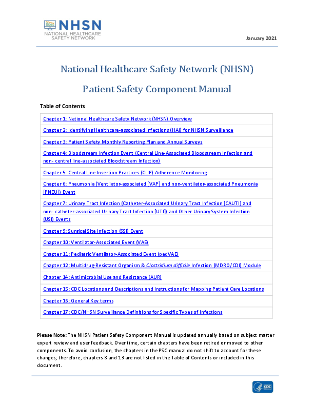
Table of Contents Chapter 1: National Healthcare Safety Network (NHSN) Overview Chapter 2: Identifying Healthcare-associated Infections (HAI) for NHSN Surveillance Chapter 3: Patient Safety Monthly Reporting Plan and Annual Surveys Chapter 4: Bloodstream Infection Event (Central Line-Associated Bloodstream Infection and non- central line-associated Bloodstream Infection) Chapter 5: Central Line Insertion Practices (CLIP) Adherence Monitoring Chapter 6: Pneumonia (Ventilator-associated [VAP] and non-ventilator-associated Pneumonia [PNEU]) Event Chapter 7: Urinary Tract Infection (Catheter-Associated Urinary Tract Infection [CAUTI] and non- catheter-associated Urinary Tract Infection [UTI]) and Other Urinary System Infection (USI) Events Chapter 9: Surgical Site Infection (SSI) Event Chapter 10: Ventilator-Associated Event (VAE) Chapter 11: Pediatric Ventilator-Associated Event (pedVAE) Chapter 12: Multidrug-Resistant Organism & Clostridium difficile Infection (MDRO/CDI) Module Chapter 14: Antimicrobial Use and Resistance (AUR) Chapter 15: CDC Locations and Descriptions and Instructions for Mapping Patient Care Locations Chapter 16: General Key terms Chapter 17: CDC/NHSN Surveillance Definitions for Specific Types of Infections
Please Note: The NHSN Patient Safety Component Manual is updated annually based on subject matter expert review and user feedback. Over time, certain chapters have been retired or moved to other components. To avoid confusion, the chapters in the PSC manual do not shift to account for these changes; therefore, chapters 8 and 13 are not listed in the Table of Contents or included in this document.
January 2021
National Healthcare Safety Network (NHSN) Overview
The NHSN is a secure, Internet-based surveillance system that expands and integrates patient and healthcare personnel safety surveillance systems managed by the Division of Healthcare Quality Promotion (DHQP) at the Centers for Disease Control and Prevention. In addition, facilities that participate in certain reporting programs operated by the Centers for Medicare and Medicaid Services (CMS) can do so through use of NHSN. Furthermore, some U.S. states use NHSN as a means for healthcare facilities to submit data on healthcare-associated infections (HAIs) and transfusion-related adverse events mandated through their specific state legislation.
NHSN enables healthcare facilities to collect and use data about HAIs, adherence to clinical practices known to prevent HAIs, the incidence or prevalence of multidrug-resistant organisms within their organizations, trends and coverage of healthcare personnel safety and vaccination, and adverse events related to the transfusion of blood and blood products.
The NHSN includes six components: Patient Safety, Long-term Care Facility, Outpatient Dialysis, Healthcare Personnel Safety, Biovigilance, and Outpatient Procedure (Figure 1).
FIGURE 1: NHSN COMPONENTS
NHSN Components
Patient Safety
Long-term Care Facility
Outpatient Dialysis
Healthcare Personnel Safety
Biovigilance
Outpatient Procedure
1 - 1
January 2021
NHSN Overview
The Patient Safety Component includes five modules that focus on events associated with medical devices, surgical procedures, antimicrobial agents used during healthcare, multidrug resistant organisms, and Coronavirus Infectious Disease 2019 (COVID-19).
� Device-associated Module: o Bloodstream Infection (CLABSI � Central line-associated bloodstream infection) o Central line insertion practices (CLIP) adherence o Urinary Tract Infection (CAUTI � Catheter-associated urinary tract infection) o Pediatric Ventilator-associated events (PedVAE) (NICU and pediatric locations only) o Ventilator-associated events (VAE) (adult locations only) o Pneumonia (VAP � Ventilator-associated pneumonia) - in pediatric locations (in-plan* or off-plan*), or NICU and adult locations (off-plan* only)
� Procedure-associated Module: o Surgical Site Infection (SSI)
� Antimicrobial Use and Resistance Module (AUR) � Multidrug-Resistant Organism and Clostridium difficile Infection (MDRO/CDI) Module � Coronavirus Infectious Disease 2019 (COVID-19) Module (off plan* only):
o Patient Impact and Hospital Capacity Pathway o Healthcare Worker Staffing Pathway o Healthcare Supply Pathway
*Note: "In-plan" surveillance means that the facility has committed to following the NHSN surveillance protocol, in its entirety, for that particular event, as shown in the facility's NHSN monthly reporting plan. "Off-plan" surveillance is surveillance that is done because a facility has decided to track a particular event for internal use. Data that are entered into NHSN "off-plan" are not included in NSHN annual reports or other NHSN publications. A facility makes no commitment to follow the NHSN protocol for "off-plan" events. Further, "off-plan" data cannot be uploaded into NHSN via Clinical Document Architecture (CDA) and must be manually entered. Instructions and standardized surveillance methods and definitions for each module of the Patient Safety Component are provided in this manual and on the NHSN website (www.cdc.gov/nhsn). Modules may be used singly or simultaneously.
The NHSN Long-term Care Facility Component provides long-term care facilities (LTCFs) with standardized surveillance methods and definitions for four modules: (1) Multidrug resistant organism (MDRO) and Clostridioides difficile Infection (CDI) laboratory-identified (LabID) Events; (2) Urinary Tract Infections (UTI); (3) Prevention Process Measures 4) Coronavirus Infectious Disease (COVID-19). The component is ideal for use by nursing homes, skilled nursing facilities, chronic care facilities, and assisted living and residential care facilities. LTCF surveillance protocols, training materials, data collection forms, instructions, and other supporting materials are provided on the Long-term Care Facility Component website: https://www.cdc.gov/nhsn/ltc/index.html.
Outpatient hemodialysis centers have several surveillance options tailored to their patients and setting in the Dialysis Component. The component consists of 3 modules: 1) Dialysis Event; (2) Prevention Process Measures; and (3) Dialysis Patient Influenza Vaccination. Facilities that treat hemodialysis outpatients should refer to the Dialysis Component instructions and standardized surveillance methods and definitions at www.cdc.gov/nhsn/dialysis/index.html.
1 - 2
January 2021
NHSN Overview
There are two modules in the Healthcare Personnel Safety (HPS) Component of NHSN: The Healthcare Personnel Exposure Module and the Healthcare Personnel Vaccination Module. The Healthcare Personnel Exposure Module includes: Blood/Body Fluid Exposure Only; Blood/Body Fluid Exposure with Exposure Management; and Influenza Exposure Management. This module is no longer available for enrollment and should only be used by facilities that have already been reporting Blood/Body Fluid Exposure and Exposure Management data to the system. The Healthcare Personnel Vaccination Module includes: Influenza Vaccination Summary. Data collected in this surveillance system can assist healthcare facilities, health systems, and public health agencies to monitor and report trends in blood/body fluid exposures, to characterize antiviral medication use for exposures to influenza, and to monitor influenza vaccination coverage among healthcare personnel. These modules may be used separately or simultaneously. Instructions and standardized surveillance methods and definitions for the Healthcare Personnel Vaccination Module is provided in the NHSN Manual: HPS Component Protocol https://www.cdc.gov/nhsn/pdfs/hps-manual/vaccination/hps-flu-vaccine-protocol.pdf
The NHSN Biovigilance Component, Hemovigilance Module facilitates national surveillance of transfusion-related recipient adverse events. The Hemovigilance Module is designed for transfusion service staff to collect data on annual facility and transfusion service characteristics, individual reports on adverse transfusion reactions, errors or accidents associated with adverse reactions, and monthly counts of transfused or discarded components. The Hemovigilance Module surveillance protocol, training materials, data collection forms, instructions, and other supporting materials are provided on the Hemovigilance Module website: www.cdc.gov/nhsn/acute-care-hospital/bio-hemo/index.html.
The Outpatient Procedure Component (OPC) includes two modules that focus on adverse events associated with surgical procedures performed in Ambulatory Surgery Centers (ASCs). The two modules include Same Day Outcome Measures and Surgical Site Infections.
� Same Day Outcome Measures (OPC-SDOM) are a grouping of outpatient care quality indicators that represent a broad range of risks encountered by patients accessing care in various outpatient settings. The four individual outcome measures are: o Patient Burn o Patient Fall o Wrong Site, Wrong Side, Wrong Patient, Wrong Procedure, Wrong Implant o All-Cause Hospital Transfer/Admission
� Surgical Site Infection (OPC-SSI) - SSI surveillance for outpatient operative procedures using the Outpatient Procedure Component (OPC) replaces the use of the Patient Safety Component SSI event chapter for ASCs.
The OPC surveillance protocols, training materials, data collection forms, instructions, and other supporting materials are provided on the Outpatient Procedure Component website: https://www.cdc.gov/nhsn/ambulatory-surgery/index.html.
1 - 3
January 2021
NHSN Overview
Surveillance Techniques
Some of the options in the following modules require active, patient-based, prospective surveillance of events and their corresponding denominator data by a trained Infection Preventionist (IP). This means that the IP shall seek out infections during a patient's stay by screening a variety of data sources, such as laboratory, pharmacy, admission/discharge/transfer, radiology/imaging, and pathology databases, as well as patient charts, including history and physical exam notes, nurses'/physicians' notes, temperature charts, etc. Others may be trained to screen data sources for these infections, but the IP must make the final determination. Laboratory-based surveillance should not be used alone, unless all possible criteria for identifying an infection are solely determined by laboratory evidence (for example, LabID event detection in the MDRO/CDI Module). Retrospective chart reviews should be used only when patients are discharged before all information can be gathered. NHSN forms should be used to collect all required data, using the NHSN definitions of each data field. To minimize the IP's data collection burden, others may be trained to collect the denominator data and process of care data (for example, central line insertion practices).
Procedure-Associated Module
Surgical site infection (SSI) monitoring is offered through this module. SSI surveillance requires active, patient-based, prospective surveillance techniques (see Surveillance Techniques above). To minimize IPs' workload of collecting denominator data, operating room data may be downloaded (see file specifications at: https://www.cdc.gov/nhsn/pdfs/ps-analysis-resources/ImportingProcedureData.pdf)
Both pre-discharge and post-discharge surveillance methods should be used to detect SSIs. Surveillance may include both inpatient and outpatient operative procedures. These methods include 1) direct examination of patients' wounds during hospitalization, or follow-up visits to either surgery clinics or physicians' offices, 2) review of medical records or surgery clinic patient records, 3) surgeon surveys by mail or telephone, and 4) patient surveys by mail or telephone (though patients may have a difficult time assessing their infections). Any combination of these methods is acceptable for use; however, CDC criteria for SSI must be applied.
Device-Associated Module
Medical instrumentation increases the risk of development of an HAI and most patients admitted for health care are exposed to some kind of medical device in the course of their treatment. Such devices include, but are not limited to, vascular and urinary catheters, and ventilators. NHSN enables facilities to monitor infectious complications associated with the use of these devices and to monitor processes related to their use which might increase infection risk. Specifically, surveillance of central line-associated bloodstream infection (CLABSI), catheter-associated urinary tract infection (CAUTI), ventilator-associated events (VAE), and/or ventilator-associated pneumonia (VAP) is possible using the NHSN. In addition, central line insertion practices (CLIP) can be monitored to inform facilities of the appropriateness of their processes and how they may relate to HAI development. See Dialysis Component for detailed instructions for Dialysis Event (DE) surveillance of hemodialysis outpatients (www.cdc.gov/nhsn/dialysis/index.html).
1 - 4
January 2021
NHSN Overview
Device-associated denominator data should be collected at the same time each day, or by weekly sampling methods, in certain locations, for CLABSI and CAUTI surveillance (see the CLABSI and CAUTI protocols for guidance). When denominator data are available from electronic databases (for example, ventilator days from respiratory therapy), these sources may be used as long as the counts are not substantially different (+/- 5%) from manually-collected counts that have been validated for a minimum of three months. See the respective device-associated event protocols for detailed surveillance instructions.
Antimicrobial Use and Resistance (AUR) Module
The use of antimicrobial agents has a direct effect on antimicrobial resistance patterns of pathogens. The observed increase in multidrug resistance is in part due to inappropriate prescription of, as well as only partial completion of courses of antibiotics.
The AUR Module allows facilities to collect information on the amount of antimicrobials that are used for patient care within their systems, as well as to collect data on the prevalence of drug-resistant organisms in their inpatient and outpatient areas. Electronic capture and reporting of microbiology and pharmacy data are the only available options for reporting data into this module.
See the Antimicrobial Use and Resistance protocol for detailed surveillance instructions.
Multidrug-resistant Organism and Clostridium difficile Infection (MDRO/CDI) Module
The NHSN MDRO/CDI Module offers a means for facilities to meet criteria and metrics that are outlined in several organizational guidelines to control and measure the spread of MDROs and CDI within their healthcare system. The module has two separate and independent reporting options, Laboratoryidentified (LabID) Event and Infection Surveillance that may be tailored to meet the needs of participating NHSN facilities.
In addition, the following process measures are available: (1) adherence to hand hygiene; (2) adherence to contact precautions when caring for patients infected or colonized with an MDRO or C. difficile; and (3) adherence to active surveillance testing (AST) of MRSA and/or VRE. Active surveillance testing outcome measures is also available in locations where AST adherence is being performed and enables facilities to use the results of AST to monitor the incidence and prevalence of positive MRSA and/or VRE cultures. See the MDRO/CDI protocol for detailed surveillance instructions.
Coronavirus Infectious Disease 2019 (COVID-19) Module
The NHSN Coronavirus Infectious Disease 2019 Module was created during the COVID-19 outbreak as a means to monitor associated impacts on the human and material resources of acute care facilities. The module allows a facility to report information on the hospital's capacity to respond to COVID-19 by reporting bed and mechanical ventilator availability as well as current and impending staffing and personal protective equipment shortages.
1 - 5
January 2021
Identifying Healthcare-associated Infections (HAI) for NHSN Surveillance
To standardize the classification of an infection as present on admission (POA) or a healthcare-associated infection (HAI), the following objective surveillance definitions and guidance are used for NHSN surveillance:
Table of Contents
General Instructions ................................................................................................................................................. 1 Infection Window Period.............................................................................................................................. 3 Date of Event (Event Date) ........................................................................................................................... 7
Present on Admission (POA) ......................................................................................................................8 Hospital Acquired Infection (HAI) ..............................................................................................................8 Location of Attribution (LOA)....................................................................................................................... ............9 Transfer Rule ............................................................................................................................................................10 Repeat Infection Timeframe....................................................................................................................... 11 Secondary BSI Attribution Period ............................................................................................................. 16 Secondary BSI Attribution Tables............................................................................................................17 Pathogen Assignment Guidance................................................................................................................. 18 Appendix....................................................................................................................................................................27
The intention of this approach is to align criteria and definitions and decrease subjectivity while maintaining epidemiologic standardization and clinical relevance. A variety of scenarios to include repeat infections of the same type, concurrent infections of differing types, and pathogen assignment in multipathogen infections are addressed. See Appendix Flow Diagram for NHSN Event Determination.
General Instructions
1. The guidance found in this Chapter is not applicable when performing SSI, VAE, PedVAE or LabID surveillance. Infection window period, Date of Event, POA, HAI, and RIT, SBAP definitions as defined in this chapter do not apply to SSI, VAE, PedVAE, or LabID Events (Table 1). Please refer to Chapters 9, 10, 11 and 12 respectively for guidance specific to these event determinations
2. Organisms belonging to the following genera are typically causes of community-associated infections and are rarely or are not known to be causes of healthcare-associated infections. They are excluded and cannot be used to meet any NHSN definition: Blastomyces, Histoplasma, Coccidioides, Paracoccidioides, Cryptococcus and Pneumocystis. Additionally, refer to the
2-1
January 2021
Identifying Healthcare-associated Infections
individual event protocols for pathogen exclusions specific to the event being reported for example, BSI, UTI, PNEU, ENDO, GIT, IAB.
3. If the date of specimen collection is on or after the date of documentation of evidence of consent AND the patient is being supported for organ donation purposes, an event identified using the specimen culture result or microbiologic non-culture based diagnostic test result should not be reported as an HAI. The patient should, however, still be included in device and patient day denominator data collection.
4. Hospice, palliative or comfort care patients are not excluded from NHSN surveillance.
5. Identification of organisms from specimens collected during post-mortem examination (autopsy) are only eligible for use in meeting the CNS/IC (Intracranial) infection definition and the PNEU infection definition using lung tissue specimen obtained by transthoracic or transbronchial biopsy immediately post-mortem. For all other NHSN definitions autopsy specimens/reports are not eligible for use.
6. Infections occurring in newborns with date of event on hospital day 1 or day 2 are considered POA. Those with date of event on day 3 or later are HAI. This includes infections acquired transplacentally (for example but not limited to herpes simplex, toxoplasmosis, rubella, cytomegalovirus, or syphilis) or as a result from passage through the birth canal. Exception: See guidance about non-reporting of CLABSIs with Group B Streptococcus during a neonate's first 6 days of life found in the Comments and Reporting Instructions section of the Bloodstream Infection Event (Central Line-Associated Bloodstream Infection and Non-central line-associated Bloodstream Infection) protocol.
7. Reactivation of a latent infection (for example but not limited to herpes, shingles, syphilis, or tuberculosis) is not considered to be an HAI.
8. For purposes of NHSN surveillance, if an observation patient is admitted to an inpatient location, the patient must be included in all surveillance events designated in the monthly reporting plan and included in patient and device day counts. The patient is being housed, monitored, and cared for in an inpatient location and therefore is at risk for acquisition of an HAI.
2-2
January 2021 Table 1: Exceptions to application of Chapter 2
Identifying Healthcare-associated Infections
See ENDO criteria in Chapter 17: CDC/NHSN Surveillance Definitions for Specific Types of Infections for endocarditis *See SSI, LabID, VAE, and PedVAE surveillance protocols
Infection Window Period
The Infection Window Period (IWP) is defined as the 7-days during which all site-specific infection criteria must be met. It includes the collection date of the first positive diagnostic test that is used as an element to meet the site-specific infection criterion, the 3 calendar days before and the 3 calendar days after (Table 2). For purposes of defining the Infection Window Period the following examples are considered diagnostic tests:
� laboratory specimen collection � imaging test � procedure or exam
Table 2: Infection Window Period
It is important to use the first diagnostic test that creates an infection window period during which all elements of the criterion can be found. See example below.
Example When meeting PNEU definition using the PNU2 criterion, identification of an eligible organism from blood or from a site-specific specimen, and an imaging test may be available. Both the organism identification
2-3
January 2021
Identifying Healthcare-associated Infections
and the imaging test are diagnostic tests. Use the first diagnostic test for which all elements of the PNU2 criterion occur within the infection window period.
In this example below, Option 1 uses the imaging test (not the blood culture) to set the infection window period. This is the first diagnostic test that creates an infection window period in which all elements of PNU2 criterion occur.
Infection Window Period Special Considerations
1. Infection criteria that do not include a diagnostic test:
For site-specific infection criteria that do not include a diagnostic test, the date of the first documented localized sign or symptom that is used as an element of the site-specific infection criterion is used to define the infection window period for example, diarrhea, site-specific pain, purulent drainage. Note that a non-specific sign or symptom for example, fever is not considered to be localized and therefore is not to be used to define the infection window period.
2-4
January 2021
Identifying Healthcare-associated Infections
For example, when meeting EMET using criterion 2, there is no diagnostic test as a part of this criterion. The date of the first documented localized sign or symptom, purulent drainage or pain or tenderness that is used as an element to meet EMET criterion 2 is to be used to set the infection window period. Fever is not a localized sign.
2. More than one criterion can be met: When more than one criterion of a site-specific infection definition is met, identify the infection window period that results in the earliest date of event.
Example A patient has purulent drainage noted at a superficial wound site on hospital day 2. It is documented on day 3 that the wound site is painful, and swelling is present. S. aureus is identified from a wound specimen with collection date on day 4. SKIN definition can be met using criterion 2a with pain, swelling and positive culture from the site-specific specimen (diagnostic test) and also met using criterion 1 with purulent drainage (sign). Using the sign of infection, purulent drainage, to set the infection window period results in Criterion 1 being met and provides the earliest date of event.
2-5
January 2021
Identifying Healthcare-associated Infections
3. Endocarditis: When meeting the Endocarditis (ENDO) definition, the Infection Window Period (IWP) is defined as the 21 days during which all site-specific infection criteria must be met. It includes the date the first positive diagnostic test that is used as an element of the ENDO infection criterion was obtained, the 10 calendars days before and the 10 calendar days after. The IWP is lengthened for ENDO to accommodate the extended diagnostic timeframe that is frequently required to reach a clinical determination of endocarditis.
2-6
January 2021
Identifying Healthcare-associated Infections
Date of Event (Event Date)
The Date of Event (DOE) is the date the first element used to meet an NHSN site-specific infection criterion occurs for the first time within the seven-day infection window period (Table 3 and Table 4).
An infection is considered Present on Admission (POA) if the date of event of the NHSN sitespecific infection criterion occurs during the POA time period, which is defined as the day of admission to an inpatient location (calendar day 1), the 2 days before admission, and the calendar day after admission. For purposes of NHSN surveillance and determination of the Repeat Infection Timeframe (as defined below) if the date of event is determined to be either of the two days prior to inpatient admission, then the date of event will be hospital day 1.
An infection is considered a Healthcare-associated Infection (HAI) if the date of event of the NHSN site-specific infection criterion occurs on or after the 3rd calendar day of admission to an inpatient location where day of admission is calendar day 1.
Note: Accurate determination of DOE is critical because DOE is used to determine:
� if an event is HAI or POA � location of attribution � device association � day 1 of the Repeat Infection Timeframe
Table 3: Date of Event and Classification Determination
2-7
January 2021
Identifying Healthcare-associated Infections
Table 4: Infection Window Period and Date of Event Note the date of event is the date the first element used to meet the site-specific infection criterion occurs for the first time in the infection window period. In the first example, it is day 2, the date the fever occurs for the first time in the infection window period and this results in a POA determination. In the second example it is day 4, the date of the diagnostic test, which is the first element in the infection window period, and this results in an HAI determination. Date of event may be, but is not always, the date of the diagnostic test which is used to set the infection window period.
Example 1
HOSPITAL DAY
1 2 Date of Event 3 4
5 6 7 8 9 10 11 12 13 14 15 16
17 18
INFECTION WINDOW PERIOD
Fever > 38.0 C Urine culture: >100,000 CFU/ ml E. coli
SUTI-POA Date of Event = 2 Pathogen = E. coli
Example 2
HOSPITAL DAY
1 2 3 4 Date of Event
5 6 7 8 9 10 11 12 13 14 15 16 17 18
INFECTION WINDOW PERIOD
Urine culture: >100,000 CFU/ml E. coli Fever > 38.0 C Fever > 38.0 C
SUTI-HAI Date of Event = 4 Pathogen = E. coli
2-8
January 2021
Identifying Healthcare-associated Infections
Notes: � Acceptable documentation includes patient-reported signs or symptoms within the POA timeframe,
documented in the medical record by a healthcare professional. Information communicated verbally from facility to facility, or information found in another facility's medical record cannot be used unless also documented in the current facility's medical record (with the exception of post �discharge SSI surveillance). For example, the following would be eligible for use if documented in the current facility's medical record:
o patient states measured fever > 38.0� C or >100.4� F occurring in the POA timeframe o nursing home reports fever prior to arrival to the hospital and occurring in the POA timeframe o patient complains of dysuria o copy of laboratory test result from another facility
� Physician diagnosis can be accepted as evidence of an infection only when physician diagnosis is an element of the specific infection definition. For example, physician diagnosis is not an element of any UTI criteria; therefore, physician diagnosis of a UTI may not be used to satisfy POA status of a UTI.
Location of Attribution (LOA)
The inpatient location where the patient was assigned on the date of event is the location of attribution (see Date of Event definition). Non-bedded patient locations, (for example, Operating Room (OR) or Interventional Radiology (IR)) are not eligible for assignment of location of attribution for HAI events. Location of attribution must be assigned to a location where denominator data (for example, patient days, device days) can be collected.
Transfer Rule (Exception to Location of Attribution)
If the date of event is on the date of transfer or discharge, or the next day, the infection is attributed to the transferring/discharging location. This is called the Transfer Rule. If the patient was in multiple locations within the transfer rule time frame, attribute the infection to the first location in which the patient was housed the day before the infection's date of event. See examples below.
� When the transfer rule is invoked following facility discharge from one facility and admission to another, receiving facilities should share information regarding the HAI with the transferring facility. Such information should include all information necessary to determine that HAI criteria are met. Sharing of HAI data between facilities promotes consistency and accuracy in reporting HAI data. Surveillance after the patient is discharged from the facility is not required. However, if discovered, any infection with a date of event (DOE) on the day of discharge or the next day is attributable to the discharging location and should be included in any data reported to NHSN for that location.
� Note: Although the transfer rule does not apply to SSI or LabID events, facilities should always share information of potential HAI events that may occur before or following transfers between facilities. Please refer to Chapter 9 and Chapter 12 for guidance regarding SSI and LabID events.
2-9
January 2021
Identifying Healthcare-associated Infections
o Location Example: Date
3/22 3/23
3/24 Date of Event
3/25
Patient Location
Unit A Unit A Unit B Unit B
Unit B
Location of Attribution
---
Unit A
--
o Facility Example: Date
3/22 3/23
3/24 Date of Event
3/25
Patient Location Facility 1 Facility 1 Facility 2 Facility 2
Facility 2
Location of Attribution
-----
Facility 1
---
o Multiple transfers within the same facility during the same admission example
In instances where a patient has been transferred to more than one location on the date of an infection, or the day before, attribute the infection to the first location in which the patient was housed the day before the infection's date of event.
Date
3/22 3/23
3/24 Date of Event
3/25
Patient Location
Unit A Unit A Unit B Unit C Unit C Unit D Unit D
Location of Attribution
-
Unit A
-
2-10
January 2021
Identifying Healthcare-associated Infections
Repeat Infection Timeframe
The Repeat Infection Timeframe (RIT) is a 14-day timeframe during which no new infections of the same type are reported.
� The RIT applies to both POA and HAI determinations. � The date of event is Day 1 of the 14-day RIT. � If criteria for the same type of infection are met and the date of event is within the 14-day RIT, a
new event is not identified or reported. � Additional pathogens recovered during the RIT from the same type of infection are added to the
event. � Note the original date of event is maintained as is the original 14-day RIT. � Device association determination and location of attribution are not to be amended. See
examples in Table 5 and Table 6 below. � The RIT will apply at the level of specific type of infection with the exception of BSI, UTI, and PNEU
where the RIT will apply at the major type of infection.
Specific Type Example:
Patients will have no more than one SKIN infection reported in a SKIN RIT, but may have overlapping or simultaneous SKIN RIT and DECU RIT
Major Type Examples: � Patients will have no more than one BSI reported in a BSI RIT (LCBI 1, LCBI 2, MBILCBI 1, MBI-LCBI 2, MBI-LCBI 3) � Patients will have no more than one PNEU reported in a PNEU RIT (PNU1, PNU2, PNU3). � Patients will have no more than one UTI reported in a UTI RIT (SUTI, ABUTI, USI)
� The RIT applies during a patient's single admission, including the day of discharge and the day after, in keeping with the Transfer Rule. An RIT does not carry over from one admission to another even if readmission is to the same facility.
� The RIT for Endocarditis (ENDO) is extended to include the remainder of the patient's current admission.
2-11
January 2021
Identifying Healthcare-associated Infections
In the example below (Table 5), the Date of Event is hospital day 4. The 14-day RIT is hospital day 4 through day 17. On hospital day 12, within the RIT, a urine culture with > 100,000 CFU/ml S. aureus is identified. The urine pathogen identified from the hospital day 12 culture is added to the originally identified infection on hospital day 4. Determination of a new infection or continuation of ongoing infection is not required. The original date of event and the RIT are maintained.
Table 5: Repeat Infection Timeframe
2-12
January 2021
Identifying Healthcare-associated Infections
In the example below (Table 6) a non-catheter associated UTI is identified with date of event on day 4. This sets an RIT day 4 -17. On day 5 a Foley catheter is inserted. On day 8, within the RIT, a urine culture with > 100,000 CFU/ml E.coli is identified. The E.coli is added to the originally identified day 4 event. The device association does not change, and the date of event and RIT are maintained.
Table 6. Repeat Infection Timeframe and Interim Device Insertion
Notes: � A patient may have negative cultures during the RIT without impact on the RIT. � Do not change the device-association determination during the RIT. � Do not change location of attribution determination during the RIT.
2-13
January 2021
Identifying Healthcare-associated Infections
Secondary BSI Attribution Period
(Refer to Appendix B, Secondary Bloodstream Infection (BSI) Guide of the BSI Event Protocol) The Secondary BSI Attribution Period*(SBAP) is the period in which a blood specimen must be collected for a secondary bloodstream infection to be attributed to a primary site infection. This period includes the Infection Window Period combined with the Repeat Infection Timeframe (RIT). It is 14-17 days in length depending upon the date of event.
For purposes of NHSN, in order for a bloodstream infection to be determined secondary to another site of infection the following requirements must be met:
An NHSN site-specific definition must be met; either one of the CDC/NHSN Surveillance Definitions for Specific Types of Infections (defined in Chapter 17), or UTI, PNEU or SSI definition.
AND
One of the following scenarios must be met:
Scenario 1: At least one organism from the blood specimen matches an organism identified from the site-specific infection that is used as an element to meet the NHSN site-specific infection criterion and the blood specimen is collected in the secondary BSI attribution period. (infection window period + repeat infection timeframe).
OR
Scenario 2: An organism identified in the blood specimen is an element that is used to meet the NHSN site-specific infection criterion, and therefore is collected during the site-specific infection window period.
*Notes:
� When meeting the Endocarditis (ENDO) definition, the secondary BSI attribution period includes the 21-day infection window period and all subsequent days of the patient's current admission.
o As a result of this lengthy ENDO secondary BSI attribution period, secondary BSI pathogen assignment for ENDO, is limited to organism(s) identified in blood specimen that match the organism(s) used to meet the ENDO definition.
For example, if the ENDO definition was met using a site-specific specimen (cardiac vegetation) or using a blood specimen where S. aureus was the identified organism and subsequently a blood specimen collected during the ENDO secondary BSI attribution period (but outside of the IWP) is positive for S. aureus and E.coli, while the S. aureus can be assigned to the ENDO event, it cannot be assumed the E.coli can be assigned as a secondary BSI pathogen. The blood organism (E.coli) does not match the organism (S. aureus) used to meet the ENDO definition. If the blood specimen can be used to meet an
2-14
January 2021
Identifying Healthcare-associated Infections
ENDO definition criterion both organisms can be assigned. Otherwise the E.coli will need to be investigated as a separate BSI and be identified as a secondary BSI to another sitespecific infection or determined to be a primary BSI.
Exception: Necrotizing enterocolitis (NEC) criteria include neither a site-specific specimen nor organism identified from blood specimen, however an exception for assigning a BSI secondary to NEC is provided.
A BSI is considered secondary to NEC if the patient meets one of the two NEC criteria AND an organism identified from blood specimen collected during the secondary BSI attribution period is an LCBI pathogen, or the same common commensal which is identified from two or more blood specimens drawn on separate occasions collected on the same or consecutive days.
2-15
January 2021
Identifying Healthcare-associated Infections
Secondary BSI Attribution Period Tables:
In the example below (Table 7), the Date of Event is hospital day 4. The 14-day RIT is hospital day 4 through day 17. The Secondary BSI Attribution Period is the Infection Window Period combined with the Repeat Infection Timeframe (RIT), 17 days in this example. The blood culture collected on hospital day 10 has a matching pathogen to the site-specific culture used to meet SUTI definition, and therefore, a secondary BSI is identified.
Table 7: Secondary BSI Attribution Period
2-16
January 2021
Identifying Healthcare-associated Infections
In the example below (Table 8), the Date of Event is hospital day 4. The 14-day RIT is hospital day 4 through day 17. The secondary BSI Attribution Period is 17 days in length. The blood culture collected on hospital day 5 is used as an element to meet the PNU2 infection definition and therefore a secondary BSI is identified.
Table 8: Secondary BSI Attribution Period
2-17
January 2021
Identifying Healthcare-associated Infections
Pathogen Assignment Guidance
The following provides guidance for reporting pathogens associated with site-specific infections that are identified during the RIT or during the secondary BSI attribution period.
� Additional eligible pathogens recovered during the RIT from the same type of infection are added to the event.
� Report all site-specific pathogens before secondary BSI pathogens. � If at least one BSI pathogen with a collection date in the secondary BSI attribution period matches
organism from a specimen (either a site-specific specimen or a blood specimen) that was used to meet a site-specific infection criterion additional eligible BSI pathogens from the same blood specimen are also considered secondary to the event. � BSI pathogens may be assigned to more than one infection source at the same time in the following scenarios. 1) Secondary BSI pathogen assigned to two different site-specific infections (see Example 1)
OR 2) Secondary BSI pathogen assigned to a site-specific infection and assigned as pathogen to a
primary BSI event (see Example 2a).
MBI-RIT Exception: An MBI-LCBI designation will not change to an LCBI event if the following criteria are met:
1. The blood culture with the non-MBI organism is collected during an existing BSI (MBI-LCBI) RIT AND
2. The blood culture with the non-MBI organism is deemed secondary to an NHSN sitespecific infection (see Example 2b).
Example 1:
K. pneumoniae is identified in a blood culture during the SBAP of a SUTI with K. pneumoniae. The patient is also recovering from COLO surgery performed at your facility in the past week and now has:
o Fever > 38.0� C, o Abdominal pain, and o CT showing abdominal abscess
These three elements, when combined with a positive blood culture, meet IAB criterion 3b. If a facility includes both UTI and SSI (for COLO) in their monthly reporting plan, a UTI and SSI will be reported, both with a secondary BSI and with pathogen K. pneumoniae.
Note: As per the SSI protocol, the SSI-IAB does not have an Infection Window Period or RIT. The secondary BSI attribution period is 17 days in duration including the date of event, 3 days prior and 13 days after the date of event.
2-18
January 2021 Cont. Example 1
Identifying Healthcare-associated Infections
2-19
January 2021
Identifying Healthcare-associated Infections
Example 2a:
On day 4 of hospital admission, S. aureus is identified in a blood culture meeting the HAI, LCBI 1 criterion. On day 8 the patient has a fever > 38.0� C and E. coli is identified in a urine culture meeting the SUTI definition. On hospital day 13, a blood culture positive for E.coli is identified. Because the blood culture occurs within both the LCBI RIT and the SUTI secondary BSI attribution period, the pathogen, E.coli is assigned to both events.
2-20
January 2021
Identifying Healthcare-associated Infections
Example 2b: On day 7 of hospital admission, E. faecalis is identified in a blood culture meeting MBI-LCBI 1 criteria. During the BSI RIT of the MBI-LCBI 1 event, a blood culture with a non-MBI organism (Staphylococcus aureus) is collected but is deemed secondary to a SKIN 2a. Because the Staphylococcus aureus (a non-MBI organism) is secondary to the SKIN 2a, the MBI-LCBI 1 designation will not change to an LCBI 1.
2-21
January 2021
Identifying Healthcare-associated Infections
� Pathogens excluded from specific infection definitions (for example. yeast in UTI, or Enterococcus spp. in PNEU) are also excluded as pathogens for BSIs secondary to that type of infection (specifically they cannot be added to one of these infections as a pathogen). The excluded organism must be accounted for as either:
1) A primary bloodstream infection (BSI/CLABSI) (see Example 3)
OR
2) A secondary BSI attributed to another primary infection (for example, to an IAB or SINU), in accordance with Appendix B, Secondary BSI Guide of the BSI Event protocol (see Example 4)
Example 3: A SUTI with Enterococcus faecalis is identified and a subsequent blood culture with yeast and E. faecalis is collected during the SUTI secondary BSI attribution period. A BSI secondary to SUTI is identified. E. faecalis is already documented as a pathogen, but the yeast will not be reported as a secondary BSI pathogen, because yeasts are excluded as organisms in the UTI definition. In this example, no other primary source of infection for which the yeast BSI can be assigned as secondary is identified. Therefore, a primary BSI with yeast only is identified.
Note: The Enterococcus faecalis is not assigned as a pathogen for the primary BSI because if an excluded organism had not been identified, a primary BSI would not have been reported.
2-22
January 2021
Identifying Healthcare-associated Infections
Example 4: A PNU2 with Acinetobacter baumannii cultured from blood is identified. Note: the positive chest imaging result is the diagnostic test that is used to define the infection window period. A subsequent blood culture with Enterococcus faecalis and A. baumannii is collected during the secondary BSI attribution period of this PNU2 event. Enterococcus faecalis will not be reported as a pathogen for the PNU2, because Enterococcus spp. are excluded as organisms in the PNEU definition. Another primary source of infection, SUTI, is found and Enterococcus faecalis is assigned as a secondary BSI pathogen.
2-23
January 2021
Identifying Healthcare-associated Infections
Determination of a secondary BSI to a primary site of infection does not set an RIT for all subsequent BSIs. If a blood culture occurs during a site specific infection's secondary BSI attribution period and it cannot be used as an element to meet the infection definition or does not have at least one matching pathogen to the site-specific infection culture used to meet the site-specific infection criterion the BSI must be evaluated as a new BSI event (see Example 5)
Example 5: A SUTI with Enterococcus faecalis is identified and a blood culture with E. faecalis collected on hospital day 11 within the SUTI secondary BSI attribution period is also identified. On hospital day 15 (also within the SUTI RIT and secondary BSI attribution period), a blood culture growing Staphylococcus aureus is identified. Because the blood growing S. aureus does not have at least one pathogen that matches the urine culture used to meet the SUTI criterion the BSI cannot be attributed as secondary to the SUTI. The BSI will need to be investigated as a new BSI event and either assigned as a secondary BSI to another primary site of infection or determined to be a primary BSI.
Note: The secondary BSI attribution period for a primary site of infection does not establish a repeat infection timeframe for all subsequent BSIs.
2-24
January 2021
Identifying Healthcare-associated Infections
� When identifying a BSI which appears to fall within a BSI-RIT, it is important to verify the initial BSI was indeed a primary BSI and not a secondary BSI to site-specific event. Only primary BSIs create a BSI RIT, therefore, incorrectly establishing a BSI-RIT for a secondary BSI event can result in the inaccurate assignment of a BSI pathogen(s) and the identification of a true CLABSI event will likely be missed (see Example 6).
Example 6: Initially a BSI was identified as POA and therefore not further investigated. Upon identification of a subsequent BSI it cannot be assumed that the POA BSI set a BSI RIT. Instead, it must be verified that the initial BSI was indeed a primary BSI and not a secondary BSI to a site-specific infection. In the example below, upon further review the initial BSI was actually determined to be a secondary BSI to a SKIN infection. The SKIN Secondary BSI Attribution Period does not capture all subsequent BSIs. In this example it can only account for BSIs that have at least one matching pathogen to the site-specific specimen (wound drainage) used to meet SKIN. The BSI on hospital day 9 does not match and it also was determined not to be secondary to another site-specific infection and therefore a CLABSI is identified.
2-25
January 2021
Identifying Healthcare-associated Infections
Note: The complete set of CDC/NHSN HAI site-specific infection criteria, and the comments and reporting instructions integral to the correct application of the criteria, can be found in Chapter 17, CDC/NHSN Surveillance Definitions for Specific Types of Infections, PNEU (Chapter 6), and UTI (Chapter 7).
2-26
January 2021 2-27
January 2021
Patient Safety Monthly Reporting Plan and Annual Surveys
Monthly Reporting Plan
The Patient Safety Monthly Reporting Plan form (CDC 57.106) is used by NHSN facilities to inform CDC which Patient Safety modules are used by that facility during a given month. This allows CDC to select the data that should be included in the aggregate data analysis used for creating national benchmarks. Data entered into NHSN may represent either "in-plan" or "off-plan" surveillance. Each participating facility must identify and enter a monthly reporting plan (MRP) to indicate the module(s) used, if any, and the events, locations and/or procedures that will be monitored in-plan. The modules and locations selected for the month represent in-plan surveillance and indicate that the NHSN surveillance protocols will be used in their entirety, for that surveillance.
� Only in-plan data are submitted to The Centers for Medicare and Medicaid Services (CMS) in accordance with CMS's Quality Reporting Programs and included in NHSN annual reports or other NHSN publications.
� "Off-plan" surveillance is surveillance performed because a facility is tracking a particular event for non-NHSN use. A facility makes no commitment to follow the NHSN protocol for "off-plan" events and such data are not included in CMS Quality Reporting Programs, NHSN annual reports or other NHSN publications.
For every month for which data are entered into NHSN, an MRP must be completed; a facility may choose the option "No NHSN Patient Safety Modules Followed this Month". The MRP should reflect reporting requirements (for example, local, state, or CMS mandates) when applicable to the facility. The MRP is the first step in indicating the data that NHSN should submit to CMS as part of the CMS Quality Reporting Programs.
Instructions for completing the Patient Safety Monthly Reporting Plan form can be found in the Table of Instructions.
Annual Facility Survey
One or more annual facility surveys must be completed upon enrollment in NHSN, activation of an NHSN component, and/or identification of select CMS-certified units. Thereafter, at the beginning of each year, a new facility survey(s) must be completed to reflect data from the prior calendar year. For example, at the beginning of 2020, an acute care hospital completes a 2019 Annual Hospital Survey containing data from 2019.
3 - 1
January 2020
Monthly Reporting Plan and Annual Surveys
Surveys must be completed by March 1st each year. If no completed annual facility survey is submitted by March 1st, no MRPS can be entered until the applicable annual survey(s) is complete. The Patient Safety
Component has separate surveys for the following types of facilities:
� Hospital (includes the following hospital types: general, acute care; critical access; oncology; orthopedic; pediatric; women's; women's and children's; military; psychiatric; and Veterans Affairs): Patient Safety Component � Annual Hospital Survey (57.103)
� Long-term Acute Care (LTAC) Hospital: Patient Safety Component � Annual Facility Survey for LTAC (57.150)
� Inpatient Rehabilitation Facility (includes free-standing rehabilitation facilities and CMS-certified inpatient rehabilitation units located within a hospital): Patient Safety Component � Annual Facility Survey for IRF (57.151)
Instructions for completing the Annual Survey form can be found in the Table of Instructions, A link to the Table of Instructions form is included on each of the annual survey forms.
3 - 2
January 2021
Bloodstream Infection Event (Central Line-Associated Bloodstream Infection and Non-central Line Associated Bloodstream Infection)
Table of Contents
Introduction................................................................................................................................................................3 Settings........................................................................................................................................................................5 Key Terms and Abbreviations...................................................................................................................................3 Definitions Specific to BSI / CLABSI Surveillance:..................................................................................................4 LCBI Hierarchy; Types of LCBIs.................................................................................................................................4 Types of Central Lines for NHSN reporting purposes...........................................................................................6 Devices Not Considered CLs for NHSN Reporting Purposes:.........................................................................6 Table 1: Laboratory-Confirmed Bloodstream Infection Criteria: ................................................................. 7 Table 2: Mucosal Barrier Injury Laboratory-Confirmed Bloodstream Infection (MBI-LCBI)..........................10 Reporting Instructions: ............................................................................................................................... 13 Blood Specimen Collection.....................................................................................................................................14 Table 3: Examples of Associating the Use of Central Lines to BSI Events (CLABSI): .................................. 16 Pathogen Exclusions and Reporting Considerations: ................................................................................. 19 Table 4: Reporting Speciated and Unspeciated Organisms Identified9 from Blood Specimens................ 20 Table 5: Examples Illustrating the MBI-LCBI Criteria for Neutropenia ....................................................... 20 Monthly Summary Data..........................................................................................................................................22 Table 6: Examples of Denominator Day counts for Device Days ............................................................... 22 Table 7: Denominator Data Collection Methods........................................................................................ 25 Data Analyses: ............................................................................................................................................ 27 Standardized Utilization Ratio (SUR):....................................................................................................................28 Rates and Ratios: ........................................................................................................................................ 29 Device Utilization Ratio...........................................................................................................................................29 Descriptive analysis .................................................................................................................................... 29 NHSN Group Analysis: ................................................................................................................................ 29 Group Analysis Resources: ......................................................................................................................... 29 Additional Resources .................................................................................................................................. 30 Table 8: CLABSI Measures Available in NHSN ............................................................................................ 31 References .................................................................................................................................................. 32 Appendix A: Partial List of MBI-LCBI Organisms ........................................................................................ 33 Appendix B: Secondary BSI Guide (not applicable to Ventilator-associated Events [VAE]) ....................... 34 Table B1: Secondary BSI Guide: List of all NHSN primary site-specific definitions available for making secondary BSI determinations using Scenario 1 or Scenario 2 .................................................................................... 38 Secondary BSI Reporting Instructions: ....................................................................................................... 39 Pathogen Assignment................................................................................................................................. 41 Example 1: Pathogen Assignment .............................................................................................................. 42
4 - 1
January 2021
Device-associated Module BSI
Example 2: Pathogen Assignment (continued)....................................................................................................43 Example 3: Pathogen Assignment (continued)....................................................................................................44 Example 4: Pathogen Assignment (continued)....................................................................................................45 Example 5: Pathogen Assignment (continued).....................................................................................................47 Figure B1: Secondary BSI Guide for eligible organisms*....................................................................................49 Figure B2: VAE Guidance for Secondary BSI Determination..............................................................................50
Disclaimer: The appearance of any product or brand names in this training protocol is for educational purposes only and is not meant to serve as an official endorsement of any such product or brand by the Centers for Disease Control and Prevention (CDC) or the United States Government. CDC and the United States Government, by mentioning any particular product or brand, is neither recommending that product or brand nor recommending against the product's or brand's use.
4 - 2
January 2021
Device-associated Module BSI
Bloodstream Infection Event (Central Line-Associated Bloodstream Infection and Non-central Line Associated Bloodstream Infection)
Introduction
Although a 46% decrease in CLABSIs has occurred in hospitals across the U.S. from 2008-2013, an estimated 30,100 central line-associated bloodstream infections (CLABSI) still occur in intensive care units and wards of U.S. acute care facilities each year.1 CLABSIs are serious infections typically causing a prolongation of hospital stay and increased cost and risk of mortality.
CLABSI can be prevented through proper insertion techniques and management of the central line. These techniques are addressed in the CDC's Healthcare Infection Control Practices Advisory Committee (CDC/HICPAC) Guidelines for the Prevention of Intravascular Catheter-Related Infections, 2011.2
Settings
Surveillance may occur in any inpatient location where denominator data can be collected, which can include critical/intensive care units (ICU), specialty care areas (SCA), neonatal units including neonatal intensive care units (NICUs), step down units, wards, and long term care units. A complete listing of inpatient locations and instructions for mapping can be found in the CDC Locations and Descriptions chapter.
Note: CLABSI surveillance after patient discharge from a facility is not required. However, if discovered, any CLABSI with a date of event (DOE) on the day of or the day after discharge is attributed to the discharging location and should be communicated to that facility to encourage appropriate NHSN reporting of CLABSIs. (See Transfer Rule, Chapter 2). Do not collect or report additional central line days after discharge.
Key Terms and Abbreviations
Refer to the NHSN Patient Safety Manual, Chapter 2 Identifying Healthcare Associated Infections in NHSN and
Chapter 16 NHSN Key Terms for definitions of the following universal concepts for conducting HAI surveillance.
I.
Date of event (DOE)
II. Healthcare associated infection (HAI)
III. Infection window period (IWP)
IV. Present on admission (POA)
V. Repeat infection timeframe (RIT)
VI. Secondary BSI attribution period (SBAP)
VII. Location of Attribution (LOA)
VIII. Transfer rule
4 - 3
January 2021
Device-associated Module BSI
Definitions Specific to BSI / CLABSI Surveillance:
Primary bloodstream infection (BSI): A Laboratory Confirmed Bloodstream Infection (LCBI) that is not secondary to an infection at another body site (see Appendix B. Secondary BSI Guide and CDC/NHSN Surveillance Definitions for Specific Types of Infection [Ch-17], UTI [Ch-7], Pneumonia (Ch-6), and SSI (Ch-9).
LCBI Hierarchy; Types of LCBIs
(see Table 1 and Table 2):
BSIs
LCBI 1
LCBI 2
LCBI 3
MBI-LCBI 1 MBI-LCBI 2 MBI-LCBI 3
Secondary BSI: A BSI that is thought to be seeded from a site-specific infection at another body site (see Appendix B. Secondary BSI Guide and CDC/NHSN Surveillance Definitions for Specific Types of Infection [Ch-17], UTI [Ch-7], Pneumonia (Ch-6), and SSI (Ch-9)
Secondary BSI Attribution Period (SBAP): the period in which a blood specimen must be collected for a secondary BSI to be attributed to a primary site of infection. This period includes the Infection Window Period (IWP) combined with the Repeat Infection Timeframe (RIT). It is 14-17 days in length depending upon the date of event (see Ch. 2 pages 2-13).
Infusion: The administration of any solution through the lumen of a catheter into a blood vessel. Infusions include continuous infusion (for example, nutritional fluids or medications), intermittent infusion (for example, IV flush), IV antimicrobial administration, and blood transfusion or hemodialysis treatment.
Access: The performance of any of the following activities during the current inpatient admission: � Line placement � Use of (entering the line with a needle or needleless device) any central line for: o Infusion o Withdrawal of blood � Use for hemodynamic monitoring
Notes: 1. If a patient is admitted to an inpatient location with a central line (CL) already in place, and it is the
patient's only CL, the day of first access in an inpatient location begins the central line day count (CL Day
4 - 4
January 2021
Device-associated Module BSI
for making central line-associated determinations. Note: simply "de-accessing" any type of central line (for example, removal of port needle but port remains in body) does not remove the patient from CLABSI surveillance nor from device day counts for reporting denominator summary data.
2. An inpatient location, for making determinations about central line access, includes but is not limited to, any department or unit within the facility that provides service to inpatients [for example, inpatient Dialysis, Operating Room (OR), Interventional Radiology, Gastroenterology Lab (GI), Cardiac Catheterization lab (CC), wards, ICUs, etc.].
3. Include any inpatient receiving dialysis in CLABSI surveillance conducted in the patient's assigned inpatient location, regardless of whether or not the patient only has one CL and dialysis staff are the only providers to access it during dialysis treatment.
Examples: CLABSIs in the following examples will be attributed to Unit A � Patient on Unit A receives onsite dialysis by contracted dialysis staff � Dialysis staff travels to Unit A to provide dialysis to Unit A patient � Patient in Unit A for inpatient care is transported to dialysis unit within the facility for dialysis
Because CLABSI events cannot be attributed to a non-bedded location, such events must be attributed to the inpatient location housing the patient.
Central line (CL): An intravascular catheter that terminates at or close to the heart, OR in one of the great vessels that is used for infusion, withdrawal of blood, or hemodynamic monitoring. Consider the following great vessels when making determinations about CLABSI events and counting CL device days:
� Aorta � Pulmonary artery � Superior vena cava � Inferior vena cava � Brachiocephalic veins � Internal jugular veins � Subclavian veins � External iliac veins � Common iliac veins � Femoral veins � In neonates, the umbilical artery/vein.
Notes: 1. Neither the type of device nor the insertion site is used to determine if a device is considered a central
line for NHSN reporting purposes. 2. At times, a CL may migrate from its original central location after confirmation of proper placement.
NHSN does not require ongoing verification of proper line placement. Therefore, once a line has been designated a CL it continues to be a CL, regardless of migration, until removed from the body or patient
4 - 5
January 2021
Device-associated Module BSI
discharge, whichever comes first. CL days are included for any CLABSI surveillance conducted in that location. 3. An introducer is an intravascular catheter, and depending on the location of the tip and its use, may be considered a CL. 4. A non-lumened intravascular catheter that terminates at or close to the heart or in a great vessel that is not used for infusion, withdrawal of blood or hemodynamic monitoring is not considered a CL for NHSN reporting purposes (for example, non-lumened pacemaker wires. Please note: there are some pacemaker wires that do have lumens, which may be considered a central line).
Types of Central Lines for NHSN reporting purposes:
1. Permanent central line: Includes: a. Tunneled catheters, including tunneled dialysis catheters b. Implanted catheters (including ports)
2. Temporary central line: A non-tunneled, non-implanted catheter 3. Umbilical catheter: A vascular catheter inserted through the umbilical artery or vein in a neonate. All
umbilical catheters are central lines.
Eligible Central Line: A CL that has been in place for more than two consecutive calendar days (on or after CL day 3), following the first access of the central line, in an inpatient location, during the current admission. Such lines are eligible for CLABSI events and remain eligible for CLABSI events until the day after removal from the body or patient discharge, whichever comes first. See Table 3 for examples.
Central line-associated BSI (CLABSI): A laboratory confirmed bloodstream infection where an eligible BSI organism is identified, and an eligible central line is present on the LCBI DOE or the day before.
Central line days: the number of days a central line has been accessed to determine if a LCBI is a CLABSI
Denominator device days: the count of central lines on an inpatient unit that is recorded in the monthly denominator summary data
Eligible BSI Organism: Any organism that is eligible for use to meet LCBI or MBI-LCBI criteria. In other words, an organism that is not an excluded pathogen for use in meeting LCBI or MBI-LCBI criteria. These organisms may or may not be included on the NHSN organism list. Please contact NHSN for guidance regarding organisms that are not included on the NHSN organism list
Devices Not Considered CLs for NHSN Reporting Purposes:
� Arterial catheters � Arteriovenous fistula � Arteriovenous graft
4 - 6
January 2021
Device-associated Module BSI
� Atrial catheters (also known as transthoracic intra-cardiac catheters, those catheters inserted directly into the right or left atrium via the heart wall)
� Extracorporeal life support (ECMO) � Hemodialysis reliable outflow (HERO) dialysis catheter � Intra-aortic balloon pump (IABP) devices � Peripheral IV or Midlines � Ventricular Assist Device (VAD)
Table 1: Laboratory-Confirmed Bloodstream Infection Criteria:
Must meet one of the following LCBI criteria:
Criterion
Comments and reporting instructions that follow the site-specific criteria provide further
explanation and are integral to the correct application of the criteria.
Once an LCBI determination is made, proceed to the MBI-LCBI definitions and determine if the corresponding MBI-LCBI criteria are also met
(for example, after meeting LCBI 2, investigate for potential MBI-LCBI 2)
LCBI 1
If LCBI 1 criteria is met, consider MBI-LCBI 1
Patient of any age has a recognized bacterial or fungal pathogen, not included on the NHSN common commensal list:
1. Identified from one or more blood specimens obtained by a culture OR 2. Identified to the genus or species level by non-culture based microbiologic testing (NCT)*
methods (for example, T2 Magnetic Resonance [T2MR] or Karius Test). Note: If blood is collected for culture within 2 days before, or 1 day after the NCT, disregard the result of the NCT and use only the result of the CULTURE to make an LCBI surveillance determination. If no blood is collected for culture within this time period, use the result of the NCT for LCBI surveillance determination.
AND
Organism(s) identified in blood is not related to an infection at another site (See Appendix B: Secondary BSI Guide).
*For the purposes of meeting LCBI-1, NCT is defined as a methodology that identifies an organism directly from a blood specimen without inoculation of the blood specimen to any culture media. For instance, NCT does not include identification by PCR of an organism grown in a blood culture bottle or any other culture media.
Notes:
1. If a patient meets both LCBI 1 and LCBI 2 criteria, report LCBI 1 with the recognized pathogen entered as pathogen #1 and the common commensal as pathogen #2.
2. No additional elements (in other words, no sign or symptom such as fever) are needed to meet LCBI 1 criteria; therefore, the LCBI 1 DOE will always be the collection date of the first positive blood specimen used to set the BSI IWP.
4 - 7
January 2021
Device-associated Module BSI
LCBI 2
If LCBI 2 criteria is met, consider MBI-LCBI 2
Patient of any age has at least one of the following signs or symptoms: fever (>38.0oC), chills, or hypotension
AND
Organism(s) identified in blood is not related to an infection at another site (See Appendix B: Secondary BSI Guide).
AND
The same NHSN common commensal is identified by a culture from two or more blood specimens collected on separate occasions (see Blood Specimen Collection).
Common Commensal organisms include, but are not limited to, diphtheroids (Corynebacterium spp. not C. diphtheria), Bacillus spp. (not B. anthracis), Propionibacterium spp., coagulase-negative staphylococci (including S. epidermidis), viridans group streptococci, Aerococcus spp. Micrococcus spp. and Rhodococcus spp. For a full list of common commensals, see the Common Commensal tab of the NHSN Organisms List.
Notes: 1. Criterion elements must occur within the 7-day IWP (as defined in Chapter 2) which includes the collection date of the positive blood specimen, the 3 calendar days before and the 3 calendar days after.
2. The two matching common commensal specimens represent a single element for use in meeting LCBI 2 criteria and the collection date of the first specimen is used to determine the BSI IWP.
3. At least one element (specifically, a sign or symptom of fever, chills or hypotension) is required to meet LCBI 2 criteria; the LCBI 2 DOE will always be the date the first element occurs for the first time during the BSI IWP, whether that be a sign or symptom or the positive blood specimen.
-
6/1 Fever > 38.0 �C
-
6/2 No LCBI element
-
6/3 No LCBI element
Single 6/4 S. epidermidis(1 of 2)
element 6/5 S. epidermidis(2 of 2)
-
6/6 No LCBI element
-
6/7 No LCBI element
LCBI 2 DOE = 6/1 -
Date of 1st diagnostic test = 6/4 -
4 - 8
January 2021
Device-associated Module BSI
LCBI 3
If LCBI 3 criteria is met, consider MBI-LCBI 2
Patient 1 year of age has at least one of the following signs or symptoms: fever (>38.0oC), hypothermia (<36.0oC), apnea, or bradycardia
AND
Organism(s) identified in blood is not related to an infection at another site (See Appendix B: Secondary BSI Guide).
AND
The same NHSN common commensal is identified by a culture from two or more blood specimens collected on separate occasions (see Blood Specimen Collection).
Common Commensal organisms include, but are not limited to, diphtheroids (Corynebacterium spp. not C. diphtheria), Bacillus spp. (not B. anthracis), Propionibacterium spp., coagulase-negative staphylococci (including S. epidermidis), viridans group streptococci, Aerococcus spp. Micrococcus spp. and Rhodococcus spp. For a full list of common commensals, see the Common Commensal tab of the NHSN Organisms List.
Notes:
1. Criterion elements must occur within the 7-day IWP (as defined in Chapter 2) which includes the collection date of the positive blood specimen, the 3 calendar days before and the 3 calendar days after.
2. The two matching common commensal specimens represent a single element for use in meeting LCBI 3 criteria and the date of the first is used to determine the BSI IWP.
3. At least one element (specifically, a sign or symptom of fever, hypothermia, apnea or bradycardia) is required to meet LCBI 3 criteria; the LCBI 3 DOE will always be the date the first element occurs for the first time during the BSI IWP whether that be a sign or symptom or the positive blood specimen.
5/31 No LCBI element
-
6/1 No LCBI element
-
6/2 No LCBI element
Single 6/3
element
S. epidermidis (1 of 2)
6/4 S. epidermidis (1 of 2)
-
6/5 Apnea documented
-
6/6 No LCBI element
Date of 1st diagnostic test = 6/3 LCBI DOE = 6/3 -
4 - 9
January 2021
Device-associated Module BSI
Table 2: Mucosal Barrier Injury Laboratory-Confirmed Bloodstream Infection (MBILCBI)
Must meet one of the following MBI-LCBI criteria
An MBI-LCBI is a subset of the LCBI criteria; therefore, a BSI event must fully meet an LCBI criterion before evaluating for the corresponding MBI-LCBI criteria.
The MBI-LCBI DOE will always be the date the prerequisite LCBI criteria was met. Abnormal ANC and WBC values reflect risk factors for acquiring an MBI-LCBI, not symptoms of infection and therefore are not used in DOE determinations.
MBI-LCBI 1
MBI-LCBI 2
MBI-LCBI 3
Patient of any age fully meets LCBI 1 criterion
Patient of any age fully meets LCBI Patient <1 year of age fully meets
2 criterion
LCBI 3 criterion
with at least one blood specimen
with at least two matching blood specimens
with ONLY intestinal organisms with ONLY Viridans Group Streptococcus and/or Rothia spp.alone but
from the NHSN MBI organism list*
no other organisms
identified by culture or nonculture based microbiologic testing method
identified by culture
AND
Patient meets at least one of the following:
1. Is an allogeneic hematopoietic stem cell transplant recipient within the past year with one of the
following documented during same hospitalization as positive blood specimen:
a. Grade III or IV gastrointestinal graft versus host disease [GI GVHD]
b. 1-liter diarrhea in a 24-hour period (or 20 mL/kg in a 24-hour period for patients <18
years of age) with onset on or within the 7 calendar days before the date the positive blood
specimen was collected. 2. Is neutropenic, defined as at least two separate days with ANC and/or WBC values <500
cells/mm3 collected within a 7-day time period which includes the collection date of the positive
blood specimen, the 3 calendar days before and the 3 calendar days after (See Table 5).
Note:
1. If a patient meets both MBI-LCBI 1 and MBI-LCBI 2 criteria (specifically has Viridans Group Streptococcus or Rothia spp. plus only other MBI organisms in the blood specimen), report
4 - 10
January 2021
Device-associated Module BSI
organisms as MBI-LCBI 1 with the recognized pathogen as pathogen #1 and the common commensal as pathogen #2.
2. Any combination of ANC and/or WBC values can be used to meet neutropenic criteria provided they are collected on separate days within the 7-day period that includes the date of the positive blood specimen, the 3 calendar days before and the 3 calendar days after.
3. When a blood specimen positive for an organism not included on the NHSN MBI organism list is collected during the BSI RIT of an MBI-LCBI, the initial MBI-LCBI event is edited to an LCBI and the identified non-MBI organism is added.
*A partial list of MBI-LCBI organisms is provided in Appendix A. See MBI organism tab on the NHSN organism list for the full list of MBI organisms.
Eligible positive blood specimens must be collected on separate occasions and limited to the following:
� Viridans Group Streptococcus identified in at least two sets of blood specimens
� Rothia spp. identified in at least two sets of blood specimens
� Viridans Group Streptococcus and Rothia spp. identified in at least two sets of blood specimens
Formula for calculating ANC if not provided by your laboratory:
� The ANC is not always reported directly in the chart � The WBC in the chart is usually reported in terms of thousand cell/mm3
ANC = Absolute Segs + Absolute Bands OR
ANC = WBC x %Segs + %Bands / 100
Example: WBC: 2 k/mm3
Segs: 20%
Bands: 20% ANC = 2000 x (20+20)/100 = 800 cells/mm3
Reporting Instructions: See below for a Summary of CLABSI Exclusions and Reporting Requirements for 2020.
� When one of the exclusions listed below is met, these events are considered LCBIs, but are NOT considered central line associated, even in the presence of a CL. In such a case, please mark the "central line field = Yes" if an eligible central line had been in place for more than 2 consecutive calendar days on the BSI DOE and is still in place on the BSI DOE or the day before. Reporting of these events to NHSN is required. Additionally, these events are not included in the CLABSI event data.
4 - 11
January 2021
Device-associated Module BSI
� In each instance, a subsequent positive blood specimen resulting in a BSI with a date of event outside of the BSI RIT must be investigated and meet the CLABSI exclusion criteria again in a new BSI IWP in order to determine it is not central line associated.
Note: Meeting LCBI criteria in all of the situations noted below will result in setting a BSI RIT and any associated device days should be included in counts for denominator summary data.
a. Extracorporeal life support (ECLS or ECMO) or Ventricular Assist Device (VAD): A BSI meeting LCBI criteria with an eligible central line where extracorporeal life support (ECMO) OR VAD are present for more than 2 days on the BSI DOE and is still in place on the DOE or the day before, will be considered an LCBI. Report such events, marking the ECMO or VAD fields as "Yes."
b. Patient Injection: A BSI meeting LCBI criteria that is accompanied by documentation of observed or suspected patient injection into the vascular access line, within the BSI IWP, will be considered an LCBI but not a CLABSI for NHSN reporting purposes. This exclusion is very specific to "INJECTION". Manipulating or tampering with the line (such as biting, picking at, sucking on, etc.) DOES NOT meet the intent of this exclusion. The documentation must state specifically that the patient was "observed injecting..." or "suspected of injecting..." the device. Insinuations or descriptive events that suggest such behavior DO NOT meet the intent of this exclusion. Report such events, marking the Patient Injection field as "Yes"
c. Epidermolysis bullosa (EB): If during the current admission, there is documentation of a diagnosis of EB report such an event, marking the EB field as "Yes."
NOTE: The Epidermolysis bullosa (EB) CLABSI exclusion is limited to the genetic forms of EB in the pediatric population.
d. Munchausen Syndrome by Proxy (MSBP): If during the current admission, there is documentation or a diagnosis of known or suspected MSBP, also known as factitious disorder imposed on another (FDIA), report such an event, marking the MSBP fields as "Yes."
e. Pus at the vascular access site: Occasionally, a patient with both a central line and another vascular access device will have pus at the other access site. If there is pus at the site of one of the following vascular access devices and a specimen collected from that site has at least one matching organism to an organism identified in blood report such events, marking the "pus at the vascular access site" field as "Yes." Vascular access devices included in this exception are limited to:
� Arterial catheters
� Arteriovenous fistulae
� Arteriovenous grafts
� Atrial catheters (also known as transthoracic intra-cardiac catheters, those catheters inserted directly into the right or left atrium via the heart wall)
� Hemodialysis reliable outflow (HERO) dialysis catheters
4 - 12
January 2021
Device-associated Module BSI
� Intra-aortic balloon pump (IABP) devices
� Non-accessed CL (those neither inserted nor used during current admission)
� Peripheral IV or Midlines
Reporting Instructions:
1. Group B Streptococcus: Group B Streptococcus identified from blood, with a date of event during the first 6 days of life, will not be reported as a CLABSI. A BSI RIT is set, and any associated device days should be included in counts for denominator summary data.
2. Do not report a BSI that has a DOE that occurs within a BSI RIT. However, add additional organisms identified that are eligible for BSI events to the initial BSI event. See RIT guidance in Chapter 2, Identifying Healthcare associated Infections or Chapter 16, Key Terms.
3. Only primary BSIs create a 14-day BSI RIT: Primary BSI example: Patient has a positive blood specimen identifying S. aureus on hospital day 6, which is not secondary to another site-specific source of infection. A subsequent positive blood specimen is collected on hospital day 12 that identifies Pseudomonas aeruginosa. Because this occurs in the BSI RIT, no new BSI event is identified or reported, and Pseudomonas is added to the initial BSI event.
4. Secondary BSIs do not create a 14-day BSI RIT:
Secondary BSI example: A SUTI with Enterococcus faecalis is identified and E. faecalis is also collected from a blood specimen on hospital day 11 within the SUTI secondary BSI attribution period. This BSI is secondary to the SUTI. Only a SUTI RIT is set, not a BSI RIT. On hospital day 15 (also within the SUTI RIT and secondary BSI attribution period), a blood culture which grows Staphylococcus aureus is collected. Because the blood growing S. aureus does not have at least one pathogen that matches the urine culture used to meet the SUTI criterion the BSI cannot be attributed as secondary to the SUTI. There is no BSI RIT in effect, therefore the BSI will need to be investigated as a new BSI event and either assigned as a secondary BSI to another primary site of infection or determined to be a primary BSI.
Note: The secondary BSI attribution period of a primary source of infection is not a "catch all" for subsequent BSIs. 5. There is no expectation that positive blood specimens collected during the present on admission (POA) timeframe be investigated. If identified, they are not reported to NHSN. However, if a subsequent positive blood specimen is collected within 14 days of a positive blood specimen collected during the POA timeframe, it is imperative that a determination be made for the original blood specimen in order to make the correct determination about the subsequent blood specimen.
4 - 13
January 2021
Device-associated Module BSI
Example 1: A patient has a positive blood specimen with E. coli that is POA 6/1. On 6/10, a subsequent positive blood specimen with K. pneumonia is collected. The 6/1 blood specimen is investigated and if determined to be a primary BSI, it sets a 14-day BSI RIT (6/1-6/14). Therefore, the 6/10 specimen is not a new BSI event and K. pneumonia is added to the POA BSI event if reported.
Example 2: A patient has a positive blood specimen that identifies S. aureus present on admission 6/1. On 6/10, a subsequent positive blood specimen with K. pneumonia is collected. To make the correct determination about the second blood specimen, the initial POA BSI event must be investigated to determine if it is primary or secondary to another site. In reviewing the chart, a right elbow culture from 5/31, also positive for S. aureus, plus the symptoms needed to meet JNT criteria 3c were documented making the 6/1 BSI secondary to JNT. The POA primary JNT infection creates a 14-day JNT RIT (6/1-6/14), during which no new JNT infections are reported. Because the subsequent blood specimen does not contain at least one matching pathogen to the specimen used to meet the JNT criteria, the positive blood with K. pneumonia cannot be attributed to the original JNT event and must be investigated as a primary or secondary BSI.
6. Purulent phlebitis confirmed with a positive semi quantitative culture of a catheter tip, but with either a negative or no blood culture is considered a CVS-VASC, not an LCBI, SST-SKIN, or an SST-ST infection.
Blood Specimen Collection
1. In LCBI criteria 2 and 3, the phrase "two or more blood specimens drawn on separate occasions" means: a. blood from at least two separate blood draws was collected on the same or consecutive calendar days, and b. two separate site preparations (decontamination steps) were performed during specimen collection.
This will reduce misidentification of contaminated blood specimens as LCBIs. For example, aseptic technique indicates that separate site decontaminations would be performed for blood specimens drawn from different sites (in other words; different venipunctures, a combination of venipuncture and lumen withdrawal, or different lumens of the same central line), or at different times. Specimens collected in this manner would therefore be considered "separate occasions".
2. Specimen Collection Considerations: Blood specimens drawn through central lines can have a higher rate of contamination than blood specimens collected through peripheral venipuncture. 3, 4 However, all positive blood specimens, regardless of the site from which they are drawn or the purpose for which they are collected, must be included when conducting in-plan CLABSI surveillance (for example, weekly blood cultures performed in hematology and oncology locations).
3. Catheter tip cultures cannot be used in place of blood specimens for meeting LCBI criteria.
4 - 14
January 2021
Device-associated Module BSI
4. In MBI-LCBI 1, 2 and 3, "No other organisms" means there is no identification of a non-MBI-LCBI pathogen (such as S. aureus) or 2 matching common commensals (such as coagulase-negative staphylococci) collected from the blood on separate occasions that would otherwise meet LCBI criteria. If this occurs, the infection does not meet MBI-LCBI criteria.
5. When a blood specimen positive for an organism not included on the NHSN MBI organism list is collected during the BSI RIT of an MBI-LCBI, the initial MBI-LCBI event is edited to an LCBI and the identified non-MBI organism is added.
� MBI-RIT Exception: An MBI-LCBI designation will not change to an LCBI event if the following criteria are met:
1. The blood culture with the non-MBI organism is collected during an existing BSI (MBI-LCBI) RIT AND
2. The blood culture with the non-MBI organism is deemed secondary to an NHSN site-specific infection
Please see Example 5 in the Secondary BSI Guide section of this protocol and Chapter 2 Pathogen Assignment (Example 2b).
4 - 15
January 2021
Device-associated Module BSI
Table 3: Examples of Associating the Use of Central Lines to BSI Events (CLABSI):
This table provides examples that illustrate: � Device association as determined by the presence of an eligible CL on the BSI DOE or the day before. � The goal of NHSN HAI surveillance is to identify risks to the patient that are the result of device use in general; therefore, NHSN will not require a BSI to be associated with a specific device when more than one line is present.
Date Patient A: Port Status Accessed
Eligible for CLABSI event
31-Mar Port in
No
No
1-Apr Port in
No
No
2-Apr Port in
Yes
No
3-Apr Port in
Yes
No
4-Apr
5-Apr
6-Apr
Port in Yes Deaccessed*
Yes-eligible CL
Port in No
Yes-eligible CL
Port in
No
Yeseligible
CL
-
-
CL Day 1
CL Day 2
CL Day 3
CL Day 4
CL Day 5
Patient A becomes eligible for a CLABSI on 4/4 because an accessed port had been in place for some portion of > 2 consecutive
calendar days making it an eligible CL on 4/4 (CL day 3). The port remains eligible for a CLABSI until it is removed, or the patient
is discharged, whichever comes first.
Date
31-Mar
1-Apr
2-Apr
3-Apr
4-Apr
5-Apr
6-Apr
Patient B: CL Status
CL in
CL in
CL in
CL in
CL in / CL out
No device
No device
Accessed
No
No
Yes
Yes
Removed
-
-
Eligible for
CLABSI
No
No
No
No
Yes-eligible CL Yes-eligible CL No
event
-
-
CL Day 1
CL Day 2
CL Day 3
-
-
Patient B becomes eligible for a CLABSI on 4/4 (CL Day 3) through 4/5. An accessed CL had been in place > 2 consecutive
calendar days making it an eligible CL on 4/4 (CL day 3). A BSI DOE on the day of or the day after device removal or patient
discharge is considered device-associated (CLABSI).
4 - 16
January 2021
Device-associated Module BSI
Date
31-Mar
1-Apr
2-Apr
3-Apr
4-Apr
5-Apr
6-Apr
Patient C: CL Status
CL in
CL in
CL in/ CL out
CL in
CL in
CL in/ CL out
No device
Accessed
Yes
Yes
Removed
Placed
Yes
Removed
-
Eligible for
CLABSI
Yes
Yes
Yes
Yes
Yes
Yes
Yes
event
CL Day 3
CL Day 4
CL Day 5
CL Day 6
CL Day 7
CL Day 8
-
Patient C was admitted to an inpatient location on 3/29 with a central line in place. Patient C becomes eligible for a CLABSI on
3/31 (CL Day 3) through 4/6 because an accessed CL had been in place > 2 consecutive calendar days. A BSI DOE occurring on
the day of or the day after device removal or patient discharge is considered a device-associated infection (CLABSI). The patient
remains eligible for a CLABSI event through 4/6 because a full calendar day did not pass without a CL in place, therefore, device
counts continue uninterrupted.
Date
31-Mar
1-Apr
2-Apr
3-Apr
4-Apr
5-Apr
6-Apr
Patient D: CL Status Accessed Eligible for CLABSI
event
CL in Yes
Yes-eligible CL
CL Day 3
CL in Yes
Yes-eligible CL
CL Day 4
CL in/ CL out Removed
Yes-eligible CL
CL Day 5
No device -
Yes-eligible CL
-
CL in Placed
No
CL Day 1
CL in Yes
No
CL Day 2
CL in
Yes Yeseligible CL
CL Day 3
Patient D was admitted to an inpatient location on 3/29 with a central line in place. Patient D becomes eligible for a CLABSI 3/31 (CL Day 3) through 4/3. An accessed CL had been in place > 2 consecutive calendar days, however, a full calendar day passed (4/3) with no CL in place, therefore, device day counts start over at CL day 1 when a new line is placed. After 4/3, the patient will not be eligible for a CLABSI event again until 4/6 when the new CL becomes an eligible CL (CL day 3).
Date
31-Mar
1-Apr
2-Apr
3-Apr
4-Apr
5-Apr
6-Apr
Patient E: CL Status
No device
CL in
CL in
CL in
CL in
CL in
CL in
Accessed
-
Placed
Yes
Yes
Yes
Yes
Yes
Eligible for
CLABSI
-
event
No
No
Yes-eligible CL
Yes-eligible CL
Yes-eligible CL
Yeseligible
CL
-
CL Day 1
CL Day 2
CL Day 3
CL Day 4
CL Day 5
CL Day 6
Patient E becomes eligible for a CLABSI on 4/3 (CL Day 3) through 4/6 because line placement is considered first access which
begins device day counts regardless of whether the line is being actively used or not and an accessed CL had been in place > 2
consecutive calendar days.
4 - 17
January 2021
Device-associated Module BSI
BOLD = change in status NOTE: The procedure for de-accessing a port involves ensuring patency of the line prior to removal of the needle which involves blood withdrawal, an IV flush and injection of an anticoagulant.
4 - 18
January 2021
Device-associated Module BSI
Pathogen Exclusions and Reporting Considerations:
1. The term "recognized pathogen" in LCBI 1 criteria refers to any organism that is not included on the NHSN common commensal list (see NHSN Master Organism List for the complete list of common commensals used for NHSN reporting purposes). Exceptions:
a. Organisms that are parasites and viruses are excluded as LCBI pathogens. This exclusion applies to meeting a primary BSI only. Viruses and parasites are eligible for use in secondary BSI determinations
b. Organisms belonging to the following genera are excluded as LCBI pathogens: Campylobacter, Salmonella, Shigella, Listeria, Vibrio and Yersinia as well as C. difficile, Enterohemorrhagic E.coli, and Enteropathogenic E. coli. These organisms are eligible for use in secondary BSI determinations but will not be reported as the sole pathogen in a primary BSI.
c. Organisms belonging to the following genera cannot be used to meet any NHSN definition: Blastomyces, Histoplasma, Coccidioides, Paracoccidioides, Cryptococcus, and Pneumocystis. These organisms are excluded because they typically cause community-associated infections and are rarely known to cause healthcare-associated infections.
2. Business rules written into the pathogen fields of the NHSN application prevent entry of a common commensal as pathogen #1 when attempting to report both a recognized pathogen and commensal identified in an LCBI 1 or MBI-LCBI 1. In order to save the event successfully, enter the recognized pathogen first as pathogen # 1 and the common commensal as pathogen #2.
3. For LCBI criteria 2 and 3, if the common commensal is identified to the species level for one blood specimen, and a companion blood specimen is identified with only a descriptive name, which is complementary to the companion culture (in other words, to the genus level), then it is assumed the organisms are the same. An organism identified to the species level should be reported along with the antibiogram, if available (see Table 4). Colony morphology, biotype, and antibiogram comparisons should not be used to determine the `sameness' of organisms because laboratory testing capabilities and protocols vary between facilities. To reduce reporting variabilities due to differences in laboratory practice only genus and species identification should be used, and they should only be reported once. If antibiograms are available and the sensitivities differ for the same organisms in separate specimens, always report the more resistant panel (see Table 4).
4. A common commensal identified in a single blood specimen is considered a contaminant. It will not be used to meet LCBI 2 or 3 criteria nor will it prevent a case from meeting MBI-LCBI criteria when the organism requirements call for" only" a specific organism or type of organism (for example, "only intestinal organisms from the MBI list").
4 - 19
January 2021
Device-associated Module BSI
Table 4: Reporting Speciated and Unspeciated Organisms Identified from Blood Specimens
Culture Report Coagulase-positive staphylococci S. epidermidis Enterococcus spp. Bacillus spp. (not anthracis) S. salivarius
Companion Culture Report S. aureus Coagulase-negative staphylococci E. faecium B. cereus Strep viridans
Report as... S. aureus S. epidermidis E. faecium B. cereus S. salivarius
Note: When identification to the species level is not provided, the genus of the organism will be reported to NHSN. When identification to the genus level is not provided, report the organism as available on the NHSN all organism list (for example, Gram-positive bacilli).
Table 5: Examples Illustrating the MBI-LCBI Criteria for Neutropenia
-
-
Pt. WBC A
Pt. ANC B
Pt. WBC C
Day Day Day Day Day Day
-7 -6 -5 -4 -3
-2
100 800 400 300 ND ND
ND 410 130 ND ND 120
100 800 400 300 ND ND
Day -1 320
110
ND
Day 1* 400 + BC* x 1 Candida spp. ND +BC* x 2 viridans strep plus fever >38�C 600 + BC* x 1 Candida spp.
Day 2
ND
110
230
Day 3
550
Day 4
600
300 320
ND 400
ND = not done; *Collection date of positive blood specimen; Italics = ANC/WBC < 500 cells/mm3; ANC/WBC < 500 cells/mm3 used to meet neutropenia for MBI-LCBI criteria
Rationale for Table 5: Patient A meets MBI-LCBI 1 criteria with neutropenia: Positive blood specimen with intestinal organism (Candida spp.) and neutropenia*. In this case, the WBC values on Day 1 = 400, and Day -1 = 320 are used.
4 - 20
January 2021
Device-associated Module BSI
Patient B meets MBI-LCBI 2 criteria with neutropenia: At least two positive blood specimens with viridans group streptococci, fever >38�C and neutropenia*. In this case, the ANC values on day -1 = 110 and Day -2 = 120 are used.
Note: Any two of Days -2, -1, 2, 3, and 4 could be used to meet this requirement since WBC and/or ANC values of <500cells/mm3 were present on those days.
Patient C meets MBI-LCBI 1 criteria with neutropenia: Positive blood specimen with intestinal organism (Candida spp.) and neutropenia*. In this case, WBC values on Day 2 = 230 and Day 4 = 400 are used.
*Neutropenia is defined as: 2 separate days of ANC or WBC <500 cells/mm3 occurring on the collection date of the positive blood specimen (Day 1) or during the 3 days before or the 3 days after Day
4 - 21
January 2021
Device-associated Module BSI
Monthly Summary Data
Numerator Data: The Primary Bloodstream Infection (BSI) form (CDC 57.108) is used to collect and report each CLABSI that is identified during the month selected for surveillance. For CLABSI surveillance, all LCBI and MBILCBI that are identified as central-line associated must be included. The Instructions for Completion of Primary Bloodstream Infection (BSI) form contains brief instructions for collection and entry of each data element on the form. The Primary BSI form includes patient demographic information and whether a central line was present, and, if so, the type of central line the patient had if appropriate to the location; these data will be used to calculate line-specific infection rates. Additional data include the specific criteria met for identifying the primary BSI, whether the patient died, organisms identified from blood specimens, and the organisms' antimicrobial susceptibilities.
Reporting Instruction: During the month of surveillance, if no CLABSI events are identified, the "Report No Events" box must be checked on the appropriate denominator summary screen, (for example, Denominators for Intensive Care Unit [ICU]/other locations [not NICU or SCA], etc.
Denominator Data: Device days and patient days are used for denominator reporting. Device-day denominator data that are collected differ according to the patient location. The following methods can be used for the collection of denominator data:
Table 6: Examples of Denominator Day counts for Device Days
This table provides examples that illustrate: � Denominator device day counts for a central line present on an inpatient location at the time of the device day count.
Date
31-Mar
1-Apr
2-Apr
3-Apr
4-Apr
5-Apr
6-Apr
Patient A:
Inpatient Location ICU CL inserted
ICU CL in
ICU CL in
ICU CL in
ICU CL in
ICU CL in
ICU CL in
Denominator
Day Counts for Device
Day 1*
Day 2
Day 3
Day 4
Day 5
Day 6
Day 7
Days
Patient A has a CL inserted in the ICU. Because the CL was inserted in an inpatient location, Day 1 will begin the
denominator day count for device days. Patient A will have 7 denominator device days for 3/31-4/6.
4 - 22
January 2021
Device-associated Module BSI
Date
31-Mar
1-Apr
2-Apr
3-Apr
4-Apr
5-Apr
6-Apr
Patient B:
ED CL in place at
time of admission
Patient admitted to
inpatient location ICU
CL in
ICU CL in
ICU CL in
ICU CL in
Inpatient Location
CL in
Inpatient Location
CL in
Denominator
Device Day
-
Day 1
Day 2
Day 3
Day 4
Day 5
Day 6
Count
Patient B has a central at the time of admission. Because Patient B is admitted to the emergency department on
3/31, the denominator device day count will not begin until the patient is transferred to the inpatient location on
4/1. Patient B will have 6 denominator device days for 4/1-4/6.
Date
31-Mar
1-Apr
2-Apr
3-Apr
4-Apr
5-Apr
6-Apr
Patient C:
Denominator Device Day
Count
Inpatient Location ICU CL in place at
time of admission
Day 1
ICU CL in
Day 2
ICU CL in/ CL out
Day 3*
ICU CL in
Day 4
ICU
ICU
ICU
CL in
CL in/ CL out No device
Day 5
Day 6*
-
Patient C has a central at the time of admission to ICU. Because Patient C is admitted to ICU on 3/31, the
denominator device day count will begin on the day of admission (3/31). Because there is no device on 4/6, the
denominator device day count will end on 4/5. Patient C will have 6 denominator device days for 3/31-4/5.
Date
31-Mar
1-Apr
2-Apr
3-Apr
4-Apr
5-Apr
6-Apr
Patient D:
Inpatient Location ICU
No device
Inpatient Location ICU CL inserted
ICU CL in
ICU CL in
ICU CL in
ICU CL in
ICU CL in
Denominator
Device Day
-
Day 1*
Day 2
Day 3
Day 4
Day 5
Day 6
Count
Patient D does not have a central line in place at the time of admission to ICU. Because there is no central line in
place on admission, the denominator device day count will not begin until the central line is placed in the inpatient
location on 4/1. Patient D will have 6 denominator device days for 4/1-4/6.
4 - 23
January 2021
Device-associated Module BSI
Date
31-Mar
1-Apr
2-Apr
3-Apr
4-Apr
5-Apr
6-Apr
Patient E:
Denominator Device Day
Count
Inpatient Location ICU
Patient admitted with nonaccessed
port
Day 1
Inpatient Location ICU
Port not accessed
Day 2
ICU Port not accessed
Day 3
ICU Port accessed
Day 4
ICU Port accessed
Day 5
ICU Port accessed
Day 6
ICU Port accessed
Day 7
Patient E has a non-accessed port at the time of admission to ICU. The denominator device day count will begin on
the date the patient is admitted to ICU (3/31). Accessing the port on 4/3 does not change the denominator device
day count. Patient E will have 7 denominator device days for 3/31-4/6.
*If the central line is in place at the time of the denominator device count, it is included in the daily
denominator device day count.
4 - 24
January 2021
Device-associated Module BSI
Table 7: Denominator Data Collection Methods
Data Collection Method
Details
Manual, Daily
Denominator data (patient days and device days) should be collected at the same time, every day, for each location performing surveillance to ensure that differing collection methods don't inadvertently result in device days being > patient days.
� For locations other than specialty care areas/oncology (SCA/ONC) and NICUs, the number of patients with at least one central line, of any type, is collected daily, at the same time each day during the month and is recorded on the Denominators for Intensive Care Unit (ICU)/Other Locations (Not NICU or SCA/ONC) form (CDC 57.118). Only the totals for the month are entered into NHSN
Notes:
1. Only one central line per patient is counted per calendar day regardless of the number of central lines present.
2. All central lines on inpatient units should be included in device day counts regardless of access.
� For specialty care areas/oncology, the number of patients with at least one central line are separated into those with permanent central lines and those with temporary central lines. The number of patients with at least one central line, of either or both type(s), is collected daily, at the same time each day during the month and is recorded on the Denominators for Specialty Care Area (SCA)/Oncology (ONC) form (CDC 57.117). Only the totals for the month are entered into NHSN. Temporary and permanent lines are reported separately in this location because permanent lines are more commonly used in this patient population and may be associated with a lower BSI rate when compared to temporary central lines. Notes:
1. Only one central line per patient is counted per calendar day regardless of the number of central lines present.
2. All central lines on inpatient units should be included in device day counts regardless of access.
3. If a patient has both a temporary and a permanent central line, only report the temporary line because it is associated with a higher risk of bloodstream infection.
The Instructions for Completion of Denominators for Intensive Care Unit (ICU)/Other Locations (Not NICU and SCA/ONC) and Instructions for
4 - 25
January 2021
Device-associated Module BSI
Data Collection Method
Details
Completion of Denominators for Specialty Care Areas (SCA)/Oncology (ONC) contain brief instructions for collection and entry of each data element on the form.
� In NICUs, the number of patients with at least one central line is stratified by birth weight in five categories because the risk of BSI varies by birth weight. These data are reported on the Denominators for Neonatal Intensive Care Unit (NICU) form (CDC 57.116).
Note:
1. Report only birth weight when entering BSI denominator data. The infant's weight at the time of BSI identification is not used and should not be reported. For example, a neonate weighs 1006 grams at birth but remains in the NICU for two months and has a body weight of 1650 grams when a CLABSI develops; enter the birth weight of 1006 grams on the BSI form.
2. All central lines on inpatient units should be included in device day counts regardless of access. The Instructions for Completion of Denominators for Neonatal Intensive Care Unit (NICU) form contains brief instructions for collection and entry of each data element on the forms.
Manual, sampled once/week (collected at the same time on the same designated day, once per week)
� To reduce staff time spent collecting surveillance data, once weekly sampling of denominator data to generate estimated central line days, may be used as an alternative to daily collection in non-oncology ICUs and wards (see Notes below). Sampling may not be used in SCA/ONC locations or NICUs. During the month, the number of patients in the location (patient-days) and the number of patients with at least one central line of any type (central line days) is collected on a designated day each week (for example, every Tuesday), and at the same time each day.
� Evaluations of this method have repeatedly shown that use of Saturday or Sunday generate the least accurate estimates of denominator data, therefore, weekend days should not be selected as the designated denominator data collection day.6-8 If the designated day is missed, collect the denominator data on the next available weekday.
� The following must be collected and entered into NHSN: 1. The monthly total for patient-days, collected daily
4 - 26
January 2021
Device-associated Module BSI
Data Collection Method
Details
2. The sampled total for patient-days 3. The sampled total central line-days
When these data are entered, the NHSN application will calculate an estimate of central line-days.
Notes:
1. To ensure the accuracy of estimated denominator data obtained by sampling, only ICU and ward location types with an average of 75 or more central line-days per month are eligible to use this method. A review of each location's central line denominator data for the past twelve months in NHSN will help determine which locations are eligible.
2. The accuracy of estimated denominator data generated by sampling can be heavily influenced by incorrect or missing data. Careful implementation of data collection following the guidance in this protocol is essential to avoid erroneous fluctuations in rates or SIRs.
Electronic
For any location, denominator data from electronic sources (in other words, central line days from electronic charting may be used only after a validation of a minimum 3 consecutive months proves the data to be within 5% (+/-) of the manually collected once-a-day counts.
When converting from one electronic counting system to another electronic counting system, the new electronic system should be validated against manual counts as above. If electronic counts for the new electronic system are not within 5% of manual counts, resume manual counting and continue working with IT staff to improve design of electronic denominator data extraction (while reporting manual counts) until concurrent counts are within 5% for 3 consecutive months. Note: This guideline is important because validating a new electronic counting system against an existing electronic system can magnify errors and result in inaccurate denominator counts. Perform the validation of electronic counts separately for each location conducting CLABSI surveillance.
Data Analyses:
All data that are entered into NHSN can be analyzed at event or summary level. The data in NHSN can be visualized and analyzed in various ways, for example, descriptive analysis reports for both the denominator and numerator data.
4 - 27
January 2021
Device-associated Module BSI
Types of CLABSI Analysis Reports
Standardized Infection Ratio (SIR): The standardized infection ratio (SIR) is calculated by dividing the number of observed events by the number of predicted events. The number of predicted events is calculated using probabilities estimated from negative binomial models constructed from 2015 NHSN data, which represents the baseline population. For more information on SIR and the CLABSI parameter estimates, please see the SIR guide: https://www.cdc.gov/nhsn/pdfs/ps-analysis-resources/nhsn-sir-guide.pdf.
SIR
=
() ()
While SIRs can be calculated for single locations, the measure also allows you to summarize your data across multiple locations, adjusting for differences in the incidence of infection among the location types. For example, you can obtain one CLABSI SIR adjusting for all locations reported. Similarly, you can obtain one CLABSI SIR for all ICUs in your facility. In addition, IRF units within Acute Care Hospitals will be separated from all other ACH locations.
For more information on using the CLABSI SIR reports, please see the troubleshooting guide: https://www.cdc.gov/nhsn/pdfs/ps-analysis-resources/clabsicauti_sirtroubleshooting.pdf. For further information regarding the p-value and 95% confidence interval, please the following guide: https://www.cdc.gov/nhsn/ps-analysis-resources/keys-to-success.html
Note: The SIR will be calculated only if the number of predicted events (numPred) is 1 to help enforce a minimum precision criterion.
Standardized Utilization Ratio (SUR):
The SUR, or standardized utilization ratio, is a summary measure used to track device use at a national, state, local, or facility level over time. The SUR adjusts for various facility and/or location-level factors that contribute to device use. The method of calculating an SUR is similar to the method used to calculate the Standardized Infection Ratio (SIR), a summary statistic used in NHSN to track healthcare-associated infections (HAIs). In device-associated HAI data analysis, the SUR compares the actual number of device days reported to what would be predicted, given the standard population (specifically, the NHSN baseline), adjusting for several factors that have been found to be significantly associated with differences in device utilization.
In other words, an SUR greater than 1.0 indicates that more device days were observed than predicted; conversely, an SUR less than 1.0 indicates that fewer device days were observed than predicted. SURs are currently calculated in NHSN for the following device types: central lines, urinary catheters, and ventilators.
More information regarding the SUR calculations can be found at:
4 - 28
January 2021
Device-associated Module BSI
https://www.cdc.gov/nhsn/pdfs/ps-analysis-resources/nhsn-sur-guide-508.pdf
Rates and Ratios:
The CLABSI rate per 1000 central line days is calculated by dividing the number of CLABSIs by the number of central line days and multiplying the result by 1000. The Central Line Utilization Ratio is calculated by dividing the number of central line days by the number of patient days. These calculations will be performed separately for different types of ICUs, specialty care areas, oncology units, and other locations in the institution. Separate rates and ratios will also be calculated for different types of central lines in specialty care areas/oncology locations and for birth weight categories in NICUs.
Device Utilization Ratio
The Central Line Utilization Ratio is calculated by dividing the number of central line catheter days by the number of patient days. These calculations will be performed separately for the different types of ICUs, specialty care areas, and other locations in the institution, except for neonatal locations. DURs are useful for the purposes of tracking device use over shorter periods of time and for internal trend analyses.
Descriptive analysis
Descriptive analysis output options of numerator and denominator data, such as line listings, frequency tables, and bar and pie charts are available in the NHSN application. CLABSI SIRs, rates, and run charts are also available. A line list, frequency table, and rate table are also available to analyze pathogens and antimicrobial susceptibility data reported for CLABSIs. Guides on using NHSN analysis features are available from: https://www.cdc.gov/nhsn/ps-analysis-resources/reference-guides.html.
NHSN Group Analysis:
NHSN Group Users can perform the same analysis as facility level users in NHSN. A few helpful tools in NHSN for groups are listed in the resources below. These tools are guides on how to start and join a Group; how to create a template to request data from facilities; how to determine the level of access granted by the facility following the previous steps, and how to analyze the facilities data.
Group Analysis Resources:
NHSN Group Users Page: https://www.cdc.gov/nhsn/group-users/index.html
Group User's Guide to the Membership Rights Report: https://www.cdc.gov/nhsn/pdfs/ps-analysisresources/GroupAnalysisWebinar.pdf
Group User's Guide to the Line Listing- Participation Alerts: https://www.cdc.gov/nhsn/pdfs/ps-analysisresources/group-alerts.pdf
4 - 29
January 2021
Device-associated Module BSI
Additional Resources
Analysis Resources: https://www.cdc.gov/nhsn/ps-analysis-resources/index.html Analysis Reference Guides: https://www.cdc.gov/nhsn/PS-Analysis-resources/reference-guides.html NHSN Training: https://www.cdc.gov/nhsn/training/index.html
4 - 30
January 2021
Device-associated Module BSI
Table 8: CLABSI Measures Available in NHSN
Measure
CLABSI SIR
MBI-LCBI SIR (ACH Only)
CLABSI Rates
MBI-LCBI Rates
Exclusions
MBI-LCBIs, ECMO, VAD, MSBP, EB, Patient selfinjection, and Pus at vascular site ECMO, VAD, MSBP, EB, Patient selfinjection, and Pus at vascular site MBI-LCBIs, ECMO, VAD, MSBP, EB, Patient selfinjection, and Pus at vascular site ECMO, VAD, MSBP, EB, Patient selfinjection, and Pus at vascular site
Calculation
The number of Observed CLABSIs The number of Predicted CLABSIs
The number of Observed MBI - LCBIs The number of Predicted MBI - LCBIs
(The
The number of CLABSIs for a location number of Central Line Days for that location
)
�
1000
(
The number of MBI_LCBIs for a location
) � 1000
The number of Central Line Days for that location
Central Line SUR
The number of Observed Central Line Days The number of Predicted Central Line Days
Central Line Days for a location
DUR
The Patient Days for that location
Application
Both location specific and summarized measure
Both location specific and summarized measure
Location specific measure only
Location specific measure only
Both location specific and summarized measure Location specific measure only
4 - 31
January 2021
References
Device-associated Module BSI
1CDC National and State Healthcare-Associated Infections Progress Report, published October 2018, available at https://www.cdc.gov/hai/data/portal/progress-report.html
2 O'Grady, NP., Alexander, M., Burns, LA., Dellinger, EP., Garland, J., Heard, SO., Maki, DG., et al. "Guidelines for the Prevention of Intravascular Catheter-related Infections". Clinical Infectious Diseases 52 (a): (2011): 1087-99.
3 Clinical and Laboratory Standards Institute (CLSI). Principles and Procedures for Blood Cultures; Approved Guideline. CLSI document M47-A. Wayne, PA: Clinical and Laboratory Standards Institute; 2007.
4Baron, EJ., Weinstein, MP., Dunne, WM., Yagupsky, P., Welch, DF., Wilson, DM. Blood Cultures; Approved Guideline. Washington, DC: ASM Press; 2005.
5 Lee, A., Mirrett, S., Reller, LB., Weinstein, MP. "Detection of Bloodstream Infections In Adults: How Many Blood Cultures are Needed?" Journal of Clinical Microbiology, Nov; 45(11): (2007): 3546-8.
6 Klevens, RM., et al. "Sampling for Collection of Central Line Day Denominators in Surveillance for Healthcare-associated Bloodstream Infections". Infection Control Hospital Epidemiology. 27: (2006):338-42.
7 Thompson, ND., et al." Evaluating the Accuracy of Sampling to Estimate Central Line�Days: Simplification of NHSN Surveillance Methods". Infection Control Hospital Epidemiology. 34(3): (2013): 221-228.
8 See, I., et al. ID Week 2012 (Abstract #1284): Evaluation of Sampling Denominator Data to Estimate Urinary Catheter- and Ventilator-Days for the NHSN. San Diego, California. October 19, 2012.
4 - 32
January 2021
Device-associated Module BSI
Appendix A: Partial List of MBI-LCBI Organisms
Abiotrophia
Escherichia (E)
Alistipes
Eubacterium
Alloscardovia
Ewingella (E)
Anaerobiospirillum
Faecalibacterium
Anaerococcus
Filifactor
Anaerorhabdus
Finegoldia
Arcobacter
Flavonifractor
Atopobium
Fusobacterium
Averyella (+E)
Gemella
Bacteroides
Geotrichum
Bifidobacterium
Granulicatella
Bilophila
Hafnia (E)
Blautia
Helcococcus
Buttiauxella (E)
Helicobacter
Klebsiella (E)
Candida
Kluyvera (E)
Capnocytophaga
Kluyveromyces
CDC Enteric Group 58 (+E) Lactobacillus
Cedecea (E)
Leclercia (E)
Citrobacter (E)
Leminorella (E)
Clostridium
Leptotrichia
Collinsella
Leuconostoc
Cronobacter (+E)
Megamonas
Dialister
Megasphaera
Dichelobacter
Mitsuokella
Edwardsiella (E)
Moellerella (E)
Eggerthella
Mogibacterium
Eggerthia
Morganella (E)
Enterobacter (E)
Obesumbacterium (+E)
Enterococcus
Odoribacter
E = Family Enterobacteriaceae
Pantoea (+E) Parabacteroides Peptostreptococcus Pichia Porphyromonas Prevotella Proteus (E) Providencia (E) Pseudoflavonifractor Pseudoramibacter Rahnella (E) Raoultella (+E) Rothia Ruminococcus Saccharomyces Sarcina Serratia (E)
Slackia Streptococcus (VGS subset) Tannerella Tatumella (E) Tetragenococcus Tissierella Trabulsiella (E) Veillonella Weissella
Yokenella (E)
Note: See complete list of MBI Pathogens including species by selecting the MBI Organisms tab at the bottom of the NHSN Organism List
4 - 33
January 2021
Device-associated Module BSI
Appendix B: Secondary BSI Guide (not applicable to Ventilator-associated Events [VAE])
The purpose of using the CDC/NHSN infection criteria is to identify and consistently categorize infections that are healthcare-associated into major and site-specific infection types. LCBI criteria include the caveat that the organism(s) identified from the blood cannot be related to infection at another site (in other words, it must be a primary BSI). One must be sure that there is no other CDC/NHSN defined primary site-specific infection that may have seeded the bloodstream secondarily; otherwise the bloodstream infection may be misclassified as a primary BSI and erroneously associated with the use of a central line, specifically called a CLABSI. For locations performing in-plan VAE surveillance, refer to Figure B2 in this appendix, as well as the VAE chapter for specific guidance on assigning a secondary BSI to a VAE. When conducting BSI surveillance, the PNEU definitions (as well as UTI, SSI and all definitions found in Chapter 17) are available for attributing a secondary BSI for any patient in any location. For example, a ventilated patient in an adult location where VAE surveillance is being conducted can have a secondary BSI assigned to VAE or PNEU. A ventilated patient in a neonatal location where in-plan PedVAP surveillance is not an option can have a secondary BSI assigned to PNEU.
Secondary BSI Scenarios: For purposes of NHSN reporting, in order for a bloodstream infection to be determined secondary to another site of infection the following requirements must be met:*
An NHSN site-specific definition must be met; either one of the CDC/NHSN Surveillance Definitions for Specific Types of Infections (defined in Chapter 17), or UTI, PNEU or SSI definitions.
AND
One of the following scenarios must be met:
Scenario 1: At least one organism from the blood specimen matches an organism identified from the sitespecific specimen that is used as an element to meet the NHSN site-specific infection criterion AND the blood specimen is collected during the secondary BSI attribution period (infection window period + repeat infection timeframe).
OR
Scenario 2: An organism identified in the blood specimen is an element that is used to meet the NHSN sitespecific infection criterion, and therefore is collected during the site-specific infection window period.
Exception Notes:
1. The necrotizing enterocolitis (NEC) definition does not include criteria for a matching site-specific specimen nor an organism identified from a blood specimen that can be used as an element to meet the NEC criteria, however an * exception for assigning a BSI secondary to NEC is provided.
b. An BSI is considered secondary to NEC if the patient meets one of the two NEC criteria AND an organism identified from a blood specimen, collected during the secondary BSI attribution period, is an LCBI pathogen, or the same common commensal identified from two or more blood specimens drawn on separate occasions that are on the same or consecutive days.
4 - 34
January 2021
Device-associated Module BSI
2. The ENDO criteria have different rules for infection window period, RIT, pathogen assignment and secondary BSI attribution period. (See ENDO criteria in Ch. 17).
� Below are examples with guidance on how to distinguish between the primary or secondary nature of a BSI. The definition of "matching organisms", important notes and reporting instructions are also provided. See Figure B1: Secondary BSI Guide for algorithmic display of the following instructions.
Scenario 1: An organism identified from the site-specific infection is used as an element to meet the sitespecific infection criterion, AND the blood specimen contains at least one matching organism to that sitespecific specimen. The positive blood specimen must be collected during the site-specific infection's secondary BSI attribution period. (For your convenience, a list of infection criteria that include a blood specimen with at least one matching pathogen to the site-specific specimen that was used as an element to meet the definition are included in Table B1).
a. Example: Patient meets NHSN criteria for a symptomatic urinary tract infection (suprapubic tenderness and >105 CFU/ml of E. coli) and blood specimen collected during the SUTI secondary BSI attribution period is positive for E. coli. This is a SUTI with a secondary BSI and the reported organism is E. coli.
b. Example: Patient meets NHSN criteria for a symptomatic urinary tract infection (suprapubic tenderness and >105 CFU/ml of E. coli) and blood specimen collected during the SUTI secondary BSI attribution period grows E. coli and P. aeruginosa. This is a SUTI with a secondary BSI and the reported organisms are E. coli and P. aeruginosa, since both site and blood specimens are positive for at least one matching pathogen.
c. Example: Patient meets NHSN criteria for a symptomatic urinary tract infection (suprapubic tenderness and >105 CFU/ml of E. coli) and a single blood specimen collected during the SUTI secondary BSI attribution period E. coli and S. epidermidis. This is a SUTI with a secondary BSI and the reported organism is only E. coli, since the single common commensal S. epidermidis positive blood specimen by itself does not meet BSI criteria.
Scenario 2: An organism identified from a blood specimen is an element used to meet the site-specific infection criterion and is collected during the site-specific infection window period. (For your convenience, a list of infection criteria that include positive blood culture as an element are included in Table B1).
a. Example: Patient becomes febrile and complains of nausea and abdominal pain. CT scan done that day shows fluid collection suggestive of infection. Blood specimen collected that day results in identification of Bacteroides fragilis. Because the patient meets IAB criterion 3b, using the identification of an organisms from the blood specimen as an element (fever, nausea or abdominal pain, positive blood specimen and CT scan showing infection in abdominal cavity), the BSI is considered secondary to IAB.
b. Example: Patient is febrile, has a new onset of cough and has positive chest imaging test indicating the presence of an infiltrate. Blood specimens collected identify Pseudomonas aeruginosa. Because the patient can meet PNU2 definition using the identification of organisms from a blood specimen
4 - 35
January 2021
Device-associated Module BSI
as one of the elements of the infection criterion (specifically, infiltrate on chest imaging test, fever, new onset of cough and organism identified from blood specimen), the BSI is considered secondary to PNEU.
Note: In situations where an NHSN infection definition can be met using more than one criterion of the infection definition, it is possible that identification of an organism from the blood and site-specific specimens may not match and a BSI may still be considered a secondary BSI. Consider the following:
a. Example: During the SSI surveillance period, a postoperative patient becomes febrile and complains of nausea and abdominal pain. CT scan done that day shows fluid collection suggestive of infection. Culture results show Escherichia coli from the T-tube drainage specimen and the blood specimen grows Bacteroides fragilis. Although the organisms in the blood culture and site-specific culture do not match for at least one organism, the blood culture is considered secondary to IAB. This is because the patient meets organ/space SSI IAB criterion 3b, using the identification of organism in a blood specimen as an element (fever, nausea or abdominal pain, organism identified from a blood specimen and CT scan showing infection in abdominal cavity). This patient also meets IAB criterion 3a using the positive site culture plus fever, and nausea or abdominal pain even though the organism involved is different from that used for IAB criterion 3b. In this case, the BSI is considered secondary to the organ/space SSI IAB and both organisms would be listed as IAB infection pathogens.
b. Example: Patient is febrile, has a new onset of cough and has positive chest imaging test indicating the presence of an infiltrate. Blood and bronchoalveolar lavage (BAL) specimens are collected. Results identify Klebsiella pneumoniae > 104 CFU/ml from the BAL and Pseudomonas aeruginosa from the blood. Although the organisms in the blood specimen and site-specific specimen do not match for at least one organism, because the patient can meet PNU2 definition using either the identification of organism from blood specimen or BAL specimen as one of the elements of the infection criterion (i.e. infiltrate on chest imaging test, fever, new onset of cough and organism identified from blood specimen or identified from BAL specimen), the blood is considered a secondary BSI to PNEU and both organisms would be listed as PNEU pathogens.
4 - 36
January 2021
Device-associated Module BSI
Note: If no matching organism is identified from the blood and the site-specific specimen, which is used to meet the site-specific infection definition, and the organism identified from the blood specimen cannot be used to meet the site-specific infection criteria, secondary BSI attribution cannot be assigned. The BSI would be primary in nature.
a. Example: Patient has pustules on their abdomen with tenderness and swelling. Purulent material is obtained from the pustules and is positive for Streptococccus Group B. A blood specimen collected the same day identifies methicillin resistant Staphylococcus aureus. Because the organisms from the site and blood specimens do not match, and there is no site-specific criterion for SKIN that includes organisms identified from blood specimen, both a site-specific infection, SKIN (criteria 1 and 2a) and a primary BSI would be reported.
b. Example: A patient has an abscess in the soft tissue around a percutaneous endoscopic gastrostomy (PEG) tube, identified by CT scan, and there is also purulent drainage from that site. No site-specific specimen was collected, but a blood specimen is positive for Staphylococcus aureus. No other sites of infection are identified. Because no culture of the site was collected, and the patient therefore cannot meet ST criterion 1, and because there is no ST criterion which uses identification of organism from blood specimen as an element, this patient has a ST infection with unknown pathogen (criterion 2), and a primary BSI with the pathogen Staphylococcus aureus for NHSN reporting purposes.
4 - 37
January 2021
Device-associated Module BSI
Table B1: Secondary BSI Guide: List of all NHSN primary site-specific definitions available for making secondary BSI determinations using Scenario 1 or Scenario 2
Scenario 1
A positive blood specimen must contain at least one eligible matching organism to the site-specific specimen
And the blood specimen is collected in the sitespecific secondary BSI attribution period
And an eligible organism identified from the sitespecific specimen is used as an element to meet the site-specific definition
Site ABUTI BONE BRST CARD CIRC CONJ DECU DISC EAR EMET ENDO EYE GE GIT IAB IC JNT LUNG MED MEN ORAL OREP PJI PNEU SA SINU SSI SKIN ST UMB UR USI SUTI VASC only as SSI VCUF
Criterion ABUTI 1 1 1 2 or 3 1 1 1
1, 3, 5 or 7 1 1 1 2a
2a, 2b (only yeast) 1 or 3a 1 1 1 1 1 1 or 3a 1 1 or 3e 2 or 3 1 1
SI, DI or OS 2a 1 1a
1a or 3a 1
1a, 1b or 2 1 3
Scenario 2
Positive blood specimen must be an element of the site-specific definition
And blood specimen is collected in the site-specific infection window period
And an eligible organism identified in a blood specimen is used as an element to meet the sitespecific definition
Site ABUTI BONE BURN DISC
ENDO
GIT IAB JNT MEN OREP PNEU SA UMB USI
Criterion ABUTI 3a 1 3a
4a, 4b, 5a or 5b (specific organisms) 6e or 7e plus other
criteria as listed 1b or 2c 2b or 3b 3c 2c or 3c 3a 2 or 3
3a
1b 3b or 4b
4 - 38
January 2021
Device-associated Module BSI
Secondary BSI Reporting Instructions:
� For reporting secondary BSI for possible VAP (PVAP), see Figure B2 and Chapter 10.
� Do not report secondary bloodstream infection for vascular (VASC) infections, Ventilator-Associated Conditions (VAC), Infection-related Ventilator-Associated Complications (IVAC), or pneumonia 1 (PNEU 1).
� When a BSI is suspected to be secondary to a lower, respiratory tract infection the BSI can be determined to be secondary to VAE or PNEU definitions. (See Figure B2).
� Site-specific organism exclusions apply to secondary BSI attribution as well.
A matching organism is defined as one of the following: 1. If genus and species are identified in both specimens, they must be the same.
a. Example: An intraabdominal specimen is used as an element to meet IAB definition and is growing Enterobacter cloacae. A blood specimen with a collection date in the IAB secondary BSI attribution period is reported to be growing Enterobacter cloacae. These are considered matching organisms.
b. Example: An intraabdominal specimen is used as an element to meet IAB definition and is growing Enterobacter aerogenes. A blood specimen with a collection date in the IAB secondary BSI attribution period is reported to be growing Enterobacter cloacae. These are NOT considered matching organisms as the species are different.
2. If one organism is less definitively identified than the other, the lesser identified organism must be identified at least to the genus level and at that level the organisms must be the same. a. Example: A surgical wound growing Pseudomonas species is used to meet deep incisional SSI criteria and a blood specimen growing Pseudomonas aeruginosa is collected in the SSI secondary BSI attribution period. The organisms are considered matching at the genus level and therefore the BSI is secondary to the SSI. b. Example: PCR identifying Enterococcus faecalis in CSF meets the MEN definition. A subsequent blood culture collected in the MEN secondary BSI attribution period is identified as Enterococcus species. The organisms are considered to be matching and therefore the BSI is secondary to MEN.
3. There are two exceptions to the definition: a. Infections meeting LCBI 2 criteria with Staphylococcus or Streptococcus Example (Staphylococcus): A patient has a fever and a previous chest tube site is reddened, swollen and a culture is collected from the soft tissue. A culture of the chest tube site is positive for Staphylococcus species. SST/ST definition is met. The next day, two blood culture sets are collected. Both are positive for coagulase negative Staphylococcus. The organisms are NOT considered matching, because Staphylococcus species could represent a coagulase negative or a
4 - 39
January 2021
Device-associated Module BSI
coagulase positive Staphylococcus. Therefore, the BSI would not be considered secondary to SST/ST. Example (Streptococcus): A patient has a fever and a previous chest tube is red and swollen and a culture is collected from the soft tissue. The chest tube site culture is reported positive for Streptococcus species. SST/ST definition is met. The next day 2 blood culture sets are collected. The blood cultures are both positive for Streptococcus, viridans group. The organisms are NOT considered matching, because Streptococcus species could represent a Streptococcus, viridans group or non- Streptococcus, viridans group. Therefore, the BSI would not be considered secondary to SST/ST.
b. In cases where an organism is identified only as "yeast" or "yeast not otherwise specified", the organism can be considered a match to other yeasts, when collected during the required timeframe, whether more fully identified or not. Example: A culture of tissue from the ulcer margin of a decubiti reported positive for yeast is used as an element to meet the DECU definition. A blood specimen collected in the secondary BSI attribution period of the DECU is reported as Candida albicans. In this example the two organisms are considered matching organisms as the organisms are complementary (i.e., Candida is a type of yeast) and because yeasts isolated from non-sterile sites are commonly not identified to the genus or genus and species level.
Note: This exception is limited to yeast. It does not apply to identification of organisms as Gram positive cocci, Gram negative rods, etc.
Example: A culture of tissue from ulcer margin of a decubiti reported positive for Gram negative rod is used as an element to meet DECU definition. A blood specimen collected in the secondary BSI attribution period of the DECU is reported as E.coli. In this example the two organisms are NOT considered matching organisms. Notes: 1. Antibiograms of the blood and potential primary site isolates do not have to match.
2. If the blood specimen by itself does not meet BSI criteria (for example, only one blood specimen positive for a common commensal), that specimen may not be used to meet secondary BSI criteria (see Scenario 1c).
4 - 40
January 2021
Device-associated Module BSI
Pathogen Assignment
� Additional pathogens identified from secondary BSIs, should be added to the pathogens reported for the primary infection type. The Secondary BSI data collection field should be checked yes.
MBI-RIT Exception: An MBI-LCBI designation will not change to an LCBI event if the following criteria are met:
1. The blood culture with the non-MBI organism is collected during an existing BSI (MBI-LCBI) RIT AND
2. The blood culture with the non-MBI organism is deemed secondary to an NHSN site-specific infection
Please see Example 5 in the Secondary BSI Guide section of this protocol and Chapter 2 Pathogen Assignment (Example 2b)
� A secondary BSI pathogen may be assigned to two different primary sites of infection (for example, UTI and an IAB infection). In example 1 below, two primary sites of infection have been identified and a blood culture is collected within both the SUTI and the IAB secondary BSI attribution period. The blood culture pathogen matches the pathogens for both primary sites of infection (SUTI and IAB). Therefore, the pathogen is reported for both primary sites of infection as a secondary bloodstream infection.
� If at least one BSI pathogen with a collection date in the secondary BSI attribution period matches an organism from a specimen that was used to meet a site-specific infection criterion (either a site-specific specimen or a blood specimen) the BSI is considered secondary to the event. However, if no matching pathogen is identified, the subsequent BSI pathogen must be evaluated and deemed primary or secondary to another site-specific infection. For example: A patient with a primary UTI with E. coli and a secondary BSI with E. coli has a subsequent positive blood specimen with yeast. Yeast is an excluded pathogen for meeting UTI criteria; therefore, the subsequent blood must be evaluated as primary or secondary to another site-specific infection.
4 - 41
January 2021
Monthly Reporting Plan and Annual Surveys
Example 1: Pathogen Assignment
Hospital
UTI
UTI
UTI Infection
IAB Infection
IAB IAB
Day (HD) SBAP RIT Window Period Window Period RIT SBAP
1
2
3
4
1
Urine culture:
>100,000 cfu/ml
K. pneumoniae
5
2
Fever > 38.0 C
6
3
7
4
8
5
Fever >38.0 C,
Abdominal pain
9
6
CT Scan:
Abdominal
abscess
10
7
Blood culture: Blood culture:
K. pneumoniae K. pneumoniae
11
8
12
9
13
10
14
11
15
12
16
13
17
14
18
19
20
21
22
23
SUTI &
IAB & Secondary
Secondary BSI BSI
DOE = HD 4
DOE = HD 8
Pathogen: K.
Pathogen: K.
pneumoniae
pneumoniae
Infection Window Period
(First positive diagnostic test, 3 days before and 3 days after)
Repeat Infection Timeframe (RIT)
(DOE = day 1)
Secondary BSI Attribution Period (SBAP) (Infection Window
Period + RIT)
Date of Event (DOE)
Date the first element occurs for the first time within the infection window
period
Pathogens excluded from specific infection definitions (for example, yeast in UTI, or Enterococcus spp. for PNEU) are also excluded as pathogens for BSIs secondary to that type of infection (they cannot be added on to one of these infections as a pathogen). In example 2 below, the excluded organism must be accounted for as either 1) a primary bloodstream infection (BSI/CLABSI) or, 2) a secondary bloodstream infection attributed to another primary infection (for example, IAB, SINU). A blood culture with yeast and E. faecalis is collected during the SUTI RIT. A BSI secondary to SUTI is identified. E. faecalis is already documented as a pathogen, but the yeast will not be reported as a secondary BSI pathogen, because yeasts are excluded as organisms in the UTI definition. Because no other primary source of infection for which the yeast BSI can be assigned as secondary is found, a primary BSI with yeast is identified.
4 - 42
January 2021
Monthly Reporting Plan and Annual Surveys
Note: The Enterococcus faecalis is not reported as a pathogen for the primary BSI because if an excluded organism had not been identified, a primary BSI would not have been reported.
Example 2: Pathogen Assignment (continued)
Hospital UTI UTI
UTI Infection
BSI Infection
BSI
Day (HD) SBAP RIT Window Period
Window Period
RIT
1
2
3
1
Dysuria
4
2
Urine culture:
> 100,000 cfu/ml
E. faecalis
5
3
6
4
7
5
8
6
9
7
10
8
11
9
Blood culture:
Blood culture:
1
E.faecalis / Yeast E. faecalis / Yeast
12
10
2
13
11
3
14
12
4
15
13
5
16
14
6
17
7
18
8
19
9
20
10
21
11
22
12
23
13
24
14
25
UTI & Secondary Primary BSI
BSI
DOE = HD 11
DOE = HD 3
Pathogen: Yeast
Pathogen: E.
faecalis
Infection Window Period
(First positive diagnostic test, 3 days before and 3 days after)
Repeat Infection Timeframe (RIT)
(date of event = day 1)
Secondary BSI Attribution Period (SBAP)
(Infection Window Period + RIT)
Date of Event (DOE)
Date the first element occurs for the first time within the infection window period
4 - 43
January 2021
Monthly Reporting Plan and Annual Surveys
Example 3: Pathogen Assignment (continued)
Hospital Day (HD) 1
IAB SBAP Admit
2
PICC
placed
3
4
5 6 7 8
9 10 11
12 13 14 15
IAB IAB Infection Window
RIT
Period
Abdominal pain &
distention
IAB Infection Window Period
US guided drainage-5L purulent peritoneal fluid: Klebsiella pneumoniae and E.coli
Abdominal pain CTS multiple liver abscesses Blood culture: C. glabrata, L. casei
jaundice, fever
IAB 1 DOE = HD 4 Pathogens: pneumoniae, E. coli
IAB 3b & Secondary BSI
K. DOE = HD 4
Pathogens:
C.
glabrata, L casei
Infection Window Period
(First positive diagnostic test, 3 days before and 3 days after)
Repeat Infection Timeframe (RIT)
(date of event = day 1)
Secondary BSI Attribution Period (SBAP)
(Infection Window Period + RIT)
Date of Event (DOE)
Date the first element occurs for the first time within the infection window period
Only one infection of a specific type (or major type for BSI, UTI and pneumonia) is reported during an RIT for that type of event. However, a new event of the same specific type (or major type for BSI, UTI and pneumonia) can be identified during an RIT if all required elements occur within a new IWP and the DOE is within the RIT of the initial event. In example 3, IAB criteria 1 is met on hospital day-4 using organisms identified from purulent fluid. During the IAB RIT (hospital day 4-hospital day 17), IAB criteria 3a is met (on hospital day 10) using two symptoms, positive imaging evidence of an abscess and a positive blood specimen. The positive blood specimen occurs within the IAB secondary BSI attribution period; therefore, it is considered secondary to IAB. The pathogens, in this case, do not have to match because another definition (IAB 3b) is fully met within a new IAB IWP (hospital day 8-hospital day 14). Because the DOE (hospital day 10) occurs within the RIT of the initial IAB 1, a new event is not reported. The DOE, RIT and device association are not changed but any additional organisms identified (C. glabrata and L. casei) are added to the initial IAB event if reported.
4 - 44
January 2021
Monthly Reporting Plan and Annual Surveys
Example 4: Pathogen Assignment (continued)
Hospital Day (HD) 1 2
3 4 5 6
GIT SBAP Admit PICC placed
7 8 9 10 11
12 13
14 15
GIT RIT
GIT Infection Window Period Fever & vomiting
GIT Infection Window Period
CT bowel abscess
Blood culture: Enterococcus faecalis X2
Blood culture: Candida glabrata
Abscess drainage: Candida glabrata Abdominal pain and nausea
GIT-2c DOE & Secondary BSI DOE= HD 1 Pathogen: E. faecalis
GIT-2a & Secondary BSI
DOE = HD 1
Pathogen:
C.
glabrata
Infection Window Period
(First positive diagnostic test, 3 days before and 3 days after)
Repeat Infection Timeframe (RIT)
(date of event = day 1)
Secondary BSI Attribution Period (SBAP)
(Infection Window Period + RIT)
Date of Event (DOE)
Date the first element occurs for the first time within the infection window period
Only one infection of a specific type (or major type for BSI, UTI and pneumonia) is reported during an RIT for that type of event. However, a new event of the same specific type (or major type for BSI, UTI and pneumonia) can be identified during an RIT if all required elements occur within a new IWP and the DOE is within the RIT of the initial event. In example 4, GIT criterion 2c is met on hospital day-1 using two symptoms, positive imaging evidence of an abscess and a positive blood specimen. During the GIT RIT (hospital day 1-hospital day 14), GIT criteria 2a is met (on hospital day 11) using two symptoms and a positive abscess culture. The positive blood specimen occurs within the GIT secondary BSI attribution period and matches the organism identified from the abscess culture. Therefore, it is considered secondary to the GIT infection. The pathogens, in this case, do not have to match because another definition (GIT 2a) is fully met within a new GIT IWP (hospital day 8-hospital day 14). Because the DOE (hospital day 11) occurs within the RIT of the initial GIT 2c, a new event is not reported. The DOE RIT and
4 - 45
January 2021
Monthly Reporting Plan and Annual Surveys
device association are not changed but any additional organism identified (C. glabrata) is added to the initial GIT event if reported.
Note: This scenario is applicable to any site-specific infection definition from Chapter 17 or major infection type including BSI, UTI or pneumonia.
4 - 46
January 2021
Monthly Reporting Plan and Annual Surveys
Example 5: Pathogen Assignment (continued)
Hospital RIT Infection Window
Day
Period
1
2
3
4
5
WBC �
400 cells/mm3
6
7
1 Blood culture:
E. faecalis
8
2
9
3
10
4 WBC �
300 cells/mm3
11
5
12
6
13
7
14
8
15
9
16
10
17
11
18
12
19
13
20
14
21
22
23
24
25
26
MBI-LCBI 1
Date of Event = HD 7
Pathogen: E. faecalis
Infection Window Period
RIT BSI
Erythema, Pain
1
Skin culture:
2
Staphylococcus aureus
3
4
5
6
7
8
9
Blood culture:
10
Staphylococcus aureus
11
12
13
14
SKIN 2a & Secondary BSI Date of Event = HD 10 Pathogen: Staphylococcus aureus
Infection Window Period
(First positive diagnostic test, 3 days before and 3 days after)
Repeat Infection Timeframe (RIT)
(date of event = day 1)
Secondary BSI Attribution Period (SBAP)
(Infection Window Period + RIT)
Date of Event (DOE)
Date the first element occurs for the first time within the infection window period
A non-MBI organism is NOT assigned to an MBI-LCBI (primary BSI) event when a blood culture with the non-MBI organism is collected during a BSI (MBI-LCBI)-RIT and deemed secondary to an NHSN site-specific infection. The MBI-LCBI designation will not change to an LCBI event. On day 7 of hospital admission, E.
4 - 47
January 2021
Monthly Reporting Plan and Annual Surveys
faecalis is identified in a blood culture meeting MBI-LCBI 1 criteria. During the BSI RIT of the MBI-LCBI 1 event, a blood culture with a non-MBI organism (Staphylococcus aureus) is collected but is deemed secondary to a SKIN 2a. Because the Staphylococcus aureus (a non-MBI organism) is secondary to the SKIN 2a, the MBI-LCBI 1 designation will not change to an LCBI 1.
4 - 48
January 2021
Monthly Reporting Plan and Annual Surveys
Figure B1: Secondary BSI Guide for eligible organisms*
(Not applicable to Ventilator-associated Events [VAE], See Figure B2)
*Exception: The necrotizing enterocolitis (NEC) definition does not include criteria for a matching site-specific specimen nor an organism identified from a blood specimen, however an exception for assigning a BSI secondary to NEC is provided. A BSI is considered secondary to NEC if the patient meets one of the two NEC criteria AND an organism identified from a blood specimen, collected during the secondary BSI attribution period, is an LCBI pathogen or the same common commensal is identified from 2 or more blood specimens drawn on separate occasions but on the same or consecutive days.
4 - 49
January 2021
Monthly Reporting Plan and Annual Surveys
Figure B2: VAE Guidance for Secondary BSI Determination
Positive blood specimen.
Lower respiratory tract infection suspected as source.
PVAP
VAC, IVAC or
No VAE
BSI secondary to PVAP per VAE
surveillance protocol *
BSI NOT secondary to PVAP
Determine if BSI is secondary to another site-specific infection
Refer to Figure B1
Determine if BSI is secondary to another site-specific infection
Refer to Figure B1
*Secondary BSIs may be reported for possible VAP (PVAP) events, provided that at least one organism identified from the blood specimen matches an organism identified from an appropriate respiratory tract specimen (including respiratory secretions, pleural fluid and lung tissue). The respiratory tract specimen must have been collected on or after the 3rd day of mechanical ventilation and within 2 calendar days before or after the day of onset of worsening oxygenation to be considered as a criterion for meeting the PVAP definitions. In addition, the blood specimen must have been collected during the 14-day event period, where day 1 is the day of onset of worsening oxygenation.
� In cases where PVAP is met with only the histopathology criterion and no culture or non-culture based test is performed on an eligible respiratory specimen, and there is also a positive blood specimen, a secondary BSI to VAE is not reported.
� In cases where a culture or non-culture based test of respiratory secretions, pleural fluid or lung tissue is performed and does not identify an organism that matches an organism identified from blood, a secondary BSI to VAE is not reported.
Note: Candida species or yeast not otherwise specified, coagulase-negative Staphylococcus species, and Enterococcus species identified from blood cannot be deemed secondary to a PVAP, unless the organism was also identified from pleural fluid or lung tissue.
4 - 50
January 2021
Central Line Insertion Practices (CLIP) Adherence Monitoring
Introduction
Central line-associated bloodstream infections (CLABSIs) may be prevented through proper placement and management of the central line.1-4 The CDC's Healthcare Infection Control Practices Advisory Committee (CDC/HICPAC) Guidelines for the Prevention of Intravascular Catheter-Related Infections, 20111 recommend evidence-based central line insertion practices known to reduce the risk of subsequent central line-associated bloodstream infection. These include hand hygiene by inserters, use of maximal sterile barriers during insertion, proper use of a skin antiseptic prior to insertion, and time to allow the skin antiseptic to dry before catheter insertion.
Several centers have found it useful to monitor adherence to evidence-based central line insertion practices as a method for identifying quality improvement opportunities and strategically targeting interventions. Feedback of adherence data has been a component of multifaceted interventions that have successfully reduced CLABSI rates.
Participation in NHSN CLIP surveillance enables participating facilities and CDC to: � Monitor central line insertion practices in individual patient care units and facilities and provide aggregate adherence data for all participating facilities; facilities have the option of recording inserter-specific adherence data � Facilitate quality improvement by identifying specific gaps in adherence to recommended prevention practices, thereby helping to target intervention strategies for reducing CLABSI rates
Participating facilities may perform surveillance for insertion practices during the following: � a month when concurrent CLABSI surveillance is being conducted � a month when no CLABSI surveillance is being conducted
If participating facilities wish to identify associations between insertion practices and outcomes (specifically, CLABSI), surveillance for insertion practices and CLABSI must be done concurrently.
Settings
Surveillance may occur in any type of patient care location where central lines are inserted.
5 - 1
January 2021
Central Line Insertion Practices
Numerator and Denominator Data
The Central Line Insertion Practices Adherence Monitoring Form (CDC 57.125) is used to collect and report central line insertion practices for every central line insertion attempt occurring during the month in the unit(s) selected for surveillance. If an insertion attempt is unsuccessful, report a new CLIP event only if a new site preparation was performed. The Table of Instructions for Completion of the Central Line Insertion Practices Adherence Monitoring Form contains directions for collection and entry of each data element on the form. The form can be completed at or near the time of insertion, either by the inserter or an observer present at the insertion (for example, a nurse assisting with the catheter insertion), or the form can be completed from documentation in the patient chart (only if all elements of the monitoring form have been incorporated into standard central line insertion procedure notes). The form includes information pertaining to demographics of the patient, information pertaining to the inserter, information on maximal sterile barriers used, the reason for central line insertion, whether the insertion was successful, skin antisepsis, hand hygiene practice before insertion, type of central line including whether it was antimicrobial coated, insertion site and, if placed because of suspected existing central line infection, the use of a guide wire. Elements of some of these data will be used to calculate adherence to recommended insertion practices.
Data Analyses
Adherence rates for specific insertion practices will be calculated by dividing the number of bundlecompliant central line insertions (numerator) by the total number of central line insertions (denominator) and multiplying the result by 100. Such calculations can also be done for a bundle of practices that have been shown to reduce the incidence of CLABSI (specifically, NHSN CLIP Bundle). In NHSN for CLIP insertions dated January 1, 2016 and forward, adherence to the bundle requires a "Yes" to all of the following:
� Hand hygiene performed � Appropriate skin prep*
o Chlorhexidine gluconate (CHG) for patients 60 days old unless there is a documented contraindication to CHG
o Povidone iodine, alcohol, CHG, or other specified for children < 60 days old � Skin prep agent has completely dried before insertion � All 5 maximal sterile barriers used
o Sterile gloves o Sterile gown o Cap o Mask worn o Large sterile drape (a large sterile drape covers the patient's entire body)
Note: These calculations are performed separately for different types of locations in the institution. Participants have the option of calculating inserter-specific adherence rates.
*The Food and Drug Administration (FDA) has labeled CHG to be used with care in premature infants and infants < 2 months of age.
5 - 2
January 2021
Central Line Insertion Practices
References
1O'Grady NP, Alexander M, Burns LA, et al. Guidelines for the prevention of intravascular catheter-related infections. Clin Infect Dis. 2011;52(9):1087-99.
2Tang HJ, Lin HL, Lin YH, Leung PO, Chuang YC, Lai CC. The impact of central line insertion bundle on central line-associated bloodstream infection. BMC Infect Dis. 2014;14:356.
3Infusion Nurses Society. Infusion Therapy Standards of Practice. J Inf Nurs. 2016;39(1S).
4Pronovost P, Needham D, Berenholtz S, et al. An intervention to decrease catheter-related bloodstream infections in the ICU. N Engl J Med. 2006;355(26):2725-2732.
5 - 3
January 2021
Pneumonia (Ventilator-associated [VAP] and nonventilator-associated Pneumonia [PNEU]) Event
Table of Contents
Introduction ...................................................................................................................................... 1 Settings .............................................................................................................................................2 Definitions ........................................................................................................................................2 Guidance for Determination of Eligible Imaging Test Evidence.......................................................3 General Comments Applicable to All Pneumonia Specific Site Criteria ...........................................3 Reporting Instructions......................................................................................................................5 Table 1: Specific Site Algorithms for Clinically Defined Pneumonia (PNU1) ....................................6 Table 2: Specific Site Algorithm for Pneumonia with Common Bacterial or Filamentous Fungal Pathogens and Specific Laboratory Findings (PNU2) .......................................................................7 Table 3: Specific Site Algorithm for Viral, Legionella, and other Bacterial Pneumonias with Definitive Laboratory Findings (PNU2).............................................................................................8 Table 4: Specific Site Algorithm for Pneumonia in Immunocompromised Patients (PNU3)............9 Figure 1: Pneumonia Flow Diagram for Patients of Any Age .........................................................10 Figure 2: Pneumonia Flow Diagram, Alternative Criteria for Infants and Children .......................11 Footnotes to Algorithms and Flow Diagrams.................................................................................12 Table 5: Threshold values for cultured specimens used in the diagnosis of pneumonia ..............15 Numerator Data .............................................................................................................................16 Denominator Data..........................................................................................................................16 Data Analyses .................................................................................................................................17 Table 6: VAP Measures Available in NHSN.....................................................................................18 References ......................................................................................................................................19
Introduction
In 2015 CDC conducted a point-prevalence survey in a sample of acute care hospitals in U.S. and determined that of the 427 health care�associated infections identified, pneumonia was the most common infection with 32% of those being ventilator associated.1 Patients receiving invasive mechanical ventilation are at risk for numerous complications, including pneumonia. Ventilator-associated pneumonia (VAP) and other healthcare-associated pneumonias are important, common healthcare-associated infections, but national surveillance for VAP has long been a challenge because of the lack of objective, reliable definitions. Due to these challenges, in January 2013 the National Healthcare Safety Network (NHSN) replaced surveillance for ventilator-associated pneumonia (VAP) in adult inpatient locations with surveillance for ventilator-associated events (VAE).2 Based on discussions with an expert working group in 2012-
6 - 1
January 2021
Device-associated Module PNEU Events
2013, NHSN also discontinued in-plan VAP surveillance in neonatal locations. As of January 2014, in-plan VAP surveillance is only available in pediatric inpatient locations.
Settings
Surveillance may occur in any inpatient pediatric location where denominator data can be collected, such as critical/intensive care units (pedICUs), specialty care areas (SCA), step-down units, wards, and long-term care units. In-plan surveillance for ventilator-associated pneumonia (pedVAP) using the criteria found in this chapter is restricted to patients of any age in pediatric locations (excludes neonatal locations). In-plan surveillance conducted for mechanicallyventilated patients in adult locations (regardless of age) will use the Ventilator-Associated Event (VAE) protocol (see VAE chapter). The PNEU definitions are still available for those units seeking to conduct off-plan PNEU surveillance for mechanically-ventilated adult, pediatric and neonatal patients and non-ventilated adult, pediatric or neonatal patients. The PNEU definitions are also available for secondary bloodstream infection assignment when performing Central LineAssociated Bloodstream Infection (CLABSI) surveillance in ventilated or non-ventilated patients in any location. A complete listing of inpatient locations and instructions for mapping can be found in the CDC Locations and Descriptions chapter.
Note: If you are following pedVAP in your monthly reporting plan it is not required to monitor for VAPs after the patient is discharged from the facility. However, if discovered, any VAPs with event date on the day of discharge or day after discharge should be reported to NHSN (see Transfer Rule in Chapter 2). No additional ventilator days are reported.
Definitions
Present on Admission (POA): Infections that are POA, as defined in Chapter 2, are not considered HAIs and therefore are never reported to NHSN.
Healthcare-associated infections (HAI): All NHSN site-specific infections must first meet the HAI definition as defined in Chapter 2 before a site-specific infection can be reported to NHSN.
Pneumonia (PNEU) is identified by using a combination of imaging, clinical and laboratory criteria. The following pages detail the various criteria that may be used for meeting the surveillance definition of healthcare-associated pneumonia (Tables 1-4 and Figures 1 and 2), general comments applicable to all site-specific criteria, and reporting instructions. Table 5 shows threshold values for cultured specimens used in the surveillance diagnosis of pneumonia.
Date of event: For a PNEU/VAP the date of event is the date when the first element used to meet the PNEU infection criterion occurred for the first time within the 7-day Infection Window Period.
6 - 2
January 2021
Device-associated Module PNEU Events
Ventilator: Any device used to support, assist or control respiration (inclusive of the weaning period) through the application of positive pressure to the airway when delivered via an artificial airway, specifically an oral/nasal endotracheal or tracheostomy tube.
Note: Ventilation and lung expansion devices that deliver positive pressure to the airway (for example: CPAP, Bipap, bi-level, IPPB and PEEP) via non-invasive means (for example: nasal prongs, nasal mask, full face mask, total mask, etc.) are not considered ventilators unless positive pressure is delivered via an artificial airway (oral/nasal endotracheal or tracheostomy tube).
Ventilator-associated pneumonia (VAP): A pneumonia where the patient is on mechanical ventilation for >2 calendar days on the date of event, with day of ventilator placement being Day 1,*
AND the ventilator was in place on the date of event or the day before.
*If the ventilator was in place prior to inpatient admission, the ventilator day count begins with the admission date to the first inpatient location.
Guidance for Determination of Eligible Imaging Test Evidence
� If only one imaging test is available, it is acceptable for this to satisfy the imaging requirement for PNEU/VAP-POA determinations regardless of whether the patient has underlying pulmonary or cardiac disease.
� When multiple imaging test results are available, persistence of imaging test evidence of pneumonia is a requirement for all patients not just those with underlying cardiac or pulmonary disease.
� When identifying persistence of imaging test evidence of pneumonia, the second imaging test must occur within seven days of the first but is not required to occur within the Infection Window Period. The date of the first eligible imaging test will be utilized when determining if the PNEU/VAP criteria are met within the infection window period. All other elements of PNEU/VAP definition must be present within the infection window period.
General Comments Applicable to All Pneumonia Specific Site Criteria
1. Physician's diagnosis of pneumonia alone is not an acceptable criterion for POA (present on admission) or HAI (healthcare-associated) pneumonia.
2. Although specific criteria are included for infants and children and immunocompromised patients, all patients may meet any of the other pneumonia sitespecific criteria.
6 - 3
January 2021
Device-associated Module PNEU Events
3. Pneumonia due to gross aspiration (for example, in the setting of intubation in the field, emergency department, or operating room) that meets the PNEU/VAP definition with a date of event during the HAI timeframe is considered healthcare-associated (HAI).
4. Multiple episodes of healthcare-associated pneumonia may occur in critically ill patients with lengthy hospital stays. When determining whether to report multiple episodes of healthcare-associated pneumonia in a single patient, follow the Repeat Infection Timeframe (RIT) guidance found in Chapter 2.
5. Excluded organisms that cannot be used to meet the PNEU/VAP definition are as follows:
a. "Normal respiratory flora," "normal oral flora," "mixed respiratory flora," "mixed oral flora," "altered oral flora" or other similar results indicating isolation of commensal flora of the oral cavity or upper respiratory tract
b. The following organisms unless identified from lung tissue or pleural fluid (where specimen was obtained during thoracentesis or initial placement of chest tube and NOT from an indwelling chest tube):
i. Any Candida species as well as a report of "yeast" that is not otherwise specified
ii. Any coagulase-negative Staphylococcus species
iii. Any Enterococcus species
6. If the excluded pathogens, any Candida species*or yeast not otherwise specified, any coagulase-negative Staphylococcus species, and any Enterococcus species are identified from blood they can only be attributed as a secondary BSI to PNEU if PNU2 or PNU3 is met with a matching organism identified from pleural fluid (where specimen was obtained during thoracentesis or initial placement of chest tube and NOT from an indwelling chest tube) or lung tissue and the blood specimen collection date is within the Secondary BSI Attribution Period (SBAP).
*The exception to this is any Candida species or yeast not otherwise specified identified from blood can be attributed as a secondary BSI to PNEU if PNU3 is met using the blood and a sputum, endotracheal aspirate, BAL or protected specimen brushing with matching Candida species and both specimens have a collection date in the Infection Window Period.
7. Additionally, because organisms belonging to the following genera are typically causes of community-associated infections and are rarely or are not known to be causes of healthcare-associated infections, they are also excluded, and cannot be used to meet any NHSN definition: Blastomyces, Histoplasma, Coccidioides, Paracoccidioides, Cryptococcus, and Pneumocystis.
8. Abbreviations used in the PNEU laboratory criteria: BAL�bronchoalveolar lavage EIA�enzyme immunoassay IFA�immunofluorescent antibody LRT�lower respiratory tract
6 - 4
January 2021
Device-associated Module PNEU Events
PMN�polymorphonuclear leukocyte RIA�radioimmunoassay
Reporting Instructions
� There is a hierarchy of specific categories within the major site pneumonia. If the patient meets criteria for more than one specific site during the infection window period or the RIT, report only one:
o If a patient meets criteria for both PNU1 and PNU2, report PNU2. o If a patient meets criteria for both PNU2 and PNU3, report PNU3. o If a patient meets criteria for both PNU1 and PNU3, report PNU3.
� Pathogens and secondary bloodstream infections can only be reported for PNU2 and PNU3 specific events.
� Report concurrent LUNG and PNEU with at least one matching organism(s) as PNEU.
6 - 5
January 2021
Device-associated Module PNEU Events
Table 1: Specific Site Algorithms for Clinically Defined Pneumonia (PNU1)
Imaging Test Evidence
Signs/Symptoms
Two or more serial chest imaging test results with at least one of the following1,2,14:
New and persistent or Progressive and persistent
� Infiltrate
� Consolidation
For ANY PATIENT, at least one of the following:
� Fever (>38.0�C or >100.4�F) � Leukopenia (4000 WBC/mm3) or leukocytosis (12,000 WBC/mm3) � For adults 70 years old, altered mental status with no other recognized cause
And at least two of the following:
� New onset of purulent sputum3 or change in character of sputum4, or increased respiratory secretions, or increased suctioning requirements
� New onset or worsening cough, or dyspnea, or tachypnea5 � Rales6 or bronchial breath sounds
� Worsening gas exchange (for example: O2 desaturations (for example: PaO2/FiO2
240)7, increased oxygen requirements, or increased ventilator demand)
� Cavitation
ALTERNATE CRITERIA, for infants 1 year old:
� Pneumatoceles, in infants 1 year old
Worsening gas exchange (for example: O2 desaturations [for example pulse oximetry <94%], increased oxygen requirements, or increased ventilator demand)
Note: In patients without underlying pulmonary or cardiac disease (for example: respiratory distress syndrome, bronchopulmonary dysplasia, pulmonary edema, or chronic obstructive pulmonary disease), one definitive imaging test result is acceptable.1
And at least three of the following:
� Temperature instability � Leukopenia (4000 WBC/mm3) or leukocytosis (15,000 WBC/mm3) and left shift
(10% band forms) � New onset of purulent sputum3 or change in character of sputum4, or increased
respiratory secretions or increased suctioning requirements � Apnea, tachypnea5 , nasal flaring with retraction of chest wall or nasal flaring with
grunting � Wheezing, rales6, or rhonchi � Cough
� Bradycardia (<100 beats/min) or tachycardia (>170 beats/min)
ALTERNATE CRITERIA, for child >1 year old or 12 years old, at least three of the following:
� Fever (>38. 0�C or >100. 4�F) or hypothermia (<36. 0�C or <96.8�F) � Leukopenia (4000 WBC/mm3) or leukocytosis (15,000 WBC/mm3) � New onset of purulent sputum3 or change in character of sputum4, or increased
respiratory secretions, or increased suctioning requirements � New onset or worsening cough, or dyspnea, apnea, or tachypnea5. � Rales6 or bronchial breath sounds
� Worsening gas exchange (for example: O2 desaturations [for example pulse oximetry
<94%], increased oxygen requirements, or increased ventilator demand)
6 - 6
January 2021
Device-associated Module PNEU Events
Table 2: Specific Site Algorithm for Pneumonia with Common Bacterial or Filamentous Fungal Pathogens and Specific Laboratory Findings (PNU2)
Imaging Test Evidence Signs/Symptoms
Laboratory
Two or more serial chest imaging test results with at least one of the following1,2,14:
New and persistent or Progressive and persistent
� Infiltrate
� Consolidation
� Cavitation
� Pneumatoceles, in infants 1 year old
Note: In patients without underlying pulmonary or cardiac disease (for example: respiratory distress syndrome, bronchopulmonary dysplasia, pulmonary edema, or chronic obstructive pulmonary disease), one definitive chest imaging test result is acceptable.1
At least one of the following:
� Fever (>38.0�C or >100.4�F) � Leukopenia (4000 WBC/mm3)
or leukocytosis (12,000 WBC/mm3)
� For adults 70 years old, altered mental status with no other recognized cause
And at least one of the following:
� New onset of purulent sputum3 or change in character of sputum4, or increased respiratory secretions, or increased suctioning requirements
� New onset or worsening cough, or dyspnea or tachypnea5
� Rales6 or bronchial breath sounds
� Worsening gas exchange (for example: O2 desaturations [for example: PaO2/FiO2 240]7, increased oxygen requirements, or increased ventilator demand)
At least one of the following:
� Organism identified from blood8,13
� Organism identified from pleural fluid9,13
� Positive quantitative culture or corresponding semi-quantitative culture result9 from minimallycontaminated LRT specimen (specifically, BAL, protected specimen brushing or endotracheal aspirate)
� 5% BAL-obtained cells contain intracellular bacteria on direct microscopic exam (for example: Gram's stain)
� Positive quantitative culture or corresponding semi-quantitative culture result9 of lung tissue
� Histopathologic exam shows at least one of the following evidences of pneumonia:
o Abscess formation or foci of consolidation with intense PMN accumulation in bronchioles and alveoli
o Evidence of lung parenchyma invasion by fungal hyphae or pseudohyphae
6 - 7
January 2021
Device-associated Module PNEU Events
Table 3: Specific Site Algorithm for Viral, Legionella, and other Bacterial Pneumonias with Definitive Laboratory Findings (PNU2)
Imaging Test Evidence Signs/Symptoms
Laboratory
Two or more serial chest imaging test results with at least one of the following1,2,14:
New and persistent or Progressive and persistent
� Infiltrate
� Consolidation
� Cavitation
� Pneumatoceles, in infants 1 year old
At least one of the following:
At least one of the following:
� Fever (>38.0�C or >100.4�F)
� Leukopenia (4000 WBC/mm3) or leukocytosis (12,000 WBC/mm3)
� For adults 70 years old, altered mental status with no other recognized cause
And at least one of the following:
� New onset of purulent sputum3 or change in character of sputum4, or increased respiratory secretions, or increased suctioning requirements
� Virus, Bordetella, Legionella, Chlamydia or Mycoplasma identified from respiratory secretions or tissue by a culture or non-culture based microbiologic testing method which is performed for purposes of clinical diagnosis or treatment (for example: not Active Surveillance Culture/Testing (ASC/AST)
� Fourfold rise in paired sera (IgG) for pathogen (for example: influenza viruses, Chlamydia)
Note: In patients without underlying pulmonary or cardiac disease (for example: respiratory distress syndrome, bronchopulmonary dysplasia, pulmonary edema, or chronic obstructive pulmonary disease), one definitive chest imaging test result is acceptable.1
� New onset or worsening cough or dyspnea, or tachypnea5
� Rales6 or bronchial breath sounds
� Worsening gas exchange (for example: O2 desaturations [for example: PaO2/FiO2 240]7, increased oxygen requirements, or increased ventilator demand)
� Fourfold rise in Legionella pneumophila serogroup 1 antibody titer to 1:128 in paired acute and convalescent sera by indirect IFA
� Detection of L. pneumophila serogroup 1 antigens in urine by RIA or EIA
6 - 8
January 2021
Device-associated Module PNEU Events
Table 4: Specific Site Algorithm for Pneumonia in Immunocompromised Patients (PNU3)
Imaging Test Evidence
Signs/Symptoms
Laboratory
Two or more serial chest imaging test results with at least one of the following1,2,14:
New and persistent or Progressive and persistent
� Infiltrate
� Consolidation
� Cavitation
� Pneumatoceles, in infants 1 year old
Note: In patients without underlying pulmonary or cardiac disease (for example: respiratory distress syndrome, bronchopulmonary dysplasia, pulmonary edema, or chronic obstructive pulmonary disease), one definitive chest imaging test result is acceptable.1
Patient who is immunocompromised (see definition in footnote10) has at least one of the following:
� Fever (>38.0�C or >100.4�F)
� For adults 70 years old, altered mental status with no other recognized cause
� New onset of purulent sputum3, or change in character of sputum4, or increased respiratory secretions, or increased suctioning requirements
� New onset or worsening cough, or dyspnea, or tachypnea5
� Rales6 or bronchial breath sounds
� Worsening gas exchange (for example: O2 desaturations [for example: PaO2/FiO2 240]7, increased oxygen requirements, or increased ventilator demand)
� Hemoptysis
� Pleuritic chest pain
At least one of the following:
� Identification of matching Candida spp from blood and one of the following: sputum, endotracheal aspirate, BAL or protected specimen brushing11,12,13
� Evidence of fungi (excluding any Candida and yeast not otherwise specified) from minimally-contaminated LRT specimen (specifically BAL, protected specimen brushing or endotracheal aspirate) from one of the following:
- Direct microscopic exam - Positive culture of fungi - Non-culture diagnostic laboratory test
OR
Any of the following from:
LABORATORY CRITERIA DEFINED UNDER PNU2
6 - 9
IMAGING
SIGNS & SYMPTOMS
January 2021
Device-associated Module PNEU Events
Figure 1: Pneumonia Flow Diagram for Patients of Any Age
Facility ID#_______________Event #______________________ Event Date __/__/___
Patient with underlying diseases1,2,14 has 2 or more imaging test results with one of the following: New & persistent OR Progressive & persistent
q Infiltrate q Consolidation q Cavitation q Pneumatoceles, in y.o
Patient without underlying diseases 1,2,14 has 1 or more imaging test results with one of the following: New & persistent OR Progressive & persistent
q Infiltrate q Consolidation q Cavitation q Pneumatoceles, in y.o.
At least one of the following:
q Fever (>38.0�C/100.4�F)
q Leukopenia
WBC/mm3) or leukocytosis
WBC/mm3)
q Altered mental status with no other cause, in
y.o.
At least two of the following: q New onset of purulent sputum3, or change in character of sputum, or respiratory secretions, or suctioning requirements4 q New onset or worsening cough, or dyspnea, or tachypnea5 q Rales 6 or bronchial breath sounds q Worsening gas exchange (e.g., O2 desats [e.g., PaO2/FiO2 < 240]7, O2 req, or ventilation demand)
At least one of the following: q New onset of purulent sputum3, or change in character of sputum, or respiratory secretions, or suctioning requirements4 q New onset or worsening cough, or dyspnea, or tachypnea5 q Rales 6 or bronchial breath sounds q Worsening gas exchange (e.g., O2 desats [e.g., PaO2/FiO2 < 240]7, O2 req, or ventilation demand
At least one of the following in an immunocompromised patient 10 :
q Fever (>38.0�C/100.4�F)
q Altered mental status with no
other cause, in
y.o.
q New onset of purulent sputum3, or
change in character of sputum, or
respiratory secretions, or
suctioning requirements4
q New onset or worsening cough, or
dyspnea, or tachypnea5
q Rales 6 or bronchial breath
sounds
q Worsening gas exchange (e.g.,
O2 desats [e.g., PaO2/FiO2 < 240]7,
O2 req, or ventilation demand)
q Hemoptysis
q Pleuritic chest pain
Immunocompromised
At least one of the following:
q Organism identified from
blood 8,13
q Organism identified from
pleural fluid13
q Positive quantitative culture or
corresponding semi-
quantitative result9 from
minimally-contaminated LRT
specimen (e.g., BAL, protected
specimen brushing, or
endotracheal aspirate)
q
BAL-obtained cells
contain intracellular bacteria
on direct microscopic exam
q Positive quantitative culture or
corresponding semi-
quantitative result9 of lung
parenchyma
q Histopathologic exam shows at
least one of the following:
Abscess formation or
foci of consolidation with
intense PMN
accumulation in
bronchioles and alveoli
Evidence of lung
parenchyma invasion by
fungal hyphae or
pseudohyphae
At least one of the following:
q Virus, Bordetella, Legionella, Chlamydia or Mycoplasma identified from respiratory
secretions or tissue by a
culture or non-culture based
microbiologic testing method
which is performed for
purposes of clinical diagnosis
or treatment (e.g., not Active
Surveillance Culture/Testing
(ASC/AST).
q 4-fold rise in paired sera (IgG)
for pathogen (e.g., Influenza
viruses, Chlamydia)
q 4-fold rise in L. pneumophila
antibody titer to
in
paired acute and convalescent
sera by indirect IFA
q Detection of Legionella
pneumophila serogroup 1
antigens in urine by RIA or EIA
At least one of the following:
q Identification of matching Candida spp. from blood and one of the following: sputum, endotracheal aspirate, BAL or protected specimen brushing
11,1 2,13
q Evidence of fungi (excluding all Candida spp. and yeast not otherwise specified) from minimally contaminated LRT specimen (specifically: BAL, protected specimen brushing or endotracheal aspirate) from one of the following: � Direct microscopic exam � Positive culture of fungi � Non-culture diagnostic laboratory test
Immunocompromised
PNU1
PNU2
PNU3
LABORATORY
6 - 10
January 2021
Device-associated Module PNEU Events
Figure 2: Pneumonia Flow Diagram, Alternative Criteria for Infants and Children
Facility ID#_______________Event #______________________ Event Date __/__/____
IMAGING
Patient with underlying diseases1,2,14 has 2 or more imaging test results with one of the following:
New & persistent OR Progressive & persistent
q Infiltrate q Consolidation q Cavitation q Pneumatoceles, in
1 y.o
Patient without underlying diseases 1,2,14 has 1 or more imaging test results with one of the following:
New & persistent OR Progressive & persistent
q Infiltrate q Consolidation q Cavitation q Pneumatoceles, in 1 y.o.
6 - 11
SIGNS & SYMPTOMS
ALTERNATE CRITERIA for Infants <1 year old
q Worsening gas exchange (e.g., O2 desaturations [e.g., pulse oximetry <94%], increased oxygen requirements, or increased ventilator demand)
AND THREE of the following:
q Temperature instability q Leukopenia ( 4000 WBC/mm3) or
leukocytosis (>15,000 WBC/mm3) and left shift (>10% band forms) q New onset of purulent sputum3 or change in character of sputum4, or increasd respiratory secretions or increased suctioning requirements. q Apnea, tachypnea5, nasal flaring with retraction of chest wall or grunting q Wheezing, rales 6 or rhonchi q Cough q Bradycardia (<100 beats/min) or tachycardia (>170 beats/min)
ALTERNATE CRITERIA for Children > 1 or 12 years old
At least THREE of the following:
q Fever (>38. 0�C or >100. 4�F) or hypothermia (<36. 0�C or <96. 8�F)
q Leukopenia ( 4000 WBC/mm3) or leukocytosis (>15,000 WBC/mm3)
q New onset of purulent sputum3, or change in character of sputum4, or increased respiratory secretions, or increased suctioning requirements4
q New onset or worsening cough, or dyspnea, or tachypnea5
q Rales 6 or bronchial breath sounds q Worsening gas exchange (e.g., O2
desats [e.g., PaO2/FiO2 < 240]7, O2 req, or ventilation demand)
PNU1
January 2021
Device-associated Module PNEU Events
Footnotes to Algorithms and Flow Diagrams
1. To help confirm difficult cases, multiple imaging test results spanning over several calendar days must be considered when determining if there is imaging test evidence of pneumonia. Pneumonia may have rapid onset and progression but does not resolve quickly. Imaging test evidence of pneumonia will persist. Rapid imaging resolution suggests that the patient does not have pneumonia, but rather a non-infectious process such as atelectasis or congestive heart failure. � In non-ventilated patients, the diagnosis of healthcare-associated pneumonia may be quite clear on the basis of signs, symptoms and a single definitive chest imaging test result. Therefore, in a patient without underlying pulmonary or cardiac disease and when there is only one imaging test available, if it is an eligible finding, the imaging test evidence requirement can be met. � In patients without underlying disease if more than one imaging test is available serial imaging test results must also be evaluated and demonstrate persistence. � In patients with underlying disease, serial chest imaging test results must be examined to help separate infectious from non-infectious pulmonary processes. In patients with pulmonary or cardiac disease (for example: interstitial lung disease or congestive heart failure), the diagnosis of pneumonia may be particularly difficult. For example: Pulmonary edema from decompensated congestive heart failure may simulate the presentation of pneumonia.
2. Note that there are many ways of describing the imaging appearance of pneumonia. Examples include, but are not limited to, "air-space disease", "focal opacification", "patchy areas of increased density". Although perhaps not specifically delineated as pneumonia by the radiologist, in the appropriate clinical setting these alternative descriptive wordings should be seriously considered as potentially positive findings. If provided and the findings are not documented as attributed to another issue (for example: pulmonary edema, chronic lung disease) they are eligible for meeting imaging test evidence of pneumonia.
3. Purulent sputum is defined as secretions from the lungs, bronchi, or trachea that contain 25 neutrophils and 10 squamous epithelial cells per low power field (x100). Refer to the table below if your laboratory reports these data semi-quantitatively or uses a different format for reporting Gram stain or direct examination results (for example: "many WBCs" or "few squamous epithelial cells"). This laboratory confirmation is required since written clinical descriptions of purulence are highly variable.
6 - 12
January 2021
Device-associated Module PNEU Events
How do I use the purulent respiratory secretions criterion if ... My laboratory reports counts of "white blood cells" or "polymorphonuclear leukocytes" or "leukocytes" rather than counts of "neutrophils"? My laboratory reports semi-quantitative results (not quantitative results) for numbers of neutrophils and squamous epithelial cells? My laboratory cannot provide additional information on how its semi-quantitative reporting corresponds to quantitative reporting ranges for neutrophils and squamous epithelial cells? My laboratory reports only the numbers of neutrophils present, without reporting the number of squamous epithelial cells?
My laboratory uses different reporting thresholds for neutrophils and squamous epithelial cells (for example: maximum report of 20 neutrophils per low power field [x100], or minimum report of 15 squamous epithelial cells per low power field [x100])? My laboratory processes respiratory specimens such as bronchoalveolar lavage fluid using a centrifugation procedure (for example, "cytospin"), and there is no quantitation or semi-quantitation of neutrophils or white blood cells in the direct examination report?
Instruction
Assume that counts of cells identified by these other descriptors (for example "white blood cells") are equivalent to counts of neutrophils, unless the laboratory tells you this is not the case. Check with the laboratory to get information about what quantitative ranges the semi-quantitative reports correspond to. Use the following direct examination results to meet the purulent respiratory secretions criterion: many, heavy, numerous 4+, or 25 neutrophils per low power field (lpf) [x100], AND no, rare, occasional, few, 1+ or 2+, or 10 squamous epithelial cells per lpf [x100] . In this situation, the purulent secretions criterion may be met using the specified quantitative and semiquantitative thresholds for neutrophils alone (specifically many, heavy, numerous, 4+, or 25 neutrophils per lpf [x100]). In this situation, the purulent secretions criterion may be met using the laboratory's specified maximum quantitative threshold for neutrophils, and/or minimum quantitative threshold for squamous epithelial cells.
In this situation, a report indicating the presence of white blood cells, without quantitation, is sufficient to meet the purulent secretions criterion.
4. Change in character of sputum refers to the color, consistency, odor and quantity.
5. In adults, tachypnea is defined as respiration rate >25 breaths per minute. Tachypnea is defined as >75 breaths per minute in premature infants born at <37 weeks gestation and until the 40th week; >60 breaths per minute in patients <2 months old; >50 breaths per minute in patients 2-12 months old; and >30 breaths per minute in children >1 year old.
6. Rales may be described as "crackles".
7. This measure of arterial oxygenation is defined as the ratio of the arterial tension (PaO2) to the inspiratory fraction of oxygen (FiO2).
6 - 13
January 2021
Device-associated Module PNEU Events
8. Any coagulase-negative Staphylococcus species, any Enterococcus species and any Candida species or yeast not otherwise specified that are identified from blood cannot be deemed secondary to a PNEU, unless the organism was also identified from pleural fluid (where specimen was obtained during thoracentesis or initial placement of chest tube and NOT from an indwelling chest tube) or lung tissue. This applies when meeting PNU2 or when meeting PNU3 with the laboratory findings found in PNU2. Identification of matching Candida spp. from blood and sputum, endotracheal aspirate, BAL or protected specimen brushing with specimen collection dates in the same IWP (see footnote 11) can be used to satisfy PNU3 definition for patients meeting the immunocompromised (see footnote 10).
9. Refer to threshold values for cultured specimens with growth of eligible pathogens (Table 5).
Notes:
� A specimen that is not obtained through an artificial airway (specifically endotracheal tube or tracheostomy) from a ventilated patient is not considered minimally contaminated and is not eligible for use in meeting the laboratory criteria for PNU2. Sputum or tracheal secretions collected from a non-ventilated patient are not minimally-contaminated specimens.
� The following organisms can only be used to meet PNEU definitions when identified from pleural fluid obtained during thoracentesis or initial placement of chest tube (not from an indwelling chest tube) or lung tissue:
o Any coagulase-negative Staphylococcus species
o Any Enterococcus species
o Any Candida species or yeast not otherwise specified. Exception: identification of matching Candida spp. from blood and sputum, endotracheal aspirate, BAL or protected specimen brushing with specimen collection dates in the same IWP can be used to satisfy PNU3 definition for immunocompromised patients (see footnote 10)
10. Immunocompromised patients include only
� those with neutropenia defined as absolute neutrophil count or total white blood cell count (WBC) <500/mm3
� those with leukemia, lymphoma or who are HIV positive with CD4 count <200 � those who have undergone splenectomy � those who have a history of solid organ or hematopoietic stem cell transplant � those on cytotoxic chemotherapy � those on enteral or parenteral administered steroids (excludes inhaled and topical steroids)
daily for >2 weeks on the date of event
11. Blood specimen and sputum, endotracheal aspirate, BAL, or protected specimen brushing specimens must have a collection date that occurs within the Infection Window Period.
12. Semi-quantitative or non-quantitative cultures of sputum obtained by deep cough, induction, aspiration, or lavage are acceptable.
6 - 14
January 2021
Device-associated Module PNEU Events
13. Identification of organism by a culture or non-culture based microbiologic testing method which is performed for purposes of clinical diagnosis or treatment (for example: not Active Surveillance Culture/Testing (ASC/AST).
14. If the imaging test result is equivocal for pneumonia, check to see if subsequent imaging tests are definitive. For example, if a chest imaging test result states infiltrate vs. atelectasis and a subsequent imaging test result is definitive for infiltrate--the initial imaging test would be eligible for use. In the absence of finding a subsequent imaging result that clarifies the equivocal finding, if there is clinical correlation then the equivocal imaging test is eligible for use.
Table 5: Threshold values for cultured specimens used in the diagnosis of pneumonia
Specimen collection/technique
Values*
Lung tissue
>104 CFU/g tissue
Bronchoscopically (B) obtained specimens
Bronchoalveolar lavage (B-BAL)
Protected BAL (B-PBAL) Protected specimen brushing (B-PSB)
--
>104 CFU/ml
>104 CFU/ml >103 CFU/ml
Nonbronchoscopically (NB) obtained (blind)specimens
NB-BAL NB-PSB Endotracheal aspirate (ETA) CFU = colony forming units g = gram ml = milliliter
----
104 CFU/ml >103 CFU/ml 105 CFU/ml
*Consult with your laboratory to determine if reported semi-quantitative results match the quantitative thresholds. In the absence of additional information available from your laboratory, a semi-quantitative result of "moderate" or "heavy" or "many" or "numerous" growth, or 2+, 3+, or 4+ growth is considered to correspond. Open-lung biopsy specimens and immediate post-mortem specimens obtained by transthoracic or transbronchial biopsy
6 - 15
January 2021
Device-associated Module PNEU Events
Numerator Data
The Pneumonia (PNEU) form (CDC 57.111) is used to collect and report each VAP that is identified during the month selected for surveillance. The Instructions for Completion of Pneumonia (PNEU) form contains brief instructions for collection and entry of each data element on the form. The pneumonia form includes patient demographic information and information on whether or not mechanically-assisted ventilation was present. Additional data include the specific criteria met for identifying pneumonia, whether the patient developed a secondary bloodstream infection, whether the patient died, the organisms identified from culture or non-culture based microbiologic testing methods, and the organisms' antimicrobial susceptibilities.
Reporting Instruction: If no VAPs are identified during the month of surveillance, the "Report No Events" box must be checked on the appropriate denominator summary screen, for example: Denominators for Intensive Care Unit (ICU)/Other Locations (Not NICU or SCA/ONC), etc.
Denominator Data
Device days and patient days are used for denominators (see Key Terms chapter). Ventilator days, which are the number of patients managed with a ventilatory device, are collected daily, at the same time each day, according to the chosen location using the appropriate form (CDC 57.116, 57.117, and 57.118). These daily counts are summed and only the total for the month is entered into NHSN. Ventilator days and patient days are collected for each of the locations where VAP is monitored. When denominator data are available from electronic sources (for example: ventilator days from respiratory therapy), these sources may be used as long as the counts are not substantially different (+/- 5%) from manually-collected counts, validated for a minimum of three months.
When converting from one electronic counting system to another electronic counting system, the new electronic system should be validated against manual counts as above. If electronic counts for the new electronic system are not within 5% of manual counts, resume manual counting and continue working with IT staff to improve design of electronic denominator data extraction (while reporting manual counts) until concurrent counts are within 5% for 3 consecutive months.
Note: This guideline is important because validating a new electronic counting system against an existing electronic system can magnify errors and result in inaccurate denominator counts.
6 - 16
January 2021
Device-associated Module PNEU Events
Data Analyses
All data that is entered into NHSN can be analyzed at event or summary level. The data in NHSN can be visualized and analyzed in various ways, specifically, descriptive analysis reports for both the denominator and numerator data.
Types of VAP Analysis Reports
VAP Rate The VAP rate per 1000 ventilator days is calculated by dividing the number of VAPs by the number of ventilator days and multiplying the result by 1000 (ventilator days).
.
VAP Rate per 1000 ventilator days = . * 1000
Device Utilization Ratio The Ventilator Utilization Ratio is calculated by dividing the number of ventilator days by the number of patient days. These calculations will be performed separately for the different types of ICUs, SCAs, and other locations in the institution.
.
DUR = .
Descriptive Analysis Output Options Descriptive analysis output options of numerator and denominator data, such as line listings, frequency tables, and bar and pie charts are also available in the NHSN application.
Line List: https://www.cdc.gov/nhsn/pdfs/ps-analysis-resources/linelists.pdf Frequency Tables: https://www.cdc.gov/nhsn/pdfs/ps-analysis-resources/frequencytables.pdf Bar Chart: https://www.cdc.gov/nhsn/pdfs/ps-analysis-resources/BarCharts.pdf Pie Chart: https://www.cdc.gov/nhsn/pdfs/ps-analysis-resources/PieChart.pdf
Guides on using NHSN analysis features are available at: www.cdc.gov/nhsn/PS-Analysisresources/reference-guides.html.
6 - 17
January 2021
Table 6: VAP Measures Available in NHSN
Measure
Calculation
Device-associated Module PNEU Events
Application
VAP Rates DUR
The number of VAPs for a location x 1000 The number of Ventilator Days for a location
The Ventilator Days for a location The Patient Days for that location
Location specific measure only
Location specific measure only
NHSN Group Analysis NHSN Group Users can perform the same analysis as facility level users in NHSN. A few helpful tools in NHSN for groups are listed in the resources below. These tools are guides on how to start and join a Group; how to create a template to request data from facilities; how to determine the level of access granted by the facility following the previous steps, and how to analyze the facilities data.
Group Analysis Resources NHSN Group Users Page: https://www.cdc.gov/nhsn/group-users/index.html Group User's Guide to the Membership Rights Report: https://www.cdc.gov/nhsn/pdfs/psanalysis-resources/GroupAnalysisWebinar.pdf Group User's Guide to the Line Listing- Participation Alerts: https://www.cdc.gov/nhsn/pdfs/psanalysis-resources/group-alerts.pdf
6 - 18
January 2021
Device-associated Module PNEU Events
References
1Magill SS, O'Leary E, Janelle SJ, et al. Changes in Prevalence of Health Care�Associated Infections in U.S. Hospitals. N Engl J Med. 2018;379(18):1732-1744. 2 Magill SS, Klompas M, Balk R, et al. Developing a new, national approach to surveillance for ventilator-associated events. Crit Care Med. 2013;41(11):2467-2475.
6 - 19
January 2021
Urinary Tract Infection (Catheter-Associated Urinary Tract Infection [CAUTI] and Non-Catheter-Associated Urinary Tract Infection [UTI]) Events
Table of Contents
Example of Associating Catheter Use to UTI:.............................................................................................. 3 Figure 1: Associating Catheter Use to UTI .................................................................................................... 3 Table 1. Urinary Tract Infection Criteria ....................................................................................................... 5 Figure 2: Identifying SUTI and ABUTI Flowchart.................................................................................................10 Monthly Summary Data .............................................................................................................................. 11 Table 2: Denominator Data Collection Methods ........................................................................................ 11 Data Analyses:............................................................................................................................................. 14 Table 3. CAUTI Measures Available in NHSN .............................................................................................. 17 References .................................................................................................................................................. 18
Introduction: Urinary tract infections (UTIs) are the fifth most common type of healthcare-associated infection, with an estimated 62,700 UTIs in acute care hospitals in 2015. UTIs additionally account for more than 9.5% of infections reported by acute care hospitals1. Virtually all healthcare-associated UTIs are caused by instrumentation of the urinary tract.
Approximately 12%-16% of adult hospital inpatients will have an indwelling urinary catheter (IUC) at some time during their hospitalization, and each day the indwelling urinary catheter remains, a patient has a 3%-7% increased risk of acquiring a catheter-associated urinary tract infection (CAUTI).2-3
CAUTI can lead to such complications as prostatitis, epididymitis, and orchitis in males, and cystitis, pyelonephritis, gram-negative bacteremia, endocarditis, vertebral osteomyelitis, septic arthritis, endophthalmitis, and meningitis in patients. Complications associated with CAUTI cause discomfort to the patient, prolonged hospital stay, and increased cost and mortality4. It has been estimated that each year, more than 13,000 deaths are associated with UTIs.5
Prevention of CAUTI is discussed in the CDC/HICPAC document, Guideline for Prevention of Catheterassociated Urinary Tract Infection.6
Settings: Surveillance may occur in any inpatient location(s) where denominator data can be collected, such as critical intensive care units (ICU), specialty care areas (SCA), step- down units, wards, inpatient rehabilitation locations, and long term acute care locations. Neonatal ICUs may participate, but only off plan (not as a part of their monthly reporting plan). A complete listing of inpatient locations and instructions for mapping are located in the CDC Locations and Descriptions chapter.
7 - 1
January 2021
Device-associated Module UTI
Note: Surveillance for CAUTI after the patient is discharged from the facility is not required. However, if discovered, any CAUTI with a date of event (DOE) on the day of discharge or the next day is attributable to the discharging location and should be included in any CAUTIs reported to NHSN for that location (see Transfer Rule Chapter 2). No additional indwelling urinary catheter days are reported.
Refer to the NHSN Patient Safety Manual, Chapter 2 Identifying Healthcare Associated Infections in NHSN and Chapter 16 NHSN Key Terms for definitions of the following universal concepts for conducting HAI surveillance.
I. Date of event (DOE) II. Healthcare associated infection (HAI) III. Infection window period (IWP) IV. Present on admission (POA) V. Repeat infection timeframe (RIT) VI. Secondary BSI attribution period (SBAP) VII. Location of Attribution (LOA) VIII. Transfer rule
Definitions:
Urinary tract infections (UTI) are defined using Symptomatic Urinary Tract Infection (SUTI) criteria, and Asymptomatic Bacteremic UTI (ABUTI). (See Table 1)
Note: UTI cannot be considered secondary to another site of infection.
Indwelling catheter: A drainage tube that is inserted into the urinary bladder through the urethra, is left in place, and is connected to a drainage bag (including leg bags). These devices are also called Foley catheters. Indwelling urinary catheters that are used for intermittent or continuous irrigation are also included in CAUTI surveillance. Condom or straight in-and-out catheters are not included nor are nephrostomy tubes, ileoconduits, or suprapubic catheters unless an indwelling urinary catheter (IUC) is also present.
Catheter-associated UTI (CAUTI): A UTI where an indwelling urinary catheter was in place for more than two consecutive days in an inpatient location on the date of event, with day of device placement being Day 1*,
AND an indwelling urinary catheter was in place on the date of event or the day before. If an indwelling urinary catheter was in place for more than two consecutive days in an inpatient location and then removed, the date of event for the UTI must be the day of device discontinuation or the next day for the UTI to be catheter-associated.
7 - 2
January 2021
Device-associated Module UTI
*If the IUC was in place prior to inpatient admission, the catheter day count that determines device � association begins with the admission date to the first inpatient location. This allows for consistency with device denominator count (see Table 2 Denominator Data Collection Methods)
Example of Associating Catheter Use to UTI:
A patient in an inpatient unit has an IUC inserted and the following day is the date of event for a UTI. Because the IUC has not been in place for more than two consecutive days in an inpatient location on the date of event, this is not a CAUTI. However, depending on the date of admission, this may be a healthcare-associated UTI and sets an RIT. Please refer to SUTI 1b: Non-CAUTI.
Notes: � SUTI 1b cannot be catheter-associated.
Indwelling urinary catheters that are removed and reinserted: If, after an IUC removal, the patient is without an IUC for at least 1 full calendar day (NOT to be read as 24 hours), then the IUC day count will start anew. If instead, a new IUC is inserted before a full calendar day has passed, the indwelling urinary catheter device day count, to determine eligibility for a CAUTI, will continue uninterrupted.
Figure 1: Associating Catheter Use to UTI
March 31
April 1 April 2
April 3 April 4
April 5 April 6
(Hospital day 3)
Patient A IUC
IUC
Day 3
Day 4
Patient B IUC Day 3
IUC Day 4
IUC
removed
(Foley Day 5) IUC removed
(IUC
Day 5)
IUC replaced (Foley Day 6)
IUC Day 7
No IUC IUC
replaced (IUC Day 1)
IUC
No IUC
removed Day 8
IUC Day 2
IUC Day 3
Rationale: NHSN surveillance for infection is not aimed at a specific device. Instead surveillance is aimed at identifying risk to the patient that is the result of device use in general.
7 - 3
January 2021
Device-associated Module UTI
Notes:
� In the examples above, Patient A is eligible for a CAUTI beginning on March 31, through April 6th, since an IUC was in place for some portion of each calendar day until April 6th. A UTI with date of event on April 6th would be a CAUTI since the IUC had been in place greater than two days and was removed the day before the date of event.
� Patient B is eligible for a CAUTI on March 31 (IUC Day 3) through April 3. The IUC had been in place for greater than two days and a HAI occurring on the day of device discontinuation or the following calendar day is considered a device-associated infection.
� If the patient did not have a CAUTI by April 3, the patient is not eligible for a CAUTI until April 6, when the second IUC had been in place for greater than two days.
7 - 4
January 2021
Device-associated Module UTI
Table 1. Urinary Tract Infection Criteria
Criterion Urinary Tract Infection (UTI) Symptomatic UTI (SUTI) Must meet at least one of the following criteria:
SUTI 1a Patient must meet 1, 2, and 3 below:
Catheterassociated Urinary Tract Infection (CAUTI) in any age patient
1. Patient had an indwelling urinary catheter that had been in place for more than 2 consecutive days in an inpatient location on the date of event AND was either: � Present for any portion of the calendar day on the date of event, OR � Removed the day before the date of event
2. Patient has at least one of the following signs or symptoms: � fever (>38.0�C � suprapubic tenderness* � costovertebral angle pain or tenderness* � urinary urgency ^ � urinary frequency ^ � dysuria ^
3. Patient has a urine culture with no more than two species of organisms identified, at least one of which is a bacterium of 105 CFU/ml (See Comments). All elements of the SUTI criterion must occur during the IWP (See IWP Definition Chapter 2 Identifying HAIs in NHSN).
When entering event into NHSN choose "INPLACE" for Risk Factor for IUC When entering event into NHSN choose "REMOVE" for Risk Factor for IUC *With no other recognized cause (see Comments) ^ These symptoms cannot be used when catheter is in place. An IUC in place could cause patient complaints of "frequency" "urgency" or "dysuria".
Note: � Fever is a non-specific symptom of infection and cannot be excluded from UTI
determination because it is clinically deemed due to another recognized cause.
7 - 5
January 2021
Device-associated Module UTI
SUTI 1b
NonCatheterassociated Urinary Tract Infection (NonCAUTI) in any age patient
Patient must meet 1, 2, and 3 below:
1. One of the following is true: � Patient has/had an indwelling urinary catheter but it has/had not been in place for more than two consecutive days in an inpatient location on the date of event OR � Patient did not have an indwelling urinary catheter in place on the date of event nor the day before the date of event
2. Patient has at least one of the following signs or symptoms:
� fever (>38�C) � suprapubic tenderness* � costovertebral angle pain or tenderness* � urinary frequency ^ � urinary urgency ^ � dysuria ^
3. Patient has a urine culture with no more than two species of organisms identified, at least one of which is a bacterium of 105 CFU/ml. (See Comments) All elements of the SUTI criterion must occur during the IWP (See IWP Definition Chapter 2 Identifying HAIs in NHSN).
When entering event into NHSN choose "NEITHER" for Risk Factor for IUC *With no other recognized cause (see Comments) ^These symptoms cannot be used when IUC is in place. An IUC in place could cause patient complaints of "frequency" "urgency" or "dysuria".
Note:
� Fever is a non-specific symptom of infection and cannot be excluded from UTI determination because it is clinically deemed due to another recognized cause.
7 - 6
January 2021
Device-associated Module UTI
SUTI 2
CAUTI or NonCAUTI in patients 1 year of age or less
Patient must meet 1, 2, and 3 below:
1. Patient is 1 year of age (with or without an indwelling urinary catheter)
2. Patient has at least one of the following signs or symptoms: � fever (>38.0�C) � hypothermia (<36.0�C) � apnea* � bradycardia* � lethargy* � vomiting* � suprapubic tenderness*
3. Patient has a urine culture with no more than two species of organisms identified, at least one of which is a bacterium of 105 CFU/ml. (See Comments) All elements of the SUTI criterion must occur during the IWP (See IWP Definition Chapter 2 Identifying HAIs in NHSN).
If patient had an IUC in place for more than two consecutive days in an inpatient location and the IUC was in place on the date of event or the previous day the CAUTI criterion is met. If no such IUC was in place, UTI (non-catheter associated) criterion is met.
*With no other recognized cause (See Comments)
Note: Fever and hypothermia are non-specific symptoms of infection and cannot be excluded from UTI determination because they are clinically deemed due to another recognized cause.
7 - 7
January 2021
Device-associated Module UTI
Comments
"Mixed flora" is not available in the pathogen list within NSHN. Therefore, it cannot be reported as a pathogen to meet the NHSN UTI criteria. Additionally, "mixed flora" represent at least two species of organisms. Therefore, an additional organism recovered from the same culture would represent more than two species of microorganisms. Such a specimen also cannot be used to meet the UTI criteria.
The following excluded organisms cannot be used to meet the UTI definition:
Any Candida species as well as a report of "yeast" that is not otherwise specified mold dimorphic fungi or parasites
An acceptable urine specimen may include these organisms as long as one bacterium of > 100,000 CFU/ml is also present. Additionally, these non-bacterial organisms identified from blood cannot be deemed secondary to a UTI since they are excluded as organisms in the UTI definition.
Suprapubic tenderness whether elicited by palpation (tenderness-sign) or provided as a subjective complaint of suprapubic pain (pain-symptom), documentation of either found in the medical record is acceptable as a part of SUTI criterion if documented in the medical record during the Infection Window Period.
Lower abdominal pain or bladder or pelvic discomfort are examples of symptoms that can be used as suprapubic tenderness. Generalized "abdominal pain" in the medical record is not to be interpreted as suprapubic tenderness as there are many causes of abdominal pain and this symptom is too general.
Left or right lower back or flank pain are examples of symptoms that can be used as costovertebral angle pain or tenderness. Generalized "low back pain" is not to be interpreted as costovertebral angle pain or tenderness.
7 - 8
January 2021
Device-associated Module UTI
Comments
Asymptomatic Bacteremic Urinary Tract Infection (ABUTI) (in any age patient)
Patient must meet 1, 2, and 3 below:
1. Patient with* or without an indwelling urinary catheter has no signs or symptoms of SUTI 1 or 2 according to age
2. Patient has a urine culture with no more than two species of organisms identified, at least one of which is a bacterium of 105 CFU/ml (see Comment section below)
3. Patient has organism identified** from blood specimen with at least one matching bacterium to the bacterium at > 100,000 CFU/ml identified in the urine specimen, or is eligible LCBI criterion 2 (without fever) and matching common commensal(s) in the urine. All elements of the ABUTI criterion must occur during the Infection Window Period (See Definition Chapter 2 Identifying HAIs in NHSN).
*Patient had an IUC in place for more than two consecutive days in an inpatient location on the date of event, and IUC was in place on the date of event or the day before. Catheter - associated ABUTI is reportable if CAUTI is in the facility's reporting plan for the location.
** Organisms identified by a culture or non-culture based microbiologic testing method which is performed for purposes of clinical diagnosis or treatment (e.g., not Active Surveillance Culture/Testing (ASC/AST).
A urine specimen with "Mixed flora" cannot be used to meet the urine criterion. Additionally, the following excluded organisms cannot be used to meet the UTI definition:
� Any Candida species as well as a report of "yeast" that is not otherwise specified � mold � dimorphic fungi or � parasites
An acceptable urine specimen may include these excluded organisms as long as one bacterium of >100,000 CFU/ml is also present. Additionally, these non-bacterial organisms identified from blood cannot be deemed secondary to a UTI since they are excluded as organisms in the UTI definition
7 - 9
January 2021
Device-associated Module UTI
Figure 2: Identifying SUTI and ABUTI Flowchart
Identifying Symptomatic Urinary Tract Infection (SUTI) & Asymptomatic Bacteremic Urinary Tract Infection (ABUTI)
Positive urine culture with no more than two species of organisms, at least one of which is a bacterium of > 105 CFU/ml. All elements of the UTI criterion must occur during the Infection Window Period (Note: if none of the
organisms present at > 105 CFU/ml are bacteria, answer = No)
No
Does not meet UTI criteria
Yes
Had an indwelling urinary catheter that had been in place for > two consecutive days and was either:
1. Present for any portion of the calendar day on date of event OR
2. Removed day before date of event?
No
Yes
At least one of the following signs or symptoms?
a. Any age patient: fever (>38.0�C), suprapubic tenderness* costovertebral angle pain*, urgency^, dysuria^, frequency^
b. Patients <1 year of age: fever (38.0�C), hypothermia (36.0�C) suprapubic tenderness*, costovertebral pain*, apnea*, bradycardia*, lethargy*, or vomiting*.
*With no other recognized cause ^These symptoms cannot be used when catheter is in place
At least one of the following signs or symptoms?
c. Any age patient: fever (>38.0�C), suprapubic tenderness* costovertebral angle pain*, urgency^, dysuria^, frequency^
d. Patients <1 year of age: fever (38.0�C), hypothermia (36.0�C) suprapubic tenderness*, costovertebral pain*, apnea*, bradycardia*, lethargy*, or vomiting*.
*With no other recognized cause ^These symptoms cannot be used when catheter is in place
Yes
Meets criteria for non- catheter associated
SUTI
No
Organism identified* from blood specimen with at least one matching bacterium to
bacterium in the urine at > 100,000 CFU/ml? *identified by a culture or non-culture based
microbiologic testing method which is performed for purposes of clinical diagnosis or treatment (e.g.,
not Active Surveillance Culture/Testing (ASC/AST).
Yes
No
Yes
No
Organism identified* from blood specimen with at least one matching bacterium to
bacterium in the urine at > 100,000 CFU/ml? *identified by a culture or non-culture based
microbiologic testing method which is performed for purposes of clinical diagnosis or treatment (e.g.,
not Active Surveillance Culture/Testing (ASC/AST).
Yes
No
Meets criteria for non-catheter
associated ABUTI
Does not meet UTI criteria
Meets criteria for catheter-
associated SUTI (CAUTI)
Meets criteria for catheter-
associated ABUTI (CAUTI)
Does not meet UTI criteria
7 - 10
January 2021
Device-associated Module UTI
Monthly Summary Data
Numerator Data: The Urinary Tract Infection (UTI) form (CDC 57.114) is used to collect and report each CAUTI that is identified during the month selected for surveillance. The Instructions for Completion of Urinary Tract Infection form include brief instructions for collection and entry of each data element on the form. The UTI form includes patient demographic information and information on whether an indwelling urinary catheter was present. Additional data include the specific criteria met for identifying the UTI, whether the patient developed a secondary bloodstream infection, whether the patient died, and the organisms isolated from cultures and their antimicrobial susceptibilities.
Reporting Instructions: If no CAUTIs are identified during the month of surveillance, the "Report No Events" box must be checked on the appropriate denominator summary screen, (for example, Denominators for Intensive Care Unit (ICU)/Other Locations (Not NICU or SCA/ONC).
Denominator Data: Device days and patient days are used for denominators (See Key Terms chapter).The method of collecting device-day denominator data may differ depending on the location of patients being monitored. The following methods may be used:
Table 2: Denominator Data Collection Methods
Denominator Data Collection Method Manual, Daily (specifically, collected at the same time every day of the month)
Details
Denominator data (patient days and device days) should be collected at the same time, every day, for each location performing surveillance to ensure that differing collection methods don't inadvertently result in device days being greater than patient days.
The Instructions for Completion of Denominators for Intensive Care Unit (ICU)/Other Locations (Not NICU and SCA/ONC) and Instructions for Completion of Denominators for Specialty Care Areas (SCA)/Oncology (ONC) contain brief instructions for collection and entry of each data element on the form.
Indwelling urinary catheter days, which are the number of patients with an indwelling urinary catheter device, are collected daily, at the same time each day, according to the chosen location using the appropriate form (CDC 57.117 and 57.118). These daily counts are summed and only the total for the month is entered into NHSN. Indwelling urinary catheter days and patient days are collected separately for each of the locations monitored.
7 - 11
January 2021
Device-associated Module UTI
Denominator Data Collection Method Manual, sampled once/week (collected at the same time on the same designated day, once per week)
Details
To reduce staff time spent collecting surveillance data, once weekly sampling of denominator data to generate estimated urinary catheter days may be used as an alternative to daily collection in non-oncology ICUs and wards (see Notes below). Sampling may not be used in SCA/ONC locations or NICUs. During the month, the number of patients in the location (patient-days) and the number of patients with an indwelling urinary catheter (urinary catheter-days) is collected on a designated day each week (for example, every Tuesday), at the same time during the month.
Evaluations of this method have repeatedly shown that use of Saturday or Sunday generate the least accurate estimates of denominator data, and, therefore, these days should not be selected as the designated day.7-9 If the day designated for the collection of sampled data is missed, collect the data on the next available day instead.
The following must be collected and entered NHSN:
1. The monthly total for patient-days, based on collection daily 2. The sampled total for patient-days 3. The sampled total urinary catheter-days
When these data are entered, the NHSN application will calculate an estimate of urinary catheter-days.
Notes:
� To ensure the accuracy of estimated denominator data obtained by sampling, only ICU and ward location types with an average of 75 or more urinary catheter-days per month are eligible to use this method. A review of each location's urinary catheter denominator data for the past 12 months in NHSN will help determine which locations are eligible.
� The accuracy of estimated denominator data generated by sampling can be heavily influenced by incorrect or missing data. Careful implementation of data collection following the guidance in this protocol is essential to avoid erroneous fluctuations in rates or Standardized Infection Ratios (SIRs).
7 - 12
January 2021
Denominator Data Collection Method Electronic
Device-associated Module UTI
Details
For any location, denominator data from electronic sources (for example, urinary catheter days from electronic charting), may be used after validation of a minimum three consecutive months proves the data to be within 5% (+/-) of the manually-collected, once a day counts.
When converting from one electronic counting system to another electronic counting system, the new electronic system should be validated against manual counts as above. If electronic counts for the new electronic system are not within 5% of manual counts, resume manual counting and continue working with IT staff to improve design of electronic denominator data extraction (while reporting manual counts) until concurrent counts are within 5% for 3 consecutive months. Note: This guideline is important because validating a new electronic counting system against an existing electronic system can magnify errors and result in inaccurate denominator counts.
Perform the validation of electronic counts separately for each location conducting CAUTI surveillance.
7 - 13
January 2021
Device-associated Module UTI
Data Analyses:
All data that is entered into NHSN can be analyzed at event or summary level. The data in NHSN can be visualized and analyzed in various ways, for example, descriptive analysis reports for both the denominator and numerator data.
Types of CAUTI Analysis Reports
Standardized Infection Ratio
The Standardized Infection Ratio (SIR) is a summary measure used to track HAIs at a national, state, or local level over time. The SIR adjusts for various facility and/or patient-level factors that contribute to HAI risk within each facility. In HAI data analysis, the SIR compares the actual number of HAIs reported to the number that would be predicted, given the standard population (i.e., NHSN baseline), adjusting for several risk factors that have been found to be significantly associated with differences in infection incidence. The number of predicted infections is calculated using probabilities from negative binomial regression models constructed from 2015 NHSN data.
()
SIR = ()
An SIR greater than 1.0 indicates that more HAIs were observed than predicted; conversely, an SIR less than 1.0 indicates that fewer HAIs were observed than predicted.
More information regarding the CAUTI SIR model and the parameter estimates can be found in the SIR Guide.
SIR Guide: https: //www.cdc.gov/nhsn/pdfs/ps-analysis-resources/nhsn-sir-guide.pdf Keys to Success: https://www.cdc.gov/nhsn/pdfs/ps-analysis-resources/keys-to-success-h.pdf
Note: The SIR will be calculated only if the number of predicted CAUTIs (numPred) is 1 to help enforce a minimum precision criterion.
While the CAUTI SIR can be calculated for single locations, the measure also allows you to summarize your data by multiple locations, adjusting for differences in the incidence of infection among the location types. For example, you will be able to obtain one CAUTI SIR adjusting for all locations reported. Similarly, you can obtain one CAUTI SIR for all ICUs in your facility.
The Standardized Utilization Ratio
The SUR, or Standardized Utilization Ratio is a summary measure used to track device use at a national, state, or local, or facility level over time. The SUR adjusts for various facility and/or location-level factors that contribute to device use. The method of calculating an SUR is similar to the method used to calculate
7 - 14
January 2021
Device-associated Module UTI
the Standardized Infection Ratio (SIR), a summary statistic used in NHSN to track healthcare-associated infections (HAIs). In device-associated HAI data analysis, the SUR compares the actual number of device days reported to what would be predicted, given the standard population (specifically, the NHSN baseline), adjusting for several factors that have been found to be significantly associated with differences in device utilization.
()
SUR = ()
In other words, an SUR greater than 1.0 indicates that more device days were observed than predicted; conversely, an SUR less than 1.0 indicates that fewer device days were observed than predicted. SURs are currently calculated in NHSN for the following device types: central lines, urinary catheters, and ventilators.
More information regarding the CAUTI SUR model and the parameter estimates can be found in the SUR Guide.
CAUTI Rate
The CAUTI rate per 1000 urinary catheter days is calculated by dividing the number of CAUTIs by the number of catheter days and multiplying the result by 1000.
.
CAUTI Rate = . * 1000
Device Utilization Ratio The Urinary Catheter Utilization Ratio is calculated by dividing the number of urinary catheter days by the number of patient days.
.
DUR =
.
These calculations will be performed separately for the different types of ICUs, specialty care areas, and other locations in the institution, except for neonatal locations. DURs are useful for the purposes of tracking device use over shorter periods of time and for internal trend analyses.
Descriptive Analysis Output Options
Descriptive analysis output options of numerator and denominator data, such as line listings, frequency tables, and bar and pie charts are available in the NHSN application. SIRs, SURs and CAUTI rates and run charts are also available.
7 - 15
January 2021
Device-associated Module UTI
Line List: https://www.cdc.gov/nhsn/pdfs/ps-analysis-resources/linelists.pdf Frequency Tables: https://www.cdc.gov/nhsn/pdfs/ps-analysis-resources/frequencytables.pdf Bar Chart: https://www.cdc.gov/nhsn/pdfs/ps-analysis-resources/BarCharts.pdf Pie Chart: https://www.cdc.gov/nhsn/pdfs/ps-analysis-resources/PieChart.pdf
Guides on using NHSN analysis features are available at: www.cdc.gov/nhsn/PS-Analysisresources/reference-guides.html.
A troubleshooting guide for the CAUTI SIR is available at: https://www.cdc.gov/nhsn/pdfs/ps-analysisresources/nhsn-sur-guide-508.pdf
NHSN Group Analysis: NHSN Group Users can perform the same analysis as facility level users in NHSN. A few helpful tools in NHSN for groups are listed in the resources below. These tools are guides on how to start and join a Group; how to create a template to request data from facilities; how to determine the level of access granted by the facility following the previous steps, and how to analyze the facilities data.
Group Analysis Resources:
NHSN Group Users Page: https://www.cdc.gov/nhsn/group-users/index.html Group User's Guide to the Membership Rights Report: https://www.cdc.gov/nhsn/pdfs/ps-analysisresources/GroupAnalysisWebinar.pdf Group User's Guide to the Line Listing- Participation Alerts: https://www.cdc.gov/nhsn/pdfs/ps-analysisresources/group-alerts.pdf
7 - 16
January 2021
Device-associated Module UTI
Table 3. CAUTI Measures Available in NHSN
Measure
Calculation
CAUTI SIR
CAUTI Rates Urinary Catheter SUR DUR
Number of Observed CAUTIs Number of Predicted CAUTIs
Number of CAUTIs per locaiton Number of Urinary Catheter Days per location
*
1000
Number of Observed Catheter Days Number of Predicted Catheter Days
Number of Catheter Days for a location Number of Patient Days for a location
Application
Both location specific and summarized measure
Location specific measure only
Both location specific and summarized measure
Location specific measure only
7 - 17
January 2021
Device-associated Module UTI
References
1Magill S., O'Leary S. Janelle D., et al. Changes in Prevalence of Health Care Associated Infection in the U.S. Hospitals. New England Journal of Medicine . 2018;379: 1732-1744.
2McGuckin M. The patient survival guide: 8 simple solutions to prevent hospital and healthcare-associated infections. New York, NY: Demos Medical Publishing; 2012.
3Lo E, Nicolle LE, Coffin SE, Gould C, Maragakis LL, Meddings J, et al. Strategies to prevent catheterassociated urinary tract infections in acute care hospitals: 2014 update. Infection Control and Hospital Epidemiology 2014; 35:464-79.
4Scott R. The Direct Medical Costs of Healthcare-Associated Infections in U.S. Hospitals and the Benefits of Prevention, 2009. Division of Healthcare Quality Promotion, National Center for Preparedness, Detection, and Control of Infectious Diseases, Coordinating Center for Infectious Diseases, Centers for Disease Control and Prevention, February 2009.
5Klevens, R., Edward, J., et al. Estimating Healthcare-associated Infections and Deaths in U.S. Hospitals. Public Health Reports. 2007;122: 160-166.
6Gould, CV., Umscheid, CA.,Agarwal, RK., Kuntz, G., Pegues, DA. "Guideline for Prevention of Catheterassociated Urinary Tract Infections". Infection Control and Hospital Epidemiology. 2010;31: 319-26.
7Klevens, R., et al. Sampling for Collection of Central Line Day Denominators in Surveillance for Healthcare-associated Bloodstream Infections. Infection Control and Hospital Epidemiology. 2006;27: 338-42.
8Thompson, N., et al. Evaluating the Accuracy of Sampling to Estimate Central Line� Days: Simplification of NHSN Surveillance Methods. Infection Control and Hospital Epidemiology. 2013;34(3): 221-228.
9See, I., et al. ID Week 2012 (Abstract #1284): Evaluation of Sampling Denominator Data to Estimate Urinary Catheter and Ventilator Days for the NHSN. San Diego, California. October 19, 2012.
7 - 18
January 2021
Surgical Site Infection Event (SSI)
Table of Contents
Introduction .................................................................................................................................... 1 Settings............................................................................................................................................ 2 Requirements.................................................................................................................................. 2 Surveillance Methods ..................................................................................................................... 3 Operative Procedure Codes ............................................................................................................ 3 Definition of an NHSN Operative Procedure ..........................................................................................4 SSI Event Details.............................................................................................................................. 5 Denominator for Procedure Details................................................................................................ 6 Table 1. Surgical Site Infection Criteria ......................................................................................... 10 Table 2. Surveillance Periods for SSI Following Selected NHSN Operative Procedure Categories15 Table 3. Specific Sites of an Organ/Space SSI ............................................................................... 16 SSI Numerator (SSI Event) Reporting ............................................................................................ 17 Table 4. NHSN Principal Operative Procedure Category Selection List ........................................ 22 SSI Denominator for Procedure Reporting ................................................................................... 23 Data Analyses................................................................................................................................ 27 References .................................................................................................................................... 32 APPENDIX. Specific event types available for SSI attribution by NHSN procedure category. ..... 33
Introduction:
In 2014, a total of 14.2 million operative procedures were performed in the inpatient setting in United States hospitals1. The CDC healthcare-associated infection (HAI) prevalence survey found that there were an estimated 110,800 surgical site infections (SSIs) associated with inpatient surgeries in 20152. Based on the 2019 HAI data results published in the NHSN's HAI Progress Report, about a 7% decrease in the standardized infection ratio (SIR) related to all NHSN operative procedure categories combined was reported between 2015 and 2019. About a 9% decrease in SIR related to the Surgical Care Improvement Project (SCIP) NHSN operative procedure categories was reported between 2015 and 20193.
While advances have been made in infection control practices, including improved operating room ventilation, sterilization methods, barriers, surgical technique, and availability of antimicrobial prophylaxis, SSIs remain a substantial cause of morbidity, prolonged
CDC 9 - 1
January 2021
Procedure-associated Module SSI Events
hospitalization, and death. SSI is associated with a mortality rate of 3%, and 75% of SSIassociated deaths are directly attributable to the SSI4. SSI is the most costly HAI type with an estimated annual cost of $3.3 billion, and is associated with nearly 1 million additional inpatientdays annually5,6.
Surveillance of SSI with feedback of appropriate data to surgeons has been shown to be an important component of strategies to reduce SSI risk7-10. A successful surveillance program includes the use of epidemiologically-sound infection definitions and effective surveillance methods, stratification of SSI rates according to risk factors associated with SSI development, and data feedback8,9. The most recent CDC and Healthcare Infection Control Practices Advisory Committee Guideline for the Prevention of Surgical Site Infection was published in 2017; this guideline provides evidence-based strategies for SSI prevention10.
Settings:
Surveillance of surgical patients will occur in any inpatient facility and/or hospital outpatient procedure department (HOPD) where the selected NHSN operative procedure(s) are performed.
Note: Ambulatory Surgery Centers (ASCs) should use the Outpatient Procedure Component (OPC) to perform SSI surveillance.
Requirements:
� Perform surveillance for SSI following at least one NHSN operative procedure category (that is included in ICD-10-PCS and/or CPT NHSN operative procedure code mapping) as indicated in the Patient Safety Monthly Reporting Plan (CDC 57.106).
� Collect SSI event (numerator) and operative procedure (denominator) data on all procedures included in the selected operative procedure categories indicated on the facility's monthly reporting plan.
� A procedure must meet the NHSN definition of an operative procedure in order to be included in SSI surveillance. All procedures included in the NHSN monthly surveillance plan are followed for superficial incisional, deep incisional, and organ/space SSI events and the type of SSI reported must reflect the deepest tissue level where SSI criteria is met during the surveillance period.
� SSI events where infection present at the time of surgery (PATOS) = YES are reported to NHSN.
� An SSI event is attributed to the facility in which the NHSN operative procedure is performed.
Note: Facilities that have identified potential SSI events that are attributable to procedures performed at a different facility should provide details of the potential events to the facility where the procedure was originally performed.
9 - 2
January 2021
Procedure-associated Module SSI Events
Surveillance Methods:
SSI monitoring requires active, patient-based, prospective surveillance. Concurrent and postdischarge surveillance methods should be used to detect SSIs following inpatient operative procedures and post-discharge surveillance for outpatient operative procedures.
For example, these methods include: � Review of medical records or surgery clinic patient records o Admission, readmission, ED, and OR logs o Patient charts for signs and symptoms of SSI Acceptable documentation includes patient-reported signs or symptoms within the SSI surveillance period, documented in the medical record by a healthcare professional. o Lab, imaging, other diagnostic test reports o Clinician/healthcare professional notes o ICD-10-CM Infection Diagnosis Codes to prompt further review � Visit the ICU and wards � talk to primary care staff � Surgeon surveys by mail or telephone � Patient surveys by mail or telephone (though patients may have a difficult time assessing their infections).
Any combination of these methods (or other methods identified by the facility) with the capacity to identify all SSIs is acceptable for use; however, NHSN criteria for SSI must be used. To minimize Infection Preventionists' (IPs) workload of collecting denominator data, operating room data may be downloaded. (See file specifications at: https://www.cdc.gov/nhsn/pdfs/ps-analysis-resources/ImportingProcedureData.pdf).
Operative Procedure Codes:
Operative procedure codes are used in health care settings as a way to communicate uniform information. This wide use of operative procedure codes allows NHSN to incorporate the operative procedure codes as a means to standardize NHSN SSI surveillance reporting. The operative procedure codes are required to determine the correct NHSN operative procedure category to be reported.
NHSN uses the following operative procedure coding systems:
� International Classification of Diseases, 10th Revision Clinical Modifications/Procedure Coding System (ICD-10-CM/PCS), as defined by the ICD-10 Coordination and Maintenance Committee of the National Center for Health Statistics and the Centers for Medicare and Medicaid Services (CMS).
9 - 3
January 2021
Procedure-associated Module SSI Events
� Current Procedural Terminology (CPT), as defined by the American Medical Association (AMA).
The mapping for ICD-10-PCS and CPT NHSN operative procedures is found in the "Supporting Materials" section of the SSI Protocol on the NHSN website. The mapping documents include a general definition for each NHSN operative procedure category as well as a description for each individual operative procedure code. Entering the operative procedure code into the NHSN application remains optional.
Note: The former NHSN Category "OTH - other" is not mapped to ICD-10-PCS and CPT NHSN operative procedure codes. For in-plan reporting purposes, an infection associated with a procedure that is not included in one of the NHSN operative procedure categories is not considered an NHSN SSI, although the infection may be investigated as a HAI.
Definition of an NHSN Operative Procedure:
An NHSN Operative Procedure is a procedure:
� that is included in the ICD-10-PCS and/or CPT NHSN operative procedure code mapping And
� takes place during an operation where at least one incision (including laparoscopic approach and cranial Burr holes) is made through the skin or mucous membrane, or reoperation via an incision that was left open during a prior operative procedure And
� takes place in an operating room (OR), defined as a patient care area that met the Facilities Guidelines Institute's (FGI) or American Institute of Architects' (AIA) criteria for an operating room when it was constructed or renovated11. This may include an operating room, C-section room, interventional radiology room, or a cardiac catheterization lab.
Exclusions: Otherwise eligible procedures that are assigned an ASA score of 6 are not eligible for NHSN SSI surveillance.
9 - 4
January 2021
Procedure-associated Module SSI Events
SSI Event Details
The Infection Window Period (IWP), Present on Admission (POA), Healthcare-Associated Infection (HAI), and Repeat Infection Timeframe (RIT) definitions do not apply to the SSI protocol. For additional POA and PATOS details, see SSI Event Reporting Instructions #2 and #3.
Date of event (DOE):
For an SSI, the date of event is the date when the first element used to meet the SSI infection criterion occurs for the first time during the SSI surveillance period. The date of event must fall within the SSI surveillance period to meet SSI criteria. The type of SSI (superficial incisional, deep incisional, or organ/space) reported and the date of event assigned must reflect the deepest tissue level where SSI criteria are met during the surveillance period. Synonym: infection date.
All elements required to meet an SSI criterion usually occur within a 7-10 day timeframe with no more than 2-3 days between elements. The elements must be relational to each other, meaning you should ensure the elements all associate to the SSI, and this can only happen if elements occur in a relatively tight timeframe. Each case differs based on the individual elements occurring and the type of SSI.
Secondary BSI Attribution Period for SSI:
The secondary BSI attribution period for SSI is a 17-day period that includes the date of SSI event, 3 days prior, and 13 days after. For detailed instructions on determining whether identification of organisms from a blood specimen represents a secondary BSI, refer to the Secondary BSI Guide (Appendix B of the BSI Event Protocol).
9 - 5
January 2021
Procedure-associated Module SSI Events
Denominator for Procedure Details
Additional guidance can be found within the Instructions for Completion of Denominator for Procedure Form (CDC 57.121).
ASA physical status:
Assessment by the anesthesiologist of the patient's preoperative physical condition using the American Society of Anesthesiologists' (ASA) Physical Status Classification System12. Patient is assigned an ASA score of 1-6 at time of surgery.
Note: Do NOT report procedures with an ASA physical status of 6 (a declared brain-dead patient whose organs are being removed for donor purposes) to NHSN.
Diabetes:
The NHSN SSI surveillance definition of diabetes indicates that the patient has a diagnosis of diabetes requiring management with insulin or a non-insulin anti-diabetic agent. This includes:
� Patients with "insulin resistance" who are on management with anti-diabetic agents. � Patients with gestational diabetes. � Patients who are noncompliant with their diabetes medications.
The ICD-10-CM diagnosis codes that reflect the diagnosis of diabetes are also acceptable for use to answer YES to the diabetes field question on the denominator for procedure entry if they are documented during the admission where the procedure is performed. These codes are found on the NHSN website in the SSI section under "Supporting Materials".
The NHSN definition of diabetes excludes patients with no diagnosis of diabetes. The definition also excludes patients who receive insulin for perioperative control of hyperglycemia but have no diagnosis of diabetes.
Duration of operative procedure:
The interval in hours and minutes between the Procedure/Surgery Start Time and the Procedure/Surgery Finish Time, as defined by the Association of Anesthesia Clinical Directors (AACD)13:
� Procedure/Surgery Start Time (PST): Time when the procedure is begun (for example, incision for a surgical procedure).
� Procedure/Surgery Finish (PF): Time when all instrument and sponge counts are completed and verified as correct, all postoperative radiologic studies to be done in the OR are completed, all dressings and drains are secured, and the physicians/surgeons have completed all procedure-related activities on the patient.
9 - 6
January 2021
Procedure-associated Module SSI Events
Emergency operative procedure:
A procedure that is documented per the facility's protocol to be an Emergency or Urgent procedure.
General anesthesia:
The administration of drugs or gases that enter the general circulation and affect the central nervous system to render the patient pain free, amnesic, unconscious, and often paralyzed with relaxed muscles. This does not include conscious sedation.
Height:
The patient's most recent height documented in the medical record in feet (ft.) and inches (in.), or meters (m).
NHSN Inpatient Operative Procedure:
An NHSN operative procedure performed on a patient whose date of admission to the healthcare facility and the date of discharge are different calendar days.
NHSN Outpatient Operative Procedure:
An NHSN operative procedure performed on a patient whose date of admission to the healthcare facility and date of discharge are the same calendar day.
Non-primary Closure:
The closure of the surgical wound in a way which leaves the skin level completely open following the surgery. Closure of any portion of the skin represents primary closure (see Primary Closure definition below). For surgeries with non-primary closure, the deep tissue layers may be closed by some means (with the skin level left open), or the deep and superficial layers may both be left completely open. Wounds with non-primary closure may or may not be described as "packed" with gauze or other material, and may or may not be covered with plastic, "wound vacs," or other synthetic devices or materials. Examples:
� Laparotomy in which the incision was closed to the level of the deep tissue layers, sometimes called "fascial layers" or "deep fascia," but the skin level was left open.
� The abdomen is left completely open after the surgery (an "open abdomen").
Primary Closure:
The closure of the skin level during the original surgery, regardless of the presence of wires, wicks, drains, or other devices or objects extruding through the incision. This category includes surgeries where the skin is closed by some means. Thus, if any portion of the incision is closed at the skin level, by any manner, a designation of primary closure should be assigned to the surgery.
9 - 7
January 2021
Procedure-associated Module SSI Events
Note: If a procedure has multiple incision/laparoscopic trocar sites and any of the incisions are closed primarily then the procedure technique is recorded as primary closed.
Scope:
An instrument used to reach and visualize the site of the operative procedure. In the context of an NHSN operative procedure, use of a scope involves creation of several small incisions to perform or assist in the performance of an operation rather than use of a traditional larger incision (specifically, open approach).
ICD-10-PCS codes can be helpful in answering this scope question. The fifth character indicates the approach to reach the procedure site:
ICD-10 5th Character
0 3
4 7 8 F
Approach
Open Percutaneous (Included only in CRAN and VSHN categories- procedures with BURR holes) Percutaneous endoscopic Via natural or artificial opening Via natural or artificial opening with endoscopic Via natural or artificial opening with percutaneous endoscopic assistance
NHSN Scope Designation
NO NO
YES NO NO YES
Note: If a procedure is coded as open and scope then the procedure should be reported to NHSN as Scope = NO. The open designation is considered a higher risk procedure.
For CPT codes, the scope question can be answered based on the procedure code description. Using HYST code 58570 as an example, the procedure code description indicates Laparoscopy, surgical, with total hysterectomy. Laparoscopy is Scope = YES.
HYST
58570
Laparoscopy, surgical, with total hysterectomy, for uterus 250 g or less
9 - 8
January 2021
Procedure-associated Module SSI Events
Trauma:
Blunt or penetrating injury occurring prior to the start of the procedure. Complex trauma cases may require multiple trips to the OR during the same admission to repair the initial trauma. In such cases, trauma = Yes.
Weight:
The patient's most recent weight documented in the medical record in pounds (lbs.) or kilograms (kg) prior to or otherwise closest to the procedure.
Wound class:
An assessment of the degree of contamination of a surgical wound at the time of the operation. Wound class should be assigned by a person involved in the surgical procedure (for example, surgeon, circulating nurse, etc.). The wound class must be applied according to the wound class schema that is adopted within each organization. The four wound classifications available include Clean, Clean-Contaminated, Contaminated, and Dirty/Infected.
Based on feedback from external experts in the field of surgery, there are a group of NHSN procedures that can never be recorded as clean. These operative procedure categories are APPY, BILI, CHOL, COLO, REC, SB, and VHYS. Therefore, for these procedures in the application clean is not an option on the drop-down menu.
All other operative procedure categories can be entered as clean procedures within the NHSN application. For example, CSEC, HYST, or OVRY can be a clean wound class if documented as such.
9 - 9
January 2021
Procedure-associated Module SSI Events
Table 1. Surgical Site Infection Criteria
Criterion
Surgical Site Infection (SSI) Superficial incisional SSI Must meet the following criteria: Date of event occurs within 30 days after any NHSN operative procedure (where day 1 = the procedure date) AND involves only skin and subcutaneous tissue of the incision AND patient has at least one of the following:
a. purulent drainage from the superficial incision. b. organism(s) identified from an aseptically-obtained specimen
from the superficial incision or subcutaneous tissue by a culture or nonculture based microbiologic testing method which is performed for purposes of clinical diagnosis or treatment (for example, not Active Surveillance Culture/Testing (ASC/AST)). c. superficial incision that is deliberately opened by a surgeon, physician* or physician designee and culture or non-culture based testing of the superficial incision or subcutaneous tissue is not performed
AND patient has at least one of the following signs or symptoms: localized pain or tenderness; localized swelling; erythema; or heat. d. diagnosis of a superficial incisional SSI by a physician* or physician designee.
* The term physician for the purpose of application of the NHSN SSI criteria may be interpreted to mean a surgeon, infectious disease physician, emergency physician, other physician on the case, or physician's designee (nurse practitioner or physician's assistant).
9 - 10
January 2021
Procedure-associated Module SSI Events
Comments
Superficial Incisional SSI
There are two specific types of superficial incisional SSIs:
1. Superficial Incisional Primary (SIP) � a superficial incisional SSI that is identified in the primary incision in a patient that has had an operation with one or more incisions (for example, C-section incision or chest incision for CBGB)
2. Superficial Incisional Secondary (SIS) � a superficial incisional SSI that is identified in the secondary incision in a patient that has had an operation with more than one incision (for example, donor site incision for CBGB)
Reporting Instructions for Superficial SSI
The following do not qualify as criteria for meeting the NHSN definition of
superficial incisional SSI:
� Diagnosis/treatment of cellulitis (redness/warmth/swelling), by itself, does not meet criterion "d" for superficial incisional SSI. Conversely, an incision that is draining or that has organisms identified by culture or non-culture based testing is not considered a cellulitis.
� A stitch abscess alone (minimal inflammation and discharge confined to the points of suture penetration).
� For an NHSN operative procedure, a laparoscopic trocar site is considered a surgical incision and not a stab wound.
� A localized stab wound or pin site infection is not considered an SSI; depending on the depth, these infections might be considered either a skin (SKIN) or soft tissue (ST) infection.
9 - 11
January 2021
Procedure-associated Module SSI Events
Deep incisional SSI Must meet the following criteria: The date of event occurs within 30 or 90 days after the NHSN operative procedure (where day 1 = the procedure date) according to the list in Table 2 AND involves deep soft tissues of the incision (for example, fascial and muscle layers) AND patient has at least one of the following:
a. purulent drainage from the deep incision. b. a deep incision that spontaneously dehisces, or is deliberately opened
or aspirated by a surgeon, physician* or physician designee AND
organism(s) identified from the deep soft tissues of the incision by a culture or non-culture based microbiologic testing method which is performed for purposes of clinical diagnosis or treatment (for example, not Active Surveillance Culture/Testing (ASC/AST)) or culture or nonculture based microbiologic testing method is not performed. A culture or non-culture based test from the deep soft tissues of the incision that has a negative finding does not meet this criterion.
AND patient has at least one of the following signs or symptoms: fever (>38�C); localized pain or tenderness. c. an abscess or other evidence of infection involving the deep incision that is detected on gross anatomical or histopathologic exam, or imaging test.
* The term physician for the purpose of application of the NHSN SSI criteria may be interpreted to mean a surgeon, infectious disease physician, emergency physician, other physician on the case, or physician's designee (nurse practitioner or physician's assistant).
9 - 12
January 2021
Procedure-associated Module SSI Events
Comments
Deep incisional SSI
There are two specific types of deep incisional SSIs:
1. Deep Incisional Primary (DIP) � a deep incisional SSI that is identified in a primary incision in a patient that has had an operation with one or more incisions (for example, C-section incision or chest incision for CBGB)
2. Deep Incisional Secondary (DIS) � a deep incisional SSI that is identified in the secondary incision in a patient that has had an operation with more than one incision (for example, donor site incision for CBGB)
9 - 13
January 2021
Procedure-associated Module SSI Events
Organ/Space SSI Must meet the following criteria:
Date of event occurs within 30 or 90 days after the NHSN operative procedure (where day 1 = the procedure date) according to the list in Table 2 AND involves any part of the body deeper than the fascial/muscle layers that is opened or manipulated during the operative procedure AND patient has at least one of the following:
a. purulent drainage from a drain that is placed into the organ/space (for example, closed suction drainage system, open drain, T-tube drain, CTguided drainage).
b. organism(s) identified from fluid or tissue in the organ/space by a culture or non-culture based microbiologic testing method which is performed for purposes of clinical diagnosis or treatment (for example, not Active Surveillance Culture/Testing (ASC/AST)).
c. an abscess or other evidence of infection involving the organ/space that is detected on gross anatomical or histopathologic exam, or imaging test evidence suggestive of infection.
AND meets at least one criterion for a specific organ/space infection site listed in Table 3. These criteria are found in the Surveillance Definitions for Specific Types of Infections (Chapter 17)
9 - 14
January 2021
Procedure-associated Module SSI Events
Table 2. Surveillance Periods for SSI Following Selected NHSN Operative Procedure Categories. Day 1 = the date of the procedure.
30-day Surveillance
Category Operative Procedure
Category Operative Procedure
AAA
Abdominal aortic aneurysm repair LAM
Laminectomy
AMP
Limb amputation
LTP
Liver transplant
APPY
Appendix surgery
NECK
Neck surgery
AVSD
Shunt for dialysis
NEPH
Kidney surgery
BILI
Bile duct, liver or pancreatic
surgery
OVRY
Ovarian surgery
CEA
Carotid endarterectomy
PRST
Prostate surgery
CHOL
Gallbladder surgery
REC
Rectal surgery
COLO
Colon surgery
SB
Small bowel surgery
CSEC
Cesarean section
SPLE
Spleen surgery
GAST
Gastric surgery
THOR
Thoracic surgery
HTP
Heart transplant
THYR
Thyroid and/or parathyroid surgery
HYST
Abdominal hysterectomy
VHYS
Vaginal hysterectomy
KTP
Kidney transplant
XLAP
Exploratory laparotomy
90-day Surveillance
Category Operative Procedure
BRST
Breast surgery
CARD
Cardiac surgery
CBGB
Coronary artery bypass graft with both chest and donor site incisions
CBGC CRAN FUSN
Coronary artery bypass graft with chest incision only Craniotomy Spinal fusion
FX
Open reduction of fracture
HER
Herniorrhaphy
HPRO Hip prosthesis
KPRO
Knee prosthesis
PACE
Pacemaker surgery
PVBY
Peripheral vascular bypass surgery
VSHN
Ventricular shunt
Notes:
� Superficial incisional SSIs are only followed for a 30-day period for all procedure types.
� Secondary incisional SSIs are only followed for a 30-day period regardless of the surveillance
period for the primary site.
9 - 15
January 2021
Procedure-associated Module SSI Events
Table 3. Specific Sites of an Organ/Space SSI
Category BONE BRST CARD
DISC
EAR EMET ENDO GIT
IAB
IC JNT LUNG
Specific Site Osteomyelitis Breast abscess or mastitis Myocarditis or pericarditis
Disc space infection
Ear, mastoid infection Endometritis Endocarditis Gastrointestinal (GI) tract infection Intraabdominal infection, not specified elsewhere Intracranial infection Joint or bursa infection Other infection of the lower respiratory tract
Category MED MEN ORAL
OREP
PJI SA SINU UR
USI
VASC VCUF -
Specific Site Mediastinitis Meningitis or ventriculitis Oral cavity infection (mouth, tongue, or gums) Deep pelvic tissue infection or other infection of the male or female reproductive tract Periprosthetic joint infection Spinal abscess/infection Sinusitis Upper respiratory tract, pharyngitis, laryngitis, epiglottitis Urinary System Infection
Arterial or venous infection Vaginal cuff infection -
(Criteria for these sites can be found in Chapter 17 (Surveillance Definitions for Specific Types of Infections)
Note: Appendix contains a list of all NHSN operative procedure categories and the site-specific SSIs that may be attributable to each category.
9 - 16
January 2021
Procedure-associated Module SSI Events
SSI Numerator (SSI Event) Reporting
Numerator Data:
All patients having any of the procedures included in the selected NHSN operative procedure category(s) are monitored for SSI. The Surgical Site Infection (SSI) form is completed for each SSI. If no SSI events are identified during the surveillance month, check the "Report No Events" field in the Missing PA Events tab of the Incomplete/Missing List.
The Instructions for Completion of the Surgical Site Infection Form (CDC 57.120) include brief instructions for collection and entry of each data element on the form. The SSI form includes patient demographic information and specific event details that pertain to the SSI event.
SSI Event Reporting Instructions:
1. Excluded organisms: Well-known community associated organisms (organisms belonging to the following genera: Blastomyces, Histoplasma, Coccidioides, Paracoccidioides, Cryptococcus and Pneumocystis) and/or organisms associated with latent infections (for example, herpes, shingles, syphilis, or tuberculosis) are excluded from meeting SSI criteria.
2. Attributing SSI to an NHSN operative procedure when there is evidence of infection at the time of the primary surgery: The Present on Admission (POA) definition does not apply to the SSI protocol. If evidence of infection is present at the time of the procedure and the patient meets SSI criteria within the SSI surveillance period following the procedure, an SSI is attributed to the procedure (for guidance on PATOS determination, see PATOS reporting instruction below).
3. Infection present at time of surgery (PATOS): PATOS is a YES/NO field on the SSI event form. PATOS denotes that there is evidence of infection visualized (seen) during the surgical procedure to which the SSI is attributed. The evidence of infection must be noted intraoperatively and documented within the narrative portion of the operative note or report of surgery.
The patient does not have to meet the NHSN definition of an SSI at the time of the procedure, but there must be documentation that there is evidence of infection present at the time of surgery.
a) Only select PATOS = YES if it applies to the depth of the SSI that is being attributed to the procedure. Example: � If a patient has evidence of an intraabdominal infection documented intraoperatively at the time of surgery and then later returns with an organ/space SSI the PATOS field would be selected as a YES.
9 - 17
January 2021
Procedure-associated Module SSI Events
� If a patient has evidence of an intraabdominal infection documented intraoperatively at the time of surgery and then later returns with a superficial or deep incisional SSI the PATOS field would be selected as a NO.
b) Examples that indicate evidence of infection include but are not limited to: abscess, infection, purulence/pus, phlegmon, or "feculent peritonitis" documented in the operative report. An appendix that has ruptured will meet PATOS = YES, if the patient has a subsequent intraabdominal organ space SSI.
c) The following verbiage alone without specific mention of infection does not meet the PATOS definition: colon perforation, contamination, necrosis, gangrene, fecal spillage, nicked bowel during procedure, or a note of inflammation.
d) The use of the ending "itis" in an operative note/report of surgery does not automatically meet PATOS, as it may only reflect inflammation which is not infectious in nature (for example, diverticulitis, peritonitis, and appendicitis).
e) Identification of an organism alone using culture or non-culture based microbiologic testing method or on a pathology report from a surgical specimen does not = PATOS (specifically, a positive culture/path report without surgical documentation of infection is not PATOS = YES).
f) Wound class cannot be used for PATOS determination.
g) Trauma resulting in a contaminated case does not necessarily meet the PATOS requirement. For example, a fresh gunshot wound to the abdomen may be a trauma with a high wound class but there would not be time for infection to develop.
Examples of PATOS application:
1. Patient is admitted with an acute abdomen. The patient is sent to the OR for an exploratory laparotomy (XLAP) where there is a finding of an abscess due to ruptured appendix and an appendix surgery (APPY) is performed. Patient returns two weeks later and meets criteria for an organ/space IAB SSI. The PATOS field would be selected as YES since an abscess was noted at time of surgery in the same tissue level as the subsequent SSI.
2. Patient is admitted with a ruptured diverticulum and a colon surgery (COLO) is performed. In the OR note the surgeon documents that there are multiple abscesses in the intraabdominal cavity. Patient returns three weeks later and meets criteria for a superficial incisional SSI. The PATOS field would be selected as NO since there was no documentation of evidence of infection of the superficial tissues at time of the COLO surgery.
9 - 18
January 2021
Procedure-associated Module SSI Events
3. During an unplanned cesarean section (CSEC) the surgeon nicks the bowel and there is contamination of the intraabdominal cavity. One week later the patient returns and meets criteria for an organ/space OREP SSI. The PATOS field would be selected as NO since there was no documentation of evidence of infection at the time of the CSEC. The colon nick was a complication but there was no infection present at time of surgery.
4. Patient undergoes a foot amputation (AMP) due to "dry-gangrene" of the foot from chronic ischemia. The patient returns two weeks later and meets criteria for a deep incisional SSI. The PATOS field would be selected as NO since there was no documentation of evidence of infection at time of the AMP. The word gangrene is not sufficient to qualify for infection.
Note: For more information about PATOS, see Quick Learn titled "Surgical Site Infection (SSI) Event PATOS � Infection Present at Time of Surgery"
4. Multiple tissue levels are involved in the infection: The type of SSI (superficial incisional, deep incisional, or organ/space) reported must reflect the deepest tissue level where SSI criteria is met during the surveillance period.
� Report infection that involves the organ/space as an organ/space SSI, whether or not it also involves the superficial or deep incision sites.
� Report infection that involves the superficial and deep incisional sites as a deep incisional SSI.
� If an SSI started as a deep incisional SSI on day 10 of the SSI surveillance period and then a week later (day 17 of the SSI surveillance period) meets criteria for an organ space SSI, the date of event would be the date of the organ/ space SSI.
5. Attributing SSI to a NHSN procedure when several are performed on different dates: If a patient has several NHSN operative procedures performed on different dates, attribute the SSI to the most recently performed NHSN operative procedure.
Note: For multiple NHSN operative procedures performed within a 24 hour period, see Denominator Reporting Instruction #9.
6. Attributing SSI to NHSN procedures that involve multiple primary incision sites: If multiple primary incision sites of the same NHSN operative procedure become infected, only report as a single SSI, and assign the type of SSI (superficial incisional, deep incisional, or organ/space) that represents the deepest tissue level where SSI criteria is met at any of the involved primary incision sites during the surveillance period. Examples:
9 - 19
January 2021
Procedure-associated Module SSI Events
� If one laparoscopic incision meets criteria for a superficial incisional SSI and another meets criteria for a deep incisional SSI, only report one deep incisional SSI.
� If one or more laparoscopic incision sites meet criteria for superficial incisional SSI but the patient also has an organ/space SSI related to the laparoscopic procedure, only report one organ/space SSI.
� If an operative procedure is limited to a single breast and involves multiple incisions in that breast that become infected, only report a single SSI.
� In a colostomy formation or reversal (take down) procedure, the stoma and other abdominal incision sites are considered primary incisions. If both the stoma and another abdominal incision site develop superficial incisional SSI, report only as one SSI (SIP).
7. Attributing SSI to NHSN procedures that have secondary incision sites: Certain procedures can involve secondary incisions (specifically the following, BRST, CBGB, CEA, FUSN, PVBY, REC, and VSHN). The surveillance period for all secondary incision sites is 30 days, regardless of the required deep incisional or organ/space SSI surveillance period for the primary incision site(s) (Table 2). Procedures meeting this designation are reported as only one operative procedure. For example:
� A saphenous vein harvest incision site in a CBGB procedure is considered the secondary incision site. One CBGB procedure is reported, the saphenous vein harvest site is monitored for 30 days after surgery for SSI, and the chest incision is monitored for 90 days after surgery for SSI. If the patient develops an SSI of the leg site (such as a superficial incisional SSI) and an SSI of the chest site (such as a deep incisional SSI) two SSIs are reported.
� A tissue harvest site (for example, Transverse Rectus Abdominis Myocutaneous [TRAM] flap) in a BRST procedure is considered the secondary incision site. One BRST procedure is reported, and if the secondary incision site gets infected, report as either SIS or DIS as appropriate.
8. SSI detected at another facility: It is required that if an SSI is detected at a facility other than the one in which the operation was performed, the IP of the index facility will be provided with enough detail so the infection can be reported to NHSN. When reporting the SSI, the index facility should indicate that Detected = RO (Readmission to facility other than where procedure was performed).
9. SSI attribution after multiple types of NHSN procedures are performed during a single trip to the OR: If more than one NHSN operative procedure category was performed through a single incision/laparoscopic sites during a single trip to the operating room, attribute the SSI to the procedure that is thought to be associated with the infection. If it is not clear, as is
9 - 20
January 2021
Procedure-associated Module SSI Events
often the case when the infection is an incisional SSI, use the NHSN Principal Operative Procedure Category Selection Lists (Table 4) to select the operative procedure to which the SSI should be attributed. For example, if a patient develops SSI after a single trip to the OR in which both a COLO and SB were performed, and the source of the SSI is not apparent, assign the SSI to the COLO procedure.
10. SSI following invasive manipulation/accession of the operative site: An SSI will not be attributed if the following 3 criteria are ALL met:
� during the post-operative period the surgical site is without evidence of infection and,
� an invasive manipulation/accession of the site is performed for diagnostic or therapeutic purposes (for example, needle aspiration, accession of ventricular shunts, accession of breast expanders) and,
� an infection subsequently develops in a tissue level which was entered during the manipulation/accession.
Tissue levels that are BELOW the deepest entered level will be eligible for SSI. For example, a superficial debridement following a COLO procedure, where the muscle/fascia and organ/space was not entered, a subsequent organ/space SSI following the debridement may be an SSI attributable to the index COLO procedure. This reporting instruction does NOT apply to closed manipulation (for example, closed reduction of a dislocated hip after an orthopedic procedure). Invasive manipulation does not include wound packing or changing of wound packing materials as part of postoperative care.
11. Reporting instructions for post-operative infection scenarios: An SSI that otherwise meets the NHSN definitions should be reported to NHSN without regard to post-operative accidents, falls, inappropriate showering or bathing practices, or other occurrences that may or may not be attributable to patients' intentional or unintentional postoperative actions. SSI should also be reported regardless of the presence of certain skin conditions (for example, dermatitis, blister, impetigo) that occur near an incision, and regardless of the possible occurrence of a "seeding" event from an unrelated procedure (for example, dental work). This instruction concerning various postoperative circumstances is necessary to reduce subjectivity and data collection burden.
9 - 21
January 2021
Procedure-associated Module SSI Events
Table 4. NHSN Principal Operative Procedure Category Selection List
(The categories with the highest risk of SSI are listed before those with lower risks.)
Priority 1 2 3 4 5 6 7 8 9 10 11 12 13 14 15 16 17 18 Priority 1 2 3 4 5 Priority 1 2 3 4 Priority 1 2
Category LTP COLO BILI SB REC KTP GAST AAA HYST CSEC XLAP APPY HER NEPH VHYS SPLE CHOL OVRY Category HTP CBGB CBGC CARD THOR Category VSHN CRAN FUSN LAM Category NECK THYR
Abdominal Operative Procedures Liver transplant Colon surgery Bile duct, liver or pancreatic surgery Small bowel surgery Rectal surgery Kidney transplant Gastric surgery Abdominal aortic aneurysm repair Abdominal hysterectomy Cesarean section Laparotomy Appendix surgery Herniorrhaphy Kidney surgery Vaginal hysterectomy Spleen surgery Gall bladder surgery Ovarian surgery Thoracic Operative Procedures Heart transplant Coronary artery bypass graft with donor incision(s) Coronary artery bypass graft, chest incision only Cardiac surgery Thoracic surgery Neurosurgical (Brain/Spine) Operative Procedures Ventricular shunt Craniotomy Spinal fusion Laminectomy Neck Operative Procedures Neck surgery Thyroid and or parathyroid surgery
9 - 22
January 2021
Procedure-associated Module SSI Events
SSI Denominator for Procedure Reporting
Denominator Data:
Denominator data are collected for each individual NHSN operative procedure category selected for monitoring on the Patient Safety Monthly Reporting Plan. For all patients having any of the procedures included in the NHSN operative procedure category(s) for which SSI surveillance is being performed during the month, complete the Denominator for Procedure form. An operative procedure code is required to determine the correct NHSN operative procedure category to be reported. The Instructions for Completion of the Denominator for Procedure Form (57.121) include brief instructions for collection and entry of each data element on the form.
Denominator Reporting Instructions:
1. Closure type: Incisional closure type does not exclude a procedure from SSI surveillance. All otherwise eligible procedures are included in the denominator reporting, regardless of closure type. Therefore, both primarily closed procedures and those that are not closed primarily must be entered into the denominator data for procedures in the facility's monthly reporting plan. If a procedure has multiple incision sites and any of the incisions are closed primarily then the procedure is entered as a primary closure. Any SSIs attributable to either primarily closed or non-primarily closed procedures must be reported.
Note: If a patient returns to the OR within 24 hours of the end of the first procedure, assign the surgical wound closure that applies when the patient leaves the OR from the first operative procedure.
2. Wound class: A high wound class is not an exclusion for denominator reporting. If the procedure meets the definition of an NHSN operative procedure it should be reported in the denominator data regardless of wound class. NHSN will use the wound class for risk adjustment, as appropriate.
3. Different operative procedure categories performed during same trip to the OR: If procedures in more than one NHSN operative procedure category are performed during the same trip to the operating room through the same or different incisions, a Denominator for Procedure form is reported for each NHSN operative procedure category being monitored. For example, if a CARD and CBGC are done through the same incision, a Denominator for Procedure form is reported for each. In another example, if following a motor vehicle accident, a patient has an open reduction of fracture (FX) and splenectomy (SPLE) performed during the same trip to the operating room and both procedure categories are being monitored, complete a Denominator for Procedure form for each.
9 - 23
January 2021
Procedure-associated Module SSI Events
EXCEPTION: If a patient has both a CBGC and CBGB during the same trip to the operating room, report only as a CBGB. Only report as a CBGC if there is only a chest incision. CBGB and CBGC are never reported for the same patient for the same trip to the operating room.
4. Duration of the procedure when more than one category of NHSN operative procedure is performed through the same incision: If more than one NHSN operative procedure category is performed through the same incision during the same trip to the operating room, record the combined duration of all procedures, which is the time from procedure/surgery start time to procedure/surgery finish time. For example, if a CBGC and a CARD are performed on a patient during the same trip to the operating room, the time from start time to finish time is reported for both operative procedures.
5. Duration of operative procedures if patient has two different NHSN operative procedures performed via separate incisions on the same trip to the OR: Try to determine the correct duration for each separate procedure (if this is documented); otherwise, take the time for both procedures and split it evenly between the two.
6. Same operative procedure category but different ICD-10-PCS or CPT codes during same trip to the OR: If procedures of different ICD-10-PCS or CPT codes from the same NHSN operative procedure category are performed through the same incision/laparoscopic sites, record only one procedure for that category. For example, a facility is performing surveillance for CARD procedures. A patient undergoes a replacement of both the mitral and tricuspid valves during the same trip to the operating room. Complete one CARD Denominator for Procedure form because both procedures are in the same operative procedure category (CARD).
7. For revision HPRO and KPRO procedures: If total or partial revision HPRO or KPRO is performed, determine if any of the ICD-10-PCS/CM diagnosis or procedure codes indicating infection (see link below) were assigned to the index joint in the 90 days prior to and including the index HPRO or KPRO revision. If any of the specified codes are assigned to the procedure, indicate on the Denominator for Procedure form that the revision was associated with `prior infection at index joint' = YES. The `prior infection at index joint' variable only applies to revision HPRO and KPRO. The cases designated `prior infection at index joint' = YES should be validated before the procedure is submitted to NHSN. This validation is necessary to ensure the code is aligned with the index joint revision. The ICD-10-PCS/CM code mapping guidance is found on the NHSN website in the SSI section under "Supporting Materials."
8. Same NHSN operative procedure category via separate incisions: For operative procedures that can be performed via separate incisions during same trip to operating room (specifically the following, AMP, BRST, CEA, FUSN, FX, HER, HPRO, KPRO, LAM, NEPH, OVRY, PVBY), separate Denominator for Procedure forms are completed. To document the duration of the procedures, indicate the procedure/surgery start time to procedure/surgery finish time for
9 - 24
January 2021
Procedure-associated Module SSI Events
each procedure separately or, alternatively, take the total time for the procedures and split it evenly between procedures.
Notes: � A COLO procedure with a colostomy formation is entered as one COLO procedure.
� Laparoscopic hernia repairs are considered one procedure, regardless of the number of hernias that are repaired in that trip to the OR. In most cases there will be only one incision time documented for this procedure. If more than one time is documented, total the durations. Open (specifically, non-laparoscopic) hernia repairs are reported as one procedure for each hernia repaired via a separate incision, (specifically, if two incisions are made to repair two defects, then two procedures will be reported). It is anticipated that separate incision times will be recorded for these procedures. If not, take the total time for both procedures and split it evenly between the two.
9. More than one operative procedure through same incision/surgical space within 24 hours: When a patient has more than one operative procedure via the same incision or into the same surgical space and the second procedure start time is within 24 hours of the first procedure finish time, report only one Denominator for Procedure form for the original procedure, combining the durations for both procedures based on the procedure start times and finish times for both procedures. For example, a patient has a CBGB lasting 4 hours. He returns to the OR six hours later for another operative procedure via the same incision (for example, CARD). The second operation has duration of 1.5 hours. Record the operative procedure as one CBGB and the duration of operation as 5 hour 30 minutes. If the wound class has changed, report the higher wound class. If the ASA class has changed, report the higher ASA class. Do not report the CARD procedure in your denominator data.
Note: When the patient returns to the OR within 24 hours of the end of the first procedure assign the surgical wound closure technique that applies when the patient leaves the OR from the first operative procedure.
10. Patient expires in the OR: If a patient expires in the operating room, do not complete a Denominator for Procedure form. This operative procedure is excluded from the denominator.
9 - 25
January 2021
Procedure-associated Module SSI Events
11. HYST or VHYS: For the purpose of NHSN SSI reporting, hysterectomy procedure codes that involve an incision made into the abdomen, including trocar insertion, are listed in the abdominal hysterectomy (HYST) category. The correct CPT hysterectomy procedure codes should be assigned by a medical record coder using current guidelines and conventions. Hysterectomy procedures should be designated as an HYST or VHYS, based on the approach of the procedure (5th character of the ICD-10 operative procedure code) that the facility's medical coder assigns to the hysterectomy procedure.
Procedure HYST
VHYS
ICD-10 5th Character
0 4 F
7 8
Approach
Open Percutaneous endoscopic Via natural or artificial opening with percutaneous endoscopic assistance Via natural or artificial opening Via natural or artificial opening with endoscopic
9 - 26
January 2021
Procedure-associated Module SSI Events
Data Analyses
Once surveillance data are collected for procedure and surgical site infections (SSIs) and entered into NHSN, they can be analyzed/visualized in various ways including descriptive analysis reports for both denominator and numerator data and the Standardized Infection Ratio (SIR) reports.
Types of SSI Analyses Reports
Descriptive analysis reports
Descriptive analysis options of numerator and denominator data are available in the NHSN application, such as line listings, frequency tables, and bar and pie charts. A line list, frequency table, and rate table are also available to analyze pathogens and antimicrobial susceptibility data reported for each SSI. Quick reference guides on these reports can be found at the bottom of this page: https://www.cdc.gov/nhsn/ps-analysisresources/reference-guides.html
SSI Rate Reports
SSI rates per 100 operative procedures are calculated by dividing the number of SSIs by the number of specific operative procedures and multiplying the results by 100. SSIs will be included in the numerator of a rate based on the date of procedure, not the date of event. Using the advanced analysis feature of the NHSN application, SSI rate calculations can be performed separately for the different types of operative procedures and stratified by the basic risk index. The universal exclusion criteria and SIR inclusion criteria do not apply in the calculation of the SSI rate. The SSI rate includes PATOS events, outpatient procedures and excludes procedures with non-primary closure techniques. More information regarding the basic risk index calculation can be found in the paper: https://www.cdc.gov/nhsn/pdfs/datastat/2009NHSNReport.pdf
SSI SIR Reports
The Standardized Infection Ratio (SIR) is calculated by dividing the number of observed infections by the number of predicted infections. The SIR will be calculated only if the number of predicted HAIs ("numPred" in the NHSN application) is 1 to help enforce a minimum precision criterion.
SIR = Observed (O) HAIs
Predicted (P) HAIs
The number of predicted infections is calculated using SSI probabilities estimated from multivariate logistic regression models constructed from NHSN data during a baseline time period, which represents a standard population's SSI experience3. The procedures/SSI occurring in adults are modeled separately from those occurring in pediatrics.
9 - 27
January 2021
Procedure-associated Module SSI Events
The SSI SIR can be generated for individual procedures for different summary time periods. While the SSI SIR can be calculated for single procedure categories and for specific surgeons, the measure also allows you to summarize your data across multiple procedure categories while adjusting for differences in the estimated probability of infection among the patients included across the procedure categories. For example, you will be able to obtain one SSI SIR adjusting for all procedures reported. Alternatively, you can obtain one SSI SIR for all COLO only within your facility.
Additional Notes about SSI SIRs 1. Closure technique: All of the SSI SIRs that use the 2006-2008 SSI baseline data will include only those procedures that were reported with a primary closure method. All of the SSI SIRs that use the 2015 baseline data will include all procedures that were reported with primary or non-primary closure methods.
2. Infection present at time of surgery (PATOS): All of the SSI SIR reports that use the 20062008 SSI baseline will include SSIs that are reported as present at time of surgery. This means that the PATOS event is included in the numerator of the SIR and the procedure from which the event occurred is included in the denominator of the SIR. All of the SSI SIR reports that use the new 2015 SSI baseline will exclude SSIs that are reported as present at time of surgery from both the numerator and denominator. Therefore, the PATOS event is excluded in the numerator of the SIR and the procedure from which the event occurred is excluded in the denominator of the SIR.
3. SIRs based on Procedure Date: SSIs will be included in the numerator of an SIR based on the date of procedure, not the date of event. This is because the procedure carries the risk for the infection/SSI.
There are three main SSI SIR Models available from NHSN, each briefly described in the table below. The first two models, the All SSI SIR and the Complex A/R SSI SIR models, are available for all NHSN operative procedures/SSI occurring in both adults and pediatric patients, while the third model, the Complex 30-day SSI SIR is available for colon and abdominal hysterectomy procedures/SSI occurring in adults only. Please see the NHSN SIR Guide for more model specific information: https://www.cdc.gov/nhsn/pdfs/ps-analysis-resources/nhsn-sir-guide.pdf
9 - 28
January 2021
Procedure-associated Module SSI Events
Table 1: Inclusion Criteria of SSI in SIR Models
All SSI SIR
� Includes separate models for inpatient and hospital outpatient
Model
procedures (under the 2015 baseline) � Includes Superficial, Deep & Organ/Space SSIs
� Superficial & Deep incisional SSIs limited to primary incisional SSIs only
� Includes SSIs identified on admission, readmission & via post-discharge
surveillance
Complex
� Includes only Deep incisional primary SSIs & Organ/Space SSIs
A/R SSI Model
� Includes only SSIs identified on Admission/Readmission to facility where procedure was performed
� Includes only inpatient procedures
� Used for the HAI Progress Report, published annually by CDC
Complex 30- � Includes only in-plan, inpatient COLO and HYST procedures in adult
day SSI model (used for CMS IPPS)
patients (specifically, 18 years of age) � Includes only deep incisional primary SSIs and organ/space SSIs with an
event date within 30 days of the procedure � Includes SSIs identified on admission, readmission & via post-discharge
surveillance
� Uses Diabetes, ASA score, gender, age, BMI, oncology hospital and closure technique to determine risk for COLO (under the 2015 baseline,
BS2)
Uses Diabetes, ASA score, age, BMI and oncology hospital to determine
risk for HYST (under the 2015 baseline, BS2)
� NOTE: The Complex 30-day SSI model, under the 2006-2008 baseline,
BS1, uses only age and ASA to determine risk for both COLO and HYST
(BS1 applies to data up to 2016)
� Used only for CMS IPPS reporting and for public reporting on Hospital
Compare
For more information on how to generate a line listing report to determine SSI inclusion criteria, please see the quick
reference guide: https://www.cdc.gov/nhsn/pdfs/ps-analysis-resources/ssi-events-line-list-qrg.pdf
In addition to the SSI inclusion criteria listed above, there are a set of exclusion criteria that are applied to procedures and associated events. The "Line List of Procedures Excluded from the SIR" is an NHSN analysis report that is intended to assist users in reviewing the procedures that are excluded from the SIRs and the reasons for the exclusion. Users can use the quick reference guide, https://www.cdc.gov/nhsn/pdfs/ps-analysis-resources/line-list-procedures-excludedsir.pdf to generate and interpret this report. This list of exclusion criteria, also called the universal exclusion criteria, applies to procedures regardless of the SSI model. Often, the reason for
9 - 29
January 2021
Procedure-associated Module SSI Events
procedure exclusion from the SIRs is due to data quality issues, which can be addressed, if applicable.
Table 2: Universal Exclusion Criteria for NHSN Operative Procedures
Universal Exclusion Criteria
Variables
Definition of Variables
Procedure excluded for missing risk factors used in risk
exclMissingVarInd
adjustment of applicable procedure category for SSI models
List of missing risk factors used in risk adjustment of
exclMissingVarList
applicable procedure category for SSI models
Procedure excluded due to procedure duration being less
than 5 minutes or exceeding the IQR5 value. Please see the
list of procedure duration cutoff points in the SSI section of
the SIR Guide: https://www.cdc.gov/nhsn/pdfs/ps-analysis-
exclDurThresholdInd
resources/nhsn-sir-guide.pdf
Procedure excluded if the patient's age at time of procedure
exclAgeGT109Ind
is 109 years or older
Procedure excluded because it was reported as an
outpatient procedure; NOTE: all outpatient procedures are
excluded from the inpatient SSI SIRs calculated using the
2015 baseline. There are separate SIR reports for procedures
performed in Hospital Outpatient Procedure Departments
exclOutpatientInd
(HOPD).
Procedures performed in pediatric patients are excluded
exclPedIndcmpx30d
from the Complex 30-day model
Procedure excluded because patient's gender was not
exclGenderOth
reported as male or female (specifically, gender = O)
Procedure is excluded if procedure code is KPRO or HPRO
and (jntRepHemi=totrev or jntRepTot=partrev) (and
exclInvalidJointRepHemi
procedure date is January 1, 2015-December 31, 2015.
Procedure excluded if the adult patient's BMI is less than 12
or greater than 60.
In pediatric patients > 18 years if BMI is less than 10.49 or
exclBMIThresholdInd
greater than 65.79**
**This BMI exclusion applies to all procedures on pediatric patients, in both applicable SSI models (All SSI and
Complex A/R). CDC Growth Charts are used to assess BMI in pediatric patients, calculated using height, weight, age
and gender. More information can be found here:
https://www.cdc.gov/nccdphp/dnpao/growthcharts/resources/sas.htm
9 - 30
January 2021
Procedure-associated Module SSI Events
NHSN Group Analysis:
NHSN Group Users can perform the same analysis as facility level users in NHSN. A few helpful tools in NHSN for groups are listed in the resources below. These tools are guides on how to start and join a Group; how to create a template to request data from facilities; how to determine the level of access granted by the facility following the previous steps, and how to analyze the facilities data.
Group Analysis Resources: � NHSN Group Users weblink: https://www.cdc.gov/nhsn/group-users/index.html � Group User's Guide to the Membership Rights Report:
https://www.cdc.gov/nhsn/pdfs/ps-analysis-resources/GroupAnalysisWebinar.pdf � Group User's Guide to the Line Listing- Participation Alerts:
https://www.cdc.gov/nhsn/pdfs/ps-analysis-resources/group-alerts.pdf
Additional Resources: � Analysis Resources:
o https://www.cdc.gov/nhsn/ps-analysis-resources/index.html o https://www.cdc.gov/nhsn/PS-Analysis-resources/reference-guides.html � NHSN Training: https://www.cdc.gov/nhsn/training/index.html
9 - 31
January 2021
Procedure-associated Module SSI Events
References
1McDermott K.W., et al., Overview of Operating Room Procedures During Inpatient Stays in U.S. Hospitals, 2014. HCUP Statistical Brief #233. December 2017. Agency for Healthcare Research and Quality, Rockville, MD. Available from: https://www.hcupus.ahrq.gov/reports/statbriefs/sb233-Operating-Room-Procedures-United-States2014.jsp
2Magill, S.S., et al., "Changes in Prevalence of Health Care-Associated Infection in U.S. Hospitals". New England Journal of Medicine, 379(18): (2018): 1732-44.
3CDC National and State Healthcare-Associated Infections Progress Report, published October 2019, available from: https://www.cdc.gov/hai/data/portal/progress-report.html
4Awad, S.S., "Adherence to surgical care improvement project measures and postoperative surgical site infections". Surgical Infection (Larchmt), 13(4): (2012): 234-7.
5Zimlichman, E., et al., "Health Care-Associated Infections. A Meta-analysis of Costs and Financial Impact on the US Health Care System". JAMA Intern Med, 173(22): (2013): 2039-46.
6de Lissovoy, G., et al., "Surgical site infection: Incidence and impact on hospital utilization and treatment costs". Am J Infect Control, 37(5): (2009): 387-97.
7Condon, R.E., et al., "Effectiveness of a surgical wound surveillance program". Archives of Surgery, 118(3): (1983): 303-7.
8Consensus paper on the surveillance of surgical wound infections. The Society for Hospital Epidemiology of America; The Association for Practitioners in Infection Control; The Centers for Disease Control; The Surgical Infection Society. Infection Control Hospital Epidemiology, 13(10): (1992): 599-605.
9Haley, R.W., et al., "The efficacy of infection surveillance and control programs in preventing nosocomial infections in US hospitals". American Journal of Epidemiology, 121(2) :(1985):182-205.
10Berr�os-Torres, SI. et al., Centers for Disease Control and Prevention Guideline for the Prevention of Surgical Site Infection. JAMA Surg, 152(8): (2017):784-91.
11 The Facility Guidelines Institute, Guidelines for design and construction of hospitals. 2018, St. Louis, MO: The Facility Guidelines Institute. 12American Society of Anesthesiologists. ASA Physical Status Classification System. Available from: http://www.asahq.org/quality-and-practice-management/standards-guidelinesand-related-resources/asa-physical-status-classification-system.
13Donham, R.T., W.J. Mazzei, and R.L. Jones, Association of Anesthesia Clinical Directors' Procedure Times Glossary. American Journal of Anesthesiology, 23(5S): (1996):S1-S12.
9 - 32
January 2021
Procedure-associated Module SSI Events
APPENDIX.
Specific event types available for SSI attribution by NHSN procedure category
Operative Procedure Category AAA - Abdominal aortic aneurysm repair
AMP - Limb amputation APPY - Appendix surgery AVSD - AV shunt for dialysis BILI - Bile duct, liver or pancreatic surgery
Specific Event Type DIP - Deep Incisional Primary ENDO - Endocarditis GIT - Gastrointestinal tract IAB - Intraabdominal, not specified elsewhere SIP - Superficial Incisional Primary VASC - Arterial or venous infection BONE - Osteomyelitis DIP - Deep Incisional Primary JNT - Joint or bursa SIP - Superficial Incisional Primary DIP - Deep Incisional Primary GIT - Gastrointestinal tract IAB - Intraabdominal, not specified elsewhere SIP - Superficial Incisional Primary DIP - Deep Incisional Primary SIP - Superficial Incisional Primary VASC - Arterial or venous infection DIP - Deep Incisional Primary GIT - Gastrointestinal tract IAB - Intraabdominal, not specified elsewhere SIP - Superficial Incisional Primary
BRST - Breast surgery CARD - Cardiac surgery
BRST - Breast abscess or mastitis DIP - Deep Incisional Primary DIS - Deep Incisional Secondary SIP - Superficial Incisional Primary SIS - Superficial Incisional Secondary BONE - Osteomyelitis CARD - Myocarditis or pericarditis DIP - Deep Incisional Primary ENDO - Endocarditis IAB - Intraabdominal, not specified elsewhere LUNG - Other infections of the lower respiratory tract MED - Mediastinitis SIP - Superficial Incisional Primary VASC - Arterial or venous infection
9 - 33
January 2021
Procedure-associated Module SSI Events
Operative Procedure Category CBGB - Coronary bypass with chest & donor incisions
CBGC - Coronary bypass graft with chest incision
CEA - Carotid endarterectomy CHOL - Gallbladder surgery COLO - Colon surgery
Specific Event Type BONE - Osteomyelitis CARD - Myocarditis or pericarditis DIP - Deep Incisional Primary DIS - Deep Incisional Secondary ENDO - Endocarditis IAB - Intraabdominal, not specified elsewhere LUNG - Other infections of the lower respiratory tract MED - Mediastinitis SIP - Superficial Incisional Primary SIS - Superficial Incisional Secondary VASC - Arterial or venous infection BONE - Osteomyelitis CARD - Myocarditis or pericarditis DIP - Deep Incisional Primary ENDO - Endocarditis IAB - Intraabdominal, not specified elsewhere LUNG - Other infections of the lower respiratory tract MED - Mediastinitis SIP - Superficial Incisional Primary VASC - Arterial or venous infection DIP - Deep Incisional Primary DIS - Deep Incisional Secondary SIP - Superficial Incisional Primary SIS - Superficial Incisional Secondary VASC - Arterial or venous infection DIP - Deep Incisional Primary GIT - Gastrointestinal tract IAB - Intraabdominal, not specified elsewhere SIP - Superficial Incisional Primary DIP - Deep Incisional Primary GIT - Gastrointestinal tract IAB - Intraabdominal, not specified elsewhere OREP - Deep pelvic tissue infection or other infection of the male or female reproductive tract SIP - Superficial Incisional Primary USI - Urinary System Infection
9 - 34
January 2021 Operative Procedure Category CRAN - Craniotomy CSEC - Cesarean section
FUSN - Spinal fusion
FX - Open reduction of fracture GAST - Gastric surgery HER - Herniorrhaphy 9 - 35
Procedure-associated Module SSI Events
Specific Event Type BONE - Osteomyelitis DIP - Deep Incisional Primary IC - Intracranial infection MEN - Meningitis or ventriculitis SINU - Sinusitis SIP - Superficial Incisional Primary DIP - Deep Incisional Primary EMET - Endometritis GIT - Gastrointestinal tract IAB - Intraabdominal, not specified elsewhere OREP - Deep pelvic tissue infection or other infection of the male or female reproductive tract SIP - Superficial Incisional Primary USI - Urinary System Infection BONE - Osteomyelitis DIP - Deep Incisional Primary DIS - Deep Incisional Secondary DISC - Disc space infection IAB - Intraabdominal, not specified elsewhere IC - Intracranial infection LUNG - Other infections of the lower respiratory tract MEN - Meningitis or ventriculitis SA - Spinal abscess/infection SIP - Superficial Incisional Primary SIS - Superficial Incisional Secondary BONE - Osteomyelitis DIP - Deep Incisional Primary JNT - Joint or bursa SIP - Superficial Incisional Primary DIP - Deep Incisional Primary GIT - Gastrointestinal tract IAB - Intraabdominal, not specified elsewhere LUNG - Other infections of the lower respiratory tract SIP - Superficial Incisional Primary DIP - Deep Incisional Primary IAB - Intraabdominal, not specified elsewhere SIP - Superficial Incisional Primary
January 2021 Operative Procedure Category HPRO - Hip prosthesis HTP - Heart transplant
HYST - Abdominal hysterectomy KPRO - Knee prosthesis KTP - Kidney transplant
LAM - Laminectomy
9 - 36
Procedure-associated Module SSI Events
Specific Event Type BONE - Osteomyelitis DIP - Deep Incisional Primary PJI - Periprosthetic joint infection SIP - Superficial Incisional Primary BONE - Osteomyelitis CARD - Myocarditis or pericarditis DIP - Deep Incisional Primary ENDO - Endocarditis IAB - Intraabdominal, not specified elsewhere LUNG - Other infections of the lower respiratory tract MED - Mediastinitis SIP - Superficial Incisional Primary VASC - Arterial or venous infection DIP - Deep Incisional Primary IAB - Intraabdominal, not specified elsewhere OREP - Deep pelvic tissue infection or other infection of the male or female reproductive tract SIP - Superficial Incisional Primary VCUF - Vaginal cuff infection BONE - Osteomyelitis DIP - Deep Incisional Primary PJI - Periprosthetic joint infection SIP - Superficial Incisional Primary DIP - Deep Incisional Primary IAB - Intraabdominal, not specified elsewhere OREP - Deep pelvic tissue infection or other infection of the male or female reproductive tract SIP - Superficial Incisional Primary USI - Urinary System Infection VASC - Arterial or venous infection BONE - Osteomyelitis DIP - Deep Incisional Primary DISC - Disc space infection IAB - Intraabdominal, not specified elsewhere IC - Intracranial infection MEN - Meningitis or ventriculitis SA - Spinal abscess/infection SIP - Superficial Incisional Primary
January 2021 Operative Procedure Category LTP - Liver transplant NECK - Neck surgery
NEPH - Kidney surgery
OVRY - Ovarian surgery
PACE - Pacemaker surgery
PRST - Prostate surgery
Procedure-associated Module SSI Events
Specific Event Type DIP - Deep Incisional Primary GIT - Gastrointestinal tract IAB - Intraabdominal, not specified elsewhere SIP - Superficial Incisional Primary VASC - Arterial or venous infection DIP - Deep Incisional Primary EAR - Ear, mastoid infection ORAL - Oral cavity infection (mouth, tongue, or gums) SIP - Superficial Incisional Primary UR - Upper respiratory tract infection, pharyngitis, laryngitis, epiglottitis DIP - Deep Incisional Primary IAB - Intraabdominal, not specified elsewhere OREP - Deep pelvic tissue infection or other infection of the male or female reproductive tract SIP - Superficial Incisional Primary USI - Urinary System Infection DIP - Deep Incisional Primary IAB - Intraabdominal, not specified elsewhere OREP - Deep pelvic tissue infection or other infection of the male or female reproductive tract SIP - Superficial Incisional Primary USI - Urinary System Infection CARD - Myocarditis or pericarditis DIP - Deep Incisional Primary ENDO - Endocarditis IAB - Intraabdominal, not specified elsewhere SIP - Superficial Incisional Primary VASC - Arterial or venous infection DIP - Deep Incisional Primary IAB - Intraabdominal, not specified elsewhere OREP - Deep pelvic tissue infection or other infection of the male or female reproductive tract SIP - Superficial Incisional Primary USI - Urinary System Infection
9 - 37
January 2021
Procedure-associated Module SSI Events
Operative Procedure Category PVBY - Peripheral vascular bypass surgery REC - Rectal surgery
SB - Small bowel surgery
SPLE - Spleen surgery THOR - Thoracic surgery
THYR - Thyroid and/or parathyroid surgery
Specific Event Type DIP - Deep Incisional Primary DIS - Deep Incisional Secondary SIP - Superficial Incisional Primary SIS - Superficial Incisional Secondary VASC - Arterial or venous infection DIP - Deep Incisional Primary DIS - Deep Incisional Secondary GIT - Gastrointestinal tract IAB - Intraabdominal, not specified elsewhere OREP - Deep pelvic tissue infection or other infection of the male or female reproductive tract SIP - Superficial Incisional Primary SIS - Superficial Incisional Secondary USI - Urinary System Infection DIP - Deep Incisional Primary GIT - Gastrointestinal tract IAB - Intraabdominal, not specified elsewhere OREP - Deep pelvic tissue infection or other infection of the male or female reproductive tract SIP - Superficial Incisional Primary USI - Urinary System Infection DIP - Deep Incisional Primary IAB - Intraabdominal, not specified elsewhere SIP - Superficial Incisional Primary
BONE - Osteomyelitis BRST - Breast abscess or mastitis DIP - Deep Incisional Primary IAB - Intraabdominal, not specified elsewhere LUNG - Other infections of the lower respiratory tract SIP - Superficial Incisional Primary DIP - Deep Incisional Primary EAR - Ear, mastoid infection GIT - Gastrointestinal tract SIP - Superficial Incisional Primary UR - Upper respiratory tract infection, pharyngitis, laryngitis, epiglottitis
9 - 38
January 2021 Operative Procedure Category VHYS - Vaginal hysterectomy
VSHN - Ventricular shunt
XLAP - Exploratory laparotomy
Procedure-associated Module SSI Events
Specific Event Type DIP - Deep Incisional Primary IAB - Intraabdominal, not specified elsewhere OREP - Deep pelvic tissue infection or other infection of the male or female reproductive tract SIP - Superficial Incisional Primary USI - Urinary System Infection VCUF - Vaginal cuff infection BONE - Osteomyelitis DIP - Deep Incisional Primary DIS - Deep Incisional Secondary IAB - Intraabdominal, not specified elsewhere IC - Intracranial infection LUNG � Other infections of the lower respiratory tract MEN - Meningitis or ventriculitis SA - Spinal abscess/infection SIP - Superficial Incisional Primary SIS - Superficial Incisional Secondary DIP - Deep Incisional Primary EMET - Endometritis GIT - Gastrointestinal tract IAB - Intraabdominal, not specified elsewhere OREP - Deep pelvic tissue infection or other infection of the male or female reproductive tract SIP - Superficial Incisional Primary USI - Urinary System Infection
9 - 39
January 2021
Ventilator-Associated Event (VAE)
For use in adult locations only
Table of Contents
Introduction .................................................................................................................................................. 1 Settings.......................................................................................................................................................... 3 Definitions ..................................................................................................................................................... 3 Table 1: Definitions of routes of administration......................................................................................... 12 Table 2: Instructions for using the purulent respiratory secretions criterion, based on laboratory
reporting of respiratory secretion direct examination results..................................................... 13 Reporting Instructions ................................................................................................................................ 15 Table 3: Threshold values for cultured specimens used in the PVAP definition ........................................ 16 Figure 1: Ventilator-Associated Events (VAE) Surveillance Algorithm........................................................ 18 Numerator and Denominator Data............................................................................................................. 19 Data Analyses.............................................................................................................................................. 21 Table 4: VAE Measures Available in NHSN ................................................................................................. 24 References .................................................................................................................................................. 25 Appendix. List of Antimicrobial Agents Eligible for IVAC, PVAP.................................................................. 26 VAE Frequently Asked Questions (FAQs) .................................................................................................... 28
Introduction
Mechanical ventilation is an essential, life-saving therapy for patients with critical illness and respiratory failure. Studies have estimated that more than 300,000 patients receive mechanical ventilation in the United States each year [1-3]. These patients are at high risk for complications and poor outcomes, including death [1-5]. Ventilator-associated pneumonia (VAP), sepsis, Acute Respiratory Distress Syndrome (ARDS), pulmonary embolism, barotrauma, and pulmonary edema are among the complications that can occur in patients receiving mechanical ventilation; such complications can lead to longer duration of mechanical ventilation, longer stays in the ICU and hospital, increased healthcare costs, and increased risk of disability and death. Mortality in patients with acute lung injury on mechanical ventilation has been estimated to range from 24% in persons 15-19 years of age to 60% for patients 85 years and older [4].
Surveillance for ventilator-associated events in the National Healthcare Safety Network (NHSN) prior to 2013 was limited to VAP. For the year 2012, VAP incidence for various types of hospital units ranged from 0.0-4.4 per 1,000 ventilator days [6]. However, there is currently no valid, reliable definition for VAP, and even the most widely used VAP criteria and definitions are neither sensitive nor specific [7-10].
A particular difficulty with many commonly used VAP definitions, including the NHSN PNEU definitions (revised in 2002), is that they require radiographic findings of pneumonia. Evidence suggests that chest radiograph findings do not accurately identify VAP. The subjectivity and variability inherent in chest
10-1
January 2021
Device-associated Module VAE
radiograph technique, interpretation, and reporting make chest imaging ill-suited for inclusion in a definition algorithm to be used for the potential purposes of public reporting, inter-facility comparisons, and pay-for-reporting and pay-for-performance programs. Another major difficulty with available VAP definitions is their reliance on specific clinical signs or symptoms, which are subjective and may be poorly or inconsistently documented in the medical record. The NHSN PNEU protocol includes multiple definition pathways and special criteria for selected patient populations (for example, children, immunocompromised patients), increasing its complexity.
The limitations of VAP surveillance definitions have implications for prevention. Valid and reliable surveillance data are necessary for assessing the effectiveness of prevention strategies. It is notable that some of the most effective measures for improving outcomes of patients on mechanical ventilation do not specifically target pneumonia prevention [11-14].
In 2011, CDC convened a Working Group composed of members of several stakeholder organizations to address the limitations of the NHSN PNEU definitions and propose a new approach to surveillance for Ventilator-Associated Events (VAE) for NHSN [15]. The organizations represented in the Working Group include: the Critical Care Societies Collaborative (the American Association of Critical-Care Nurses, the American College of Chest Physicians, the American Thoracic Society, and the Society for Critical Care Medicine); the American Association for Respiratory Care; the Association of Professionals in Infection Control and Epidemiology; the Council of State and Territorial Epidemiologists; the Healthcare Infection Control Practices Advisory Committee's Surveillance Working Group; the Infectious Diseases Society of America; and the Society for Healthcare Epidemiology of America.
The VAE surveillance definition algorithm developed by the Working Group and implemented in the NHSN in January 2013 is based on objective, streamlined, and potentially automatable criteria that identify a broad range of conditions and complications occurring in mechanically-ventilated adult patients [16]. Several modifications to the VAE definitions have been made since January 2013. These modifications address issues raised by NHSN users and discussed with the Working Group. There are three definition tiers within the VAE algorithm: 1) Ventilator-Associated Condition (VAC); 2) Infectionrelated Ventilator-Associated Complication (IVAC); and 3) Possible VAP (PVAP). Data indicate that streamlined, objective algorithms to detect ventilator-associated complications (similar to the VAC tier of the VAE algorithm) are easily implemented, can make use of electronic health record systems to automate event detection, and identify events that are clinically important and associated with outcomes such as ICU and hospital length of stay and mortality [16,17]. Research suggests that most VACs are due to pneumonia, ARDS, atelectasis, and pulmonary edema [16]. These are significant clinical conditions that may be preventable. VAE rates and event characteristics in 2014 in adult inpatient locations reporting data to NHSN have been published [18].
NOTE: The VAE definition algorithm is for use in surveillance; it is not a clinical definition algorithm and is not intended for use in the clinical management of patients. Examples provided throughout this protocol and in the VAE "Frequently-Asked Questions" are for illustration purposes only and are not intended to represent actual clinical scenarios.
10-2
January 2021
Device-associated Module VAE
Settings
Inpatient locations eligible to participate in VAE surveillance are those adult locations in acute care hospitals, long term acute care hospitals, and inpatient rehabilitation facilities where denominator data (ventilator and patient days) can be collected for patients. Such locations may include critical/intensive care units (ICU), specialty care areas (SCA), step-down units, and wards. A complete listing of adult inpatient locations can be found in Chapter 15.
NOTE: Non-acute care mapped locations in acute care facilities (chronic care units in acute care facilities) are not eligible to participate in VAE surveillance.
NOTE: It is not required to monitor for VAEs after discharge if a patient is transferred to another facility while still on mechanical ventilation. However, VAEs discovered within 2 calendar days of discharge (where the day of discharge is day 1) should be reported to NHSN. No additional ventilator days are reported.
Definitions
VAE: VAEs are identified by using a combination of objective criteria: deterioration in respiratory status after a period of stability or improvement on the ventilator, evidence of infection or inflammation, and laboratory evidence of respiratory infection. The following pages outline the criteria that must be used for meeting the VAE surveillance definitions (Figure 1). To report VAEs, use the Ventilator-Associated Event (VAE) form (CDC 57.112) and Instructions for Completion of Ventilator-Associated Event Form.
NOTE: Patients must be mechanically ventilated for at least 4 calendar days to fulfill VAE criteria (where the day of intubation and initiation of mechanical ventilation is day 1). The earliest date of event for VAE (the date of onset of worsening oxygenation) is day 3 of mechanical ventilation. Line lists of VAE data elements demonstrating scenarios that meet and do not meet the VAE definitions are presented in "Frequently-Asked Questions (FAQs)" number (no.) 2 at the end of this protocol.
NOTE: The baseline period of stability or improvement on the ventilator is defined as the 2 calendar days immediately preceding the first day of increased daily minimum PEEP or FiO2, and must be characterized by 2 calendar days of stable or decreasing daily minimum FiO2 or PEEP values (specifically the daily minimum PEEP or FiO2 on the second day of the baseline period of stability or improvement must be equal to or less than the daily minimum PEEP or FiO2 on the first day of the baseline period of stability or improvement). The definitions of "daily minimum PEEP" and "daily minimum FiO2" are included below. Note that the minimum daily PEEP or FiO2 used for VAE surveillance is the lowest setting during a calendar day that was maintained for > 1 hour (see daily minimum PEEP and FiO2 definitions for exception to 1 hour requirement).
For the purposes of VAE surveillance, PEEP values between 0 cmH2O and 5 cmH2O will be considered equivalent. This means that patients with daily minimum PEEP values from 0 to 5
10-3
January 2021
Device-associated Module VAE
cmH2O must then have an increase in the daily minimum PEEP to at least 8 cmH2O, sustained for at least 2 calendar days, to meet the VAC definition.
EXAMPLE: In the example below, the baseline period is defined by mechanical ventilation (MV) days 1 through 4 (shaded in light gray), and the period of worsening oxygenation by MV days 5 and 6 (shaded in darker gray), where the daily minimum PEEP is 3 cmH2O greater than the daily minimum PEEP of the first day in the baseline period. Note that there is no VAC on MV day 3, because PEEP values 0-5 cmH2O are considered equivalent for the purposes of this surveillance.
MV Day
1 2 3 4 5 6
Daily minimum PEEP (cmH2O)
0 (5) 0 (5) 5 5 8 8
Daily minimum FiO2 (oxygen concentration, %)
VAE
1.00 (100%)
-
0.50 (50%)
-
0.50 (50%)
-
0.50 (50%)
-
0.50 (50%)
VAC
0.50 (50%)
-
EXAMPLE: In the example below, the baseline period is defined by mechanical ventilation (MV) days 1 through 4 (shaded in light gray), and the period of worsening oxygenation by MV days 5 and 6 (shaded in darker gray), where the daily minimum PEEP is 3 cmH2O greater than the daily minimum PEEP of the first day in the baseline period. In this example, note that MV days 14 are considered a baseline period even though the daily minimum PEEP increases from 0 to 3 to 5 cmH2O during this time period--because PEEP values from 0-5 cmH2O are considered equivalent for the purposes of this surveillance.
MV Day
1 2 3 4 5 6
Daily minimum PEEP (cmH2O)
0 (5) 0 (5) 3 (5) 5 8 8
Daily minimum FiO2 (oxygen concentration, %)
VAE
1.00 (100%)
-
0.50 (50%)
-
0.50 (50%)
-
0.50 (50%)
-
0.50 (50%)
VAC
0.50 (50%)
-
EXAMPLE: In the example below, the baseline period is defined by mechanical ventilation (MV) days 3 and 4 (shaded in light gray), and the period of worsening oxygenation by MV days 5 and 6 (shaded in darker gray), where the daily minimum FiO2 is 0.20 (20 points) over the daily minimum FiO2 of the first day in the baseline period.
10-4
January 2021
Device-associated Module VAE
MV Day
1 2 3 4 5 6
Daily minimum PEEP (cmH2O)
8 6 5 5 6 6
Daily minimum FiO2 (oxygen concentration, %)
VAE
1.00 (100%)
-
0.50 (50%)
-
0.40 (40%)
-
0.40 (40%)
-
0.70 (70%)
VAC
0.70 (70%)
-
EXAMPLE: In the example below, there is no VAC, because the FiO2 on MV day 4 is higher than the FiO2 on MV day 3 (and therefore not stable or decreasing) � even though the FiO2 on MV days 3 and 4 meets the 20-point threshold when compared with the daily minimum FiO2 on MV days 5 and 6.
MV Day
1 2 3 4 5 6
Daily minimum PEEP (cmH2O)
8 6 5 5 6 6
Daily minimum FiO2 (oxygen concentration, %)
1.0 (100%) 0.50 (50%) 0.35 (35%) 0.40 (40%) 0.70 (70%) 0.70 (70%)
VAE
No event -
NOTE: Patients on high frequency ventilation, extracorporeal life support, or paracorporeal membrane oxygenation are EXCLUDED from VAE surveillance during periods of time when the support is in place the entire calendar day (see FAQ no. 22 at the end of this protocol).
NOTE: Patients who are receiving a conventional mode of mechanical ventilation while in the prone position and patients who are receiving a conventional mode of mechanical ventilation while receiving nitric oxide therapy, helium-oxygen mixtures (heliox), or epoprostenol therapy are INCLUDED in VAE surveillance.
NOTE: Patients on Airway Pressure Release Ventilation (APRV) or related modes (see FAQ nos. 22 and 23 at the end of this protocol), are INCLUDED, but when this mode is in use the VAE period of stability or improvement on the ventilator and the period of worsening oxygenation should be determined by changes in FiO2 only, since changes in PEEP as indicated in this surveillance algorithm may not be applicable to APRV. In addition, patients with VAE who are on APRV or related modes of mechanical ventilation can optionally be indicated as such on the VAE form (CDC 57.112).
NOTE: VAEs are defined by a 14-day period, starting on the day of onset of worsening oxygenation (the event date, day 1). A new VAE cannot be identified or reported until this 14day period has elapsed. See FAQ no. 4 at the end of this protocol.
10-5
January 2021
Device-associated Module VAE
Date of Event: The date of onset of worsening oxygenation. This is defined as the first calendar day in which the daily minimum PEEP or FiO2 increases above the thresholds outlined in the VAE definition algorithm (specifically day 1 of the required 2-day period of worsening oxygenation following a 2-day period of stability or improvement on the ventilator).
EXAMPLE: A patient is intubated in the Emergency Room for severe community-acquired pneumonia and admitted to the MICU (day 1). The patient stabilizes and improves on days 2-5, with a daily minimum FiO2 of 0.35 (35%) on days 4 and 5. On day 6, the patient experiences respiratory deterioration, and requires a minimum FiO2 of 0.60 (60%) on days 6 and 7, meeting the criteria for a VAC. The date of the VAC event is day 6.
NOTE: The "date of event" is NOT the date on which all VAE criteria have been met. It is the first day (of a 2-day period) on which either of the worsening oxygenation thresholds (for PEEP or FiO2) is met.
VAE Window Period: This is the period of days around the event date (specifically the day of onset of worsening oxygenation) within which other VAE criteria must be met. It is usually a 5-day period and includes the 2 days before, the day of, and the 2 days after the VAE event date (specifically the first day of worsening oxygenation, the day of VAE onset). There is an exception, however, in which the VAE Window Period is only 3 or 4 days, as follows:
In cases where the VAE event date corresponds to MV day 3 or day 4, the window period described above may only be a 3-day or a 4-day window, because it can NOT include any days before the 3rd day of MV. For example, if the VAE event date is MV day 3, then the window period includes only the day of VAE onset and the 2 days after VAE onset (because the 2 days before VAE onset are before the 3rd day of MV).
Positive End-Expiratory Pressure (PEEP): "A technique used in respiratory therapy in which airway pressure greater than atmospheric pressure is achieved at the end of exhalation by the introduction of a mechanical impedance to exhalation" [19]. In patients on mechanical ventilation, PEEP is one of the key parameters that can be adjusted depending on the patient's oxygenation needs, and is typically in the range of 0 to 15 cmH2O. A sustained increase (defined later in this protocol) in the daily minimum PEEP of 3 cmH2O following a period of stability or improvement on the ventilator is one of two criteria that can be used in meeting the VAC definition. For the purposes of this surveillance, PEEP values from 0 to 5 cmH2O are considered equivalent.
Fraction of Inspired Oxygen (FiO2): The fraction of oxygen in inspired gas. For example, the FiO2 of ambient air is 0.21; the oxygen concentration of ambient air is 21%. In patients on mechanical ventilation, the FiO2 is one of the key parameters that can be adjusted depending on the patient's oxygenation needs, and is typically in the range of 0.30 (oxygen concentration of 30%) to 1.0 (oxygen concentration of 100%). A sustained increase (defined later in this protocol) in the daily minimum FiO2 of 0.20 (20%) following a period of stability or improvement on the ventilator is the second of the two criteria that can be used in meeting the VAC definition.
10-6
January 2021
Device-associated Module VAE
Daily Minimum PEEP: The lowest value of PEEP during a calendar day that is set on the ventilator and maintained for > 1 hour. This requirement that the daily minimum PEEP be the lowest setting maintained for > 1 hour will ensure that units monitoring and recording PEEP settings hourly or more frequently than once per hour are able to apply the VAE surveillance PEEP criterion in a standardized way. In the event that ventilator settings are monitored and recorded less frequently than once per hour, the daily minimum PEEP is simply the lowest value of PEEP set on the ventilator during the calendar day. In circumstances where there is no value that is documented to have been maintained for > 1 hour (for example, the lowest value of PEEP is set late in the calendar day, mechanical ventilation is discontinued early in the calendar day, PEEP settings are changed very frequently throughout the calendar day) the daily minimum PEEP should default to the lowest PEEP setting during the calendar day (regardless of how long that setting was maintained). For example, a patient who is intubated and started on mechanical ventilation at 11:30 pm on June 1, with a PEEP setting of 10 cmH2O from 11:30 pm to midnight, would have a daily minimum PEEP of 10 cmH2O on June 1 for the purposes of VAE surveillance.
NOTE: In units tracking PEEP settings every hour or more frequently than every hour, there must be sufficient consecutive recordings of a specific PEEP setting to meet the minimum required duration of > 1 hour. For example, in units tracking PEEP every 15 minutes, 5 consecutive recordings of PEEP at a certain level would be needed to meet the required > 1 hour minimum duration (for example, at 09:00, 09:15, 09:30, 09:45, and 10:00). In units tracking PEEP every 30 minutes, 3 consecutive recordings of PEEP at a certain level would be needed to meet the required > 1 hour minimum duration (for example, at 09:00, 09:30, and 10:00). In units tracking PEEP every hour, 2 consecutive recordings of PEEP at a certain level would be needed to meet the required > 1 hour minimum duration (for example, at 09:00 and 10:00).
EXAMPLE: The patient is intubated at 6 pm. PEEP is set at the following values through the remainder of the calendar day:
Time PEEP (cmH2O)
6 pm 10
7 pm 8
8 pm 5
9 pm 5
10 pm 8
11 pm 8
In this example, the daily minimum PEEP for the purposes of VAE surveillance is 5 cmH2O. PEEP settings are being monitored and recorded every hour. There are two consecutive hours where the PEEP setting is noted to be 5 cmH2O (8 pm and 9 pm), and therefore required minimum duration of > 1 hour is met.
EXAMPLE: The patient is intubated at 6 pm. PEEP is set at the following values through the remainder of the calendar day:
Time PEEP (cmH2O)
6 pm 8
7 pm 8
8 pm 5
9 pm 8
10 pm 5
11 pm 8
10-7
January 2021
Device-associated Module VAE
In this example, the daily minimum PEEP for the purposes of VAE surveillance is 8 cmH2O. PEEP settings are being monitored and recorded every hour. Although the lowest PEEP is 5 cmH2O, it is recorded at two non-consecutive time points only (8 pm, then 10 pm), and so the required > 1 hour minimum duration is not met. There are two consecutive hours where the PEEP setting is noted to be 8 cmH2O (6 pm and 7 pm), and therefore the required minimum duration of > 1 hour is met to allow use of this setting as the daily minimum value for VAE surveillance.
EXAMPLE: PEEP is set at the following values through the course of a calendar day:
Time PEEP (cmH2O)
12 am 5
4 am 8
8 am 5
12 pm 8
4 pm 8
8 pm 10
In this example, the daily minimum PEEP is 5 cmH2O. PEEP settings are being monitored and recorded every 4 hours; therefore, the lowest recorded PEEP setting for the calendar day is the value used in VAE surveillance.
EXAMPLE: You are reviewing a patient's ventilator settings on Wednesday morning to determine the daily minimum PEEP values for Monday and Tuesday. The MICU monitors and records PEEP settings for mechanically-ventilated patients every 30 minutes. You see that the lowest PEEP setting on Monday (5 cmH2O) was recorded at 11:30 pm when the episode of mechanical ventilation was initiated for this patient. The patient remained at this PEEP setting for an additional 30 minutes on Tuesday morning, and was then maintained on PEEP 10 cmH2O for the rest of the day on Tuesday. What do you record as the daily minimum PEEP for Monday and for Tuesday? In this example, the only PEEP setting recorded on Monday was 5 cmH2O. Because there is no value on Monday that has been maintained for > 1 hour, the lowest (and only) setting of 5 cmH20 is recorded as the daily minimum PEEP for that calendar day. On Tuesday, the daily minimum PEEP should be recorded as 10 cmH2O, which is the lowest PEEP setting maintained for > 1 hour on Tuesday.
Day Monday Tuesday Tuesday Tuesday Tuesday Tuesday
Time 23:30 00:00 00:30 01:00 01:30 02:00 through 23:30
PEEP (cmH2O) 5 5 5 10 10 10
Daily Minimum FiO2: The lowest value of FiO2 during a calendar day that is set on the ventilator and maintained for > 1 hour. This requirement that the daily minimum FiO2 be the lowest setting maintained for > 1 hour will ensure that units monitoring and recording FiO2 settings hourly or more frequently than once per hour are able to apply the VAE surveillance FiO2 criterion in a standardized way. In the event that ventilator settings are monitored and recorded less frequently than once per hour, the daily
10-8
January 2021
Device-associated Module VAE
minimum FiO2 is simply the lowest value of FiO2 set on the ventilator during the calendar day. In circumstances where there is no value that is documented to have been maintained for > 1 hour (for example, the lowest value of FiO2 is set late in the calendar day, mechanical ventilation is discontinued early in the calendar day, FiO2 settings are changed very frequently throughout the calendar day) the daily minimum FiO2 should default to the lowest FiO2 setting during the calendar day (regardless of how long that setting was maintained). For example, a patient who is intubated and started on mechanical ventilation at 11:30 pm on June 1, with a FiO2 setting of 0.30 from 11:30 pm to midnight, would have a daily minimum FiO2 of 0.30 on June 1 for the purposes of VAE surveillance.
NOTE: In units tracking FiO2 settings every hour or more frequently than every hour, there must be sufficient consecutive recordings of a specific FiO2 setting to meet the minimum required duration of > 1 hour. For example, in units tracking FiO2 every 15 minutes, 5 consecutive recordings of FiO2 at a certain level would be needed to meet the required > 1 hour minimum duration (for example, 09:00, 09:15, 09:30, 09:45, and 10:00). In units tracking FiO2 every 30 minutes, 3 consecutive recordings of FiO2 at a certain level would be needed to meet the required > 1 hour minimum duration (for example, 09:00, 09:30, and 10:00). In units tracking FiO2 every hour, 2 consecutive recordings of FiO2 at a certain level would be needed to meet the required > 1 hour minimum duration (for example, 09:00 and 10:00).
EXAMPLE: The patient is intubated at 6 pm. FiO2 is set at the following values through the remainder of the calendar day:
Time FiO2
6 pm 1.0
7 pm 0.8
8 pm 0.5
9 pm 0.5
10 pm 0.8
11 pm 0.8
In this example, the daily minimum FiO2 for the purposes of VAE surveillance is 0.5. FiO2 settings are being monitored and recorded every hour. There are two consecutive hours where the FiO2 setting is noted to be 0.5 (8 pm and 9 pm), and therefore required minimum duration of > 1 hour is met.
EXAMPLE: The patient is intubated at 6 pm. FiO2 is set at the following values through the remainder of the calendar day:
Time FiO2
6 pm 0.8
7 pm 0.8
8 pm 0.5
9 pm 0.8
10 pm 0.5
11 pm 0.8
In this example, the daily minimum FiO2 for the purposes of VAE surveillance is 0.8. FiO2 settings are being monitored and recorded every hour. Although the lowest FiO2 is 0.5, it is recorded at two non-consecutive time points only (8 pm, and then 10 pm), and so the required 1 hour minimum duration is not met. There are two consecutive hours where the FiO2 setting is noted to be 0.8 (6 pm and 7 pm), and therefore the required minimum duration of > 1 hour is met to allow use of this setting as the daily minimum value for VAE surveillance.
10-9
January 2021
Device-associated Module VAE
EXAMPLE: FiO2 is set at the following values through the course of a calendar day:
Time FiO2
2 pm 1.0
4 pm 0.60
6 pm 0.40
8 pm 0.50
10 pm 0.55
12 am 0.60
In this example, the patient was intubated at 2 pm. The daily minimum FiO2 is 0.40. FiO2 settings are being monitored and recorded every 2 hours; therefore, the lowest recorded FiO2 setting for the calendar day is the value used in VAE surveillance.
EXAMPLE: You are reviewing a patient's ventilator settings on Friday morning to determine the daily minimum FiO2 value for Thursday. The patient was intubated and initiated on mechanical ventilation at 21:45 hours on Thursday. The ICU monitored and recorded FiO2 settings for the patient every 15 minutes during the remainder of the day on Thursday. Based on the information recorded in the table below, what should you record as the daily minimum FiO2 for Thursday? In this example, since there is no setting that is maintained for > 1 hour during the calendar day, the daily minimum FiO2 for Thursday is 0.70 (70%). This is the lowest value of FiO2 set on the ventilator during the calendar day.
Day Thursday
Time 21:45 22:00 22:15 22:30 22:45 23:00 23:15 23:30 23:45
FiO2 Intubated; 1.0 1.0 0.90 0.90 0.70 0.80 0.85 0.85 0.85
Ventilator: A device used to support, assist, or control respiration (inclusive of the weaning period) through the application of positive pressure to the airway when delivered via an artificial airway, specifically oral/nasal endotracheal or tracheostomy tube.
NOTE: Ventilation and lung expansion devices that deliver positive pressure to the airway (for example, CPAP, BiPAP, bi-level, IPPB, and PEEP) via non-invasive means (for example, nasal prongs, nasal mask, full face mask, total mask, etc.) are not considered ventilators unless positive pressure is delivered via an artificial airway (oral/nasal endotracheal or tracheostomy tube).
Episode of Mechanical Ventilation: Defined as a period of days during which the patient was mechanically ventilated for some portion of each consecutive day.
10-10
January 2021
Device-associated Module VAE
NOTE: A break in mechanical ventilation of at least one full calendar day, followed by reintubation and/or reinitiation of mechanical ventilation during the same hospitalization, defines a new episode of mechanical ventilation.
EXAMPLE: A patient is intubated and mechanical ventilation is initiated at 11 pm on hospital day 1. The patient remains intubated and mechanically ventilated from hospital days 2-10. The patient is extubated at 9 am on hospital day 11, and remains extubated on hospital day 12. The patient is reintubated and mechanical ventilation is reinitiated on hospital day 13. The patient remains intubated and mechanically ventilated from hospital day 14-18. This patient has had two episodes of mechanical ventilation (days 1-11 and days 13-18), separated by at least one full calendar day off of mechanical ventilation.
New Antimicrobial Agent: Defined as any agent listed in the Appendix that is initiated on or after the third calendar day of mechanical ventilation AND in the VAE Window Period (specifically, the period typically defined by the 2 calendar days before, the day of, and the 2 calendar days after the onset date of the VAE). The agent is considered new for the purposes of this definition if it was NOT given to the patient on either of the 2 days preceding the current start date.
EXAMPLE: A patient is intubated and mechanically ventilated on hospital day 1 in the MSICU. Ceftriaxone and azithromycin are started on day 1 and administered daily. After 3 days of improving respiratory status, the patient's oxygenation deteriorates on days 4 and 5, with a daily minimum PEEP that is 4 cmH2O higher than it was on days 2 and 3. Criteria for the VAC definition are met; the date of the event is hospital day 4. Ceftriaxone is discontinued and meropenem is begun on day 5. Azithromycin is continued. In this case, meropenem is a new antimicrobial agent: 1) it was begun on day 5 of mechanical ventilation, and 2) within the VAE Window Period (on the day after VAE onset), and 3) it was not given to the patient on either of the 2 days preceding the current start date. By contrast, ceftriaxone and azithromycin would not be considered new antimicrobial agents, since they were begun on day 1 of mechanical ventilation and continued daily into the VAE Window Period.
The antimicrobial agent(s) must have been given by one of the routes of administration outlined in Table 1, and therapy with one or more new antimicrobial agents must be continued for at least 4 calendar days (referred to as 4 "qualifying antimicrobial days" or "QADs"). For further guidance on identification of new antimicrobial agents and on how to determine whether the requirement for 4 QADs is met, refer to FAQs nos. 6-10 at the end of this protocol.
10-11
January 2021
Device-associated Module VAE
Table 1: Definitions of routes of administration
Route of Administrationa Definitionb
Intravenous
An intravascular route that begins with a vein.
Intramuscular
A route that begins within a muscle.
Digestive Tract
A route that begins anywhere in the digestive tract extending
from the mouth through rectum.
Respiratory Tract
A route that begins within the respiratory tract, including the
oropharynx and nasopharynx.
aOther routes of administration are excluded (for example, antibiotic locks, intraperitoneal, intraventricular, irrigation, topical). bDefinitions per SNOMED Reference Terminology
Qualifying Antimicrobial Day (QAD): A day on which the patient was administered an antimicrobial agent that was determined to be "new" within the VAE Window Period. Four consecutive QADs are needed to meet the IVAC antimicrobial criterion--starting within the VAE Window Period. Days on which a new antimicrobial agent is administered count as QADs. Days between administrations of a new antimicrobial agent also count as QADs as long as there is a gap of no more than 1 calendar day between administrations. For example, if levofloxacin is given on VAE Day 1, has not been given in the 2 preceding calendar days, and is given again on VAE Days 3, 5, and 7, there are 7 QADs--because the days between levofloxacin doses also count as QADs. By contrast, days between administrations of different antimicrobial agents do NOT count as QADs; for example, if levofloxacin is given to the patient on VAE Days -2 and -1 only, no antimicrobials are given on VAE Day 1, and meropenem is given only on VAE Day 2 (remember there is no VAE Day 0), then there are not 4 consecutive QADs. VAE Days -2 and -1 count as 2 consecutive QADs, but VAE Day 1 cannot be counted as a QAD because it is a day between different antimicrobial agents. For further guidance on identification of new antimicrobial agents and on how to determine whether the requirement for 4 QADs is met, refer to FAQ nos. 6-10 at the end of this protocol.
Purulent Respiratory Secretions: Defined as secretions from the lungs, bronchi, or trachea that contain 25 neutrophils and 10 squamous epithelial cells per low power field [lpf, x100].
NOTE: Some clinical laboratories may use different results reporting formats for direct examinations of respiratory secretions. Additional instructions for using the purulent respiratory secretions criterion are provided in Table 2, below (see also FAQ no. 19 at the end of this protocol).
10-12
January 2021
Device-associated Module VAE
Table 2: Instructions for using the purulent respiratory secretions criterion, based on laboratory reporting of respiratory secretion direct examination results.
How do I use the purulent respiratory secretions criterion if ... My laboratory reports counts of "white blood cells" or "polymorphonuclear leukocytes" or "leukocytes" rather than counts of "neutrophils"?
My laboratory reports semi-quantitative results (not quantitative results) for numbers of neutrophils and squamous epithelial cells? My laboratory cannot provide additional information on how its semi-quantitative reporting corresponds to quantitative reporting ranges for neutrophils and squamous epithelial cells?
My laboratory reports only the numbers of neutrophils present, without reporting the number of squamous epithelial cells?
My laboratory uses different reporting thresholds for neutrophils and squamous epithelial cells (for example, maximum report of 20 neutrophils per low power field [x100], or minimum report of 15 squamous epithelial cells per low power field [x100])? My laboratory processes respiratory specimens such as bronchoalveolar lavage fluid using a centrifugation procedure (for example, "cytospin"), and there is no quantitation or semi-quantitation of neutrophils or white blood cells in the direct examination report?
Instruction
Assume that counts of cells identified by these other descriptors (for example, "white blood cells") are equivalent to counts of neutrophils, unless the laboratory tells you this is not the case.
Check with the laboratory to get information about what quantitative ranges the semiquantitative reports correspond to.
Use the following direct examination results to meet the purulent respiratory secretions criterion: many, heavy, numerous, 4+, or 25 neutrophils per low power field (lpf) [x100], AND no, rare, occasional, few, 1+ or 2+, or 10 squamous epithelial cells per lpf [x100] [20].
In this situation, the purulent secretions criterion may be met using the specified quantitative and semi-quantitative thresholds for neutrophils alone (specifically many, heavy, numerous, 4+, or 25 neutrophils per lpf [x100]).
In this situation, the purulent secretions criterion may be met using the laboratory's specified maximum quantitative threshold for neutrophils, and/or minimum quantitative threshold for squamous epithelial cells.
In this situation, a report indicating the presence of white blood cells, without quantitation, is sufficient to meet the purulent secretions criterion.
10-13
January 2021
Device-associated Module VAE
Location of Attribution: The inpatient location where the patient was assigned on the date of the VAE, which is further defined as the date of onset of worsening oxygenation.
EXAMPLE: Patient is intubated and ventilated in the Operating Room on hospital day 1, and then is admitted post-operatively to the SICU on hospital day 1, still on the ventilator. On hospital day 3, the patient experiences the onset of worsening oxygenation, manifested by an increase in the daily minimum FiO2 of 0.20 (20%). On day 4 (also the 4th day of mechanical ventilation) the patient meets criteria for a VAC. This is reported to NHSN as a VAC for the SICU.
EXCEPTION: Transfer Rule: If a VAE develops on the day of transfer or the day following transfer from one inpatient location to another in the same facility or to a new facility (where the day of transfer is day 1), the event is attributed to the transferring location. This is called the Transfer Rule, and examples are shown below.
EXAMPLE: Patient on a ventilator in the SICU who has had improving oxygenation for 3 days is transferred to the MICU, still on the ventilator. On the day of transfer, after the patient has arrived in the MICU, the patient experiences an acute decompensation, requiring an increase of 0.30 (30 points) in FiO2 that persists during the following calendar day. VAC criteria are met on calendar day 2 in the MICU. Because the onset of worsening oxygenation occurred on the day of transfer to the MICU, the VAC event is attributed to the SICU.
EXAMPLE: Patient is extubated in the MICU and transferred to the medical stepdown unit on hospital day 6. The next day, while in the stepdown unit (day 7), the patient experiences worsening oxygenation and is reintubated and transferred back to the MICU. Criteria for VAC are met the next day (day 8). In this case, the day prior to extubation and the day of extubation (hospital days 5 and 6) count as the required 2-day period of stability or improvement. The day of reintubation (day 7) and the following day (day 8) count as the required 2-day period of worsening oxygenation. Because the onset of worsening oxygenation occurred on the day following transfer out of the MICU, the event is reported to NHSN as a VAC for the MICU.
EXAMPLE: Patient intubated and mechanically ventilated for 8 days in the MSICU of Hospital A is transferred for further care on day 8 to the MSICU of Hospital B. The patient was stable on the ventilator in Hospital A from days 3-8. On the day of transfer to Hospital B (day 1 in Hospital B), the patient's respiratory status deteriorates. The patient's respiratory status continues to worsen on the day after transfer (day 2 in Hospital B), the patient meets criteria for VAC on hospital day 3. The date of the event is day 2 in Hospital B, the first day of the period of worsening oxygenation meeting VAE PEEP or FiO2 thresholds. The infection preventionist (IP) from Hospital B calls the Hospital A IP to report that this patient was admitted to Hospital B with a VAC. This VAC should be reported to NHSN for and by Hospital A, and attributed to the Hospital A MSICU. Date of event was day following transfer. No additional ventilator days are reported by Hospital A.
10-14
January 2021
Device-associated Module VAE
Reporting Instructions
(additional guidance may be found in the FAQs at the end of this protocol)
� Conducting in-plan VAE surveillance means assessing patients for the presence of ALL events included in the algorithm--from VAC to IVAC to PVAP. At this time, a unit conducting in-plan VAE surveillance cannot decide, for example, that only surveillance for VAC (and not for IVAC or PVAP) will be performed.
� There is a hierarchy of definitions within VAE: o If a patient meets criteria for VAC and IVAC, report as IVAC. o If a patient meets criteria for VAC, IVAC, and PVAP, report PVAP.
� Do not upgrade an event using findings that occur outside the VAE Window Period.
� If the date of event (date of onset of worsening oxygenation) is on or after the date of
documentation of evidence of consent AND the patient is being supported for organ donation
purposes, the event should not be reported as a VAE.
� Pathogens are not reported for VAC or IVAC events.
� Secondary BSIs are not reported for VAC or IVAC events (see FAQ no. 11 at the end of this protocol).
� Pathogens may be reported for PVAP events, provided they are isolated or identified from appropriate specimen types according to the requirements of the algorithm and are NOT on the list of excluded organisms and culture or non-culture based microbiologic testing method results: o Excluded organisms and culture or non-culture based microbiologic testing method results that cannot be used to meet the PVAP definition are as follows: � "Normal respiratory flora," "normal oral flora," "mixed respiratory flora," "mixed oral flora," "altered oral flora" or other similar results indicating isolation of commensal flora of the oral cavity or upper respiratory tract � Any Candida species or yeast not otherwise specified; any coagulase-negative Staphylococcus species; and any Enterococcus species, when identified from sputum, endotracheal aspirates, bronchoalveolar lavage, or protected specimen brushings specimens. These organisms can be reported as PVAP pathogens if identified from lung tissue or pleural fluid (where specimen was obtained during thoracentesis or initial placement of chest tube and NOT from an indwelling chest tube) o Additionally, because organisms belonging to the following genera are typically causes of community-associated respiratory infections and are rarely or are not known to be causes of healthcare-associated infections, they are also excluded, and cannot be used to meet the PVAP definition when isolated from any eligible specimen type (to include lung tissue and pleural fluid): Blastomyces, Histoplasma, Coccidioides, Paracoccidioides, Cryptococcus, and Pneumocystis.
� There are three criteria that can be used to meet the PVAP definition (Figure 1): o Criterion 1: Positive culture meeting specific quantitative or semi-quantitative threshold (Table 3); o Criterion 2: Purulent respiratory secretions AND identification of organisms NOT meeting the quantitative or semi-quantitative thresholds specified in Table 3; o Criterion 3: (one of the following)
10-15
January 2021
Device-associated Module VAE
� Organisms identified from pleural fluid specimen (where specimen was obtained during thoracentesis or initial placement of chest tube and NOT from an indwelling chest tube)
� Positive lung histopathology � Lower respiratory specimen cytology findings suggestive of infection � Positive diagnostic test for Legionella species or selected respiratory viruses. � See Table 3 for the required quantitative culture thresholds meeting the PVAP definition (Criterion 1). Note that if your laboratory reports semi-quantitative culture results, you should check with your laboratory to confirm that semi-quantitative results match the quantitative thresholds noted in Table 3 (see also FAQ no. 24 at the end of this protocol).
Table 3: Threshold values for cultured specimens used in the PVAP definition
Specimen collection/technique
Values
Lung tissue
104 CFU/g tissue*
Bronchoscopically (B) obtained specimens
Bronchoalveolar lavage (B-BAL) Protected BAL (B-PBAL) Protected specimen brushing (B-PSB)
104 CFU/ml* 104 CFU/ml* 103 CFU/ml*
Nonbronchoscopically (NB) obtained (blind) specimens
NB-BAL NB-PSB
104 CFU/ml* 103 CFU/ml*
Endotracheal aspirate (ETA)
105 CFU/ml*
CFU = colony forming units, g = gram, ml = milliliter *Or corresponding semi-quantitative result (see FAQ no. 24 at the end of this protocol)
� Secondary BSIs may be reported for PVAP events, provided that at least one organism identified from the blood matches an organism isolated from an appropriate respiratory tract specimen (including respiratory secretions, pleural fluid, and lung tissue). The respiratory tract specimen must have been collected on or after the 3rd day of mechanical ventilation and within 2 calendar days before or after the day of onset of worsening oxygenation to be considered as a criterion for meeting the PVAP definition. In addition, the organisms identified from blood must have been collected during the 14-day event period, where day 1 is the day of onset of worsening oxygenation (See FAQ no.13 at the end of this protocol). o In cases where PVAP is met with only the histopathology criterion and no culture or nonculture based testing is performed on an eligible respiratory specimen, and there is also a positive blood specimen a secondary BSI is not reported. o In cases where a culture or non-culture based testing of respiratory secretions, pleural fluid, or lung tissue is performed and does not identify an organism that matches an organism identified from blood, a secondary BSI is not reported. o A matching organism is defined as one of the following:
1. If genus and species are identified in both specimens, they must be the same.
10-16
January 2021
Device-associated Module VAE
a.
Example: A blood specimen resulted with Enterobacter cloacae and a BAL
specimen resulted with Enterobacter cloacae are matching organisms.
b. Example: A blood specimen resulted with Enterobacter cloacae and a BAL
specimen resulted with Enterobacter agglomerans are NOT matching organisms
as the species are different.
2. If the organism is less definitively identified in one specimen than the other, the lesser identified organism must be identified to at least the genus level and at that level the organisms must be the same.
a.
Example: A BAL resulted with Pseudomonas spp. and a blood specimen resulted
with Pseudomonas aeruginosa are considered a match at the genus level and
therefore the BSI can be reported as secondary BSI to VAE
Exception: In cases where an organism is identified only as "yeast" or "yeast not otherwise specified", the organism can be considered a match to other yeasts, when collected during the required timeframe, whether more fully identified or not.
Example: A blood specimen reported as Candida albicans and a lung tissue resulted with yeast not otherwise specified are considered to have matching organisms. In this example the two organisms are considered matching organisms because the organisms are complementary (specifically Candida is a type of yeast). NOTE: This exception is limited to yeast. It does not apply to identification of organisms as Gram positive cocci, Gram negative rods, etc.
NOTE: Any Candida species or yeast not otherwise specified, any coagulasenegative Staphylococcus species, and any Enterococcus species identified from blood cannot be deemed secondary to a PVAP, unless the organism was also identified from pleural fluid or lung tissue.
10-17
January 2021
Device-associated Module VAE
Figure 1: Ventilator-Associated Events (VAE) Surveillance Algorithm
Patient has a baseline period of stability or improvement on the ventilator, defined by 2 calendar days of stable or decreasing daily minimum* FiO2 or PEEP values. The baseline period is defined as the 2 calendar days immediately preceding the first day of increased daily minimum PEEP or FiO2. *Daily minimum defined by lowest value of FiO2 or PEEP during a calendar day that is maintained for > 1 hour.
After a period of stability or improvement on the ventilator, the patient has at least one of the following indicators of worsening oxygenation: 1) Increase in daily minimum* FiO2 of 0.20 (20 points) over the daily minimum FiO2 of the first day in the baseline period, sustained for 2
calendar days. 2) Increase in daily minimum* PEEP values of 3 cmH2O over the daily minimum PEEP of the first day in the baseline period, sustained for 2
calendar days. *Daily minimum defined by lowest value of FiO2 or PEEP during a calendar day that is maintained for > 1 hour. Daily minimum PEEP values of 0-5 cmH2O are considered equivalent for the purposes of VAE surveillance.
Ventilator-Associated Condition (VAC)
On or after calendar day 3 of mechanical ventilation and within 2 calendar days before or after the onset of worsening oxygenation, the patient meets both of the following criteria:
1) Temperature > 38 �C or < 36�C, OR white blood cell count 12,000 cells/mm3 or 4,000 cells/mm3. AND 2) A new antimicrobial agent(s) (see Appendix for eligible antimicrobial agents) is started and is continued for 4 qualifying antimicrobial days
(QAD).
Infection-related Ventilator-Associated Complication (IVAC)
On or after calendar day 3 of mechanical ventilation and within 2 calendar days before or after the onset of worsening oxygenation, ONE of the following criteria is met (taking into account organism exclusions specified in the protocol):
1) Criterion 1: Positive culture of one of the following specimens, meeting quantitative or semi-quantitative thresholds as outlined in protocol, without requirement for purulent respiratory secretions: � Endotracheal aspirate, 105 CFU/ml or corresponding semi-quantitative result � Bronchoalveolar lavage, 104 CFU/ml or corresponding semi-quantitative result � Lung tissue, 104 CFU/g or corresponding semi-quantitative result � Protected specimen brush, 103 CFU/ml or corresponding semi-quantitative result
2) Criterion 2: Purulent respiratory secretions (defined as secretions from the lungs, bronchi, or trachea that contain 25 neutrophils and 10 squamous epithelial cells per low power field [lpf, x100]) PLUS organism identified from one of the following specimens (to include qualitative culture, or quantitative/semi-quantitative culture without sufficient growth to meet Criterion #1): � Sputum � Endotracheal aspirate � Bronchoalveolar lavage � Lung tissue � Protected specimen brush
3) Criterion 3: One of the following positive tests:
� Organism identified from pleural fluid (where specimen was obtained during thoracentesis or initial placement of chest tube and NOT from an indwelling chest tube)
� Lung histopathology, defined as: 1) abscess formation or foci of consolidation with intense neutrophil accumulation in bronchioles and alveoli; 2) evidence of lung parenchyma invasion by fungi (hyphae, pseudohyphae, or yeast forms); 3) evidence of infection with the viral pathogens listed below based on results of immunohistochemical assays, cytology, or microscopy performed on lung tissue
� Diagnostic test for Legionella species � Diagnostic test on respiratory secretions for influenza virus, respiratory syncytial virus, adenovirus, parainfluenza virus,
rhinovirus, human metapneumovirus, coronavirus If the laboratory reports semi-quantitative results, those results must correspond to the quantitative thresholds. Refer to Table 2 and 3.
Possible Ventilator-Associated Pneumonia (PVAP)
10-18
January 2021
Device-associated Module VAE
Numerator and Denominator Data
Numerator Data: The Ventilator-Associated Event (VAE) form (CDC 57.112) is used to collect and report each VAE that is identified during the month selected for surveillance. The Instructions for Completion of Ventilator-Associated Event Form includes brief instructions for collection and entry of each data element on the form. The VAE form includes patient demographic information and information on the start date and location of initiation of mechanical ventilation. Additional data include the specific criteria met for identifying VAE, whether the patient developed a secondary bloodstream infection, whether the patient died, and, where applicable, the organisms detected and their antimicrobial susceptibilities.
Reporting Instruction: If no VAEs are identified during the month of surveillance, the "Report No Events" box must be checked on the appropriate denominator summary screen, for example, Denominators for Intensive Care Unit (ICU)/Other Locations (Not NICU or SCA), etc.
Denominator Data: Device days and patient days are used for denominators (see Chapter 16 Key Terms). Ventilator days, which are the numbers of patients managed with ventilatory devices, are collected daily, at the same time each day, according to the chosen location using the appropriate form (CDC 57.117 and 57.118). These daily counts are summed and only the total for the month is entered into NHSN. Ventilator and patient days are collected for each of the locations monitored. When denominator data are available from electronic sources (for example, ventilator days from respiratory therapy), these sources may be used as long as the counts are not substantially different (+/- 5%) from manually-collected counts, prevalidated for a minimum of 3 consecutive months.
When converting from one electronic counting system to another electronic counting system, the new electronic system should be validated against manual counts as above. If electronic counts for the new electronic system are not within 5% of manual counts, resume manual counting and continue working with IT staff to improve design of electronic denominator data extraction (while reporting manual counts) until concurrent counts are within 5% for 3 consecutive months.
NOTE: This guideline is important because validating a new electronic counting system against an existing electronic system can magnify errors and result in inaccurate denominator counts.
NOTE: All ventilator days are counted, including ventilator days for patients on mechanical ventilation for < 3 days, and patients on high frequency ventilation and other therapies excluded from VAE surveillance. Patients with tracheostomies who are undergoing weaning from mechanical ventilation using tracheostomy collar trials are included in ventilator day counts as long as they spend some portion of the day on mechanical ventilation at a time that overlaps with the daily time during which ventilator day counts are performed.
NOTE: In addition to the total number of patients on ventilators on each day of surveillance, the number of patients on ventilators who are on the APRV mode of mechanical ventilation or related modes (which is a subset of all patients on ventilators) can optionally be indicated on the appropriate form (CDC 57.117 and 57.118). See FAQ nos. 22 and 23 at the end of this protocol.
10-19
January 2021
Device-associated Module VAE
Collection of an additional denominator, episodes of mechanical ventilation (EMV), is optionally available for VAE surveillance. The EMV denominator represents the sum of the number of episodes of mechanical ventilation that occurred in that location during the month. A single episode of mechanical ventilation for each patient is to be counted only once per month. Do note, it is possible for a patient to have more than one episode of ventilation occur during a month (for example, discontinuation of mechanical ventilation for greater than 1 calendar day followed by re-initiation of mechanical ventilation). The EMV denominator is determined by counting all patients in the location who are on mechanical ventilation on the first day of the month regardless of eligibility for inclusion in VAE surveillance. Then, on each subsequent day of the month, count each additional patient that is started on mechanical ventilation. This would include those that are admitted to the location already on mechanical ventilation, those that are newly ventilated, and any previously ventilated patients who have new episodes of mechanical ventilation occurring during the same month. The sum of the count for the first day and each subsequent day of the month is entered in NHSN.
EXAMPLE: On January 1, there are 5 patients on mechanical ventilation in the MICU (2 patients were started on mechanical ventilation on December 24, 2 patients on December 31, and 1 patient on January 1). During the rest of the month, the following are noted: 1 patient is started on mechanical ventilation on January 8; 2 patients are transferred to the MICU on mechanical ventilation on January 15, and 1 patient who was previously ventilated (from January 1 through January 12) goes back on mechanical ventilation on January 20. No other patients are on mechanical ventilation during the month of January. The number of EMV for January is nine. This is calculated as follows: 5 patients (on mechanical ventilation on the first day of the month) + 4 patients who were either started on mechanical ventilation, transferred into the MICU on mechanical ventilation, or re-initiated on mechanical ventilation after being off of the vent for at least 1 calendar day = 9 EMV.
10-20
January 2021
Device-associated Module VAE
Data Analyses
All data that is entered into NHSN can be analyzed at event or summary level. The data in NHSN can be visualized and analyzed in various ways, specifically, descriptive analysis reports for both the denominator and numerator data.
Types of VAE Analysis Reports
The Standardized Infection Ratio
The Standardized Infection Ratio (SIR) is a summary measure used to track HAIs at a national, state, or local level over time. The SIR adjusts for various facility and/or patient-level factors that contribute to HAI risk within each facility. In HAI data analysis, the SIR compares the actual number of HAIs reported to the number that would be predicted, given the standard population (specifically, the NHSN baseline), adjusting for several risk factors that have been found to be significantly associated with differences in infection incidence. The number of predicted infections in NHSN is calculated based on the 2015 national aggregate data and is adjusted for each facility using variables found to be significant predictors of HAI incidence (called as NHSN baseline). NHSN uses negative binomial regression model to perform the VAE SIR calculations.
()
SIR = ()
An SIR will be created for each VAE Category, IVAC Plus, and Total VAE.
++
Total VAE SIR =
+
IVAC Plus SIR =
A SIR greater than 1.0 indicates that more HAIs were observed than predicted; conversely, a SIR less than 1.0 indicates that fewer HAIs were observed than predicted.
More information regarding the VAE SIR model and the parameter estimates can be found in The NHSN Guide to the SIR.
NOTE: The SIR will be calculated only if the number of predicted VAEs (numPred) is 1 to help enforce a minimum precision criterion. This rule was instituted to avoid the calculation and interpretation of statistically imprecise SIRs, which typically have extreme values.
10-21
January 2021
Device-associated Module VAE
While the VAE SIR can be calculated for single locations, the measure also allows you to summarize your data by multiple locations, adjusting for differences in the incidence of VAEs among the location types. For example, you can calculate one VAE SIR adjusting for all locations reported. Similarly, you can calculate one VAE SIR for all specialty care areas in your facility.
The Standardized Utilization Ratio
The Standardized Utilization Ratio (SUR) is a summary measure used to track device use at a national, state, or local, or facility level over time. The SUR adjusts for various facility and/or location-level factors that contribute to device use. The method of calculating a SUR is similar to the method used to calculate the Standardized Infection Ratio (SIR), a summary statistic used in NHSN to track healthcare-associated infections (HAIs). In device-associated HAI data analysis, the SUR compares the actual number of device days reported to what would be predicted, given the standard population (specifically, the NHSN baseline), adjusting for several factors that have been found to be significantly associated with differences in device utilization.
()
SUR = ()
In other words, a SUR greater than 1.0 indicates that more device days were observed than predicted; conversely, a SUR less than 1.0 indicates that fewer device days were observed than predicted. SURs are currently calculated in NHSN for the following device types: central lines, urinary catheters, and ventilators.
More information regarding the VAE SUR model and the parameter estimates can be found in The NHSN Guide to the SUR.
VAE Rate
The VAE rate per 1000 ventilator days is calculated by dividing the number of VAEs by the number of ventilator days and multiplying the result by 1000 (ventilator days).
.
VAE Rate per 1000 ventilator days = . * 1000
The VAE rate per 100 episodes of mechanical ventilation (EMV) is calculated by dividing the number of VAEs by the number of episodes of mechanical ventilation and multiplying the result by 100 (episodes of mechanical ventilation).
.
VAE Rate per 100 EMV = . * 100
10-22
January 2021
Device-associated Module VAE
Rates and SIRs that may be appropriate for use in public reporting, inter-facility comparisons, and pay-forreporting/pay-for-performance programs are the overall VAE rate (where the numerator consists of all events meeting at least the VAC definition). Rates and SIRs that may be appropriate for internal use within an individual unit or facility include the "IVAC-plus" rate (where the numerator consists of all events meeting at least the IVAC definition),and rates of specific event types (for example, events meeting only the VAC definition, events meeting only the IVAC definition, events meeting only the PVAP definition). The Ventilator Utilization Ratio is calculated by dividing the number of ventilator days by the number of patient days. These calculations will be performed separately for the different types of ICUs, SCAs, and other locations in the institution.
Device Utilization Ratio
The Ventilator or Device Utilization Ratio (DUR) is calculated by dividing the number of ventilator days by the number of patient days. These calculations will be performed separately for the different types of ICUs, SCAs, and other locations in the institution.
.
DUR = .
Descriptive Analysis Output Options
Descriptive analysis output options of numerator and denominator data, such as line listings, frequency tables, and bar and pie charts are available in the NHSN application. SIRs, SURs and VAE rates and run charts are also available.
Line List: https://www.cdc.gov/nhsn/pdfs/ps-analysis-resources/linelists.pdf Frequency Tables: https://www.cdc.gov/nhsn/pdfs/ps-analysis-resources/frequencytables.pdf Bar Chart: https://www.cdc.gov/nhsn/pdfs/ps-analysis-resources/BarCharts.pdf Pie Chart: https://www.cdc.gov/nhsn/pdfs/ps-analysis-resources/PieChart.pdf
Guides on using NHSN analysis features are available at: www.cdc.gov/nhsn/PS-Analysisresources/reference-guides.html. A troubleshooting guide for the VAE SIR is available at: https://www.cdc.gov/nhsn/pdfs/ps-analysisresources/nhsn-sir-guide.pdf A troubleshooting guide for the VAE SUR is available at: https://www.cdc.gov/nhsn/pdfs/ps-analysisresources/nhsn-sur-guide-508.pdf
10-23
January 2021
Table 4. VAE Measures Available in NHSN
Measure
Calculation
VAE SIR
The number of Observed VAEs The number of Predicted VAEs
Device-associated Module VAE
Application
Both location specific and summarized measure
VAE Rates
The number of VAEs for a location
__ x 1000
The number of Ventilator Days for a location
Location specific measure only
Ventilator SUR
The number of Observed Ventilator Days The number of Predicted Ventilator Days
DUR
The Ventilator Days for a location The Patient Days for that location
Both location specific and summarized measure
Location specific measure only
NHSN Group Analysis
NHSN Group Users can perform the same analysis as facility level users in NHSN. A few helpful tools in NHSN for groups are listed in the resources below. These tools are guides on how to start and join a Group; how to create a template to request data from facilities; how to determine the level of access granted by the facility following the previous steps; and how to analyze the facilities data.
Group Analysis Resources
NHSN Group Users Page: https://www.cdc.gov/nhsn/group-users/index.html Group User's Guide to the Membership Rights Report: https://www.cdc.gov/nhsn/pdfs/ps-analysisresources/GroupAnalysisWebinar.pdf Group User's Guide to the Line Listing- Participation Alerts: https://www.cdc.gov/nhsn/pdfs/ps-analysisresources/group-alerts.pdf
10-24
January 2021
Device-associated Module VAE
References
1) Behrendt CE. Acute respiratory failure in the United States: incidence and 31-day survival. Chest 2000;118:11005.
2) Kahn JM, Goss CH, Heagerty PJ, et al. Hospital volume and the outcomes of mechanical ventilation. N Engl J Med 2006;355:41-50.
3) Wunsch H, Linde-Zwirble WT, Angus DC, Hartman ME, Milbrandt EB, Kahn JM. The epidemiology of mechanical ventilation use in the United States. Crit Care Med 2010;38:1947-53.
4) Rubenfeld GD, Caldwell E, Peabody E, et al. Incidence and outcomes of acute lung injury. N Engl J Med 2005;353:1685-93.
5) Esteban A, Anzueto A, Frutos F, et al. Characteristics and outcomes in adult patients receiving mechanical ventilation: a 28-day international study. JAMA 2002;287:345-55.
6) Dudeck MA, Weiner LM, Allen-Bridson K, et. al. National Healthcare Safety Network (NHSN) Report, Data Summary for 2012, Device-associated Module. Am J Infect Control 2013;41:1148-66.
7) Klompas M. Does this patient have ventilator-associated pneumonia? JAMA 2007;297:1583-93. 8) Klompas M. Interobserver variability in ventilator-associated pneumonia surveillance. Am J Infect Control
2010;38:237-9. 9) Klompas M, Kulldorff M, Platt R. Risk of misleading ventilator-associated pneumonia rates with use of standard
clinical and microbiological criteria. Clin Infect Dis 2008;46:1443-6. 10) Zilberberg MD, Shorr AF. Ventilator-associated pneumonia: the clinical pulmonary infection score as a surrogate
for diagnostics and outcome. Clin Infect Dis 2010;51 Suppl 1:S131-5. 11) Girard T, Kress JP, Fuchs BD, et al. Efficacy and safety of a paired sedation and ventilator weaning protocol for
mechanically ventilated patients in intensive care (Awakening and Breathing Controlled trial): a randomised controlled trial. Lancet 2008;371:126-34. 12) Str�m T, Martinussen T, Toft P. A protocol of no sedation for critically ill patients receiving mechanical ventilation. Lancet 2010;375:475-80. 13) The Acute Respiratory Distress Syndrome Network. Ventilation with lower tidal volumes as compared with traditional tidal volumes for acute lung injury and the acute respiratory distress syndrome. N Engl J Med 2000;342:1301-8. 14) Schweickert WD, Pohlman MC, Pohlman AS, et al. Early physical and occupational therapy in mechanically ventilated, critically ill patients: a randomised controlled trial. Lancet 2009;373:1874-82. 15) Magill SS, Klompas M, Balk R, et al. Developing a new, national approach to surveillance for ventilatorassociated events. Crit Care Med 2013;41:2467-75. 16) Klompas M, Khan Y, Kleinman K, et al. Multicenter evaluation of a novel surveillance paradigm for complications of mechanical ventilation. PLoS One 2011;6:e18062. 17) Klompas M, Magill S, Robicsek A, et al. Objective surveillance definitions for ventilator-associated pneumonia. Crit Care Med 2012;40(12):3154-61. 18) Magill SS, Li Q, Gross C, et al. Incidence and characteristics of ventilator-associated events reported to the National Healthcare Safety Network in 2014. Crit Care Med 2016;44(12):2154-62., 19) Stedman's medical dictionary. (28th ed). (2005). Philadelphia: Lippincott, Williams, & Wilkins. 20) Garcia, LS (Ed.). (2010). Clinical Microbiology Procedures Handbook. Herndon, VA: ASM Press, page 3.2.1.16
10-25
January 2021
Device-associated Module VAE
Appendix. List of Antimicrobial Agents Eligible for IVAC, PVAP
Antimicrobial Agent AMIKACIN AMPHOTERICIN B AMPHOTERICIN B LIPOSOMAL AMPICILLIN AMPICILLIN/SULBACTAM ANIDULAFUNGIN AZITHROMYCIN AZTREONAM BALOXAVIR MARBOXIL CASPOFUNGIN CEFAZOLIN CEFEPIME CEFIDEROCOL CEFOTAXIME CEFOTETAN CEFOXITIN CEFTAROLINE CEFTAZIDIME CEFTAZIDIME/AVIBACTAM CEFTOLOZANE/TAZOBACTAM CEFTRIAXONE CEFUROXIME CIPROFLOXACIN CLARITHROMYCIN CLINDAMYCIN COLISTIMETHATE DALBAVANCIN DELAFLOXACIN DOXYCYCLINE ERAVACYCLINE ERTAPENEM FLUCONAZOLE FOSFOMYCIN GEMIFLOXACIN GENTAMICIN IMIPENEM/CILASTATIN
10-26
January 2021
IMIPENEM/CILASTATIN/RELEBACTAM ISAVUCONAZONIUM ITRACONAZOLE LEFAMULIN LEVOFLOXACIN LINEZOLID MEROPENEM MEROPENEM/VABORBACTAM METRONIDAZOLE MICAFUNGIN MINOCYCLINE MOXIFLOXACIN NAFCILLIN OMADACYCLINE ORITAVANCIN OSELTAMIVIR OXACILLIN PENICILLIN G PERAMIVIR PIPERACILLIN/TAZOBACTAM PLAZOMICIN POLYMYXIN B POSACONAZOLE QUINUPRISTIN/DALFOPRISTIN REMDESIVIR RIFAMPIN SULFAMETHOXAZOLE/TRIMETHOPRIM TEDIZOLID TELAVANCIN TETRACYCLINE TIGECYCLINE TOBRAMYCIN VANCOMYIN, intravenous only VORICONAZOLE ZANAMIVIR
10-27
Device-associated Module VAE
January 2021
Device-associated Module VAE
VAE Frequently Asked Questions (FAQs)
1) When should I use VAE? Are there circumstances in which I should still use PNEU? � VAE surveillance is location based, and restricted to adult inpatient units only. � Pediatric and neonatal units are excluded from VAE surveillance (even in circumstances where a pediatric unit may occasionally care for patients who are 18 years of age and older). � Locations mapped to mixed age CDC location codes are excluded from VAE surveillance. � Ventilated patients who are 18 years of age and older and who are cared for in pediatric units should be included in any in-plan PedVAP and/or PedVAE surveillance for that location.
NOTE: It is NOT recommended to include in VAE surveillance young children housed in adult ICU locations who are not thought to be physiologically similar to the location's adult patient population. Facilities may want to evaluate their location mapping to be sure that locations are mapped appropriately to the correct CDC location codes. In circumstances where the populations of adults and children cared for in the same physical location is more mixed (for example, 50% adult patients and 50% pediatric patients), it is recommended that facilities weigh the possibility of establishing a virtual pediatric location for the purposes of surveillance. More information on virtual locations and location mapping can be found here: www.cdc.gov/nhsn/PDFs/pscManual/15LocationsDescriptions_current.pdf
� While on high frequency ventilation, extracorporeal life support, or paracorporeal membrane oxygenation, patients are EXCLUDED from VAE surveillance.
NOTE: Patients who are receiving a conventional mode of mechanical ventilation while in the prone position and patients who are receiving a conventional mode of mechanical ventilation while receiving nitric oxide therapy, helium-oxygen mixtures (heliox), or epoprostenol therapy are INCLUDED.
NOTE: Patients on Airway Pressure Release Ventilation (APRV) and related modes of mechanical ventilation (see FAQ nos. 22 and 23 at the end of this protocol) are INCLUDED; however, during periods of time while the patient is on APRV, the VAE period of stability or improvement on the ventilator and the period of worsening oxygenation should be determined by changes in FiO2 only, since changes in PEEP as indicated in this surveillance algorithm may not be applicable to APRV. In addition, patients with VAE who are on APRV or a related mode of mechanical ventilation at the time of VAE onset can be optionally indicated as such on the VAE Form (CDC 57.112).
� In-plan surveillance for ventilator-associated PNEU may still be conducted for pediatric patients ONLY ("PedVAP" surveillance).
� The PNEU definitions are still available for those units seeking to conduct off-plan PNEU/VAP surveillance for patients of any age and for assignment of a secondary BSI.
10-28
January 2021
Device-associated Module VAE
2) I am having difficulty visualizing how to arrange the VAE data elements to facilitate easy identification of events. Can you provide some additional guidance? � For units in which VAE surveillance will be conducted manually, we recommend that you organize the necessary data elements in a table or spreadsheet to assist in identifying VAEs. There are a number of different ways in which to organize the data � you may consider limiting your spreadsheet to just include the daily minimum PEEP and FiO2 values, and then, if a VAC event is identified, utilize other data sources to gather information on the data elements included in the IVAC and PVAP definitions. Alternatively, you may choose to include columns for all data elements (from VAC through PVAP) in a single spreadsheet.
NOTE: For most patients under surveillance for VAE, the only data elements you will need to record are the ventilator days, minimum daily PEEP, and minimum daily FiO2. The maximum and minimum daily temperatures and white blood cell counts only need to be recorded for those patients who are identified as having met criteria for VAC. The antimicrobial criterion only needs to be assessed for those patients with VAC and with an abnormal temperature or white blood cell count that meets the criteria within the IVAC definition. Microbiology and related data elements included as criteria in the PVAP definition only need to be assessed for those patients who have met the IVAC definition.
NOTE: Keep in mind that the baseline period of stability or improvement on the ventilator is defined as the 2 calendar days immediately preceding the first day of increased daily minimum PEEP or FiO2, and must be characterized by 2 calendar days of stable or decreasing daily minimum FiO2 or PEEP values (specifically the daily minimum PEEP or FiO2 on the second day of the baseline period of stability or improvement must be equal to or less than the daily minimum PEEP or FiO2 on the first day of the baseline period of stability or improvement). Keep in mind, too, that PEEP values of 0 to 5 cmH2O are considered equivalent for the purposes of VAE surveillance. This means that any daily minimum value of 0 to 5 cmH2O will be evaluated as if it were 5 cmH2O when determining whether a VAC has occurred or not. Also, the daily minimum PEEP or FiO2 is defined as the lowest setting during a calendar day that is maintained for > 1 hour.
EXAMPLE: In the table below, the data elements used to meet VAC, IVAC, and PVAP definition are organized in a fashion that facilitates identification of an event, highlighted in the shaded region. In this example, MV days 3 and 4 constitute the baseline period, with stable minimum PEEP of 5 cmH2O on each day. On MV days 5 and 6, the daily minimum PEEP is 8 cmH2O, which meets the VAC criterion for worsening oxygenation. If we scan across the table, we can see that the IVAC temperature/white blood cell count criterion is not met (there are no temperatures < 36�C or > 38�C, and no white blood cell counts 4,000 cells/mm3 or 12,000 cells/mm3) � so even though the patient was started on a new antimicrobial agent and continued on that agent for 4 calendar days, IVAC is not met. Therefore, this event would be reported as a VAC, with the date of event being MV day 5.
10-29
January 2021
Device-associated Module VAE
Patient MV Day PEEPmin FiO2min Tempmin Tempmax WBCmin WBCmax Abx Specimen Polys / Epis Organism VAE
1
1
10
1.0
37.1
37.6
4.3
4.3
None
--
--
--
--
1
2
5
0.60
36.8
37.2
4.6
4.6
None
--
--
--
--
1
3
5
0.40
37.0
37.9
5.4
5.4
None
--
--
--
--
1
4
5
0.40
36.5
37.3
9.2
9.2
Yes
--
--
--
--
1
5
8
0.50
36.3
36.9
8.4
8.4
Yes
ETA
25 / 10 S. aureus VAC
1
6
8
0.40
37.2
37.5
8.5
8.8
Yes
--
--
--
-
1
7
5
0.40
37.8
37.9
7.6
7.6
Yes
--
--
--
-
MV = mechanical ventilation. PEEPmin = Daily minimum PEEP. FiO2min = Daily minimum FiO2. Tempmin = Daily minimum temperature. Tempmax = Daily maximum temperature. WBCmin = Daily minimum white blood cell count. WBCmax = Daily maximum white blood cell count. Abx = antimicrobial agents. Polys / epis = Polymorphonuclear leukocytes and squamous epithelial cells from respiratory specimen.
EXAMPLE: In the table below, by scanning across the data elements, you can see that there are no periods in which there is a stable, 2-day baseline period followed by a 2-day period where the PEEP or FiO2 are increased 3 cmH2O or 20 points over the first day in the baseline period. On MV days 2 and 3, the PEEP values are 7 cmH2O and 6 cmH2O respectively, and then increase to 9 cmH2O on MV days 4 and 5 � but the difference between day 4 or day 5 and day 2 is only 2 cmH2O, rather than the required 3 cmH2O. Also, the gradual increase in FiO2 from the time of initiation of mechanical ventilation means that there are not two days on which the FiO2 is at least 20 points higher than on the 2 previous days. Therefore, although the temperature and white blood cell counts exceed the required thresholds for IVAC on several occasions, and the patient appears to have received a new antimicrobial agent for several days in the setting of a positive blood culture, the VAC definition is not met, and so no VAE is reported.
Patient 2 2 2 2 2 2 2 2 2 2 2 2 2 2
MV Day 1 2 3 4 5 6 7 8 9 10 11 12 13 14
PEEPmin 5 7 6 9 9 8 6 6 5 5 5 5 7 8
FiO2min 0.30 0.30 0.45 0.45 0.60 0.60 0.75 0.75 0.80 0.75 0.70 0.60 0.60 0.60
Tempmin 37.1 36.8 37.0 36.5 36.3 37.2 37.8 38.2 38.5 37.4 37.2 37.3 37.2 37.0
Tempmax 37.6 37.2 37.9 37.3 36.9 37.5 37.9 38.4 38.9 38.1 37.9 37.5 37.8 37.7
WBCmin 4.3 4.6 5.4 9.2 8.4 8.5 7.6 10.5 12.7 12.9 9.4 9.5 8.2 8.6
WBCmax 4.3 4.6 5.4 9.2 8.4 8.8 7.6 11.9 12.7 12.9 9.4 9.5 8.2 8.6
Abx Specimen Polys / Epis Organism VAE
None
--
--
--
--
None
--
--
--
--
None
--
--
--
--
None
--
--
--
--
None
ETA
25 / 10 S. aureus --
None
--
--
--
--
None
--
--
--
--
Yes
Blood
--
S. aureus --
Yes
--
--
--
--
Yes
--
--
--
--
Yes
--
--
--
--
Yes
--
--
--
--
Yes
--
--
--
--
Yes
--
--
--
--
3) Is there a hierarchy of reporting for VAE? How do I know whether to report a VAC, an IVAC, or a PVAP?
� Conducting in-plan VAE surveillance means assessing patients for the presence of ALL events included in the algorithm--from VAC to IVAC to PVAP. At this time, a unit participating in in-plan
10-30
January 2021
Device-associated Module VAE
VAE surveillance cannot decide, for example, that only surveillance for VAC (and not for IVAC or PVAP) will be performed.
� There is a hierarchy of definitions within VAE: o If a patient meets criteria for VAC and IVAC, report as IVAC. o If a patient meets criteria for VAC, IVAC, and PVAP, report PVAP.
4) How do I determine the duration of a VAE? Can a patient have more than one VAE during a hospitalization?
� Patients may have multiple VAEs during a single hospitalization. The event period is defined by the 14-day period that starts on the date of onset of worsening oxygenation. VAE criteria met during that 14-day period are attributed to the current VAE.
EXAMPLE: Patient is intubated and mechanical ventilation is initiated in the MICU (day 1). The patient is stable during the following 4 calendar days (days 2 through 5). On days 6 and 7 the patient's minimum daily FiO2 is increased more than 0.20 (20 points) over the first day in the baseline period, therefore meeting the VAC FiO2 threshold. The VAC episode is defined by the period encompassing days 6 through 19 (14 days, starting on day 1 of worsening oxygenation, which in this case is day 6). If the patient were to experience a period of stability or improvement on the ventilator on days 18 and 19, followed by another 2-day period of worsening on days 20 and 21, a new VAE would be reported, since the second period of worsening oxygenation has occurred more than 14 days after the start of the initial period of worsening oxygenation.
5) Sometimes patients are intubated, extubated, and reintubated several times during a single hospitalization. How do I define an episode of mechanical ventilation, and can a VAE occur in a patient who has recently been extubated?
� An episode of mechanical ventilation is defined as a period of days during which the patient was mechanically ventilated for some portion of each consecutive day during the period.
EXAMPLE: A patient is intubated and mechanically ventilated on hospital day 1. The patient remains on mechanical ventilation from hospital day 2 through 12 noon on hospital day 6. At noon on hospital day 6, the patient is extubated. The patient remains extubated on hospital day 7 and is then reintubated on hospital day 8. In this case, the first episode of mechanical ventilation is defined by days 1 through 6. Since the patient was extubated on day 6 and remained extubated for a full calendar day on day 7, the reintubation of the patient on day 8 defines the start of a second episode of mechanical ventilation. See figure, below.
Hosp Day No.
1
2
3
4
MV Episode
1
1
1
1
MV Day No.
1
2
3
4
5
6
7
8
9
10
1
1
--
2
2
2
5
6--extubated at noon
--
1--reintubated
2
3
1 full calendar day off mechanical ventilation, followed by reintubation, defines a new episode of mechanical ventilation.
10-31
January 2021
Device-associated Module VAE
EXAMPLE: A patient is intubated and mechanically ventilated on hospital day 1. The patient remains on mechanical ventilation from hospital day 2 through hospital day 6 at 12 noon. At noon on hospital day 6, the patient is extubated. The patient is reintubated at 9 pm on hospital day 7 and remains intubated and mechanically ventilated till 2 pm on day 10. The patient is extubated at 2 pm on day 10 and remains extubated until hospital discharge on day 15. In this case, there is only a single episode of mechanical ventilation, defined by days 1 through 10, because the patient was extubated on day 6 but reintubated the next calendar day (day 7). See figure, below.
Hosp Day No.
1
2
3
4
5
6
7
8
9
10
MV Episode
1
1
1
1
1
1
1
1
1
1
MV Day No.
1
2
3
4
5
6--extubated at 7--reintubated
noon
at 9 pm
8
9
10--extubated at 2 pm
Patient was reintubated on the calendar day following extubation (days 6-7). Because there is not 1 calendar day off mechanical ventilation, there is only 1 episode of mechanical ventilation.
� A VAE can occur in a patient who has been extubated and is then reintubated, subject to the amount of time the patient was off the ventilator, as noted in the examples below.
EXAMPLE: A patient is intubated and mechanically ventilated on hospital day 1. The patient remains on mechanical ventilation from hospital day 2 through 12 noon on hospital day 6. At noon on hospital day 6, the patient is extubated. The patient remains extubated on hospital day 7 and is then reintubated on hospital day 8. In this case, because the patient has been extubated for 1 full calendar day (day 7), the "VAE clock" starts over with reintubation on hospital day 8. To meet VAE during this second episode of mechanical ventilation, the patient would have to have at least 2 days of stability or improvement and at least 2 days of worsening oxygenation on the ventilator; therefore, the earliest date on which the patient could meet VAE criteria would be hospital day 11 (stable or improving settings on days 8 and 9, increased ventilator settings on days 10 and 11). The VAE event date would be reported as day 10--the first day of worsening oxygenation meeting VAE criteria. See figure, below.
Hosp Day No. 1
2
3
4
5
6
7
8
9
10
11
MV Episode
1
1
1
1
1
1
--
2
2
2
2
MV Day No.
1
2
3
4
5
6--extubated at noon
--
1-reintubated
2
3
4
Day 1 of
Day 2 of
Day 1 of
Day 2 of
VAE Criterion --
--
--
--
--
--
--
stability or
stability or
worsening
worsening
improvement improvement oxygenation oxygenation
EXAMPLE: A patient is intubated and mechanically ventilated on hospital day 1. The patient remains on mechanical ventilation from hospital day 2 through 12 noon on hospital day 6, when the patient is extubated. The patient is reintubated at 9 pm on hospital day 7. In this case, there is no "new" episode of mechanical ventilation, since there was not a full, ventilator-free calendar day. Therefore, the period of worsening oxygenation may be determined to have started on day
10-32
January 2021
Device-associated Module VAE
7, the day of reintubation, as long as PEEP or FiO2 criteria are met. PEEP and FiO2 data from hospital days 5 and 6 (through the time of extubation) may be used to determine whether a period of stability and improvement occurred, and these data may be compared to PEEP and FiO2 data obtained from the time of reintubation on day 7 and beyond to determine whether at least 2 days of worsening oxygenation occurred. The earliest that the patient could meet VAE criteria would be day 8 (assuming stable or improving ventilator settings on days 5 and 6, and two days of worsening oxygenation meeting criteria on days 7 and 8). The VAE event date would be reported as day 7--the first day of worsening oxygenation meeting VAE criteria. See figure, below.
Hosp Day No. 1 2 3 4
5
6
7
8
9
10
MV Episode
1 1 1 1
1
1
1
1
1
1
MV Day No.
1 2 3 4
5
6--extubated at noon
7--reintubated at 9 pm
8
9
10
Day 1 of
Day 2 of
Day 1 of
Day 2 of
VAE Criterion -- -- -- --
stability or
stability or
worsening
worsening
--
--
improvement improvement
oxygenation
oxygenation
� A patient may also meet criteria for VAC while intubated, and then meet criteria for IVAC (or PVAP) following extubation.
EXAMPLE: A patient is intubated and mechanically ventilated on hospital day 1. The patient remains on mechanical ventilation till 11 am on hospital day 10, when the patient is extubated. Criteria for VAC are met during the episode of mechanical ventilation, based on 2 days of stability or improvement (MV days 5 and 6) followed by 2 days of worsening oxygenation (MV days 7 and 8). The date of the event is MV day 7, the day of onset of worsening oxygenation. Within the 2 days before and 2 days after the day of onset of worsening oxygenation, the patient has a temperature of 38.4�C, and a new antimicrobial agent is started (meropenem, on MV day 9--see FAQ no. 6-10 at the end of this protocol). The new antimicrobial agent is continued for at least 4 days (hospital days 8 through 11). Therefore, even though the patient was extubated on hospital day 10 and remained extubated on hospital day 11 (the day on which all IVAC criteria were fulfilled), the event should be reported as an IVAC. See figure, below.
Hosp Day No. MV Day No. VAE Criterion
Antimicrobial agent
4 4
-Ceftriaxone
5 5
Day 1 of stability or improvement
Ceftriaxone
6 6
Day 2 of stability or improvement
Ceftriaxone
7 7
Day 1 of worsening oxygenation
Ceftriaxone
8 8
Day 2 of worsening oxygenation
Meropenem
9
10
11
9
Extubated at
--
11 am
Temp 38.4�C
--
--
Meropenem Meropenem Meropenem
Patient has fulfilled all IVAC criteria, and IVAC should be reported. Date of the IVAC event is hospital day/MV day 7.
10-33
January 2021
Device-associated Module VAE
6) What antimicrobial agents are included in the IVAC definition? � See the Appendix for a list of the antimicrobial agents eligible for consideration in the IVAC definition (as well as the PVAP definition). � See Table 1 for eligible routes of administration.
7) How do I figure out if an antimicrobial agent is "new" for the IVAC definition?
� A new antimicrobial agent is defined as any agent listed in the Appendix that is initiated on or after 3 days of mechanical ventilation AND in the VAE Window Period (defined by the two days before, the day of, and the two days after the onset date of the VAE--as long as all of these days are on or after the 3rd day of mechanical ventilation). The agent is considered new for the purposes of this definition if it was NOT given to the patient on either of the 2 days preceding the current start date. The agent must be administered via one of the routes listed in Table 1. See the example in the figure below:
MV Day No.
4
5
6
7
8
9
10
11
VAE Criterion
-
-
-
Onset (day 1) of worsening
Day 2 of worsening oxygenation
-
-
-
oxygenation meeting VAE PEEP
meeting VAE PEEP or FiO2
or FiO2 thresholds
thresholds
Example of the 5-day period during which the first dose of a new antimicrobial agent must be given to meet requirements of IVAC definition
EXAMPLE: A single dose of intravenous vancomycin is given to a patient on the VAE onset date (which is the day of onset of worsening oxygenation meeting VAE criteria, in this case MV day 7), and was not given to the patient during the 2 previous days (MV days 5 and 6). Vancomycin is therefore considered a new antimicrobial agent. See figure below.
MV Day No. VAE Criterion
Antimicrobial agent
4 --
None
5 Day 1 of stability or improvement
None
6 Day 2 of stability or improvement
None
7 Day 1 of worsening oxygenation Single dose of vancomycin ordered and administered
8 Day 2 of worsening oxygenation
None
9 -
None
10
-
Single dose of vancomycin ordered and administered
A single dose of vancomycin is ordered and administered to the patient within the period defined by the two days before, the day of, and the two days after the VAE onset date. Note that no vancomycin was given in the 2 preceding days, and so vancomycin is
a "new" antimicrobial agent for the purposes of the VAE definition.
EXAMPLE: If meropenem is given to a patient on the VAE onset date (which is the day of onset of worsening oxygenation meeting VAE criteria, in this case MV day 7), and was not given to the patient during the 2 previous days (MV days 5 and 6), then meropenem is considered a new antimicrobial agent (see figure below). Note that the patient is also receiving ceftriaxone, and receives doses during the 5-day period around the onset of worsening oxygenation (first dose during the 5-day period was on MV day 5). However, because ceftriaxone was given to the patient
10-34
January 2021
Device-associated Module VAE
the day before the 5-day period (on MV day 4), ceftriaxone does not count as a new antimicrobial agent for the purposes of the IVAC definition.
MV Day No. VAE Criterion
Antimicrobial agent
4 -Ceftriaxone
5 Day 1 of stability or improvement
Ceftriaxone
6 Day 2 of stability or improvement
Ceftriaxone
7 Day 1 of worsening oxygenation
Meropenem
8 Day 2 of worsening oxygenation
Meropenem
9 Meropenem
10 Meropenem
First dose of meropenem during the 5-day period around the onset of worsening oxygenation. Note that no meropenem was given in the 2 preceding days, and so meropenem is a "new" antimicrobial agent for the purposes of the VAE definition.
8) I have figured out that a new antimicrobial agent was given to the patient. How do I determine whether it was continued for 4 days?
� Make sure you are using the Medication Administration Record. You need to know which antimicrobial agents were actually administered to the patient. Antimicrobial orders or dispensing information is not sufficient.
� You do not need to know the dose or frequency of administration.
� Four consecutive Qualifying Antimicrobial Days (QADs)--starting within the VAE Window Period-- are needed to meet the IVAC criterion. A QAD is a day on which the patient was administered an antimicrobial agent that was determined to be "new" within the VAE Window Period. Days between administrations of a new antimicrobial agent also count as QADs as long as there is a gap of no more than 1 calendar day between administrations of the same antimicrobial agent. For example, if levofloxacin is given on VAE Day 1, has not been given in the 2 preceding calendar days, and is given again on VAE Days 3, 5, and 7, there are 7 QADs--because the days between levofloxacin doses also count as QADs.
� The requirement for 4 consecutive QADs can be met with 4 days of therapy with the same antimicrobial (with a gap of no more than 1 calendar day between administrations of that antimicrobial)--or it can be met with 4 days of therapy with multiple antimicrobial agents, as long as each antimicrobial was started within the VAE Window Period.
EXAMPLE: In the figure below, meropenem would meet the antimicrobial criterion of the IVAC definition because at least one dose was given on 4 consecutive days.
MV Day No. VAE Criterion
Antimicrobial agent QAD
1 --
Ceftriaxone No
2 Day 1 of Stability or improvement Ceftriaxone
3 Day 2 of stability or improvement Ceftriaxone
4 Day 1 of worsening oxygenation Meropenem
No
No
Yes
5 Day 2 of worsening oxygenation Meropenem
Yes
6 -
Meropenem Yes
7 -
Meropenem Yes
10-35
January 2021
Device-associated Module VAE
EXAMPLE: In the figure below, the 3 drugs shown in bold lettering all qualify as new antimicrobial agents, and therefore the antimicrobial criterion of IVAC is met, since the patient is given 4 consecutive days of new antimicrobial agents.
MV Day No. VAE Criterion
Antimicrobial agent QAD
1 --
Ceftriaxone
2 Day 1 of Stability or improvement Ceftriaxone
No
No
3 Day 2 of stability or improvement Ceftriaxone
No
4 Day 1 of worsening oxygenation Meropenem
Yes
5 Day 2 of worsening oxygenation Imipenem/ Cilastatin
Yes
6 -
Piperacillin/ Tazobactam
Yes
7 -
Piperacillin/ Tazobactam
Yes
EXAMPLE: In the figure below, levofloxacin is a new antimicrobial agent (it was started during the VAE Window Period, on MV day 3, and was not given in the 2 days preceding the first day of administration). There are gaps of no more than 1 calendar days between days on which levofloxacin is given, and so the intervening days also count as QADs. In this example, there are 5 QADs (MV days 3-7); therefore, the antimicrobial criterion of IVAC is met.
MV Day No. VAE Criterion
Antimicrobial agent QAD
1
2
3
4
5
6
7
--
Day 1 of
Day 2 of
Day 1 of
Day 2 of
--
---
Stability or
stability or
worsening
worsening
improvement improvement oxygenation oxygenation
--
--
Levofloxacin
--
Levofloxacin
--
Levofloxacin
No
No
Yes
Yes
Yes
Yes
Yes
9) There are many patients in my ICU with renal insufficiency and/or who are receiving hemodialysis. These patients may receive certain antimicrobial agents on an infrequent dosing schedule (for example, every 48 hours). How do I determine whether they have received 4 consecutive days of new antimicrobial therapy?
� See above. You do not need to know the patient's renal function, the dose of the antimicrobial agent, or the frequency of administration. The antimicrobial criterion rules remain the same, regardless of whether patients have renal dysfunction or not.
10) What if the patient is being given one-time doses of intravenous vancomycin? How do I take that into account when using the IVAC surveillance definition? � The rules for determining whether the antimicrobial criterion is met do not require that you know the dose or frequency of administration. � Make sure that vancomycin qualifies as a new antimicrobial agent--that it was not given in the 2 days preceding the day on which vancomycin was given during the VAE Window Period. � Check to see whether there are 4 consecutive QADs with vancomycin; if there are gaps of no more than 1 calendar day between days on which vancomycin is given, the intervening days may be counted as QADs. If there are gaps of longer than 1 calendar day between days of vancomycin therapy, the requirement for 4 consecutive QADs cannot be met using vancomycin alone--but make sure to check whether the 4 consecutive QAD requirement is met by considering any other antimicrobials being administered to the patient. See the example in the figure below:
10-36
January 2021
Device-associated Module VAE
EXAMPLE: A patient is given a single dose of vancomycin 1 gram IV on MV day 5. Since vancomycin was started on or after day 3 of mechanical ventilation, and no vancomycin was administered on MV days 2, 3, or 4, vancomycin qualifies as a new antimicrobial agent. A second, single dose of vancomycin 1 gram IV is administered on MV day 8. Because there is a gap of more than 1 calendar day between days of vancomycin administration (there is a gap of 2 days in this example), the requirement for 4 consecutive QADs is not met, and therefore the IVAC antimicrobial criterion is not met.
MV Day No. VAE Criterion
Antimicrobial agent QAD
2 --
None
No
3 --
None
No
4 Day 1 of Stability or improvement
None
No
5 Day 2 of stability or improvement Vancomycin 1 gram IV x 1
dose Yes
6 Day 1 of worsening oxygenation
None
No
7 Day 2 of worsening oxygenation
None
No
8 -
Vancomycin 1 gram IV x 1
dose Yes
9 -
None
No
11) Can I report pathogens or secondary BSIs for VAC and IVAC? � Pathogens are NOT reported for VAC or IVAC events. � Secondary BSIs are NOT reported for VAC or IVAC events.
EXAMPLE: A patient hospitalized and mechanically ventilated in the MICU for 14 days develops worsening oxygenation following a 7-day period of stability on the ventilator. VAC criteria are met on hospital day 15 (stable ventilator settings on days 12 and 13, increased ventilator settings on days 14 and 15). The onset date is day 14. The white blood cell count is noted to be 15,500 cells/mm3 on day 14. Meropenem and intravenous vancomycin are begun on day 15, administered through the patient's right-sided central line, which was inserted on ICU admission. The antibiotics continue to be administered on day 18, meeting IVAC criteria. Endotracheal aspirate cultures done on days 15 and 16 grow scant upper respiratory flora. A blood culture collected on day 15 is positive for Klebsiella oxytoca. There are no other signs or symptoms of infection. This patient should be reported as having an IVAC and a central line-associated BSI if the BSI cannot be attributed as secondary to another primary site of infection. The BSI cannot be reported as secondary to the IVAC event.
12) Can I report pathogens for PVAP?
� Pathogens may be reported for PVAP events, provided they are isolated or identified from appropriate specimen types according to the requirements of the algorithm and are NOT on the list of excluded organisms and culture results: o Excluded organisms and culture results that cannot be used to meet the PVAP definition are as follows: "Normal respiratory flora," "normal oral flora," "mixed respiratory flora," "mixed oral flora," "altered oral flora" or other similar results indicating isolation of commensal flora of the oral cavity or upper respiratory tract; any Candida species or yeast not otherwise specified; any coagulase-negative Staphylococcus species; and any Enterococcus species, when identified from sputum, endotracheal aspirates, bronchoalveolar lavage, or protected specimen brushings. Only eligible pathogens
10-37
January 2021
Device-associated Module VAE
identified from eligible specimens with a collection date occurring in the VAE Window Period can be reported.
NOTE: When any Candida species or yeast not otherwise specified, any coagulase-negative Staphylococcus species, or any Enterococcus species are identified from lung tissue or pleural fluid, these organisms may be reported as PVAP pathogens.
Additionally, because organisms belonging to the following genera are usually causes of community-associated respiratory infections and rarely or are not known to be causes of healthcare-associated infections, they are also excluded, and cannot be used to meet the PVAP definition when isolated from any eligible specimen type (to include lung tissue and pleural fluid): Blastomyces, Histoplasma, Coccidioides, Paracoccidioides, Cryptococcus, and Pneumocystis.
� See Table 3 for the required quantitative culture thresholds associated with various specimen types in the PVAP definition. Note that if your laboratory reports semi-quantitative culture results, you should check with your laboratory to confirm that semi-quantitative results match the quantitative thresholds noted in Table 3.
13) Can I report secondary BSIs for PVAP? � Secondary BSIs may be reported for PVAP events, provided that the organism identified from blood specimen matches an organism identified from an appropriate respiratory tract specimen (including respiratory secretions, pleural fluid, and lung tissue). The respiratory tract specimen must have been collected within 2 calendar days before or after the day of onset of worsening oxygenation to be considered as a criterion for meeting the PVAP definition. In addition, the positive blood specimen must have been collected during the 14-day event period, where day 1 is the day of onset of worsening oxygenation. o In cases where PVAP is met with only the histopathology criterion and no culture or nonculture based test is performed on an eligible respiratory specimen, and there is also a positive blood specimen, a secondary BSI for VAE is not reported. o In cases where a culture or non-culture based test of respiratory secretions, pleural fluid, or lung tissue is performed and does not identify an organism that matches an organism identified from blood, a secondary BSI for VAE is not reported.
NOTE: Any Candida species or yeast not otherwise specified, any coagulase-negative Staphylococcus species, and any Enterococcus species identified from blood cannot be deemed secondary to a PVAP, unless the organism was also identified from pleural fluid or lung tissue.
EXAMPLE: Patient hospitalized and mechanically ventilated in the MICU for 14 days develops worsening oxygenation following a 7-day period of stability on the ventilator. VAC criteria are met on hospital day 15 (stable ventilator settings on days 12 and 13, increased ventilator settings on days 14 and 15). The onset date is day 14. The white blood cell count is noted to be 15,500 cells/mm3 on day 14. Meropenem and vancomycin are begun on day 15, administered through the patient's right-sided central line (inserted on ICU admission). The
10-38
January 2021
Device-associated Module VAE
antibiotics continue to be administered on day 18, meeting IVAC criteria. Endotracheal aspirate specimens collected on days 15 and 16 grow 105 CFU/ml Klebsiella oxytoca. A blood culture collected on day 15 is positive for K. oxytoca. This patient should be reported as having a PVAP with a secondary BSI due to K. oxytoca.
EXAMPLE: Patient hospitalized and mechanically ventilated in the MICU for 14 days develops worsening oxygenation following a 7-day period of stability on the ventilator. VAC criteria are met on hospital day 15 (stable ventilator settings on days 12 and 13, increased ventilator settings on days 14 and 15). The onset date is day 14. The white blood cell count is noted to be 15,500 cells/mm3 on day 14. Meropenem and vancomycin are begun on day 15, administered through the patient's right-sided central line (inserted on ICU admission). The antibiotics continue to be administered on day 18, meeting IVAC criteria. A thoracentesis is performed on day 15 at the patient's bedside using aseptic technique. Pleural fluid is sent for culture and grows Candida albicans. A blood culture collected on day 16 is positive for C. albicans. This patient should be reported as having a PVAP with a secondary BSI due to C. albicans.
EXAMPLE: Patient hospitalized and mechanically ventilated in the MICU for 14 days develops worsening oxygenation following a 7-day period of stability on the ventilator. VAC criteria are met on hospital day 15 (stable ventilator settings on days 12 and 13, increased ventilator settings on days 14 and 15). The onset date is day 14. The white blood cell count is noted to be 15,500 cells/mm3 on day 14. Meropenem and vancomycin are begun on day 15, administered through the patient's right-sided central line (inserted on ICU admission). The antibiotics continue to be administered on day 18, meeting IVAC criteria. An endotracheal aspirate collected on day 15 is a good quality specimen, with 25 neutrophils and 10 squamous epithelial cells per low power field, and grows Staphylococcus aureus (qualitative result). A blood culture collected on day 24 is positive for S. aureus and for coagulase-negative staphylococci (CoNS). This patient should be reported as having a PVAP, with S. aureus reported as the pathogen. A secondary BSI should also be reported for the PVAP, since the positive blood culture was collected within the 14-day period of the VAE, and an organism isolated from blood (S. aureus) matched an organism isolated from culture of the endotracheal aspirate. The CoNS also isolated from the blood culture on day 24 is not reported as a pathogen for the PVAP because it is an excluded organism.
14) Can I only report pathogens if they are isolated in cultures of appropriate specimens? What about pathogens identified by non-culture-based diagnostic testing? � PVAP incorporates results of non-culture-based microbiological diagnostic testing. For PVAP, pathogens that are grown in culture OR selected pathogens that are identified as a result of other laboratory testing (for example, antigen testing, PCR, immunohistochemistry, etc.) should be reported. Do not limit reporting to just those organisms isolated in culture. For example, influenza A identified by polymerase chain reaction (PCR) in a patient meeting PVAP criteria should be reported as a pathogen for that event.
10-39
January 2021
Device-associated Module VAE
15) The "PVAP" Criterion 3 includes "positive diagnostic tests" for Legionella species, and selected viruses. What kinds of diagnostic tests can be used to meet the definition? � Diagnostic testing practices may vary from facility to facility and change over time as better tests are developed. Listed here are some examples of diagnostic tests for specific pathogens included in the PVAP definition. Positive results of these tests may be used in meeting the PVAP definition. Your facility may use other testing methods; positive results obtained using these methods may also be appropriate for use in meeting the PVAP definition. If you have a question regarding a diagnostic test method, check with your laboratory. � For Legionella species, positive results of any of the following, performed on the appropriate specimen: urinary antigen, Legionella-specific respiratory culture, paired serology (4-fold rise in titer between acute and convalescent specimens), direct fluorescent antibody stain, immunohistochemistry stain, or nucleic acid detection assays (such as PCR) performed on a respiratory specimen. � For respiratory viruses (influenza, respiratory syncytial virus [RSV], parainfluenza viruses, human metapneumovirus, coronaviruses, rhinoviruses and adenovirus), positive results for any of the following: o Performed on an appropriate respiratory specimen � PCR or other viral nucleic acid detection methods, antigen detection methods, including rapid tests, viral cell culture, or o Performed on appropriate pathologic specimens � immunohistochemical assays, cytology, microscopy, or o Performed on appropriately timed paired sera (acute and convalescent) � serological assays demonstrating seroconversion or a significant rise in antibody titer.
16) What about pneumonitis that occurs in a mechanically-ventilated patient and is determined to be due to herpes simplex virus (HSV) or cytomegalovirus (CMV)? Can these infections be reported as VAEs? � In most cases pneumonitis due to HSV and CMV represents reactivation of a latent infection, and therefore would not be considered healthcare-associated, according to the NHSN definition of a healthcare-associated infection. As it relates to VAE surveillance, laboratory confirmation of HSV or CMV would not be used to meet PVAP.
17) Are there any culture results or microorganisms that CANNOT be used to meet the PVAP definition? � The following pathogens and culture results may NOT be used to meet the definition and may NOT be reported as causes of PVAP when they are identified from sputum, endotracheal aspirates, bronchoalveolar lavages, or protected specimen brushings: o Culture results reported as "Normal respiratory flora," "normal oral flora," "mixed respiratory flora," "mixed oral flora," "altered oral flora," or other similar results indicating isolation of commensal flora of the oral cavity or upper respiratory tract o Any Candida species or yeast not otherwise specified o Any coagulase-negative Staphylococcus species o Any Enterococcus species
NOTE: These organisms are excluded because they are common upper respiratory tract commensals, colonizers, or contaminants, and are unusual causes of VAP. Their exclusion from the surveillance definitions should NOT be used in clinical decision-making regarding patient treatment. Providers must independently determine the clinical significance of these organisms identified from respiratory specimens and the need for treatment.
10-40
January 2021
Device-associated Module VAE
NOTE: When any Candida species or yeast not otherwise specified, any coagulase-negative Staphylococcus species or any Enterococcus species are identified from lung tissue or pleural fluid, these organisms may be reported as PVAP pathogens.
Additionally, because organisms belonging to the following genera are typically causes of community-associated respiratory infections and are rarely or are not known to be causes of healthcare-associated infections, they are also excluded, and cannot be used to meet the PVAP definition when isolated from any eligible specimen type (to include lung tissue and pleural fluid): Blastomyces, Histoplasma, Coccidioides, Paracoccidioides, Cryptococcus, and Pneumocystis.
� When sputum, endotracheal aspirate, bronchoalveolar lavage, or protected specimen brushing culture or non-culture based testing results are mixed and contain one or more of the excluded pathogens in addition to one or more non-excluded pathogens, the culture may be used to meet the PVAP definition (depending on whether a qualitative, semi-quantitative, or quantitative culture was performed, and whether the semi-quantitative or quantitative CFU/ml thresholds were met) BUT only the non-excluded pathogen(s) should be reported.
EXAMPLE: Patient intubated and mechanically ventilated in the MSICU meets IVAC criteria on day 8 of mechanical ventilation. On the day after the onset of worsening oxygenation, an endotracheal aspirate is collected. The Gram stain shows 25 neutrophils and 10 squamous epithelial cells per low power field, and the culture grows "heavy Staphylococcus aureus" and "heavy Candida albicans." This patient should be reported as having a PVAP (Criterion 1) due to Staphylococcus aureus � as long as the semi-quantitative result "heavy" is equivalent to the quantitative threshold of 105 CFU/ml for endotracheal aspirates. If the semi-quantitative result is not equivalent to the quantitative threshold of 105 CFU/ml for endotracheal aspirates, the patient should still be reported as PVAP (Criterion 2). Candida albicans from the endotracheal aspirate culture is not reported, because it is an excluded result.
18) What about organisms identified from pleural fluid and lung tissue specimens? Can I report any pathogen identified from a lung tissue, or from a pleural fluid specimen, assuming the specimen was obtained during thoracentesis or at the time of chest tube insertion? � Any pathogen identified from lung tissue, when that lung tissue was obtained during an open lung biopsy, video-assisted thoracoscopic surgery, or via other transthoracic or transbronchial biopsy approach, may be reported with the exception of the excluded pathogens belonging to the following genera: Blastomyces, Histoplasma, Coccidioides, Paracoccidioides, Cryptococcus, and Pneumocystis. � Any pathogen identified from pleural fluid, when that fluid was obtained during thoracentesis or at the time of initial chest tube insertion, may be reported with the exception of the excluded pathogens belonging to the following genera: Blastomyces, Histoplasma, Coccidioides, Paracoccidioides, Cryptococcus, and Pneumocystis.
19) How are "purulent respiratory secretions" defined? � Purulent respiratory secretions used to meet Criterion 2 of the PVAP definition are defined as:
10-41
January 2021
Device-associated Module VAE
o Secretions from the lungs, bronchi, or trachea with 25 neutrophils and 10 squamous epithelial cells per low power field [lpf, x100].
o If the laboratory reports semi-quantitative results, you should check with your laboratory to be certain that the semi-quantitative results match the quantitative thresholds noted above.
� If your laboratory is not able to provide additional information on how a semi-quantitative reporting system corresponds to quantitative reporting ranges for neutrophils and squamous epithelial cells, here is some guidance from the Clinical Microbiology Procedures Handbook (3rd ed., 2010)*:
1+ = occasional or rare = < 1 cell per low power field [lpf, x100] 2+ = few = 1-9 cells per low power field [lpf, x100] 3+ = moderate = 10-25 cells per low power field [lpf, x100] 4+ = heavy = > 25 cells per low power field [lpf, x100]
*Reference: Garcia, LS (Ed.). (2010). Clinical Microbiology Procedures Handbook. Herndon, VA: ASM Press, page 3.2.1.16.
o With this range in mind, and in the absence of additional information from your laboratory, "purulent respiratory secretions" are defined as secretions that contain many, heavy, numerous, 4+ or 25 neutrophils per low power field [lpf, x100] AND no, rare, occasional, few, 1+ or 2+, or 10 squamous epithelial cells per low power field [lpf, x100].
� If your laboratory uses a different reporting format for results of direct examination of respiratory secretions, you may still be able to use the purulent respiratory secretions in meeting the PVAP definition. See the instructions available in the VAE Protocol, Table 2.
20) What is the definition of "positive lung histopathology" that can be used to meet the PVAP definition? � If the lung tissue specimen was obtained via open lung biopsy, video-assisted thoracoscopic surgery, or via other transthoracic or transbronchial biopsy approach, it is eligible for consideration in meeting the PVAP definition (Criterion 3). � Histopathological findings that can be used to meet the PVAP definition include: o Abscess formation or foci of consolidation with intense neutrophil accumulation in bronchioles and alveoli; o Evidence of lung parenchyma invasion by fungi (hyphae, pseudohyphae, or yeast forms); o Evidence of infection with the viral pathogens listed in FAQ no. 14 (at the end of this protocol) based on results of immunohistochemical assays, cytology, or microscopy performed on lung tissue. � Additionally, lower respiratory specimen cytology findings suggestive of infection are eligible for consideration in meeting the PVAP definition (Criterion 3).
21) I am still having trouble understanding the time frame that defines a VAE. Can you explain what is meant by this statement that appears in the algorithm: "On or after calendar day 3 of mechanical ventilation and within 2 calendar days before or after the onset of worsening oxygenation"? � The intent of these criteria is to determine whether a VAC is due to an infectious process (IVAC) and/or pneumonia (PVAP) by looking for corroborating inflammatory and infectious signs at the time of VAC onset. The criterion, "on or after calendar day 3" is intended to exclude inflammatory and infectious signs present on the first two days of mechanical ventilation because they are
10-42
January 2021
Device-associated Module VAE
more likely to be due to pre-existing conditions than ventilator-acquired complications. The criterion, "within 2 calendar days before or after the onset of worsening oxygenation," is intended to identify infectious and inflammatory signs that arise at the same time as VAC and may therefore point to the cause of the VAC. � The figures below illustrate the time frame that defines a VAE. The event date is the first day of worsening oxygenation, defined by the PEEP and FiO2 thresholds outlined in the algorithm. The event date defines the time frame within which all other criteria must be met. In the examples below, the shaded area defines the VAE Window Period in which IVAC criteria (temperature or white count abnormalities, plus a new antimicrobial agent started and continued for at least 4 days) must be met, and in which a PVAP criterion must be met.
NOTE: Keep in mind that VAE criteria must be met based on specimens collected or antimicrobial agents started after day 2 of mechanical ventilation.
EXAMPLE 1: When the onset date of the VAE occurs early in the course of mechanical ventilation (for example, day 3 or 4 of mechanical ventilation), the period in which certain inflammatory and infectious criteria are sought for IVAC and PVAP is shorter, because the first 2 days of mechanical ventilation are excluded from the normal 5 day window surrounding the day of increased ventilator support.
MV Day No. Worsening oxygenation
Temperature abnormality or white blood cell count abnormality Antimicrobial agent
Purulent respiratory secretions, positive culture, positive histopathology
1
2
3
4
5
6
7
--
Day 1 of
Day 2 of
Day 1 of
Day 2 of
-
-
Stability or
stability or
worsening
worsening
improvement improvement oxygenation oxygenation
-
An abnormal temperature or white blood cell count, according to
-
-
the algorithm parameters, must be documented within this shaded
period
-
-
New agent must be started on any day within this shaded period,
-
and then continued for at least 4 days
-
-
Specimen must be collected on any day within this shaded
-
period
EXAMPLE 2: When the onset date of the VAE occurs later in the course of mechanical ventilation, the period in which certain criteria must be met is a day longer, because the patient has already been on mechanical ventilation for more than 3 days and therefore inflammatory and infectious signs arising anywhere in the full 5-day window surrounding the day of increased ventilator settings can count towards IVAC and PVAP.
10-43
January 2021
Device-associated Module VAE
MV Day No. Worsening oxygenation
Temperature abnormality or white blood cell count abnormality Antimicrobial agent
Purulent respiratory secretions, positive culture, positive histopathology
10
11
12
13
14
15
16
--
Day 1 of
Day 2 of
Day 1 of
Day 2 of
-
-
Stability or
stability or
worsening
worsening
improvement improvement oxygenation oxygenation
-
An abnormal temperature or white blood cell count, according to the algorithm
-
parameters, must be documented within this shaded period
-
New agent must be started on any day within this shaded period, and then
-
continued for at least 4 days
-
-
Specimen must be collected on any day within this shaded period
22) Providers in my ICU use different types of mechanical ventilation for different patients. Can you explain the circumstances in which mechanically-ventilated patients are to be excluded from VAE surveillance, and the circumstances in which mechanically-ventilated patients should be included in VAE surveillance?
� VAE surveillance is restricted to adult inpatient locations. Patients on mechanical ventilation who are in adult inpatient locations in acute care and long-term acute care hospitals and inpatient rehabilitation facilities are eligible for inclusion in VAE surveillance.
� Patients are excluded from VAE surveillance during periods of time when they are receiving high frequency ventilation, or if they are receiving extracorporeal life support or paracorporeal membrane oxygenation (for example extracorporeal membrane oxygenation - ECMO). Patients may be on these types of support for a portion of a calendar day, but not for the entire calendar day. In these instances, the patient is eligible for inclusion in VAE surveillance during the portion of the calendar day when the patient was being mechanically ventilated using a conventional type of mechanical ventilation. Ventilator settings documented while on a conventional mode of ventilation are to be used to select daily minimum PEEP and FiO2 values for the calendar day.
� Patients are included in surveillance if they are on a ventilator (as defined in the VAE surveillance protocol), and are being mechanically ventilated through an endotracheal or tracheostomy tube using a conventional mode of mechanical ventilation (such as volume controlled, pressure controlled, or pressure support mechanical ventilation). o Patients on conventional mechanical ventilation who are receiving nitric oxide, heliumoxygen mixtures (heliox), or epoprostenol therapy are included in surveillance. o Patients on conventional mechanical ventilation who are being ventilated in the prone position are included in surveillance.
� Patients are also included in surveillance if they are on a ventilator (as defined in the VAE surveillance protocol), and are being mechanically ventilated through an endotracheal or tracheostomy tube using Airway Pressure Release Ventilation (APRV) or related modes. Some terms that are used to indicate APRV or a related mode of mechanical ventilation include (but may not be limited to): BiLevel, Bi Vent, BiPhasic, PCV+, and DuoPAP. o For patients on APRV or related modes the entire calendar day, the period of worsening oxygenation following a period of stability or improvement on the ventilator that is required for identification of a VAE will be defined by the FiO2 criterion within the VAE surveillance definition algorithm. The PEEP criterion may not be applicable in patients on APRV or related modes of mechanical ventilation. o For patients on APRV or related modes for a portion of the calendar day identification of a VAE can be determined in either the PEEP or FiO2 parameter. However, only ventilator
10-44
January 2021
Device-associated Module VAE
settings documented during the calendar day while on a conventional mode of ventilation are to be used to select the daily minimum PEEP. � If you have questions about mechanical ventilation, you should check with the Respiratory Care or Respiratory Therapy and/or Critical Care departments in your facility.
23) Do I need to indicate if a patient was on APRV at the time of VAE onset, and do I need to indicate the number of patients on APRV in my ICU for each day of VAE surveillance? � If the VAE occurred in a patient on Airway Pressure Release Ventilation (APRV) or a related mode of mechanical ventilation (for example, BiLevel, Bi Vent, BiPhasic, PCV+, DuoPAP) at the time of VAE onset, responding "Yes" in the "APRV" field is optional on the VAE Form (CDC 57.112). Otherwise, indicate "No." � On the appropriate denominator form (CDC 57.117 or 57.118), in the column for "Number of patients on a ventilator," you will see that there are two sub-columns. In the sub-column, "Total patients," enter the total number of patients on a ventilator on that day. It is optional to provide the "Number on APRV," in the sub-column. If provided, enter the number for the subset of patients on a ventilator on that day who are on the APRV mode of mechanical ventilation or related modes (for example, BiLevel, Bi Vent, BiPhasic, PCV+, DuoPAP) at the time the count is performed. If there are no patients on APRV or a related mode of mechanical ventilation, enter "0" (zero).
24) My laboratory only performs semi-quantitative cultures of lower respiratory tract specimens and cannot provide me with additional guidance to help me know what semi-quantitative culture result corresponds to the quantitative thresholds specified in Criterion 1 of the PVAP definition. Can you provide more information? For the purposes of this surveillance, and in the absence of additional information available from your laboratory, a semi-quantitative result of "moderate" "many" "numerous" or "heavy" growth, or 2+, 3+ or 4+ growth, meets the PVAP definition (Criterion 1).
10-45
Pediatric Ventilator-Associated Event (PedVAE)
For use in neonatal and pediatric locations only
January 2021
Table of Contents
Introduction.................................................................................................................................................. 1 Settings ......................................................................................................................................................... 3 Definitions .................................................................................................................................................... 3 Reporting Instructions ................................................................................................................................ 11 Figure 1: Pediatric Ventilator-Associated Events (PedVAE) Surveillance Algorithm .................................. 12 Numerator and Denominator Data ............................................................................................................ 13 Data Analyses ..............................................................................................................................................14 Table 1: PedVAE Measures Available in NHSN....................................................................................................15 References .................................................................................................................................................. 17 Appendix. List of Eligible Antimicrobial Agents .......................................................................................... 18
Introduction
Mechanical ventilation is an essential, life-saving therapy for patients with critical illness and respiratory failure. Hundreds of thousands of patients receive mechanical ventilation in the United States each year [1-3]. These patients are at high risk for complications and poor outcomes, including death [1-5]. Ventilator-associated pneumonia (VAP), sepsis, Acute Respiratory Distress Syndrome (ARDS), pulmonary embolism, barotrauma, and pulmonary edema are among the complications that can occur in patients receiving mechanical ventilation. Such complications can lead to longer duration of mechanical ventilation, longer stays in the ICU and hospital, increased healthcare costs, and increased risk of disability and death. In preterm neonates, prolonged mechanical ventilation for respiratory distress syndrome can contribute to the development of chronic lung disease [6]. Prolonged mechanical ventilation in extremely low birthweight infants is also associated with neurodevelopmental delay [7].
Surveillance for ventilator-associated events in the National Healthcare Safety Network (NHSN) prior to 2013 was limited to VAP. Traditional VAP definitions, including the NHSN PNEU definitions (revised in 2002), have well-described limitations [8-11]. They typically require radiographic evidence of pneumonia, although data suggest that chest radiograph findings do not accurately identify VAP. The subjectivity and variability inherent in chest radiograph technique, interpretation, and reporting make chest imaging illsuited for inclusion in a definition algorithm to be used for the potential purposes of public reporting, inter-facility comparisons, and pay-for-reporting and pay-for-performance programs. Another major difficulty with available VAP definitions is their reliance on specific clinical signs or symptoms, which are subjective and may be poorly or inconsistently documented in the medical record.
11-1
January 2021
Device-associated Module PedVAE
The limitations of VAP surveillance definitions have implications for prevention. Valid and reliable surveillance data are necessary for assessing the effectiveness of prevention strategies. It is notable that some effective measures for improving outcomes of patients on mechanical ventilation do not specifically target pneumonia prevention [12-15].
In 2011, CDC organized a working group composed of members of several stakeholder organizations to address the limitations of the NHSN PNEU definitions and propose a new approach to surveillance for Ventilator-Associated Events (VAE) for NHSN [16], focusing on adult patients. The organizations represented in the working group included: the Critical Care Societies Collaborative (the American Association of Critical-Care Nurses, the American College of Chest Physicians, the American Thoracic Society, and the Society for Critical Care Medicine); the American Association for Respiratory Care; the Association of Professionals in Infection Control and Epidemiology; the Council of State and Territorial Epidemiologists; the Healthcare Infection Control Practices Advisory Committee's Surveillance Working Group; the Infectious Diseases Society of America; and the Society for Healthcare Epidemiology of America.
The VAE surveillance definition algorithm developed by the working group was implemented in the NHSN in January 2013 and is available for use in adult locations only. The definition algorithm is based on objective, streamlined, and potentially automatable criteria that identify a broad range of conditions and complications occurring in mechanically-ventilated patients in adult locations. Data indicate that streamlined, objective algorithms to detect ventilator-associated events are easily implemented, can make use of electronic health record systems to automate event detection, and identify events that are clinically important and associated with outcomes such as ICU and hospital length of stay and mortality [17, 18]. Research suggests that most VAEs in adult patients are due to pneumonia, ARDS, atelectasis, and pulmonary edema [17]. These are significant clinical conditions that may be preventable. VAE rates and event characteristics in 2014 in adult inpatient locations reporting data to NHSN have been published [19].
VAE surveillance was not initially made available for use in neonatal and pediatric locations, based on the recommendations of a separate working group that CDC organized in 2012 to consider whether the VAE surveillance approach could also be used in neonatal and pediatric inpatient populations. This working group included representatives from the following organizations: the American Academy of Pediatrics (AAP) Committee on the Fetus and Newborn and Committee on Infectious Diseases; the AAP Section on Critical Care and Section on Pediatric Pulmonology; the American Association of Critical-Care Nurses; the American College of Chest Physicians Pediatric Chest Medicine Network; the American Thoracic Society Scientific Assembly on Pediatrics; the American Association for Respiratory Care; the Children's Hospital Association; the Association of Professionals in Infection Control and Epidemiology; the Council of State and Territorial Epidemiologists; the Pediatric Infectious Diseases Society; the Pediatric Cardiac Intensive Care Society; the Society for Healthcare Epidemiology of America; the Society of Critical Care Medicine; and the Vermont-Oxford Network. In mid-2013, this working group determined that there were insufficient data to inform development of a pediatric VAE definition. Further working group discussions were postponed until 2015, following publication of the results of a study on pediatric VAE definition criteria [20]. This study demonstrated that events defined by changes in the fraction of inspired oxygen (FiO2) and Mean Airway Pressure (MAP) were associated with increases in patient length of stay as well as
11-2
January 2021
Device-associated Module PedVAE
mortality [20]. After additional discussion with the working group, CDC decided to move forward with pediatric VAE (PedVAE) development and implementation in NHSN. NOTE: The PedVAE definition algorithm is for use in surveillance; it is not a clinical definition algorithm and is not intended for use in the clinical management of patients. Examples provided throughout this protocol are for illustration purposes only and are not intended to represent actual clinical scenarios.
Settings
Inpatient locations eligible to participate in PedVAE surveillance are those neonatal and pediatric locations in acute care hospitals, long term acute care hospitals, and inpatient rehabilitation facilities where denominator data (ventilator and patient days) can be collected for patients. Such locations may include critical/intensive care units (ICU), specialty care areas (SCA), step-down units, and wards. A complete listing of neonatal and pediatric inpatient locations can be found in Chapter 15.
NOTE: All patients in the neonatal and pediatric inpatient locations found in Chapter 15 are included regardless of patient's age.
NOTE: Non-acute care mapped locations in acute care facilities (chronic care units in acute care facilities) are not eligible to participate in PedVAE surveillance.
NOTE: It is not required to monitor for PedVAEs after discharge if a patient is transferred to another facility while still on mechanical ventilation. However, PedVAEs discovered within 2 calendar days of discharge (where the day of discharge is day 1) should be reported to NHSN. No additional ventilator days are reported. See Transfer Rule below (page 11) for details on reporting.
Definitions
PedVAE: PedVAEs are identified by deterioration in respiratory status after a period of stability or improvement on the ventilator. The following pages outline the criteria that must be used for meeting the PedVAE surveillance definitions (Figure 1). To report PedVAEs, use the Pediatric Ventilator-Associated Event form (CDC 57.113) and Instructions for Completion of Pediatric Ventilator-Associated Event Form.
NOTE: Patients must be mechanically ventilated for at least 4 calendar days to fulfill PedVAE criteria (where the day of intubation and initiation of mechanical ventilation is day 1). The earliest date of event for PedVAE (the date of onset of worsening oxygenation) is day 3 of mechanical ventilation.
NOTE: The baseline period of stability or improvement on the ventilator is defined as the 2 calendar days immediately preceding the first day of increased daily minimum MAP or FiO2, and must be characterized by 2 calendar days of stable or decreasing daily minimum FiO2 or MAP values (specifically, the daily minimum MAP or FiO2 on the second day of the baseline period of stability or improvement must be equal to or less than the daily minimum MAP or FiO2 on the first day of the baseline period of stability or improvement). The definitions of "daily minimum MAP" and "daily minimum FiO2" are included below. Note that the daily minimum MAP is the lowest value documented during a calendar day, and the daily minimum FiO2 is the lowest value documented
11-3
January 2021
Device-associated Module PedVAE
during a calendar day that was maintained for > 1 hour (see daily minimum FiO2 definitions for exception to the > 1 hour requirement).
NOTE: For the purposes of surveillance, in patients < 30 days old, MAP values of 0-8 cmH2O are considered equivalent; therefore, any day on which the daily minimum MAP was 0-8 cmH2O would be assigned a daily minimum value of 8 cmH2O, and an increase in the daily minimum MAP to at least 12 cmH2O, sustained for 2 calendar days, would be needed to meet the PedVAE definition.
For the purposes of surveillance, in patients 30 days old, MAP values of 0-10 cmH2O are considered equivalent; therefore, any day on which the daily minimum MAP was 0-10 cmH2O would be assigned a daily minimum value of 10 cmH2O, and an increase in the daily minimum MAP to at least 14 cmH2O, sustained for 2 calendar days, would be needed to meet the PedVAE definition.
EXAMPLE: In the example below, in a patient < 30 days old, the baseline period is defined by mechanical ventilation (MV) days 1 through 4 (shaded in light gray), and the period of worsening oxygenation by MV days 5 and 6 (shaded in darker gray), where the daily minimum MAP is 4 cmH2O greater than the daily minimum MAP during the baseline period (keeping in mind that daily minimum MAP values 0-8 cmH2O in a patient < 30 days should be considered to be equal to 8 cmH2O for the purposes of surveillance.)
MV Day
1 2 3 4 5 6
Daily minimum MAP (cmH2O)
7 (8) 7 (8) 8 8 12 12
Daily minimum FiO2 (oxygen concentration, %)
1.00 (100%) 0.50 (50%) 0.50 (50%) 0.50 (50%) 0.50 (50%) 0.50 (50%)
PedVAE
-
EXAMPLE: In the example below, the baseline period is defined by mechanical ventilation (MV) days 3 and 4 (shaded in light gray), and the period of worsening oxygenation by MV days 5 and 6 (shaded in darker gray), where the daily minimum FiO2 is 0.25 (25 points) over the daily minimum FiO2 during the baseline period.
MV Day
1 2 3 4 5 6
Daily minimum MAP (cmH2O)
12 11 9 9 11 11
Daily minimum FiO2 (oxygen concentration, %)
1.00 (100%) 0.50 (50%) 0.40 (40%) 0.40 (40%) 0.70 (70%) 0.70 (70%)
PedVAE
-
11-4
January 2021
Device-associated Module PedVAE
EXAMPLE: In the example below, there is no PedVAE, because the FiO2 on MV day 4 is higher than the FiO2 on MV day 3 (and therefore not stable or decreasing) � even though the FiO2 on MV days 3 and 4 meets the 25-point threshold when compared with the daily minimum FiO2 on MV days 5 and 6.
MV Day
1 2 3 4 5 6
Daily minimum MAP (cmH2O)
12 11 9 9 11 11
Daily minimum FiO2 (oxygen concentration, %)
1.00 (100%) 0.50 (50%) 0.35 (35%) 0.40 (40%) 0.70 (70%) 0.70 (70%)
PedVAE
No event -
NOTE: Patients on extracorporeal life support or paracorporeal membrane oxygenation are EXCLUDED from PedVAE surveillance during periods of time when the support is in place the entire calendar day.
NOTE: Patients on high-frequency oscillatory or jet ventilation are INCLUDED in PedVAE surveillance. Additionally, patients who are receiving a conventional mode of mechanical ventilation or high frequency oscillatory or jet ventilation while in the prone position are INCLUDED in PedVAE surveillance, and patients who are receiving a conventional mode of mechanical ventilation or high frequency oscillatory or jet ventilation while receiving surfactant, corticosteroids, nitric oxide therapy, helium-oxygen mixtures (heliox), or epoprostenol therapy are also INCLUDED in PedVAE surveillance.
NOTE: PedVAEs are defined by a 14-day period, starting on the day of onset of worsening oxygenation (the event date, day 1). A new PedVAE cannot be identified or reported until this 14-day period has elapsed.
Date of Event: The date of onset of worsening oxygenation. This is defined as the first calendar day in which the daily minimum MAP or FiO2 increases above the thresholds outlined in the PedVAE definition algorithm (specifically, day 1 of the required 2-day period of worsening oxygenation following a 2-day period of stability or improvement on the ventilator).
EXAMPLE: A patient is intubated in the Emergency Room for severe community-acquired pneumonia and admitted to the PICU (day 1). The patient stabilizes and improves on days 2-5, with a daily minimum FiO2 of 0.35 (35%) on days 4 and 5. On day 6, the patient experiences respiratory deterioration, and requires a minimum FiO2 of 0.60 (60%) on days 6 and 7, meeting the criteria for a PedVAE. The date of the PedVAE event is day 6.
NOTE: The "date of event" is NOT the date on which all PedVAE criteria have been met. It is the first day (of a 2-day period) on which either of the worsening oxygenation thresholds (for MAP or FiO2) is met.
11-5
January 2021
Device-associated Module PedVAE
Mean Airway Pressure (MAP): The average pressure exerted on the airway and lungs from the beginning of inspiration until the beginning of the next inspiration [21]. In patients on mechanical ventilation, MAP is the most powerful influence on oxygenation and is determined by positive end-expiratory pressure (PEEP), peak inspiratory pressure (PIP), inspiratory time and frequency [22]. A sustained increase in the daily minimum MAP of 4 cmH2O following a period of stability or improvement on the ventilator is one of two criteria that can be used in meeting the PedVAE definition.
Fraction of Inspired Oxygen (FiO2): The fraction of oxygen in inspired gas. For example, the FiO2 of ambient air is 0.21; the oxygen concentration of ambient air is 21%. In patients on mechanical ventilation, the FiO2 is one of the key parameters that can be adjusted depending on the patient's oxygenation needs and is typically in the range of 0.21 (oxygen concentration of 21%) to 1.0 (oxygen concentration of 100%). A sustained increase (defined later in this protocol) in the daily minimum FiO2 of 0.25 (25%) following a period of stability or improvement on the ventilator is the second of the two criteria that can be used in meeting the PedVAE definition.
Daily Minimum MAP: The lowest value of MAP during a calendar day. When determining the daily minimum MAP value, round MAP readings in the following manner: a MAP of 10.00 � 10.49 is rounded to 10 and a MAP of 10.50 � 10.99 is rounded to 11. For example, a patient who is intubated and started on mechanical ventilation at 9:30 pm on June 1, with a MAP of 10.35 cmH2O and a MAP of 10.54 cmH2O at 11:30 pm would have a daily minimum MAP of 10 cmH2O on June 1 for the purposes of PedVAE surveillance.
EXAMPLE: The patient (< 30 days old) is intubated at 6 pm. MAP values through the remainder of the calendar day are as follows:
Time MAP (cmH2O)
6 pm 12
7 pm 11
8 pm 9
9 pm 9
10 pm 11
11 pm 11
In this example, the daily minimum MAP for the purposes of PedVAE surveillance is 9 cmH2O.
EXAMPLE: The patient is intubated at 6 pm. MAP values are as follows through the remainder of the calendar day:
Time MAP (cmH2O)
6 pm 12
7 pm 12
8 pm 10
9 pm 12
10 pm 10
11 pm 12
In this example, the daily minimum MAP for the purposes of PedVAE surveillance is 10 cmH2O. This is the lowest value recorded during the calendar day. When making daily minimum MAP determinations the value does not need to be maintained for > 1 hour.
EXAMPLE: MAP values are as follows through the course of a calendar day for a patient < 30 days old:
11-6
January 2021
Device-associated Module PedVAE
Time MAP (cmH2O)
1 am 9
4 am 11
8 am 9
12 pm 11
4 pm 11
8 pm 12
In this example, the daily minimum MAP is 9 cmH2O.
EXAMPLE: You are reviewing a < 30-day-old patient's ventilator data on Wednesday morning to determine the daily minimum MAP values for Monday and Tuesday. The PICU monitors and records MAP every 30 minutes. You see that the lowest MAP on Monday (9 cmH2O) was recorded at 11:30 pm when the episode of mechanical ventilation was initiated for this patient. The patient remained at this MAP for an additional 30 minutes on Tuesday morning, and was then at MAP 12 cmH2O for the rest of the day on Tuesday. What do you record as the daily minimum MAP for Monday and for Tuesday? The lowest (and only) value of 9 cmH20 is recorded as the daily minimum MAP for Monday. On Tuesday, the daily minimum MAP should also be recorded as 9 cmH2O, as it is the lowest value recorded on Tuesday.
Day Monday Tuesday Tuesday Tuesday Tuesday Tuesday
Time 23:30 00:00 00:30 01:00 01:30 02:00 through 23:30
MAP (cmH2O) 9 9 9 12 12 12
Daily Minimum FiO2: The lowest value of FiO2 during a calendar day that is set on the ventilator and maintained for > 1 hour. This requirement that the daily minimum FiO2 be the lowest setting maintained for > 1 hour will ensure that units monitoring and recording FiO2 settings hourly or more frequently than once per hour are able to apply the PedVAE surveillance FiO2 criterion in a standardized way. In the event that ventilator settings are monitored and recorded less frequently than once per hour, the daily minimum FiO2 is simply the lowest value of FiO2 set on the ventilator during the calendar day. Similarly, in circumstances where there is no value that has been maintained for > 1 hour (for example, the lowest value of FiO2 is set late in the calendar day, mechanical ventilation is discontinued early in the calendar day, FiO2 settings are changed very frequently throughout the calendar day) the daily minimum FiO2 is the lowest value of FiO2 set on the ventilator during the calendar day (regardless of how long that setting was maintained). For example, a patient who is intubated and started on mechanical ventilation at 11:30 pm on June 1, with a FiO2 setting of 0.30 from 11:30 pm to midnight, would have a daily minimum FiO2 of 0.30 on June 1 for the purposes of PedVAE surveillance.
NOTE: In units tracking FiO2 settings every hour or more frequently than every hour, there must be sufficient consecutive recordings of a specific FiO2 setting to meet the minimum required duration of > 1 hour. For example, in units tracking FiO2 every 15 minutes, 5 consecutive recordings of FiO2 at a certain level would be needed to meet the required > 1 hour minimum duration (for example,
11-7
January 2021
Device-associated Module PedVAE
09:00, 09:15, 09:30, 09:45, and 10:00). In units tracking FiO2 every 30 minutes, 3 consecutive recordings of FiO2 at a certain level would be needed to meet the required > 1 hour minimum duration (for example, 09:00, 09:30, and 10:00). In units tracking FiO2 every hour, 2 consecutive recordings of FiO2 at a certain level would be needed to meet the required > 1 hour minimum duration (for example, 09:00 and 10:00).
EXAMPLE: The patient is intubated at 6 pm. FiO2 is set at the following values through the remainder of the calendar day:
Time FiO2
6 pm 1.0
7 pm 0.8
8 pm 0.5
9 pm 0.5
10 pm 0.8
11 pm 0.8
In this example, the daily minimum FiO2 for the purposes of PedVAE surveillance is 0.5. FiO2 settings are being monitored and recorded every hour. There are two consecutive hours where the FiO2 setting is noted to be 0.5 (8 pm and 9 pm), and therefore required minimum duration of > 1 hour is met.
EXAMPLE: The patient is intubated at 6 pm. FiO2 is set at the following values through the remainder of the calendar day:
Time FiO2
6 pm 0.8
7 pm 0.8
8 pm 0.5
9 pm 0.8
10 pm 0.5
11 pm 0.8
In this example, the daily minimum FiO2 for the purposes of PedVAE surveillance is 0.8. FiO2 settings are being monitored and recorded every hour. Although the lowest FiO2 is 0.5, it is recorded at two non-consecutive time points only (8 pm, and then 10 pm), and so the required > 1 hour minimum duration is not met. There are two consecutive hours where the FiO2 setting is noted to be 0.8 (6 pm and 7 pm), and therefore the required minimum duration of 1 hour is met to allow use of this setting as the daily minimum value for PedVAE surveillance.
EXAMPLE: FiO2 is set at the following values through the course of a calendar day:
Time FiO2
2 pm 1.0
4 pm 0.60
6 pm 0.40
8 pm 0.50
10 pm 0.55
12 am 0.60
In this example, the patient was intubated at 2 pm. The daily minimum FiO2 is 0.40. FiO2 settings are being monitored and recorded every 2 hours; therefore, the lowest recorded FiO2 setting for the calendar day is the value used in PedVAE surveillance.
EXAMPLE: You are reviewing a patient's ventilator settings on Friday morning to determine the daily minimum FiO2 value for Thursday. The patient was intubated and initiated on mechanical ventilation at 21:45 hours on Thursday. The ICU monitored and recorded FiO2 settings for the patient every 15 minutes during the remainder of the day on Thursday. Based on the
11-8
January 2021
Device-associated Module PedVAE
information recorded in the table below, what should you record as the daily minimum FiO2 for Thursday? In this example, since there is no setting that is maintained for > 1 hour during the calendar day, the daily minimum FiO2 for Thursday is 0.70 (70%). This is the lowest value of FiO2 set on the ventilator during the calendar day.
Day Thursday
Time 21:45 22:00 22:15 22:30 22:45 23:00 23:15 23:30 23:45
FiO2 Intubated; 1.0 1.0 0.90 0.90 0.70 0.80 0.85 0.85 0.85
Ventilator: Any device used to support, assist, or control respiration (inclusive of the weaning period) through the application of positive pressure to the airway when delivered via an artificial airway, specifically an oral/nasal endotracheal or tracheostomy tube.
NOTE: Ventilation and lung expansion devices that deliver positive pressure to the airway (for example: CPAP, Bipap, bi-level, IPPB, and PEEP) via non-invasive means (for example: nasal prongs, nasal mask, full face mask, total mask, etc.) are not considered ventilators unless positive pressure is delivered via an artificial airway (oral/nasal endotracheal or tracheostomy tube).
Episode of Mechanical Ventilation: Defined as a period of days during which the patient was mechanically ventilated for some portion of each consecutive day. NOTE: A break in mechanical ventilation of at least one full calendar day, followed by reintubation and/or
reinitiation of mechanical ventilation during the same hospitalization, defines a new episode of mechanical ventilation.
EXAMPLE: A patient is intubated and mechanical ventilation is initiated at 11 pm on hospital day 1. The patient remains intubated and mechanically ventilated from hospital days 2-10. The patient is extubated at 9 am on hospital day 11 and remains extubated on hospital day 12. The patient is reintubated and mechanical ventilation is reinitiated on hospital day 13. The patient remains intubated and mechanically ventilated from hospital day 14-18. This patient has had two episodes of mechanical ventilation (days 1-11 and days 13-18), separated by at least one full calendar day off of mechanical ventilation.
Location of Attribution: The inpatient location where the patient was assigned on the date of the VAE, which is further defined as the date of onset of worsening oxygenation.
11-9
January 2021
Device-associated Module PedVAE
EXAMPLE: Patient is intubated and ventilated in the Operating Room on hospital day 1, and then is admitted post-operatively to the NICU on hospital day 1, still on the ventilator. On hospital day 3, the patient experiences the onset of worsening oxygenation, manifested by an increase in the daily minimum FiO2 of 0.25 (25%). On day 4 (also the 4th day of mechanical ventilation) the patient meets criteria for a PedVAE. This is reported as a PedVAE for the NICU.
EXCEPTION: Transfer Rule: If a PedVAE develops on the day of transfer or the day following transfer from one inpatient location to another in the same facility or to a new facility (where the day of transfer is day 1), the event is attributed to the transferring location. This is called the Transfer Rule, and examples are shown below.
EXAMPLE: Patient is extubated in the PICU and transferred to the medical stepdown unit on hospital day 6. The next day, while in the stepdown unit (day 7), the patient experiences worsening oxygenation and is reintubated and transferred back to the PICU. Criteria for PedVAE are met the next day (day 8). In this case, the day prior to extubation and the day of extubation (hospital days 5 and 6) count as the required 2-day period of stability or improvement. The day of reintubation (day 7) and the following day (day 8) count as the required 2-day period of worsening oxygenation. Because the onset of worsening oxygenation occurred on the day following transfer out of the PICU, the event is reported as a PedVAE for the PICU.
EXAMPLE: Patient intubated and mechanically ventilated for 8 days in the NICU of Hospital A is transferred for further care on day 8 to the NICU of Hospital B. The patient was stable on the ventilator in Hospital A from days 3-8. On the day of transfer to Hospital B (day 1 in Hospital B), the patient's respiratory status deteriorates. The day after transfer (day 2 in Hospital B), the patient meets criteria for PedVAE. The date of the event is day 1 in Hospital B, the first day of the period of worsening oxygenation meeting PedVAE MAP or FiO2 thresholds. The infection preventionist (IP) from Hospital B calls the Hospital A IP to report that this patient was admitted to Hospital B with a PedVAE. This PedVAE should be reported by Hospital A, and attributed to the Hospital A NICU. No additional ventilator days are reported by Hospital A.
11-10
January 2021
Device-associated Module PedVAE
Reporting Instructions
� Conducting in-plan PedVAE surveillance means assessing patients for the presence of events meeting the PedVAE definition.
� If the date of event (date of onset of worsening oxygenation) is on or after the date of documentation of evidence of consent AND the patient is being supported for organ donation purposes, the event should not be reported as a PedVAE.
� Secondary BSIs are not reported or attributable to a PedVAE. � Clinical findings associated with a PedVAE may assist in better understanding the etiology and
focusing efforts to prevent PedVAEs [23-25]. Should a facility choose to provide the following information, the PedVAE form includes optional data fields to report:
o Clinical diagnoses or events that were associated with the PedVAE. Note that multiple events may be reported for a single PedVAE.
o Antimicrobial agents listed in the Appendix that are administered on the date of event or within the 2 days before or 2 days after the event. The name of the specific antimicrobial agent and the administration initiation date may also be reported.
o Pathogens detected by culture or non-culture-based microbiological testing of upper or lower respiratory specimens with a specimen collection date on the date of event or within the 2 days before or 2 days after the date of event or in blood with a specimen collection date within the 2 days before the date of event and up to 13 days after the date of event.
Note: Because organisms belonging to the following genera are typically causes of community-associated respiratory infections and are rarely or are not known to be causes of healthcare-associated infections, they are excluded, and cannot be reported: Blastomyces, Histoplasma, Coccidioides, Paracoccidioides, Cryptococcus, and Pneumocystis
o Legionella or Streptococcus pneumoniae detected by urine antigen testing with a date of specimen collection on the date of event or within the 2 days before or 2 days after the event.
11-11
January 2021
Device-associated Module PedVAE
Figure 1: Pediatric Ventilator-Associated Events (PedVAE) Surveillance Algorithm
Patient has a baseline period of stability or improvement on the ventilator, defined by 2 calendar days of stable or decreasing daily minimum* FiO2 or MAP values. The baseline period is defined as the 2 calendar days immediately preceding the first day of increased daily minimum MAP or FiO2.
*Daily minimum FiO2 is defined as the lowest value of FiO2 documented during a calendar day that is maintained for > 1 hour. Daily minimum MAP is the lowest value documented during the calendar day. For patients < 30 days old, daily minimum MAP values 0-8 cm H2O are considered equal to 8 cmH2O for the purposes of surveillance. For patients 30 days old, daily minimum MAP values 0-10 cmH2O are considered equal to 10 cmH2O for the purposes of surveillance.
After a period of stability or improvement on the ventilator, the patient has at least one of the following indicators of worsening oxygenation: 1) Increase in daily minimum FiO2 of 0.25 (25 points) over the daily minimum FiO2 of the first day in
the baseline period, sustained for 2 calendar days. 2) Increase in daily minimum MAP values of 4 cmH2O over the daily minimum MAP of the first day in
the baseline period, sustained for 2 calendar days.
Pediatric Ventilator-Associated Event (PedVAE)
11-12
January 2021
Device-associated Module PedVAE
Numerator and Denominator Data
Numerator Data: The Pediatric Ventilator-Associated Event form (CDC 57.113) is used to collect and report each PedVAE that is identified during the month selected for surveillance. The Instructions for Completion of Pediatric Ventilator-Associated Event Form includes brief instructions for collection and entry of each data element on the form. The PedVAE form includes patient demographic information and information on the start date and location of initiation of mechanical ventilation. Additional data include the specific criteria met for identifying PedVAE, information about whether the patient was on antimicrobial drugs or had pathogens detected in culture or non-culture-based microbiological testing, whether the patient died, and, where applicable, the organisms detected and their antimicrobial susceptibilities.
Denominator Data: Device days and patient days are used for denominators (see Chapter 16 Key Terms). Ventilator days, which are the numbers of patients managed with ventilatory devices, are collected daily, at the same time each day, according to the chosen location using the appropriate form (CDC 57.116 [NICU] or CDC 57.117 [Specialty Care Areas] or CDC 57.118 [ICU/Other Locations]). These daily counts are summed and only the total for the month is reported. Ventilator and patient days are collected for each of the locations monitored. When denominator data are available from electronic sources (for example, ventilator days from respiratory therapy), these sources may be used as long as the counts are not substantially different (+/- 5%) from manually-collected counts, pre-validated for a minimum of 3 months.
When converting from one electronic counting system to another electronic counting system, the new electronic system should be validated against manual counts as above. If electronic counts for the new electronic system are not within 5% of manual counts, resume manual counting and continue working with IT staff to improve design of electronic denominator data extraction (while reporting manual counts) until concurrent counts are within 5% for 3 consecutive months. Note: This guideline is important because validating a new electronic counting system against an existing electronic system can magnify errors and result in inaccurate denominator counts.
NOTE: All ventilator days are counted, including ventilator days for patients on mechanical ventilation for < 3 days, and ventilator days for patients on extracorporeal life support or paracorporeal membrane oxygenation who are excluded from PedVAE surveillance. Patients with tracheostomies who are undergoing weaning from mechanical ventilation using tracheostomy collar trials are included in ventilator day counts as long as they spend some portion of the day on mechanical ventilation at a time that overlaps with the daily time during which ventilator day counts are performed.
Collection of an additional denominator, episodes of mechanical ventilation (EMV), is optionally available for PedVAE surveillance. The EMV denominator represents the sum of the number of episodes of mechanical ventilation that occurred in that location during the month. A single episode of mechanical ventilation for each patient is to be counted only once per month. Do note, it is possible for a patient to have more than one episode of ventilation occur during a month (for example, discontinuation of mechanical ventilation for greater than 1 calendar day followed by reinitiation of mechanical ventilation). The EMV denominator is determined by counting all patients in the location
11-13
January 2021
Device-associated Module PedVAE
who are on mechanical ventilation on the first day of the month regardless of eligibility for inclusion in PedVAE surveillance. Then, on each subsequent day of the month, count each additional patient that is started on mechanical ventilation. This would include those that are admitted to the location already on mechanical ventilation, those that are newly ventilated, and any previously ventilated patients who have new episodes of mechanical ventilation occurring during the same month. The sum of the count for the first day and each subsequent day of the month is reported.
EXAMPLE: On January 1, there are 5 patients on mechanical ventilation in the PICU (2 patients were started on mechanical ventilation on December 24, 2 patients on December 31, and 1 patient on January 1). During the rest of the month, the following are noted: 1 patient is started on mechanical ventilation on January 8; 2 patients are transferred to the PICU on mechanical ventilation on January 15; and 1 patient who was previously ventilated (from January 1 through January 12) goes back on mechanical ventilation on January 20. No other patients are on mechanical ventilation during the month of January. The number of EMV for January is nine. This is calculated as follows: 5 patients (on mechanical ventilation on the first day of the month) + 4 patients who were either started on mechanical ventilation, transferred into the PICU on mechanical ventilation, or reinitiated on mechanical ventilation after being off of the vent for at least 1 calendar day = 9 EMV.
Data Analyses
All data that is entered into NHSN can be analyzed at event or summary level. The data in NHSN can be visualized and analyzed in various ways, specifically, descriptive analysis reports for both the denominator and numerator data.
Types of PedVAE Analysis Reports
PedVAE Rate The PedVAE rate per 1000 ventilator days is calculated by dividing the number of PedVAEs by the number of ventilator days and multiplying the result by 1000 (ventilator days).
.
PedVAE Rate per 1000 ventilator days = . * 1000
The PedVAE rate per 100 episodes of mechanical ventilation (EMV) is calculated by dividing the number of PedVAEs by the number of episodes of mechanical ventilation and multiplying the result by 100 (episodes of mechanical ventilation).
.
PedVAE Rate per 100 EMV = . * 100
11-14
January 2021
Device-associated Module PedVAE
Device Utilization Ratio The Ventilator or Device Utilization Ratio (DUR) is calculated by dividing the number of ventilator days by the number of patient days. These calculations will be performed separately for the different types of ICUs, SCAs, and other locations in the institution.
.
DUR = .
Descriptive Analysis Output Options Descriptive analysis output options of numerator and denominator data, such as line listings, frequency tables, and bar and pie charts are also available in the NHSN application.
� Line List: https://www.cdc.gov/nhsn/pdfs/ps-analysis-resources/linelists.pdf � Frequency Tables: https://www.cdc.gov/nhsn/pdfs/ps-analysis-resources/frequencytables.pdf � Bar Chart: https://www.cdc.gov/nhsn/pdfs/ps-analysis-resources/BarCharts.pdf � Pie Chart: https://www.cdc.gov/nhsn/pdfs/ps-analysis-resources/PieChart.pdf
Guides on using NHSN analysis features are available at: https://www.cdc.gov/nhsn/PS-Analysis-resources/reference-guides.html
Table 1: PedVAE Measures Available in NHSN
Measure
Calculation
Application
PedVAE Rates (Ventilator Days)
The number of PedVAEs for a location x 1000 The number of Ventilator Days for a location
Location specific measure only
PedVAE Rates (EMV)
The number of PedVAEs for a location x 100 The number of EMV for a location
DUR
Number of Ventilator Days for a location Number of Patient Days for that location
Location specific measure only
Location specific measure only
11-15
January 2021
Device-associated Module PedVAE
NHSN Group Analysis
NHSN Group Users can perform the same analysis as facility level users in NHSN. A few helpful tools in NHSN for groups are listed in the resources below. These tools are guides on how to start and join a Group; how to create a template to request data from facilities; how to determine the level of access granted by the facility following the previous steps; and how to analyze the facilities data.
Group Analysis Resources:
� NHSN Group Users Page: https://www.cdc.gov/nhsn/group-users/index.html � Group User's Guide to the Membership Rights Report:
https://www.cdc.gov/nhsn/pdfs/ps-analysis-resources/GroupAnalysisWebinar.pdf � Group User's Guide to the Line Listing- Participation Alerts:
https://www.cdc.gov/nhsn/pdfs/ps-analysis-resources/group-alerts.pdf
11-16
January 2021
Device-associated Module PedVAE
References
1) Behrendt CE. Acute respiratory failure in the United States: incidence and 31-day survival. Chest 2000;118:1100-5. 2) Kahn JM, Goss CH, Heagerty PJ, et al. Hospital volume and the outcomes of mechanical ventilation. N Engl J Med
2006;355:41-50. 3) Wunsch H, Linde-Zwirble WT, Angus DC, Hartman ME, Milbrandt EB, Kahn JM. The epidemiology of mechanical ventilation
use in the United States. Crit Care Med 2010;38:1947-53. 4) Rubenfeld GD, Caldwell E, Peabody E, et al. Incidence and outcomes of acute lung injury. N Engl J Med 2005;353:1685-93. 5) Esteban A, Anzueto A, Frutos F, et al. Characteristics and outcomes in adult patients receiving mechanical ventilation: a 28-
day international study. JAMA 2002;287:345-55. 6) Fraser J, Walls M, McGuire W. Respiratory complications of preterm birth. BMJ 2004;329:962-5 7) Walsh MC, Morris BH, Wrage LA, et al. Extremely low birthweight neonates with protracted ventilation: mortality and 18-
month neurodevelopmental outcomes. J Pediatrics 2005;146:798-804 8) Klompas M. Does this patient have ventilator-associated pneumonia? JAMA 2007;297:1583-93. 9) Klompas M. Interobserver variability in ventilator-associated pneumonia surveillance. Am J Infect Control 2010;38:237-9. 10) Klompas M, Kulldorff M, Platt R. Risk of misleading ventilator-associated pneumonia rates with use of standard clinical and
microbiological criteria. Clin Infect Dis 2008;46:1443-6. 11) Zilberberg MD, Shorr AF. Ventilator-associated pneumonia: the clinical pulmonary infection score as a surrogate for
diagnostics and outcome. Clin Infect Dis 2010;51 Suppl 1:S131-5. 12) Girard T, Kress JP, Fuchs BD, et al. Efficacy and safety of a paired sedation and ventilator weaning protocol for mechanically
ventilated patients in intensive care (Awakening and Breathing Controlled trial): a randomised controlled trial. Lancet 2008;371:126-34. 13) Str�m T, Martinussen T, Toft P. A protocol of no sedation for critically ill patients receiving mechanical ventilation. Lancet 2010;375:475-80. 14) The Acute Respiratory Distress Syndrome Network. Ventilation with lower tidal volumes as compared with traditional tidal volumes for acute lung injury and the acute respiratory distress syndrome. N Engl J Med 2000;342:1301-8. 15) Schweickert WD, Pohlman MC, Pohlman AS, et al. Early physical and occupational therapy in mechanically ventilated, critically ill patients: a randomised controlled trial. Lancet 2009;373:1874-82. 16) Magill SS, Klompas M, Balk R, et al. Developing a new, national approach to surveillance for ventilator-associated events. Crit Care Med 2013;41:2467-75. 17) Klompas M, Khan Y, Kleinman K, et al. Multicenter evaluation of a novel surveillance paradigm for complications of mechanical ventilation. PLoS One 2011;6:e18062. 18) Klompas M, Magill S, Robicsek A, et al. Objective surveillance definitions for ventilator-associated pneumonia. Crit Care Med 2012;40(12):3154-61. 19) Magill SS, Li Q, Gross C, et al. Incidence and characteristics of ventilator-associated events reported to the National Healthcare Safety Network in 2014. Crit Care Med 2016;44(12):2154-62. 20) Cocoros NM, Kleinman K, Priebe GP, et al. Ventilator-Associated Events in Neonates and Children--A New Paradigm. Crit Care Med. 2016 Jan;44:14-22. 21) Heulitt M, Clement KC. (2015). Respiratory Mechanics in the Mechanically Ventilated Patient. In Pediatric and Neonatal Mechanical Ventilation (p. 303). Rimensberger PC. New York City, NY: Springer Publishing. 22) Donn SM, Sinha SK. (2015). Ventilator Modes. In Pediatric and Neonatal Mechanical Ventilation (p. 162). Rimensberger PC. New York City, NY: Springer Publishing. 23) Cocoros NM, Priebe GP, Gray JE, et al. Factors Associated with Pediatric Ventilator-Associated Conditions in Six U. S. Hospitals: A Nested Case-Control Study. Pediatric Crit Care Med 2017 Nov;18(11):e536-e545. 24) Karandikar MV, coffin SE, Priebe GP, et al. Variability in antimicrobial use in pediatric ventilator-associated events. Infect Control Hosp Epidemiol. 2019 Jan;40(1):32-39. 25) Vaewpanich J, Akcan-Arikan, A, Coss-Bu, JA, et al. Fluid Overload and Kidney Injusty Score as a Predictor for VentilatorAssociated Events. Front Pediatr. 2019 May;22;7:204.
11-17
January 2021
Appendix. List of Eligible Antimicrobial Agents
Antimicrobial Agent AMIKACIN AMPHOTERICIN B AMPHOTERICIN B LIPOSOMAL AMPICILLIN AMPICILLIN/SULBACTAM ANIDULAFUNGIN AZITHROMYCIN AZTREONAM BALOXAVIR MARBOXIL CASPOFUNGIN CEFAZOLIN CEFEPIME CEFIDEROCOL CEFOTAXIME CEFOTETAN CEFOXITIN CEFTAROLINE CEFTAZIDIME CEFTAZIDIME/AVIBACTAM CEFTOLOZANE/TAZOBACTAM CEFTRIAXONE CEFUROXIME CIPROFLOXACIN CLARITHROMYCIN CLINDAMYCIN COLISTIMETHATE DALBAVANCIN DELAFLOXACIN DOXYCYCLINE ERAVACYCLINE ERTAPENEM FLUCONAZOLE FOSFOMYCIN GEMIFLOXACIN GENTAMICIN IMIPENEM/CILASTATIN IMIPENEM/CILASTATIN/RELABACTAM ISAVUCONAZONIUM ITRACONAZOLE
11-18
Device-associated Module PedVAE
January 2021
LEFAMULIN LEVOFLOXACIN LINEZOLID MEROPENEM MEROPENEM/VABORBACTAM METRONIDAZOLE MICAFUNGIN MINOCYCLINE MOXIFLOXACIN NAFCILLIN OMADACYCLINE ORITAVANCIN OSELTAMIVIR OXACILLIN PENICILLIN G PERAMIVIR PIPERACILLIN/TAZOBACTAM PLAZOMICIN POLYMYXIN B POSACONAZOLE QUINUPRISTIN/DALFOPRISTIN REMDESIVIR RIFAMPIN SULFAMETHOXAZOLE/TRIMETHOPRIM TEDIZOLID TELAVANCIN TETRACYCLINE TIGECYCLINE TOBRAMYCIN VANCOMYCIN, intravenous only VORICONAZOLE ZANAMIVIR
Device-associated Module PedVAE
11-19
January 2021
Multidrug-Resistant Organism & Clostridioides difficile Infection (MDRO/CDI) Module
Table of Contents
Background..................................................................................................................................................... 2 Table 1. Core and Supplemental Reporting Choices for MDRO and CDI Module................................3 Section I: Core Reporting.............................................................................................................................. 5 Laboratory-identified (LabID) Event Reporting..........................................................................................5 Figure 1. MDRO Test Result Algorithm for All Specimens Laboratory-identified (Labid) Events....... 8 Figure 2. MDRO Test Result Algorithm for Blood Specimens Only Laboratory-identified (Labid) Events..... 9 Table 2: Reporting Options for the MDRO Module (Non-CDI)............................................................... 10 MDRO Data Analysis......................................................................................................................................15 1b: Clostridioides difficile (C. difficile) Labid Event Reporting................................................................ 21 Figure 3. C. difficile Test Result Algorithm for Laboratory identified (Labid) Events........................... 22 Table 3: Reporting Options for C. difficile Labid Event............................................................................. 23 C difficile (CDI) Data Analysis........................................................................................................................27 Table 4: Measures Delivered to CMS for Facilities Participating in Quality Reporting Programs MRSA Bloodstream Infection and C. difficile Labid Events..................................................................................34 Infection Surveillance Reporting..................................................................................................................34 2a. MDRO Infection Surveillance Reporting..............................................................................................35 2b. Clostridioides difficile Infection Surveillance Reporting.................................................................... 36 Section II. Supplemental Reporting............................................................................................................. 38 1. Prevention Process Measures Surveillance............................................................................................38 b. Monitoring Adherence to Gown and Gloves Use as Part of Contact Precautions............................39 c. Monitoring Adherence to Active Surveillance Testing...........................................................................40 2. Active Surveillance Testing Outcome Measures.....................................................................................42 Appendix 1. Guidance for Handling MDRO and CDI Module Infection Surveillance and Labid Event42 Reporting When Also Following Other NHSN Modules............................................................................45 Appendix 2: Counts Involving Observation Patients................................................................................. 47 Appendix 3: Differentiating Between Labid Event and Infection Surveillance..................................... 51
12-1
January 2021
MDRO & CDI Module
Background:
Methicillin-resistant Staphylococcus aureus (MRSA), vancomycin-resistant Enterococcus spp. (VRE), and certain gram-negative bacilli have increased in prevalence in U.S. hospitals over the last three decades, and have important implications for patient safety. There is concern about these multidrugresistant organisms (MDROs), as options for treating patients with these infections are often extremely limited, and MDRO infections are associated with increased lengths of stay, costs, and mortality. Many of these traits have also been observed for Clostridioides difficile infection (CDI). The Healthcare Infection Control Practices Advisory Committee (HICPAC) has approved guidelines for the control of MDROs. 1 These guidelines are available at https://www.cdc.gov/infectioncontrol/guidelines/MDRO/index.html). The MDRO and C. difficile module of NHSN can provide a tool to assist facilities in meeting some of the criteria outlined in the guidelines. In addition, many of the metrics used in this module are consistent with "Recommendations for Metrics for Multidrug-Resistant Organisms in Healthcare Settings: SHEA/HICPAC Position Paper."2
Clostridioides difficile (C. difficile) is responsible for a spectrum of C. difficile infections (CDI), including uncomplicated diarrhea, pseudomembranous colitis, and toxic megacolon, which can, in some instances, lead to sepsis and even death. Although CDI represents a subset of gastrointestinal tract infections in the current CDC definitions for HAIs, specific standard definitions for CDI 3 should be incorporated to obtain a more complete understanding of how C. difficile is being transmitted in a healthcare facility.
As outlined in the HICPAC guideline1, these MDRO and C. difficile pathogens may require specialized monitoring to evaluate if intensified infection control efforts are required to reduce the occurrence of these organisms and related infections. The goal of this module is to provide a mechanism for facilities to report and analyze these data that will inform infection prevention professionals of the impact of targeted prevention efforts.
This module contains two core reporting options for MDRO and C. difficile � Laboratory Identified (LabID) Event reporting and Infection Surveillance reporting. These reporting options function as two separate and independent reporting methods - one focused on laboratory-based reporting and the second on infection criteria based surveillance reporting. Reporting options are summarized in Table 1. Participants may choose either one or both of these reporting options and then may also choose to participate in any of the supplemental monitoring methods described in Table 1.
See Appendix 3: Differentiating Between LabID Event and Infection Surveillance for key differences between the two options.
12-2
January 2021
MDRO & CDI Module
Table 1. Core and Supplemental Reporting Choices for MDRO and CDI Module
Reporting Choices
MRSA or MRSA/MSSA
Core Proxy Infection
Measures LabID Event Choose 1 organism AND/OR Infection Surveillance Choose 1 organism Supplemental
Method A, B, C, D
A, B Method
Prevention Process
Measures
Options:
� Hand Hygiene
B
Adherence
� Gown and Gloves
Use Adherence
B
� Active
Surveillance
Testing (AST)
Adherence
B
AST Outcome
Measures
� Incident and
B
Prevalent Cases
using AST
N/A � not available or contraindicated
MDRO
CephR-Klebsiella, CRE (E. coli,
VRE
Enterobacter, Klebsiella),
Acinetobacter spp. (MDR)
Method
Method
CDI C. difficile Method
A, B, C, D
A, B, C, D
�A, B, C
A, B Method
B B
B B
A, B Method
B B
N/A N/A
�A, B Method
B B
N/A N/A
�No surveillance for C. difficile will be performed in Neonatal Intensive Care Units (NICU), Specialty Care Nurseries (SCN), babies in LDRP (Labor, Delivery, Recovery, and Post-partum), well-baby nurseries, or well-baby clinics. And, if conducting facility-wide monitoring (Method C), the denominator counts (admissions, patient-days, encounters) for these locations must be removed.
12-3
January 2021
MDRO & CDI Module
Reporting Method (must choose to monitor by LabID Event or Infection Surveillance reporting before supplemental methods can also be used for monitoring):
A: Facility-wide by location. Report for each location separately and cover all locations in a facility. This reporting method requires the most effort, but provides the most detail for local and national statistical data.
B: Selected locations within the facility (1 or more). Report separately for one or more specific locations within a facility. This includes reporting individual events and denominator data for each of the selected locations. This reporting method is ideal for use during targeted prevention programs.
Note: MDRO "Blood Specimens Only" monitoring is the only MDRO LabID event reporting option for IRF, ED and 24-hr Observation locations. For Inpatient locations other than IRF, ED and 24-hr Observation (examples: IPF, Medical, Surgical, etc.) `All Specimens" monitoring is the only MDRO LabID event reporting option.
C: Overall facility-wide. Report individual LabID events from each inpatient location and total denominator counts for the entire facility. Options include:
(1) Overall Facility-wide Inpatient (FacWideIN) to cover all inpatient locations where denominator data are collected. When using FacWideIN reporting, facilities must also include location specific reporting for outpatient emergency department (adult and pediatric) and 24-hr Observation location(s).
Note: When following FacWideIN, facilities must enter denominators for all inpatient locations physically located in the hospital, as well as denominators for all inpatient locations minus any inpatient rehabilitation facility (IRF) and inpatient psychiatric facility (IPF) locations with separate CCNs. Totals reported should not include facilities affiliated with the hospital that are enrolled separately in NHSN. Additionally, separate denominator data will be required to capture encounters for each mapped emergency department and 24-hr observation location.
(2) Overall Facility-wide Outpatient (FacWideOUT) to cover all outpatient locations affiliated with the facility where encounters are captured. Facilities may choose to monitor FacWideOUT in addition to FacWideIN monitoring.
D: Overall facility-wide: Blood Specimens Only. This method is available for MDRO LabID Events only and targets the most invasive events. Report individual LabID events from each inpatient location and total denominator counts for the entire facility. Options include:
(1) Overall Facility-wide Inpatient (FacWideIN) to cover all inpatient locations. Using this option, facilities must also include location specific reporting for each outpatient emergency department (specifically, adult and pediatric) and 24-hr observation location(s).
12-4
January 2021
MDRO & CDI Module
Note: When following FacWideIN, facilities must enter denominators for all inpatient locations physically located in the hospital, as well as denominators for all inpatient locations minus any inpatient rehabilitation facility (IRF) and inpatient psychiatric facility (IPF) locations with separate CCNs. Totals reported should not include facilities affiliated with the hospital that are enrolled separately in NHSN. Additionally, separate denominator data will be required to capture encounters for each mapped emergency department and 24-hr observation location.
(2) Overall Facility-wide Outpatient (FacWideOUT) to cover all outpatient locations affiliated with the facility. Facilities may choose to monitor FacWideOUT in addition to FacWideIN monitoring.
Section I: Core Reporting
Laboratory-Identified (LabID) Event Reporting
Introduction: LabID Event reporting option allows laboratory testing data to be used without clinical evaluation of the patient, and therefore is a much less labor-intensive method to track MDROs and C. difficile. These provide proxy infection measures of MDRO and/or C. difficile healthcare acquisition, exposure burden, and infection burden based almost exclusively on laboratory data and limited admission date data, including patient care location. LabID Event reporting is ONLY for collecting and tracking positive laboratory results (for example, positive cultures) that are collected for "clinical" purposes (specifically for diagnosis and treatment). This means that the results of laboratory specimens collected for active surveillance testing (AST) purposes only should not be reported as LabID Events.
Key points for LabID Event Reporting: � LabID Events can be monitored at the overall facility-wide level for inpatient areas (FacWideIN),
and/or at the overall facility-wide level for outpatient areas (FacWideOUT). � At the Overall facility-wide levels and for IRF, ED, and 24-hour observation, MDROs can be monitored
for All Specimen types or for Blood Specimens Only. All other locations can only monitor for All Specimen types. � LabID Events can be monitored for specific locations and require unique denominator data from each of the specific locations (specifically, facility-wide locations monitored separately [Method A] allowing for both facility-wide and location-specific data, or by selected locations only [Method B]). � A facility choosing to conduct FacWideIN surveillance for LabID Events must also follow locationspecific surveillance for that same organism in each outpatient emergency department (pediatric and adult) and 24-hour observation location.
Laboratory and admission data can be used to calculate a variety of distinct proxy measures including: admission prevalence rate and overall patient prevalence rate (measures of exposure burden), MDRO bloodstream infection incidence rate (measure of infection burden and healthcare acquisition), overall MDRO infection/colonization incidence rate (measure of healthcare acquisition), and CD incidence rate (measure of infection burden and healthcare acquisition).
12-5
January 2021
MDRO & CDI Module
Use NHSN forms to collect all required data, using the definitions of each data field as indicated in the Tables of Instructions. When denominator data are available from electronic databases, these sources may be used only after a validation of a minimum 3 consecutive months proves the data to be within 5% (+/-) of the manually conducted once a day counts.
1A: MDRO LabID Event Reporting
Methodology: Facilities may choose to monitor one or more of the following MDROs: MRSA, MRSA and MSSA, VRE, CephR- Klebsiella, CRE, and/or multidrug-resistant Acinetobacter spp. (see definitions below). For S. aureus, both the resistant (MRSA) and the susceptible (MSSA) phenotypes can be tracked to provide concurrent measures of the susceptible pathogens as a comparison to those of the resistant pathogens in a setting of active MRSA prevention efforts.
Note: No Active Surveillance Culture/Testing (ASC/AST) results are to be included in this reporting of individual results (See General Key Terms chapter). AST tracking should be recorded under Process & Outcome Measures.
MDRO Definitions: MDROs included in this module are defined below.
MRSA:
Includes S. aureus cultured from any specimen that tests oxacillin-resistant, cefoxitinresistant, or methicillin-resistant by standard susceptibility testing methods, or any laboratory finding of MRSA (includes but not limited to PCR or other molecular based detection methods).
MSSA:
S. aureus cultured from a specimen testing susceptible to oxacillin, cefoxitin, or methicillin by standard susceptibility testing method.
VRE:
Enterococcus faecalis, Enterococcus faecium, or Enterococcus species unspecified
(only those not identified to the species level) that is resistant to vancomycin, by
standard susceptibility testing methods or a laboratory finding of VRE (includes but not
limited to PCR or other molecular based detection methods).
CephRKlebsiella:
Klebsiella oxytoca or Klebsiella pneumoniae testing non-susceptible (specifically, either resistant or intermediate) to ceftazidime, cefotaxime, ceftriaxone, cefepime, ceftazidime/avibactam, or ceftolozane/tazobactam.
CRE:
Any Escherichia coli, Klebsiella oxytoca, Klebsiella pneumoniae, Klebsiella aerogenes
or Enterobacter spp. testing resistant to imipenem, meropenem, doripenem,
ertapenem, meropenem/vaborbactam, or imipenem/relebactam by standard
susceptibility testing methods (specifically, minimum inhibitory concentrations of 4
mcg/mL for doripenem, imipenem, meropenem, meropenem/vaborbactam, and
imipenem/relebactam or 2 mcg/mL for ertapenem) OR by production of a
carbapenemase (specifically, KPC, NDM, VIM, IMP, OXA-48) demonstrated using a
12-6
January 2021
MDRO & CDI Module
recognized test (examples: polymerase chain reaction, metallo--lactamase test, modified-Hodge test, Carba-NP). Note: For in-plan CRE surveillance, facilities must conduct surveillance for all three organisms CRE-E.coli, CRE-Enterobacter, and CREKlebsiella (Klebsiella oxytoca, Klebsiella aerogenes and Klebsiella pneumoniae).
MDR-
Any Acinetobacter spp. testing non-susceptible (specifically, either resistant or
Acinetobacter: intermediate) to at least one agent in at least 3 antimicrobial classes of the following 6
antimicrobial classes:
Class Aminoglycosides: Carbapenems:
Fluoroquinolones:
Antimicrobial Amikacin Gentamicin Tobramycin Imipenem Meropenem Doripenem
Ciprofloxacin Levofloxacin
Class -lactam/-lactam -lactamase inhibitor combination: Cephalosporins:
Sulbactam:
Antimicrobial Piperacillin/tazobactam
Cefepime Ceftazidime Cefoxitin Ceftriaxone Ampicillin/sulbactam
Settings: MDRO LabID Event reporting can occur in any location: inpatient or outpatient.
Requirements: Facilities choose at least one of the reporting methods listed below and report data
Note: Facilities must indicate each reporting choice chosen for the calendar month on the Patient Safety Monthly Reporting Plan (CDC 57.106).
For each MDRO being monitored, all MDRO test results are evaluated using either the algorithm in Figure 1 (All Specimens) or Figure 2 (Blood Specimens only) to determine reportable LabID events for each calendar month, and for each facility location as determined by the reporting method chosen. If monitoring all specimens, all first MDRO isolates (chronologically) per patient, per month, per location are reported as a LabID event regardless of specimen source [EXCLUDES tests related to active surveillance testing] (Figure 1); if a duplicate MDRO isolate is from blood, or if monitoring blood specimens only, it is reported as a LabID event only if it represents a unique blood source [specifically, no prior isolation of the MDRO in blood from the same patient and location in less than or equal to 2 weeks, even across calendar months] (Figures 1 & 2). As a general rule, at a maximum, there should be no more than 3 blood isolates reported, which would be very rare. If monitoring all specimens and a blood isolate is entered as the first specimen of the month, then no non-blood specimens can be entered that month for that patient and location. Report each LabID Event individually on a separate form.
12-7
January 2021
MDRO & CDI Module
FIGURE 1. MDRO TEST RESULT ALGORITHM FOR ALL SPECIMENS LABORATORYIDENTIFIED (LABID) EVENTS
MDRO isolate from any specimen (except AST specimens) per patient and location
1st in calendar month per patient, per location, per MDRO
Yes
No
LabID Event (Nonduplicate isolate)
Duplicate MDRO isolate
Source = Blood for patient
No
Not a LabID Event
and
Yes
Prior (+) same MDRO from blood in 14 days from
same location (including across calendar months)
Yes Not a LabID Event
No
LabID Event (Unique blood source MDRO)
12-8
January 2021
MDRO & CDI Module
Figure 2. MDRO Test Result Algorithm for Blood Specimens Only LaboratoryIdentified (LabID) Events
MDRO isolate from blood per patient and location
No LabID Event
Prior (+) same
MDRO from blood
in 14 days from
same patient and
location (including
across calendar
months)
Yes
Duplicate MDRO test
Not a LabID Event
12-9
January 2021
MDRO & CDI Module
Table 2: Reporting Options for the MDRO Module (non-CDI)
Method Facility-wide by location
Note: Must monitor All Specimen sources Selected locations
Numerator Data Reporting by Location
Enter each MDRO LabID Event reported by location
Denominator Data Reporting
Report separate denominators for each location in the facility as specified in the NHSN Monthly Reporting Plan
Note: Must monitor All Specimen sources with the exception of IRF units, 24hour observation, and emergency department
Overall Facility-wide Inpatient (FacWideIN), All Specimen
Overall Facility-wide Outpatient (FacWideOUT), All Specimen
Overall Facility-wide Inpatient (FacWideIN), Blood Specimen Only
Enter each MDRO LabID Event reported by selected locations
Enter each MDRO LabID Specimen Event from all inpatient locations AND separately for outpatient emergency department, and 24-hour observation location(s)
Enter each MDRO LabID Event from all affiliated outpatient locations separately
Enter each MDRO LabID Blood Specimen Event from all inpatient locations AND separately for outpatient emergency department, and 24-hour observation location(s)
Report separate denominators for each Selected location(s) monitored as specified in the NHSN Monthly Reporting Plan
Report total denominator data for all inpatient locations physically located in the hospital (for example, total number of admissions and total number of patient days), as well as denominators for all inpatient locations minus inpatient rehabilitation facility and inpatient psychiatric facility locations with separate CCNs � Separate denominators should be entered
for each mapped outpatient emergency department and 24-hour observation location(s) Report total denominator data for all outpatient locations (for example, total number of encounters, including ED and OBS encounters in addition to other outpatient locations) Report total denominator data for all inpatient locations physically located in the hospital (for example, total number of admissions and total number of patient days), as well as denominators for all locations minus inpatient rehabilitation facility and inpatient psychiatric facility locations with separate CCNs � Separate denominators should be entered for each mapped outpatient emergency department and 24-hour observation location(s)
12-10
January 2021
MDRO & CDI Module
Definitions: MDRO Isolate: Any specimen, obtained for clinical decision making, testing positive for an MDRO (as defined above). Note: Excludes tests related to active surveillance testing.
Duplicate MDRO Isolate: If monitoring all specimens, any subsequent MDRO isolate from the same patient and location after the first isolate of the specific MDRO during a calendar month, regardless of specimen source, except unique blood source (Figure 1). For blood isolates: � Any MDRO blood isolate from the same patient and location, following a previous MDRO blood isolate within 14 days across calendar months & readmission to the same location. � There should be 14 days with no blood isolates for the patient and specific location before another blood event is entered into NHSN for the patient and location. � The date of specimen collection is considered Day 1.
EXAMPLE: On January 2, a newly admitted ICU patient has a positive MRSA urine culture. The following week, while still in the ICU, the same patient has MRSA cultured from an infected decubitus ulcer. The MRSA wound culture is considered a duplicate MDRO isolate, since it is the second non-blood MRSA isolate collected from the same patient and location during the same calendar month.
Unique Blood Source: A MDRO isolate from blood in a patient with no prior positive blood culture for the same MDRO and location in less than or equal to 14 days, even across calendar months and different facility admissions (Figure 2). There should be 14 days with no positive blood culture result for the patient, MDRO, and location before another Blood LabID Event is entered into NHSN for the patient, MDRO, and location for blood specimen only monitoring. All unique blood source isolates must be reported to NHSN (if your facility chooses this type of surveillance); however, not all unique blood source isolates will be counted in the FacWideIN Standardized Infection Ratio (SIR) and analysis reports. Refer to page 20 of this protocol for information about which LabID events are counted in the SIRs. Additionally, if following all specimens, the first MDRO for the patient, month, and location should be reported. The date of specimen collection is considered Day 1.
Note: NHSN recommends that facilities keep an internal line listing log of all positive isolates for reference in LabID event reporting which will assist in decision making around the 14-day reporting rule which is location specific.
12-11
January 2021
MDRO & CDI Module
EXAMPLE: Monitoring Blood Specimens only with multiple isolates from same location
On January 1, an ICU patient has a positive MRSA urine culture which is not entered into NHSN because blood specimens only are being monitored. On January 2, while in the same location (ICU), the same patient has a positive MRSA blood culture which is entered into NHSN. This starts the 14-day count. On January 5, while in the same location (ICU), the same patient has another positive MRSA blood culture which is not entered into NHSN because it has not been 14 days since the original positive MRSA blood culture while in the same location. The January 5 positive blood culture starts a new 14-day count. On January 19, while in the same location (ICU), the same patient has another positive MRSA blood culture. The January 19 MRSA blood culture is entered into NHSN because it has been more than 14 days since the patient's most recent positive blood culture (January 5) while in the same location (January 19 is day 15).
EXAMPLE: Monitoring All Specimens with multiple isolates from same location
On January 1, an ICU patient has positive MRSA urine culture which is entered into NHSN because it is the first MDRO isolate of the month for this patient. On January 2, while in the same location (ICU), the same patient has a positive MRSA blood culture which is entered into NHSN because it is the first positive MRSA blood isolate for the month. No other non-blood MRSA isolates should be reported for the month for this patient and location as these would represent duplicate isolates. Any additional MRSA positive blood isolates for the month should be reported following the same 14-day rule as when reporting Blood Specimens only. Subsequent months should be reported in the same manner.
12-12
January 2021
MDRO & CDI Module
Laboratory-Identified (LabID) Event: All non-duplicate MDRO isolates from any specimen source and unique blood source MDRO isolates. [EXCLUDES tests related to active surveillance testing]. Even if reporting at the Facility Wide level (FacWideIN or FacWideOUT), all reporting must follow rules by location for reporting.
Note: A LabID Event calculator is available on the NHSN website to help with data entry decision making around the 14-day rule, which is location specific.
EXAMPLE #1: Monitoring Blood Specimens only with isolates from ED & inpatient location
EXAMPLE #2: Monitoring All Specimens
If monitoring blood specimens for FacWideIN (which requires surveillance in the emergency department and 24-hour observation locations), a patient has a positive MRSA laboratory isolate while in the emergency department (ED). This specimen represents a MRSA LabID Event and should be entered for the outpatient emergency department. The next calendar day, the same patient is admitted to ICU and three days later, has a second positive MRSA blood specimen. This specimen also represents a unique LabID Event, because it is the first positive blood specimen in this location (ICU). Note: while this patient has two LabID Events, the second specimen taken from the ICU will be removed from most analysis reports.
If monitoring all specimens, on January 2, a newly admitted ICU patient with no previous positive laboratory isolates during this admission has a positive MRSA urine culture. This specimen represents a LabID Event since it is the first MRSA isolate for the patient, the location, and the calendar month.
EXAMPLE #3: Monitoring All Specimens with isolates from ED & inpatient location
If monitoring all specimens for FacWideIN surveillance, on January 2, a VRE wound culture is collected from the facility's own ED. The patient is then admitted to 4W the next calendar day. The ED culture result must be entered as an outpatient LabID event for the ED location for January 2, as the ED location is included in FacWideIN surveillance and reporting.
EXAMPLE #4: Monitoring Blood Specimens only with multiple blood isolates
If monitoring blood specimens only, on January 26, a newly admitted ICU patient with no previous positive laboratory isolates during this admission has a positive MRSA urine culture which is not entered as a LabID Event since blood specimens only are being monitored. The following day, while in the same location, the same patient has a positive MRSA blood culture. This specimen represents a LabID Event since it is a unique blood source (the first MRSA blood isolate for the same patient and same location). While remaining in ICU, the same patient has another positive blood culture on February 5. This does not represent a new LabID Event since it has not been more than14 days since the most recent MRSA positive blood isolate for this patient and location.
12-13
January 2021
MDRO & CDI Module
Reporting Instructions: � All LabID Events must be reported by location � LabID event reporting is separate and independent of events reported through MDRO Infection Surveillance reporting and/or HAIs reported through the Device-associated and/or Procedureassociated Modules. � For instructions on unique reporting scenarios, see Appendix 1. Guidance for Handling MDRO and CDI Module Infection Surveillance and LabID Event Reporting When Also Following Other NHSN Modules � For additional reporting information, see Appendix 3. Differentiating Between LabID Event and Infection Surveillance
Numerator Data: Data will be reported using the Laboratory-identified MDRO or CDI Event form (CDC 57.128).
Denominator Data: Patient days and admissions (for inpatient locations), and encounters (for outpatient locations) are reported using the MDRO and CDI Monthly Denominator Form (CDC 57.127).
Reporting FacWideIN Denominators: Line 1: Facilities will enter total patient days and total patient admissions to reflect all inpatient locations physically located in the hospital.
Line 2: The second line of denominator data entry should reflect all inpatient locations minus inpatient rehabilitation units (IRF units) and inpatient psychiatric units (IPF units) with a separate CCN.
Line 3: The third line of denominator data entry should reflect all inpatient locations minus inpatient rehabilitation units (IRF units) and inpatient psychiatric units (IPF units) with a separate CCN minus babybased locations (for example, NICU, well baby nursery, etc.). The totals should not include other facility types within the hospital that are enrolled and reporting separately (for example, LTAC). See Table of Instructions for completion instructions.
Note: For Acute Care Hospitals completing FacWideIN surveillance, additional guidance on denominator reporting is available here: https://www.cdc.gov/nhsn/pdfs/cms/acutecare-mrsa-cdi-labiddenominatorreporting.pdf. A quick learn instructional video is available here: https://www.youtube.com/watch?v=p917TeQfV8c.
FacWideOUT, Emergency Departments, 24 hour observation units, and other outpatient units: monthly denominator data are reported as encounters. An encounter is defined as any patient visit to an outpatient location. Each patient counts once regardless of time spent in the location.
Note:
For NHSN reporting purposes, the `date admitted to the facility' is HD 1. When determining a patient's admission dates to both the facility and specific inpatient location, the NHSN user must take into account any days spent in an inpatient location, as these days contribute to exposure risk. NHSN defines an inpatient as any patient cared
12-14
January 2021
MDRO & CDI Module
for/housed on an inpatient location. Local status may differ from NHSN definition; all days spent in an inpatient unit, regardless of local admission status and/or billing status are included in the counts of admissions and inpatient days for the facility and specific location; for NHSN reporting purposes, the date admitted to the facility is the calendar date that the patient physically locates to an inpatient location. For further information on counting patient days and admissions, see Appendix 2: Determining Patient Days for Summary Data Collection: Observation vs. Inpatients.
MDRO Data Analysis: All event and summary (denominator) data that are entered into NHSN can be analyzed. After a user generates analysis datasets in the application, all data entered for the facility up until that time are made available in the analysis reports. The data in NHSN can be visualized and analyzed in various ways. For example, descriptive analysis reports for numerator (LabID Events) and denominator (for example, patient days, admissions) data, such as line lists, frequency tables, and bar and pie charts are available. In addition, measures of MDRO incidence and prevalence are available in rate tables and SIR reports.
Categorizing MDRO LabID Events Based on data provided on the LabID Event form, each event will be categorized by NHSN. Refer to the "Onset" variable in the NHSN Line List. Onset is assigned based on the location of specimen collection, the date admitted to facility, and date specimen collected, as applicable.
� Community-Onset (CO): LabID Event specimen collected in an outpatient location or an inpatient location less than or equal to 3 days after admission to the facility (specifically, days 1, 2, or 3 of admission).
� Healthcare Facility-Onset (HO): LabID Event specimen collected more than 3 days after admission to the facility (specifically, on or after day 4). Thus, all HO LabID Events will have occurred more than 48 hours after admission.
Rate Tables Rate tables are available for each organism in the MDRO Module. Various prevalence and incidence rates can be calculated at the month-level or higher. Note: Incomplete records in NHSN will trigger an "Alert" on the facility's homepage. All records identified by an "Alert" will be excluded from the rate tables until the Alert is resolved.
The following section describes the various rates calculated for MDRO LabID event surveillance.
Note: FacWideIN MDRO rates utilize the FacWideIN denominators (patient days and admissions) reported for the facility minus admissions and patient days from inpatient rehabilitation facility (IRF) and inpatient psychiatric facility (IPF) locations with unique CCNs. This represents the patient days and admissions entered on Line 2 of the FacWideIN denominator form. For NHSN reporting purposes, IRFs/IPFs located within an acute care hospital (ACH) are recognized as an inpatient location for the ACH; therefore,
12-15
January 2021
MDRO & CDI Module
admissions/discharges from ACH to IRF/IPF and vice versa are considered `transfers', specifically, the hospitalization is considered a `continuous' stay for event reporting.
Proxy Measures for Exposure Burden of MDROs � All specimens:
Inpatient Reporting: � Admission Prevalence Rate = Number of 1st LabID Events per patient per month identified less than or equal to 3 days after admission to the location (if monitoring by inpatient location), or the facility (if monitoring by overall facility-wide inpatient=FacWideIN) / Number of patient admissions to the location or facility x 100
� Location Percent Admission Prevalence that is Community-Onset = Number of Admission Prevalent LabID Events to a location that are CO / Total number Admission Prevalent LabID Events x 100
� Location Percent Admission Prevalence that is Healthcare Facility-Onset = Number of Admission Prevalent LabID Events to a location that are HO / Total number of Admission Prevalent LabID Events x 100
� Overall Patient Prevalence Rate = Number of 1st LabID Events per patient per month regardless of time spent in location (specifically prevalent + incident, if monitoring by inpatient location), or facility (specifically, CO + HO, if monitoring by overall facility-wide inpatient=FacWideIN) / Number of patient admissions to the location or facility x 100
Outpatient Reporting:
� Outpatient Prevalence Rate = Number of 1st LabID Events per patient per month for the location (if monitoring by outpatient location), or the facility (if monitoring by overall facility-wide outpatient = FacWideOUT) / Number of patient encounters for the location or facility x 100
Measures for MDRO Bloodstream Infection: Calculated when monitoring either all specimens or blood specimens only. Note: Except for certain locations (specifically, inpatient rehabilitation facilities, emergency departments, and 24-hour observation locations), the blood specimens only option can only be used at the FacWideIN and FacWideOUT levels.
Inpatient Reporting: � MDRO Bloodstream Infection Admission Prevalence Rate = Number of all unique blood source LabID Events per patient per month identified less than or equal to 3 days after admission to the location (if monitoring by inpatient location), or facility (if monitoring by overall FacWideIN) / Number of patient admissions to the location or facility x 100 o Note: For MRSA Bacteremia FacWideIN surveillance, this is the CO rate that is used in the risk adjustment calculations of the MRSA bacteremia SIR. The numerator excludes any event in which the patient had a prior positive event in the previous 14 days.
12-16
January 2021
MDRO & CDI Module
� MDRO Bloodstream Infection Incidence Rate = Number of all unique blood source LabID Events per patient per month identified more than 3 days after admission to the location (if monitoring by inpatient location), or facility (if monitoring by overall facility-wide inpatient=FacWideIN) / Number of patient admissions to the location or facility x 100
� MDRO Bloodstream Infection Incidence Density Rate = Number of all unique blood source LabID Events per patient per month identified more than 3 days after admission to the location (if monitoring by inpatient location), or facility (if monitoring by overall facility-wide inpatient=FacWideIN) / Number of patient days for the location or facility x 1,000
� MDRO Bloodstream Infection Overall Patient Prevalence Rate = Number of 1st Blood LabID Events per patient per month regardless of time spent in location (specifically, prevalent + incident, if monitoring by inpatient location), or facility (specifically, CO + HO, if monitoring by overall facilitywide inpatient=FacWideIN) / Number of patient admissions to the location or facility x 100
MRSA Bloodstream Reporting for CMS-certified Inpatient Rehabilitation Facilities (IRFs) mapped as units within a hospital:
� Inpatient MRSA Bacteremia Incidence Density Rate for IRF units: Number of all incident blood source MRSA LabID events identified > 3 days after location admission to an IRF unit and where the patient had no positive MRSA bacteremia LabID Events in the prior 14 days in any CMS-certified IRF unit / Total number of patient days for IRF unit(s) x 1,000
Outpatient Reporting:
� Combined MRSA Bloodstream Infection Outpatient Prevalence Rate for ED and 24 hour Observation Locations = Number of unique blood source MRSA LabID events identified in an ED or 24 hour observation location / Total patient encounters in ED and 24 hour observation location(s) x 100 o Note: For MRSA Bacteremia FacWideIN surveillance, this outpatient rate is used in the risk adjustment calculations of the MRSA bacteremia SIR. The numerator excludes any event in which the patient had a prior positive event in the previous 14 days in an ED or 24-hour observation location.
� MDRO Bloodstream Infection Outpatient Prevalence Rate = Number of all unique blood source LabID Events per patient per month for the location (if monitoring by outpatient location), or the facility (if monitoring by overall facility-wide outpatient=FacWideOUT) / Number of patient encounters for the location or facility x 100
Measures for MDRO-CRE surveillance: The above incidence and prevalence rates are calculated separately for each species of CRE (specifically, Klebsiella, E.coli, and Enterobacter) as well as for all species combined. The following additional metric is available for CRE LabID event reporting:
Percent Positive for Carbapenemase: number CRE positive for carbapenemase / number CRE tested for carbapenemase x 100
12-17
January 2021
MDRO & CDI Module
Proxy Measures for MDRO Healthcare Acquisition:
� Overall MDRO Infection/Colonization Incidence Rate = Number of 1st LabID Events per patient per month among those with no documented prior evidence of previous infection or colonization with this specific organism type from a previously reported LabID Event, and identified more than 3 days after admission to the location (if monitoring by inpatient location), or facility (if monitoring by overall facility-wide inpatient=FacWideIN) / Number of patient admissions to the location or facility x 100
� Overall MDRO Infection/Colonization Incidence Density Rate = Number of 1st LabID Events per patient per month among those with no documented prior evidence of previous infection or colonization with this specific organism type from a previously reported LabID Event, and identified >3 days after admission to the location (if monitoring by inpatient location), or facility (if monitoring by overall facility-wide inpatient=FacWideIN) / Number of patient days for the location or facility x 1,000
MRSA Bacteremia LabID Event SIR Reports
SIRs are only available for FacWideIN surveillance of MRSA bacteremia and C. difficile. Unit-specific SIRs are not available. The section below is specific to the MRSA SIR. Information about the C. difficile SIR is available on page 29.
The standardized infection ratio (SIR) is calculated by dividing the number of observed events by the number of predicted events. The number of predicted events is calculated using probabilities estimated from negative binomial models constructed from 2015 NHSN data, which represents the baseline population. P-values and 95% confidence intervals are calculated in NHSN using a mid-P Exact Test to provide a statistical comparison between the number of observed events and the number of predicted events. In the NHSN application, the number of predicted events is referred to as "numPred". The SIR will be calculated only if the number of predicted events (numPred) is greater than or equal to 1 to help enforce a minimum precision criterion. Note: Incomplete records in NHSN will trigger an "Alert" on the facility's homepage. All records identified by an "Alert" will be excluded from the SIRs until the Alert is resolved.
Separate MRSA SIR reports exist in NHSN for each facility type:
For acute care hospitals (ACHs), critical access hospitals (CAHs), long-term acute care hospitals (LTACHs), and PPS-exempt cancer hospitals (PCHs):
� FacWideIN MRSA Bacteremia SIR = Number of all unique blood source MRSA LabID Events identified in a non-IRF/IPF inpatient location more than 3 days after admission to the facility (specifically, HO MRSA blood events with no prior MRSA blood event for that patient in the previous 14 days) / Number of predicted HO MRSA blood LabID Events
12-18
January 2021
MDRO & CDI Module
o Notes: An HO MRSA bacteremia LabID event will not be counted in the SIR if the same patient had a prior positive MRSA bacteremia in the previous 14 days. This 14-day deduplication crosses calendar months. More information about which events are counted in the numerator of the FacWideIN SIR can be found here: https://www.cdc.gov/nhsn/pdfs/ps-analysis-resources/mrsacdi_tips.pdf
o The acute care hospital MRSA SIR is only calculated on the quarter-level or higher, due to the requirements for risk adjustment*.
o The MRSA SIR reports located in the CMS Reports folder for LTACHs will not contain any data beyond 2018 Q3. See page 31 of this protocol, and the June 2019 NHSN Newsletter, for more information.
For free-standing inpatient rehabilitation facilities (IRFs):
� FacWideIN MRSA Bacteremia SIR = Number of all unique blood source MRSA LabID Events identified in a non-IPF location in which specimen collection occurred greater than 3 days after admission to the facility (specifically, HO MRSA blood events with no prior MRSA blood event for that patient in the previous 14 days) / Number of predicted HO MRSA blood LabID Events o Notes: An HO MRSA bacteremia LabID event will not be counted in the SIR if the same patient had a prior positive MRSA bacteremia in the previous 14 days. This 14-day deduplication crosses calendar months. More information about which events are counted in the numerator of the FacWideIN SIR can be found here: https://www.cdc.gov/nhsn/pdfs/ps-analysis-resources/mrsacdi_tips.pdf o The MRSA SIR reports located in the CMS Reports folder for IRFs will not contain any data beyond 2018 Q3. See page 33 of this protocol, and the June 2019 NHSN Newsletter, for more information.
For IRF units located within a hospital:
� MRSA Bacteremia SIR for IRF Units = Number of all unique blood source MRSA LabID Events identified more than 3 days after location admission to the IRF unit and where the patient had no positive MRSA bacteremia LabID Event in the prior 14 days in any CMS-certified IRF unit / Number of predicted MRSA blood LabID Events in the IRF unit(s) o Notes: A MRSA bacteremia LabID event from the IRF unit will not be counted in the SIR if the same patient had a prior positive MRSA bacteremia in the previous 14 days in an IRF unit. This 14-day de-duplication crosses calendar months. Data from all IRF Units within the hospital are combined. More information about which events are counted in the numerator of the IRF Unit SIR can be found here: https://www.cdc.gov/nhsn/pdfs/psanalysis-resources/mrsacdi_tips.pdf o The MRSA SIR reports located in the CMS Reports folder for IRFs will not contain any data beyond 2018 Q3. See page 33 of this protocol, and the June 2019 NHSN Newsletter, for more information.
The CMS IRFQR and LTCHQR Programs no longer requires submission of data for MRSA bacteremia starting with 2018 Q4 data. However, IRFs and LTACHs may still be required to report MRSA bacteremia data in response to a state or local reporting mandate, or may choose to continue this surveillance
12-19
January 2021
MDRO & CDI Module
voluntarily. The SIR reports located in the general MDRO/CDI � LabID Event analysis folder will contain all data reported, beyond 2018 Q3.
*For more information on the SIR, its methodology, and the MRSA and/or CDI parameter estimates, please see the SIR guide: https://www.cdc.gov/nhsn/pdfs/ps-analysis-resources/nhsn-sir-guide.pdf.
NHSN Group Analysis: NHSN Group Users can perform the same analysis as facility level users in NHSN. A few helpful tools in NHSN for groups are listed in the resources below. These tools are guides on how to start and join a Group; how to create a template to request data from facilities; how to determine the level of access granted by the facility following the previous steps, and how to analyze the facilities data.
Group Analysis Resources: NHSN Group Users Page: https://www.cdc.gov/nhsn/group-users/index.html Group User's Guide to the Membership Rights Report: https://www.cdc.gov/nhsn/pdfs/ps-analysisresources/GroupAnalysisWebinar.pdf Group User's Guide to the Line Listing- Participation Alerts: https://www.cdc.gov/nhsn/pdfs/ps-analysisresources/group-alerts.pdf
Additional Analysis Resources - CMS reporting resources (checklists, etc.): https://www.cdc.gov/nhsn/cms/index.html - Keys to Success with NHSN Data: https://www.cdc.gov/nhsn/ps-analysis-resources/keys-tosuccess.html - NHSN Training Website: https://www.cdc.gov/nhsn/training/index.html - NHSN Analysis Resources: https://www.cdc.gov/nhsn/ps-analysis-resources/index.html
12-20
January 2021
MDRO & CDI Module
1B: Clostridioides difficile (C. difficile) LabID Event Reporting
Methodology: Facilities may choose to monitor C. difficile where C. difficile testing in the laboratory is performed routinely only on unformed (specifically, conforming to the shape of the container) stool samples. C. difficile LabID events may be monitored from all available inpatient locations, emergency departments and 24 hour observation locations as well as all available affiliated outpatient locations where care is provided to patients post discharge or prior to admission (for example, outpatient clinics and/or physician offices using the same patient identification system for the patient as the admitting facility).
Settings: C. difficile LabID Event reporting can occur in any location: inpatient or outpatient. Surveillance will NOT be performed in NICU, SCN, babies in LDRP, well-baby nurseries, or well-baby clinics. If LDRP locations are being monitored, baby counts must be removed when compiling total facility counts.
Requirements: All C. difficile test results are evaluated using the algorithm in Figure 3. Facilities must choose one or more of the reporting choices listed in Table 3 below and report data accordingly.
12-21
January 2021
MDRO & CDI Module
Figure 3. C. difficile Test Result Algorithm for Laboratory Identified (LabID) Events
(+) C. difficile test result per patient and location
No
LabID Event
Prior (+) in 14
days from same
patient and
location
(including across
calendar
months)
Yes
Duplicate C. difficile test
Not a LabID Event
12-22
January 2021
MDRO & CDI Module
Table 3: Reporting Options for C. difficile LabID Event
Method Facility-wide by location Selected locations
Overall Facility-wide Inpatient (FacWideIN)
Overall Facility-wide Outpatient (FacWideOUT)
Numerator Data Reporting by Location
Enter each CDiff LabID Event reported by location
Enter each CDiff LabID Event reported by selected locations
Enter each CDiff LabID Event from all inpatient locations AND separately for outpatient emergency department and 24hour observation location(s)
Enter each CDiff LabID Event from all affiliated outpatient locations separately
Denominator Data Reporting
Report separate denominators for each location in the facility as specified in the NHSN Monthly Reporting Plan Report separate denominators for selected locations monitored as specified in the NHSN Monthly Reporting Plan Report total denominator data for all inpatient locations physically located in the hospital (for example, total number of admissions and total number of patient days), minus inpatient rehabilitation facility and inpatient psychiatric facility locations with unique CCNs � Separate denominators should be entered to
capture encounters for each mapped outpatient emergency department and 24hour observation location(s) Report total denominator data for all outpatient locations (for example, total number of encounters including ED and OBS encounters in addition to other outpatient locations)
Note: Facilities must indicate each reporting choice chosen for the calendar month on the Patient Safety Monthly Reporting Plan (CDC 57.106).
12-23
January 2021
MDRO & CDI Module
Definitions:
CD-positive laboratory assay: A positive laboratory test result for C. difficile toxin A and/or B, (includes molecular assays [PCR] and/or toxin assays) tested on an unformed stool specimen (must conform to the container) OR A toxin-producing C. difficile organism detected by culture or other laboratory means performed on an unformed stool sample (must conform to the container).
Note: � When using a multi-step testing algorithm for CDI on the same unformed stool specimen, the finding
of the last test performed on the specimen that is documented in the patient medical record will determine if the CDI positive laboratory assay definition is met.
Examples of Multi-step Testing Interpretations (does not consider prior positives):
Multi-step Testing Same Specimen
Example A
Last test
Example B
Last test
Example C
Last test
Example D
Last test
Testing Step Test 1 Test 2 Test 3 Test 1 Test 2 Test 3 Test 1 Test 2 Test 3 Test 1 Test 2 Test 3
Testing Method
NAAT GDH EIA NAAT GDH EIA GDH EIA NAAT GDH EIA NAAT
Documented Findings Negative Positive Positive Positive Positive Negative Positive Negative Positive Positive Positive Negative
Eligible LabID Event? Yes
No
Yes
No
Duplicate C. difficile-positive test: � Any C. difficile toxin-positive laboratory result from the same patient and location, following a
previous C. difficile toxin-positive laboratory result within 14 days even across calendar months and readmissions to the same facility location. � There should be 14 days with no C. difficile toxin-positive laboratory result for the patient and specific location before another C. difficile LabID Event is entered into NHSN for the patient and location. � The date of specimen collection is considered Day 1.
12-24
January 2021
MDRO & CDI Module
Note: NHSN recommends each facility keep an internal line listing log of all positive specimens as a reference in LabID event reporting to ensure the 14-day rule is applied correctly. The 14-day rule for LabID event reporting is specific to the location and resets each time a patient transfers to a new inpatient location.
EXAMPLE: On January 1, an ICU patient has a C. difficile toxin-positive laboratory result which is entered into NHSN. On January 4, while in the same location (ICU), the same patient has another positive C. difficile toxin-positive laboratory result which is not entered into NHSN because it is a duplicate for the patient and location (has not been more than 14 days since the original C. difficile toxin-positive laboratory result while in the same location). On January 16, while in the same location (ICU), the same patient has another C. difficile toxin-positive laboratory result. While it has been more than 14 days since the initial positive C. difficile toxin-positive laboratory result was entered into NHSN (January 1) for the same patient and same location, it has not been more than 14 days since the patient's most recent C. difficile toxin-positive laboratory result (January 4) while in the same location. Therefore, the C. difficile toxin-positive laboratory result for January 16 is not entered into NHSN. On January 31, the patient has another C. difficile toxin-positive laboratory result while in the same location (ICU). Since it has been more than 14 days since the patient's most recent C. difficile toxin-positive laboratory result (January 16) while in the same location, this event is entered into NHSN.
Laboratory-Identified (LabID) Event: All non-duplicate C. difficile toxin-positive laboratory results. Even if reporting at the facility-wide level (FacWideIN or FacWideOUT), all reporting must follow rules by location for reporting.
Notes: � A LabID Event calculator is available on the NHSN website to help with data entry decision making
around the location specific 14-day rule. � If a facility is participating in FacWideIN surveillance and reporting, the facility must also conduct
separate location-specific surveillance in all outpatient emergency department and 24-hour observation locations. This means LabID Events for the same organism and LabID Event type must be reported from these locations even if the patient is not subsequently admitted to an inpatient location during the same encounter. � All emergency department and 24-hour observation locations must be identified and mapped as outpatient locations within NHSN. For more information about mapping locations, see Chapter 15 in the NHSN manual.
12-25
January 2021
MDRO & CDI Module
Reporting Instructions: All C. difficile LabID Events must be reported by location and separately and independently of Events reported using the C. difficile Infection Surveillance reporting option and/or HAI reporting.
Numerator: Data will be reported using the Laboratory-Identified MDRO or CDI Event form (CDC 57.128).
Denominator Data: Patient days and admissions (for inpatient locations), and encounters (for outpatient locations) are reported using the MDRO and CDI Monthly Denominator Form (CDC 57.127).
Reporting FacWideIN Denominators: Line 1: Facilities will enter total patient days and total patient admissions to reflect all inpatient locations physically located in the hospital.
Line 2: The second line of denominator data entry should reflect all inpatient locations minus inpatient rehabilitation units (IRF units) and inpatient psychiatric units (IPF units) with a separate CCN.
Line 3: The third line of denominator data entry should reflect all inpatient locations minus inpatient rehabilitation units (IRF units) and inpatient psychiatric units (IPF units) with a separate CCN minus babybased locations (for example, NICU, well baby nursery, etc.). The totals should not include other facility types within the hospital that are enrolled and reporting separately (for example, LTAC). See Table of Instructions for completion instructions.
Note: For Acute Care Hospitals completing FacWideIN surveillance, additional guidance on denominator reporting is available here: https://www.cdc.gov/nhsn/pdfs/cms/acutecare-mrsa-cdi-labiddenominatorreporting.pdf
Primary CDI Test Method: The response for the primary test type used to identify CDI should reflect the testing method used on the majority (more than 50%) of stool specimens tested during the quarter. The primary test type is reported on the FacWideIN and CMS-certified IRF unit denominator forms on the third month of each quarter (March, June, September, and December). See below for a list of hypothetical scenarios on how to determine the accurate CDI test method to report to NHSN.
Example 1: At Facility A, the laboratory used either NAAT or EIA when testing specimens for CDI during the quarter. The decision to use either NAAT or EIA for a particular specimen was made based on pre-determined criteria set by the facility. For all specimens tested during this quarter, the facility noted that NAAT was used in 75% of specimens tested. EIA was used in 25% of specimens tested. Regardless of testing selection criteria, the appropriate response for primary test type for this quarter is NAAT because NAAT was used for the majority of specimens.
Example 2: At Facility B, the laboratory uses "GDH plus EIA for toxin, followed by NAAT for discrepant results" as the standard testing process for specimens during the quarter. In a single quarter, GDH plus EIA was used in 55% of specimens tested. The remaining specimens (45%) had
12-26
January 2021
MDRO & CDI Module
discrepant results between GDH and EIA, and thus were reflexed to NAAT. The appropriate response for the primary test type for this quarter is "GDH antigen plus EIA for toxin" since the majority of specimens were not tested by NAAT.
FacWideOUT and ED/24-hour Observation locations reporting: Denominator data is provided using encounters. An encounter is defined as a patient visit to an outpatient location for care. Each visit counts as one encounter.
For NHSN reporting purposes, the `date admitted to the facility' is HD 1. When determining a patient's admission dates to both the facility and specific inpatient location, the NHSN user must take into account any days spent in an inpatient location as these days contribute to exposure risk. NHSN defines an inpatient as any patient cared for/housed on an inpatient location. Local status may differ from NHSN definition; all days spent in an inpatient unit, regardless of local admission status and/or billing status are included in the counts of admissions and inpatient days for the facility and specific location; for NHSN reporting purposes, the date admitted to the facility is the calendar date that the patient physically locates to an inpatient location. For further information on counting patient days and admissions, see Appendix 2: Determining Patient Days for Summary Data Collection: Observation vs. Inpatients
C. Difficile (CDI) Data Analysis:
All CDI event and summary (denominator) data that are entered into NHSN can be analyzed. After a user generates analysis datasets in the application, all data entered for their facility up until that time are made available in the analysis reports. The data in NHSN can be visualized and analyzed in various ways. For example, descriptive analysis reports for numerator (CDI Events) and denominator (for example, patient days, admissions) data, such as line lists, frequency tables, and bar and pie charts are available. In addition, measures of CDI incidence and prevalence are available in rate tables and SIR reports.
CDI Event Categorization Based on data provided on the CDI LabID Event form, each event will be categorized by NHSN. Refer to the "Onset" variable in the NHSN Line List. Onset is assigned based on the location of specimen collection, the date admitted to facility, date of specimen collection, and previous discharge, as applicable.
� Community-Onset (CO): LabID Event meeting one of the following criteria: o A) collected in an outpatient location in which the patient was not previously discharged from an inpatient location within the same facility less than or equal to 28 days prior to current date of specimen collection o B) collected in an inpatient location less than or equal to 3 days after admission to the facility (specifically, days 1, 2, or 3 of admission).
� Community-Onset Healthcare Facility-Associated (CO-HCFA): CO LabID Event collected from an inpatient or an outpatient location from a patient who was discharged from the facility less than
12-27
January 2021
MDRO & CDI Module
or equal to 28 days prior to current date of stool specimen collection. The previous discharge must have been from an inpatient location within the same facility (in other words, an outpatient visit does not qualify as "admitted", and therefore is not used to set the timeline for CO-HCFA).
� Healthcare Facility-Onset (HO): LabID Event collected from an inpatient location more than 3 days after admission to the facility (specifically, on or after day 4).
In addition to the onset categorization, CDI LabID Events are further categorized by NHSN as Incident or Recurrent. Refer to the the `cdiAssay' variable in the NHSN Line List.
� Incident CDI LabID Event: Any CDI LabID Event from a specimen obtained more than 56 days after the most recent CDI LabID Event (or with no previous CDI LabID Event documented) for that patient. Note: the date of first specimen collection is considered day 1.
� Recurrent CDI LabID Event: Any CDI LabID Event from a specimen obtained more than 14 days and less than or equal to 56 days after the most recent CDI LabID Event for that patient. Note: the date of first specimen collection is considered day 1.
� CdiAssay will be unassigned, or "blank", for any CDI LabID event that was collected less than or equal to 14 days after the most recent CDI LabID event for that patient.
Note: Beginning in 2015, for FacWideIN surveillance, cdiAssay is assigned based on Events from inpatient locations, emergency departments, and 24-hour observation locations. For data reported prior to 2015, cdiAssay was assigned based on events from within the same setting only. For example, in 2014, if performing both FacWideIN and FacWideOUT surveillance, cdiAssay of inpatient CDI LabID Events was determined by a review of previously-entered CDI LabID Events from inpatient locations only.
Rate Tables FacWideIN and location-specific rate tables are available for CDI. Various prevalence and incidence rates can be calculated at the month-level or higher. Note: Incomplete records in NHSN will trigger an "Alert" on the facility's homepage. All records identified by an "Alert" will be excluded from the rate tables until the Alert is resolved.
Note: FacWideIN CDI rates utilize the FacWideIN denominators (patient days and admissions) reported for the facility minus admissions and patient days from the following: IRF and IPF locations with unique CCNs separate from the reporting facility, neonatal ICUs, special care nurseries, and well-baby locations. This represents the patient days and admissions entered on Line 3 of the FacWideIN denominator form. For NHSN reporting purposes, IRFs/IPFs located within an acute care hospital (ACH) are recognized as an inpatient location for the ACH; therefore, admissions/discharges from ACH to IRF/IPF and vice versa are considered `transfers', specifically, the hospitalization is considered a `continuous' stay for event reporting.
12-28
January 2021
MDRO & CDI Module
The following section describes the various measures calculated for CDI LabID event surveillance.
CDI Prevalence Rates:
� Inpatient Admission Prevalence Rate = Number of non-duplicate CDI LabID Events per patient per month identified less than or equal to 3 days after admission to the location (if monitoring by inpatient location), or facility (if monitoring by overall facility-wide inpatient=FacWideIN) (includes CO and CO-HCFA events) / Number of patient admissions to the location or facility x 100 o Note: See "CDIF_admPrevRate" in the NHSN Rate Tables.
� Community-Onset Admission Prevalence Rate = Number of inpatient CDI LabID events that are CO, per month, in the facility / Number of patient admissions to the facility x 100 Note: See "CDI_COprevRate" in the NHSN Rate Tables. This calculation is only accurate for overall FacWideIN reporting. For CDI FacWideIN surveillance, this is the CO rate that is used in the risk adjustment calculations of the CDI SIR.
� Inpatient Percent Admission Prevalence that is Community-Onset = Number of Admission Prevalent LabID Events to a location that are CO / Total number Admission Prevalent LabID Events x 100 o Note: See "CDIF_pctAdmPrevCO" in the NHSN Rate Tables. This percentage is available for unit-specific CDI surveillance and is calculated separately for each applicable unit. The numerator in this formula does not include CDI LabID events labeled as CO-HCFA.
� Inpatient Percent Admission Prevalence that is Community-Onset Healthcare Facility-Associated = Number of Admission Prevalent LabID Events to a location that are CO-HCFA / Total number Admission Prevalent LabID Events x 10 o Note: See "CDIF_pctAdmPrevCOHCFA". This percentage is available for unit-specific CDI surveillance and is calculated separately for each applicable unit.
� Inpatient Percent Admission Prevalence that is Healthcare Facility-Onset = Number of Admission Prevalent LabID Events to a location that are HO / Total number of Admission Prevalent LabID Events x 100 o Note: See "CDIF_pctAdmPrevHO" in the NHSN Rate Tables. This percentage is available for unit-specific CDI surveillance and is calculated separately for each applicable unit.
� Inpatient Overall Patient Prevalence Rate = Number of 1st CDI LabID Events per patient per month regardless of time spent in location (specifically, prevalent + incident, if monitoring by inpatient location), or facility (specifically, CO + CO-HCFA + HO, if monitoring by FacWideIN) / Number of patient admissions to the location or facility x 100 o Note: See "CDIF_prevRate" in the NHSN Rate Tables.
� Outpatient Prevalence Rate = Number of all non-duplicate CDI LabID Events per patient per month for the location (if monitoring by outpatient location), or the facility (if monitoring by
12-29
January 2021
MDRO & CDI Module
overall facility-wide outpatient=FacWideOUT) / Number of patient encounters for the location or facility x 100
� Combined Outpatient Prevalence Rate for ED and 24 hour Observation Locations = Total number of unique CO CDI LabID events identified in an ED or 24 hour observation location / Total patient encounters in ED and 24 hour observation location(s) x 100 o Note: The numerator excludes any event in which the patient had a prior positive CDI event in the previous 14 days in an ED or 24-hour observation location. Date of first specimen collection is considered "Day 1".
CDI Incidence Rates
� Inpatient Location CDI Incidence Rate = Number of Incident CDI LabID Events per month identified more than 3 days after admission to the location / Number of patient days for the location x 10,000
o Note: See "CDIF_incRate" in the NHSN Rate Tables. This rate is only available for locationspecific CDI surveillance.
� Inpatient Facility CDI Healthcare Facility-Onset Incidence Rate = Number of all Incident HO CDI LabID Events per month in the facility / Number of patient days for the facility x 10,000 o Note: See "CDIF_HOIncRate" in the NHSN Rate Tables. (This calculation is only available for FacWideIN reporting.)
� Inpatient Facility CDI Combined Incidence Rate = Number of all Incident HO and CO-HCFA CDI LabID Events per month in the facility / Number of patient days for the facility x 10,000 o Note: See "CDIF_facIncRate" in the NHSN Rate Tables. (This calculation is only available for FacWideIN reporting.)
� Inpatient CDI Incidence Density Rate for IRF units: Number of all incident CDI LabID events identified more than 3 days after location admission to an IRF unit and where the patient had no positive CDI LabID events in the prior 14 days in any CMS-certified IRF unit / Total number of patient days for IRF units x 10,000 o Note: See "CDIF_IRFIncRate" in the NHSN Rate Tables. This rate is only available for CMScertified IRF units located within an acute care or critical access hospital
12-30
January 2021
MDRO & CDI Module
CDI LabID Event SIR Reports
SIRs are only available for FacWideIN surveillance of MRSA bacteremia and C. difficile. Unit-specific SIRs are not available. The section below is specific to the CDI SIR. For more information about the MRSA SIR, refer to page 20.
The standardized infection ratio (SIR) is calculated by dividing the number of observed events by the number of predicted events. The number of predicted events is calculated using probabilities estimated from negative binomial models constructed from 2015 NHSN data, which represents the baseline population. P-values and 95% confidence intervals are calculated in NHSN using a mid-P Exact Test to provide a statistical comparison between the number of observed events and the number of predicted events. In the NHSN application, the number of predicted events is referred to as "numPred". The SIR will be calculated only if the number of predicted events (numPred) is greater than or equal to 1 to help enforce a minimum precision criterion. Note: Incomplete records in NHSN will trigger an "Alert" on the facility's homepage. All records identified by an "Alert" will be excluded from the SIRs until the Alert is resolved.
The CDI SIRs are only calculated at the quarter level or higher in order to account for the quarterlyreporting of CDI test type. Note that SIRs will not be calculated for a quarter until the CDI test type has been reported. The risk adjustment model for some facility types also utilizes a quarterly communityonset prevalence rate, which requires that all 3 months of data entry are complete in NHSN before an SIR is calculated. When the FacWideIN or IRF Unit MDRO denominator form is completed for the last month of each quarter, users are asked to report the primary type of test that was used to identify CDI in the hospital for that quarter. That test type is then used in the calculation of the FacWideIN or IRF Unit CDI SIR for that quarter. The test type selected should reflect the testing methodology used for clinical decision making.
Separate CDI SIR reports exist in NHSN for each facility type:
For acute care hospitals (ACHs), critical access hospitals (CAHs), long-term acute care hospitals (LTACHs), and PPS-exempt cancer hospitals (PCHs):
� FacWideIN CDI SIR = Number of all incident CDI LabID Events identified in a non-IRF/IPF location more than 3 days after admission to the facility) / Number of predicted Incident HO CDI LabID Events o Note: More information about which events are counted in the FacWideIN CDI SIR can be found here: https://www.cdc.gov/nhsn/pdfs/ps-analysis-resources/mrsacdi_tips.pdf
12-31
January 2021
MDRO & CDI Module
For free-standing Inpatient Rehabilitation Facilities:
� FacWideIN CDI SIR = Number of all incident CDI LabID Events identified in a non-IPF location more than 3 days after admission to the facility/ Number of predicted Incident HO CDI LabID Events o Note: More information about which events are counted in the FacWideIN CDI SIR can be found here: https://www.cdc.gov/nhsn/pdfs/ps-analysis-resources/mrsacdi_tips.pdf
For CMS-certified Inpatient Rehabilitation Facility Units located within a hospital:
IRF units within a hospital that participate in the CMS Inpatient Rehabilitation Facility Quality Reporting Program will be given a CDI SIR separate from the FacWideIN SIR for the acute care hospital. The SIR will be sent to CMS on behalf of IRF units participating in the CMS IRF Quality Reporting Program.
� CDI SIR for IRF units: Number of all CDI LabID events identified more than 3 days after location admission to an IRF unit and where the patient had no positive CDI LabID events in the prior 14 days in any CMS-certified IRF unit / Number of predicted CDI LabID events in the IRF unit(s)
o Note: This SIR is only available for CMS-certified IRF units located within an acute care or critical access hospital. The CDI SIR for IRF Units is only calculated at the quarter level or higher in order to account for the quarterly-reporting of CDI test type. Note that SIRs will not be calculated for a quarter until the CDI test type has been reported. When the IRF Unit's MDRO denominator form is completed for the last month of each quarter, users are asked to report the primary type of test that was used to identify CDI for that quarter. That test type is then used in the calculation of the IRF Unit's CDI SIR for that quarter. More information about which events are counted in the IRF Unit's CDI SIR can be found here: https://www.cdc.gov/nhsn/pdfs/ps-analysis-resources/mrsacdi_tips.pdf
*For more information on the SIR, its methodology, and the MRSA and/or CDI parameter estimates, please see the SIR guide: https://www.cdc.gov/nhsn/pdfs/ps-analysis-resources/nhsn-sir-guide.pdf.
NHSN Group Analysis: NHSN Group Users can perform the same analysis as facility level users in NHSN. A few helpful tools in NHSN for groups are listed in the resources below. These tools are guides on how to start and join a Group; how to create a template to request data from facilities; how to determine the level of access granted by the facility following the previous steps, and how to analyze the facilities data.
Group Analysis Resources: NHSN Group Users Page: https://www.cdc.gov/nhsn/group-users/index.html Group User's Guide to the Membership Rights Report: https://www.cdc.gov/nhsn/pdfs/ps-analysisresources/GroupAnalysisWebinar.pdf Group User's Guide to the Line Listing- Participation Alerts: https://www.cdc.gov/nhsn/pdfs/ps-analysisresources/group-alerts.pdf
12-32
January 2021
MDRO & CDI Module
Additional Analysis Resources - CMS reporting resources (checklists, etc.): https://www.cdc.gov/nhsn/cms/index.html - Keys to Success with NHSN Data: https://www.cdc.gov/nhsn/ps-analysis-resources/keys-tosuccess.html - NHSN Training Website: https://www.cdc.gov/nhsn/training/index.html - NHSN Analysis Resources: https://www.cdc.gov/nhsn/ps-analysis-resources/index.html
12-33
January 2021
MDRO & CDI Module
Table 4: Measures Delivered to CMS For Facilities Participating in Quality Reporting Programs MRSA Bloodstream Infection and C. difficile LabID Events
Facility Type
CMS Quality Reporting Program
MRSA Bloodstream Infection LabID Event Measure Sent to CMS
C. difficile LabID Event Measure
Sent to CMS
General Acute Care Hospitals
Inpatient Quality Reporting Program
Long Term Care Hospitals (referred to as Long Term Acute Care Hospitals in NHSN)
Long Term Care Hospital Quality Reporting Program
FacWideIN MRSA Bacteremia SIR
NONE*
FacWideIN CDI SIR FacWideIN CDI SIR
Inpatient Rehabilitation Facilities (IRFs)
PPS-Exempt Cancer Hospitals (PCHs)
Inpatient Rehabilitation Facility Quality Reporting Program
IRF units within a hospital: NONE*
Free-standing IRFs: NONE*
PPS-Exempt Cancer Hospital Quality Reporting Program
FacWideIN MRSA Bacteremia SIR
IRF units within a hospital: CDI SIR for IRF Units
Free-standing IRFs: FacWideIN CDI SIR
FacWideIN CDI SIR
*Starting with 2018 Q4 data, CMS removed the requirement for IRFs and LTACs to report MRSA bacteremia LabID Events as part of the CMS Quality Reporting Program. However, MRSA bacteremia LabID Event analysis reports, including the SIR, are still available to all facilities.
Infection Surveillance Reporting
Introduction: The Infection Surveillance reporting option for MDRO and C. difficile infections enables users to utilize the CDC/NHSN healthcare-associated infections definitions for identifying and reporting infections associated with MDROs and/or C. difficile. Surveillance must occur from at least one patient care area and requires active, patient-based, prospective surveillance of the chosen MDRO(s) and/or C. difficile infections (CDIs) by a trained Infection Preventionist (IP). This means that the IP shall seek to confirm and classify infections caused by the chosen MDRO(s) and/or C. difficile for monitoring during a patient's stay in at least one patient care location during the surveillance period. These data will enhance the ability of NHSN to aggregate national data on MDROs and CDIs.
12-34
January 2021
MDRO & CDI Module
2A. MDRO Infection Surveillance Reporting
Methodology: Facilities may choose to monitor one or more of the following MDROs: MRSA, MRSA and MSSA, VRE, CephR- Klebsiella, CRE (CRE-Klebsiella, CRE-E. coli, and CRE-Enterobacter), and multidrugresistant Acinetobacter spp. (See definitions in Section I, Option 1A). For S. aureus, both the resistant (MRSA) and the susceptible (MSSA) phenotypes can be tracked to provide concurrent measures of the susceptible pathogens as a comparison to those of the resistant pathogens in a setting of active MRSA prevention efforts. Note: No Active Surveillance Culture/Testing (ASC/AST) results are to be included in this reporting of individual results.
Settings: Infection Surveillance can occur in any inpatient location where such infections may be identified and where denominator data can be collected, which may include critical/intensive care units (ICU), specialty care areas (SCA), neonatal units, step-down units, wards, and chronic care units. In Labor, Delivery, Recovery, & Post-partum (LDRP) locations, where mom and babies are housed together, users must count both mom and baby in the denominator. If moms only are being counted, then multiply moms times two to include both mom and baby in denominators.
Requirements: Surveillance for all types of NHSN-defined healthcare-associated infections (HAIs), regardless if HAI is included in "in-plan" or "off- plan" surveillance, of the MDRO selected for monitoring in at least one location in the healthcare facility as indicated in the Patient Safety Monthly Reporting Plan (CDC 57.106).
Definitions: MDROs included in this module are defined in Section I, Option 1A. Refer to CDC/NHSN Surveillance Definitions for Specific Types of Infections for infection site criteria.
Location of Attribution and Transfer Rule applies � See Identifying HAIs in NHSN chapter (Chapter 2).
Reporting Instructions: If participating in MDRO/CDI Infection Surveillance and/or LabID Event Reporting, along with the reporting of HAIs through the Device-Associated and/or Procedure-Associated Modules, see Appendix 1: Guidance for Handling MDRO/CDI Module Infection Surveillance and LabID Event Reporting When Also Following Other NHSN Modules, for instructions on unique reporting scenarios.
Numerator Data: Number of healthcare-associated infections, by MDRO type. Infections are reported on the appropriate NHSN forms: Primary Bloodstream Infection, Pneumonia, Ventilator-Associated Event, Urinary Tract Infection, Surgical Site Infection, or MDRO or CDI Infection Event (CDC 57.108, 57.111, 57.112, 57.114, 57.120, and 57.126, respectively.). See the Table of Instructions, located in each of the applicable chapters, for completion instructions.
Denominator Data: Number of patient days and admissions. Patient days and admissions are reported by location using the MDRO and CDI Monthly Denominator Form (CDC 57.127). See Table of Instructions for completion instructions.
Data Analysis: Data are stratified by time (for example, month, quarter, etc.) and patient care location.
12-35
January 2021
MDRO & CDI Module
MDRO Infection Incidence Rate = Number of HAIs by MDRO type / Number of patient days x 1000
2B. Clostridioides difficile Infection Surveillance Reporting
Methodology: C. difficile Infection (CDI) Surveillance, reporting on all NHSN-defined healthcare-associated CDIs from at least one patient care area, is one reporting option for C. difficile (part of your facility's Monthly Reporting Plan). These data will enhance the ability of NHSN to aggregate national data on CDIs.
Settings: Infection Surveillance will occur in any inpatient location where denominator data can be collected, which may include critical/intensive care units (ICU), specialty care areas (SCA), step-down units, wards, and chronic care units. Surveillance will NOT be performed in Neonatal Intensive Care Units (NICU), Specialty Care Nurseries (SCN), babies in LDRP, or well-baby nurseries. If LDRP locations are being monitored, baby counts must be removed.
Requirements: Surveillance for CDI must be performed in at least one location in the healthcare institution as indicated in the Patient Safety Monthly Reporting Plan (CDC 57.106).
Definitions: Report all healthcare-associated infections where C. difficile, identified by a positive toxin result including toxin producing gene [PCR]), is the associated pathogen, according to the Repeat Infection Timeframe (RIT) rule for HAIs (See Identifying HAIs in NHSN chapter). Refer to specific definitions in CDC/NHSN Surveillance Definitions for Specific Types of Infections chapter for C. difficile gastrointestinal system infection (GI-CDI).
HAI cases of CDI that meet criteria for a healthcare-associated infection should be reported as Clostridioides difficile gastrointestinal system infection (GI-CDI). Report the pathogen as C. difficile on the MDRO or CDI Infection Event form (CDC 57.126). If the patient develops GI-CDI, and GI-GE or GI-GIT, report the GI-CDI and the GI-GE or GI-GIT only if additional enteric organisms are identified and applicable criteria are met. Note: CDI laboratory-identified event (LabID Event) categorizations (for example, recurrent CDI assay, incident CDI assay, healthcare facility-onset, community-onset, community-onset healthcare facility-associated) do not apply to HAIs including C. difficile associated gastrointestinal system infections (GI-CDI). Each new GI-CDI must be reported according to the HAI rules outlined in Identifying HAIs in NHSN chapter 2.
CDI Complications: CDI in a case patient within 30 days after CDI symptom onset with at least one of the following:
1. Admission to an intensive care unit for complications associated with CDI (for example: for shock that requires vasopressor therapy);
2. Surgery (for example, colectomy) for toxic megacolon, perforation, or refractory colitis AND/OR
3. Death caused by CDI within 30 days after symptom onset and occurring during the hospital admission.
12-36
January 2021
MDRO & CDI Module
Location of Attribution and Transfer Rule apply to Infection Surveillance � See Identifying HAIs in NHSN chapter.
Numerator Data: Number of healthcare-associated C. difficile infections. Infections are reported on the MDRO or CDI Infection Event form (CDC 57.126). See Tables of Instructions for completion instructions.
Denominator Data: Number of patient days and admissions by location are reported using the MDRO and CDI Monthly Denominator Form (CDC 57.127). See Tables of Instructions for completion instructions.
C. difficile Infections:
Numerator: The total number of HAI CDI cases identified during the surveillance month for a location.
Denominator: The total number of patient days and admissions during the surveillance month for a location.
Data Analysis: Data are stratified by time (for example, month, quarter, etc.) and by patient care location.
C. difficile Infection Incidence Rate = Number of HAI CDI cases / Number of patient days x 10,000
12-37
January 2021
MDRO & CDI Module
Section II. Supplemental Reporting 1. Prevention Process Measures Surveillance
a. Monitoring Adherence to Hand Hygiene
Introduction: This option will allow facilities to monitor adherence to hand hygiene after a healthcare worker (HCW) has contact with a patient or inanimate objects in the immediate vicinity of the patient. Research studies have reported data suggesting that improved after-contact hand hygiene is associated with reduced MDRO transmission. While there are multiple opportunities for hand hygiene during patient care, for the purpose of this option, only hand hygiene after contact with a patient or inanimate objects in the immediate vicinity of the patient will be observed and reported. (http://www.cdc.gov/handhygiene/)
Settings: Surveillance will occur in any location: inpatient or outpatient.
Requirements: Surveillance for adherence to hand hygiene in at least one location in the healthcare institution for at least one calendar month as indicated in the Patient Safety Monthly Reporting Plan (CDC 57.106). This should be done in patient care locations also selected for Infection Surveillance or LabID Event reporting.
In participating patient care locations, perform at least 30 different unannounced observations after contact with patients for as many individual HCWs as possible. For example, try to observe all types of HCWs performing a variety of patient care tasks during the course of the month, not only nurses, or not only during catheter or wound care. No personal identifiers will be collected or reported. Definitions:
Antiseptic handwash: Washing hands with water and soap or other detergents containing an antiseptic agent.
Antiseptic hand-rub: Applying an antiseptic hand-rub product to all surfaces of the hands to reduce the number of microorganisms present.
Hand hygiene: A general term that applies to either: handwashing, antiseptic hand wash, antiseptic hand rub, or surgical hand antisepsis.
Handwashing: Washing hands with plain (specifically, non-antimicrobial) soap and water.
Numerator: Hand Hygiene Performed = Total number of observed contacts during which a HCW touched either the patient or inanimate objects in the immediate vicinity of the patient and appropriate hand hygiene was performed.
Denominator: Hand Hygiene Indicated = Total number of observed contacts during which a HCW touched either the patient or inanimate objects in the immediate vicinity of the patient and therefore, appropriate hand hygiene was indicated. Hand hygiene process measure data are reported using the MDRO and CDI Monthly Denominator Form (CDC 57. 127). See Tables of Instructions for completion instructions.
12-38
January 2021
MDRO & CDI Module
Data Analysis: Data are stratified by time (for example, month, quarter, etc.) and patient care location.
Hand Hygiene Percent Adherence = Number of contacts for which hand hygiene was performed / Number of contacts for which hand hygiene was indicated x 100
b. Monitoring Adherence to Gown and Gloves Use as Part of Contact Precautions
Introduction: This option will allow facilities to monitor adherence to gown and gloves use when a HCW has contact with a patient or inanimate objects in the immediate vicinity of the patient, when that patient is on Transmission-based Contact Precautions. While numerous aspects of adherence to Contact Precautions could be monitored, this surveillance option is only focused on the use of gown and gloves. (http://www.cdc.gov/ncidod/dhqp/gl_isolation_contact.html)
Settings: Surveillance can occur in any of 4 types of inpatient locations: (1) intensive care units (ICU), (2) specialty care areas, (3) neonatal intensive care units (NICU), and (4) any other inpatient care location in the institution (for example, surgical wards).
Requirements: Surveillance for adherence to gown and gloves use in at least one location in the healthcare institution for at least 1 calendar month as indicated in the Patient Safety Monthly Reporting Plan (CDC 57.106). Ideally, this should be done in patient care locations also selected for Infection Surveillance or LabID Event reporting.
Among patients on Transmission-based Contact Precautions in participating patient care locations, perform at least 30 unannounced observations. A total of thirty different contacts must be observed monthly among HCWs of varied occupation types. For example, try to observe all types of HCWs performing a variety of patient care tasks during the course of the month, not only nurses, or not only during catheter or wound care. Both gown and gloves must be donned appropriately prior to contact for compliance. No personal identifiers will be collected or reported.
Definitions:
Gown and gloves use: In the context of Transmission-based Contact Precautions, the donning of both a gown and gloves prior to contact with a patient or inanimate objects in the immediate vicinity of the patient. Both a gown and gloves must be donned appropriately prior to contact for compliance.
Numerator: Gown and Gloves Used = Total number of observed contacts between a HCW and a patient or inanimate objects in the immediate vicinity of a patient on Transmission-based Contact Precautions for which gown and gloves had been donned appropriately prior to the contact.
Denominator: Gown and Gloves Indicated = Total number of observed contacts between a HCW and a
12-39
January 2021
MDRO & CDI Module
patient on Transmission-based Contact Precautions or inanimate objects in the immediate vicinity of the patient and therefore, gown and gloves were indicated.
Gown and gloves use process measure data are reported using the MDRO and CDI Monthly Denominator Form (CDC 57.127). See Tables of Instructions for completion instructions.
Data Analysis: Data are stratified by time (for example, month, quarter, etc.) and patient care location. Gown and Glove Use Percent Adherence = Number of contacts for which gown and gloves were used appropriately / Number of contacts for which gown and gloves were indicated x 100
c. Monitoring Adherence to Active Surveillance Testing
Introduction: This option will allow facilities to monitor adherence to active surveillance testing (AST) of MRSA and/or VRE, using culturing or other methods.
Settings: Surveillance will occur in any of 4 types of inpatient locations: (1) intensive care units (ICU), (2) specialty care areas, (3) neonatal intensive care units (NICU), and (4) any other inpatient care location in the institution (for example, surgical wards).
Requirements: Surveillance of AST adherence in at least one location in the healthcare facility for at least one calendar month as indicated in the Patient Safety Monthly Reporting Plan (CDC 57.106). A facility may choose to report AST for MRSA and/or VRE in one or multiple patient care locations, as the facility deems appropriate. Ideally, this should be done in patient care locations also selected for Infection Surveillance or LabID Event reporting. To improve standardization of timing rules for AST specimen collection, classify admission specimens as those obtained on day 1 (admission date), day 2, or day 3 (specifically, less than or equal to 3 days). Classify discharge/transfer AST specimens as those collected on or after day 4 (specifically, more than 3 days).
Definitions:
AST Eligible Patients: Choose one of two methods for identifying patients that are eligible for AST: All = All patients in the selected patient care area regardless of history of MRSA or VRE infection or colonization. OR NHx = All patients in the selected patient care area who have NO documented positive MRSA or VRE infection or colonization during the previous 12 months (as ascertained by either a facility's laboratory records or information provided by referring facilities); and no evidence of MRSA or VRE during stay in the patient care location (specifically, they are not in Contact Precautions).
Timing of AST: Choose one of two methods for reporting the timing of AST: Adm = Specimens for AST obtained less than or equal to 3 days after admission, OR Both = Specimens for AST obtained less than or equal to 3 days after admission and, for patients'
12-40
January 2021
MDRO & CDI Module
stays of more than 3 days, at the time of discharge/transfer. Discharge/transfer AST should include all discharges (including discharges from the facility or to other wards or deaths) and can include the most recent weekly AST if performed more than 3 days after admission to the patient care location. Discharge/transfer AST should not be performed on patients who tested positive on AST admission.
Numerator and Denominator Data: Use the MDRO and CDI Monthly Denominator Form (CDC 57.127) to indicate: 1) AST was performed during the month for MRSA and/or VRE, 2) AST-eligible patients, and 3) the timing of AST. No personal identifiers will be collected or reported. See Tables of Instructions for completion instructions.
Numerator: For each month during which AST is performed: Admission AST Performed = Number of patients eligible for admission AST who had a specimen obtained for testing less than or equal to 3 days after admission, AND/OR Discharge/Transfer AST Performed = For patients' stays more than 3 days, the number of discharged or transferred patients eligible for AST who had a specimen obtained for testing prior to discharge, not including the admission AST.
Denominator: For each month during which AST is performed: Admission AST Eligible = Number of patients eligible for admission AST (All or NHx), AND/OR Discharge/Transfer AST Eligible = Number of patients eligible for discharge/transfer AST (All or NHx) AND in the facility location more than 3 days AND negative if tested on admission.
Data Analysis: Data are stratified by patient care location and time (for example, month, quarter, etc.), according to AST-eligible patients monitored and the timing of AST.
Admission AST Percent Adherence = Number of patients with admission AST Performed / Number of patients admission AST eligible x 100
Discharge/transfer AST Percent Adherence = Number of patients with discharge/transfer AST performed / Number of patients discharge/transfer AST eligible x 100
12-41
January 2021
MDRO & CDI Module
2. Active Surveillance Testing Outcome Measures
Introduction: This option will allow facilities to use the results of AST to monitor the prevalent and incident rates of MRSA and/or VRE colonization or infection. This information will assist facilities in assessing the impact of intervention programs on MRSA or VRE transmission.
Settings: Surveillance will occur in any of 4 types of inpatient locations: (1) intensive care units (ICU), (2) specialty care, (3) neonatal intensive care units (NICU), and (4) any other inpatient care location in the institution (for example, surgical wards).
Requirements: Surveillance for prevalent and/or incident MRSA or VRE cases in at least one location in the healthcare facility for at least one calendar month as indicated in the Patient Safety Monthly Reporting Plan (CDC 57.106). This can be done ONLY in locations where AST adherence is being performed. A minimum AST adherence level will be required for the system to calculate prevalence and incidence. A facility may choose to report AST for MRSA and/or VRE in one or multiple patient care locations, as the facility deems appropriate. Ideally, this should be done in patient care locations also selected for Infection Surveillance or LabID Event reporting. To improve standardization of timing rules for AST specimen collection, classify admission specimens as those obtained on day 1 (admission date), day 2, or day 3 (specifically, less than or equal to 3 days). Classify discharge/transfer AST specimens as those collected on or after day 4 (specifically, more than 3 days). Only the first specimen positive for MRSA or VRE from a given patient in the patient care location is counted, whether obtained for AST or as part of clinical care. If an Admission AST specimen is not collected from an eligible patient, assume the patient has no MRSA or VRE colonization.
Definitions:
AST Admission Prevalent case: Known Positive = A patient with documentation on admission of MRSA or VRE colonization or infection in the previous 12 months (specifically, patient is known to be colonized or infected as ascertained by either a facility's laboratory records or information provided by referring facilities). (All MRSA or VRE colonized patients currently in a location during the month of surveillance should be considered "Known Positive"), OR Admission AST or Clinical Positive = A patient with MRSA or VRE isolated from a specimen collected for AST less than or equal to 3 days after admission or from clinical specimen obtained less than or equal to 3 days after admission (specifically, MRSA or VRE cannot be attributed to this patient care location).
AST Incident case: A patient with a stay more than 3 days: With no documentation on admission of MRSA or VRE colonization or infection during the previous 12 months (as ascertained either by the facility's laboratory records or information provided by referring facilities); including admission AST or clinical culture obtained less than or equal to 3 days after admission (specifically, patient without positive specimen),
12-42
January 2021
MDRO & CDI Module
AND With MRSA or VRE isolated from a specimen collected for AST or clinical reasons more than 3 days after admission to the patient care location or at the time of discharge/transfer from the patient care location (including discharges from the facility or to other locations or deaths).
MRSA colonization: Carriage of MRSA without evidence of infection (for example, nasal swab test positive for MRSA, without signs or symptoms of infection).
AST Eligible Patients: Choose one of two methods for identifying patients' eligible for AST: All = All patients in the selected patient care area regardless of history of MRSA or VRE infection or colonization, OR NHx = All patients in the selected patient care area who have NO documented positive MRSA or VRE infection or colonization during the previous 12 months (as ascertained either by the facility's laboratory records or information provided by referring facilities); and no evidence of MRSA or VRE during stay in the patient care location.
Timing of AST: Choose one of two methods for reporting the timing of AST: Adm = Specimens for AST obtained less than or equal to 3 days after admission, OR Both = Specimens for AST obtained less than or equal to 3 days after admission and, for patients' stays of more than 3 days, at the time of discharge/transfer. Discharge/transfer AST should include all discharges (including discharges from the facility or to other wards or deaths) and can include the most recent weekly AST if performed more than 3 days after admission to the patient care location. Discharge/transfer AST should not be performed on patients who tested positive on AST admission.
Numerator and Denominator Data: Use the MDRO and CDI Monthly Denominator Form (CDC 57.127) to indicate: 1) AST outcomes monitoring and adherence was performed during the month for MRSA and/or VRE, 2) AST eligible patients, and 3) the timing of AST. No personal identifiers will be collected or reported. See Tables of Instructions for completion instructions.
If only admission AST is performed, only prevalent cases of MRSA or VRE can be detected in that patient care location. If both admission and discharge/transfer AST are performed, both prevalent and incident cases can be detected. No personal identifiers will be collected or reported.
Admission Prevalent Case: Numerator Sources: (1) Known Positive; (2) Admission AST or Clinical Positive = Cases less than or equal to 3 days after admission Denominator Source: Total number of admissions
Incident Case: Numerator: Discharge/transfer AST or Clinical Positive = Cases more than 3 days after admission and without positive test result(s) on admission
12-43
January 2021
MDRO & CDI Module
Denominator: Total number of patient days
Note: For research purposes calculating patient-days at risk (specifically, excluding patient-days in which patients were known to be MRSA or VRE colonized or infected) may be a preferable denominator, but for surveillance purposes and ease of aggregating, total number of patient days is required for this module.
Data Analysis: Data are stratified by patient care location and time (for example, month, quarter, etc.) according to the eligible patients monitored and timing of AST.
AST Admission Prevalence rate = For Eligible patients = All: Number of admission AST or clinical positive / Number of admissions x 100
For Eligible patients = NHx: Number of admission AST or clinical positive + Number of known positive / Number of admissions x 100
AST Incidence rate = Number of discharge/transfer AST or clinical positive / Number of patient days x 1000
1HICPAC, Management of Multidrug-Resistant Organisms in Healthcare Settings. <https://www.cdc.gov/hicpac/index.html>.
2Cohen AL, et al. Infection Control and Hospital Epidemiology. Oct 2008; 29:901-913.
3McDonald LC, Coignard B, Dubberke E, Song X. Horan T, Kutty PK. Recommendations for surveillance of Clostridium difficile-associated disease. Infection Control Hospital Epidemiology 2007; 28:140-5.
4 Clinical Practice Guidelines for Clostridium difficile Infection in Adults and Children: 2017 Update by the Infectious Diseases Society of America (IDSA) and Society for Healthcare Epidemiology of America (SHEA); L Clifford McDonald, Dale N Gerding,Stuart Johnson, Johan S Bakken, Karen C Carroll,Susan E Coffin, Erik R Dubberke, Kevin W Garey, Carolyn V Gould , Ciaran Kelly, Vivian Loo, Julia Shaklee Sammons, Thomas J Sandora, Mark H Wilcox ; Clinical Infectious Diseases, Volume 66, Issue 7, 19 March 2018, Pages 987�994,
12-44
January 2021
MDRO & CDI Module
Appendix 1. Guidance for Handling MDRO and CDI Module Infection Surveillance and LabID Event Reporting When Also Following Other NHSN Modules
If a facility is monitoring CLABSIs, CAUTIs, VAPs, or VAEs within the Device-Associated Module and/or SSIs within the Procedure-Associated Module and is also monitoring MDROs (for example, MRSA) in the MDRO and CDI Module, then there are a few situations where reporting the infection or LabID event may be confusing. The following scenarios provide guidance to keep the counts and rates consistent throughout your facility and between all of the NHSN Modules. These rules apply to the reporting of "Big 5" infections (BSI, UTI, PNEU, VAE, and SSI) caused by an MDRO selected for monitoring.
Device-Associated Module with MDRO and CDI Module
Scenario 1: Facility is following CLABSI, CAUTI, VAP, or VAE along with MDRO Infection Surveillance and possibly LabID Event Reporting in the same location: Healthcare-associated Infection identified for this location.
1. Report the infection (BSI, UTI, PNEU, or VAE). 2. Answer "Yes" to the MDRO infection question. This fulfills the infection reporting requirements of both modules in one entry and lets the NHSN reporting tool know that this infection should be included in both the Device-Associated and the MDRO infection datasets and rates. 3. If following LabID event reporting in the same location, report also (separately) as a LabID Event
(if meets the MDRO protocol criteria for LabID event).
Scenario 2: Facility is following BSI (CLABSI), UTI (CAUTI), PNEU/VAP, or VAE along with MDRO Infection Surveillance and possibly LabID Event Reporting in multiple locations: The event date for the infection is the day of patient transfer from one location (the transferring location) to another location (the new location), or the next day.
1. Report the infection (BSI, UTI, PNEU and VAE) and attribute to the transferring location, if transferring location was following that Event Type (BSI, UTI, PNEU, VAE) on the day of Event, which occurred on the date of transfer, or the following day.
2. Answer "Yes" to the MDRO infection question, if the transferring location was following that MDRO on the day of Event, which occurred on the date of transfer, or the following day.
3. If, on the date of culture collection, the new location is following LabID event reporting, report also (separately) as a LabID Event and attribute to the new location (if meets the MDRO protocol criteria for LabID event).
Procedure-Associated Module with MDRO and CDI Module
Note: SSIs are associated with a procedure and not a patient location, but MDROs are connected with the patient location.
12-45
January 2021
MDRO & CDI Module
Scenario 3: Facility is following SSI along with MDRO Infection Surveillance and possibly LabID Event Reporting: Patient has surgery, is transferred to a single unit for the remainder of the stay, and during the current stay acquires an SSI.
1. Report the infection (SSI) and attribute to the post-op location. 2. Answer "Yes" to the MDRO infection question, if the post-op location is following that MDRO
during the month of the date of event. 3. If following LabID event reporting in the post-op location, report also (separately) as a LabID Event
(if meets the MDRO protocol criteria for LabID event).
Scenario 4: Facility is following SSI along with MDRO Infection Surveillance and possibly LabID Event Reporting: Patient has surgery, is either discharged immediately (outpatient) or transferred to a unit (inpatient), is discharged, and subsequently is readmitted with an SSI.
1. Report the infection (SSI) and attribute to the discharging (post-op) location (not the readmission location).
2. Answer "Yes" to the MDRO infection question, if the discharging (post-op) location was following that MDRO during the Date of Event.
3. If following LabID event reporting in the readmitting location or outpatient clinic where the specimen was collected, report also (separately) as a LabID Event (if meets the MDRO protocol criteria for LabID event).
12-46
January 2021
MDRO & CDI Module
Appendix 2: Counts Involving Observation Patients
In response to questions regarding counting "observation" patients, the following guidance is offered.
For the purpose of NHSN surveillance and reporting, an "observation" location (for example, 24-hour observation area) is considered an outpatient unit, and time spent in this type of unit does not ever contribute to any inpatient counts (specifically, patient days, device days, admissions). Stays in such outpatient units represent "encounters" for the purposes of outpatient surveillance for LabID Event monitoring in the MDRO/CDI module.
The NHSN instructions for recording the number of patients in an inpatient unit state that for each day of the month selected, at the same time each day, the number of patients on the unit should be recorded. This procedure should be followed regardless of the patient's admission status as an observation patient or an inpatient.
Key point -- it is the patient's physical location and NOT the patient's admission status as an "observation" patient that determines whether the patient counts for an inpatient location or the 24 hour observation location
1. Observation patient in observation location: When an observation patient is housed in a location that is mapped as a 24-hr Observation area, they should not be included in any inpatient counts. These areas are considered outpatient locations.
2. Observation patient in inpatient location: a. If an observation patient is transferred to an inpatient location: � LabID event reporting -- Only patient days in the inpatient location are to be included in patient day counts for the location or FacWideIN. These counts should be inclusive of all patients housed in the inpatient location, regardless of their status as an observation patient.
� Device-associated surveillance -- Device-day denominator data accrue beginning when the patient arrives in any inpatient location where surveillance is occurring, in accordance with the location's device-count methods.
b. If an observation patient is admitted to an inpatient location, the patient must be included in all surveillance events designated in the monthly reporting plan and included in patient and device day counts. The patient is being housed, monitored, and cared for in an inpatient location and therefore is at risk for acquisition of an HAI. The facility assignment of the patient as an observation patient or an inpatient has no bearing for the purpose of counting.
Below is an example of attributing patient days to a patient admitted to an inpatient location, regardless of whether the facility considers the patient an observation patient or an inpatient.
12-47
January 2021
MDRO & CDI Module
The examples show counts taken at: A) 12:00 am and B) 11:00 pm. A. Count at 12:00 am (midnight):
Date 01/01
Mr X Pt Day Mr X admitted at 8:00 pm
Mr Y Pt Day Mr Y admitted at 12:00 am
Mr X not counted because the count for 01/01/10 was taken at 12:00 am on 01/01
10 and he was not yet admitted
Mr Y is counted because the count for 01/01 was taken at 12:00 am and that is
when he was admitted
01/02 01/03 01/04 01/05
Total
X 1 2 3 Mr X discharged at 5:00 pm 4 Counted for 01/05 because he was in the hospital at 12:00 am on 01/05 when the count for that day was taken 4 patient days
1 2 3 4 Mr Y discharged at 12:01 am 5 Counted for 01/05 because he was in the hospital at 12:00 am on 01/05 when the count for that day was taken 5 patient days
If we use the same admission dates and times for Mr. X, but a different time is selected for the patient day count, say 11:00 pm, the total number of days in the count will be the same; they will simply be coming from different dates.
When converting from one electronic counting system to another electronic counting system, the new electronic system should be validated against manual counts as above. If electronic counts for the new electronic system are not within 5% of manual counts, resume manual counting and continue working with IT staff to improve design of electronic denominator data extraction (while reporting manual counts) until concurrent counts are within 5% for 3 consecutive months. Note: This guideline is important because validating a new electronic counting system against an existing electronic system can magnify errors and result in inaccurate denominator counts.
12-48
January 2021
MDRO & CDI Module
B. Count at 11:00 pm:
Date 01/01
Mr X Mr X admitted at 8:00 am
01/02 01/03 01/04 01/05
MR X discharged at 5:00 pm
Total
-
Pt Day Counted because the count for 01/01 is taken
at 11:00 pm on 01/01 and he is in the hospital at that time 1 2 3 4
Not counted for 01/05 because he was not in the hospital at 11:00 pm on 01/05 when the
count for that day was taken X
4 patient days
Determining Admission Counts for Summary Data Collection:
In response to questions regarding how to count number of admissions, the following guidance is offered. How you operationalize this guidance will depend on how you are obtaining the data for your counts.
Recognizing that there are a variety of ways in which patient day and admission counts are obtained for a facility and for specific locations, this guidance is offered to assist with standardization within and across facilities. It is most important that whatever method is used by a facility, it should be used each and every month for consistency of data and metrics.
If admissions are calculated electronically, the data must be checked to ensure that all appropriate patients are included or excluded from those counts and that, for three consecutive months, your electronic data are within +/- 5% of the number obtained by manual counts. If these counts are more than 5% discrepant, then you will need to evaluate and discuss with your IT staff to determine the cause of the discrepancies and methods to address them. The main goal is to accurately count patients in the denominators that may contribute to the numerator.
See below for specific examples:
1. Facility-Wide Inpatient Admission Count: Include any new patients that are assigned to a bed in any inpatient location within the facility regardless of billing status. Qualification as a new patient means that the patient was not present on the previous calendar day. The daily admission counts are summed at the end of the calendar month for a monthly facility-wide inpatient admission count.
2. Inpatient Location-Specific Admission Count: Include any new patients that are assigned to a bed in the specific inpatient location. Qualification as a new patient means that the patient was not present
12-49
January 2021
MDRO & CDI Module
in the specific inpatient location on the previous calendar day. The daily admission counts are summed at the end of the calendar month for a monthly inpatient location-specific admission count.
Any patient who meets criteria for new inclusion should be counted, regardless of whether they are coded by the facility as an inpatient or as an observation patient.
Below is an example of manually counting location-specific and facility-wide admission counts related to a patient admitted to an inpatient location and transferred to multiple patient locations during his hospital stay. The example show counts taken at 11:00 pm.
Example: Counts at 11:00 pm:
Unit
Date/Time Mr. X
Placed on Inpatient Unit
SICU
10/08 � 10:00am
(facility admission)
MICU
10/13 � 9:15am
Surgical Ward Medical Ward
10/13 � 11:30am 10/25 � 1:30pm
Date/Time Mr. X Transferred Out of Inpatient Unit 10/13 � 9:00am
10/13 � 11:00am
10/25 � 1:00pm
10/26 � 10:00am (facility discharge)
Inpatient Location-Specific Admission Count 1 Adm for SICU
Not present and so not counted
1 Adm for Surgical Ward 1 Adm for Medical Ward
Inpatient FacilityWide Admission Count 1 Adm for FacWideIN Same Adm, and also not present so not counted Same Adm so not counted Same Adm so not counted
12-50
January 2021
MDRO & CDI Module
Appendix 3: Differentiating Between LabID Event and Infection Surveillance
Topic
LabID Event
Infection Surveillance (using HAI surveillance definitions)
Protocol
LabID Event protocol in Chapter 12 of NHSN manual
Infection Surveillance protocol in Chapter 12 of NHSN manual and HAI site-specific definitions in NHSN manual (for example, BSI, UTI, SSI, PNEU, VAE, and GI-CDI and other HAI definitions)
Signs & Symptoms
NONE. Laboratory and admission data, without clinical evaluation of patient
Combination of laboratory data and clinical evaluation of patient (signs/symptoms)
Surveillance Rules
Denominator Reporting
Categorization of Infections
� HAI and POA do NOT apply � Transfer Rule does NOT apply � Location = location of patient at
time of specimen collection � Event date = specimen collection
date
� Number of patient days and admissions
� Can be reported by specific location or facility-wide, depending on reporting option(s) selected
� Inpatient and/or outpatient
� HAI and POA do apply � Transfer Rule applies � See NHSN protocol for details regarding
location and date of event
� Device days and patient days must be collected separately for each monitored location
� Inpatient reporting only
� Events categorized based on inpatient or outpatient and admission and specimen collection dates � Healthcare Facility-Onset (HO) � Community-Onset (CO) � Community-Onset Healthcare FacilityAssociated (CO-HCFA) for C. difficile only
� HO,CO, and CO-HCFA (if applicable) LabID Events must be reported to NHSN
� Additional categorizations are applied to C. difficile, which include Incident CDI event and Recurrent CDI event. Both must be reported to NHSN.
� HAI protocols used � Events are either HAI or not, therefore
LabID Event categorizations do not apply � Only HAIs are reported to NHSN
12-51
January 2021
Antimicrobial Use and Resistance (AUR) Module
Contents
Antimicrobial Use and Resistance (AUR) Module ................................................................................1 Introduction .................................................................................................................................................. 1 1. Antimicrobial Use (AU) Option.................................................................................................................. 2
Introduction ......................................................................................................................................... 2 Requirements ....................................................................................................................................... 3 Data Analyses ....................................................................................................................................... 8 References.......................................................................................................................................... 14 Appendix A. Table of Instructions: Antimicrobial Use Option ........................................................... 15 Appendix B. List of Antimicrobials...................................................................................................... 17 Appendix C. Example Calculations of Antimicrobial Days .................................................................. 21 Appendix D: List of SAARs .................................................................................................................. 24 Appendix E: Antimicrobial Groupings for SAAR & Rate Table Calculations ....................................... 27 2. Antimicrobial Resistance (AR) Option..................................................................................................... 34 Introduction ....................................................................................................................................... 34 Requirements ..................................................................................................................................... 35 Data Analyses ..................................................................................................................................... 41 References.......................................................................................................................................... 47 Appendix F. List of Eligible Organisms for the NHSN AR Option ........................................................ 48 Appendix G. Technical and Isolate Based Report Variables............................................................... 54 Appendix H. Denominator Data Variables ......................................................................................... 56 Appendix I. NHSN AR Option Phenotype Definitions......................................................................... 57
Introduction
This module contains two options: one focused on antimicrobial use and the second on antimicrobial resistance. To participate in either option, facility personnel responsible for reporting antimicrobial use (AU) or resistance (AR) data to the National Healthcare Safety Network (NHSN) must coordinate with their pharmacy and/or laboratory information software providers to configure their system to generate standard formatted file(s) to be imported into NHSN. The format provided for data submission follows the Health Level 7 (HL7) Clinical Document Architecture (CDA) standard.7 Manual data entry is not available for the AUR Module. Facilities can participate in one (AU or AR) or both (AU and AR) options at any given time.
Purpose The NHSN AUR Module provides a mechanism for facilities to report and facilities and NHSN Group users to analyze antimicrobial use and/or resistance data to inform benchmarking, reduce antimicrobial resistant infections through antimicrobial stewardship, and interrupt transmission of resistant pathogens at individual facilities or facility networks.6
14-1
January 2021
1. Antimicrobial Use (AU) Option
Introduction
Antimicrobial resistance rates continue to increase in hospitals across the United States.1 One of the five CDC core actions to combat the spread of antimicrobial resistance is improving the use of antimicrobials.2 Studies show that providing timely and reliable feedback of information to clinicians regarding their prescribing practices, such as through antimicrobial usage reports, can improve appropriateness of antimicrobial use.3-5
Objectives: The primary objective of the Antimicrobial Use (AU) Option is to facilitate risk-adjusted interand intra-facility antimicrobial use benchmarking. A secondary objective is to evaluate antimicrobial use trends over time at the facility and national levels.
Methodology: The primary antimicrobial use metric reported to the AU Option is antimicrobial days per 1,000 days present. An antimicrobial day (also known as day of therapy) is defined by any amount of a specific antimicrobial agent administered in a calendar day to a particular patient as documented in the electronic medication administration record (eMAR) and/or bar coding medication record (BCMA) (refer to Numerator Data section starting on page 14-4 for more information); all antimicrobial days for a specific agent administered across a population are summed in aggregate.8-11 Days present are defined as the aggregate number of patients housed in a patient care location or facility anytime throughout a day during a calendar month (refer to Denominator Data section starting on page 14-6 for more information). For each facility, the numerator (antimicrobial days) is aggregated by month for each patient care location and overall for inpatient areas facility-wide (specifically, facility-wide inpatient or FacWideIN). Similarly, the denominator (days present) is calculated for the corresponding patient carelocation-month or facility-wide inpatient-month.
A secondary antimicrobial use metric, antimicrobial days per 100 admissions, is reported to the AU Option for facility-wide inpatient (FacWideIN) data. The numerator and denominators are further defined below and must adhere to the data format prescribed by the HL7 CDA Implementation Guide developed by the CDC and HL7.7 Manual data entry is not available for the NHSN AU Option.
Settings: All inpatient facilities (for example, general acute care hospitals, critical access hospitals, children's hospitals, oncology hospitals, long term acute care hospitals, inpatient rehabilitation facilities, inpatient psychiatric hospitals) enrolled in NHSN and reporting to the Patient Safety Component can participate in the AU Option. Facilities must have the ability to collect the numerator and denominator data electronically and upload those data into NHSN using the required CDA specifications. NHSN does not currently support the submission of data into the AU Option from ambulatory surgery centers, long term care facilities (specifically, skilled nursing facilities, nursing homes) or outpatient dialysis facilities.
14-2
January 2021
NHSN strongly encourages the submission of data from all NHSN-defined inpatient locations (including procedural areas like operating rooms), facility-wide inpatient (FacWideIN), and select outpatient acute care settings (specifically, outpatient emergency department [ED], pediatric emergency department [ED], and 24-hour observation area) from which the numerator and denominator data can be accurately captured. The AU Option does not accept data from other outpatient locations such as outpatient clinics. The FacWideIN record should contain data from all inpatient locations and inpatient procedural areas from which the numerator and denominator can be accurately captured. A comprehensive submission will enable a facility to optimize inter- and/or intra-facility comparisons among specific wards, combined wards, and facility-wide data. NHSN delineates a CDC-defined designation (CDC Location) for patient care areas/locations where patients have similar disease conditions or are receiving care for similar medical or surgical specialties. Each facility location is "mapped" to one CDC Location within the NHSN facility. The specific CDC Location code is determined by the type of patients cared for in that area according to the NHSN location mapping algorithm for acuity level and service type. The patient care areas include adult, pediatric, and neonatal units as defined by NHSN Codes. See the NHSN Locations chapter for more information regarding location mapping. Note: use the same patient care locations throughout NHSN for both AUR and HAI reporting. Facilities should not map separate locations only for AUR reporting.
Requirements
Each month: 1. The facility must indicate the specific locations from which they plan to submit antimicrobial use data in the Patient Safety Monthly Reporting Plan. a. When reporting AU Option data from inpatient and outpatient locations, list FacWideIN, each individual inpatient location, and each individual outpatient location as separate rows in the plan. 2. The CDA files submitted by the facility contain all data fields outlined in the Table of Instructions (Appendix A) for each location. 3. The facility uploads data via CDA files for all locations indicated in the Monthly Reporting Plan. a. Submit one file for each individual patient care location as well as a separate file for FacWideIN. As an example, a facility with three patient care locations will upload three separate files for each individual location and one additional file for FacWideIN for a total of four files per month.
NHSN recommends the facility uploads data into NHSN for a given calendar month by the end of the subsequent calendar month.
14-3
January 2021
Numerator Data (Antimicrobial Days):
Antimicrobial Days (also known as Days of Therapy): Defined as the aggregate sum of days for which any amount of a specific antimicrobial agent was administered to individual patients as documented in the eMAR and/or BCMA.8-11 Appendix B provides the full list of antimicrobial agents collected in the NHSN AU Option. Aggregate antimicrobial days are reported monthly for inpatient locations, FacWideIN, and three select outpatient acute care settings (specifically, outpatient ED, pediatric ED, and 24-hour observation area) for select antimicrobial agents and stratified by route of administration (specifically, intravenous, intramuscular, digestive, and respiratory).
Refer to Table 1 and Table 2 for the definitions of drug-specific antimicrobial days and stratification based on route of administration. For example, when a provider administers a patient 1 gram Vancomycin intravenously twice daily for three days, three "Vancomycin Days (total)" and three "Vancomycin Days (IV)" are counted when stratified by intravenous route of administration. Please note antimicrobials that have an extended half-life, such as Dalbavancin and Oritavancin, are only counted as an antimicrobial day on the day of administration. Similarly, in the case of renal impairment, antimicrobials such as Vancomycin are only counted as an antimicrobial day on the day of administration. Table 3 summarizes the data elements for numerator calculation. Appendix C provides additional examples of antimicrobial day calculation.
A value (specifically, a number greater than or equal to "1", "0", or "NA") must be reported for every antimicrobial agent and route of administration listed in Appendix B for every location record for each month. Antimicrobial agents and routes of administration cannot be left blank. Facilities should report "0" (zero) antimicrobial days when no aggregate use occurred during a given reporting period for a specific antimicrobial agent/route (for example, Zanamivir via the respiratory route) and that agent/route can be accurately captured in the eMAR or BCMA system.
Please note, facilities should report "NA" (Not Applicable) only when the administrations for an agent/route cannot be electronically captured at that facility (specifically, data are not available for a specific antimicrobial agent/route). Furthermore, consistently report "NA" across all locations and FacWideIN. For example, if a facility was unable to electronically capture Amikacin administered via the respiratory route (in the event of using the IV formulation for inhalation), the facility would report "NA" for the respiratory route of Amikacin for all individual locations and FacWideIN. Facilities should only use "NA" for non-formulary agents when those agents, if administered, cannot be accurately electronically captured. If use of non-formulary agents can be accurately electronically captured, no use of those agents in each location/month would be reported as "0" (zero). The NHSN Team expects use of "NA" at a given facility to remain consistent across months (in other words, we would expect facilities unable to electronically capture data for a given agent/route to consistently report "NA"). Facilities should not switch back and forth between reporting a use value great than or equal to zero and "NA".
14-4
January 2021
Table 1. Classification and Definition of Routes of Administration for Antimicrobial Days
Classification: Route of Administrationa
Definitionb
Intravenous (IV)
An intravascular route that begins with a vein.
Intramuscular (IM)
A route that begins within a muscle.
Digestive Tract
A route that begins anywhere in the digestive tract extending from the mouth through rectum.c
Respiratory Tract
A route that begins within the respiratory tract, including the
oropharynx and nasopharynx.
a Other routes of administration are excluded from the AU Option reporting (for example, antibiotic
locks, intraperitoneal, intrapleural, intraventricular, irrigation, topical) and should not be included in the
total antimicrobial days nor the sub-stratification of the routes of administration.
b Definitions were drawn from SNOMED qualifier value hierarchy. Refer to the CDA Antimicrobial Use
(AU) Toolkit for specific codes corresponding to each route of administration.
c For example, rectal administration of Vancomycin.
Table 2. Example Stratification of Antimicrobial Days by Route of Administration
Month/
Drug-specific Antimicrobial Days
Year-
Antimicrobial
Location
Agent
Total a
IV
IM
Digestiveb Respiratory
Month/
Tobramycin Tobramycin Tobramycin Tobramycin Tobramycin
Year
Tobramycin
Days
Days
Days
Days
Days
Location
(Total)
(IV)
(IM)
(Digestive) (Respiratory)
01/2019
-
Med Ward
1
1
0
0
1
a Drug-specific antimicrobial days (total) attributes one antimicrobial day for any route of administration.
For example, if a healthcare professional administered Tobramycin to a patient intravenously and via a
respiratory route on the same day, the antimicrobial days would be counted as "one Tobramycin Day
(Total)" and the stratification by route of administration would be "one Tobramycin Day (IV)" and "one
Tobramycin Day (Respiratory)".
b Tobramycin is used for an example of route stratification only and is not FDA approved for
administration via the digestive route.
14-5
January 2021
Table 3. Data Elements for Antimicrobial Days
Data Element Details
Antimicrobial Agents
Defined as select antimicrobial agents and stratified by route of administration (specifically, intravenous, intramuscular, digestive, and respiratory). Refer to Appendix B for a complete list of antimicrobials. The list of select antimicrobials will evolve with time as new agents become commercially available and old agents are removed from the market. Topical antimicrobial agents are not included in the NHSN AU Option.
Data source
Antimicrobial days are derived from antimicrobial administration data documented in the eMAR and/or BCMA only. Usage derived from other data sources (for example, pharmacy orders, doses dispensed, doses billed) cannot be submitted.
Location
Antimicrobial days are aggregated for each inpatient location, facility-wide inpatient, and three select outpatient acute-care settings (specifically, outpatient ED, pediatric ED, and 24-hour observation area) per the NHSN location definitions.
Time Unit
Antimicrobial days for a specific antimicrobial agent and stratification by route of administration are aggregated monthly per location.
Denominator Data (Days Present and Admissions): The numerator will be analyzed against the denominators of days present (all locations) and admissions (for facility-wide inpatient [FacWideIN] only). The denominators are further defined below.
Days present: Days present are defined as the time period during which a given patient is at risk for antimicrobial exposure in a given patient location. The definition of days present differs from the definition of patient days used in other NHSN modules. Days present is further defined below in context of calculation for patient care location-specific analyses and facility-wide inpatient analyses. Please note that a separate calculation for days present is required for each patient care location compared to facility-wide inpatient.
For patient care location-specific analyses, days present are calculated as the number of patients who were present, regardless of patient status (for example, inpatient, observation), for any portion of each day during a calendar month for a patient care location. The aggregate measure is calculated by summing days present for that location and month. The day of admission, discharge, and transfer to and from locations will be included in the days present count. Below are examples that illustrate appropriate days present calculation:
� A patient admitted to the medical ward on Monday and discharged two days later on Wednesday contributes three days present in the medical ward because the patient was present in that specific location at some point during each of the three calendar days (specifically, Monday, Tuesday, and Wednesday).
� On the day a patient is transferred from a medical critical care unit to a medical ward, the patient contributes one day present in the medical critical care unit and one day present in the medical ward because the patient was present in both locations at some point during
14-6
January 2021
that calendar day. Similarly, a patient contributes days present to the operating room or ED if data are submitted from these locations. � One patient can only contribute one day present for a specific location per calendar day. One patient cannot contribute more than one day present to any one unique location on the same day but can contribute a day present to two different locations on the same day. For example, a patient transferred from the surgical ward to the operating room and back to the surgical ward in a calendar day contributes one day present to the surgical ward and one day present to the operating room.
For facility-wide inpatient (FacWideIN) analyses, days present are calculated as the number of patients who were present in an inpatient location within the facility for any portion of each day during a calendar month. The aggregate measure is calculated by summing up all the days present for facility-wide inpatient for a given month. Thus, a sum of days present from locationspecific analyses would be higher than days present for the facility (FacWideIN), because transfers between wards can account for multiple location "days present" for a given patient on a single calendar day. Therefore, it is not permissible to sum the individual days present for location-specific analyses to achieve the facility-wide inpatient (FacWideIN) days present count. The calculation must be a separate summation for facility-wide inpatient analyses.
Please note that only inpatient locations in which both the antimicrobial days (numerator) and the days present (denominator) can be accurately electronically captured should be included in the FacWideIN counts. Additionally, outpatient locations (ED, pediatric ED, and 24-hr observation) should not be included in FacWideIN counts.
Admissions: Admissions are defined as the aggregate number of patients admitted to an inpatient location within the facility (facility-wide inpatient) starting on first day of each calendar month through the last day of the calendar month. A patient admitted to an inpatient unit would be counted as an admission even if they were discharged that same calendar day. In the AU Option, admissions are reported only for facility-wide inpatient (FacWideIN).
Table 4. Location-specific and Facility-wide Inpatient Metrics Patient Care Location-Specific Analyses Rate of Antimicrobial Days per 1,000 Days Present
� 1000
Notes:
� One patient can contribute only one day present per calendar day for each specific location. � Summed total may be higher when compared to facility-wide count (reflecting transfers
between locations).
14-7
January 2021
Facility-wide Inpatient Analyses Rate of Antimicrobial Days per 1,000 Days Present
� 1000
Notes:
� One patient can contribute only one day present per calendar day for a facility. Thus, one denominator is obtained for all inpatient locations in an entire facility.
� The day present measure for facility-wide inpatient should be lower when compared to sum total from location-specific comparison.
� Only include inpatient units where both the antimicrobial days (numerator) and the days present (denominator) can be accurately electronically captured.
� Exclude outpatient locations.
Rate of Antimicrobial Days per 100 Admissions
� 100
Notes:
� Only calculated for facility-wide inpatient for the AU Option. � Only include inpatient units where both the antimicrobial days (numerator) and the days
present and admissions (denominators) can be accurately electronically captured. � Exclude outpatient locations.
Data Analyses
All AU Option data reported to NHSN can be analyzed. After generating analysis datasets within NHSN, users can view reported data using various NHSN analysis reports to visualize and analyze data in more detail. For example, descriptive analysis reports such as line lists, bar charts and pie charts are available. In addition, measures of antimicrobial use are available in rate tables and SAAR reports.
Types of AU Option Analysis Reports
Standardized Antimicrobial Administration Ratio (SAAR): The Standardized Antimicrobial Administration Ratio (SAAR) is a metric developed by CDC to analyze and report antimicrobial use data in summary form. The SAAR is calculated by dividing observed antimicrobial use by predicted antimicrobial use.
14-8
January 2021
=
The observed antimicrobial use is the number of days of therapy, or antimicrobial days, reported by a facility for a specified category of antimicrobial agents in a specified group of patient care locations. The predicted antimicrobial use is calculated using predictive models developed by CDC and applied to nationally aggregated 2017 adult and pediatric or 2018 neonatal AU data reported to NHSN from the same group of patient care location types. Separate predictive models are developed for each specific antimicrobial agent category.
The SAAR can be generated for 22 antimicrobial agent categories (7 adult, 8 pediatric, and 7 neonatal) and 17 specific NHSN location types (8 adult, 5 pediatric, and 4 neonatal), for a total of 47 possible SAARs (see Appendix D), each of which can serve as a high-value target or high-level indicator for antimicrobial stewardship programs. The antimicrobial agent categories were determined by CDC with input from external adult, pediatric, and neonatal infectious disease physicians and pharmacists. The SAAR agent categories are listed below. The specific antimicrobial agents in each category can be found in Appendix E.
� Adult SAAR antimicrobial agent categories o All antibacterial agents o Broad spectrum antibacterial agents predominantly used for hospital-onset infections o Broad spectrum antibacterial agents predominantly used for community-acquired infections o Antibacterial agents predominantly used for resistant Gram-positive infections (e.g., MRSA) o Narrow spectrum beta-lactam agents o Antibacterial agents posing the highest risk for CDI (not mutually exclusive, agents may overlap with other categories) o Antifungal agents predominantly used for invasive candidiasis
� Pediatric SAAR antimicrobial agent categories o All antibacterial agents o Broad spectrum antibacterial agents predominantly used for hospital-onset infections o Broad spectrum antibacterial agents predominantly used for community-acquired infections o Antibacterial agents predominantly used for resistant Gram-positive infections (e.g., MRSA) o Narrow spectrum beta-lactam agents o Azithromycin o Antibacterial agents posing the highest risk for CDI (not mutually exclusive, agents may overlap with other categories) o Antifungal agents predominantly used for invasive candidiasis
14-9
January 2021
� Neonatal SAAR antimicrobial agent categories o All neonatal antibacterial agents o Vancomycin predominantly used for treatment of late-onset sepsis o Broad spectrum antibacterial agents predominantly used for hospital-onset infections o Third generation Cephalosporins o Ampicillin predominantly used for treatment of early-onset sepsis o Aminoglycosides predominantly used for treatment of early-onset and late-onset sepsis o Fluconazole predominantly used for candidiasis
At present, SAARs are available to facilities that have submitted AU data from one of the 17 eligible adult, pediatric, and neonatal SAAR location types included in Table 5. In the future, as more facilities submit AU data, the NHSN Team plans to develop SAARs for additional location types.
Table 5. Location types able to generate SAARs
CDC Location Type Adult Locations Medical Critical Care Surgical Critical Care Medical-Surgical Critical Care Medical Ward Surgical Ward Medical-Surgical Ward ONC General Hematology-Oncology Ward Adult Step Down Unit Pediatric Locations Pediatric Medical Critical Care Pediatric Medical-Surgical Critical Care Pediatric Medical Ward Pediatric Surgical Ward Pediatric Medical-Surgical Ward Neonatal Locations Step down Neonatal Nursery Neonatal Critical Care (Level II/III) Neonatal Critical Care (Level III) Neonatal Critical Care (Level IV)
CDC Location Code
IN:ACUTE:CC:M IN:ACUTE:CC:S IN:ACUTE:CC:MS IN:ACUTE:WARD:M IN:ACUTE:WARD:S IN:ACUTE:WARD:MS IN:ACUTE:WARD:ONC_HONC IN:ACUTE:STEP
IN:ACUTE:CC:M:PED IN:ACUTE:CC:MS_PED IN:ACUTE:WARD:M_PED IN:ACUTE:WARD:S_PED IN:ACUTE:WARD:MS_PED
IN:ACUTE:STEP:NURS IN:ACUTE:CC_STEP:NURS IN:ACUTE:CC:NURS IN:ACUTE:CC:NURS_IV
NSHN Healthcare Service Location (HL7) Code
1027-2 1030-6 1029-8 1060-3 1072-8 1061-1 1232-8 1099-1
1044-7 1045-4 1076-9 1086-8 1081-9
1041-3 1039-7 1040-5 1269-0
14-10
January 2021
A high SAAR that achieves statistical significance (specifically, a SAAR value statistically significantly larger than 1.0) may indicate antimicrobial over-use. A SAAR that is not statistically different from 1.0 indicates antimicrobial use is equivalent to the referent population's antimicrobial use. A low SAAR that achieves statistical significance may indicate antimicrobial under-use. Please note, a SAAR alone is not a definitive measure of the appropriateness or judiciousness of antimicrobial use, and any SAAR may warrant further investigation. For example, a SAAR above 1.0 that does not achieve statistical significance may be associated with meaningful excess of antimicrobial use and further investigation may be needed. Also, a SAAR that is statistically different from 1.0 does not mean that further investigation will be productive.
SAARs can be produced by month, quarter, half year, or year or cumulative time periods. The SAAR report can be modified to show SAARs by a specific location or a subset of location types. However, keep in mind that SAARs can only be generated and/or modified to show data for the 17 select location types listed above in Table 5.
Additional detail and guidance for the SAARs are available in the resources listed below: SAAR Guide: https://www.cdc.gov/nhsn/pdfs/ps-analysis-resources/aur/au-saar-guide-508.pdf
Keys to Success with the SAAR: https://www.cdc.gov/nhsn/ps-analysis-resources/keys-to-successsaar.html SAAR Table: https://www.cdc.gov/nhsn/pdfs/ps-analysis-resources/aur/AU-QRG-SAARTables.pdf SAAR Table � by Location: https://www.cdc.gov/nhsn/pdfs/ps-analysis-resources/aur/AU-QRGSAARTables-Location.pdf SAAR Bar Chart in Excel: https://www.cdc.gov/nhsn/pdfs/ps-analysis-resources/aur/au-qrg-saarbartable-location-508.pdf
Rates: As a supplement to the SAARs, rate tables showing the pooled mean rates and percentile distributions of specific antimicrobials for specific adult, pediatric and neonatal locations are available. Adult and pediatric SAAR location types can generate rates for antimicrobials predominantly used for extensively antimicrobial resistant bacteria. This rate table shows the antimicrobial days per 1,000 days present for a grouping of five specific drugs (listed in Appendix E) along with the pooled mean rate and percentile distributions for the 25th, 50th, 75th, and 90th percentiles based on the 2017 baseline adult and pediatric AU data. Rates can also be generated for well baby and step down neonatal nurseries for select antimicrobial groupings. These rate tables show the antimicrobial days per 1,000 days present for specific antimicrobial groupings (listed in Appendix E) along with the pooled mean rate and percentile distributions for the 25th, 50th, 75th, and 90th percentiles based on the 2018 baseline neonatal AU data.
SAAR Baseline Rate Tables: https://www.cdc.gov/nhsn/pdfs/ps-analysis-resources/aur/au-qrg-ratetabledrugs-508.pdf
14-11
January 2021
Additionally, users can generate basic rate tables as incidence density rates of antimicrobial days per 1,000 days present stratified by patient care location and facility-wide inpatient. A rate of antimicrobial days per 100 admissions can also be generated for facility-wide inpatient only. Default rate tables can be generated by antimicrobial category (specifically, antibacterial, antifungal, anti-influenza, antiviral) and class (for example, aminoglycosides, carbapenems, cephalosporins) for the most recent month of data submitted or all months of data submitted for FacWideIN or each individual location. Modifications can be made to any rate table to show specific months or locations. Specific rate tables can also be modified to produce a rate per individual antimicrobial, select antimicrobials within the same class, and select antimicrobials within different classes.
Rate Table � by location: https://www.cdc.gov/nhsn/pdfs/ps-analysis-resources/aur/AU-QRGRateTables-Location.pdf Rate Table � FacWideIN: https://www.cdc.gov/nhsn/pdfs/ps-analysis-resources/aur/AU-QRGRateTables-FACWIDEIN.pdf
Descriptive analysis: Line Lists: Line lists are the most customizable AU Option analysis report. The default line lists show the total antimicrobial days and the sub-stratification of routes of administration for each antimicrobial as well as the days present and admissions for each month and location of data submitted. Default line lists can be generated for the most recent month of data submitted or all months of data submitted, for FacWideIN or each individual location. Users can modify any line list to show specific months, locations, antimicrobials, and/or routes of administration. The line lists are the most helpful AU Option report when validating the data.
Line List: https://www.cdc.gov/nhsn/pdfs/ps-analysis-resources/aur/AU-QRG-LineList.pdf
Bar Charts & Pie charts: Bar charts and pie charts provide visualizations of the antimicrobial use within a facility. Default bar charts and pie charts can be generated for the most recent month of data submitted or all months of data submitted for FacWideIN or each individual location. There is also a bar chart that shows selected agent distribution by month.
Bar Chart: https://www.cdc.gov/nhsn/pdfs/ps-analysis-resources/aur/AU-QRG-BarChart.pdf Bar Chart � Selected drugs: https://www.cdc.gov/nhsn/pdfs/ps-analysis-resources/aur/AU-QRGBarChart-drugs-508.pdf Pie Chart: https://www.cdc.gov/nhsn/pdfs/ps-analysis-resources/aur/AU-QRG-PieChart.pdf
All AU Option data analysis reports can be exported from NHSN in various formats including Excel, CSV, SAS.
14-12
January 2021 NHSN Group Analysis: NHSN Group users can visualize and analyze AU data shared with them by member facilities using NHSN analysis reports. In addition to the Analysis Quick Reference Guides (QRGs) referenced in each section above and available from in the Antimicrobial Use and Resistance Module Reports section of the Analysis Quick Reference Guide page, Groups can find Group-specific resources on the NHSN Group Users page. Additional Analysis Resources: Users can find recorded training sessions and Quick Learn videos highlighting AU Option analysis reports in the Training section of the AUR Module page.
14-13
January 2021
References
1. Weiner LM, Webb AK, Limbago B, et al. Antimicrobial-resistant pathogens associated with adult healthcare-associated infections: summary of data reported to the National Healthcare Safety Network at the Centers for Disease Control and Prevention, 2015-2017. Infect Control Hosp Epidemiol 2020;41:1-18.
2. Centers for Disease Control and Prevention. Antibiotic Resistance Threats in the United States, 2019. Available at: https://www.cdc.gov/drugresistance/pdf/threats-report/2019-ar-threatsreport-508.pdf.
3. Davey P, Marwick CA, Scott CL, et al. Interventions to improve antibiotic prescribing practices for hospital inpatients. Cochrane Database Syst Rev 2017:2;CD003543.
4. Ansari F, Gray K, Nathwani D, et al. Outcomes of an intervention to improve hospital antibiotic prescribing; interrupted time series with segmented regression analysis. J Antimicrob Chemother 2003;52:842-8.
5. Solomon DH, Van Houten L, Glynn RJ. Academic detailing to improve use of broad-spectrum antibiotics at an academic medical center. Arch Inter Med 2001;161:1897-902.
6. Dellit TH, Owens RC, McGowan JE, et al. Infectious Diseases Society of America and the Society for Healthcare Epidemiology of America Guidelines for Developing an Institutional Program to Enhance Antimicrobial Stewardship. Clin Infect Dis 2007;44:159-77.
7. National Healthcare Safety Network (NHSN) Patient Safety Component: Clinical Document Architecture. http://www.cdc.gov/nhsn/cdaportal/index.html
8. Schwartz DN, Evans RS, Camins B, et al. Deriving measures of intensive care unit antimicrobial use from computerized pharmacy data: methods, validation, and overcoming barriers. Infect Control Hosp Epidemiol 2011;32:472-80.
9. Polk RE, Fox C, Mahoney A, Letcavage J, MacDougall C. Measurement of adult Antibacterial Drug Use in 130 US Hospitals: Comparison of Defined Daily Dose and Days of Therapy. Clin Infect Dis 2007;44:664-70.
10. Kuster SP, Ledergerber B, Hintermann A, et al. Quantitative antibiotic use in hospitals: comparison of measurements, literature review, and recommendations for standards of reporting. Infection 2008; 6:549-59.
11. Berrington A. Antimicrobial prescribing in hospitals: be careful what you measure. J Antimicrob Chemother 2010:65:163-168.
12. CLSI. 2020 Performance standards for antimicrobial susceptibility testing, 30th edition. CLSI document M100-ED20. Wayne, PA: Clinical and Laboratory Standards Institute; 2020.
14-14
January 2021
Appendix A. Table of Instructions: Antimicrobial Use Option
Data Field Facility OIDa Application OIDb
SDS Validation ID
Vendor Software Name Software Version
Vendor Name
Month Year Location Numerator: Antimicrobial days per month per location
Data Field Description Required. Must be assigned to facility and included in the CDA data file prior to submission to NHSN. Required in 2021. Must be assigned to a vendor's software application and included in the AU CDA data file prior to submission to NHSN. The Application OID should be obtained by the software vendor and is distinct from the Facility OID. Required in 2021. The Synthetic Data Set (SDS) Validation ID will be provided to the AU CDA vendor by the AUR Module Team upon confirmation that the AU Summary SDS Excel file passed validation as part of the AU SDS initiative.c Optional. Vendor software name is the name of the software application that generates the AU CDA file. NHSN collects this information to more effectively troubleshoot CDA files when needed. Optional. Software version is the version of the software application that generates the AU CDA file. NHSN collects this information to more effectively troubleshoot CDA files when needed. Optional. Vendor name is the name of the vendor that owns the software application that generates the AU CDA file. NHSN collects this information to more effectively troubleshoot CDA files when needed. Required. Record the 2-digit month during which the data were collected for this location. Required. Record the 4-digit year during which the data were collected for this location. Required. The patient care location for which the data are being uploaded.
Required. Antimicrobial days are defined as the aggregate sum of the days of therapy for which a specific antimicrobial was administered. These are required to be extracted from electronic medication administration record (eMAR) and/or bar coding medication record (BCMA). Antimicrobial days are collected for select antimicrobial agents (refer to Appendix B) and stratified by route of administration.
Denominator(s): Required.
Days present
Days present are defined as risk for antimicrobial exposure per each day of the calendar month stratified by location. For patient care location-specific analyses, days present is calculated as the number of patients who were present for any portion of each day during a calendar month for a patient care location. For facilitywide inpatient analyses, days present are calculated as the number of patients who were present in an inpatient location within the facility for any portion of each day during a calendar month.
14-15
January 2021
Antibiotic Use and Resistance Module AUR
Data Field
Data Field Description
Admissions
Admissions are defined as the aggregate number of patients admitted to an
inpatient location within the facility (facility-wide inpatient) starting on first day of
each calendar month through the last day of the calendar month. A patient
admitted to an inpatient unit would be counted as an admission even if they were
discharged that same calendar day. In the AU Option, admissions are only reported
for facility-wide inpatient.
a Facilities interested in submitting data to NHSN via CDA must obtain a Facility OID (object identifier).
More information on how to obtain an OID for your facility can be found on the CDA Submission Support
Portal.
b Starting in 2021, AU CDA files will be required to include an Application OID (object identifier) as part of
the AU Option Synthetic Data Set initiative. More information on how to obtain an Application OID can be
found on the Vendor (Application) Object Identifier page.
c More detailed information about the AU Synthetic Data Set validation process can be found on the CDA
Submission Support Portal's Innovation Tools page.
14-16
January 2021
Antibiotic Use and Resistance Module AUR
Appendix B. List of Antimicrobials
Please note that mapping of standardized terminology (RXNORM) are provided in the Information Data Model (IDM) found in the Antimicrobial Use Toolkit. The list of NHSN drug codes as well as the drug values used for the development of the CDA files can be found here: Eligible Antimicrobials.
Antimicrobial Agent AMANTADINE
AMIKACIN AMIKACIN LIPOSOMALb AMOXICILLIN AMOXICILLIN/ CLAVULANATE AMPHOTERICIN B AMPHOTERICIN B LIPID COMPLEX AMPHOTERICIN B LIPOSOMAL AMPICILLIN AMPICILLIN/ SULBACTAM ANIDULAFUNGIN AZITHROMYCIN AZTREONAM BALOXAVIR MARBOXIL
CASPOFUNGIN CEFACLOR CEFADROXIL CEFAZOLIN CEFDINIR CEFEPIME CEFIXIME CEFOTAXIME CEFOTETAN CEFOXITIN CEFPODOXIME
Antimicrobial Category
Anti-influenza
Antibacterial Antibacterial Antibacterial Antibacterial
Antifungal Antifungal
Antimicrobial Classa
M2 ion channel inhibitors Aminoglycosides
Aminoglycosides
Penicillins
-lactam/ -lactamase inhibitor combination Polyenes
Polyenes
Antimicrobial Subclassa
Aminopenicillin
Antifungal
Polyenes
Antibacterial Antibacterial
Antifungal Antibacterial Antibacterial Anti-influenza
Antifungal Antibacterial Antibacterial Antibacterial Antibacterial Antibacterial Antibacterial Antibacterial Antibacterial Antibacterial Antibacterial
Penicillins -lactam/ -lactamase inhibitor combination Echinocandins Macrolides Monobactams Polymerase acidic endonuclease inhibitors Echinocandins Cephalosporins Cephalosporins Cephalosporins Cephalosporins Cephalosporins Cephalosporins Cephalosporins Cephalosporins Cephalosporins Cephalosporins
Aminopenicillin
Cephalosporin 2nd generation Cephalosporin 1st generation Cephalosporin 1st generation Cephalosporin 3rd generation Cephalosporin 4th generation Cephalosporin 3rd generation Cephalosporin 3rd generation Cephamycin Cephamycin Cephalosporin 3rd generation
14-17
January 2021
Antibiotic Use and Resistance Module AUR
Antimicrobial Agent CEFPROZIL CEFTAROLINE
CEFTAZIDIME CEFTAZIDIME/AVIBACTAM
CEFTOLOZANE/ TAZOBACTAM CEFIDEROCOL CEFTRIAXONE CEFUROXIME CEPHALEXIN CHLORAMPHENICOL CIPROFLOXACIN CLARITHROMYCIN CLINDAMYCIN COLISTIMETHATE COLISTINc DALBAVANCIN DAPTOMYCIN DELAFLOXACIN DICLOXACILLIN
DOXYCYCLINE ERAVACYCLINE ERTAPENEM ERYTHROMYCIN FIDAXOMICIN FLUCONAZOLE FOSFOMYCIN GEMIFLOXACIN GENTAMICIN IMIPENEM/ CILASTATIN IMIPENEM/CILASTATIN/ RELEBACTAM
Antimicrobial Category
Antibacterial Antibacterial
Antibacterial Antibacterial
Antibacterial
Antibacterial Antibacterial Antibacterial Antibacterial Antibacterial Antibacterial Antibacterial Antibacterial Antibacterial Antibacterial Antibacterial Antibacterial Antibacterial Antibacterial
Antibacterial Antibacterial Antibacterial Antibacterial Antibacterial Antifungal Antibacterial Antibacterial Antibacterial Antibacterial
Antibacterial
Antimicrobial Classa
Cephalosporins Cephalosporins
Cephalosporins -lactam/ -lactamase inhibitor combination -lactam/ -lactamase inhibitor combination Cephalosporins Cephalosporins Cephalosporins Cephalosporins Phenicols Fluoroquinolones Macrolides Lincosamides Polymyxins Polymyxins Glycopeptides Lipopeptides Fluoroquinolones Penicillins
Tetracyclines Tetracyclines Carbapenems Macrolides Macrocyclic Azoles Fosfomycins Fluoroquinolones Aminoglycosides Carbapenems
Antimicrobial Subclassa
Cephalosporin 2nd generation Cephalosporins with antiMRSA activity Cephalosporin 3rd generation
Siderophore Cephalosporin 3rd generation Cephalosporin 2nd generation Cephalosporin 1st generation
Lipoglycopeptides
Penicillinase-stable penicillins Fluorocycline
-lactam/ -lactamase inhibitor combination
14-18
January 2021
Antibiotic Use and Resistance Module AUR
Antimicrobial Agent ISAVUCONAZONIUM ITRACONAZOLE LEFAMULIN LEVOFLOXACIN LINEZOLID MEROPENEM MEROPENEM/ VABORBACTAM METRONIDAZOLE MICAFUNGIN MINOCYCLINE MOXIFLOXACIN NAFCILLIN
NITROFURANTOIN OMADACYCLINE ORITAVANCIN OSELTAMIVIR
OXACILLIN
PENICILLIN G PENICILLIN V PERAMIVIR
PIPERACILLIN/ TAZOBACTAM PLAZOMICIN POLYMYXIN B POSACONAZOLE QUINUPRISTIN/ DALFOPRISTIN REMDESIVIR RIFAMPIN RIMANTADINE
Antimicrobial Category
Antifungal Antifungal Antibacterial Antibacterial Antibacterial Antibacterial Antibacterial
Antibacterial Antifungal Antibacterial Antibacterial Antibacterial
Antibacterial Antibacterial Antibacterial Anti-influenza
Antibacterial
Antibacterial Antibacterial Anti-influenza
Antibacterial
Antibacterial Antibacterial Antifungal Antibacterial
Antimicrobial Classa
Azoles Azoles Pleuromutilins Fluoroquinolones Oxazolidinones Carbapenems -lactam/ -lactamase inhibitor combination Nitroimidazoles Echinocandins Tetracyclines Fluoroquinolones Penicillins
Nitrofurans Tetracyclines Glycopeptides Neuraminidase inhibitors Penicillins
Penicillins Penicillins Neuraminidase inhibitors -lactam/ -lactamase inhibitor combination Aminoglycosides Polymyxins Azoles Streptogramins
Antimicrobial Subclassa
Penicillinase-stable penicillins Aminomethylcycline Lipoglycopeptides Penicillinase-stable penicillins Penicillin Penicillin
Antiviral Antibacterial Anti-influenza
Nucleotide Analog Rifampin M2 ion channel inhibitors
14-19
January 2021
Antibiotic Use and Resistance Module AUR
Antimicrobial Agent SULFAMETHOXAZOLE/ TRIMETHOPRIM TEDIZOLID
Antimicrobial Category
Antibacterial
Antibacterial
Antimicrobial Classa
Folate pathway inhibitors Oxazolidinones
Antimicrobial Subclassa
TELAVANCIN
Antibacterial Glycopeptides
Lipoglycopeptides
TETRACYCLINE
Antibacterial Tetracyclines
TIGECYCLINE
Antibacterial Glycylcyclines
TINIDAZOLE
Antibacterial Nitroimidazoles
TOBRAMYCIN
Antibacterial Aminoglycosides
VANCOMYCIN
Antibacterial Glycopeptides
Glycopeptide
VORICONAZOLE
Antifungal
Azoles
ZANAMIVIR
Anti-influenza Neuraminidase
inhibitors
a Adapted from CLSI M10012
b While reported separately in the CDA file, Amikacin Liposomal will be rolled up and reported in the NHSN
AU Option analysis reports with Amikacin.
c While reported separately in the CDA file, Colistin will be rolled up and reported in the NHSN AU Option
analysis reports with Colistimethate.
14-20
January 2021
Antibiotic Use and Resistance Module AUR
Appendix C. Example Calculations of Antimicrobial Days
Example 1. Example eMAR and Calculation of Antimicrobial Days This example illustrates the antimicrobial days calculation for a patient receiving 1 gram Meropenem intravenously every 8 hours and 1000mg Amikacin intravenously every 24 hours in the medical ward. Table 1 provides an example of administered doses for this patient documented in eMAR. Table 2 illustrates the calculation of Meropenem and Amikacin days by antimicrobial (total) and stratified by route of administration based on the administered doses of Meropenem and Amikacin documented in eMAR. Table 3 illustrates the contribution of this patient's antimicrobial days to the aggregate monthly report per patient care location.
Table 1. Example eMAR for patient housed in Medical Ward
Monday
Tuesday
Medical Ward
December 28
December 29
Meropenem 1g
Given: 2300
Given: 0700
intravenously every 8 hours
Given: 1500
Given: 2300
Amikacin 1000mg
Given: 2300
Given: 2300
intravenously every 24 hours
Wednesday December 30 Given: 0700
--
Table 2. Example of calculation of antimicrobial days
Calculation
Monday December 28
Tuesday December 29
Wednesday December 30
Drug-specific Antimicrobial Meropenem Days = 1 Meropenem Days = 1
Days (total)
Amikacin Days = 1
Amikacin Days = 1
Meropenem Days = 1 Amikacin Days = 0
Drug-specific Antimicrobial Meropenem Days (IV) Meropenem Daysa
Meropenem Days
Days Stratified by Route of = 1
(IV) = 1
(IV) = 1
Administration
Amikacin Days
Amikacin Days
Amikacin Days
(IV) = 1
(IV) = 1
(IV) = 0
a Please note, despite receiving three administrations of Meropenem on December 29, the patient only
contributed one total Meropenem antimicrobial day per calendar day.
Table 3. Example of antimicrobial days per month per patient care location
Drug-specific Antimicrobial Days
Month/
Antimicrobial
Year-Location
Agent
Total
IV
IM
Digestive
December
Meropenem
3
3
0
0
Medical Ward
December
Amikacin
2
2
0
0
Medical Ward
Respiratory 0
0
14-21
January 2021
Antibiotic Use and Resistance Module AUR
Example 2. Differences in Calculations for Patient Care Location and Facility-Wide Inpatient for a Patient Transferred Between Patient Care Locations
This example illustrates the antimicrobial days calculation for a patient receiving 1 gram Vancomycin every 8 hours that was transferred from the MICU to a medical ward on December 1. Table 1 provides an example of doses documented in eMAR administered to this patient in the MICU and Medical Ward. Table 2 illustrates the calculation of Vancomycin days by antimicrobial (total) and stratified by route of administration based on the administered doses of Vancomycin documented in eMAR. One Vancomycin day is attributed to both the MICU and Medical Ward locations since administrations took place in both units during the calendar day. Further, despite receiving two administrations of Vancomycin in the Medical Ward, the patient only attributes one total Vancomycin antimicrobial day for the Medical Ward per calendar day. Table 3 shows the contribution of this patient's Vancomycin days to the aggregate monthly report per location and facility-wide inpatient. Note that while the patient attributes one total Vancomycin day for both the MICU and the Medical Ward on December 1, only one total Vancomycin day can be attributed to the FacWideIN count that calendar day.
Table 1. Example eMAR for patient transferred from MICU to Medical Ward on December 1
Tuesday
Tuesday
December 1
December 1
eMAR
Location: MICU
Location: Medical Ward
Vancomycin 1g intravenously every 8
Given: 0700
Given: 1500
hours
Given: 2300
Table 2. Example of calculation of antimicrobial days for December 1
Tuesday
December 1
Calculation
Location: MICU
Drug-specific Antimicrobial Days
Vancomycin Days = 1
(total)
Drug-specific Antimicrobial Days
Vancomycin Days (IV) = 1
Stratified by Route of Administration
Tuesday December 1 Location: Medical Ward Vancomycin Days = 1
Vancomycin Days (IV) = 1
Table 3. Example of antimicrobial days per month per patient care location and facility-wide inpatient
contributed from December 1
Month/
Drug-specific Antimicrobial Days
Year-Location
Antimicrobial
Agent
Total
IV
IM Digestive Respiratory
December
Vancomycin
1
1
0
0
0
MICU
December
Vancomycin
1
1
0
0
0
Medical Ward
December
Vancomycin
1
1
0
0
0
Facility-wide inpatient
14-22
January 2021
Antibiotic Use and Resistance Module AUR
Example 3. Calculation of Antimicrobial Days for a Patient Care Location when a Patient Admission extends over Two Different Months
This example illustrates the antimicrobial days calculation for a patient receiving 1 gram Ceftriaxone intravenously every 24 hours for two days in the Surgical Ward (but spanning different months). Table 1 provides an example of administered doses for this patient documented in eMAR. Table 2 illustrates the calculation of Ceftriaxone days by antimicrobial (total) and stratification of route of administration based upon the administered doses of Ceftriaxone documented in eMAR. Table 3 illustrates the contribution of this patient's Ceftriaxone days to the aggregate monthly report per patient care location.
Note: The patient's FacWideIN admission (denominator) would be attributed to the month the patient was first admitted to an inpatient location within the facility. In the scenario highlighted here, the patient would attribute 1 admission to December and no admission to January (specifically, the patient would not be counted in the total January admissions count). The patient would continue to contribute one day present for each day the patient was in the location/facility.
Table 1. Example eMAR for patient housed in Surgical Ward
Thursday
December 31
eMAR
Location: Surgical Ward
Ceftriaxone 1g intravenously
Given: 0800
every 24 hours
Friday January 1 Location: Surgical Ward Given: 0800
Table 2. Example of calculation of antimicrobial days
Thursday
December 31
Calculation
Location: Surgical Ward
Drug-specific Antimicrobial
Ceftriaxone Day = 1
Days (total)
Drug-specific Antimicrobial
Ceftriaxone Day (IV) = 1
Days Stratified by Route of
Administration
Friday January 1 Location: Surgical Ward Ceftriaxone Day = 1
Ceftriaxone Day (IV) = 1
Table 3. Example of antimicrobial days per month per patient care location
Drug-specific Antimicrobial Days
Month/
Antimicrobial
Year-Location
Agent
Total
IV
IM
Digestive
December/
Ceftriaxone
1
1
0
0
Surgical Ward
January/
Ceftriaxone
1
1
0
0
Surgical Ward
Respiratory 0
0
14-23
January 2021
Antibiotic Use and Resistance Module AUR
Appendix D: List of SAARsa
Table 1. Adult SAARs SAAR Antimicrobial Agent Category All antibacterial agents Broad spectrum antibacterial agents predominantly used for hospital-onset infections
Broad spectrum antibacterial agents predominantly used for community-acquired infections
Antibacterial agents predominantly used for resistant Gram-positive infections (e.g., MRSA)
Narrow spectrum betalactam agents
Antibacterial agents posing the highest risk for CDI
Locations All Adult SAAR Locations Adult Medical, Medical-Surgical, Surgical ICUs Adult Medical, Medical-Surgical, Surgical Wards Adult Step Down Units Adult General Hematology-Oncology Wards Adult Medical, Medical-Surgical, Surgical ICUs Adult Medical, Medical-Surgical, Surgical Wards Adult Step Down Units Adult General Hematology-Oncology Wards Adult Medical, Medical-Surgical, Surgical ICUs Adult Medical, Medical-Surgical, Surgical Wards Adult Step Down Units Adult General Hematology-Oncology Wards Adult Medical, Medical-Surgical, Surgical ICUs Adult Medical, Medical-Surgical, Surgical Wards Adult Step Down Units Adult General Hematology-Oncology Wards Adult Medical, Medical-Surgical, Surgical ICUs Adult Medical, Medical-Surgical, Surgical Wards Adult Step Down Units Adult General Hematology-Oncology Wards
SAAR Type in NHSN Adult_All-Antibacterial_2017 Adult_BSHO_ICU_2017
Adult_BSHO_Ward_2017
Adult_BSHO_Step_2017 Adult_BSHO_ONC_2017
Adult_BSCA_ICU_2017
Adult_BSCA_Ward_2017
Adult_BSCA_Step_2017 Adult_BSCA_ONC_2017
Adult_GramPos_ICU_2017
Adult_GramPos_Ward_2017
Adult_GramPos_Step_2017 Adult_GramPos_ONC_2017
Adult_NSBL_ICU_2017
Adult_NSBL_Ward_2017
Adult_NSBL_Step_2017 Adult_NSBL_ONC_2017
Adult_CDI_ICU_2017
Adult_CDI_Ward_2017
Adult_CDI_Step_2017 Adult_CDI_ONC_2017
14-24
January 2021
Antibiotic Use and Resistance Module AUR
SAAR Antimicrobial Agent Category Antifungal agents predominantly used for invasive candidiasis
Locations Adult Medical, Medical-Surgical, Surgical ICUs Adult Medical, Medical-Surgical, Surgical Wards Adult Step Down Units Adult General Hematology-Oncology Wards
SAAR Type in NHSN Adult_Antifungal_ICU_2017
Adult_Antifungal_Ward_2017
Adult_Antifungal_Step_2017 Adult_Antifungal_ONC_2017
Table 2: Pediatric SAARs SAAR Antimicrobial Agent Category All antibacterial agents Broad spectrum antibacterial agents predominantly used for hospital-onset infections Broad spectrum antibacterial agents predominantly used for community-acquired infections Antibacterial agents predominantly used for resistant Gram-positive infections (e.g., MRSA) Narrow spectrum betalactam agents
Azithromycin
Antibacterial agents posing the highest risk for CDI
Locations All Pediatric locations Pediatric Medical and Medical-Surgical ICUs Pediatric Medical, Medical-Surgical, Surgical Wards Pediatric Medical and Medical-Surgical ICUs Pediatric Medical, Medical-Surgical, Surgical Wards
Pediatric Medical and Medical-Surgical ICUs Pediatric Medical, Medical-Surgical, Surgical Wards Pediatric Medical and Medical-Surgical ICUs Pediatric Medical, Medical-Surgical, Surgical Wards Pediatric Medical and Medical-Surgical ICUs Pediatric Medical, Medical-Surgical, Surgical Wards Pediatric Medical and Medical-Surgical ICUs Pediatric Medical, Medical-Surgical, Surgical Wards
SAAR Type in NHSN Ped_All-Antibacterial_2017 Ped_BSHO_ICU_2017 Ped_BSHO_Ward_2017 Ped_BSCA_ICU_2017 Ped_BSCA_Ward_2017
Ped_GramPos_ICU_2017 Ped_GramPos_Ward_2017 Ped_NSBL_ICU_2017 Ped_NSBL_Ward_2017 Ped_Azith_ICU_2017 Ped_Azith_Ward_2017 Ped_CDI_ICU_2017 Ped_CDI_Ward_2017
14-25
January 2021
Antibiotic Use and Resistance Module AUR
SAAR Antimicrobial Agent Category Antifungal agents predominantly used for invasive candidiasis
Locations Pediatric Medical and Medical-Surgical ICUs Pediatric Medical, Medical-Surgical, Surgical Wards
SAAR Type in NHSN Ped_Antifungal_ICU_2017
Ped_Antifungal_Ward_2017
Table 3: Neonatal SAARs SAAR Antimicrobial Agent Category All antibacterial agents
Vancomycin predominantly used for treatment of lateonset sepsis
Broad spectrum antibacterial agents predominantly used for hospital-onset infections Third generation Cephalosporins
Ampicillin predominantly used for treatment of early-onset sepsis
Aminoglycosides predominantly used for treatment of early-onset and late-onset sepsis Fluconazole predominantly used for candidiasis
Locations Step down Neonatal Nursery, Neonatal Critical Care (Level II/III), Neonatal Critical Care (Level III), Neonatal Critical Care (Level IV) Step down Neonatal Nursery, Neonatal Critical Care (Level II/III), Neonatal Critical Care (Level III), Neonatal Critical Care (Level IV) Step down Neonatal Nursery, Neonatal Critical Care (Level II/III), Neonatal Critical Care (Level III), Neonatal Critical Care (Level IV) Step down Neonatal Nursery, Neonatal Critical Care (Level II/III), Neonatal Critical Care (Level III), Neonatal Critical Care (Level IV) Step down Neonatal Nursery, Neonatal Critical Care (Level II/III), Neonatal Critical Care (Level III), Neonatal Critical Care (Level IV) Step down Neonatal Nursery, Neonatal Critical Care (Level II/III), Neonatal Critical Care (Level III), Neonatal Critical Care (Level IV) Neonatal Critical Care (Level II/III), Neonatal Critical Care (Level III), Neonatal Critical Care (Level IV)
SAAR Type in NHSN Neo_All-antibacterial_2018
Neo_Vancomycin_2018
Neo_BSHO_2018
Neo_3GCephalosporins_2018 Neo_Ampicillin_2018
Neo_Aminoglycosides_2018
Neo_Fluconazole_2018
a Users can find 2014 baseline SAAR details here: https://www.cdc.gov/nhsn/pdfs/ps-analysisresources/aur/saar-2014-508.pdf.
14-26
January 2021
Antibiotic Use and Resistance Module AUR
Appendix E: Antimicrobial Groupings for SAAR & Rate Table Calculationsa
Adult SAAR Antimicrobial Agent Categories
Adult All antibacterial agents All antibacterial agents in the AUR protocol except:
� AMIKACIN LIPOSOME � CEFIDEROCOL � COLISTIN � DELAFLOXACIN � ERAVACYCLINE � IMIPENEM/CILATATIN/RELEBACTAM � LEFAMULIN � MEROPENEM/VABORBACTAM � OMADACYCLINE � PIPERACILLIN � PLAZOMICIN � TICARCILLIN/CLAVULANATE
Adult Broad spectrum antibacterial agents predominantly used for hospital-onset infections � AMIKACIN (IV only) � AZTREONAM (IV only) � CEFEPIME � CEFTAZIDIME � DORIPENEM � GENTAMICIN (IV only) � IMIPENEM/CILASTATIN � MEROPENEM � PIPERACILLIN/TAZOBACTAM � TOBRAMYCIN (IV only)
Adult Broad spectrum antibacterial agents predominantly used for community-acquired infections � CEFACLOR � CEFDINIR � CEFIXIME � CEFOTAXIME � CEFPODOXIME � CEFPROZIL � CEFTRIAXONE � CEFUROXIME
14-27
January 2021
Antibiotic Use and Resistance Module AUR
� CIPROFLOXACIN � ERTAPENEM � GEMIFLOXACIN � LEVOFLOXACIN � MOXIFLOXACIN
Adult Antibacterial agents predominantly used for resistant Gram-positive infections (e.g., MRSA) � CEFTAROLINE � DALBAVANCIN � DAPTOMYCIN � LINEZOLID � ORITAVANCIN � QUINUPRISTIN/DALFOPRISTIN � TEDIZOLID � TELAVANCIN � VANCOMYCIN (IV only)
Adult Narrow spectrum beta-lactam agents � AMOXICILLIN � AMOXICILLIN/CLAVULANATE � AMPICILLIN � AMPICILLIN/SULBACTAM � CEFADROXIL � CEFAZOLIN � CEFOTETAN � CEFOXITIN � CEPHALEXIN � DICLOXACILLIN � NAFCILLIN � OXACILLIN � PENICILLIN G � PENICILLIN V
Adult Antibacterial agents posing the highest risk for CDI This category contains antimicrobials that are part of other SAAR categories.
� CEFDINIR � CEFEPIME � CEFIXIME � CEFOTAXIME � CEFPODOXIME � CEFTAZIDIME � CEFTRIAXONE
14-28
January 2021
Antibiotic Use and Resistance Module AUR
� CIPROFLOXACIN � CLINDAMYCIN � GEMIFLOXACIN � LEVOFLOXACIN � MOXIFLOXACIN
Adult Antifungal agents predominantly used for invasive candidiasis � ANIDULAFUNGIN � CASPOFUNGIN � FLUCONAZOLE � MICAFUNGIN
Adult Rate Table
Adult Antibacterial agents predominantly used for extensively antibiotic resistant bacteria � CEFTAZIDIME/AVIBACTAM � CEFTOLOZANE/TAZOBACTAM � COLISTIMETHATE (IV only) � POLYMYXIN B (IV only) � TIGECYCLINE
Pediatric SAAR Antimicrobial Agent Categories
Pediatric All antibacterial agents All antibacterial agents in the AUR protocol except:
� AMIKACIN LIPOSOME � CEFIDEROCOL � COLISTIN � DELAFLOXACIN � ERAVACYCLINE � IMIPENEM/CILATATIN/RELEBACTAM � LEFAMULIN � MEROPENEM/VABORBACTAM � OMADACYCLINE � PIPERACILLIN � PLAZOMICIN � TICARCILLIN/CLAVULANATE
Pediatric Broad spectrum antibacterial agents predominantly used for hospital-onset infections � AMIKACIN (IV only) � AZTREONAM (IV only) � CEFEPIME
14-29
January 2021
Antibiotic Use and Resistance Module AUR
� CEFTAZIDIME � CIPROFLOXACIN � DORIPENEM � ERTAPENEM � GEMIFLOXACIN � IMIPENEM/CILASTATIN � LEVOFLOXACIN � MEROPENEM � MOXIFLOXACIN � PIPERACILLIN/TAZOBACTAM � TOBRAMYCIN (IV only)
Pediatric Broad spectrum antibacterial agents predominantly used for community-acquired infections � AMOXICILLIN/CLAVULANATE � AMPICILLIN/SULBACTAM � CEFACLOR � CEFDINIR � CEFIXIME � CEFOTAXIME � CEFPODOXIME � CEFPROZIL � CEFTRIAXONE � CEFUROXIME
Pediatric Antibacterial agents predominantly used for resistant Gram-positive infections (e.g., MRSA) � CEFTAROLINE � CLINDAMYCIN � DALBAVANCIN � DAPTOMYCIN � LINEZOLID � ORITAVANCIN � QUINUPRISTIN/DALFOPRISTIN � TEDIZOLID � TELAVANCIN � VANCOMYCIN (IV only)
Pediatric Narrow spectrum beta-lactam agents � AMOXICILLIN � AMPICILLIN � CEFADROXIL � CEFAZOLIN � CEFOTETAN � CEFOXITIN � CEPHALEXIN
14-30
January 2021
Antibiotic Use and Resistance Module AUR
� DICLOXACILLIN � NAFCILLIN � OXACILLIN � PENICILLIN G � PENICILLIN V
Pediatric Azithromycin � AZITHROMYCIN
Pediatric Antibacterial agents posing the highest risk for CDI This category contains antimicrobials that are part of other SAAR categories.
� CEFDINIR � CEFEPIME � CEFIXIME � CEFOTAXIME � CEFPODOXIME � CEFTAZIDIME � CEFTRIAXONE � CIPROFLOXACIN � CLINDAMYCIN � GEMIFLOXACIN � LEVOFLOXACIN � MOXIFLOXACIN
Pediatric Antifungal agents predominantly used for invasive candidiasis � ANIDULAFUNGIN � CASPOFUNGIN � FLUCONAZOLE � MICAFUNGIN
Pediatric Rate Table
Pediatric Antibacterial agents predominantly used for extensively antibiotic resistant bacteria � CEFTAZIDIME/AVIBACTAM � CEFTOLOZANE/TAZOBACTAM � COLISTIMETHATE (IV only) � POLYMYXIN B (IV only) � TIGECYCLINE
14-31
January 2021
Antibiotic Use and Resistance Module AUR
Neonatal SAAR Antimicrobial Agent Categories
Neonatal All antibacterial agents All antibacterial agents in the AUR protocol except:
� AMIKACIN LIPOSOME � CEFIDEROCOL � CHLORAMPHENICOL � COLISTIN � DALBAVACIN � DELAFLOXICIN � DORIPENEM � DOXYCYCLINE � ERAVACYCLINE � ERYTHROMYCIN/SULFISOXAZOLE � GEMIFLOXACIN � IMIPENEM/CILASTATIN/RELEBACTAM � MEROPENEM/VABORBACTAM � MINOCYCLINE � OMADACYCLINE � ORITIVANCIN � PIPERACILLIN � PLAZOMICIN � TETRACYCLINE � TIGECYCLINE
Neonatal Vancomycin predominantly used for treatment of late-onset sepsis � VANCOMYCIN (IV only)
Neonatal Broad spectrum antibacterial agents predominantly used for hospital-onset infections � CEFEPIME (IV only) � ERTAPENEM (IV only) � IMIPENEM/CILASTATIN (IV only) � MEROPENEM (IV only) � PIPERACILLIN/TAZOBACTAM (IV only)
Neonatal Third generation Cephalosporins � CEFOTAXIME (IV only) � CEFTAZIDIME (IV only) � CEFTRIAXONE (IV only)
Neonatal Ampicillin predominantly used for treatment of early-onset sepsis � AMPICILLIN (IV only)
14-32
January 2021
Antibiotic Use and Resistance Module AUR
Neonatal Aminoglycosides predominantly used for treatment of early-onset and late-onset sepsis � AMIKACIN (IV only) � GENTAMICIN (IV only) � TOBRAMYCIN (IV only)
Neonatal Fluconazole predominantly used for candidiasis � FLUCONAZOLE (IV and oral only)
Neonatal Rate Tables
Fluconazole predominantly used for candidiasis used in Level II step down neonatal nurseries � FLUCONAZOLE
Ampicillin predominantly used for treatment of early-onset sepsis used in well baby nurseries � AMPICILLIN (IV only)
Aminoglycosides predominantly used for treatment of early-onset and late-onset sepsis used in well baby nurseries
� AMIKACIN (IV Only) � GENTAMICIN (IV Only) � TOBRAMYCIN (IV Only)
a Users can find 2014 baseline SAAR details here: https://www.cdc.gov/nhsn/pdfs/ps-analysisresources/aur/saar-2014-508.pdf.
14-33
2. Antimicrobial Resistance (AR) Option
January 2021
Introduction
The proportion of isolates resistant to specific antimicrobial agents is a common measure of antimicrobial resistance. Proportion resistant (%R) can aid in clinical decision making (hospital antibiograms) and assessing the impact of transmission prevention and antimicrobial stewardship success, although the measure may not be very sensitive to measuring success of short-term efforts. Proportion resistant also facilitates local or regional assessment of progression or improvement of a particular resistance problem to guide local or regional transmission prevention efforts. Validity of local and regional assessments of the magnitude of a particular resistance phenotype can be improved by using standardized methodology for aggregating proportion resistant.
Objectives: 1. Facilitate antimicrobial resistance data evaluation using a standardized approach to: a. Provide local practitioners with an improved awareness of a variety of antimicrobial resistance problems to aid in clinical decision making and prioritize transmission prevention efforts. b. Provide facility-specific measures in context of a regional and national perspective (specifically, benchmarking) that can inform decisions to accelerate transmission prevention efforts and reverse propagation of emerging or established resistant pathogens. 2. Allow regional and national assessment of antimicrobial resistant organisms of public health importance, including ecologic and infection burden assessment.
Methodology: The AR Option reports antimicrobial resistance data as a proportion.1 The proportion resistant is defined as the number of resistant isolates divided by the number of isolates tested for the specific antimicrobial agent being evaluated. For each facility, the numerator (specifically, number of resistant isolates) is derived from isolate-level reports submitted. The ultimate source of the isolate data included in these reports is the laboratory information system (LIS). Laboratory results data from the electronic health record system (EHRs) can be used to populate the AR Option numerator records submitted to NHSN in healthcare settings where the LIS is directly connected to the EHRs. The AR Option obtains denominators of patient days and admissions from the ADT system (or similar system that allows for electronic access of required data elements). The sections below further define the numerator and denominator, which must adhere to the data format prescribed by the Health Level 7 (HL7) CDA Implementation Guide developed by the CDC and HL7.2 Manual data entry is not available for the AR Option.
Settings: All inpatient facilities (for example, general acute care hospitals, critical access hospitals, children's hospitals, oncology hospitals, long term acute care hospitals, inpatient rehabilitation facilities) enrolled in NHSN and using the Patient Safety Component can participate in the AR Option. Participating facilities must be able to collect the numerator and denominator data electronically and upload those data into NHSN using the required CDA specifications. NHSN does not currently support AR Option data submission
14-34
January 2021
Antibiotic Use and Resistance Module AUR
from long term care facilities (specifically, skilled nursing facilities and nursing homes) nor outpatient dialysis facilities.
NHSN strongly encourages reporting specimens at each facility from all NHSN defined inpatient locations (including inpatient procedural areas like operating rooms) and three select outpatient locations: Emergency Department (ED), Pediatric ED, and 24-hour Observation Area. The AR Option does not accept specimens collected in other outpatient location types, such as outpatient clinics. The denominators of patient days and admissions are only reported at the facility-wide inpatient level (FacWideIN). Denominator data are not reported for outpatient locations. Previous experience with AUR Module implementation suggests that reporting from all NHSN patient care locations is easier than reporting from selected locations.
Requirements
Each month:
1. The facility must indicate they plan to submit AR Option data on the Patient Safety Monthly Reporting Plan.
a. The facility must add FacWideIN to the plan to report AR Option data from inpatient locations. Individual inpatient locations should not be listed in the AR Option plan.
b. The facility must add each outpatient location separately to report AR Option data from the three select outpatient locations types.
2. The facility must report two record types for each month of surveillance. a. One event file for each isolate-based report. i. Isolate is defined as a population of a single organism observed in a culture obtained from a patient specimen. ii. Each AR Option event file contains the specific location of specimen collection. iii. Note: If the facility has no AR Events to report (specifically, there were no isolates that met the AR Option inclusion criteria), the facility can select the box on the NHSN Alert screen to report "No AR Events". More information can be found here: https://www.cdc.gov/nhsn/pdfs/ps-analysis-resources/aur/AR-QRG-NoEvents-508.pdf. b. One summary file for the FacWideIN denominator data report.
NHSN recommends AR Option data be submitted to NHSN for a given calendar month by the end of the subsequent calendar month. However, facilities should wait at least seven calendar days following the end of the month before submitting data to ensure the lab completed all susceptibility testing and reported results back to the EHRs.
Isolate-based report
The facility must report all required data each month for each eligible isolate-based report (See Appendix F). The facility should only consider specimens collected in an inpatient or select outpatient location (ED, pediatric ED, and 24-hour observation) for eligibility. Additionally, the facility should only report
14-35
January 2021
Antibiotic Use and Resistance Module AUR
specimens to the AR Option with susceptibility testing. For example, if a facility isolates Candida albicans from a urine specimen but does not perform susceptibility testing on that isolate, the isolate is not eligible for reporting to the AR Option.
The facility should report all eligible isolates that meet the reporting guidelines outlined in this protocol to NHSN regardless of the antimicrobial resistance of the isolated organism. This means that even isolates that are susceptible to all required antimicrobials are eligible to be reported to the AR Option. Additionally, isolates in which all the NHSN required antimicrobials were not tested, but at least one nonrequired drug was tested, are eligible to be reported into NHSN. For example, if a facility tested a Staphylococcus aureus isolate for the non-required drug Telithromycin and none of the other 26 NHSN required antimicrobials were tested, that isolate would still be considered eligible for reporting to the AR Option. This is consistent with CLSI M39 Guidance on reporting cumulative susceptibility test results.3 Nonculture based organism identification results (for example, T2 Magnetic Resonance [T2MR] or Karius Test) should not be submitted.
Report two distinct events based on specimens obtained in inpatient and select outpatient locations with susceptibility testing performed:
1. Each eligible organism isolated from an invasive source (blood or cerebrospinal fluid [CSF]) per patient, per 14 day period even across calendar months: a. There should be 14 days with no positive culture result from the laboratory for the patient and specific organism before the facility enters another invasive source AR Event into NHSN for the patient and specific organism. NOTE: The date of specimen collection is considered Day 1. b. After >14 days have passed with no positive culture results for that specific organism, the facility can report another positive culture from an invasive source with that specific organism as an AR Event. For example, if a facility obtained a positive blood culture from a patient on January 1, the earliest another invasive specimen could be reported to NHSN for that same patient and organism would be January 15 (assuming there were no positive blood or CSF cultures in the interim).
2. The first eligible organism isolated from any eligible non-invasive culture source (lower respiratory or urine), per patient, per month. a. Only one AR event is allowed per calendar month for the same patient/organism for lower respiratory or urine specimens.
Note: The AR Option 14 day rule starts with the day of specimen collection and applies only to those specimens collected in an inpatient location or select outpatient location (ED, pediatric ED, or 24-hour observation area) in the reporting facility. Outpatient locations other than the ED, pediatric ED, and 24hour observation area (for example, wound clinic or outpatient laboratory) should not be included in the 14 day rule. Further, cultures obtained while the patient was at another healthcare facility should not be included in the 14 day calculations.
A. Eligible organisms include: Facilities and vendors should refer to the AR Option Pathogen Roll-up Workbook found in the Antimicrobial Resistance Toolkit for eligible organisms for AR Option reporting and the complete list of
14-36
January 2021
Antibiotic Use and Resistance Module AUR
their associated SNOMED codes. All organisms in the Workbook are eligible for reporting. Refer to the AR Option Pathogen Roll-up Reference Guide, also found in the AR Toolkit, for guidance using the workbook and determining which SNOMED codes are accepted into NHSN.
Eligible organisms include: � All Acinetobacter species � Candida albicans � Candida auris � Candida glabrata � Candida parapsilosis � Candida tropicalis � Citrobacter amalonaticus � Citrobacter freundii � Citrobacter koseri (Citrobacter diversus) � All Enterobacter species � All Enterococcus species � Escherichia coli � Klebsiella aerogenes (Enterobacter aerogenes) � Klebsiella oxytoca � Klebsiella pneumoniae � Morganella morganii � Proteus mirabilis � Proteus penneri � Proteus vulgaris � Pseudomonas aeruginosa � Serratia marcescens � Staphylococcus aureus � Stenotrophomonas maltophilia � Streptococcus agalactiae (Group B Streptococcus) � Streptococcus pneumoniae
B. Specimen Sources
Eligible specimen source groups include blood, CSF, urine, and lower respiratory. Facilities and vendors should refer to the Information Data Model (IDM) found in the Antimicrobial Resistance Toolkit for the complete list of eligible specimen sources and their associated SNOMED codes. Facilities should only report those SNOMED codes listed in the AR Specimen Source value set on the Specimen Source tab in the IDM. Do not include SNOMED children specimen types unless specifically listed. Wound specimens are not eligible for reporting into the AR Option.
1. Eligible invasive specimen sources include cerebrospinal fluid (CSF) and blood specimens. (Table 1)
Note: Report blood or CSF cultures growing the same eligible specific organism (genus and species or genus only if the species has not been identified) only if the patient had no positive blood or
14-37
January 2021
Antibiotic Use and Resistance Module AUR
CSF culture result with that specific organism (genus and species or genus only if the species has not been identified) within the last 14 days, even across calendar months.
2. Eligible non-invasive specimen sources include lower respiratory (for example, sputum, endotracheal, bronchoalveolar lavage) and urine specimens.
Table 1: Example of 14 day rule for a specific organism from a single patient in an inpatient location
Date
Lab Result
Reported to NHSN?
Justification
January 1
Staphylococcus aureus Yes isolated from blood culture
Patient's first blood culture of inpatient admission; Staphylococcus aureus is isolated; facility reports AR Event into NHSN.
January 4
Staphylococcus aureus No isolated from blood culture
It has been less than 14 days since the last positive culture (January 1) from the patient isolating Staphylococcus aureus.
January 16
Staphylococcus aureus No isolated from CSF culture
It has been less than 14 days since the last positive culture (January 4) from the patient isolating Staphylococcus aureus.
January 31
Staphylococcus aureus Yes isolated from blood culture
It has more than 14 days since the last positive culture (January 16) from the patient isolating Staphylococcus aureus; facility reports AR Event into NHSN.
The facility should evaluate all isolate test results using either the algorithm in Figure 1 (Invasive specimens) or Figure 2 (Non-invasive specimens) to determine reportable AR events for each calendar month.
� For eligible invasive specimens, there should be 14 days with no positive culture result from the laboratory for the patient and specific organism before the facility enters another invasive source AR Event into NHSN for the patient and specific organism (Figure 1). Based on the 14 day rule, at a maximum, a patient would have no more than three invasive isolates per specific organism reported per month.
� For eligible non-invasive specimens, the facility should report all first non-invasive isolates (chronologically) per patient, per month, per organism as an AR Event (Figure 2).
C. Required Data
Required data include data available from the LIS, EHRs, and administrative data systems. The set of variables for each isolate consists of a variable to identify the NHSN facility, specimen-/patient-related data, and antimicrobial susceptibility data as outlined below.
14-38
January 2021
Antibiotic Use and Resistance Module AUR
For additional information on each variable please see Appendix G.
� Facility identifier o Unique NHSN Facility ID (Object Identifier [OID] is used in the CDA)
� Specimen-/Patient-related data o Patient identifier o Date of birth o Gender o Date admitted to facility (use the encounter date if event occurred in outpatient location) o Specimen collection date o Specimen source o Location code (mapped to CDC location codes) o Isolate identifier (unique isolate ID in the electronic laboratory report) o Organism (Appendix F)
� Antimicrobial susceptibility data o Antimicrobial (Appendix F) o Penicillin-binding protein 2a-agglutination (PBP2a) (required only if Staphylococcus aureus) o Polymerase chain reaction (PCR) mec-gene (required only if Staphylococcus aureus) o E-test sign o E-test value & unit of measure o Interpretation of E-test o Minimum Inhibitory Concentration (MIC) sign o MIC value & unit of measure o Interpretation of MIC test o Disk diffusion (Kirby-Bauer or KB) test sign o Disk diffusion (KB) test value & unit of measure o Interpretation of disk diffusion (KB) test o Final interpretation result
Note: While many of these fields are required in the CDA report, facilities unable to electronically obtain the results of the individual laboratory tests (specifically, E-test, MIC, Disk diffusion [KB]) may still report AR Option data by using "NA" to indicate "Not Tested" for these specific tests as long as the final interpretation result can be provided for each antimicrobial tested. Facilities unable to electronically obtain the results of the PBP2a-agglutination and/or PCR mec-gene tests for Staphylococcus aureus may report "Unknown" for these specific tests. Facilities should not employ manual means of data collection to report AR Option data to NHSN.
D. Reporting Guidelines � Interpretation of test results (E-test, MIC test, Disk diffusion [KB] test) includes the following results: o S = Susceptible o S-DD = Susceptible-Dose Dependent o I = Intermediate
14-39
January 2021
Antibiotic Use and Resistance Module AUR
o R = Resistant o NS = Non-Susceptible o NA = Not Tested
Note: After upload into NHSN, Not Tested values appear as "N". o Specific to Gentamicin and Streptomycin results for Enterococcus testing:
S = Susceptible/Synergistic R = Resistant/Not Synergistic � Facilities should only report final or corrected susceptibility testing to NHSN. Do not report preliminary laboratory results for NHSN AR Option reporting. � In circumstances where different breakpoints are required, rely on the specimen source to determine which susceptibility results to report. o If the specimen source is CSF, report the meningitis breakpoint susceptibility. o If the specimen source is blood, urine, or lower respiratory, report the non-meningitis breakpoint susceptibility.
E. Removal of Same Day Duplicates
Multiple isolates of the same organism from the same specimen may produce conflicting results. Facilities should only report one isolate to NHSN, retaining the unique nature of the test results. Facilities must follow the rules listed below to ensure removal of duplicate isolate reports. Duplicates are defined as same species or genus, when identification to species level is not provided, isolated from the same source type (specifically, invasive or non-invasive) from the same patient on the same day.
Select the isolate to report to NHSN based on these rules (see Figure 3): � For invasive source isolate selection, select CSF isolates over blood isolates. � For non-invasive source isolate selection, select lower respiratory isolates over urine isolates. � Eliminate isolates on same day without susceptibility test results. Only report isolates with complete/final laboratory testing to NHSN. � Do not merge test results across multiple isolates (specifically, don't summarize results across different isolates tested on same day). � If two isolates from the same day have conflicting susceptibilities to the panel of antimicrobials tested, report the isolate with the most resistant final interpretation (NS > R > I > S-DD > S > NA). If the lab validated susceptibility results of both isolates but did not provide a final interpretation, report the isolate with the higher amount of drug resistance based on the number of antimicrobials testing "NS" or "R". If two or more isolates have the same number of antimicrobials testing "NS" or "R", report the isolate that was the first entered into the LIS. o For example, a facility isolated Candida albicans from two blood specimens collected from the same patient on the same calendar day and the lab validated susceptibility results from both isolates. The first isolate tested "R" to three of the eight antimicrobials and the second isolate tested "R" to four of the eight antimicrobials. The facility should report the second isolate to NHSN because it showed the higher amount of resistance. � If the lab performs the same test on the same isolate but the two tests produce conflicting results, report the final interpretation provided by the lab. If the lab did not provide a final interpretation, then report the most resistant interpretation (NS > R > I > S-DD > S > NA) for that specific antimicrobial.
14-40
January 2021
Antibiotic Use and Resistance Module AUR
o For example, if a facility performs two E-tests for the same drug on the same isolate and one produces "Intermediate" while the other produces "Susceptible", report "Intermediate" as the final interpretation for that specific drug susceptibility.
� If the lab performs specific antimicrobial tests on the same isolate, they produce conflicting susceptibility interpretations, and the laboratory did not provide a final summary interpretation, report the most resistant specific test interpretation as the final interpretation (NS > R > I > S-DD > S > NA) for that specific antimicrobial. o For example, if drug susceptibility results produced MIC = Resistant and E-Test = Intermediate but the lab did not provide a final interpretation, report "Resistant" as the final interpretation for that specific antimicrobial susceptibility.
Denominator Data
For each month, report combined denominator data for all inpatient locations within the facility (facilitywide inpatient [FacWideIN]): (See Appendix H for details)
1. Patient Days: Number of patients present in the facility at the same time on each day of the month, summed across all days in the month.
2. Admissions: Number of patients admitted to an inpatient location in the facility each month. A patient admitted to an inpatient unit would be counted as an admission even if they were discharged that same calendar day.
Note: Neither the patient days nor admissions denominators should include the counts from outpatient locations (ED, pediatric ED, and 24-hour observation area). NHSN does not require a denominator record for the three outpatient locations.
Minimizing Bias & Bypassing Suppression The hospital LIS is the ultimate source of antimicrobial susceptibility test results, but in some healthcare facilities not all susceptibility results acquired or stored in a LIS are readily available for reporting to NHSN. Concerted efforts are needed to obtain antimicrobial resistance data for purposes of reporting to NHSN that, due to a practice referred to as suppression, might be withheld from clinical end users. This practice can serve to control costs or to prevent overuse of some antimicrobial agents, but it also can exert an adverse impact on antimicrobial resistance reporting to public health surveillance systems and infection control programs.4 Suppression can lead to significant biases in the antimicrobial resistance data available for surveillance or infection control. Facilities should make every effort to report all antimicrobial resistance data that meet the NHSN protocol requirements, regardless of whether those data are suppressed from clinical end users.
Data Analyses
Facilities and groups can analyze all AR Option data reported to NHSN. After generating analysis datasets within NHSN, users can view all reported data in the NHSN analysis reports. The data in NHSN can be visualized and analyzed in many ways. For example, descriptive analysis reports such as line lists and bar charts are available. In addition, measures of antimicrobial resistance are available in rate tables and antibiogram reports.
14-41
January 2021
Antibiotic Use and Resistance Module AUR
Types of AR Option Analysis Reports
Facility-wide Antibiogram: The facility-wide antibiogram table displays the calculated percent susceptible (see Table 2) for each organism-antimicrobial combination. Users can stratify the antibiogram table by specimen source, time period, and/or by specific antimicrobial or organism. Note: A facility must have tested and reported at least 30 isolates for a specific organism/antimicrobial combination in the given time period for results to appear in the NHSN antibiogram report.
Antibiogram: https://www.cdc.gov/nhsn/pdfs/ps-analysis-resources/aur/ar-qrg-antibiogram-508.pdf
Table 2. Facility-wide Antibiogram Facility-wide: standard report for facility and group user % susceptible is calculated for each organism-antimicrobial pairing:
% = #
Antimicrobial Resistance (AR) Events Two reports list the AR Events reported into the NHSN AR Option.
Line List: Users can generate a line list to show all AR Events reported into NHSN for a given time period. The line list is the most customizable type of AR Option analysis report. The line list is also the most helpful AR Option report for data validation.
Line List: https://www.cdc.gov/nhsn/pdfs/ps-analysis-resources/aur/AR-QRG-LineList.pdf
Bar Chart: Users can generate a bar chart to show all AR Events reported into NHSN for a given time period. By default the bar chart will show the number of AR Events by organism over the most recent 12 month time period. Users can modify the bar chart to show the number of Antimicrobial Resistant Organisms based on the AR Option phenotype definitions (Appendix I).
Bar Chart: https://www.cdc.gov/nhsn/pdfs/ps-analysis-resources/aur/ar-qrg-barchart-508.pdf
Antimicrobial Resistant (AR) Organisms Three reports use the AR Option phenotype definitions (Appendix I) to determine Antimicrobial Resistant Organisms.
Line List: Users can generate a line list to show all AR Organisms that meet the AR Option phenotype definitions for a given time period. The default line list shows each AR Organism reported to NHSN, patient information, specimen collection date, and the location where the specimen was collected.
14-42
January 2021
Antibiotic Use and Resistance Module AUR
AR Organisms Line List: https://www.cdc.gov/nhsn/pdfs/ps-analysis-resources/aur/ar-qrgorganisms-linelist-508.pdf
Frequency Table: Users can generate a frequency table to show the number of AR Events meeting the AR Option phenotype definitions in a given time period. While the table default is to display events by month, modifications can be made to display the data by quarter, half year, year or cumulative time periods.
AR Organisms Frequency Table: https://www.cdc.gov/nhsn/pdfs/ps-analysis-resources/aur/ar-qrgfreq-508.pdf
Rate Table: Users can generate a rate table to display the percent of resistant isolates by AR Option phenotype. The percent resistant is calculated by dividing the number of resistant isolates over the number of isolates tested multiplied by 100.
# # � 100
AR Organisms Rate Table: https://www.cdc.gov/nhsn/pdfs/ps-analysis-resources/aur/ar-qrgratetable-508.pdf
Users can also export AR Option data from NHSN in various formats including Excel, CSV, and SAS.
Additional analysis reports will be available in future releases. Requests for additional reports can be sent to: NHSN@cdc.gov.
NHSN Group Analysis: NHSN Group users can visualize and analyze AR data shared with them by member facilities using NHSN analysis reports. In addition to the Analysis Quick Reference Guides (QRGs) available in the Antimicrobial Use and Resistance Module Reports section of the Analysis Quick Reference Guide page. Groups can find Group-specific resources on the NHSN Group Users page.
Additional Analysis Resources: Users can also find recorded training sessions and Quick Learn videos highlighting AR Option analysis reports in the Training section of the AUR Module page.
14-43
January 2021
Antibiotic Use and Resistance Module AUR
Figure 1. Test Result Algorithm for Invasive Specimen Reporting
Figure 2. Test Result Algorithm for Non-Invasive Specimen Reporting 14-44
January 2021 Figure 3. Reporting Algorithm for Same Day Duplicates
Antibiotic Use and Resistance Module AUR
14-45
January 2021
Antibiotic Use and Resistance Module AUR
14-46
January 2021
Antibiotic Use and Resistance Module AUR
References
1. Schwaber MJ, De-Medina T, and Carmeli Y. Epidemiological interpretation on antibiotic resistance studies � what are we missing? Nat Rev Microbiol 2004;2:979-83.
2. National Healthcare Safety Network (NHSN) Patient Safety Component: Clinical Document Architecture. http://www.cdc.gov/nhsn/cdaportal/index.html
3. CLSI. Analysis and Presentation of Cumulative Antimicrobial Susceptibility Test Data; Approved Guideline � Third Edition. CLSI document M39-A3. Wayne, PA: Clinical and Laboratory Standards; 2009.
4. Council of State and Territorial Epidemiologists (CSTE). Recommendations for strengthening public health surveillance of antimicrobial resistance in the United States. https://cdn.ymaws.com/www.cste.org/resource/resmgr/PS/13-SI-01.pdf. Accessed October 1, 2015.
14-47
January 2021
Antibiotic Use and Resistance Module AUR
Appendix F. List of Eligible Organisms for the NHSN AR Option
Please note that standardized terminology (SNOMED) mappings are provided in the Antimicrobial Resistance Toolkit. Facilities and vendors should refer to the AR Option Pathogen Roll-up Workbook found in the Antimicrobial Resistance Toolkit for the eligible organisms for AR Option reporting and the complete list of their associated SNOMED codes. Testing methods should follow most recent CLSI guidance as appropriate.
Organism
Acinetobacter (All Acinetobacter species noted in the AR Option Pathogen Roll-up Workbook)
Specimen Type Blood, Urine, Lower Respiratory, CSF
Candida albicans Candida auris Candida glabrata Candida parapsilosis Candida tropicalis
Additional Agents for Urine Blood, Urine, CSF Note: Lower respiratory will not be collected for Candida spp.
Additional Agents for Urine
Antimicrobial Agents
Amikacin Ampicillin-sulbactam Cefepime Cefotaxime Ceftazidime Ceftriaxone Ciprofloxacin Colistin Doripenem Doxycycline Gentamicin Imipenem with Cilastatin Levofloxacin Meropenem Minocycline Piperacillin-tazobactam Polymyxin B Tobramycin Trimethoprim-sulfamethoxazole
Tetracycline
Anidulafungin Caspofungin Fluconazole Flucytosine Itraconazole Micafungin Posaconazole Voriconazole
None
Continued on the next page
14-48
January 2021
Antibiotic Use and Resistance Module AUR
Organism
Citrobacter amalonaticus Citrobacter freundii Citrobacter koseri (Citrobacter diversus) Enterobacter
(All Enterobacter species noted in the AR Option Pathogen Roll-up Workbook) Escherichia coli Klebsiella aerogenes Klebsiella oxytoca Klebsiella pneumoniae Morganella morganii Proteus mirabilis Proteus penneri Proteus vulgaris Serratia marcescens
Specimen Type Blood, Urine, Lower Respiratory, CSF
Additional Agents for Urine
Antimicrobial Agents
Amikacin Amoxicillin-clavulanic acid Ampicillin Ampicillin-sulbactam Aztreonam Cefazolin (urine or non-urine breakpoints)a Cefepime Cefotaxime Cefotetan Cefoxitin Ceftazidime Ceftazidime-avibactam Ceftolozane-tazobactam Ceftriaxone Cefuroxime Chloramphenicol Ciprofloxacin Colistin Doripenem Ertapenem Gentamicin Imipenem with Cilastatin Levofloxacin Meropenem Piperacillin-tazobactam Polymyxin B Tetracycline Trimethoprim-sulfamethoxazole Tobramycin Fosfomycin Nitrofurantoin Sulfisoxazole Trimethoprim
Continued on the next page
14-49
January 2021
Antibiotic Use and Resistance Module AUR
Organism Enterococcus
(All Enterococcus species noted in the AR Option Pathogen Roll-up Workbook) Enterococcus faecalis Enterococcus faecium
Pseudomonas aeruginosa
Specimen Type Blood, Urine, Lower Respiratory, CSF
Antimicrobial Agents
Ampicillin Dalbavancin Daptomycin Gentamicin Linezolid Oritavancin Penicillinb Streptomycin Tedizolid Telavancin Vancomycin
Additional Agents for Urine Note: Exclude Gentamicin and Streptomycin Blood, Urine, Lower Respiratory, CSF
Additional Agents for Urine
Note: For Gentamicin and Streptomycin only: Synergistic = Susceptible Non-synergistic = Resistant Ciprofloxacin Fosfomycin Levofloxacin Nitrofurantoin Tetracycline Amikacin Aztreonam Cefepime Ceftazidime Ceftazidime-avibactam Ceftolozane-tazobactam Ciprofloxacin Colistin Doripenem Gentamicin Imipenem with Cilastatin Levofloxacin Meropenem Piperacillin-tazobactam Polymyxin B Tobramycin None
Continued on the next page
14-50
January 2021 Organism Staphylococcus aureus
Stenotrophomonas maltophilia
Antibiotic Use and Resistance Module AUR
Specimen Type Blood, Urine, Lower Respiratory, CSF
Additional Agents for Urine Blood, Urine, Lower Respiratory, CSF Additional Agents for Urine
Antimicrobial Agents
Azithromycin Cefoxitin Ceftaroline Chloramphenicol Ciprofloxacin Clarithromycin Clindamycin Dalbavancin Daptomycin Doxycycline Erythromycin Gentamicin Levofloxacin Linezolid Minocycline Moxifloxacin Oritavancin Oxacillin or Nafcillinc Penicillinb Rifampin Tedizolid Telavancin Tetracycline Trimethoprim-sulfamethoxazole Vancomycin Nitrofurantoin Sulfisoxazole Trimethoprim Ceftazidime Chloramphenicol Levofloxacin Minocycline Trimethoprim-sulfamethoxazole None
Continued on the next page
14-51
January 2021
Organism Streptococcus agalactiae (Group B Streptococci)
Antibiotic Use and Resistance Module AUR
Specimen Type Blood, Urine, Lower Respiratory, CSF
Additional Agents for Urine
Antimicrobial Agents
Ampicillin Cefepime Cefotaxime Ceftaroline Ceftriaxone Chloramphenicol Clindamycin Dalbavancin Daptomycin Erythromycin Levofloxacin Linezolid Oritavancin Penicillinb Tedizolid Telavancin Vancomycin None
Continued on the next page
14-52
January 2021
Antibiotic Use and Resistance Module AUR
Organism Streptococcus pneumoniae
Specimen Type Blood, Urine, Lower Respiratory, CSF
Additional Agents for Urine
Antimicrobial Agents
Amoxicillin Amoxicillin-clavulanic acid Cefepime (meningitis or non-meningitis breakpoints)d Cefotaxime (meningitis or nonmeningitis breakpoint)d Ceftaroline Ceftriaxone (meningitis or nonmeningitis breakpoint)d Cefuroxime (parenteral breakpoint) Chloramphenicol Clindamycin Doxycycline Ertapenem Erythromycin Gemifloxacin Imipenem with Cilastatin Levofloxacin Linezolid Meropenem Moxifloxacin Penicillinb (meningitis or non-meningitis breakpoint)d Penicillin Vb (oral breakpoint) Rifampin Tetracycline Trimethoprim-sulfamethoxazole Vancomycin None
a If the LIS produces urine and non-urine breakpoint results, rely on the specimen source to determine which susceptibility results to report. If the specimen source is urine, report the urine breakpoint susceptibility. If the specimen source is blood, CSF, or lower respiratory, report the non-urine breakpoint susceptibility. b If the LIS does not differentiate between Penicillin G and Penicillin V, list susceptibility results under Penicillin G and indicate that Penicillin V was not tested (NA). c For Staphylococcus aureus susceptibility testing, if the LIS tests Nafcillin instead of Oxacillin, report Nafcillin susceptibility results as Oxacillin. d If the LIS produces meningitis and non-meningitis breakpoint results, rely on the specimen source to determine which susceptibility results to report. If the specimen source is CSF, report the meningitis breakpoint susceptibility. If the specimen source is blood, urine, or lower respiratory, report the nonmeningitis breakpoint susceptibility.
14-53
January 2021
Appendix G. Technical and Isolate Based Report Variables
NAME
DESCRIPTION OF FIELD
CODE VALUE LIST
LEVEL OF REQUIREMENT
Facility OIDa
Must be assigned to facility and included in -
Required
the importation file prior to submission to
NHSN.
Patient ID
Date of Birth Gender Date admitted to facility
Specimen collection date Specimen source Location
Isolate identifier Organism
Alphanumeric patient ID assigned by the hospital and may consist of any combination of numbers and/or letters. This ID remains the same for the patient across all visits and admissions. The date of the patient's birth including month, day, and year. M (Male), F (Female), O (Other) to indicate the gender of the patient. Date patient was admitted to the inpatient facility including month, day, and year. Notes:
� Use the encounter date if event occurred in an outpatient location.
� If patient was discharged from the ED then later admitted on a subsequent calendar day, any specimens collected during the first ED visit should use the original encounter date as the admission date for that AR Event.
Date the specimen was collected including month, day, and year. Specimen source from which the isolate was recovered (urine, lower respiratory, blood, CSF). Patient care area where patient was located when the laboratory specimen was collected. Use patient location obtained from administrative data system (ADT). Isolate identifier unique for each isolate within laboratory. Also referred to as the accession number. Organism identified from specimen (Appendix F).
-
-
SNOMED CDC Location Codes SNOMED
Required
Required Required Required
Required Required Required Required Required
14-54
January 2021
Antibiotic Use and Resistance Module AUR
NAME
DESCRIPTION OF FIELD
CODE VALUE
LEVEL OF
LIST
REQUIREMENT
Antimicrobialb Antimicrobial(s) tested for susceptibility
RxNorm
Required
(Appendix F defines agents by organism and
specimen source)
PBP2a-
Result for PBP2a-agglutination (only if SA)
-
Conditional (for
agglutination Pos/Neg/Unk
Staph aureus)
PCR mec-gene Result for PCR mec-gene (only if SA)
-
Conditional (for
Pos/Neg/Unk
Staph aureus)
E-test signc
E-test sign
-
Optionally
Required
E-test
E-test (Value in micrograms/liter). Use '.' as -
Optionally
value/units of decimal delimiter, for example, 0.25
Required
measure
Interpretation Interpretation result of the E-test
-
Required
of E-test
susceptibility test performed
MIC signc
MIC sign
-
Optionally
Required
MIC value/units MIC (Value in micrograms/liter). Use '.' as
-
Optionally
of measure
decimal delimiter, for example, 0.25
Required
Interpretation Interpretation result of the MIC susceptibility -
Required
of MIC test
test performed
Disk diffusion Disk diffusion (KB) sign
-
Optionally
(KB) signc
Required
Disk diffusion Disk diffusion (KB) value in millimeters
-
Optionally
(KB) value/units
Required
of measure
Interpretation Interpretation result of the disk diffusion (KB) -
Required
of Disk diffusion susceptibility test performed
(KB) test
Final
Final interpretation result of all different
-
Required
Interpretation susceptibility tests performed
result
a Facilities interested in submitting data to NHSN via CDA must obtain a Facility OID (object identifier).
More information on how to obtain an OID for your facility can be found on the CDA Submission Support
Portal.
b At this time, the R1 Norm Implementation Guide uses RxNorm codes to report antimicrobials for the AR
Option. NHSN plans to move to antimicrobial/test expressed as LOINC codes in a future version of the
Implementation Guide used for the AR Option.
c Refer to the HL7 Implementation Guide for specifics on how to code these values in the CDA report.
Note: While many of these specific test results (specifically, E-test, MIC, Disk diffusion [KB]) are required
in the CDA report, facilities unable to electronically obtain these results may still participate by using `NA'
to signify `Not Tested'. Facilities should not employ manual means of data collection.
14-55
January 2021
Antibiotic Use and Resistance Module AUR
Appendix H. Denominator Data Variables
DESCRIPTION OF FIELD Facility Wide Inpatient Denominator
LEVEL OF REQUIREMENT
Facility OIDa Location
Must be assigned to facility and included in the importation file prior to submission to NHSN.
FacWideIN
Required Required
Month
2-Digit month
Required
Year
4-Digit year
Required
Patient Days
For facility wide inpatient locations enter the total number of patient days collected at the same time each day combined for the month. All the facility's inpatient locations with an overnight stay should be included where denominators can be accurately collected.
Required
Admission Count
Enter the total number of admissions for all facility inpatient locations combined for the month. A patient admitted to an inpatient unit would be counted as an admission even if they were discharged that same calendar day. All the facility's inpatient locations with an overnight stay where denominators can be accurately collected should be included.
Required
a Facilities interested in submitting data to NHSN via CDA must obtain a Facility OID (object identifier). More information on how to obtain an OID for your facility can be found on the CDA Submission Support Portal.
14-56
January 2021
Antibiotic Use and Resistance Module AUR
Appendix I. NHSN AR Option Phenotype Definitions
Note: The phenotypes defined here for the AR Option only may not match phenotype definitions used in
other NHSN Modules.
Phenotype Name
Phenotype Code
Phenotype Definition
Methicillin-resistant MRSA_AR Staphylococcus aureus
Staphylococcus aureus that has tested Resistant (R) to at least one of the following: oxacillin or cefoxitin
Carbapenem-resistant Enterobacteriaceae (expanded) (Note: The family Enterobacteriaceae are now referred to by their order, Enterobacterales)
Carbapenem-resistant Enterobacteriaceae (E. coli, Klebsiella, or Enterobacter) (Note: The family Enterobacteriaceae are now referred to by their order, Enterobacterales) Carbapenem-resistant E.coli
Carbapenem-resistant Enterobacter spp. and Klebsiella aerogenes
Carbapenem-resistant Klebsiella pneumoniae/oxytoca
Carbapenem-nonsusceptible
CREexpanded_AR
CREall_AR
CREecoli_AR CREenterobacter_AR CREklebsiella_AR carbNS_PA_AR
Any Citrobacter amalonaticus, Citrobacter freundii, Citrobacter koseri, Enterobacter spp., E. coli, Klebsiella aerogenes, Klebsiella oxytoca, Klebsiella pneumoniae, and Serratia marcescens that has tested Resistant (R) to at least one of the following: imipenem, meropenem, doripenem, or ertapenem OR Any Proteus mirabilis, Proteus penneri, Proteus vulgaris, and Morganella morganii that has tested Resistant (R) to at least one of the following: meropenem, doripenem, or ertapenem Any Escherichia coli, Klebsiella aerogenes, Klebsiella oxytoca, Klebsiella pneumoniae, or Enterobacter spp. that has tested Resistant (R) to at least one of the following: imipenem, meropenem, doripenem, or ertapenem
Any Escherichia coli that has tested Resistant (R) to at least one of the following: imipenem, meropenem, doripenem, or ertapenem Any Enterobacter spp. or Klebsiella aerogenes that has tested Resistant (R) to at least one of the following: imipenem, meropenem, doripenem, or ertapenem Any Klebsiella oxytoca or Klebsiella pneumoniae that has tested Resistant (R) to at least one of the following: imipenem, meropenem, doripenem, or ertapenem Pseudomonas aeruginosa that has tested either Intermediate (I) or Resistant (R) to at least one of
14-57
January 2021
Antibiotic Use and Resistance Module AUR
Phenotype Name
Pseudomonas aeruginosa
Phenotype Code
Phenotype Definition
the following: imipenem, meropenem, or doripenem
Extended-spectrum cephalosporin-resistant E.coli Extended-spectrum cephalosporin-resistant Klebsiella pneumoniae/oxytoca Multidrug-resistant Pseudomonas aeruginosa
ESCecoli_AR ESCklebsiella_AR MDR_PA_AR
Carbapenem-nonsusceptible Acinetobacter spp.
carbNS_Acine_AR
Any Escherichia coli that has tested Resistant (R) to at least one of the following: cefepime, ceftriaxone, cefotaxime, or ceftazidime. Any Klebsiella oxytoca or Klebsiella pneumoniae that has tested Resistant (R) to at least one of the following: cefepime, ceftriaxone, cefotaxime, or ceftazidime. Pseudomonas aeruginosa that has tested either Intermediate (I) or Resistant (R) to at least one drug in at least three of the following five categories:
1. Extended-spectrum cephalosporin (cefepime, ceftazidime)
2. Fluoroquinolones (ciprofloxacin, levofloxacin)
3. Aminoglycosides (amikacin, gentamicin, tobramycin)
4. Carbapenems (imipenem, meropenem, doripenem)
5. Piperacillin/tazobactam Any Acinetobacter spp. that has tested either Intermediate (I) or Resistant (R) to at least one of the following: imipenem, meropenem, or doripenem
14-58
January 2021
Antibiotic Use and Resistance Module AUR
Phenotype Name
Multidrug-resistant Acinetobacter spp.
Phenotype Code MDR_Acine_AR
Phenotype Definition
Any Acinetobacter spp. that has tested either Intermediate (I) or Resistant (R) to at least one drug in at least three of the following six categories:
Vancomycin-resistant VREfaecalis_AR Enterococcus faecalis
Vancomycin-resistant VREfaecium_AR Enterococcus faecium
Fluconazole-resistant Candida albicans/auris/glabrata /parapsilosis/tropicalis Drug-resistant Streptococcus pneumoniae
FR_Candi_AR DR_SP_AR
1. Extended-spectrum cephalosporin (cefepime, ceftazidime, ceftriaxone, cefotaxime)
2. Fluoroquinolones (ciprofloxacin, levofloxacin)
3. Aminoglycosides (amikacin, gentamicin, tobramycin)
4. Carbapenems (imipenem, meropenem, doripenem)
5. Piperacillin/tazobactam 6. Ampicillin/sulbactam Enterococcus faecalis that has tested Resistant (R) to vancomycin
Enterococcus faecium that has tested Resistant (R) to vancomycin
Any Candida albicans, Candida auris, Candida glabrata, Candida parapsilosis, or Candida tropicalis that has tested Resistant (R) to fluconazole
Streptococcus pneumoniae that has tested either Resistant (R) to at least one of the antimicrobials listed in the NHSN AR Option defined drug panel
14-59
January 2021
CDC Locations and Descriptions and Instructions for Mapping Patient Care Locations
CDC Locations and Descriptions and Instructions for Mapping Patient Care Locations
Table of Contents
Instructions for Mapping Patient Care Locations in NHSN ........................................................................... 2 Appendix: Creation and Management of Locations in NHSN ....................................................................... 7 Master CDC Locations and Descriptions ....................................................................................................... 9 Inpatient Locations ....................................................................................................................................... 9 Acute Care Facilities General ........................................................................................................................ 9
Adult Critical Care Units ..........................................................................................................................9 Pediatric Critical Care Units...................................................................................................................11 Neonatal Units.......................................................................................................................................12 Specialty Care Areas (SCA) ....................................................................................................................16 Adult Wards...........................................................................................................................................16 Pediatric Wards .....................................................................................................................................20 Step Down Units....................................................................................................................................22 Mixed Acuity Units ................................................................................................................................23 Operating Rooms ..................................................................................................................................25 Chronic Care Units (Previously named Long Term Care) ......................................................................26 Long Term Care Facilities ............................................................................................................................ 27 Long Term Acute Care Facilities.................................................................................................................. 28 Inpatient Rehabilitation Facilities ............................................................................................................... 29 Oncology Facilities ...................................................................................................................................... 30 Inpatient Psychiatric Facilities..................................................................................................................... 33 Outpatient Locations .................................................................................................................................. 34 Outpatient Ambulatory Surgery Centers .................................................................................................... 34 Acute Care Facilities General ...................................................................................................................... 36 Acute Settings........................................................................................................................................36 Clinic (non-acute) Settings ....................................................................................................................38 Miscellaneous Outpatient Settings .......................................................................................................47 Outpatient Dialysis Facilities ....................................................................................................................... 47 Miscellaneous Areas ................................................................................................................................... 47 Facility-Wide Locations ............................................................................................................................... 48 Community Locations ................................................................................................................................. 48 Non-Patient Care Locations ........................................................................................................................ 50
15 - 1
January 2021
CDC Locations and Descriptions and Instructions for Mapping Patient Care Locations
Instructions for Mapping Patient Care Locations in NHSN
NHSN requires that facilities map each patient care area in their facility to one or more locations as defined by NHSN in order to report surveillance data collected from these areas. This document functions as a decision-making tool when determining the appropriate CDC location for NHSN surveillance, as defined in the NHSN Manual. This process should be followed when adding any new unit to NHSN for surveillance and should be repeated for any unit when there has been a significant change in patient mix (for example, merging of units, taking on a new service).
Step 1: Define the acuity level for the location
Is this patient care area comprised of at least 80% of patients that are of the same acuity level? 1
YES
NO
Proceed to Step 2 and map to a location type of that acuity level using the NHSN 80% Rule
for that specific type.2
Can this patient care area be split into 2 or more locations in NHSN for the purposes of
surveillance � also referred to as "virtual locations"? 3
YES
Proceed to Step 2 and create locations in NHSN for each of
the acuity levels, using the NHSN 80% Rule.2
NO
Map to a CDC Mixed Acuity location.4
List of Acuity Levels: Adult Critical Care Units Pediatric Critical Care Units Neonatal Critical Care Units Specialty Care Areas (SCA)/Oncology Adult Wards Pediatric Wards Neonatal Wards Step Down Units
Mixed Acuity Units Operating Rooms Chronic Care Long Term Acute Care Rehabilitation Outpatient (ACUTE) Locations Clinic (Nonacute) Settings
15 - 2
January 2021
CDC Locations and Descriptions and Instructions for Mapping Patient Care Locations
Step 2: Define the type of service for the location
General
Is this patient care area a general medical, surgical, or medical/surgical unit? Or is it comprised of patients from a specific service
type (for example, burn, cardiac)? 1
Specific
If general medical or surgical, are more than 60% of patients either
medical or surgical?
If the unit is comprised of patients of a specific service type,
does this unit meet the NHSN 80% Rule for locations2?
YES NO
Create a location in NHSN that is mapped to the majority type (specifically, medical or surgical)
The mix of patients should then be a 50/50 to 60/40 mix of
medical and surgical patients
Create a location in NHSN that is mapped to a combined medical/surgical CDC location
Create a location in NHSN that is mapped to that CDC location
type
YES
Create locations in NHSN for each of these specific service
virtual locations
Can this single unit be split into 2 or more
units in NHSN for the purposes of surveillance
� also referred to as "virtual locations3"?
NO
Is the mix of patients in this unit approximately a 50/50 to 60/40 mix of combined medical and
surgical?
YES
Create a location in NHSN that is mapped to a combined
medical/surgical unit
NO
Create a location in NHSN that is mapped to
the location of the majority type
(specifically, greater than 60%) - either medical or
surgical
15 - 3
January 2021
CDC Locations and Descriptions and Instructions for Mapping Patient Care Locations
Please see the CDC Location descriptions for definitions of each CDC Location used for NHSN surveillance in this chapter.
1. Patient mix: When determining the appropriate CDC Location mapping for a unit, facilities should review the patient mix in that unit for the last full calendar year. If a full year is not available, facilities should review patient mix based on the data they have available for that unit. When determining the acuity level, as well as the specific service type of a location, the acuity billing data (if available) should be used. Admission/transfer diagnosis can also be used to determine location mapping if billing data is not available. Facilities, when possible, should use 1 years' worth of data to make this determination. If that is not available, a shorter period of at least 3 months is acceptable, but every effort should be made to collect and analyze greater periods of time in the future. The acuity billing data is considered the most accurate depiction of the patient's illness and reason for being admitted to a particular unit.
2. NHSN 80% Rule: Each patient care area in a facility that is monitored in NHSN is "mapped" to one CDC Locations. The specific CDC Location code is determined by the type of patients cared for in that area according to the 80% Rule. That is, if 80% of patients are of a certain type (for example, pediatric patients with orthopedic problems) then that area is designated as that type of location (in this case, an Inpatient Pediatric Orthopedic Ward).
3. Virtual locations: Virtual locations are created in NHSN when a facility is unable to meet the 80% rule for location designation in a single physical unit but would like to report their NHSN surveillance data for each of the major, specific patient types in that unit. The use of virtual locations is recommended only for those physical units that are geographically split by patient service or those in which beds are designated by service. For example, a facility has an ICU � called 5 West � that is comprised of approximately 50% neurology patients and 50% neurosurgery patients. Additionally, the neurology patients are housed in beds 1 thru 10 and the neurosurgery patients are housed in beds 11 thru 20. Rather than map as a medical/surgical critical care unit, the facility decides to create 2 new locations in NHSN:
5WEST_N: Neurologic Critical Care (10 beds) 5WEST_NS: Neurosurgical Critical Care (10 beds) This facility will collect and enter data for 5WEST_N and 5WEST_NS separately. The facility will also be able to obtain rates and standardized infection ratios (SIRs) for each location separately. Note that the data collected and reported for each virtual location will be limited to the designated 10 beds assigned (specifically, overflow from 5WEST_N into 5WEST_NS will be counted with 5WEST_NS). For those facilities that use an electronic source for collecting their data, we recommend that you discuss compatibility of virtual locations in NHSN with your facility's EHR contact prior to reporting data for these locations.
4. Mixed Acuity Unit: This location is intended for those units comprised of patients with varying levels of acuity.
NOTE: Mapping a location in NHSN to the CDC "Mixed Acuity" designation may have implications on data that your facility reports for the CMS Hospital Inpatient Quality Reporting Program and/or your state's reporting mandate(s). Although a Mixed Acuity location may have ICU beds and ICU patients, it is not considered an ICU location type for the purposes of NHSN reporting and therefore, would not be included in any ICU-specific reporting requirements. Mixed Acuity units are also excluded from ward-specific reporting requirements. For information about how this location designation may impact your facility's compliance with CMS HAI reporting measures, please contact your Quality Improvement Organization
15 - 4
January 2021
CDC Locations and Descriptions and Instructions for Mapping Patient Care Locations
(QIO). For information about how this location designation may impact your facility's compliance with your state mandate (if applicable), please contact your state HAI coordinator: www.cdc.gov/HAI/statebased/index.html.
Examples
Example 1: An ICU that is 85% Burn patients, 15% Trauma
CDC Location: Burn Critical Care (IN:ACUTE:CC:B) Why? Meets 80% rule for critical care acuity level and 80% rule for specific service (burn)
Example 2: An ICU that is 55% medical and 45% surgical
CDC Location: Medical/Surgical Critical Care (IN:ACUTE:CC:MS) Why? Meets 80% rule for critical care acuity level and does not meet the 60% rule for designation as either medical or surgical service level alone, therefore, use combined medical/surgical designation
Example 3: A unit that is comprised of 60% medical inpatients and 40% general observation patients
CDC Location: Medical Ward (IN:ACUTE:WARD:M)
Why? This is a special scenario due to the mix of inpatients and outpatients in this unit. A location where at least 51% of the patients have been formally admitted to the facility should be mapped as in inpatient unit, rather than an outpatient observation unit. The 60% rule for general service and the 80% rule for specific service still apply when deciding on the specific type of inpatient location to use; this location met the 60% rule for medical service. All patients housed in this unit should be included in the surveillance efforts for this location.
Example 4: An ICU that is 40% Neurosurgical, 40% Surgical, and 20% Medical
Option 1 - Single CDC Location: Surgical Critical Care Why? Meets 80% rule for critical care acuity level and does not meet the 80% rule for a specific service level alone, but when surgical patients are combined, that total does equal 80%.
Option 2 - Multiple CDC Virtual Locations: Neurosurgical Critical Care and Surgical Critical Care, with the medical patients reported with the Surgical Critical Care location since the general surgical designation is the least specific of the two
Why? By splitting this unit into 2 virtual locations, each meets the 80% rule for critical care acuity level, and one meets the 80% rule for designation as Neurosurgical Critical Care, while the other meets the 60% rule as general surgical service (when combining surgical and medical patients).
Example 5: A unit that is comprised of 60% Medical ICU and 40% Step Down patients
Option 1 - Single CDC Location: Mixed Acuity Unit Why? This location is not comprised of at least 80% of the patients of the same acuity level and therefore meets the single location definition of a mixed acuity unit. Note that this location is not considered an ICU location type for the purposes of NHSN reporting and therefore, would not be included in any ICU-specific reporting requirements.
15 - 5
January 2021
CDC Locations and Descriptions and Instructions for Mapping Patient Care Locations
Option 2 - Multiple CDC Virtual Locations: Medical Critical Care and Step Down Unit Why? By splitting this unit into 2 virtual locations, each meets the 80% rule for the appropriate acuity level, and each meets the 80% rule for type of service.
Example 6: A pediatric ward that is comprised of 70% neurosurgical patients and 30% orthopedic patients
Option 1 - Single CDC Location: Pediatric Surgical Ward Why? Meets 80% rule for ward-level acuity and does not meet the 80% rule for a specific service level alone but meets the 60% rule for general surgical service.
Option 2 - Multiple CDC Virtual Locations: Pediatric Neurosurgical Ward and Pediatric Orthopedic Ward
Why? By splitting this unit into 2 virtual locations, each meets the 80% rule for the appropriate acuity level, and each meets the 80% rule for type of service.
Surge and/or overflow units, whether newly opened or repurposed from a previously mapped location, should follow the above guidance and be included in facility mapping. Examples of surge/overflow mapping can be found here: https://www.cdc.gov/nhsn/pdfs/covid19/location-mapping-508.pdf.
15 - 6
January 2021
CDC Locations and Descriptions and Instructions for Mapping Patient Care Locations
Appendix: Creation and Management of Locations in NHSN
Create New Locations:
If there are any operational locations in your hospital that are not already set-up in NHSN, you will need to create these locations for the purposes of NHSN surveillance and reporting.
Locations can be set up by following these steps: 1. Go to Facility > Locations. 2. On the Locations screen, enter a location code ("Your Code") and location label ("Your Label"). 3. Select a CDC Location Description from the drop-down menu. NOTE: When selecting a CDC Location Description, your location must meet the 80% Rule in order to be assigned as that CDC Location Description. 4. Make sure the Status is set to "Active" and then enter the number of beds that are set up and staffed in that location. 5. Once all information for this new location is entered, click `Add'.
Manage Existing Locations:
Facilities should make sure that the only locations with an "Active" status in NHSN are those that are operational units within the facility. The number of beds indicated for each location should also be checked for accuracy and, if necessary, updated to reflect the current number of beds set up and staffed.
Location information can be updated by following these steps: 1. Go to Facility > Locations. 2. On the Locations screen, click `Find'. 3. Review the information that appears in the Location Table at the bottom of the screen. Review the Status of each location, as well as Bed size. 4. If a location's information needs to be updated, click the location code under the "Your Code" column; the location's information will auto-fill in the fields above the Location Table. 5. Make any modifications to the Status and/or Bed size, then click `Save'.
Manage Physically Moved Locations
Units within a facility may physically move to another area of the same facility and be given a different name. If the staff are moving with these locations, and the type of patients remains the same (specifically, the only difference is the geographical location and/or Bed size), then it's recommended to change "Your Code" and "Your Label" (and Bed size, if appropriate) on the existing location records. These fields can be updated by following the instructions for "Manage Existing Locations" above. Updating the value of "Your Code" will also update all previously-entered records for these locations, allowing for continuous analysis and reporting.
Inaccurate CDC Location Description
Please note that the CDC Location Description cannot be edited after a location is mapped in NHSN. If you believe that the CDC Location Description assigned to your existing location is incorrect, there are additional steps you will need to follow, depending on the scenario:
Scenario 1: The patient population in this unit has changed such that the current CDC Location Description, using the 80% rule, is inaccurate.
15 - 7
January 2021
CDC Locations and Descriptions and Instructions for Mapping Patient Care Locations
Solution: Because the patient population has changed, a new location should be created in NHSN and should be mapped to a CDC Location Description that most accurately reflects the type of patients receiving care in that location, using the 80% rule. The old location should be put into "Inactive" status. When creating a new location, you will need to use a different "Your Code" and "Your Label" value. Note that data that have been reported from inactive locations can continue to be analyzed within NHSN for the months during which they appear in the Monthly Reporting Plans. Please note that these inactive locations will not be linked to new, active locations.
Scenario 2: The CDC Location Description initially assigned has been inaccurate for much, if not all, of the reporting to NHSN, based on the updated location guidance for 2016.
Solution: Users cannot change the CDC Location Description on existing locations within NHSN. Facilities should ensure that the locations set up in NHSN are accurate for 2016 reporting per the updated guidance. If a new CDC Location Description is needed, users must create a new location in NHSN and inactivate the old location, per the instructions above. Note that data for the old location can still be analyzed, but these data will not be connected to data reported under the new location. To connect data to the new location, facility administrators must edit the older location event and summary records to the newly created locations. This must be done before the old location is put into "Inactive" status. Once the new location is active, facilities need to change their monthly reporting plan to record the change and capture the new location data.
15 - 8
January 2021
CDC Locations and Descriptions and Instructions for Mapping Patient Care Locations
Master CDC Locations and Descriptions
CDC Location Label
NHSN Healthcare Service Location
Code
ACUTE CARE FACILITIES GENERAL
CDC Location Code
INPATIENT LOCATIONS
Location Description
Adult Critical Care Units
Burn Critical Care
1026-4
Medical Cardiac Critical Care 1028-0
Medical Critical Care
1027-2
Medical-Surgical Critical Care 1029-8
Neurologic Critical Care
1035-5
Neurosurgical Critical Care
1031-4
IN:ACUTE:CC:B IN:ACUTE:CC:C IN:ACUTE:CC:M IN:ACUTE:CC:MS IN:ACUTE:CC:N IN:ACUTE:CC:NS
Critical care area for the care of patients with significant/major burns.
Critical care area for the care of patients with serious heart problems that DO NOT require heart surgery.
Critical care area for the care of patients who are being treated for nonsurgical conditions.
Critical care area for the care of patients with medical and/or surgical conditions.
Critical care area for the care of patients with lifethreatening neurologic diseases.
Critical care area for the surgical management of patients with severe neurologic diseases or those at risk for neurologic injury as a result of surgery.
15 - 9
January 2021
CDC Locations and Descriptions and Instructions for Mapping Patient Care Locations
CDC Location Label
NHSN Healthcare Service Location
Code
Oncology Medical Critical Care 1223-7
CDC Location Code IN:ACUTE:CC:ONC_M
Oncology Surgical Critical Care 1224-5
IN:ACUTE:CC:ONC_S
Oncology Medical-Surgical Critical Care
1225-2
Prenatal Critical Care
1034-8
IN:ACUTE:CC:ONC_MS IN:ACUTE:CC:PNATL
Respiratory Critical Care
1033-0
Surgical Cardiothoracic Critical 1032-2 Care
Surgical Critical Care
1030-6
IN:ACUTE:CC:R IN:ACUTE:CC:CT IN:ACUTE:CC:S
Location Description
Critical care area for the care of oncology patients who are being treated for nonsurgical conditions related to their malignancy.
Critical care area for the evaluation and management of oncology patients with serious illness before and/or after cancer-related surgery.
Critical care area for the care of oncology patients with medical and/or surgical conditions related to their malignancy.
Critical care area for the care of pregnant patients with complex medical or obstetric problems requiring a high level of care to prevent the loss of the fetus and to protect the life of the mother.
Critical care area for the evaluation and treatment of patients with severe respiratory conditions.
Critical care area for the care of patients following cardiac and/or thoracic surgery.
Critical care area for the evaluation and management of patients with serious illness before and/or after surgery.
15 - 10
January 2021
CDC Locations and Descriptions and Instructions for Mapping Patient Care Locations
CDC Location Label Trauma Critical Care
NHSN Healthcare Service Location
Code
1025-6
CDC Location Code IN:ACUTE:CC:T
Pediatric Critical Care Units
ONC Pediatric Critical Care
1233-6
Pediatric Burn Critical Care
1042-1
Pediatric Surgical Cardiothoracic Critical Care
1043-9
Pediatric Medical Critical Care 1044-7
Pediatric Medical-Surgical Critical Care
1045-4
Pediatric Neurosurgical Critical 1046-2 Care
IN:ACUTE:CC:ONC_PED
IN:ACUTE:CC:B_PED IN:ACUTE:CC:CT_PED IN:ACUTE:CC:M_PED IN:ACUTE:CC:MS_PED IN:ACUTE:CC:NS_PED
Location Description
Critical care area for the care of patients who require a high level of monitoring and/or intervention following trauma or during critical illness related to trauma.
Critical care area for the care of oncology patients 18 years old who are being treated for surgical or nonsurgical conditions related to their malignancy.
Critical care area for the care of patients 18 years old with significant/major burns.
Critical care area for the care of patients 18 years old following cardiac and thoracic surgery.
Critical care area for the care of patients 18 years old who are being treated for nonsurgical conditions.
Critical care area for the care of patients 18 years old with medical and/or surgical conditions.
Critical care area for the surgical management of patients 18 years old with severe neurologic diseases or those at risk for neurologic injury as a result of surgery.
15 - 11
January 2021
CDC Locations and Descriptions and Instructions for Mapping Patient Care Locations
CDC Location Label
Pediatric Respiratory Critical Care
NHSN Healthcare Service Location
Code
1047-0
CDC Location Code IN:ACUTE:CC:R_PED
Pediatric Surgical Critical Care 1048-8
IN:ACUTE:CC:S_PED
Pediatric Trauma Critical Care 1049-6
IN:ACUTE:CC:T_PED
Neonatal Units
Well Newborn-- Nursery (Level 1038-9 I)
IN:ACUTE:WARD:NURS
Location Description
Critical care area for the evaluation and treatment of patients 18 years old with severe respiratory conditions.
Critical care area for the evaluation and management of patients 18 years old with serious illness before and/or after surgery.
Critical care area for the care of patients 18 years old who require a high level of monitoring and/or intervention following trauma or during critical illness related to trauma.
Hospital area for evaluation and postnatal care of healthy newborns. May include neonatal resuscitation and stabilization of ill newborns until transfer to a facility at which specialty neonatal care is provided.
15 - 12
January 2021
CDC Locations and Descriptions and Instructions for Mapping Patient Care Locations
CDC Location Label Special Care Nursery (Level II)
NHSN Healthcare Service Location
Code
1041-3
CDC Location Code IN:ACUTE:STEP:NURS
Location Description
The capabilities of Level II, listed below, are from the American Academy of Pediatrics definitions of levels of neonatal care.1
Level II special care nursery Level I capabilities plus:
� Provide care for infants born 32 wks. gestation and weighing 1500 g who have physiologic immaturity or who are moderately ill with problems that are expected to resolve rapidly and are not anticipated to need subspecialty services on an urgent basis
� Provide care for infants convalescing after intensive care
� Provide mechanical ventilation for brief duration (<24 h) or continuous positive airway pressure or both
� Stabilize infants born before 32 wks. gestation and weighing less than 1500 g until transfer to a neonatal intensive care facility
15 - 13
January 2021 CDC Location Label
Neonatal Critical Care (Level II/III) Neonatal Critical Care (Level III)
15 - 14
CDC Locations and Descriptions and Instructions for Mapping Patient Care Locations
NHSN Healthcare Service Location
Code
1039-7
CDC Location Code IN:ACUTE:CC_STEP: NURS
1040-5
IN:ACUTE:CC:NURS
-
-
Location Description
Combined nursery housing both Level II and III newborns and infants, as per the NHSN level definitions above and below. This is analogous to a mixed acuity unit specifically for Neonatal Critical Care patients.
A hospital neonatal intensive care unit (NICU) organized with personnel and equipment to provide continuous life support and comprehensive care for extremely highrisk newborn infants and those with complex and critical illness.
The capabilities of Level III, listed below, are from the American Academy of Pediatrics definitions of levels of neonatal care.1
Level III NICU Level II capabilities plus: � Provide sustained life support � Provide comprehensive care for infants born <
32 wks. gestation and weighing <1500 g and infants born at all gestational ages and birth weights with critical illness � Provide prompt and readily available access to a full range of pediatric medical subspecialists, pediatric surgical specialists, pediatric
January 2021 CDC Location Label
Neonatal Critical Care (Level IV)
CDC Locations and Descriptions and Instructions for Mapping Patient Care Locations
NHSN Healthcare Service Location
Code
CDC Location Code
Location Description
anesthesiologists, and pediatric ophthalmologists � Provide a full range of respiratory support that may include conventional and/or high-frequency ventilation and inhaled nitric oxide � Perform advanced imaging, with interpretation on an urgent basis, including computed tomography, MRI, and echocardiography
1269-0
IN:ACUTE:CC:NURS_IV
Critical care area for the care of newborns and infants with serious illness requiring Level IV care; area is supervised by a neonatologist
Level IV Level III capabilities plus: � Located within an institution with the capability to
provide surgical repair of complex congenital or acquired conditions
� Maintain a full range of pediatric medical subspecialists, pediatric surgical subspecialists, and pediatric subspecialists at the site
� Facilitate transport and provide outreach education
15 - 15
January 2021
CDC Locations and Descriptions and Instructions for Mapping Patient Care Locations
CDC Location Label
NHSN Healthcare Service Location
Code
CDC Location Code
Specialty Care Areas (SCA)
Dialysis Specialty Care Area 1198-1
IN:ACUTE:SCA:DIAL
Pediatric Dialysis Specialty Care Area
1091-8
Pediatric Solid Organ
1093-4
Transplant Specialty Care Area
IN:ACUTE:SCA:DIAL_PED IN:ACUTE:SCA:SOTP_PED
Solid Organ Transplant Specialty Care Area
1092-6
IN:ACUTE:SCA:SOTP
Adult Wards
Antenatal Care Ward
1205-4
Behavioral Health/Psych Ward 1051-2
Burn Ward
1052-0
IN:ACUTE:WARD: ANTENAT IN:ACUTE:WARD:BHV IN:ACUTE:WARD:B
Location Description
Specialty care area for the care of patients who require acute dialysis as a temporary measure. Specialty care area for the care of patients 18 years old who require acute dialysis as a temporary measure. Specialty care area for the postoperative care of patients 18 years old who have had a solid organ transplant (for example, heart/lung, kidney, liver, pancreas). Specialty care area for the postoperative care of patients >18 years old who have had a solid organ transplant (for example, heart/lung, kidney, liver, pancreas).
Hospital area for observation, evaluation, treatment or surgery of high-risk pregnancy patients. Area for the evaluation and treatment of patients with acute psychiatric or behavioral disorders. Area for the evaluation and treatment of patients who have burns.
15 - 16
January 2021
CDC Location Label
Ear, Nose, Throat Ward Gastrointestinal Ward Genitourinary Ward Gerontology Ward Gynecology Ward Jail Unit
Labor and Delivery Ward Labor, Delivery, Recovery, Postpartum Suite Medical Ward
CDC Locations and Descriptions and Instructions for Mapping Patient Care Locations
NHSN Healthcare Service Location
Code
1053-8
CDC Location Code IN:ACUTE:WARD:ENT
1054-6
IN:ACUTE:WARD:GI
1055-3
IN:ACUTE:WARD:GU
1056-1
IN:ACUTE:WARD:GNT
1057-9
IN:ACUTE:WARD:GYN
1171-8
IN:ACUTE:WARD:JAL
1058-7 1059-5
1060-3
IN:ACUTE:WARD:LD IN:ACUTE:WARD:LD_PP
IN:ACUTE:WARD:M
Location Description
Area for the evaluation, treatment, or surgery of patients with ear, nose, or throat disorders.
Area for the evaluation, treatment, or surgery of patients with disorders of the gastrointestinal tract.
Area for the evaluation, treatment, or surgery of patients with disorders of the genitourinary system.
Area for the evaluation, treatment, or surgery of patients with age-related diseases.
Area for the evaluation, treatment, or surgery of female patients with reproductive tract disorders.
Overnight stay patient care area of a hospital or correctional facility used only for those who are in custody of law enforcement during their treatment.
Area where women labor and give birth.
Suite used for labor, delivery, recovery and postpartum care -- all within the same suite.
Area for the evaluation and treatment of patients with medical conditions or disorders.
15 - 17
January 2021
CDC Locations and Descriptions and Instructions for Mapping Patient Care Locations
CDC Location Label Medical-Surgical Ward Neurology Ward Neurosurgical Ward
NHSN Healthcare Service Location
Code
1061-1
CDC Location Code IN:ACUTE:WARD:MS
1062-9
IN:ACUTE:WARD:N
1063-7
IN:ACUTE:WARD:NS
Oncology Leukemia Ward
1226-0
Oncology Lymphoma Ward 1228-6
Oncology Leukemia/Lymphoma Ward
Oncology Solid Tumor Ward
1229-4 1230-2
Oncology Hematopoietic Stem 1231-0 Cell Transplant Ward
Oncology General Hematology-Oncology Ward
1232-8
IN:ACUTE:WARD: ONC_LEUK IN:ACUTE:WARD:ONC_ LYMPH IN:ACUTE:WARD: ONC_LL
IN:ACUTE:WARD:ONC_ST
IN:ACUTE:WARD: ONC_HSCT
IN:ACUTE:WARD: ONC_HONC
Location Description
Area for the evaluation of patients with medical and/or surgical conditions.
Area for the evaluation and treatment of patients with neurologic disorders.
Area for the care of patients whose primary reason for admission is to have neurosurgery or to be cared for by a neurosurgeon after head or spinal trauma.
Area for the evaluation and treatment of patients with leukemia.
Area for the evaluation and treatment of patients with lymphoma.
Area for the evaluation and treatment of patients with leukemia and/or lymphoma.
Area for the evaluation and treatment of oncology patients with solid tumors.
Area for the care of patients who undergo stem cell transplant for the treatment of cancers and/or blood or immune system disorders.
Area for the evaluation and treatment of patients with cancer and/or blood disorders.
15 - 18
January 2021
CDC Locations and Descriptions and Instructions for Mapping Patient Care Locations
CDC Location Label Ophthalmology Ward
NHSN Healthcare Service Location
Code
1064-5
CDC Location Code IN:ACUTE:WARD:OPH
Orthopedic Ward
1065-2
Orthopedic Trauma Ward
1066-0
Plastic Surgery Ward
1067-8
Postpartum Ward Pulmonary Ward
1068-6 1069-4
Rehabilitation Ward (within Hospital)
1070-2
IN:ACUTE:WARD:ORT IN:ACUTE:WARD:T_ORT IN:ACUTE:WARD:PLS IN:ACUTE:WARD:PP IN:ACUTE:WARD:PULM IN:ACUTE:WARD:REHAB
School Infirmary
1172-6
IN:ACUTE:WARD:IFM
Location Description
Area for the care of patients whose primary reason for admission is to have eye surgery or to be cared for by an ophthalmologist after eye trauma.
Area for the evaluation, treatment, or surgery on bones, joints, and associated structures by an orthopedist.
Area for the evaluation and treatment of patients with orthopedic injuries or disorders.
Area for the care of patients who have reconstructive surgery performed by a plastic surgeon.
Area for the care of patients recovering from childbirth.
Area for the evaluation and treatment of patients with respiratory system conditions or disorders.
Area for the evaluation and restoration of function to patients who have lost function due to acute or chronic pain, musculoskeletal problems, stroke, or catastrophic events resulting in complete or partial paralysis.
Overnight stay patient care area of a school infirmary or health center (for example, private residential school or college campus).
15 - 19
January 2021
CDC Locations and Descriptions and Instructions for Mapping Patient Care Locations
CDC Location Label Stroke (Acute) Ward Surgical Ward Telemetry Ward Vascular Surgery Ward
NHSN Healthcare Service Location
Code
1071-0
CDC Location Code IN:ACUTE:WARD:STRK
1072-8
IN:ACUTE:WARD:S
1208-8
IN:ACUTE:WARD:TEL
1073-6
IN:ACUTE:WARD:VS
Location Description
Area for the evaluation, stabilization, and treatment of patients who have experienced an acute stroke. Area for the evaluation and treatment of patients who have undergone a surgical procedure. Hospital area dedicated to providing evaluation and treatment of patients requiring continuous cardiac monitoring Area for the evaluation and treatment of patients who have undergone vascular surgery.
Pediatric Wards
Adolescent Behavioral Health Ward
1075-1
Oncology Pediatric Hematopoietic Stem Cell Transplant Ward
1234-4
Oncology Pediatric General Hematology/Oncology Ward
1235-1
Pediatric Behavioral Health Ward
1077-7
IN:ACUTE:WARD: BHV_ADOL
IN:ACUTE:WARD: ONC_HSCT_PED
IN:ACUTE:WARD: ONC_HONC_PED IN:ACUTE:WARD:BHV_PED
Area for the evaluation and treatment of patients 13-18 years old with acute psychiatric or behavioral disorders.
Area for the care of patients 18 years old who undergo stem cell transplant for the treatment of cancers and/or blood or immune system disorders.
Area for the evaluation and treatment of patients 18 years old with cancer and/or blood disorders.
Area for the evaluation and treatment of patients 18 years old with acute psychiatric or behavioral disorders.
15 - 20
January 2021
CDC Locations and Descriptions and Instructions for Mapping Patient Care Locations
CDC Location Label Pediatric Burn Ward
NHSN Healthcare Service Location
Code
1078-5
CDC Location Code IN:ACUTE:WARD:B_PED
Pediatric Ear, Nose, Throat Ward
1079-3
Pediatric Genitourinary Ward 1080-1
IN:ACUTE:WARD: ENT_PED IN:ACUTE:WARD: GU_PED
Pediatric Medical Ward
1076-9
IN:ACUTE:WARD:M_PED
Pediatric Medical-Surgical Ward
Pediatric Neurology Ward
1081-9 1082-7
IN:ACUTE:WARD: MS_PED IN:ACUTE:WARD:N_PED
Pediatric Neurosurgical Ward 1083-5
IN:ACUTE:WARD:NS_PED
Pediatric Orthopedic Ward
1084-3
IN:ACUTE:WARD: ORT_PED
Location Description
Area for the evaluation and treatment of patients 18 years old who have tissue injury caused by burns.
Area for the evaluation and treatment of patients 18 years old with disorders of the ear, nose, and/or throat.
Area for the evaluation and treatment of patients 18 years old with disorders of the genitourinary system.
Area for the evaluation and treatment of patients 18 years old with medical conditions or disorders.
Area for the evaluation and treatment of patients 18 years old with medical and/or surgical conditions.
Area for the evaluation and treatment of patients 18 years old with neurologic disorders.
Area for care of patients 18 years old whose primary reason for admission is to have neurosurgery or to be cared for by a neurosurgeon after head or spinal trauma.
Area for the evaluation and treatment of patients 18 years old with orthopedic injuries or disorders.
15 - 21
January 2021
CDC Locations and Descriptions and Instructions for Mapping Patient Care Locations
CDC Location Label
Pediatric Rehabilitation Ward (within Hospital)
NHSN Healthcare Service Location
Code
1085-0
CDC Location Code IN:ACUTE:WARD: REHAB_PED
Location Description
Area for the evaluation and restoration of function to patients 18 years old who have lost function due to acute or chronic pain, musculoskeletal problems, stroke, or catastrophic events resulting in complete or partial paralysis.
Pediatric Surgical Ward
1086-8
IN:ACUTE:WARD:S_PED
Area for the evaluation and treatment of patients 18 years old who have undergone a surgical procedure.
Step Down Units
Adult Step Down Unit
1099-1
Oncology Step Down Unit
1227-8
Pediatric Step-Down Unit
1100-7
IN:ACUTE:STEP IN:ACUTE:STEP:ONC IN:ACUTE:STEP:PED
Area for adult patients who are hemodynamically stable and can benefit from close supervision and monitoring, such as frequent pulmonary toilet, vital signs, and/or neurologic and neurovascular checks.
Area for oncology patients who are hemodynamically stable and can benefit from close supervision and monitoring, such as frequent pulmonary toilet, vital signs, and/or neurologic and neurovascular checks.
Area for patients 18 years old who are hemodynamically stable and can benefit from close supervision and monitoring, such as frequent pulmonary toilet, vital signs, and/or neurologic and neurovascular checks.
15 - 22
January 2021
CDC Locations and Descriptions and Instructions for Mapping Patient Care Locations
CDC Location Label
Mixed Acuity Units
Adult Mixed Acuity Unit
Pediatric Mixed Acuity Unit
NHSN Healthcare Service Location
Code
CDC Location Code
1210-4
IN:ACUTE:MIXED: ALL_ADULT
1211-2
IN:ACUTE:MIXED: ALL_PEDS
Location Description
Hospital area for the evaluation and treatment of adult patients whose conditions are of varying levels of acuity (for example, critical care, ward-level care, step-down type care, etc.). Such a care area may be comprised of patients followed by different hospital services (for example, coronary, medical, surgical, etc.). This care area may or may not include ''acuity adaptable'' or ''universal'' beds (specifically, this model of patient care allows a patient to stay in same bed during all phases of his care, from critical care through lower levels of care).
Hospital area for the evaluation and treatment of pediatric patients whose conditions are varying levels of acuity (for example, critical care, ward-level care, step down type care, etc.). Such a care area may be comprised of patients followed by different hospital services (for example, coronary, medical, surgical, etc.). This care area may or may not include "acuity adaptable" or "universal" beds (specifically, this model of patient care allows a patient to stay in the same bed during all phases of his care, from critical care through lower levels of care).
15 - 23
January 2021
CDC Locations and Descriptions and Instructions for Mapping Patient Care Locations
CDC Location Label Mixed Age Mixed Acuity Unit
NHSN Healthcare Service Location
Code
1212-0
CDC Location Code IN: ACUTE:MIXED:ALL
Oncology Mixed Acuity Unit (all ages)
1236-9
IN: ACUTE:MIXED:ONC
Location Description
Hospital area for the evaluation and treatment of a mixture of adult and pediatric patients whose conditions are of varying levels of acuity (for example, critical care, ward-level care, step-down type care, etc.). Such a care area may be comprised of patients followed by different hospital services (for example, coronary, medical, surgical, etc.). This care area may or may not include ''acuity adaptable'' or ''universal'' beds (specifically, this model of patient care allows a patient to stay in same bed during all phases of his care, from critical care through lower levels of care).
-Area for the evaluation and treatment of a mixture of adult and pediatric oncology patients whose conditions are of varying levels of acuity (for example, critical care, ward-level care, step-down type care, etc.). This care area may or may not include "acuity adaptable" or "universal" beds (specifically, this model of patient care allows a patient to stay in the same bed during all phases of care, from critical care through lower levels of care).
15 - 24
January 2021
CDC Locations and Descriptions and Instructions for Mapping Patient Care Locations
CDC Location Label
Operating Rooms
Cardiac Catheterization Room/Suite
NHSN Healthcare Service Location
Code
CDC Location Code
1005-8
IN:ACUTE:OR:CATH
Cesarean Section Room/Suite 1095-9
IN:ACUTE:OR:LD
Interventional Radiology
1203-9
Operating Room/Suite
1096-7
IN:ACUTE:OR:RAD IN:ACUTE:OR
Location Description
A room or rooms in a hospital equipped for the performance of heart catheterizations for diagnostic or therapeutic purposes. Operating Room requirements for air changes, temperature, humidity and surfaces must be met.
A room or suite in a hospital equipped for the performance of obstetric and gynecologic surgeries and for the care of the neonate immediately after birth. Operating Room requirements for air changes, temperature, humidity and surfaces must be met.
A room where diagnostic or therapeutic radiology procedures are done on outpatients or inpatients. Operating room requirements for air changes, temperature, humidity, and surfaces must be met.
A room or suite in a hospital equipped for the performance of surgical operations. Requirements for air changes, temperature, humidity, and surfaces must be met.
15 - 25
January 2021
CDC Locations and Descriptions and Instructions for Mapping Patient Care Locations
CDC Location Label
Post Anesthesia Care Unit/Recovery Room
NHSN Healthcare Service Location
Code
1097-5
CDC Location Code IN:ACUTE:OR_STEP
Location Description
Area designated for monitoring patients for immediate effects of anesthesia before either going home or on to an in-patient care area.
Chronic Care Units (Previously named Long Term Care)
NOTE: These location descriptions should only be used to define chronic care units that share a CCN with the affiliated acute care hospital. NHSN does not
specifically define "extended periods of time", which is used to describe many of the chronic care units. NHSN leaves this to the facility's discretion. Chronic care
units are traditionally non-medical wards where dedicated care is given towards those patients with pre-existing or long-term illness, as opposed to acute care
which is concerned with short term or severe illness. Skilled nursing facility (SNF) units located within a hospital that have a CCN that is different from the acute
care hospital should be enrolled as a separate NHSN facility within the NHSN Long Term Care Facility Component and use the long-term care locations defined on
pages 28-29.
Inpatient Hospice
1165-0
IN:NONACUTE:LTC:HSP
Area where palliative care is provided to the dying
patient.
Chronic Alzheimer's Unit
1103-1
IN:NONACUTE:LTC:ALZ
Area where care is provided to persons diagnosed with Alzheimer's syndrome for extended periods of time.
Chronic Behavioral Health/Psych Unit
1104-9
IN:NONACUTE:LTC:BHV
Area where care is provided to patients with psychiatric or behavioral-disorder diagnoses for extended periods of time.
15 - 26
January 2021
CDC Locations and Descriptions and Instructions for Mapping Patient Care Locations
CDC Location Label Chronic Rehabilitation Unit
NHSN Healthcare Service Location
Code
1105-6
CDC Location Code IN:NONACUTE:LTC: REHAB
Chronic Care Unit
1102-3
Ventilator Dependent Unit
1164-3
LONG TERM CARE FACILITIES
Long Term Care Facility Inpatient Hospice Unit
1254-2
Long Term Care Facility Dementia Unit
1255-9
Long Term Care Facility Psychiatric Unit
1256-7
IN:NONACUTE:LTC IN:NONACUTE:LTC:R
IN:NONACUTE:LTCF:HSP IN:NONACUTE:LTCF:DEM IN:NONACUTE:LTCF:PSY
Location Description
Area where evaluation and restoration of function is provided to patients who have lost function due to acute or chronic pain, musculoskeletal problems, stroke, or catastrophic events resulting in complete or partial paralysis.
Area where care provided for patients with chronic disease or disabilities for extended periods of time.
Area where care is provided to patients whose respirations depend on the use of a ventilator for extended periods of time.
A unit or designated area which provides palliative and supportive care services to individuals diagnosed with life limiting (terminal) conditions.
A unit or designated area which provides specialized care for individuals diagnosed with dementia or related conditions, including Alzheimer's disease.
Unit or designated area which provides specialized care for individuals diagnosed with psychiatric or behavioral disorders.
15 - 27
January 2021
CDC Locations and Descriptions and Instructions for Mapping Patient Care Locations
CDC Location Label
Long Term Care Facility Skilled Nursing-Short Term Rehabilitation
NHSN Healthcare Service Location
Code
1257-5
CDC Location Code IN:NONACUTE:LTCF: REHAB
Long Term Care Facility General Nursing Unit
1258-3
IN:NONACUTE:LTCF:GEN
Long Term Care Facility Ventilator Dependent Unit
1259-1
Long Term Care Facility Bariatric Unit
1260-9
LONG TERM ACUTE CARE FACILITIES
Long Term Acute Care Intensive Care Unit
1220-3
IN:NONACUTE:LTCF:VEN IN:NONACUTE:LTCF:BAR IN:ACUTE:CC:LTAC
Location Description
A unit or designated area which primarily provides short term (90 days), medical, skilled nursing or rehabilitation services to individuals requiring restorative care following recent hospitalization.
A unit or designated area which primarily provides nursing, rehabilitative or custodial services to individuals with varying levels of chronic conditions or disability requiring long term (90 days) support.
A unit or designated area which provides nursing and respiratory care to individuals who require mechanical ventilation.
A unit or designated area which provides specialized care for individuals who are preparing for or have undergone bariatric surgery.
Critical care area specializing in the evaluation, treatment, and management of patients that require high observance/acuity and/or special care that are suffering medically complex conditions or who have suffered recent catastrophic illness or injury and require and extended stay in an acute care environment.
15 - 28
January 2021
CDC Locations and Descriptions and Instructions for Mapping Patient Care Locations
CDC Location Label Long Term Acute Care Ward
NHSN Healthcare Service Location
Code
1221-1
CDC Location Code IN:ACUTE:WARD:LTAC
Long Term Acute Care Intensive Care Unit
1222-9
IN:ACUTE:CC:LTAC_PED
Long Term Acute Care Pediatric Ward
1214-6
IN:ACUTE:WARD:LTAC_PED
INPATIENT REHABILITATION FACILITIES
Rehabilitation Ward (within freestanding Inpatient Rehabilitation Facility)
1217-9
IN:ACUTE:IRF
Location Description
Hospital area for the evaluation and treatment of patients suffering medically complex conditions or who have suffered recent catastrophic illness or injury and require an extended stay in an acute care environment.
Critical care area specializing in the evaluation, treatment, and management of patients 18 years old, that require high observation/acuity and/or special care that are suffering medically complex conditions or who have suffered recent catastrophic illness or injury and require an extended stay in an acute care environment.
Hospital area for the evaluation and treatment of patients <= 18 years old, suffering medically complex conditions or who have suffered recent catastrophic illness or injury, and require an extended stay in an acute care environment
Hospital area for evaluation, treatment, and restoration of function to patients have lost function due to acute or chronic pain, musculoskeletal problems, stroke, brain or spinal cord dysfunction, or catastrophic events resulting in complete or partial paralysis.
15 - 29
January 2021
CDC Locations and Descriptions and Instructions for Mapping Patient Care Locations
CDC Location Label
Pediatric Rehabilitation Ward (within freestanding Inpatient Rehabilitation Facility)
NHSN Healthcare Service Location
Code
1218-7
CDC Location Code IN:ACUTE:IRF:PED
ONCOLOGY FACILITIES
Oncology Medical Critical Care 1223-7
Oncology Surgical Critical Care 1224-5
Oncology Medical-Surgical Critical Care
1225-2
Oncology Pediatric Critical Care
1233-6
Oncology Leukemia Ward
1226-0
IN:ACUTE:CC:ONC_M IN:ACUTE:CC:ONC_S IN:ACUTE:CC:ONC_MS IN:ACUTE:CC:ONC_PED IN:ACUTE:WARD: ONC_LEUK
Location Description
Hospital area for evaluation, treatment, and restoration of function to patients 18 years old who have lost function due to acute or chronic pain, musculoskeletal problems, stroke, brain or spinal cord dysfunction, or catastrophic events results in complete or partial paralysis.
Critical care area for the care of oncology patients who are being treated for nonsurgical conditions related to their malignancy.
Critical care area for the evaluation and management of oncology patients with serious illness before and/or after cancer-related surgery.
Critical care area for the care of oncology patients with medical and/or surgical conditions related to their malignancy.
Critical care area for the care of oncology patients 18 years old who are being treated for surgical or nonsurgical conditions related to their malignancy.
Area for the evaluation and treatment of patients with leukemia.
15 - 30
January 2021
CDC Locations and Descriptions and Instructions for Mapping Patient Care Locations
CDC Location Label Oncology Lymphoma Ward
NHSN Healthcare Service Location
Code
1228-6
CDC Location Code IN:ACUTE:WARD:ONC_ LYMPH
Location Description
Area for the evaluation and treatment of patients with lymphoma.
Oncology LeukemiaLymphoma Ward
1229-4
IN:ACUTE:WARD: ONC_LL
Area for the evaluation and treatment of patients with leukemia and/or lymphoma.
Oncology Solid Tumor Ward 1230-2
IN:ACUTE:WARD:ONC_ST
Area for the evaluation and treatment of oncology patients with solid tumors.
Oncology Hematopoietic Stem 1231-0 Cell Transplant Ward
IN:ACUTE:WARD: ONC_HSCT
Area for the care of patients who undergo stem cell transplant for the treatment of cancers and/or blood or immune system disorders.
Oncology General Hematology-Oncology Ward
1232-8
IN:ACUTE:WARD: ONC_HONC Area for the evaluation and treatment of patients with cancer and/or blood disorders.
Oncology Pediatric Hematopoietic Stem Cell Transplant Ward
1234-4
IN:ACUTE:WARD: ONC_HSCT_PED
Area for the care of patients 18 years old who undergo stem cell transplant for the treatment of cancers and/or blood or immune system disorders.
Oncology Pediatric General Hematology-Oncology Ward
1235-1
IN:ACUTE:WARD: ONC_HONC_PED
Area for the evaluation and treatment of patients 18 years old with cancer and/or blood disorders.
Oncology Step Down Unit
1227-8
IN:ACUTE:STEP:ONC
Area for oncology patients who are hemodynamically stable and can benefit from close supervision and monitoring, such as frequent pulmonary toilet, vital signs, and/or neurologic and neurovascular checks.
15 - 31
January 2021
CDC Locations and Descriptions and Instructions for Mapping Patient Care Locations
CDC Location Label
Oncology Mixed Acuity Unit (all ages)
NHSN Healthcare Service Location
Code
1236-9
CDC Location Code IN:ACUTE:MIXED:ONC
Location Description
Area for the evaluation and treatment of a mixture of adult and pediatric oncology patients whose conditions are of varying levels of acuity (for example, critical care, ward-level care, step down type care, etc.). This care area may or may not include ''acuity adaptable'' or ''universal'' beds (specifically, this model of patient care allows a patient to stay in same bed during all phases of care, from critical care through lower levels of care).
In addition to the 14 ONC specific locations, HOSP-ONC facilities can also use the following locations within NHSN (Location codes and descriptions can be found in the appropriate section of the master location table):
Inpatient Locations � Operating Rooms: � Cardiac Catheterization Room/Suite � Interventional Radiology � Inpatient Operating Room/Suite � Post-Anesthesia Care Unit/Recovery Room � Facility-wide Areas: � FACWIDEIN � Miscellaneous Areas: � Pulmonary Function Testing � Treatment Room � Transport Service � Float
Outpatient Locations � Acute Care
15 - 32
January 2021
CDC Locations and Descriptions and Instructions for Mapping Patient Care Locations
CDC Location Label
NHSN Healthcare Service Location
Code
CDC Location Code
� 24-Hour Observation Area � Ambulatory Surgery Center � Emergency Department � Outpatient Pediatric Surgery Center � Outpatient Plastic Surgery Center � Outpatient Surgery Recovery Room/Post-Anesthesia Care Unit � Pediatric Emergency Department � Clinic (Nonacute) Settings � Infusion Center � Occupational Health Clinic � Outpatient Hematology/Oncology Clinic � Pediatric Hematology/Oncology Clinic � Radiology (includes Nuclear Medicine) � Specimen Collection Area (Healthcare) � Community Locations � Home Care � Home-based Hospice � Location outside facility � All Non-Patient Care Locations as designated on page 50 in the location table
Location Description
INPATIENT PSYCHIATRIC FACILITIES
HOSP-PSYCH facilities can use the following locations within NHSN (Location codes and descriptions can be found in the appropriate section of the master location table):
Inpatient Locations � Adult Wards � Behavioral Health /Psych Ward � Jail Unit
15 - 33
January 2021
CDC Locations and Descriptions and Instructions for Mapping Patient Care Locations
CDC Location Label
NHSN Healthcare Service Location
Code
CDC Location Code
� Medical Ward � Medical/Surgical Ward � Pediatric Wards � Adolescent Behavioral Health/Psych Ward � Pediatric Behavioral Health/Psych Ward � Mixed Acuity Locations � Adult Mixed Acuity � Pediatric Mixed Acuity � Chronic Care Units � Chronic Alzheimer's Unit � Chronic Behavioral Health/Psych Unit � Facility-wide Areas: � FACWIDEIN � FACWIDEOUT
OUTPATIENT LOCATIONS
OUTPATIENT AMBULATORY SURGERY CENTERS
Location Description
Ambulatory Surgery Center 1243-5
OUT:ASC:OR
Area that is equipped for the performance of surgical operations; can be attached to an ACH or free-standing and has a separate ASC CCN. Operating Room requirements for air changes, temperature, humidity and surfaces must be met.
15 - 34
January 2021
CDC Locations and Descriptions and Instructions for Mapping Patient Care Locations
CDC Location Label
Ambulatory Surgery Recovery Room
NHSN Healthcare Service Location
Code
1245-0
CDC Location Code OUT:ASC:OR_STEP
Location Description
Area designated in an ASC for monitoring patients for the immediate effects of anesthesia.
Outpatient Ambulatory Pediatric Surgery Center
1246-8
Outpatient Ambulatory Plastic 1247-6 Surgery Center
Pediatric Outpatient Operating 1248-4 Room/Suite (Attached)
OUT:ASC:OR:PED
Area, in an ASC, that is equipped for the performance of surgical operations for persons 18 years old; may be free-standing or part of a hospital. Operating Room requirements for air changes, temperature, humidity, and surfaces must be met. Patients do not stay overnight.
OUT:ASC:OR:PLS
Area, in an ASC, that is equipped for the performance of plastic surgery operations; may be free-standing or part of a hospital. Operating Room requirements for air changes, temperature, humidity and surfaces must be met. Patients do not stay overnight.
OUT:ACUTE:OR:HOPD_A_PED
A room or suite equipped for the performance of pediatric surgical operations that is physically within the walls of the affiliated ACH. It is considered a hospital outpatient department used for outpatient pediatric surgical procedures. Requirements for air changes, temperature, humidity, and surfaces must be met.
15 - 35
January 2021
CDC Locations and Descriptions and Instructions for Mapping Patient Care Locations
CDC Location Label
NHSN Healthcare Service Location
Code
Pediatric Outpatient Operating 1249-2 Room/Suite (Detached)
CDC Location Code OUT:ACUTE:OR:HOPD_D_PED
Location Description
A room or suite equipped for the performance of pediatric surgical operations that is not physically attached to the affiliated ACH (could be on the same campus or miles away). It is considered a hospital outpatient department used for outpatient pediatric surgical procedures. Requirements for air changes, temperature, humidity, and surfaces must be met.
Outpatient Hemodialysis Clinic 1268-2 - Acute Kidney Injury
OUT:NONACUTE:CLINIC:DIAL_A An outpatient setting where Acute Kidney Injury patients
KI
are evaluated and receive dialysis several times weekly.
ACUTE CARE FACILITIES GENERAL
Acute Settings
24-Hour Observation Area
1162-7
OUT:ACUTE:WARD
Emergency Department
1108-0
OUT:ACUTE:ED
Area where patients are monitored for suspected or non-life-threatening conditions for 24 hours or less. More than 50% of patients in this location must be outpatients who are not expected to be admitted to an inpatient unit.
Area that provides emergency medical services; top priority is given to those with life-threatening illness or injury.
15 - 36
January 2021
CDC Location Label
Mobile Emergency Services/EMS Post-Anesthesia Care Unit
Outpatient Operating Room/Suite_(Attached)
Outpatient Operating Room/Suite(Detached)
Pediatric Emergency Department
CDC Locations and Descriptions and Instructions for Mapping Patient Care Locations
NHSN Healthcare Service Location
Code
1174-2
CDC Location Code OUT:ACUTE:MOBILE:UE
1169-2
OUT:ACUTE:OR_STEP
1242-7
OUT:ACUTE:OR:HOPD_A
1244-3
OUT:ACUTE:OR:HOPD_D
1109-8
OUT:ACUTE:ED:PED
Location Description
Mobile unit that provides clinical and emergency medical services to patients who require them in the pre-hospital setting.
Area designated for monitoring patients for the immediate effects of anesthesia before being sent home.
A room or suite equipped for the performance of surgical operations that is physically within the walls of the affiliated ACH. It is considered a hospital outpatient department used for outpatient surgical procedures. Requirements for air changes, temperature, humidity, and surfaces must be met. A room or suite equipped for the performance of surgical operations that is not physically attached to the affiliated ACH (could be on the same campus or miles away). It is considered a hospital outpatient department used for outpatient surgical procedures. Requirements for air changes, temperature, humidity, and surfaces must be met. Area that provides emergency medical services to patients 18 years old; top priority is given to those with life-threatening illness or injury.
15 - 37
January 2021
CDC Locations and Descriptions and Instructions for Mapping Patient Care Locations
CDC Location Label Urgent Care Center
NHSN Healthcare Service Location
Code
1160-1
CDC Location Code OUT:ACUTE:CLINIC:UE
Clinic (non-acute) Settings
Allergy Clinic
1110-6
Behavioral Health Clinic
1145-2
Blood Collection Center
1147-8
OUT:NONACUTE:CLINIC: ALRG
OUT:NONACUTE:CLINIC: BHV
OUT:NONACUTE:CLINIC: BLOOD
Cardiac Rehabilitation Center 1112-2
OUT:NONACUTE:CLINIC: C_REHAB
Cardiology Clinic
1113-0
OUT:NONACUTE:CLINIC:C
Location Description
Area that provides medical care services for illnesses and injuries that are not life-threatening.
An outpatient setting for the purpose of providing services to patients with allergies.
An outpatient setting for the purpose of providing services to patients with psychiatric or behavior disorders.
An outpatient setting where blood is collected from donors. This does not include donation centers temporarily set up in non-clinical settings (for example, schools, churches) or mobile blood collection centers.
An outpatient setting where patients with cardiac disease, in partnership with a multidisciplinary team of health professionals, are encouraged and supported to achieve and maintain optimal physical health through exercise, nutritional, and psychological counseling.
An outpatient setting for the evaluation and treatment of patients with cardiac problems.
15 - 38
January 2021
CDC Locations and Descriptions and Instructions for Mapping Patient Care Locations
CDC Location Label Continence Clinic Dermatology Clinic
NHSN Healthcare Service Location
Code
1148-6
CDC Location Code
OUT:NONACUTE:CLINIC: CON
1115-5
OUT:NONACUTE:CLINIC: DERM
Diabetes-Endocrinology Clinic 1116-3
Ear, Nose, Throat Clinic
1126-2
Endoscopy Suite
1007-4
OUT:NONACUTE:CLINIC: DIAB
OUT:NONACUTE:CLINIC: ENT
OUT:NONACUTE:DIAG:GI
Family Medicine Clinic
1117-1
Genetics Clinic
1122-1
OUT:NONACUTE:CLINIC: FAM
OUT:NONACUTE:CLINIC: GEN
Location Description
An outpatient setting for the evaluation and treatment of patients with incontinence problems.
An outpatient setting for the evaluation and treatment of patients with dermatologic conditions by a dermatologist.
An outpatient setting for the evaluation, education, and treatment of persons with diabetes.
An outpatient setting for the evaluation and treatment of conditions related to the ear, nose, and/or throat.
An area where endoscopic procedures (for example, upper gastrointestinal, lower gastrointestinal endoscopies, bronchoscopy) are performed on outpatients and/or inpatients. Patient care and processing of equipment may take place in this location.
An outpatient setting for patients who are managed by a family practice physician or group of physicians. Does not include private physician practice.
An outpatient setting for testing and counseling of patients with genetic or hereditary disorders.
15 - 39
January 2021
CDC Locations and Descriptions and Instructions for Mapping Patient Care Locations
CDC Location Label Gynecology Clinic Holistic Medicine Center
NHSN Healthcare Service Location
Code
1121-3
CDC Location Code
OUT:NONACUTE:CLINIC: GYN
1161-9
OUT:NONACUTE:CLINIC: HOL
Hyperbaric Oxygen Center
1017-3
Infusion Center
1018-1
Mobile Blood Collection Center
1176-7
OUT:NONACUTE:CLINIC: HBO
OUT:NONACUTE:CLINIC: FUS
OUT:NONACUTE:MOBILE: BLOOD
Mobile MRI/CT
1175-9
OUT:NONACUTE: MOBILE_DIAG:RAD
Neurology Clinic
1123-9
OUT:NONACUTE:CLINIC:N
Location Description
An outpatient setting for the evaluation and treatment of women with reproductive tract conditions.
An outpatient setting where alternative healthcare practices are used, focusing on the physical, mental, emotional, social and spiritual aspects of health.
An outpatient setting where therapeutic hyperbaric oxygen is administered.
An outpatient setting for the administration of fluids, blood products and medications.
A self-contained mobile unit such as a bus or trailer that is specifically designed and equipped for the collection of blood and blood products from public donors. This unit typically moves from location to location.
A self-contained mobile unit such as a bus or trailer that is equipped with MRI or CT radiologic equipment and that may be moved between healthcare locations (for example, hospitals, clinics).
An outpatient setting for the diagnosis, evaluation, and treatment of persons with neurologic disorders.
15 - 40
January 2021
CDC Locations and Descriptions and Instructions for Mapping Patient Care Locations
CDC Location Label Occupational Health Clinic
NHSN Healthcare Service Location
Code
1151-0
CDC Location Code
OUT:NONACUTE:CLINIC: OCC
Occupational Therapy Clinic 1152-8
OUT:NONACUTE:CLINIC: OT_REHAB
Ophthalmology Clinic Orthopedic Clinic Ostomy Clinic
1124-7 1125-4 1149-4
OUT:NONACUTE:CLINIC: OPH
OUT:NONACUTE:CLINIC: ORT
OUT:NONACUTE:CLINIC: OST
Dental Clinic
1150-2
OUT:NONACUTE:CLINIC: DENT
Location Description
An outpatient setting where workplace physicals, workplace injury management and immunological evaluations take place
An outpatient setting where persons with injury or disability are helped to resume activities of daily living with exercise, massage, and other therapies.
An outpatient setting for the diagnosis, evaluation and treatment of ophthalmologic disorders.
An outpatient setting for the diagnosis, evaluation and treatment of orthopedic disorders.
An outpatient setting for the management of persons who have had surgical procedure for removing normal bodily wastes through a surgical opening (stoma) on the abdominal wall.
An outpatient setting that provides dental services, including preventive teeth cleaning, emergency treatment, and comprehensive oral care. This may be a private or group practice or a teaching facility for dentists and/or dental hygienists.
15 - 41
January 2021
CDC Locations and Descriptions and Instructions for Mapping Patient Care Locations
CDC Location Label Gastrointestinal Clinic
NHSN Healthcare Service Location
Code
1118-9
CDC Location Code OUT:NONACUTE:CLINIC:GI
Location Description
An outpatient setting for the diagnosis, evaluation, and treatment of conditions related to the gastrointestinal tract. Usually includes an endoscopy suite.
Hematology-Oncology Clinic 1200-5
OUT:NONACUTE:CLINIC: HONC
An outpatient setting for the diagnosis, evaluation, and treatment of persons with hematologic and/or oncologic disorders. This may include chemotherapy or blood/blood products infusion services.
Outpatient Hemodialysis Clinic 1219-5 *BV Component USE ONLY
OUT:NONACUTE:CLINIC: HD (in inpatient facility)
An outpatient setting where chronic hemodialysis patients are evaluated and receive dialysis several times weekly. - IP facilities
HIV Clinic
1154-4
OUT:NONACUTE:CLINIC: HIV
An outpatient setting for the diagnosis, evaluation, and treatment of persons who are HIV positive or who have AIDS.
Medical Clinic
1120-5
OUT:NONACUTE:CLINIC:M
An outpatient setting for the diagnosis, evaluation and treatment of medical disorders.
Rehabilitation Clinic
1155-1
OUT:NONACUTE:CLINIC: REHAB
An outpatient setting where persons with injury or disability are evaluated and treated to resume activities of daily living, speech and language skills, and maximum physical function. This may include social and psychological evaluation and treatment
15 - 42
January 2021
CDC Locations and Descriptions and Instructions for Mapping Patient Care Locations
CDC Location Label
Pain Clinic Pediatric Behavioral Health Clinic
NHSN Healthcare Service Location
Code
1127-0
CDC Location Code
OUT:NONACUTE:CLINIC: PAIN
1146-0
OUT:NONACUTE:CLINIC: BHV_PED
Pediatric Cardiology Center 1129-6
OUT:NONACUTE:CLINIC: PED_C
Pediatric Clinic Pediatric Dental Clinic
1128-8 1130-4
OUT:NONACUTE:CLINIC: PED
OUT:NONACUTE:CLINIC: DENT_PED
Pediatric Dermatology Clinic 1131-2
Pediatric DiabetesEndocrinology Clinic
1132-0
OUT:NONACUTE:CLINIC: DERM_PED
OUT:NONACUTE:CLINIC: DIAB_PED
Location Description
An outpatient setting for the evaluation and treatment of persons with chronic or intractable pain.
An outpatient setting for the evaluation and management of persons 18 years old with psychiatric or behavior disorders.
An outpatient setting for the evaluation and management of persons 18 years old with cardiac disorders.
An outpatient setting for the evaluation and treatment of persons 18 years old.
An outpatient setting that provides dental services, including preventive teeth cleaning, emergency treatment, and comprehensive oral care to persons 18 years old. This may be a private or group practice or a teaching facility for dentists and/or dental hygienists.
An outpatient setting for the evaluation and management of persons 18 years old with dermatologic disorders.
An outpatient setting for the evaluation and management of persons 18 years old with diabetes or other endocrine disorders.
15 - 43
January 2021
CDC Locations and Descriptions and Instructions for Mapping Patient Care Locations
CDC Location Label
Pediatric Gastrointestinal Clinic Pediatric HematologyOncology Clinic
NHSN Healthcare Service Location
Code
1119-7
CDC Location Code
OUT:NONACUTE:CLINIC: GI_PED
1136-1
OUT:NONACUTE:CLINIC: HONC_PED
Pediatric Nephrology Clinic
1137-9
OUT:NONACUTE:CLINIC: PGU_PED
Pediatric Orthopedic Clinic
1133-8
OUT:NONACUTE:CLINIC: ORT_PED
Pediatric Rheumatology Clinic 1138-7
Pediatric Scoliosis Clinic
1134-6
OUT:NONACUTE:CLINIC: RHEUM_PED
OUT:NONACUTE:CLINIC: SCOL_PED
Physical Therapy Clinic
1202-1
OUT:NONACUTE:CLINIC: PT_REHAB
Physician's Office
1141-1
OUT:NONACUTE:CLINIC
Location Description
An outpatient setting for the evaluation and treatment of persons 18 years old with gastrointestinal disorders.
An outpatient setting for the evaluation and treatment of persons 18 years old with cancer and/or blood disorders.
An outpatient setting for the evaluation and treatment of persons 18 years old with disorders of the genitourinary tract.
An outpatient setting for the evaluation and treatment of persons 18 years old with fractures or other orthopedic disorders.
An outpatient setting for the evaluation and treatment of persons 18 years old with rheumatology disorders.
An outpatient setting for the evaluation and treatment of persons 18 years old with scoliosis or other growth disorders of the spine.
An outpatient setting where persons with injury or disability are helped to obtain maximum physical function.
A physician's office practice.
15 - 44
January 2021
CDC Locations and Descriptions and Instructions for Mapping Patient Care Locations
CDC Location Label Podiatry Clinic Prenatal Clinic Pulmonary Clinic Pulmonary Function Testing Radiology
Rheumatology Clinic School or Prison Infirmary
NHSN Healthcare Service Location
Code
1140-3
CDC Location Code
OUT:NONACUTE:CLINIC: POD
1156-9
OUT:NONACUTE:CLINIC: PNATL
1157-7
OUT:NONACUTE:CLINIC: PULM
1009-0
OUT:NONACUTE:DIAG: PULM
1008-2
OUT:NONACUTE:DIAG: RAD
1142-9 1170-0
OUT:NONACUTE:CLINIC: RHEUM
OUT:NONACUTE:CLINIC: IFM
Location Description
An outpatient setting for the evaluation and treatment of patients with conditions or disorders of the feet.
An outpatient setting for the evaluation and treatment of pregnant women.
An outpatient setting for the evaluation and treatment of persons with disorders of the respiratory tract.
Area where the evaluation of a patient's respiratory status takes place.
An area where diagnostic or therapeutic radiologic procedures are done on outpatients and/or inpatients. Operating room requirements for air changes, temperature, humidity, and surfaces are NOT met. (includes Nuclear Medicine)
An outpatient setting for the evaluation and treatment of persons with autoimmune disorders, primarily rheumatoid arthritis.
Area in a school or correctional facility that provides medical care to students/inmates. This area is not staffed or equipped for overnight stay patients.
15 - 45
January 2021
CDC Locations and Descriptions and Instructions for Mapping Patient Care Locations
CDC Location Label Speech Therapy Clinic Surgical Services Clinic
NHSN Healthcare Service Location
Code
1158-5
CDC Location Code
OUT:NONACUTE:CLINIC: ST_REHAB
1143-7
OUT:NONACUTE:CLINIC:S
Well Baby Clinic
1139-5
Wound Center
1144-5
Wound Ostomy Continence Clinic
1159-3
OUT:NONACUTE:CLINC: NURS
OUT:NONACUTE:CLINIC: WND
OUT:NONACUTE:CLINIC: WND_OST_CONT
Therapeutic Apheresis Clinic 1207-0
OUT:NONACUTE:CLINIC: THERAPHERSIS
Location Description
An outpatient setting for the evaluation and treatment of persons with brain injury to maximize their speech, swallow, and language functions.
An outpatient setting for the preoperative evaluation and the postoperative management of patients undergoing a surgical procedure.
An outpatient setting for the examination and treatment of normal newborns.
An outpatient setting for the evaluation and treatment of persons with acute or chronic wounds.
An outpatient area that provides acute and rehabilitative care for people with selective disorders of the gastrointestinal, genitourinary, and integumentary (skin) systems.
Outpatient setting where blood is collected from patients and therapeutic apheresis procedures are performed.
15 - 46
January 2021
CDC Locations and Descriptions and Instructions for Mapping Patient Care Locations
CDC Location Label
NHSN Healthcare Service Location
Code
CDC Location Code
Location Description
Miscellaneous Outpatient Settings
Specimen Collection Area (Healthcare)
1019-9
Transport Service
1178-3
OUT:NA:LAB:SPEC OUT:NONACUTE:MOBILE
An area within a healthcare facility where procedures are performed to collect blood, tissue, and other specimens for diagnostic purposes.
Mobile unit used to transport patients to their home or from one healthcare setting to another non-emergently.
OUTPATIENT DIALYSIS FACILITIES
(Available for use in outpatient ambulatory hemodialysis facilities only)
Outpatient Hemodialysis Clinic 1153-6
OUT:NONACUTE:CLINIC: DIAL
Home Hemodialysis
1262-1
COMM:NONACUTE: HOME:DIAL
An outpatient setting for maintenance hemodialysis patients where they are evaluated and dialyzed incenter.
Hemodialysis performed by an appropriately trained patient (and the patient's caregiver) and at home.
Float Morgue/Autopsy Room
1206-2
MISCELLANEOUS AREAS
(Mainly used for Healthcare Personnel Safety component)
IN:ACUTE:FLOAT
VALID IN HPS COMPONENT ONLY
1189-0
NONPTC:NA:LAB: PATH_MORG An area within a facility that is used for the storage and/or postmortem examination of deceased persons.
15 - 47
January 2021
CDC Locations and Descriptions and Instructions for Mapping Patient Care Locations
CDC Location Label Sleep Study Unit
NHSN Healthcare Service Location
Code
1020-7
CDC Location Code
IN:NONACUTE:CLINIC: SLEEP
Location Description
Area where patients stay overnight and are evaluated for sleep disorders. (for inpatients and outpatients)
Treatment Room
1209-6
IN:ACUTE:SUPPORT: TREAT
A room in a patient care unit, in which various treatments or procedures requiring special equipment are performed, such as removing sutures, draining a hematoma, packing a wound, or performing an examination.
FACILITY-WIDE LOCATIONS
(Available only for Laboratory Identified Event Reporting [LABID] and Antimicrobial Use and Resistance [AUR] Module)
Facility-wide Inpatient (FacWideIN)
Facility-wide Outpatient (FacWideOUT)
1250-0 1251-8
FACWIDEIN FACWIDEOUT
Facility-wide Inpatient (FacWIDEIn) Facility-wide Outpatient (FacWIDEOut)
Blood Collection (Blood Drive 1195-7 Campaign)
COMMUNITY LOCATIONS
COMM:NONACUTE:CLINIC:BLO OD
A location not designed or equipped to perform healthcare functions (for example, school gym or shopping mall) that has been set up specifically to collect donations of blood and blood products from the public.
15 - 48
January 2021 CDC Location Label
Home Care
Home-based Hospice Location outside facility Specimen Collection Area (Community)
CDC Locations and Descriptions and Instructions for Mapping Patient Care Locations
NHSN Healthcare Service Location
Code
1192-4
CDC Location Code COMM:NONACUTE: HOME
Location Description
A patient's home location where medical services including routine noninvasive and other invasive procedures (for example, insertion of indwelling urinary catheter, insertion of IV line) are performed by healthcare workers and family members under the supervision of a licensed independent practitioner (for example, MD, CNP, PA).
1194-0
COMM:NONACUTE:HOME:HSP
A patient's home location where end-of-life services are performed by healthcare workers, family members, and volunteers.
1204-7
COMM:NOTFAC
A location outside this facility, including unknown outside location.
1196-5
COMM:NA:LAB:SPEC
A location not designed or equipped to perform healthcare functions (for example, school gym or shopping mall) that has been set up specifically to collect body fluids for healthcare testing. Examples would be blood sugar or cholesterol screening clinics.
15 - 49
January 2021
CDC Locations and Descriptions and Instructions for Mapping Patient Care Locations
CDC Location Label
NHSN Healthcare Service Location
Code
CDC Location Code
Location Description
NON-PATIENT CARE LOCATIONS
(Non-Patient Care Areas available for use in Biovigilance or Healthcare Personnel Safety Components only)
Administrative Areas
1184-1
NONPTC:NA:SUPPORT: ADMIN
Areas within a healthcare facility where administrative functions take place. No patient care takes place in these areas.
Assisted Living Area
1106-4
NONPTC:NA:HOME
A location where persons live and have available to them housekeeping, meal preparation, transportation, and other non-medical services. Patient care is not done in this area.
Blood Bank
1185-8
NONPTC:NA:LAB:BLOOD
An area within a healthcare facility that may collect, store, and distribute blood and blood products, and performs diagnostic tests on blood/components to determine compatibilities.
Central Sterile Supply
1186-6
NONPTC:NA:SUPPORT: CSS
An area within a healthcare facility where durable medical equipment is cleaned/decontaminated, wrapped, sterilized, and stored in preparation for patient use.
Central Trash Area
1187-4
NONPTC:NA:SUPPORT: TRASH
An area adjacent to a healthcare facility where biohazardous and non-biohazardous wastes are collected in preparation for transport to a landfill or incineration.
15 - 50
January 2021
CDC Locations and Descriptions and Instructions for Mapping Patient Care Locations
CDC Location Label
Centralized Transfusion Service
NHSN Healthcare Service Location
Code
1261-7
CDC Location Code NONPTC:NA:LAB:CTS
Clinical Chemistry Laboratory 1011-6
NONPTC:NA:LAB:CHEM
Facility Grounds
1188-2
General Laboratory Hematology Laboratory
1010-8 1012-4
Histology-Surgical Pathology Laboratory
1013-2
NONPTC:NA:SUPPORT: GRNDS NONPTC:NA:LAB NONPTC:NA:LAB:H
NONPTC:NA:LAB: HIST_PATH
Location Description
A location outside the facility that stores, manipulates, issues, and/or performs compatibility testing on blood and blood products (for example, a contracted transfusion service or a separate hospital that provides transfusion services for your facility).
An area within a diagnostic laboratory that performs general clinical chemistry analysis (clinical biochemistry), endocrinology, therapeutic substance monitoring, toxicology, blood pH and blood gas analysis, urinalysis and urine pregnancy testing.
Any outdoor area adjacent to a healthcare facility that belongs to the facility (for example, sidewalks, parking ramps, lawns).
An area that encompasses all clinical divisions within a diagnostic laboratory.
An area within a diagnostic laboratory that determines the specific properties of blood (for example, CBC, white blood count).
An area within a diagnostic laboratory that uses highpower microscopy to evaluate cells and tissues for the presence or absence of disease.
15 - 51
January 2021
CDC Locations and Descriptions and Instructions for Mapping Patient Care Locations
CDC Location Label
Housekeeping/Environmental Services
NHSN Healthcare Service Location
Code
1182-5
CDC Location Code NONPTC:NA:SUPPORT: HSKP
Laundry Room
1183-3
NONPTC:NA:SUPPORT: LAUN
Microbiology Laboratory
1014-0
NONPTC:NA:LAB:MICRO
Pharmacy
Physical Plant Operations Center
1179-1 1181-7
NONPTC:NA:SUPPORT: PHARM
NONPTC:NA:SUPPORT: ENG
Public Area in Facility
1180-9
NONPTC:NA:SUPPORT: PUB
Serology Laboratory
1015-7
NONPTC:NA:LAB:SER
Location Description
An area within a healthcare facility where the activities of housekeeping/environmental services staff are coordinated, and supplies are stored.
An area within a healthcare facility where laundry is sorted, washed, dried, and prepared for transport and use.
An area within a laboratory that performs diagnostic tests to determine the presence or absence of bacteria and their related properties.
An area within a healthcare facility where medications are prepared and labeled for patient use.
An area within a healthcare facility where construction, renovation, and maintenance staff activities and supplies are coordinated. They may also include areas of machinery and equipment.
Any indoor area within a healthcare facility that is not used for patient care and that is available to the public (for example, waiting rooms, cafeterias, hallways).
An area within a diagnostic laboratory that performs blood tests to determine the presence or absence of certain diseases or the levels of immunity.
15 - 52
January 2021 CDC Location Label
Soiled Utility Area Virology Laboratory
CDC Locations and Descriptions and Instructions for Mapping Patient Care Locations
NHSN Healthcare Service Location
Code
1190-8
CDC Location Code NONPTC:NA:SUPPORT:SOILED
Location Description
Area where used and/or soiled disposable or durable medical equipment is stored and/or cleaned in preparation for disposal or reprocessing/reuse.
1016-5
NONPTC:NA:LAB:VIR
An area within a diagnostic laboratory that performs tests and/or culturing to determine the presence or absence of specific viruses.
References
1. American Academy of Pediatrics. Policy Statement Levels of Neonatal Care. Pediatrics 2012; 130 (3): 587-597.
15 - 53
January 2021
General Key Terms
Definitions specific to individual protocols are found in the respective protocol.
Term
Definition
Active Surveillance Culture/Testing (ASC/AST)
For purposes of NHSN surveillance, Active Surveillance Culture/Testing (ASC/AST) refers to testing that is intended to identify the presence/carriage of microorganisms for the purpose of instituting or discontinuing isolation precautions (for example, nasal swab for MRSA, rectal swab for VRE), or monitoring for eradication of a carrier state. ASC/AST does NOT include identification of microorganisms with cultures or tests performed for diagnosis and treatment purposes (for example, specimens collected from sterile body sites including blood specimens). Also, see Surveillance cultures.
Apnea
See Vital Signs.
Aseptically obtained Specimen obtained in a manner to prevent introduction of organisms from the surrounding tissues.
Birthweight
Weight of the infant at the time of birth. Birthweight should not be changed as the infant gains weight. The NHSN birthweight categories are as follows: A = 750 g; B = 751-1000 g; C = 1001-1500 g; D = 1501-2500 g; E = >2500 g.
CDC location
A CDC-defined designation given to a patient care area housing patients who have similar disease conditions or who are receiving care for similar medical or surgical specialties. Each facility location that is monitored is "mapped" to one CDC Location. The specific CDC Location code is determined by the type of patients cared for in that area according to the 80% Rule. The 80% Rule requires that 80% of the patients in a location are of a certain acuity level and service type (for example, if 80% of the patients in a ward level area are pediatric patients receiving orthopedic care, this area should be designated as an Inpatient Pediatric Orthopedic Ward). When mapping facility locations to CDC locations, use the following points:
� Acuity billing data (if available) is the most reliable and objective method of determining appropriate location mapping.
� Admission/transfer diagnosis can also be used to determine location mapping if billing data is not available.
� When possible, facilities should use one year's worth of data to make this determination. If that is not available, a shorter period of at least 3 months is acceptable, but every effort should be made to collect and analyze greater periods of time consistently in the future, using the same method.
Also, see Virtual Location in the Locations and Descriptions chapter.
16 - 1
January 2021
Key Terms
Term
Definition
For detailed instructions on how to map locations, see "Instructions for Mapping Patient Care Locations in NHSN" in the Locations and Descriptions chapter.
Clinical correlation
Physician documentation of antimicrobial treatment for site-specific infection related to equivocal findings (not clearly identified) of infection on imaging test.
For example, when applying intraabdominal infection (IAB) criterion "3b", the finding of `fluid collection seen in the lower abdominal cavity' on an imaging test, may or may not represent an infection. This finding is not clearly identified as an infection and should be confirmed with clinical evidence that an infection is present. In the case of IAB criterion "3b", the clinical evidence that is required, is physician documentation of antimicrobial therapy for treating the intraabdominal infection.
Date of event (DOE)
The date the first element used to meet an NHSN site-specific infection criterion occurs for the first time within the seven-day infection window period or SSI surveillance period. Synonyms: infection date, date of infection, event date.
In the case of a process measure, the date the process or intervention was performed (for example, the day a central line was inserted is the date of CLIP event).
This definition does not apply to LabID Event, PedVAE or VAE. See Date of event for VAE, SSI, and LabID Event, PedVAE in respective protocols.
Days present
The denominator days present is only used in the AUR Module. See Antimicrobial Use and Resistance (AUR) Module.
Device-associated infection
An infection meeting the HAI definition is considered a device-associated HAI (for example, associated with the use of a ventilator, central line, or indwelling urinary catheter) if the device was in place for >2 calendar days on the date of event and was also in place on the date of event or the day before.
If the device was in place for >2 calendar days and then removed, the date of event must be the day of discontinuation or the next day to be device associated. For a patient who has a central line in place on hospital admission, day of first inpatient access is considered Device Day 1. For a patient who has a ventilator or urinary catheter in place on the day of admission, Device Day 1 is day of admission.
Device days
A count of the number of patients with a specific device in a patient care location during a time period. This count can be determined electronically or manually by a daily count or weekly sampling. See Denominator Data section within individual protocols.
Died
The patient died during the current facility admission.
16 - 2
January 2021
Key Terms
Term
Definition
Event contributed to The event either directly caused death or exacerbated an existing disease
death
condition that then led to death as evidenced by available documentation (for
example, death/discharge note, autopsy report, etc.).
Event date
See Date of event.
Equivocal imaging
Findings from medical imaging studies that do not conclusively identify an infection or infectious process. Imaging findings such as these require additional conclusive clinical evidence that an infection is present, such as physician documentation of antimicrobial therapy for treating the infection or infectious process.
Fever
See Vital signs.
Gross anatomical exam
Evidence of infection elicited or visualized on physical examination or observed during an invasive procedure. This includes findings elicited on physical examination of a patient during admission or subsequent assessments of the patient and may include findings noted during a medical/invasive procedure dependent upon the location of the infection as well as the NHSN infection criterion.
Examples:
� An intraabdominal abscess will require an invasive procedure to visualize the abscess.
� Visualization of pus or purulent drainage (includes from a drain).
� SSI only: Abdominal pain or tenderness post Cesarean section (CSEC) or hysterectomy (HYST or VHYS) is sufficient evidence of infection detected without an invasive procedure to meet general Organ Space SSI criterion "c". Allowing the documentation of abdominal pain or tenderness as gross anatomic evidence of infection to meet general Organ Space SSI criterion "c" allows the SSI determination to move on to meet a sitespecific organ/space infection criteria
Note: Imaging test evidence of infection cannot be applied to meet gross anatomic evidence of infection. Imaging test evidence has distinct findings in the HAI definitions. (For example, IAB "3b").
Healthcare-associated infection (HAI)
An infection is considered a HAI if the date of event of the NHSN site-specific infection criterion occurs on or after the 3rd calendar day of admission to an inpatient location where day of admission to an inpatient location is calendar day 1. See Identifying HAIs chapter.
Note: Rules for HAI do not apply to SSI, VAE, or LabID Events.
Hypotension
See Vital signs.
16 - 3
January 2021
Key Terms
Term
Definition
Infant
A patient who is 1 year ( 365 days) of age.
Infection date
See Date of Event.
Infection window period (IWP)
The 7 days during which all site-specific infection criteria must be met. It includes the date the first positive diagnostic test that is used as an element of the sitespecific infection criterion was obtained, the 3 calendar days before, and the 3 calendar days after.
Inpatient location See Location.
In-plan surveillance
Facility has indicated in their NHSN Monthly Reporting Plan that the NHSN surveillance protocol(s) will be used, in its entirety for the full month, for that particular HAI event type. Only in-plan data are submitted to CMS in accordance with CMS's Quality Reporting Programs and are included in NHSN annual reports or other NHSN publications.
Intensive care unit (ICU)
Also known as a Critical Care Unit, the ICU is a nursing care area that provides intensive observation, diagnostic and therapeutic procedures for adults and/or children who are critically ill. An ICU excludes nursing areas that provide stepdown, intermediate care or telemetry only. Specialty care areas are also excluded (see definition).
The type of ICU is determined by the type of patients cared for in that unit according to the 80% Rule �which means 80% of the patients in a location are of a certain type. For example, if 80% of the patients in an area are patients receiving critical care for trauma, this area should be designated as an Inpatient Trauma Critical Care Unit. When an ICU houses roughly equal populations of medical and surgical patients (a 50/50 to 60/40 mix), it is called a medical/surgical ICU.
Location
The patient care area to which a patient is assigned while receiving care in the healthcare facility.
Note: Only mapped inpatient locations where denominator data are collected can be used for attribution and reporting infection events via the Device-associated Module. Operating rooms (including cardiac catheter labs, C-section rooms, and interventional radiology), emergency departments and outpatient locations are not valid locations for attribution of device-associated infection events (see Location of Attribution). Also, see CDC Location.
Location of attribution
(LOA)
The inpatient location where the patient was assigned on the date of event (see also Date of Event and Transfer Rule terms). Non-bedded patient locations, (for example, PACU or OR) are not eligible for assignment of location of attribution for HAI events. Location of attribution must be a location where denominator data can be collected. See individual HAI protocol(s) for additional details.
16 - 4
January 2021
Key Terms
Term
Definition
Neonate
A patient who is 30 days of age.
Non-culture based microbiologic testing
Identification of microorganisms using a method of testing other than a culture. Culture based testing require inoculation of a specimen to culture media, incubation and observation for actual growth of microorganisms. Depending on the organism identified, culturing can take several days to weeks for a final report. In contrast, non-culture based testing methods generally provide faster results, which can assist with early diagnosis and tailoring of antimicrobial therapy. Examples of non-culture based testing include but are not limited to PCR (polymerase chain reaction) and ELISA (Enzyme-linked immunosorbent assay).
With the exception of Active Surveillance Culture/Testing (ASC/AST), any test methodology (culture or non-culture based) that provides a final laboratory report in the medical record and identifies an organism, is eligible for use in meeting an NHSN infection definition.
Off-plan surveillance
Facility has not indicated in their NHSN Monthly Reporting Plan that the NHSN surveillance protocol(s) will be used, in its entirety, for that particular HAI event type. Off-plan data are not submitted to CMS in accordance with CMS's Quality Reporting Programs and are not included in NHSN annual reports or other NHSN publications.
Patient days
A count of the number of patients in a patient care location during a defined time period. This count can be determined electronically or manually by a daily count or, depending on the location type, weekly sampling. See Denominator Data section within individual protocols.
Present on admission (POA)
An infection meeting an NHSN site-specific infection criterion] with a date of event that occurs on the day of admission to an inpatient location (calendar day 1), the 2 days before admission, or the calendar day after admission (POA time period). See Identifying HAIs chapter.
Note: Rules for POA do not apply to SSI, VAE, or LabID Events.
Physician
For purpose of NHSN surveillance, the term physician includes physician or physician's designee, specifically, nurse practitioner or physician's assistant.
Repeat infection timeframe (RIT)
The 14-day timeframe during which no new infections of the same type are reported.
Rules for applying RIT: � Applies to both POA and HAI event determinations. � The date of event is Day 1 of the 14-day RIT. � If criteria for the same type of infection are met and the date of event is within the 14-day RIT, a new event is not identified or reported.
16 - 5
January 2021
Key Terms
Term
Definition
� Additional pathogens recovered during the RIT from the same type of infection are added to the event and the original date of event is maintained as is the original 14-day RIT.
� Device association determination and location of attribution are not amended.
� Do not apply to SSI, VAE, or LabID Events.
See Identifying HAIs chapter.
Secondary BSI attribution period
(SBAP)
The period in which a blood specimen must be collected for a secondary bloodstream infection to be attributed to a primary site infection. This period includes the Infection Window Period (IWP) combined with the Repeat Infection Timeframe (RIT) for the primary type of infection. It is 14-17 days in length depending upon the date of event.
Notes: � Secondary BSI Attribution Period does not apply to VAE or LabID Events.
� The Secondary BSI Attribution Period for SSI is a 17-day period that includes the date of event of the SSI, 3 days prior to the date of event, and 13 days after the SSI date of event.
Standardized Infection Ratio (SIR)
Summary measure used to track HAIs over time. It compares the number of reported HAIs to the number of predicted HAIs, based on NHSN baseline data. The SIR adjusts for several factors that may impact the risk of acquiring an HAI. See the SIR Guide for more information.
Surveillance cultures
Those cultures reported as part of a facility's infection prevention and control surveillance are not used in patient diagnosis and treatment. Surveillance cultures include but are not limited to stool cultures for vancomycin-resistant Enterococci (VRE) and/or nasal swabs for methicillin-resistant Staphylococcus aureus (MRSA) surveillance. These cultures are also called active surveillance cultures or testing (ASC/AST).
Note: Positive cultures collected from sterile body sites including blood specimens are not surveillance cultures and are eligible for use in meeting NHSN HAI, LabID, VAE, and SSI event criteria. Also, see Active Surveillance Culture/Testing (ASC/AST).
Teaching hospital
NHSN defines three types of teaching hospitals:
� Major: Facility has a program for medical students and post-graduate medical training.
� Graduate: Facility has a program for post-graduate medical training (residency and/or fellowships).
� Undergraduate: Facility has a program for medical / nursing students only.
16 - 6
January 2021
Key Terms
Term
Temperature Temperature
instability Transfer rule
Vital signs
See Vital signs. See Vital signs.
Definition
The process of assigning location of attribution when the date of event is on the date of transfer or discharge, or the next day; the infection is attributed to the transferring/discharging location. If the patient was housed in multiple locations within the transfer rule time frame, attribute the infection to the first location in which the patient was housed the day before the infection's date of event.
Note: Transfer rule for HAI do not apply to LabID Events
Clinical measurements used to assess a patient's essential body functions. If a specific vital sign parameter is not stated in a CDC/NHSN HAI definition or criterion (for example, hypotension and temperature instability) the facility should use the vital sign parameter(s) as stated in its policies and procedures for clinical practices.
Notes: � For fever, NHSN does have a stated value; the facility should use the
temperature documented in the patient's medical record. There is no conversion of temperature based on route of collection.
� For apnea in ventilated patients < 1 year of age, apnea cannot be determined by changes /adjustments in ventilator settings or by worsening oxygenation.
16 - 7
January 2021
CDC/NHSN Surveillance Definitions for Specific Types of Infections
Introduction
This chapter contains the CDC/NHSN surveillance definitions and criteria for all specific types of infections. This chapter also provides additional required criteria for the specific infection types that constitute organ space surgical site infections (Refer to Chapter 9 Appendix for specific event types available for organ space SSI attribution for each NHSN operative procedure category). Comments and reporting instructions that follow the site-specific criteria provide further explanation and are integral to the correct application of the criteria. Refer to Chapter 2 (Identifying HAIs in NHSN) for specific guidance for making HAI determinations.
Infection criteria contained in this chapter may be necessary for determining whether a positive blood specimen represents a primary bloodstream infection (BSI) or is secondary to a different type of infection (see Appendix B Secondary Bloodstream Infection (BSI) Guide). A BSI that is identified as secondary to another site of infection must meet one of the infection criteria detailed in this chapter and meet other requirements. Secondary BSIs are not reported as Laboratory Confirmed Bloodstream Infections in NHSN, nor can they be associated with the use of a central line.
NOTES:
� See individual protocol chapters for infection criteria for urinary tract infections (UTI), bloodstream infections (BSI), pneumonia (PNEU), ventilator-associated infections (VAE), and surgical site infections (SSI).
� For NHSN reporting purposes, the term, "organism(s)", in this chapter includes viruses.
� Organisms belonging to the following genera cannot be used to meet any NHSN definition: Blastomyces, Histoplasma, Coccidioides, Paracoccidioides, Cryptococcus and Pneumocystis. These organisms are typically causes of community-associated infections and are rarely known to cause healthcare-associated infections, and therefore are excluded.
� Antibiograms of the blood and isolates from potential primary sites of infection do not have to match for purposes of determining the source of BSIs (see "matching organisms" below).
� A matching organism is defined as one of the following:
1. If genus and species are identified in both specimens, they must be the same. a. Example: An intraabdominal specimen is used as an element to meet IAB definition and is growing Enterobacter cloacae. A blood specimen with a
17 - 1
January 2021
Surveillance Definitions
collection date in the IAB secondary BSI attribution period is reported to be growing Enterobacter cloacae. These are considered matching organisms.
b. Example: An intraabdominal specimen is used as an element to meet IAB definition and is growing Enterococcus faecium. A blood specimen with a collection date in the IAB secondary BSI attribution period is reported to be growing Enterococcus faecalis. These are NOT considered matching organisms as the species are different.
2. If the organism is less definitively identified in one specimen than the other, the lesser identified organism must be identified to at least the genus level and at that level the organisms must be the same.
a. Example: A surgical wound growing Pseudomonas species is used to meet deep incisional SSI criteria and a blood specimen growing Pseudomonas aeruginosa is collected in the SSI secondary BSI attribution period. The organisms are considered matching at the genus level and therefore the BSI is secondary to the SSI.
b. Example: PCR identifying Enterococcus faecalis in CSF meets the MEN definition. A subsequent blood culture collected in the MEN secondary BSI attribution period is identified as Enterococcus species. The organisms are considered to be matching and therefore the BSI is secondary to MEN.
3. There are two exceptions to the definition: a. Infections meeting LCBI 2 criteria with Staphylococcus or Streptococcus:
Example-(Staphylococcus): A patient has a fever and a previous chest tube site is reddened, swollen and a culture is collected from the soft tissue. The chest tube site culture is reported positive for Staphylococcus species. SST/ST definition is met. The next day 2 blood culture sets are collected. The blood cultures are both positive for coagulase-negative Staphylococcus. The organisms are NOT considered matching, because Staphylococcus species could represent a coagulase-negative or a coagulase-positive Staphylococcus. Therefore, the BSI would not be considered secondary to SST/ST.
Example-(Streptococcus): A patient has a fever and a previous chest tube is reddened swollen and a culture is collected from the soft tissue. The chest tube site culture is reported positive for Streptococcus species. SST/ST definition is met. The next day, 2 blood culture sets are collected. The blood cultures are both positive for Streptococcus, viridans group. The organisms are NOT considered matching, because Streptococcus species could represent a Streptococcus, viridans group or non- Streptococcus, viridans group. Therefore, the BSI would not be considered secondary to SST/ST.
17 - 2
January 2021
Surveillance Definitions
b. In cases where an organism is identified only as "yeast" or "yeast not otherwise specified", the organism can be considered a match to other yeasts, when collected during the required timeframe, whether more fully identified or not.
Example: A culture of tissue from the ulcer margin of a decubiti reported positive for yeast is used as an element to meet DECU definition. A blood specimen collected in the secondary BSI attribution period of the DECU is reported as Candida albicans. In this example, the two organisms are considered matching organisms as the organisms are complementary (specifically, Candida is a type of yeast) and because yeasts isolated from nonsterile sites are commonly not identified to the genus or genus and species level.
NOTE: This exception is limited to yeast. It does not apply to identification of organisms as Gram positive cocci, Gram negative rods, etc.
Example: A culture of tissue from ulcer margin of a decubiti reported positive for Gram negative rod is used as an element to meet DECU definition. A blood specimen collected in the secondary BSI attribution period of the DECU is reported as E.coli. In this example the two organisms are NOT considered matching organisms.
Examples for Determining Matching Organisms (correct selection for NHSN reporting is bolded)
Identification # 1
Identification # 2
Bacteroides vulgatus
Bacteroides fragilis
Enterococcus faecalis
Enterococcus
Enterococcus faecium
Enterococcus faecalis
Pseudomonas species
Pseudomonas aeruginosa
Coagulase-negative Staphylococcus Staphylococcus aureus
Staphylococcus epidermidis
Coagulase-negative Staphylococcus
Staphylococcus species
Coagulase-positive Staphylococcus
Streptococcus species
Streptococcus Viridans Group
Yeast
Candida species
Matching Organisms Yes or No No Yes No Yes No Yes No No Yes
17 - 3
January 2021
Surveillance Definitions
Infection criteria used for NHSN healthcare-associated infection surveillance have been grouped into 14 major types with some further categorized into specific infection types. For example, there are three specific types of central nervous system infections (intracranial infection, meningitis or ventriculitis, and spinal abscess/infection) that are grouped under the major type of CNS�Central Nervous System.
Infection criteria are listed in alphabetical order, according to their (abbreviated) major codes, and the criteria for each of the specific types of infection follow it.
Table of Contents
Introduction ......................................................................................................................................1 CRITERIA FOR SPECIFIC TYPES OF INFECTION........................................................................................4 BJ-BONE AND JOINT INFECTION..........................................................................................................6 BONE-Osteomyelitis...................................................................................................................................................6 DISC-Disc space infection.................................................................................................................................6 JNT-Joint or bursa infection (not for use as Organ/Space SSI after HPRO or KPRO procedures)....................7 PJI � Periprosthetic Joint Infection (for use as Organ/Space SSI following HPRO and KPRO only) .................7 CNS-CENTRAL NERVOUS SYSTEM INFECTION ......................................................................................8 IC-Intracranial infection (brain abscess, subdural or epidural infection, encephalitis) ...................................8 MEN-Meningitis or ventriculitis .......................................................................................................................9 SA-Spinal abscess/infection (spinal abscess, spinal subdural or epidural infection).....................................10 CVS-CARDIOVASCULAR SYSTEM INFECTION ......................................................................................11 CARD-Myocarditis or pericarditis...................................................................................................................11 ENDO-Endocarditis ...........................................................................................................................11 MED-Mediastinitis .........................................................................................................................................14 VASC-Arterial or venous infection excluding infections involving vascular access devices with organisms identified in the blood ...................................................................................................................................15 EENT-EYE, EAR, NOSE, THROAT, or MOUTH INFECTION .....................................................................16 CONJ-Conjunctivitis ........................................................................................................................................ 16 EAR-Ear, mastoid infection ............................................................................................................................17 EYE-Eye infection, other than conjunctivitis..................................................................................................18 ORAL-Oral cavity infection (mouth, tongue, or gums) ..................................................................................18 SINU-Sinusitis .................................................................................................................................................18 UR-Upper respiratory tract infection, pharyngitis, laryngitis, epiglottitis .....................................................19 GI-GASTROINTESTINAL SYSTEM INFECTION ......................................................................................19 CDI- Clostridioides difficile Infection..............................................................................................................19 GE-Gastroenteritis (excluding C. difficile infections) .....................................................................................20 GIT-Gastrointestinal tract infection (esophagus, stomach, small and large bowel, and rectum) excluding gastroenteritis, appendicitis, and C. difficile infection ..................................................................................21
17 - 4
January 2021
Surveillance Definitions
IAB-Intraabdominal infection, not specified elsewhere, including gallbladder, bile ducts, liver (excluding viral hepatitis), spleen, pancreas, peritoneum, retroperitoneal, subphrenic or subdiaphragmatic space, or other intraabdominal tissue or area not specified elsewhere ......................................................................22 NEC-Necrotizing Enterocolitis..................................................................................................................................23 LRI- LOWER RESPIRATORY INFECTION, OTHER THAN PNEUMONIA ....................................................24 LUNG-Other infection of the lower respiratory tract and pleural cavity.......................................................24 REPR-REPRODUCTIVE TRACT INFECTION...........................................................................................24 EMET-Endometritis ........................................................................................................................................24 EPIS-Episiotomy infection ..............................................................................................................................25 OREP- Deep pelvic tissue infection or other infection of the male or female reproductive tract (for example, epididymis, testes, prostate, vagina, ovaries, uterus) including chorioamnionitis, but excluding vaginitis, endometritis or vaginal cuff infections...........................................................................................25 VCUF-Vaginal cuff infection ...........................................................................................................................25 SST-SKIN AND SOFT TISSUE INFECTION .............................................................................................26 BRST-Breast infection or mastitis...................................................................................................................26 BURN-Burn infection......................................................................................................................................26 CIRC-Newborn circumcision infection ...........................................................................................................27 DECU-Decubitus ulcer infection (also known as pressure injury infection), including both superficial and deep infections ..............................................................................................................................................27 SKIN-Skin infection (skin and /or subcutaneous) excluding decubitus ulcers, burns, and infections at vascular access sites (See VASC). ...................................................................................................................27 ST-Soft tissue infection (muscle and/or fascia [for example, necrotizing fasciitis, infectious gangrene, necrotizing cellulitis, infectious myositis, lymphadenitis, lymphangitis, or parotitis]) excluding decubitus ulcers, burns, and infections at vascular access sites (See VASC). ................................................................28 UMB-Omphalitis............................................................................................................................................. 29 USI � URINARY SYSTEM INFECTION (kidney, ureter, bladder, urethra, or perinephric space excluding UTI [see Chapter 7].) ................................................................................................................................ 29
17 - 5
January 2021
Surveillance Definitions
BJ-BONE AND JOINT INFECTION
BONE-Osteomyelitis
Osteomyelitis must meet at least one of the following criteria:
1. Patient has organism(s) identified from bone by culture or non-culture based microbiologic testing method which is performed for purposes of clinical diagnosis and treatment, for example, not Active Surveillance Culture/Testing (ASC/AST).
2. Patient has evidence of osteomyelitis on gross anatomic or histopathologic exam. 3. Patient has at least two of the following localized signs or symptoms: fever (>38.0�C), swelling*, pain
or tenderness*, heat*, or drainage* And at least one of the following:
a. organism(s) identified from blood by culture or non-culture based microbiologic testing method which is performed for purposes of clinical diagnosis and treatment, for example, not Active Surveillance Culture/Testing (ASC/AST). AND imaging test evidence suggestive of infection (for example, x-ray, CT scan, MRI, radiolabel scan [gallium, technetium, etc.]), which if equivocal is supported by clinical correlation, specifically, physician documentation of antimicrobial treatment for osteomyelitis.
b. imaging test evidence suggestive of infection (for example, x-ray, CT scan, MRI, radiolabel scan [gallium, technetium, etc.]), which if equivocal is supported by clinical correlation, specifically, physician documentation of antimicrobial treatment for osteomyelitis.
* With no other recognized cause
Reporting instructions � Report mediastinitis following cardiac surgery that is accompanied by osteomyelitis as SSI-MED rather than SSI-BONE. � If a patient meets both organ space JNT and BONE report the SSI as BONE. � After an HPRO or a KPRO if a patient meets both organ space PJI and BONE report the SSI as BONE.
DISC-Disc space infection
Vertebral disc space infection must meet at least one of the following criteria:
1. Patient has organism(s) identified from vertebral disc space by culture or non-culture based microbiologic testing method, which is performed for purposes of clinical diagnosis and treatment, for example, not Active Surveillance Culture/Testing (ASC/AST).
2. Patient has evidence of vertebral disc space infection on gross anatomic or histopathologic exam. 3. Patient has at least one of the following: fever (>38.0�C) or pain* at the involved vertebral disc space
And at least one of the following: a. organism(s) identified from blood by culture or non-culture based microbiologic testing method which is performed for purposes of clinical diagnosis and treatment, for example, not Active Surveillance Culture/Testing (ASC/AST)
17 - 6
January 2021
Surveillance Definitions
AND imaging test evidence suggestive of infection (for example, x-ray, CT scan, MRI, radiolabel scan [gallium, technetium, etc.]), which if equivocal is supported by clinical correlation, specifically, physician documentation of antimicrobial treatment for vertebral disc space infection. b. imaging test evidence suggestive of infection (for example, x-ray, CT scan, MRI, radiolabel scan [gallium, technetium, etc.]), which if equivocal is supported by clinical correlation, specifically, physician documentation of antimicrobial treatment for vertebral disc space infection.
* With no other recognized cause
JNT-Joint or bursa infection (not for use as Organ/Space SSI after HPRO or KPRO procedures)
Joint or bursa infections must meet at least one of the following criteria:
1. Patient has organism(s) identified from joint fluid or synovial biopsy by culture or non-culture based microbiologic testing method which is performed for purposes of clinical diagnosis and treatment, for example, not Active Surveillance Culture/Testing (ASC/AST).
2. Patient has evidence of joint or bursa infection on gross anatomic or histopathologic exam. 3. Patient has at least two of the following: swelling*, pain* or tenderness*, heat*, evidence of effusion*,
or limitation of motion*. And at least one of the following:
a. elevated joint fluid white blood cell count (per reporting laboratory's reference range) OR positive leukocyte esterase test strip of joint fluid.
b. organism(s) and white blood cells seen on Gram stain of joint fluid. c. organism(s) identified from blood by culture or non-culture based microbiologic testing method
which is performed for purposes of clinical diagnosis and treatment, for example, not Active Surveillance Culture/Testing (ASC/AST). d. imaging test evidence suggestive of infection (for example, x-ray, CT scan, MRI, radiolabel scan [gallium, technetium, etc.]), which if equivocal is supported by clinical correlation, specifically, physician documentation of antimicrobial treatment for joint or bursa infection.
* With no other recognized cause
Reporting instruction � If a patient meets both organ space JNT and BONE report the SSI as BONE.
PJI � Periprosthetic Joint Infection (for use as Organ/Space SSI following HPRO and KPRO only)
Joint or bursa infections must meet at least one of the following criteria:
1. Two positive periprosthetic specimens (tissue or fluid) with at least one matching organism, identified by culture or non-culture based microbiologic testing method which is performed for purposes of clinical diagnosis and treatment, for example, not Active Surveillance Culture/Testing (ASC/AST).
2. A sinus tract* communicating with the joint identified on gross anatomic exam.
17 - 7
January 2021
Surveillance Definitions
3. Having three of the following minor criteria: a. elevated serum C-reactive protein (CRP; >100 mg/L) and erythrocyte sedimentation rate (ESR; >30 mm/hr.) b. elevated synovial fluid white blood cell (WBC; >10,000 cells/L) count OR "++" (or greater) change on leukocyte esterase test strip of synovial fluid. c. elevated synovial fluid polymorphonuclear neutrophil percentage (PMN% >90%) d. positive histological analysis of periprosthetic tissue (>5 neutrophils (PMNs) per high power field). e. organism(s) identified from a single positive periprosthetic specimen (tissue or fluid) by culture or non-culture based microbiologic testing method which is performed for purposes of clinical diagnosis and treatment, for example, not Active Surveillance Culture/Testing (ASC/AST).
* A sinus tract is defined as a narrow opening or passageway that can extend in any direction through soft tissue and results in dead space with potential for abscess formation.
Comments:
� A matching organism is defined on page 17-1. Organism(s) identified from hip or knee hardware can be used to meet criterion 1.
� The NHSN definition of PJI is closely adapted from the Musculoskeletal Infection Society's (MSIS's) definition of PJI (Proceedings of the International Consensus Meeting on Periprosthetic Joint Infection, 2013).
� The standard laboratory cutoff values in criteria 3a - 3d are provided by NHSN for HPRO and KPRO SSI surveillance purposes only. The NHSN laboratory cutoffs are not intended to guide clinicians in the actual clinical diagnosis and management of acute or chronic PJI. Clinicians should refer to the MSIS consensus definition for clinical use.
Reporting Instruction � After an HPRO or a KPRO if a patient meets both organ space PJI and BONE report the SSI as BONE.
CNS-CENTRAL NERVOUS SYSTEM INFECTION
IC-Intracranial infection (brain abscess, subdural or epidural infection, encephalitis)
Intracranial infection must meet at least one of the following criteria:
1. Patient has organism(s) identified from brain tissue or dura by a culture or non-culture based microbiologic testing method which is performed for purposes of clinical diagnosis or treatment, for example, not Active Surveillance Culture/Testing (ASC/AST).
2. Patient has an abscess or evidence of intracranial infection on gross anatomic or histopathologic exam. 3. Patient has at least two of the following signs or symptoms: headache*, dizziness*, fever (>38.0�C),
localizing neurologic signs*, changing level of consciousness*, or confusion* And at least one of the following:
17 - 8
January 2021
Surveillance Definitions
a. organism(s) seen on microscopic examination of brain or abscess tissue obtained by needle aspiration or during an invasive procedure or autopsy.
b. imaging test evidence suggestive of infection (for example, ultrasound, CT scan, MRI, radionuclide brain scan, or arteriogram), which if equivocal is supported by clinical correlation, specifically, physician documentation of antimicrobial treatment for intracranial infection.
c. diagnostic single antibody titer (IgM) or 4-fold increase in paired sera (IgG) for organism.
4. Patient 1 year of age has at least two of the following signs or symptoms: fever (>38.0�C), hypothermia (<36.0�C), apnea*, bradycardia*, localizing neurologic signs*, or changing level of consciousness*, for example, irritability, poor feeding, lethargy And at least one of the following: a. organism(s) seen on microscopic examination of brain or abscess tissue obtained by needle aspiration or during an invasive procedure or autopsy. b. imaging test evidence suggestive of infection, (for example, ultrasound, CT scan, MRI, radionuclide brain scan, or arteriogram), which if equivocal is supported by clinical correlation, specifically, physician documentation of antimicrobial treatment for intracranial infection. c. diagnostic single antibody titer (IgM) or 4-fold increase in paired sera (IgG) for organism.
* With no other recognized cause
Reporting instructions � Report as MEN if meningitis (MEN) and encephalitis (IC) are present together. � Report as IC if meningitis (MEN) and a brain abscess (IC) are present together after operation. � Report as SA if meningitis (MEN) and spinal abscess/infection (SA) are present together.
MEN-Meningitis or ventriculitis
Meningitis or ventriculitis must meet at least one of the following criteria:
1. Patient has organism(s) identified from cerebrospinal fluid (CSF) by a culture or non-culture based microbiologic testing method which is performed for purposes of clinical diagnosis or treatment for example, not Active Surveillance Culture/Testing (ASC/AST).
2. Patient has at least two of the following: i. fever (>38.0�C) or headache (Note: Elements of "i" alone may not be used to meet the two required elements) ii. meningeal sign(s)* iii. cranial nerve sign(s)*
And at least one of the following: a. increased white cells, elevated protein, and decreased glucose in CSF (per reporting laboratory's reference range). b. organism(s) seen on Gram stain of CSF. c. organism(s) identified from blood by a culture or non-culture based microbiologic testing method which is performed for purposes of clinical diagnosis or treatment, for example, not Active Surveillance Culture/Testing (ASC/AST). d. diagnostic single antibody titer (IgM) or 4-fold increase in paired sera (IgG) for organism
3. Patient 1 year of age has at least two of the following elements:
17 - 9
January 2021
Surveillance Definitions
i. fever (>38.0�C), hypothermia (<36.0�C�), apnea*, bradycardia*, or irritability* (Note: Elements of "i" alone may not be used to meet the required two elements).
ii. meningeal signs* iii. cranial nerve signs* And at least one of the following:
a. increased white cells, elevated protein, and decreased glucose in CSF (per reporting laboratory's reference range).
b. organism(s) seen on Gram stain of CSF. c. organism(s) identified from blood by a culture or non-culture based microbiologic testing
method which is performed for purposes of clinical diagnosis or treatment, for example, not Active Surveillance Culture/Testing (ASC/AST). d. diagnostic single antibody titer (IgM) or 4-fold increase in paired sera (IgG) for organism.
* With no other recognized cause
Reporting instructions � Report CSF shunt infection as SSI-MEN if it occurs within 90 days of placement; if later or after manipulation/access, it is considered CNS-MEN but is not reportable as an SSI. � Report as MEN if meningitis (MEN) and encephalitis (IC) are present together. � Report as IC if meningitis (MEN) and a brain abscess (IC) are present together after operation. � Report as SA if meningitis (MEN) and spinal abscess/infection (SA) are present together.
SA-Spinal abscess/infection (spinal abscess, spinal subdural or epidural infection)
Spinal abscess/infection must meet at least one of the following criteria:
1. Patient has organism(s) identified from abscess or from purulent material found in the spinal epidural or subdural space by a culture or non-culture based microbiologic testing method which is performed for purposes of clinical diagnosis or treatment, for example, not Active Surveillance Culture/Testing (ASC/AST).
2. Patient has an abscess or other evidence of spinal infection on gross anatomic or histopathologic exam.
3. Patient has at least one of the following localized signs or symptoms: fever (>38.0�C), back pain* or tenderness*, radiculitis*, paraparesis*, or paraplegia* And at least one of the following: a. organism(s) identified from blood by a culture or non-culture based microbiologic testing method which is performed for purposes of clinical diagnosis or treatment, for example, not Active Surveillance Culture/Testing (ASC/AST) AND imaging test evidence suggestive of spinal abscess/infection, which if equivocal is supported by clinical correlation, specifically, physician documentation of antimicrobial treatment for spinal abscess/infection. b. imaging test evidence suggestive of a spinal abscess/infection (for example, myelography, ultrasound, CT scan, MRI, or other scans [gallium, technetium, etc.]) which if equivocal is
17 - 10
January 2021
Surveillance Definitions
supported by clinical correlation, specifically, physician documentation of antimicrobial treatment for spinal abscess/infection.
* With no other recognized cause
� Report as SA if meningitis (MEN) and spinal abscess/infection (SA) are present together after operation.
CVS-CARDIOVASCULAR SYSTEM INFECTION
CARD-Myocarditis or pericarditis
Myocarditis or pericarditis must meet at least one of the following criteria:
1. Patient has organism(s) identified from pericardial tissue or fluid by a culture or non-culture based microbiologic testing method which is performed for purposes of clinical diagnosis or treatment, for example, not Active Surveillance Culture/Testing (ASC/AST).
2. Patient has at least two of the following signs or symptoms: fever (>38.0�C), chest pain*, paradoxical pulse*, or increased heart size* And at least one of the following: a. abnormal EKG consistent with myocarditis or pericarditis. b. evidence of myocarditis or pericarditis on histologic exam of heart tissue. c. 4-fold rise in paired sera from IgG antibody titer. d. pericardial effusion identified by echocardiogram, CT scan, MRI, or angiography.
3. Patient 1 year of age has at least two of the following signs or symptoms: fever (>38.0�C), hypothermia (<36.0�C), apnea*, bradycardia*, paradoxical pulse*, or increased heart size* And at least one of the following: a. abnormal EKG consistent with myocarditis or pericarditis. b. histologic examination of heart tissue shows evidence of myocarditis or pericarditis. c. 4-fold rise in paired sera from IgG antibody titer. d. pericardial effusion identified by echocardiogram, CT scan, MRI, or angiography.
* With no other recognized cause
ENDO-Endocarditis
When meeting the Endocarditis (ENDO) definition:
� The ENDO Infection Window Period is defined as the 21 days during which all site-specific infection criteria must be met. It includes the date the first positive diagnostic test that is used as an element of the ENDO criterion was obtained, the 10 calendars days before and the 10 calendar days after. The Infection Window Period is lengthened for this event to accommodate the extended diagnostic timeframe that is frequently required to reach a clinical determination of endocarditis.
� The RIT for Endocarditis (ENDO) is extended to include the remainder of the patient's current admission.
17 - 11
January 2021
Surveillance Definitions
� When meeting the Endocarditis (ENDO) definition, the secondary BSI attribution period includes the 21-day infection window period and all subsequent days of the patient's current admission.
o As a result of this lengthy secondary BSI attribution period, secondary BSI pathogen assignment for ENDO, is limited to organism(s) identified in blood specimen that match the organism(s) used to meet the ENDO definition.
Example: If the ENDO definition was met using a site-specific specimen (for example, cardiac vegetation) or using a blood specimen with S. aureus as the identified organism, if a blood specimen collected during the ENDO secondary BSI attribution period is positive for S. aureus and E.coli, while the S. aureus can be assigned to the ENDO event, it cannot be assumed the E.coli can be assigned as a secondary BSI pathogen. The blood organism (E. coli) does not match the organism (S. aureus) used to meet the ENDO definition. If the blood specimen can be used to meet an ENDO definition criterion both organisms can be assigned. Otherwise the E. coli will need to be investigated as a separate BSI and identified as a secondary BSI to another site-specific infection or determined to be a primary BSI.
Endocarditis of a natural or prosthetic heart valve must meet at least one of the following criteria:
1. Organism(s) identified from cardiac vegetation*, embolized vegetation (for example, solid-organ abscess) documented as originating from cardiac source, or intracardiac abscess by a culture or nonculture based microbiologic testing method which is performed for purposes of clinical diagnosis or treatment, for example, not Active Surveillance Culture/Testing (ASC/AST).
2. Organism(s) seen on histopathologic examination of cardiac vegetation*, embolized vegetation, for example, solid organ abscess, documented as originating from cardiac source, or intracardiac abscess.
3. Endocarditis seen on histopathologic examination of cardiac vegetation* or intracardiac abscess. 4. At least one of the following echocardiographic evidence of endocarditis* :
i. vegetation on cardiac valve or supporting structures ii. intracardiac abscess iii. new partial dehiscence of prosthetic valve And at least one of the following:
a. typical infectious endocarditis organism(s) (specifically, Viridans group streptococci, Streptococcus bovis, Haemophilus spp., Actinobacillus actinomycetemcomitans, Cardiobacterium hominis, Eikenella corrodens, Kingella spp., Staphylococcus aureus, Enterococcus spp.) identified from 2 matching blood collections drawn on separate occasions with no more than 1 calendar day between specimens by a culture or nonculture based microbiologic testing method which is performed for purposes of clinical diagnosis or treatment, for example, not Active Surveillance Culture/Testing (ASC/AST).
b. Coxiella burnetii identified by anti-phase I IgG antibody titer >1:800 or identified from blood by a culture or non-culture based microbiologic testing method which is performed for purposes of clinical diagnosis or treatment ,for example, not Active Surveillance Culture/Testing (ASC/AST).
5. At least three of the following: i. prior endocarditis, prosthetic valve, uncorrected congenital heart disease, history of rheumatic heart disease, hypertrophic obstructive cardiomyopathy, or known IV drug use. �
17 - 12
January 2021
Surveillance Definitions
ii. fever (>38.0�C) iii. vascular phenomena: major arterial emboli (specifically, embolic stroke, renal infarct, splenic
infarct or abscess, digital ischemic/gangrene from embolic source), septic pulmonary infarcts, mycotic aneurysm (documented by imaging, seen in surgery, or described in gross pathological specimen), intracranial hemorrhage, conjunctival hemorrhages, or Janeway's lesions documented. iv. immunologic phenomena: glomuleronephritis (documented in chart, or white cell or red blood cell casts on urinalysis), Osler's nodes, Roth's spots, or positive rheumatoid factor. And at least one of the following:
a. typical infectious endocarditis organism(s) (specifically, Viridans group streptococci, Streptococcus bovis, Haemophilus spp., Actinobacillus actinomycetemcomitans, Cardiobacterium hominis, Eikenella corrodens, Kingella spp., Staphylococcus aureus, Enterococcus spp.) identified from 2 matching blood collections drawn on separate occasions with no more than 1 calendar day between specimens by a culture or nonculture based microbiologic testing method which is performed for purposes of clinical diagnosis or treatment, for example, not Active Surveillance Culture/Testing (ASC/AST).
b. Coxiella burnetii identified by anti-phase I IgG antibody titer >1:800 or identified from blood by a culture or non-culture based microbiologic testing method which is performed for purposes of clinical diagnosis or treatment, for example, not Active Surveillance Culture/Testing (ASC/AST).
6. At least one of the following*:
i. vegetation on cardiac valve or supporting structures seen on echocardiogram ii. intracardiac abscess seen on echocardiogram iii. new partial dehiscence of prosthetic valve seen on echocardiogram And at least three of the following:
a. prior endocarditis, prosthetic valve, uncorrected congenital heart disease, history of rheumatic heart disease, hypertrophic obstructive cardiomyopathy, or known IV drug use.
�
b. fever (>38.0�C) c. vascular phenomena: major arterial emboli (specifically, embolic stroke, renal infarct,
splenic infarct or abscess, digital ischemic/gangrene from embolic source), septic pulmonary infarcts, mycotic aneurysm (documented by imaging, seen in surgery, or described in gross pathological specimen), intracranial hemorrhage, conjunctival hemorrhages, or Janeway's lesions documented. d. immunologic phenomena: glomuleronephritis (documented in chart, or white cell or red blood cell casts on urinalysis), Osler's nodes, Roth's spots, or positive rheumatoid factor. e. identification of organism(s) from the blood by at least one of the following methods: � recognized pathogen(s) identified from blood by a culture or non-culture based
microbiologic testing method which is performed for purposes of clinical diagnosis or treatment, for example, not Active Surveillance Culture/Testing (ASC/AST). � same common commensal organism(s) identified from 2 blood collections drawn on separate occasions on the same or consecutive days by a culture or non-culture based microbiologic testing method which is performed for purposes of clinical diagnosis or treatment, for example, not Active Surveillance Culture/Testing (ASC/AST). 7. All of the following criteria:
17 - 13
January 2021
Surveillance Definitions
a. prior endocarditis, prosthetic valve, uncorrected congenital heart disease, history of rheumatic heart disease, hypertrophic obstructive cardiomyopathy, or known IV drug use.�
b. fever (>38.0�C) c. vascular phenomena: major arterial emboli (specifically, embolic stroke, renal infarct,
splenic infarct or abscess, digital ischemic/gangrene from embolic source), septic pulmonary infarcts, mycotic aneurysm (documented by imaging, seen in surgery, or described in gross pathological specimen), intracranial hemorrhage, conjunctival hemorrhages, or Janeway's lesions documented. d. immunologic phenomena: glomuleronephritis (documented in chart, or white cell or red blood cell casts on urinalysis), Osler's nodes, Roth's spots, or positive rheumatoid factor. e. identification of organism(s) from the blood by at least one of the following methods: � recognized pathogen(s) identified from blood by a culture or non-culture based
microbiologic testing method which is performed for purposes of clinical diagnosis or treatment, for example, not Active Surveillance Culture/Testing (ASC/AST). � same common commensal organism(s) identified from 2 blood collections drawn on separate occasions on the same or consecutive days by a culture or non-culture based microbiologic testing method which is performed for purposes of clinical diagnosis or treatment, for example, not Active Surveillance Culture/Testing (ASC/AST).
Reporting Instructions
* Cardiac vegetation can be found on a cardiac valve, pacemaker/defibrillator lead or ventricular assist device (VAD) components within the heart.
The following can also meet the definition of a "cardiac vegetation":
� Positive culture from a cardiac valve, pacemaker/defibrillator lead or ventricular assist device (VAD) components within the heart.
Which if equivocal is supported by clinical correlation (specifically, physician documentation of antimicrobial treatment for endocarditis).
� Elements of 5i, 6a and 7a documented during the current admission:
� May be documented outside of the ENDO infection window period or SSI surveillance period.
� Should not be used to set the ENDO date of event.
MED-Mediastinitis
Mediastinitis must meet at least one of the following criteria:
1. Patient has organism(s) identified from mediastinal tissue or fluid by a culture or non-culture based microbiologic testing method which is performed for purposes of clinical diagnosis or treatment, for example, not Active Surveillance Culture/Testing (ASC/AST).
2. Patient has evidence of mediastinitis on gross anatomic or histopathologic exam. 3. Patient has at least one of the following signs or symptoms: fever (>38.0�C), chest pain*, or sternal
instability. * And at least one of the following:
17 - 14
January 2021
Surveillance Definitions
a. purulent drainage from mediastinal area b. mediastinal widening on imaging test 4. Patient 1 year of age has at least one of the following signs or symptoms: fever (>38.0�C), hypothermia (<36.0�C), apnea*, bradycardia*, or sternal instability* And at least one of the following: a. purulent drainage from mediastinal area. b. mediastinal widening on imaging test.
* With no other recognized cause
Comment: � The mediastinal space is the area under the sternum and in front of the vertebral column, containing the heart and its large vessels, trachea, esophagus, thymus, lymph nodes, and other structures and tissues. It is divided into anterior, middle, posterior, and superior regions.
Reporting instruction � Report mediastinitis (MED) following cardiac surgery that is accompanied by osteomyelitis as SSI-MED rather than SSI-BONE.
VASC-Arterial or venous infection excluding infections involving vascular access devices with organisms identified in the blood
Note: If a patient meets the criteria for an LCBI in the presence of an arterial or vascular infection (VASC) report as an LCBI not as a VASC. **
Arterial or venous infection must meet at least one of the following criteria:
1. Patient has organism(s) from extracted arteries or veins identified by a culture or non-culture based microbiologic testing method which is performed for purposes of clinical diagnosis or treatment, for example, not Active Surveillance Culture/Testing (ASC/AST).
2. Patient has evidence of arterial or venous infection on gross anatomic or histopathologic exam. 3. Patient has at least one of the following signs or symptoms: fever (>38.0�C), pain*, erythema*, or heat
at involved vascular site* AND More than 15 colonies cultured from intravascular cannula tip using semi-quantitative culture method. 4. Patient has purulent drainage at involved vascular site. 5. Patient 1 year of age has at least one of the following signs or symptoms: fever (>38.0�C), hypothermia (<36.0�C), apnea*, bradycardia*, lethargy*, pain*, erythema*, or heat at involved vascular site* AND More than 15 colonies cultured from intravascular cannula tip using semi-quantitative culture method.
* With no other recognized cause
17 - 15
January 2021
Surveillance Definitions
Reporting instructions � Report infections of an arteriovenous graft, shunt, fistula or intravascular cannulation site without organism(s) identified from blood as CVS-VASC. � Report Organ Space VASC infections as an SSI and not an LCBI when you have an SSI with secondary BSI. � Report intravascular infections with organism(s) identified from the blood and meeting the LCBI criteria, as BSI-LCBI.
** Occasionally, a patient with both a central line and another vascular access device will have pus at the other access site. If the BSI meets the CLABSI criteria and BOTH of the following are present within the infection window period, mark the vascular access data field, "Matching organism is identified in blood and from a site-specific specimen, both collected within the infection window period and pus is present at one of the following vascular sites from which the specimen was collected" as "Yes" and risk factor "Central line" as "Yes":
� Pus at the site AND � Specimen collected from the site of one of the following, has at least one matching organism to
organism(s) identified in a blood specimen: o Arterial catheters o Arteriovenous fistula o Arteriovenous graft o Atrial catheters (also known as transthoracic intra-cardiac catheters, those catheters inserted directly into the right or left atrium via the heart wall) o Hemodialysis reliable outflow (HERO) dialysis catheters o Intra-aortic balloon pump (IABP) devices o Non-accessed central line (not accessed nor inserted during the hospitalization) o Peripheral IV or Midlines
EENT-EYE, ear, nose, throat, or mouth infection
CONJ-Conjunctivitis
Conjunctivitis must meet at least one of the following criteria:
1. Patient has organism(s) identified from conjunctival scraping or purulent exudate obtained from the conjunctiva or contiguous tissues, (for example, eyelid, cornea, meibomian glands, or lacrimal glands) by a culture or non-culture based microbiologic testing method which is performed for purposes of clinical diagnosis or treatment, for example, not Active Surveillance Culture/Testing (ASC/AST).
2. Patient has pain or redness of conjunctiva or around eye. And at least one of the following: a. WBCs and organism(s) seen on Gram stain of exudate. b. purulent exudate. c. multinucleated giant cells seen on microscopic examination of conjunctival exudate or scrapings. d. diagnostic single antibody titer (IgM) or 4-fold increase in paired sera (IgG) for organism.
17 - 16
January 2021
Surveillance Definitions
Reporting instructions � Report other infections of the eye as EYE. � Do not report chemical conjunctivitis, caused by silver nitrate (AgNO3), as a healthcare�associated infection. � Do not report a separate case of conjunctivitis (CONJ) that occurs as a part of another viral illness (for example, UR).
EAR-Ear, mastoid infection
Ear and mastoid infections must meet at least one of the following criteria:
Otitis externa must meet at least one of the following criteria: 1. Patient has organism(s) identified from purulent drainage from ear canal by a culture or non-culture based microbiologic testing method which is performed for purposes of clinical diagnosis or treatment, for example, not Active Surveillance Culture/Testing (ASC/AST). 2. Patient has at least one of the following: fever (>38.0�C), pain*, or erythema* AND organism(s) seen on Gram stain of purulent drainage from ear canal.
Otitis media must meet at least one of the following criteria: 3. Patient has organism(s) identified from fluid from middle ear obtained during an invasive procedure (for example, tympanocentesis) by a culture or non-culture based microbiologic testing method which is performed for purposes of clinical diagnosis or treatment, for example, not Active Surveillance Culture/Testing (ASC/AST). 4. Patient has at least two of the following: fever (>38.0�C), pain *, inflammation*, retraction* or decreased mobility of eardrum*, or fluid behind eardrum*.
Otitis interna must meet at least one of the following criteria: 5. Patient has organism(s) identified from fluid from inner ear obtained during an invasive procedure by a culture or non-culture based microbiologic testing method which is performed for purposes of clinical diagnosis or treatment, for example, not Active Surveillance Culture/Testing (ASC/AST). 6. Patient has a physician diagnosis of inner ear infection.
Mastoiditis must meet at least one of the following criteria: 7. Patient has organism(s) identified from fluid or tissue from mastoid by a culture or non-culture based microbiologic testing method which is performed for purposes of clinical diagnosis or treatment, for example not Active Surveillance Culture/Testing (ASC/AST). 8. Patient has at least two of the following: fever (>38.0�C), pain or tenderness*, post auricular swelling*, erythema*, headache*, or facial paralysis*. And at least one of the following: a. organism(s) seen on Gram stain of fluid or tissue from mastoid. b. imaging test evidence suggestive of infection (for example, CT scan), which if equivocal is supported by clinical correlation, specifically, physician documentation of antimicrobial treatment for mastoid infection.
* With no other recognized cause
17 - 17
January 2021
Surveillance Definitions
EYE-Eye infection, other than conjunctivitis
An infection of the eye, other than conjunctivitis, must meet at least one of the following criteria:
1. Patient has organism(s) identified from anterior or posterior chamber or vitreous fluid by a culture or non-culture based microbiologic testing method which is performed for purposes of clinical diagnosis or treatment, for example, not Active Surveillance Culture/Testing (ASC/AST).
2. Patient has at least two of the following signs or symptoms with no other recognized cause: eye pain, visual disturbance, or hypopyon AND physician initiates antimicrobial therapy within two days of onset or worsening of symptoms.
ORAL-Oral cavity infection (mouth, tongue, or gums)
Oral cavity infections must meet at least one of the following criteria:
1. Patient has organism(s) identified from abscess or purulent material from tissues of oral cavity by a culture or non-culture based microbiologic testing method which is performed for purposes of clinical diagnosis or treatment, for example, not Active Surveillance Culture/Testing (ASC/AST).
2. Patient has an abscess or other evidence of oral cavity infection found on invasive procedure, gross anatomic exam, or histopathologic exam.
3. Patient has at least one of the following signs or symptoms with no other recognized cause: ulceration, raised white patches on inflamed mucosa, or plaques on oral mucosa. And at least one of the following: a. virus identified from mucosal scrapings or exudate by a culture or non-culture based microbiologic testing method which is performed for purposes of clinical diagnosis or treatment, for example, not Active Surveillance Culture/Testing (ASC/AST) b. multinucleated giant cells seen on microscopic examination of mucosal scrapings or exudate c. diagnostic single antibody titer (IgM) or 4-fold increase in paired sera (IgG) for organism. d. fungal elements seen on microscopic exam of mucosal scrapings or exudate (for example, Gram stain, KOH). e. physician initiates antimicrobial therapy within 2 days of onset or worsening of symptoms.
Reporting instruction � Report healthcare�associated primary herpes simplex infections of the oral cavity as ORAL; recurrent herpes infections are not healthcare associated.
SINU-Sinusitis
Sinusitis must meet at least one of the following criteria:
1. Patient has organism(s) identified from fluid or tissue from the sinus cavity obtained during an invasive procedure by a culture or non-culture based microbiologic testing method which is performed for purposes of clinical diagnosis or treatment, for example, not Active Surveillance Culture/Testing (ASC/AST).
17 - 18
January 2021
Surveillance Definitions
2. Patient has at least one of the following signs or symptoms: fever (>38.0�C), pain or tenderness over the involved sinus*, headache*, purulent exudate*, or nasal obstruction* AND Imaging test evidence of sinusitis (for example, x-ray, CT scan).
* With no other recognized cause
UR-Upper respiratory tract infection, pharyngitis, laryngitis, epiglottitis
Upper respiratory tract infections must meet at least one of the following criteria:
1. Patient has at least two of the following signs or symptoms: fever (>38.0�C), erythema of pharynx*, sore throat*, cough*, hoarseness*, or purulent exudate in throat* And at least one of the following: a. organism(s) identified from upper respiratory site [specifically: larynx, pharynx, and epiglottis] by a culture or non-culture based microbiologic testing method which is performed for purposes of clinical diagnosis or treatment, for example, not Active Surveillance Culture/Testing (ASC/AST). Note: excludes sputum and tracheal aspirate because these are not upper respiratory specimens. b. diagnostic single antibody titer (IgM) or 4-fold increase in paired sera (IgG) for organism. c. physician diagnosis of an upper respiratory infection.
2. Patient has an abscess on gross anatomical or histopathologic exam or imaging test. 3. Patient 1 year of age has at least two of the following signs or symptoms: fever (>38.0�C),
hypothermia (<36.0�C), apnea*, bradycardia*, nasal discharge*, or purulent exudate in throat* And at least one of the following:
a. organism(s) identified from upper respiratory site [specifically larynx, pharynx, and epiglottis] by a culture or non-culture based microbiologic testing method which is performed for purposes of clinical diagnosis or treatment, for example, not Active Surveillance Culture/Testing (ASC/AST). Note: excludes sputum and tracheal aspirate because they are not upper respiratory specimens.
b. diagnostic single antibody titer (IgM) or 4-fold increase in paired sera (IgG) for organism. c. physician diagnosis of an upper respiratory infection.
* With no other recognized cause
GI-GASTROINTESTINAL SYSTEM INFECTION
CDI- Clostridioides difficile Infection
Clostridioides difficile infection must meet at least one of the following criteria:
1. Positive test for toxin-producing C. difficile on an unformed stool specimen (conforms to the shape of the container).
2. Patient has evidence of pseudomembranous colitis on gross anatomic (includes endoscopic exams) or histopathologic exam.
17 - 19
January 2021
Surveillance Definitions
Note: � When using a multi-testing methodology for CD identification, the result of the last test finding, which
is placed onto the patient medical record, will determine if GI-CDI criterion 1 is met.
Comments: � The date of event for CDI criterion 1, will always be the specimen collection date of the unformed stool, specifically, not the date of onset of unformed stool. � A positive test for toxin-producing C. difficile and an unformed stool specimen is a single element, and both are required to meet criterion.
Reporting instructions � Report the CDI and the GE or GIT if additional enteric organism(s) are identified and criteria are met for GE or GIT. � Report each new GI-CDI according to the Repeat Infection Timeframe (RIT) rule for HAIs (see NHSN HAI definitions in Chapter 2 for further details and guidance). � CDI laboratory-identified event (LabID Event) categorizations (for example, recurrent CDI assay, incident CDI assay, healthcare facility-onset, community-onset, community-onset healthcare facility-associated) do not apply to HAIs; including C. difficile associated gastrointestinal infections (GI-CDI).
GE-Gastroenteritis (excluding C. difficile infections)
Gastroenteritis must meet at least one of the following criteria:
1. Patient has an acute onset of diarrhea (liquid stools for > 12 hours) and no likely noninfectious cause (for example, diagnostic tests, therapeutic regimen other than antimicrobial agents, acute exacerbation of a chronic condition, or psychological stress information).
2. Patient has at least two of the following signs or symptoms: nausea*, vomiting*, abdominal pain*, fever (>38.0�C), or headache* And at least one of the following: a. an enteric pathogen is identified from stool or rectal swab by a culture or non-culture based microbiologic testing method which is performed for purposes of clinical diagnosis or treatment, for example, not Active Surveillance Culture/Testing (ASC/AST). b. an enteric pathogen is detected by microscopy on stool c. diagnostic single antibody titer (IgM) or 4-fold increase in paired sera (IgG) for organism.
* With no other recognized cause
Comment: � The reference to "enteric pathogens" describes pathogens that are not considered to be normal flora of the intestinal tract. Enteric pathogens identified on culture or with the use of other diagnostic laboratory tests include Salmonella, Shigella, Yersinia, Campylobacter, Listeria, Vibrio, Enteropathogenic or Enterohemorrhagic E. coli or Giardia.
17 - 20
January 2021
Surveillance Definitions
Reporting instruction � Report only GI-GIT using the event date as that of GI-GIT if the patient meets criteria for both GIGE and GI-GIT.
GIT-Gastrointestinal tract infection (esophagus, stomach, small and large bowel, and rectum) excluding gastroenteritis, appendicitis, and C. difficile infection
Gastrointestinal tract infections, excluding, gastroenteritis and appendicitis, must meet at least one of the following criteria:
1. Patient has one of the following: a. an abscess or other evidence of gastrointestinal tract infection on gross anatomic or histopathologic exam. b. abscess or other evidence of gastrointestinal tract infection on gross anatomic or histopathologic exam (See Reporting Instructions) AND organism(s) identified from blood by a culture or non-culture based microbiologic testing method, which is performed for purposes of clinical diagnosis or treatment, for example, not Active Surveillance Culture/Testing (ASC/AST). The organism(s) identified in the blood must contain at least one MBI organism. (See Appendix A of the BSI protocol).
2. Patient has at least two of the following signs or symptoms compatible with infection of the organ or tissue involved: fever (>38.0�C), nausea*, vomiting*, pain*or tenderness*, odynophagia*, or dysphagia* And at least one of the following: a. organism(s) identified from drainage or tissue obtained during an invasive procedure or from drainage from an aseptically-placed drain by a culture or non-culture based microbiologic testing method which is performed for purposes of clinical diagnosis or treatment, for example, not Active Surveillance Culture/Testing (ASC/AST). b. organism(s) seen on Gram stain or fungal elements seen on KOH stain or multinucleated giant cells seen on microscopic examination of drainage or tissue obtained during an invasive procedure or from drainage from an aseptically-placed drain. c. organism(s) identified from blood by a culture or non-culture based microbiologic testing method which is performed for purposes of clinical diagnosis or treatment, for example, not Active Surveillance Culture/Testing (ASC/AST). The organism(s) identified in the blood must contain at least one MBI organism (See Appendix A of the BSI protocol) AND imaging test evidence suggestive of gastrointestinal infection (for example, endoscopic exam, MRI, CT scan), which if equivocal is supported by clinical correlation, specifically, physician documentation of antimicrobial treatment for gastrointestinal tract infection. d. imaging test evidence suggestive of gastrointestinal infection (for example, endoscopic exam, MRI, CT scan), which if equivocal is supported by clinical correlation, specifically, physician documentation of antimicrobial treatment for gastrointestinal tract infection.
* With no other recognized cause
17 - 21
January 2021
Surveillance Definitions
Reporting instructions
� Report only GI-GIT using the event date as that of GI-GIT if the patient meets criteria for both GIGE and GI-GIT.
� For GIT 1b: If an organism is identified on histopathologic exam, the blood specimen must contain a matching organism.
� In patients > 1 year, pneumatosis intestinalis is considered an equivocal imaging finding for a gastrointestinal tract infection (GIT). For patients 1 year, please review the NEC criteria.
IAB-Intraabdominal infection, not specified elsewhere, including gallbladder, bile ducts, liver (excluding viral hepatitis), spleen, pancreas, peritoneum, retroperitoneal, subphrenic or subdiaphragmatic space, or other intraabdominal tissue or area not specified elsewhere
Intraabdominal infections must meet at least one of the following criteria:
1. Patient has organism(s) identified from an abscess or from purulent material from intraabdominal space by a culture or non-culture based microbiologic testing method which is performed for purposes of clinical diagnosis or treatment, for example, not Active Surveillance Culture/Testing (ASC/AST).
2. Patient has at least one of the following: a. abscess or other evidence of intraabdominal infection on gross anatomic or histopathologic exam. b. abscess or other evidence of intraabdominal infection on gross anatomic or histopathologic exam (See Reporting Instructions) AND organism(s) identified from blood by a culture or non-culture based microbiologic testing method, which is performed for purposes of clinical diagnosis or treatment, for example, not Active Surveillance Culture/Testing (ASC/AST). The organism(s) identified in the blood must contain at least one MBI organism. (See Appendix A of the BSI protocol)
3. Patient has at least two of the following: fever (>38.0�C), hypotension, nausea*, vomiting*, abdominal pain or tenderness*, elevated transaminase level(s)*, or jaundice* And at least one of the following: a. organism(s) seen on Gram stain and/or identified from intraabdominal fluid or tissue obtained during invasive procedure or from an aseptically-placed drain in the intraabdominal space (for example, closed suction drainage system, open drain, T-tube drain, CT guided drainage) by a culture or non-culture based microbiologic testing method which is performed for purposes of clinical diagnosis or treatment, for example, not Active Surveillance Culture/Testing (ASC/AST). b. organism(s) identified from blood by a culture or non-culture based microbiologic testing method which is performed for purposes of clinical diagnosis or treatment, for example, not Active Surveillance Culture/Testing (ASC/AST). The organism(s) identified in the blood must contain at least one MBI organism (See Appendix A of the BSI protocol) AND imaging test evidence suggestive of infection (for example, ultrasound, CT scan, MRI, ERCP, radiolabel scans [gallium, technetium, etc.] or on abdominal x-ray), which if equivocal is
17 - 22
January 2021
Surveillance Definitions
supported by clinical correlation, specifically, physician documentation of antimicrobial treatment for intraabdominal infection.
* With no other recognized cause
Reporting instructions
� Biliary ductal dilatation is considered an equivocal finding for cholangitis. � For IAB 2b: If an organism is identified on histopathologic exam, the blood specimen must contain
a matching organism. � Do not report pancreatitis (an inflammatory syndrome characterized by abdominal pain, nausea,
and vomiting associated with high serum levels of pancreatic enzymes) unless it is determined to be infectious in origin. � Eligible laboratory results that represent transaminase levels include: serum glutamic oxaloacetic transaminase (SGOT), serum glutamic pyruvic transaminase (SGPT), alanine transaminase (ALT) or aspartate transaminase (AST). Consider the requirement for elevated transaminase level(s) met if at least one is elevated as per the normal range provided by the laboratory.
NEC-Necrotizing enterocolitis
Necrotizing enterocolitis in infants (1 year of age) must meet one of the following criteria:
1. Infant has at least one of the clinical and one of the imaging test findings from the lists below: At least one clinical sign: a. bilious aspirate** (see Note) b. vomiting c. abdominal distention d. occult or gross blood in stools (with no rectal fissure)
And at least one imaging test finding which if equivocal is supported by clinical correlation (specifically, physician documentation of antimicrobial treatment for NEC):
a. Pneumatosis intestinalis b. Portal venous gas (Hepatobiliary gas) c. Pneumoperitoneum **Note: Bilious aspirate from a transpyloric feeding tube should be excluded
2. Surgical NEC: Infant has at least one of the following surgical findings: a. surgical evidence of extensive bowel necrosis (>2 cm of bowel affected). surgical evidence of pneumatosis intestinalis with or without intestinal perforation.
Reporting instructions
� Necrotizing enterocolitis (NEC) criteria include neither a site-specific specimen nor organism identified from blood specimen, however an exception for assigning a BSI secondary to NEC is provided. A BSI is considered secondary to NEC if the patient meets one of the two NEC criteria AND an organism identified from blood specimen collected during the secondary BSI attribution period is an LCBI pathogen, or the same common commensal is identified from two or more blood specimens drawn on separate occasions collected on the same or consecutive days.
17 - 23
January 2021
Surveillance Definitions
� Pneumatosis is considered an equivocal abdominal imaging finding for Necrotizing enterocolitis. o Examples of abdominal imaging include KUB, ultrasound, or an abdominal x-ray.
� NEC criteria cannot be met in patients > 1 year of age. Review GIT for eligibility.
LRI- LOWER RESPIRATORY INFECTION, OTHER THAN PNEUMONIA LUNG-Other infection of the lower respiratory tract and pleural cavity
Other infections of the lower respiratory tract must meet at least one of the following criteria:
1. Patient has organism(s) seen on Gram stain of lung tissue or pleural fluid or identified from lung tissue or pleural fluid (when pleural fluid was obtained during thoracentesis or initial placement of chest tube and NOT from an indwelling chest tube) by a culture or non-culture based microbiologic testing method which is performed for purposes of clinical diagnosis or treatment, for example, not Active Surveillance Culture/Testing (ASC/AST).
2. Patient has a lung abscess or other evidence of infection (for example, empyema) on gross anatomic or histopathologic exam.
3. Patient has imaging test evidence of abscess or infection (excludes imaging test evidence of pneumonia) which if equivocal is supported by clinical correlation, specifically, physician documentation of antimicrobial treatment for lung infection).
Reporting instruction � If patient meets LUNG and PNEU report as PNEU only, unless the LUNG is a surgical site organ/space infection, in which case, report both PNEU and SSI-LUNG.
REPR-REPRODUCTIVE TRACT INFECTION EMET-Endometritis
Endometritis must meet at least one of the following criteria:
1. Patient has organism(s) identified from endometrial fluid or tissue by a culture or non-culture based microbiologic testing method which is performed for purposes of clinical diagnosis or treatment, for example, not Active Surveillance Culture/Testing (ASC/AST).
2. Patient has at least two of the following signs or symptoms: fever (>38.0�C), pain or tenderness (uterine or abdominal) *, or purulent drainage from uterus.
* With no other recognized cause
Reporting instructions � Do not report an HAI chorioamnionitis as EMET (see OREP). � Do not report subsequent postpartum endometritis after a vaginal delivery as an HAI if a patient is admitted with POA chorioamnionitis (OREP). (See next bullet for endometritis following a Csection). � Report as an organ space SSI-EMET if a C-section was performed on a patient with chorioamnionitis, and the patient later develops endometritis.
17 - 24
January 2021
Surveillance Definitions
EPIS-Episiotomy infection
Episiotomy infections must meet at least one of the following criteria:
1. Postvaginal delivery patient has purulent drainage from the episiotomy. 2. Postvaginal delivery patient has an episiotomy abscess.
OREP- Deep pelvic tissue infection or other infection of the male or female reproductive tract (for example, epididymis, testes, prostate, vagina, ovaries, uterus) including chorioamnionitis, but excluding vaginitis, endometritis or vaginal cuff infections
Other infections of the male or female reproductive tract must meet at least one of the following criteria:
1. Patient has organism(s) identified from tissue or fluid from affected site (excludes urine and vaginal swabs) by a culture or non-culture based microbiologic testing method which is performed for purposes of clinical diagnosis or treatment, for example, not Active Surveillance Culture/Testing (ASC/AST).
2. Patient has an abscess or other evidence of infection of affected site on gross anatomic or histopathologic exam.
3. Patient has suspected infection of one of the listed OREP sites and two of the following localized signs or symptoms: fever (>38.0�C), nausea*, vomiting*, pain or tenderness*, or dysuria* And at least one of the following: a. organism(s) identified from blood by a culture or non-culture based microbiologic testing method which is performed for purposes of clinical diagnosis or treatment, for example, not Active Surveillance Culture/Testing (ASC/AST). b. physician initiates antimicrobial therapy within two days of onset or worsening of symptoms.
* With no other recognized cause
Reporting instructions � Report endometritis as EMET. � Report vaginal cuff infections as VCUF. � If patient has epididymitis, prostatitis, or orchitis and meets OREP criteria, and they also meet UTI criteria, report UTI only, unless the OREP is a surgical site organ/space infection, in which case, only OREP should be reported.
VCUF-Vaginal cuff infection
Vaginal cuff infections must meet at least one of the following criteria:
1. Post hysterectomy patient has purulent drainage from the vaginal cuff on gross anatomic exam. 2. Post hysterectomy patient has an abscess or other evidence of infection at the vaginal cuff on gross
anatomic exam.
17 - 25
January 2021
Surveillance Definitions
3. Post hysterectomy patient has organism(s) identified from fluid or tissue obtained from the vaginal cuff by a culture or non-culture based microbiologic testing method which is performed for purposes of clinical diagnosis or treatment, for example, not Active Surveillance Culture/Testing (ASC/AST).
Reporting instruction � Report vaginal cuff infections as SSI-VCUF.
SST-SKIN AND SOFT TISSUE INFECTION BRST-Breast infection or mastitis
A breast abscess or mastitis must meet at least one of the following criteria:
1. Patient has organism(s) identified from affected breast tissue or fluid obtained by invasive procedure by a culture or non-culture based microbiologic testing method which is performed for purposes of clinical diagnosis or treatment, for example, not Active Surveillance Culture/Testing (ASC/AST).
2. Patient has a breast abscess or other evidence of infection on gross anatomic or histopathologic exam.
3. Patient has fever (>38.0�C) and local inflammation of the breast, AND Physician initiates antimicrobial therapy within 2 days of onset or worsening of symptoms.
Reporting instruction
� For SSI after a BRST procedure: if the infection is in the subcutaneous region report as a superficial incisional SSI, and if the infection involves the muscle/fascial level report as a deep incisional SSI.
� BRST Criterion 3, above, cannot meet organ/space Surgical Site Infections.
BURN-Burn infection
Burn infections must meet the following criteria:
1. Patient has a change in burn wound appearance or character, such as rapid eschar separation, or dark brown, black, or violaceous discoloration of the eschar, AND Organism(s) identified from blood by a culture or non-culture based microbiologic testing method which is performed for purposes of clinical diagnosis or treatment, for example, not Active Surveillance Culture/Testing (ASC/AST).
Reporting instructions � Report BURN in the setting of an infected burn covered with a temporary graft or dressing. � In the setting of a permanent skin graft (autograft) over a burn wound, use the SKIN or ST criteria.
17 - 26
January 2021
Surveillance Definitions
CIRC-Newborn circumcision infection
Circumcision infection in a newborn (30 days old) must meet at least one of the following criteria:
1. Newborn has purulent drainage from circumcision site. 2. Newborn has at least one of the following signs or symptoms at circumcision site: erythema*,
swelling*, or tenderness*, AND Pathogen identified from circumcision site by a culture or non-culture based microbiologic testing method which is performed for purposes of clinical diagnosis or treatment, for example, not Active Surveillance Culture/Testing (ASC/AST). 3. Newborn has at least one of the following signs or symptoms at circumcision site: erythema*, swelling*, or tenderness*, AND Common commensal is identified from circumcision site by a culture or non-culture based microbiologic testing method which is performed for purposes of clinical diagnosis or treatment, for example, not Active Surveillance Culture/Testing (ASC/AST), AND Physician initiates antimicrobial therapy within two days on onset or worsening of symptoms.
* With no other recognized cause
DECU-Decubitus ulcer infection (also known as pressure injury infection), including both superficial and deep infections
Decubitus ulcer infections must meet the following criterion:
1. Patient has at least two of the following signs or symptoms: erythema*, tenderness*, or swelling of decubitus wound edges*, AND Organism(s) identified from needle aspiration of fluid or biopsy of tissue from ulcer margin by a culture or non-culture based microbiologic testing method which is performed for purposes of clinical diagnosis or treatment, for example, not Active Surveillance Culture/Testing (ASC/AST).
* With no other recognized cause
SKIN-Skin infection (skin and /or subcutaneous) excluding decubitus ulcers, burns, and infections at vascular access sites (See VASC).
Skin infections must meet at least one of the following criteria:
1. Patient has at least one of the following: � purulent drainage � pustules � vesicles
17 - 27
January 2021
Surveillance Definitions
� boils (excluding acne) 2. Patient has at least two of the following localized signs or symptoms: pain* or tenderness*, swelling*,
erythema*, or heat* And at least one of the following:
a. organism(s) identified from aspirate or drainage from affected site by a culture or non-culture based testing method which is performed for purposes of clinical diagnosis and treatment for example, not Active Surveillance Culture/Testing (ASC/AST). Identification of 2 or more common commensal organisms without a recognized pathogen is not eligible for use. Common Commensal organisms include, but not are not limited to, diphtheroids (Corynebacterium spp. not C. diphtheria), Bacillus spp. (not B. anthracis), Propionibacterium spp., coagulase-negative staphylococci (including S. epidermidis), viridans group streptococci, Aerococcus spp., Micrococcus spp, and Rhodococcus spp. For a full list of Common Commensals see the Common Commensal tab of the NHSN organisms list.
b. multinucleated giant cells seen on microscopic examination of affected tissue. c. diagnostic single antibody titer (IgM) or 4-fold increase in paired sera (IgG) for organism.
* With no other recognized cause
Reporting instructions � Do not report acne as a skin/soft tissue HAI. � Report SKIN or ST criteria in the setting of a permanent skin graft (autograft) over a burn wound. � Apply the site-specific definition (not SKIN) for the following: � Report omphalitis in infants as UMB. � Report infections of the circumcision site in newborns as CIRC. � For decubitus ulcers, apply the DECU infection. � Report infected burns as BURN. � Report BURN in the setting of an infected burn covered with a temporary graft or dressing. � Report breast abscesses or mastitis as BRST. � Report localized infection at a vascular access site as a VASC unless there is an organism identified from blood, meeting LCBI criteria, which should instead be reported as an LCBI (see VASC definition).
ST-Soft tissue infection (muscle and/or fascia [for example, necrotizing fasciitis, infectious gangrene, necrotizing cellulitis, infectious myositis, lymphadenitis, lymphangitis, or parotitis]) excluding decubitus ulcers, burns, and infections at vascular access sites (See VASC).
Soft tissue infections must meet at least one of the following criteria: 1. Patient has organism(s) identified from tissue or drainage from affected site by a culture or non-
culture based microbiologic testing method which is performed for purposes of clinical diagnosis or treatment, for example, not Active Surveillance Culture/Testing (ASC/AST) 2. Patient has purulent drainage at affected site. 3. Patient has an abscess or other evidence of infection on gross anatomic or histopathologic exam
17 - 28
January 2021
Surveillance Definitions
Reporting instructions � Report SKIN or ST criteria in the setting of a permanent skin graft (autograft) over a burn wound. � Apply the site-specific definitions identified below (not ST) for the following: Report infected decubitus ulcers as DECU. Report infected burns as BURN. Report BURN in the setting of an infected burn covered with a temporary graft or dressing. Report infection of deep pelvic tissues as OREP. Report localized infection at a vascular access site as a VASC unless there is an organism identified from blood, then it should be reported as an LCBI (see VASC definition).
UMB-Omphalitis
Omphalitis in a newborn (30 days old) must meet at least one of the following criteria:
1. Patient has erythema or drainage from umbilicus And at least one of the following: a. organism(s) identified from drainage or needle aspirate by a culture or non-culture based microbiologic testing method which is performed for purposes of clinical diagnosis or treatment, for example, not Active Surveillance Culture/Testing (ASC/AST) b. organism(s) identified from blood by a culture or non-culture based microbiologic testing method which is performed for purposes of clinical diagnosis or treatment, for example, not Active Surveillance Culture/Testing (ASC/AST).
2. Patient has erythema and purulence at the umbilicus
Reporting instruction � Report infection of the umbilical artery or vein related to umbilical catheterization as VASC if there is no accompanying organism identified from blood specimen. However, if the patient meets criteria for LCBI, report as a LCBI (see VASC).
USI � URINARY SYSTEM INFECTION (kidney, ureter, bladder, urethra, or perinephric space excluding UTI [see Chapter 7].)
Urinary system infections must meet at least one of the following criteria:
1. Patient has organism(s) identified from fluid (not urine) or tissue from affected site by a culture or non-culture based microbiologic testing method which is performed for purposes of clinical diagnosis or treatment, for example, not Active Surveillance Culture/Testing (ASC/AST)
2. Patient has an abscess or other evidence of infection on gross anatomical exam, during invasive procedure, or on histopathologic exam.
3. Patient has one of the following signs or symptoms: � fever (>38.0�C) � localized pain or tenderness*
And at least one of the following: a. purulent drainage from affected site
17 - 29
January 2021
Surveillance Definitions
b. organism(s) identified from blood by a culture or non-culture based microbiologic testing method which is performed for purposes of clinical diagnosis or treatment, for example, not Active Surveillance Culture/Testing (ASC/AST). AND imaging test evidence suggestive of infection, for example, ultrasound, CT scan, magnetic resonance imaging [MRI], or radiolabel scan [gallium, technetium]), which if equivocal is supported by clinical correlation, specifically, physician documentation of antimicrobial treatment for urinary system infection.
4. Patient <1 year of age has at least one of the following signs or symptoms: � fever (>38.0�C) � hypothermia (<36.0�C) � apnea* � bradycardia* � lethargy* � vomiting*
And at least one of the following: a. purulent drainage from affected site b. organism(s) identified from blood by a culture or non-culture based microbiologic testing method which is performed for purposes of clinical diagnosis or treatment, for example, not Active Surveillance Culture/Testing (ASC/AST) AND imaging test evidence suggestive of infection, for example, ultrasound, CT scans, magnetic resonance imaging [MRI], or radiolabel scan [gallium, technetium]), which if equivocal is supported by clinical correlation, specifically, physician documentation of antimicrobial treatment for urinary system infection.
* With no other recognized cause
Reporting instructions � Report infections following circumcision in newborns as SST-CIRC.
17 - 30