Thermo Scientific Pierce™ BCA Protein Assay Kit
Catalog Numbers: 23225 and 23227
Document Part No.: 2161296 | Publication No.: MAN0011430 | Revision: B.0
⚠️ WARNING! Read the Safety Data Sheets (SDSs) and follow the handling instructions. Wear appropriate protective eyewear, clothing, and gloves. Safety Data Sheets (SDSs) are available from thermofisher.com/support.
Introduction
The Thermo Scientific Pierce BCA Protein Assay Kit is a detergent-compatible formulation based on bicinchoninic acid (BCA) for the colorimetric detection and quantitation of total protein. This method combines the well-known reduction of Cu+2 to Cu+1 by protein in an alkaline medium (the biuret reaction) with the highly sensitive and selective colorimetric detection of the cuprous cation (Cu+1) using a unique reagent containing bicinchoninic acid (see reference 1 on page 4). The purple-colored reaction product of this assay is formed by the chelation of two molecules of BCA with one cuprous ion. This water-soluble complex exhibits a strong absorbance at 562 nm that is nearly linear with increasing protein concentrations over a broad working range (20–2000 µg/mL). The BCA method is not a true end-point method; that is, the final color continues to develop. However, following incubation, the rate of continued color development is sufficiently slow to allow large numbers of samples to be assayed together.
The macromolecular structure of protein, the number of peptide bonds, and the presence of four particular amino acids (cysteine, cystine, tryptophan, and tyrosine) are reported to be responsible for color formation with BCA (see reference 2 on page 4). Studies with di-, tri-, and tetrapeptides suggest that the extent of color formation is caused by more than the mere sum of individual color-producing functional groups (see reference 2 on page 4). Accordingly, protein concentrations are generally determined and reported with reference to standards of a common protein such as bovine serum albumin (BSA). A series of dilutions of known concentration are prepared from the protein and assayed alongside the unknowns before the concentration of each unknown is determined based on the standard curve. If precise quantitation of an unknown protein is required, it is advisable to select a protein standard that is similar in quality to the unknown; for example, a bovine gamma globulin (BGG) standard (see "Related products" on page 3) may be used when assaying immunoglobulin samples.
Two assay procedures are presented. Of these, the Test Tube Procedure requires a larger volume (0.1 mL) of protein sample; however, because it uses a sample to working reagent ratio of 1:20 (v/v), the effect of interfering substances is minimized. The Microplate Procedure affords the sample handling ease of a microplate and requires a smaller volume (10–25 µL) of protein sample; however, because the sample to working reagent ratio is 1:8 (v/v), it offers less flexibility in overcoming interfering substance concentrations and obtaining low levels of detection.
Note: For peptide sample concentration measurements, use the Thermo Scientific Pierce Quantitative Fluorometric Peptide Assay or the Pierce Quantitative Colorimetric Peptide Assay Kit (see Related Thermo Scientific Products).
Preparation of Standards and Working Reagent
Preparation of Diluted Albumin (BSA) Standards
Dilute the contents of one Albumin Standard (BSA) ampule into several clean vials, preferably using the same diluent as the samples.
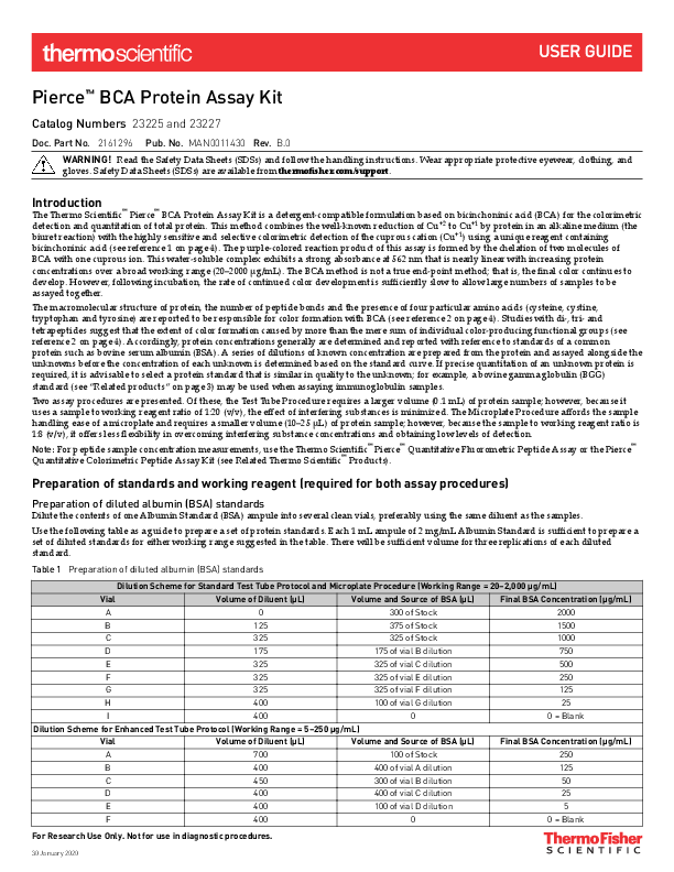
Use the following table as a guide to prepare a set of protein standards. Each 1 mL ampule of 2 mg/mL Albumin Standard is sufficient to prepare a set of diluted standards for either working range suggested in the table. There will be sufficient volume for three replications of each diluted standard.
Table 1: Preparation of Diluted Albumin (BSA) Standards
| Vial | Volume of Diluent (µL) | Volume and Source of BSA (µL) | Final BSA Concentration (µg/mL) |
|---|---|---|---|
| A | 0 | 300 of Stock | 2000 |
| B | 125 | 375 of Stock | 1500 |
| C | 325 | 325 of Stock | 1000 |
| D | 175 | 175 of vial B dilution | 750 |
| E | 325 | 325 of vial C dilution | 500 |
| F | 325 | 325 of vial E dilution | 250 |
| G | 325 | 325 of vial F dilution | 125 |
| H | 400 | 100 of vial G dilution | 25 |
| I | 400 | 0 | 0 = Blank |
Dilution Scheme for Enhanced Test Tube Protocol (Working Range = 5–250 µg/mL)
| Vial | Volume of Diluent (µL) | Volume and Source of BSA (µL) | Final BSA Concentration (µg/mL) |
|---|---|---|---|
| A | 700 | 100 of Stock | 250 |
| B | 400 | 400 of vial A dilution | 125 |
| C | 450 | 300 of vial B dilution | 50 |
| D | 400 | 400 of vial C dilution | 25 |
| E | 400 | 100 of vial D dilution | 5 |
| F | 400 | 0 | 0 = Blank |
For Research Use Only. Not for use in diagnostic procedures.
Preparation of the BCA Working Reagent (WR)
1. Use the following formula to determine the total volume of WR required:
(# standards + # unknowns) * (# replicates) * (volume of WR per sample) = total volume WR required
Example: for the standard test-tube procedure with 3 unknowns and 2 replicates of each sample:
(9 standards + 3 unknowns) * (2 replicates) * (2 mL) = 48 mL WR required
Note: 2.0 mL of the WR is required for each sample in the test-tube procedure, while only 200 µl of WR reagent is required for each sample in the microplate procedure.
2. Prepare WR by mixing 50 parts of BCA Reagent A with 1 part of BCA Reagent B (50:1, Reagent A:B). For the above example, combine 50 mL of Reagent A with 1mL of Reagent B.
Note: When Reagent B is first added to Reagent A, turbidity is observed that quickly disappears upon mixing to yield a clear, green WR. Prepare sufficient volume of WR based on the number of samples to be assayed. The WR is stable for several days when stored in a closed container at room temperature (RT).
Procedure Summary
Test-tube Procedure (Sample to WR Ratio = 1:20)
Procedure Steps:
- Pipette 0.1 mL of each standard and unknown sample replicate into an appropriately labeled test tube.
- Add 2.0 mL of the WR to each tube and mix well.
- Cover and incubate tubes at selected temperature and time:
- Standard Protocol: 37°C for 30 minutes (working range = 20–2000 µg/mL)
- RT Protocol: RT for 2 hours (working range = 20–2000 µg/mL)
- Enhanced Protocol: 60°C for 30 minutes (working range = 5–250 µg/mL)
- Cool all tubes to RT.
- With the spectrophotometer set to 562 nm, zero the instrument on a cuvette filled only with water, then measure the absorbance of all the samples within 10 minutes.
- Subtract the average 562 nm absorbance measurement of the Blank standard replicates from the 562nm absorbance measurement of all other individual standard and unknown sample replicates.
- Prepare a standard curve by plotting the average Blank-corrected 562 nm measurement for each BSA standard vs. its concentration in µg/mL. Use the standard curve to determine the protein concentration of each unknown sample.
Notes:
- Increasing the incubation time or temperature increases the net 562 nm absorbance for each test and decreases both the minimum detection level of the reagent and the working range of the protocol.
- Use a water bath to heat tubes for either Standard (37°C incubation) or Enhanced (60°C incubation) Protocol. Using a forced-air incubator can introduce significant error in color development because of uneven heat transfer.
- Because the BCA assay does not reach a true end point, color development will continue even after cooling to RT. However, because the rate of color development is low at RT, no significant error will be introduced if the 562 nm absorbance measurements of all tubes are made within 10 minutes of each other.
Microplate Procedure (Sample to WR Ratio = 1:8)
Procedure Steps:
- Pipette 25 µL of each standard or unknown sample replicate into a microplate well (working range = 20–2000 µg/mL). (For example, Thermo Scientific Pierce 96-Well Plates, Product No. 15041).
- Add 200 µL of the WR to each well and mix plate thoroughly on a plate shaker for 30 seconds.
- Cover plate and incubate at 37°C for 30 minutes.
- Cool plate to RT. Measure the absorbance at or near 562 nm on a plate reader.
- Subtract the average 562 nm absorbance measurement of the Blank standard replicates from the 562 nm measurements of all other individual standard and unknown sample replicates.
- Prepare a standard curve by plotting the average Blank-corrected 562 nm measurement for each BSA standard vs. its concentration in µg/mL. Use the standard curve to determine the protein concentration of each unknown sample.
Notes:
- If sample size is limited, 10 µL of each unknown sample and standard can be used (sample to WR ratio = 1:20). However, the working range of the assay in this case is limited to 125–2000 µg/mL.
- Wavelengths from 540–590 nm have been used successfully with this method.
- Because plate readers use a shorter light path length than cuvette spectrophotometers, the Microplate Procedure requires a greater sample to WR ratio to obtain the same sensitivity as the standard Test Tube Procedure. If higher 562 nm measurements are desired, increase the incubation time to 2 hours.
- Increasing the incubation time or ratio of sample volume to WR increases the net 562 nm measurement for each well and lowers both the minimum detection level of the reagent and the working range of the assay. As long as all standards and unknowns are treated identically, such modifications may be useful.
- If using curve-fitting algorithms associated with a microplate reader, a four-parameter (quadratic) or best-fit curve provides more accurate results than a purely linear fit. If plotting results by hand, a point-to-point curve is preferable to a linear fit to the standard points.
Related Products
| Cat. NO. | Product |
|---|---|
| 15041 | Pierce™ 96-Well Plates, 100/pkg. |
| 15075 | Reagent Reservoirs, 200/pkg. |
| 15036 | Sealing Tape for 96-Well Plates, 100/pkg. |
| 23209 | Albumin Standard Ampules, 2mg/mL, 10 x 1mL ampules, containing bovine serum albumin (BSA) |
| 23208 | Pre-Diluted Protein Assay Standards: Bovine Serum Albumin (BSA) Set, 7 x 3.5mL |
| 23212 | Bovine Gamma Globulin Standard, 2mg/mL, 10 x 1mL ampules |
| 23213 | Pre-Diluted Protein Assay Standards, (BGG) Set, 7 x 3.5mL aliquots |
| 23246 | Pierce™ Detergent Compatible (Bradford)™ Assay Kit |
| 23235 | Pierce™ Micro BCA™ Protein Assay Kit |
| 23290 | Pierce™ Quantitative Fluorometric Peptide Assay |
| 23275 | Pierce™ Quantitative Colorimetric Peptide Assay |
| 23236 | Coomassie Plus™ (Bradford)™ Assay Kit |
| 23215 | Compat-Able™ Protein Assay Preparation Reagent Set |
| 23250 | Pierce™ BCA Protein Assay Kit-Reducing Agent Compatible |
Additional Information
A. Please visit our website for additional information including the following items:
- Tech Tip: Eliminate interfering substances from samples for BCA Protein Assay
B. Alternative Total Protein Assay Reagents
If interference by a reducing substance or metal-chelating substance contained in the sample cannot be overcome, try the Thermo Scientific Coomassie Plus (Bradford) Assay Kit (Product No. 23236), which is less sensitive to such substances.
C. Cleaning and Re-using Glassware
Exercise care when re-using glassware. All glassware must be cleaned and given a thorough final rinse with ultrapure water.
D. Response characteristics for different proteins
Each of the commonly used total protein assay methods exhibits some degree of varying response toward different proteins. These differences relate to amino acid sequence, pI, structure, and the presence of certain side chains or prosthetic groups that can dramatically alter the protein's color response. Most protein assay methods use BSA or immunoglobulin (IgG) as the standard against which the concentration of protein in the sample is determined (Figure 1). However, if great accuracy is required, prepare the standard curve from a pure sample of the target protein.
Description of Figure 1: The graph displays typical protein-to-protein variation in color response. The X-axis represents Protein Concentration in µg/mL, ranging from 0 to 2,000. The Y-axis represents Net Absorbance at 562 nm (Net A(562 nm)), ranging from 0 to 3. Two curves are plotted: BSA (represented by squares and a solid line) and BGG (represented by circles and a dashed line). Both curves show an increasing trend, indicating higher absorbance with higher protein concentration. The BGG curve generally shows a slightly higher absorbance than the BSA curve at equivalent protein concentrations, particularly at higher concentrations.
Typical protein-to-protein variation in color response is listed in Table 2. All proteins were tested at 1000 µg/mL using the 30-minute/37°C Test Tube Protocol. The average net color response for BSA was normalized to 1.00, and the average net color response of the other proteins is expressed as a ratio to the response of BSA.
Table 2: Protein-to-Protein Variation
Absorbance ratios (562 nm) for proteins relative to BSA using the standard test tube protocol.
Ratio = (Avg "test" net Abs.) / (avg. BSA net Abs.)
| Protein Tested | Ratio |
|---|---|
| Albumin, bovine serum | 1.00 |
| Aldolase, rabbit muscle | 0.85 |
| α-Chymotrypsinogen, bovine | 1.14 |
| Cytochrome C, horse heart | 0.83 |
| Gamma globulin, bovine | 1.11 |
| IgG, bovine | 1.21 |
| IgG, human | 1.09 |
| IgG, mouse | 1.18 |
| IgG, rabbit | 1.12 |
| IgG, sheep | 1.17 |
| Insulin, bovine pancreas | 1.08 |
| Myoglobin, horse heart | 0.74 |
| Ovalbumin | 0.93 |
| Transferrin, human | 0.89 |
| Average Ratio | 1.02 |
| Standard Deviation | 0.15 |
| Coefficient of Variation | 14.7% |
Cited References
- Smith, P.K., et al. (1985). Measurement of protein using bicinchoninic acid. Anal Biochem 150:76-85.
- Wiechelman, K., et al. (1988). Investigation of the bicinchoninic acid protein assay: Identification of the groups responsible for color formation. Anal Biochem 175:231-7.
- Kessler, R. and Fanestil, D. (1986). Interference by lipids in the determination of protein using bicinchoninic acid. Anal Biochem 159:138-42.
- Brown, R., et al. (1989). Protein measurement using bicinchoninic acid: elimination of interfering substances. Anal Biochem 180:136-9.
Product References
- Adilakshami, T. and Laine, R.O. (2002). Ribosomal protein S25 mRNA partners with MTF-1 and La to provide a p53-mediated mechanism for survival or death. J Biol Chem 277:4147-51.
- Fischer, T., et al. (1999). Clathrin-coated vesicles bearing GAIP possess GTPase-activating protein activity in vitro. Proc Nat Acad Sci 96:6722-7.
- Prozialeck, W.C., et al. (2002). Chlamydia trachomatis disrupts N-cadherin-dependent cell-cell junctions and sequester β-catenin in human cervical epithelial cells. Infection and Immunity 70:2605-13.
- Roberts, K.P., et al. (2002). A comparative analysis of expression and processing of the rat epididymal fluid and sperm-bound forms of proteins D and E. Biology of Reproduction 67:525-33.
Troubleshooting and FAQs
Visit our online FAQ database for tips and tricks for conducting your experiment, troubleshooting information, and FAQs. The online FAQ database is frequently updated to ensure accurate and thorough content.
- For troubleshooting information and FAQs for this product: https://www.thermofisher.com/bcafaqs
- To browse the database and search using keywords: thermofisher.com/faqs
Limited Product Warranty
Life Technologies Corporation and/or its affiliate(s) warrant their products as set forth in the Life Technologies' General Terms and Conditions of Sale at www.thermofisher.com/us/en/home/global/terms-and-conditions.html. If you have any questions, please contact Life Technologies at www.thermofisher.com/support.
