PLM_Manual Ethicon Wound Closure Manual 101702
User Manual: manual pdf -FilePursuit
Open the PDF directly: View PDF ![]() .
.
Page Count: 125 [warning: Documents this large are best viewed by clicking the View PDF Link!]

WOUND
CLOSURE
MANUAL

CONTRIBUTING EDITOR
David, L. Dunn, M.D., Ph. D.
Jay Phillips Professor and Chairman of Surgery,
University of Minnesota
We thank Dr. Dunn for his contributions to the Wound Closure
Manual. Dr. Dunn is currently the Jay Phillips Professor and
Chairman of Surgery at the University of Minnesota. This
department has a long-standing tradition and has attained
national and international recognition for excellence in training
academic general surgeons and surgical scientists. He is also the
Division Chief of General Surgery, Head of Surgical Infectious
Diseases, Director of Graduate Studies, and Residency Program
Director of the Department of Surgery.
Dr. Dunn has published over 400 articles and book chapters in the
areas of Surgical Infectious Diseases and Transplantation. He has
received regional and nationwide recognition in several
academic organizations and is a Past-President of the Surgical
Infection Society, the Association for Academic Surgery, the
Minnesota Chapter of the American College of Surgeons, the
Society of University Surgeons and the Society of University
Surgeons Foundation.

PREFACE
his manual has been prepared for the medical professional who
would like to learn more about the practice of surgery–the
dynamics of tissue healing, the principles of wound closure, and the
materials available to today’s practitioners. Most important, it
touches on some of the critical decisions which must be made on a
daily basis to help ensure proper wound closure.
ETHICON, INC., a Johnson & Johnson company, is the world’s
leading marketer of surgical sutures and is the only U.S. company
that offers an adhesive with microbial protection as an alternative
to sutures for topical skin closure.
ETHICON enjoys a reputation for developing quality products to
enhance the lives of patients and for providing outstanding service
to customers. We hope you find this manual useful. But, above all,
we hope that it reflects our high regard for the men and women
who have chosen the medical profession as a career.
ETHICON, Inc.
T
~
~

TABLE OF CONTENTS
1WOUND HEALING
AND MANAGEMENT
The Wound.......................................................... 2
Recovery of Tensile Strength............................ 2
Patient Factors that Affect Wound Healing......... 2
Surgical Principles....................................... 4
Classification of Wounds................................... 5
Types of Wound Healing................................... 6
Healing by Primary Intention........................ 6
Healing by Second Intention.......................... 7
Delayed Primary Closure.............................. 7
2THE SUTURE
What is a Suture?............................................... 10
Personal Suture Preference............................... 10
Suture Characteristics....................................... 11
Size and Tensile Strength.............................. 11
Monofilament vs. Multifilament.................... 11
Absorbable vs. Nonabsorbable Sutures.............. 12
Specific Suturing Materials.............................. 13
Synthetic Absorbable Sutures......................... 14
Nonabsorbable Sutures................................ 16
Synthetic Nonabsorbable Sutures.................... 17
Common Suturing Techniques....................... 18
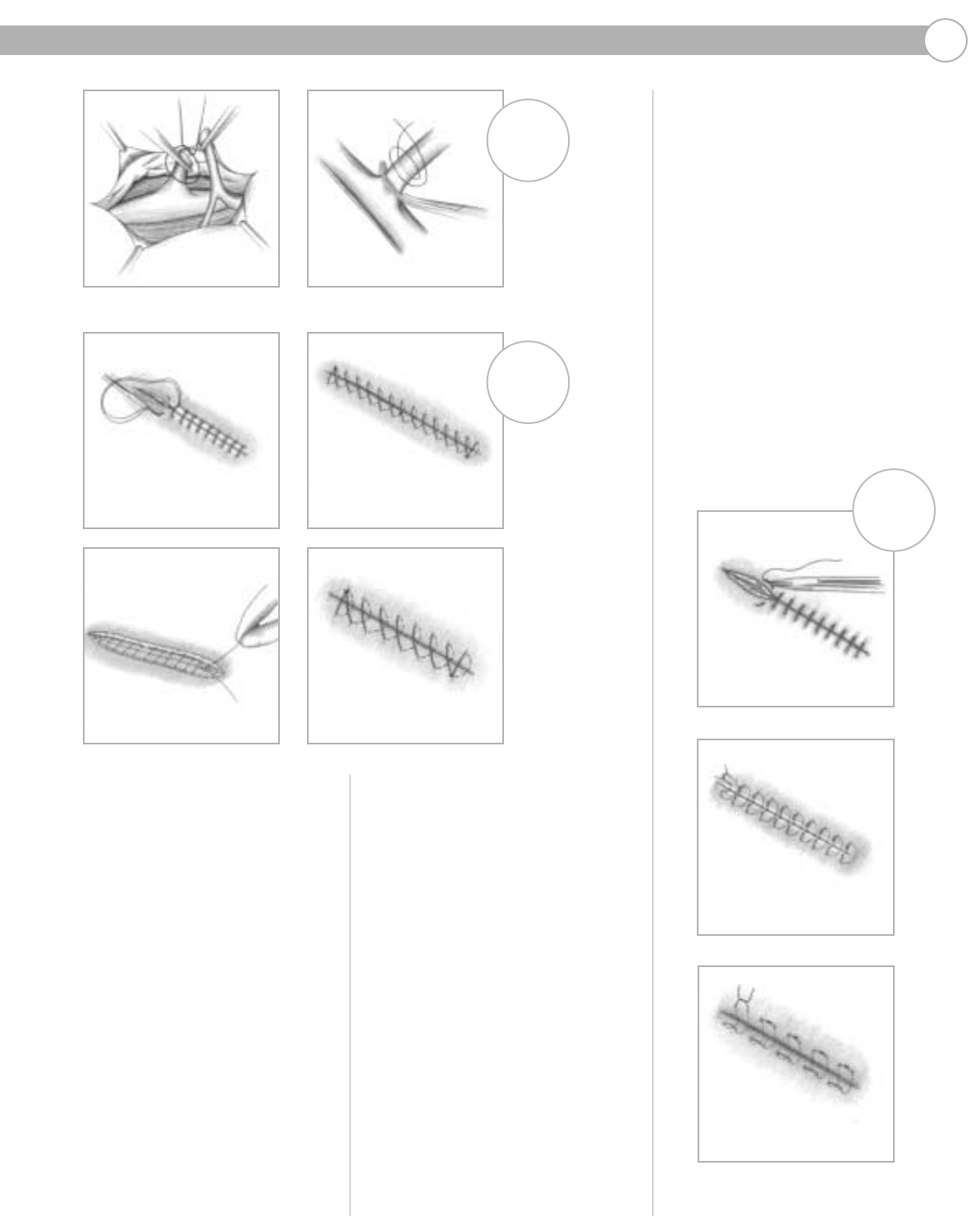
Ligatures................................................. 18
The Primary Suture Line............................. 18
Continuous Sutures.................................... 19
Interrupted Sutures.................................... 19
Deep Sutures............................................ 22
Buried Sutures .......................................... 22
Purse-String Sutures................................... 22
Subcuticular Sutures................................... 22
The Secondary Suture Line........................... 22
Stitch Placement........................................ 23
Knot Tying......................................................... 24
Knot Security............................................ 24
Knot Tying Techniques Most Often Used .......... 25
Square Knot............................................. 25
Surgeon’s or Friction Knot............................ 26
Deep Tie................................................. 26
Ligation Using a Hemostatic Clamp............... 26
Instrument Tie.......................................... 26
Endoscopic Knot Tying Techniques.................. 26
Cutting the Secured Sutures.......................... 26
Suture Removal................................................. 26
Suture Handling Tips....................................... 27
Suture Selection Procedure ............................. 27
Surgery within the Abdominal Wall Cavity....... 28
Closing the Abdomen.................................. 30
Closing Contaminated or Infected Wounds........ 40
3THE SURGICAL NEEDLE
Elements of Needle Design ............................. 42
Principles of Choosing a Surgical Needle..... 43
Anatomy of a Needle........................................ 44
The Needle Eye......................................... 45
The Needle Body....................................... 46
Straight Needle......................................... 46
Half-Curved Needle................................... 46
Curved Needle.......................................... 46
Compound Curved Needle........................... 47
The Needle Point ...................................... 47
Types of Needles............................................... 47
Conventional Cutting Needles....................... 48
Reverse Cutting Needles............................... 49
Side Cutting Needles................................... 49
Taper Point Needles................................... 50
Taper Surgical Needles................................ 50
Blunt Point Needles.................................... 51
Needleholders.................................................... 51
Needleholder Use....................................... 52
Placing the Needle in Tissue ......................... 52
Needle Handling Tips...................................... 54

4PACKAGING
An Integral Part of the Product ..................... 56
Relay* Suture Delivery System....................... 56
Modular Storage Racks................................ 56
Dispenser Boxes......................................... 56
Primary Packets........................................ 57
E-Pack* Procedure Kit..................................... 59
Expiration Date................................................. 60
Suture Sterilization........................................... 60
Anticipating Suture Needs............................... 61
Sterile Transfer of Suture Packets.................... 61
Suture Preparation in the Sterile Field............. 62
Suture Handling Technique.......................... 63
5TOPICAL SKIN ADHESIVES
Dermabond* topical skin adhesive................ 68
6OTHER SURGICAL
PRODUCTS
Adhesive Tapes................................................... 74
Indications and Usage................................. 74
Application.............................................. 74
After Care and Removal.............................. 74
Skin Closure Tapes..................................... 75
Polyester Fiber Strips.................................. 75
Umbilical Tape ........................................ 75
Surgical Staples................................................. 76
Indications and Usage................................. 76
Aftercare and Removal................................ 76
PROXIMATE* Skin Staplers........................ 76
Looped Suture................................................... 77
Retention Suture Devices................................ 77
7PRODUCT TERMS
AND TRADEMARKS
8PRODUCT INFORMATION
9INDEX
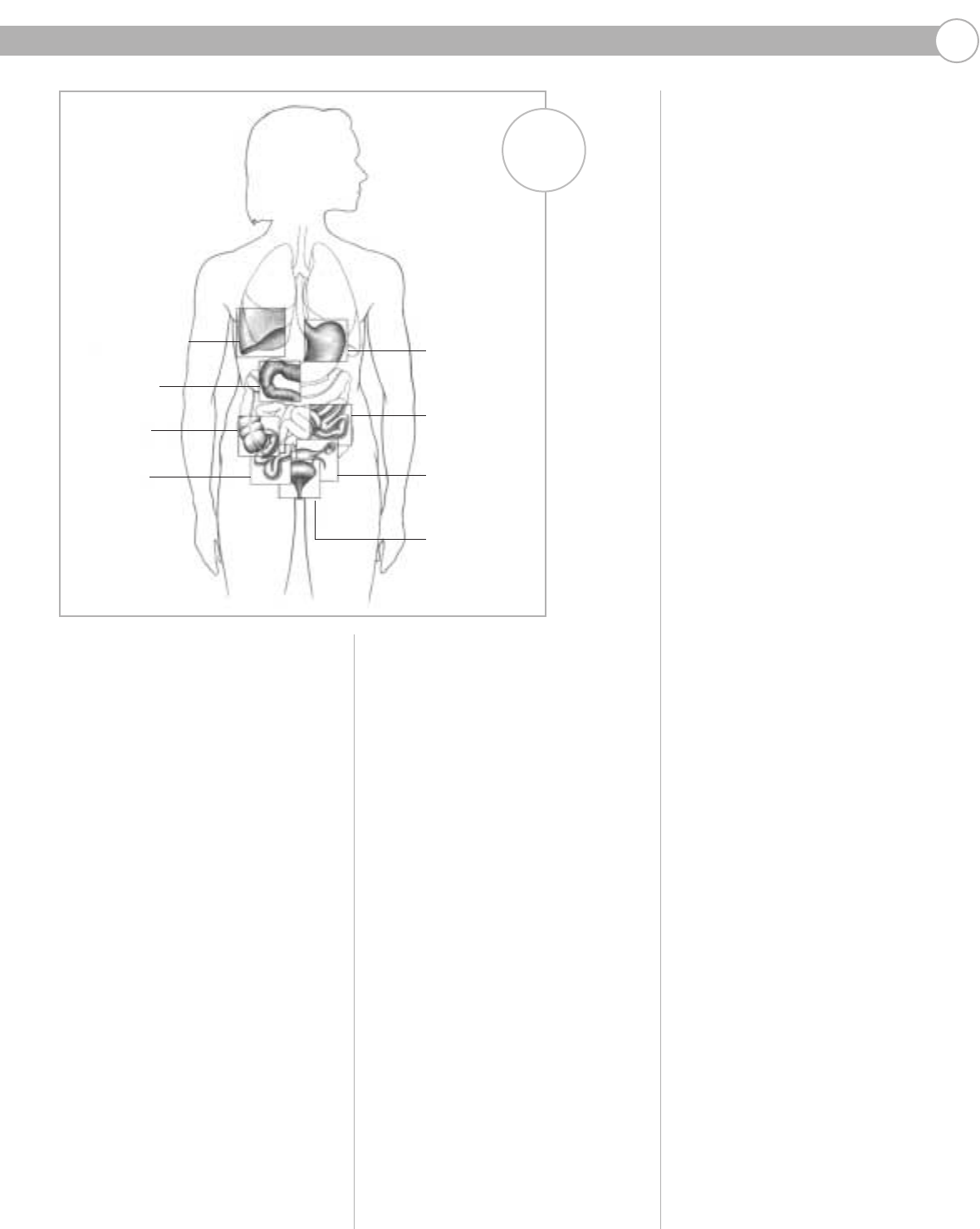
WOUND HEALING
AND MANAGEMENT
CHAPTER 1

WOUND HEALING & MANAGEMENT
2
THE WOUND
Injury to any of the tissues of
the body, especially that caused
by physical means and with
interruption of continuity is defined
as a wound.1Though most often
the result of a physical cause, a
burn is also considered a wound.
Both follow the same processes
towards the restoration to
health – otherwise known
as healing.1
Wound healing is a natural and
spontaneous phenomenon. When
tissue has been disrupted so severely
that it cannot heal naturally
(without complications or possible
disfiguration) dead tissue and
foreign bodies must be removed,
infection treated, and the tissue
must be held in apposition until the
healing process provides the wound
with sufficient strength to withstand
stress without mechanical support.
A wound may be approximated
with sutures, staples, clips, skin
closure strips, or topical adhesives.
Tissue is defined as a collection of
similar cells and the intercellular
substances surrounding them.
There are four basic tissues in the
body: 1) epithelium; 2) connective
tissues, including blood, bone and
cartilage; 3) muscle tissue; and
4) nerve tissue. The choice of
wound closure materials and the
techniques of using them are prime
factors in the restoration of
continuity and tensile strength to
the injured tissues during the
healing process.
The parameters for measuring the
strength of normal body tissue are:
• Tensile Strength—The load per
cross-sectional area unit at the
point of rupture, relating to the
nature of the material rather than
its thickness.
• Breaking Strength—The load
required to break a wound regard-
less of its dimension, the more
clinically significant measurement.
• Burst Strength—The amount of
pressure needed to rupture a
viscus, or large interior organ.
The rate at which wounds regain
strength during the wound healing
process must be understood as a
basis for selecting the most
appropriate wound closure material.
RECOVERY OF
TENSILE STRENGTH
Tensile strength affects the tissue's
ability to withstand injury but is
not related to the length of time it
takes the tissue to heal. As collagen
accumulates during the reparative
phase, strength increases rapidly but
it is many months before a plateau
is reached.2Until this time, the
wound requires extrinsic support
from the method used to bring it
together – usually sutures. While
skin and fascia (the layer of firm
connective tissue covering muscle)
are the strongest tissues in the body,
they regain tensile strength slowly
during the healing process. The
stomach and small intestine, on the
other hand, are composed of much
weaker tissue but heal rapidly.
Variations in tissue strength may
also be found within the same
organ. Within the colon, for
example, the sigmoid region is
approximately twice as strong as the
cecum—but both sections heal at
the same rate. Factors that affect
tissue strength include the size, age,
and weight of the patient, the
thickness of tissue, the presence of
edema, and duration (the degree to
which the tissue has hardened in
response to pressure or injury).
PATIENT FACTORS THAT
AFFECT WOUND HEALING
The goal of wound management
is to provide interventions that
efficiently progress wounds through
the biologic sequence of repair or
regeneration. The patient's overall
health status will affect the speed of
the healing process. The following
are factors that should be considered
by the surgical team prior to and
during the procedure. 2,3,4
✦AGE — With aging, both skin
and muscle tissue lose their tone
and elasticity. Metabolism also
slows, and circulation may be
impaired. But aging alone is not
a major factor in chronic wound
healing. Aging and chronic
disease states often go together,
and both delay repair processes
due to delayed cellular response
to the stimulus of injury, delayed
collagen deposition, and
decreased tensile strength in the
remodeled tissue. All of these
factors lengthen healing time.
✦WEIGHT — Obese patients
of any age have, excess fat at the
wound site that may prevent
securing a good closure. In
addition, fat does not have a rich
blood supply, making it the most
vulnerable of all tissues to trauma
and infection.
✦NUTRITIONAL STATUS —
Overall malnutrition associated
with chronic disease or cancer,
or specific deficiencies in
carbohydrates, proteins, zinc, and
vitamins A, B, and C can impair
the healing process. Adequate
nutrition is essential to support
cellular activity and collagen
synthesis at the wound site.
✦DEHYDRATION — If the
patient's system has been
depleted of fluids, the resulting
electrolyte imbalance can affect
cardiac function, kidney
function, cellular metabolism,
oxygenation of the blood, and
hormonal function. These effects
will not only impact upon the
patient's overall health status and
recovery from surgery but may
also impair the healing process.
✦INADEQUATE BLOOD
SUPPLY TO THE WOUND
SITE — Oxygen is necessary for
cell survival and, therefore,
healing. Skin healing takes place
most rapidly in the face and
neck, which receive the greatest
blood supply, and most slowly in
the extremities. The presence of
any condition that compromises
the supply of blood to the
wound, such as poor circulation
to the limbs in a diabetic patient
or arteriosclerosis with vascular
compromise, will slow and can
even arrest the healing process.
✦IMMUNE RESPONSES —
Because the immune response
protects the patient from
infection, immunodeficiencies
may seriously compromise the
outcome of a surgical procedure.
Patients infected with HIV, as
well as those who have recently
undergone chemotherapy or who
have taken prolonged high
dosages of catabolic steroids, may
have debilitated immune systems.
Some patients have allergies to
specific suturing materials, metal
alloys, or latex. These, on the
other hand, will cause a height-
ened immune response in the
form of an allergic reaction.
This may also interfere with the
healing process. Therefore,
the surgeon should always
check beforehand on a
patient's allergies.
✦CHRONIC DISEASE —
A patient whose system has
already been stressed by chronic
illness, especially endocrine
disorders, diabetes, malignancies,
localized infection, or debilitating
injuries will heal more slowly and
will be more vulnerable to post
surgical wound complications.
All of these conditions merit
concern, and the surgeon must
consider their effects upon the
tissues at the wound site, as well
as their potential impact upon
the patient's overall recovery
from the procedure.
Malignancies, in addition, may
alter the cellular structure of
tissue and influence the
surgeon's choice of methods and
closure materials.
✦RADIATION THERAPY —
Radiation therapy to the surgical
site prior to or shortly after
surgery can produce considerable
impairment of healing and lead
to substantial wound complica-
tions. Surgical procedures for
malignancies must be planned
to minimize the potential for
these problems.
CHAPTER 1 3
* Trademark
RELATIVE
TISSUE
STRENGTH
Stomach
(Weak)
Small
intestine
(Weak)
Female
reproductive
organs
(Weak)
Bladder
(Weak)
Lower
respiratory
tract (Weak)
Duodenum
(Strong)
Cecum
(Weak)
Ileum
(Weak)
FIGURE
1

SURGICAL PRINCIPLES
Many factors that affect the healing
process can be controlled by the
surgical team in the operating room,
by the obstetrical team in labor and
delivery, or by the emergency team
in the trauma center. Their first
priority is to maintain a sterile
and aseptic technique to prevent
infection. Organisms found
within a patient's own body most
commonly cause postoperative
infection, but microorganisms
carried by medical personnel also
pose a threat. Whatever the source,
the presence of infection will deter
healing. In addition to concerns
about sterility, the following must
be taken into consideration when
planning and carrying out an
operative procedure.3
✦THE LENGTH AND
DIRECTION OF THE
INCISION — A properly
planned incision is sufficiently
long to afford sufficient optimum
exposure. When deciding upon
the direction of the incision, the
surgeon must bear the following
in mind:
• The direction in which wounds
naturally heal is from side-to-
side, not end-to-end.
• The arrangement of tissue fibers
in the area to be dissected will
vary with tissue type.
• The best cosmetic results may be
achieved when incisions are made
parallel to the direction of the
tissue fibers. Results may vary
depending upon the tissue
layer involved.
✦DISSECTION
TECHNIQUE — When incising
tissue, a clean incision should
be made through the skin with
one stroke of evenly applied
pressure on the scalpel. Sharp
dissection should be used to cut
through remaining tissues. The
surgeon must preserve the
integrity of as many of the
underlying nerves, blood vessels,
and muscles as possible.
TISSUE HANDLING —
Keeping tissue trauma to a
minimum promotes faster
healing. Throughout the
operative procedure, the surgeon
must handle all tissues very
gently and as little as possible.
Retractors should be placed with
care to avoid excessive pressure,
since tension can cause serious
complications: impaired blood
and lymph flow, altering of the
local physiological state of the
wound, and predisposition to
microbial colonization.
HEMOSTASIS — Various
mechanical, thermal, and
chemical methods are available to
decrease the flow of blood and
fluid into the wound site.
Hemostasis allows the surgeon to
work in as clear a field as possible
with greater accuracy. Without
adequate control, bleeding from
transected or penetrated vessels
or diffused oozing on large
denuded surfaces may interfere
with the surgeon's view of
underlying structures.
Achieving complete hemostasis
before wound closure also will
prevent formation of postopera-
tive hematomas. Collections of
blood (hematomas) or fluid
(seromas) in the incision can
prevent the direct apposition of
tissue needed for complete union
of wound edges. Furthermore,
these collections provide an ideal
culture medium for microbial
growth and can lead to serious
infection.
When clamping or ligating a
vessel or tissue, care must be
taken to avoid excessive tissue
damage. Mass ligation that
involves large areas of tissue may
produce necrosis, or tissue death,
and prolong healing time.
MAINTAINING MOISTURE
IN TISSUES — During long
procedures, the surgeon may
periodically irrigate the wound
with warm physiologic (normal)
saline solution, or cover exposed
surfaces with saline-moistened
sponges or laparotomy tapes to
prevent tissues from drying out.
REMOVAL OF NECROTIC
TISSUE AND FOREIGN
MATERIALS — Adequate
debridement of all devitalized
tissue and removal of inflicted
foreign materials are essential
to healing, especially in traumatic
wounds. The presence of
fragments of dirt, metal, glass,
etc., increases the probability
of infection.
CHOICE OF CLOSURE
MATERIALS — The surgeon
must evaluate each case individu-
ally, and choose closure material
which will maximize the
opportunity for healing and
minimize the likelihood of
WOUND HEALING
4
✦
✦
✦
✦
✦
infection. The proper closure
material will allow the surgeon
to approximate tissue with as
little trauma as possible, and with
enough precision to eliminate
dead space. The surgeon's
personal preference will play a
large role in the choice of closure
material; but the location of the
wound, the arrangement of tissue
fibers, and patient factors influ-
ence his or her decision as well.
CELLULAR RESPONSE TO
CLOSURE MATERIALS —
Whenever foreign materials such
as sutures are implanted in tissue,
the tissue reacts. This reaction
will range from minimal to
moderate, depending upon the
type of material implanted. The
reaction will be more marked if
complicated by infection, allergy,
or trauma.
Initially, the tissue will deflect the
passage of the surgeon's needle
and suture. Once the sutures
have been implanted, edema of
the skin and subcutaneous tissues
will ensue. This can cause
significant patient discomfort
during recovery, as well as
scarring secondary to ischemic
necrosis. The surgeon must take
these factors into consideration
when placing tension upon the
closure material.
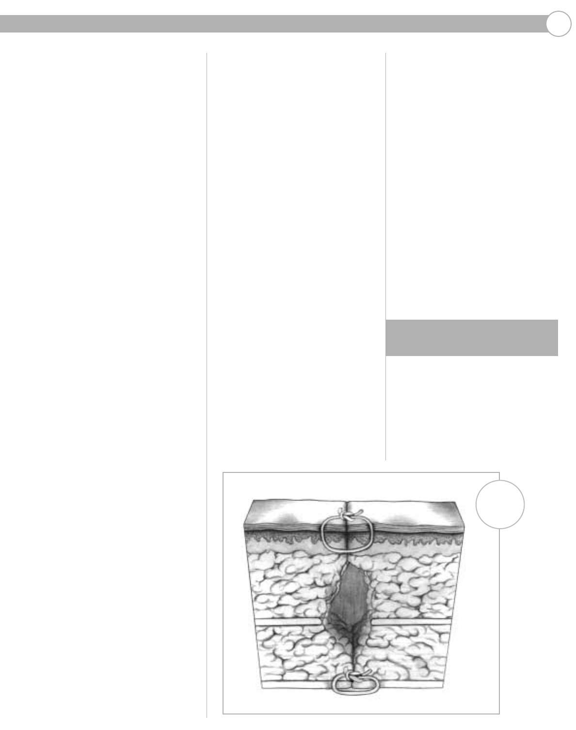
ELIMINATION OF DEAD
SPACE IN THE WOUND —
Dead space in a wound results
from separation of portions of
the wound beneath the skin
edges which have not been
closely approximated, or from air
or fluid trapped between layers of
tissue. This is especially true in
the fatty layer which tends to
lack blood supply. Serum or
blood may collect, providing an
ideal medium for the growth
of microorganisms that cause
infection. The surgeon may
elect to insert a drain or apply
a pressure dressing to help
eliminate dead space in the
wound postoperatively.
CLOSING TENSION —
While enough tension must be
applied to approximate tissue and
eliminate dead space, the sutures
must be loose enough to prevent
exaggerated patient discomfort,
ischemia, and tissue necrosis
during healing.
POSTOPERATIVE
DISTRACTION FORCES —
The patient's postoperative
activity can place undue stress
upon a healing incision.
Abdominal fascia will be placed
under excessive tension after
surgery if the patient strains to
cough, vomit, void, or defecate.
Tendons and the extremities may
also be subjected to excessive
tension during healing. The
surgeon must be certain that
the approximated wound is
adequately immobilized to
prevent suture disruption
for a sufficient period of time
after surgery.
IMMOBILIZATION —
Adequate immobilization of the
approximated wound, but not
necessarily of the entire anatomic
part, is mandatory after surgery
for efficient healing and minimal
scar formation.
CLASSIFICATION
OF WOUNDS
The Centers for Disease Control
and Prevention (CDC), using an
adaptation of the American College
of Surgeons’ wound classification
schema, divides surgical wounds
into four classes: clean wounds,
clean-contaminated wounds,
CHAPTER 1 5
✦
✦
✦
✦
DEAD SPACE
IN A WOUND
✦
FIGURE
2
* Trademark

contaminated wounds and dirty or
infected wounds.5A discussion of
each follows.
Seventy-five percent of all wounds
(which are usually elective surgical
incisions) fall into the clean wounds
category—an uninfected operative
wound in which no inflammation
is encountered and the respiratory,
alimentary, genital, or uninfected
urinary tracts are not entered.
These elective incisions are made
under aseptic conditions and are
not predisposed to infection.
Inflammation is a natural part of
the healing process and should be
differentiated from infection in
which bacteria are present and
produce damage.
Clean wounds are closed by primary
union and usually are not drained.
Primary union is the most desirable
method of closure, involving the
simplest surgical procedures and
the lowest risk of postoperative
complications. Apposition of tissue
is maintained until wound tensile
strength is sufficient so that sutures
or other forms of tissue apposition
are no longer needed.
Clean-contaminated wounds are
operative wounds in which the
respiratory, alimentary, genital, or
urinary tracts are entered under
controlled conditions and without
unusual contamination. Specifically,
operations involving the biliary
tract, appendix, vagina, and
oropharynx are included in this
category provided no evidence
of infection or major break in
technique is encountered.
Appendectomies, cholecystectomies,
and hysterectomies fall into this
category, as well as normally
clean wounds which become
contaminated by entry into a
viscus resulting in minimal spillage
of contents.
Contaminated wounds include
open, traumatic wounds or injuries
such as soft tissue lacerations, open
fractures, and penetrating wounds;
operative procedures in which gross
spillage from the gastrointestinal
tract occurs; genitourinary or biliary
tract procedures in the presence
of infected urine or bile; and
operations in which a major break
in aseptic technique has occurred
(as in emergency open cardiac
massage). Microorganisms
multiply so rapidly that within
6 hours a contaminated wound
can become infected.
Dirty and infected wounds have
been heavily contaminated or
clinically infected prior to the
operation. They include perforated
viscera, abscesses, or neglected
traumatic wounds in which
devitalized tissue or foreign material
have been retained. Infection
present at the time of surgery can
increase the infection rate of any
wound by an average of four times.
TYPES OF
WOUND HEALING
The rate and pattern of healing falls
into three categories, depending
upon the type of tissue involved
and the circumstances surrounding
closure. Timeframes are generalized
for well-perfused healthy soft
tissues, but may vary.
HEALING BY
PRIMARY INTENTION
Every surgeon who closes a wound
would like it to heal by primary
union or first intention, with
minimal edema and no local
infection or serious discharge. An
incision that heals by primary
intention does so in a minimum of
time, with no separation of the
wound edges, and with minimal
scar formation. This takes place in
three distinct phases:2,3
Inflammatory (preparative) –
During the first few days, an
inflammatory response causes an
outpouring of tissue fluids, an
accumulation of cells and
fibroblasts, and an increased blood
supply to the wound. Leukocytes
and other cells produce proteolytic
enzymes which dissolve and remove
damaged tissue debris. These are
the responses which prepare the site
of injury for repair. The process
lasts 3 to 7 days. Any factor which
interferes with the progress, may
interrupt or delay healing. During
the acute inflammatory phase, the
tissue does not gain appreciable
tensile strength, but depends solely
upon the closure material to hold it
in approximation.
Proliferative – After the
debridement process is well along,
fibroblasts begin to form a collagen
matrix in the wound known as
granulation tissue. Collagen, a
protein substance, is the chief
constituent of connective tissue.
Collagen fiber formation determines
the tensile strength and pliability of
the healing wound. As it fills with
new blood vessels, the granulation
becomes bright, beefy, red tissue.
The thick capillary bed which fills
WOUND HEALING
6
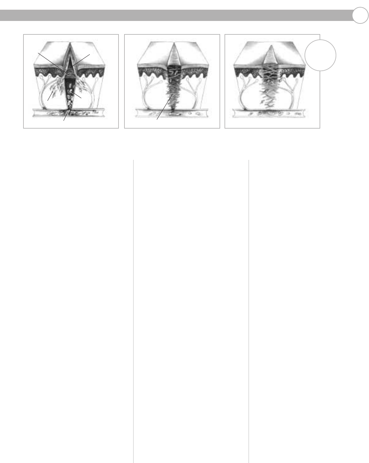
the matrix, supplies the nutrients
and oxygen necessary for the wound
to heal. This phase occurs from
day 3 onward.
In time, sufficient collagen is laid
down across the wound so that it
can withstand normal stress. The
length of this phase varies with the
type of tissue involved and the
stresses or tension placed upon the
wound during this period.
Wound contraction also occurs dur-
ing this phase. Wound contraction
is a process that pulls the wound
edges together for the purpose of
closing the wound. In essence, it
reduces the open area, and if
successful, will result in a smaller
wound with less need for repair by
scar formation. Wound contraction
can be very beneficial in the closure
of wounds in areas such as the but-
tocks or trochanter but can be very
harmful in areas such as the hand
or around the neck and face, where
it can cause disfigurement and
excessive scarring.3
Surgical wounds that are closed
by primary intention have minimal
contraction response. Skin grafting
is used to reduce avoided contrac-
tion in undesirable locations.
Remodelling – As collagen deposi-
tion is completed, the vascularity of
the wound gradually decreases and
any surface scar becomes paler. The
amount of collagen that is finally
formed – the ultimate scar – is
dependent upon the initial volume
of granulation tissue.2
HEALING BY
SECOND INTENTION
When the wound fails to heal by
primary union, a more complicated
and prolonged healing process takes
place. Healing by second intention
is caused by infection, excessive
trauma, tissue loss, or imprecise
approximation of tissue.3
In this case, the wound may be left
open and allowed to heal from the
inner layer to the outer surface.
Granulation tissue forms and
contains myofibroblasts. These
specialized cells help to close the
wound by contraction. This
process is much slower than primary
intention healing. Excessive
granulation tissue may build up
and require treatment if it protrudes
above the surface of the wound,
preventing epithelialization.
DELAYED PRIMARY
CLOSURE
This is considered by many
surgeons to be a safe method of
management of contaminated, as
well as dirty and infected traumatic
wounds with extensive tissue loss
and a high risk of infection. This
method has been used extensively in
the military arena and has proven
successful following excessive
trauma related to motor vehicle
accidents, shooting incidents, or
infliction of deep, penetrating
knife wounds.3
The surgeon usually treats these
injuries by debridement of
nonviable tissues and leaves the
wound open, inserting gauze
packing which is changed twice a
day. Patients sedation or a return to
the operating room with general
anesthesia generally is only required
in the case of large, complex
wounds. Wound approximation
using adhesive strips, previously
placed but untied sutures, staples
after achieving local anesthesia can
occur within 3-5 days if the wound
demonstrates no evidence of
infection and the appearance of red
granulation tissue. Should this not
CHAPTER 1 7
* Trademark
PHASES OF
WOUND
HEALING
FIGURE
3
Damaged
tissue
debris Tissue fluids
Fibroblasts Proteolytic
enzymes
Increased blood supply Collagen fibers
PHASE 1–
Inflammatory response and
debridement process
PHASE 2–
Collagen formation
(scar tissue)
PHASE 3–
Sufficient collagen laid down

occur, the wound is allowed to
heal by secondary intention. When
closure is undertaken, skin edges
and underlying tissue must be accu-
rately and securely approximated.
IN THE
NEXT SECTION
The materials, devices, and
techniques used to repair wounded
tissue will be discussed at length.
As you will see, the number of
options available is extensive. But
no matter how many choices the
surgeon has, his or her objective
remains singular: to restore the
patient to health with as little
operative trauma as possible and
an excellent cosmetic result.
REFERENCES
1. Stedman’s Medical Dictionary,
27th edition, 2000
2. Henry, Michael and Thompson,
Jeremy: Clinical Surgery, W.B.
Saunders, 2001
3. Skerris, David A.: Mayo Clinic
Basic Surgery Skills, Mayo Clinic
Scientific Press, 1999
4. Sussman, Carrie: Wound Care,
Aspen Publishers, 1998
5. NNIS Manual, CDC,
MHA, 2000
WOUND HEALING
8
THE SUTURE
CHAPTER 2

WHAT IS
A SUTURE?
The word "suture" describes any
strand of material used to ligate (tie)
blood vessels or approximate (bring
close together) tissues. Sutures are
used to close wounds. Sutures and
ligatures were used by both the
Egyptians and Syrians as far back as
2,000 B.C. Through the centuries, a
wide variety of materials—silk,
linen, cotton, horsehair, animal
tendons and intestines, and wire
made of precious metals—have been
used in operative procedures. Some
of these are still in use today.
The evolution of suturing material
has brought us to a point of refine-
ment that includes sutures designed
for specific surgical procedures.
Despite the sophistication of
today's suture materials and surgical
techniques, closing a wound still
involves the same basic procedure
used by physicians to the Roman
emperors. The surgeon still uses a
surgical needle to penetrate tissue
and advance a suture strand to its
desired location.
Successful use of suture materials
depends upon the cooperation of
the suture manufacturer and the
surgical team.
The manufacturer must have a
thorough knowledge of surgical
procedures, anticipate the surgical
team's needs, and produce suture
materials that meet these
stringent criteria:
• They must have the greatest
tensile strength consistent with
size limitations.
• They must be easy to handle.
• They must be secured in
packaging which presents them
sterile for use, in excellent
condition, and ensures the
safety of each member of the
surgical team.
The nurse must maintain the
sterility of sutures when storing,
handling, and preparing them for
use. The integrity and strength of
each strand must remain intact
until it is in the surgeon's hands.
The surgeon must select suture
materials appropriate for the
procedure and must place them
in the tissues in a manner consistent
with the principles that promote
wound healing.
With the manufacturer and
surgical team working in concert,
the patient reaps the final
benefit...the wound is closed in a
manner that promotes optimum
healing in minimum time.
PERSONAL SUTURE
PREFERENCE
Most surgeons have a basic
"suture routine," a preference for
using the same material(s) unless
circumstances dictate otherwise.
The surgeon acquires skill,
proficiency, and speed in handling
by using one suture material
repeatedly—and may choose
the same material throughout his
or her entire career.
A number of factors may influence
the surgeon’s choice of materials:
• His or her area of specialization.
• Wound closure experience during
clinical training.
• Professional experience in the
operating room.
• Knowledge of the healing
characteristics of tissues and
organs.
• Knowledge of the physical and
biological characteristics of
various suture materials.
• Patient factors (age, weight,
overall health status, and the
presence of infection).
Surgical specialty plays a
primary role in determining
suture preference. For example,
obstetrician/gynecologists frequently
prefer coated VICRYL* RAPIDE
(polyglactin 910) suture for
episiotomy repair and coated
VICRYL* (polyglactin 910)
suture and MONOCRYL*
(poliglecaprone 25) suture for all
tissue layers except, possibly, skin.
Most orthopaedic surgeons use
coated VICRYL suture, PDS* II
(polydioxanone) suture, and
ETHIBOND* EXCEL polyester
suture. Many plastic surgeons
prefer ETHILON* nylon suture,
VICRYL* suture, or MONOCRYL
suture. Many neurosurgeons
prefer coated VICRYL suture or
NUROLON* braided nylon suture.
But no single suture material is used
by every surgeon who practices within
a specialty.
The surgeon's knowledge of the
physical characteristics of suture
material is important. As the
requirements for wound support
vary with patient factors, the nature
THE SUTURE
10

of the procedure, and the type of
tissue involved, the surgeon will
select suture material that will retain
its strength until the wound heals
sufficiently to withstand stress on
its own.
SUTURE
CHARACTERISTICS
The choice of suture materials gen-
erally depends on whether the
wound closure occurs in one or
more layers. In selecting the most
appropriate sutures, the surgeon
takes into account the amount of
tension on the wound, the number
of layers of closure, depth of suture
placement, anticipated amount of
edema, and anticipated timing of
suture removal.
Optimal suture qualities include:
1. High uniform tensile strength,
permitting use of finer sizes.
2. High tensile strength retention
in vivo, holding the wound
securely throughout the critical
healing period, followed by
rapid absorption.
3. Consistent uniform diameter.
4. Sterile.
5. Pliable for ease of handling and
knot security.
6. Freedom from irritating
substances or impurities for
optimum tissue acceptance.
7. Predictable performance.
SIZE AND TENSILE
STRENGTH
Size denotes the diameter of the
suture material. The accepted
surgical practice is to use the
smallest diameter suture that
will adequately hold the mending
wounded tissue. This practice
minimizes trauma as the suture is
passed through the tissue to effect
closure. It also ensures that the
minimum mass of foreign material
is left in the body. Suture size is
stated numerically; as the number of
0s in the suture size increases, the
diameter of the strand decreases. For
example, size 5-0, or 00000, is
smaller in diameter than size 4-0, or
0000. The smaller the size, the less
tensile strength the suture will have.
Knot tensile strength is measured by
the force, in pounds, which the
suture strand can withstand before
it breaks when knotted. The tensile
strength of the tissue to be mended
(its ability to withstand stress)
determines the size and tensile
strength of the suturing material the
surgeon selects. The accepted rule is
that the tensile strength of the
suture need never exceed the tensile
strength of the tissue. However,
sutures should be at least as strong
as normal tissue through which they
are being placed.
MONOFILAMENT VS.
MULTIFILAMENT STRANDS
Sutures are classified according to
the number of strands of which
they are comprised. Monofilament
sutures are made of a single strand
of material. Because of their
simplified structure, they encounter
less resistance as they pass
through tissue than multifilament
suture material. They also resist
harboring organisms which may
cause infection.
These characteristics make
monofilament sutures well-suited
to vascular surgery. Monofilament
sutures tie down easily. However,
because of their construction,
extreme care must be taken when
handling and tying these sutures.
Crushing or crimping of this suture
type can nick or create a weak spot
in the strand. This may result in
suture breakage.
Multifilament sutures consist of
several filaments, or strands, twisted
or braided together. This affords
greater tensile strength, pliability, and
flexibility. Multifilament sutures may
also be coated to help them pass rela-
tively smoothly through tissue and
enhance handling characteristics.
Coated multifilament sutures are
well-suited to intestinal procedures.
CHAPTER 2 11
METRIC MEASURES AND U.S.P.
SUTURE DIAMETER EQUIVALENTS TABLE
1
U.S.P. Size
Natural
Collagen
Synthetic
Absorbables
Nonabsorbable
Materials
11-0
–––
–––
0.1
10-0
0.2
0.2
0.2
9-0
0.3
0.3
0.3
8-0
0.5
0.4
0.4
7-0
0.7
0.5
0.5
6-0
1.0
0.7
0.7
5-0
1.5
1.0
1.0
4-0
2.0
1.5
1.5
3-0
3.0
2.0
2.0
2-0
3.5
3.0
3.0
0
4.0
3.5
3.5
1
5.0
4.0
4.0
2
6.0
5.0
5.0
3
7.0
6.0
6.0
4
8.0
6.0
6.0
5
–––
7.0
7.0
6
–––
–––
8.0
* Trademark

ABSORBABLE VS.
NONABSORBABLE SUTURES
Sutures are classified according to
their degradation properties.
Sutures that undergo rapid degrada-
tion in tissues, losing their tensile
strength within 60 days, are
considered absorbable sutures.
Sutures that generally maintain
their tensile strength for longer than
60 days are nonabsorbable sutures.
Absorbable sutures may be used to
hold wound edges in approximation
temporarily, until they have healed
sufficiently to withstand normal
stress. These sutures are prepared
either from the collagen of healthy
mammals or from synthetic
polymers. Some are absorbed
rapidly, while others are treated or
chemically structured to lengthen
absorption time. They may also be
impregnated or coated with agents
that improve their handling
properties, and colored with an
FDA-approved dye to increase
visibility in tissue. Natural
absorbable sutures are digested by
body enzymes which attack and
break down the suture strand.
Synthetic absorbable sutures are
hydrolyzed—a process by which
water gradually penetrates the
suture filaments, causing the
breakdown of the suture's polymer
chain. Compared to the enzymatic
action of natural absorbables,
hydrolyzation results in a lesser
degree of tissue reaction following
implantation.
During the first stage of the
absorption process, tensile strength
diminishes in a gradual, almost
linear fashion. This occurs over the
first several weeks postimplantation.
The second stage often follows with
considerable overlap, characterized
by loss of suture mass. Both stages
exhibit leukocytic cellular responses
which serve to remove cellular
debris and suture material from the
line of tissue approximation.
The loss of tensile strength and
the rate of absorption are separate
phenomena. A suture can lose
tensile strength rapidly and yet be
absorbed slowly—or it can
maintain adequate tensile strength
through wound healing, followed
by rapid absorption. In any case,
the strand is eventually completely
dissolved, leaving no detectable
traces in tissue.
Although they offer many
advantages, absorbable sutures also
have certain inherent limitations.
If a patient has a fever, infection,
or protein deficiency, the suture
absorption process may accelerate,
causing too rapid a decline in tensile
strength. In addition, if the sutures
become wet or moist during
handling, prior to being implanted
in tissue, the absorption process
may begin prematurely. Similarly,
patients with impaired healing
are often not ideal candidates
for this type of suture. All of
these situations predispose to
postoperative complications, as
the suture strand will not maintain
adequate strength to withstand
stress until the tissues have
healed sufficiently.
Nonabsorbable sutures are those
which are not digested by body
enzymes or hydrolyzed in body
tissue. They are made from a variety
of nonbiodegradable materials and
are ultimately encapsulated or
walled off by the body’s fibroblasts.
Nonabsorbable sutures ordinarily
remain where they are buried
THE SUTURE
12
ABSORBABLE
SUTURES:
BASIC RAW
MATERIALS
Surgical Gut
Plain
Chromic
Fast Absorbing
Polyglactin 910
Uncoated (VICRYL*
(polyglactin 910) Suture)
Coated (coated VICRYL*
(polyglactin 910) suture),
(coated VICRYL* RAPIDE
(polyglactin 910) suture)
Polyglycolic Acid
Poliglecaprone 25
(MONOCRYL*
(poliglecaprone 25) suture)
Polyglyconate
Polydioxanone (PDS* II
(polydioxanone) suture)
TABLE
2
Submucosa of sheep
intestine or serosa of beef
intestine
Copolymer of glycolide and
lactide with polyglactin 370
and calcium stearate, if coated
Homopolymer of glycolide
Copolymer of glycolide and
epsilon-caprolactone
Copolymer of glycolide and
trimethylene carbonate
Polyester of poly (p-dioxanone)
SUTURE RAW MATERIAL

within the tissues. When used for
skin closure, they must be removed
postoperatively. Nonabsorbable
sutures may be used in a variety
of applications:
• Exterior skin closure, to be
removed after sufficient healing
has occurred.
• Within the body cavity, where
they will remain permanently
encapsulated in tissue.
• Patient history of reaction to
absorbable sutures, keloidal
tendency, or possible tissue
hypertrophy.
• Prosthesis attachment
(i.e., defibrillators, pacemakers,
drug delivery mechanisms).
Nonabsorbable sutures are com-
posed of single or multiple filaments
of metal, synthetic, or organic fibers
rendered into a strand by spinning,
twisting, or braiding. Each strand is
substantially uniform in diameter
throughout its length, conforming
to the United States Pharmacopeia
(U.S.P.) limitations for each size.
Nonabsorbable sutures have been
classified by the U.S.P. according to
their composition. In addition,
these sutures may be uncoated
or coated, uncolored, naturally
colored, or dyed with an FDA-
approved dye to enhance visibility.
SPECIFIC SUTURING
MATERIALS
The materials and products
described here embody the most
current advances in the manufacture
of surgical sutures. They are
grouped as either absorbable or
nonabsorbable for easy reference.
Absorbable Sutures
Surgical Gut
Absorbable surgical gut is classified
as either plain or chromic. Both
types consist of processed strands of
highly purified collagen. The
percentage of collagen in the suture
determines its tensile strength and
its ability to be absorbed by the
body without adverse reaction.
Noncollagenous material can cause
a reaction ranging from irritation to
rejection of the suture. The more
pure collagen throughout the
length of the strand, the less foreign
material there is introduced into
the wound.
ETHICON* surgical gut sutures are
manufactured from between 97%
and 98% pure ribbons of collagen.
To meet U.S.P. specifications,
processed ribbons of the submucosa
layer of sheep intestine or the
serosa layer of beef intestine are
electronically spun and polished
into virtually monofilament strands
of various sizes, with minimum and
maximum limits on diameter for
each size. The ETHICON exclusive
TRU-GAUGING process produces
a uniform diameter to within an
accuracy of 0.0002 inch
(0.0175mm) along the entire
length of every strand, eliminating
high and low spots. High and low
spots can cause the suture to fray
or chatter when knots are tied
down, resulting in a knot that is
not positioned properly or tied
securely. Most protein-based
absorbable sutures have a tendency
to fray when tied.
TRU-GAUGING ensures that
ETHICON surgical gut sutures
possess uniform high tensile
strength, virtually eliminating the
possibility of fray or breaking. Their
unexceeded strength and surface
smoothness allow the surgeon to
"snug down" the suture knot to
achieve optimum tension.
The rate of absorption of surgical
gut is determined by the type of
CHAPTER 2 13
* Trademark
NON-
ABSORBABLE
SUTURES: RAW
MATERIALS
Surgical Silk
Stainless Steel Wire
Nylon (ETHILON* nylon
suture, NUROLON* nylon
suture)
Polyester Fiber
Uncoated (MERSILENE*
polyester fiber suture)
Coated (ETHIBOND* EXCEL
polyester suture
Polypropylene (PROLENE*
polypropylene suture)
Poly(hexafluoropropylene-VDF)
(PRONOVA* poly(hexafluoro-
propylene-VDF) suture)
Raw silk spun by silkworm
Specially formulated
iron-chromium-nickel-
molybdenum alloy
Polyamide polymer
Polymer of polyethylene
terephthalate (may be coated)
Polymer of propylene
Polymer blend of poly(vinylidene
fluoride) and poly(vinylidene fluo-
ride-cohexafluoropropylene)
SUTURE RAW MATERIAL
TABLE
3

gut being used, the type and
condition of the tissue involved,
and the general health status of the
patient. Surgical gut may be used in
the presence of infection, although
it may be absorbed more rapidly
under this condition.
Plain surgical gut is rapidly
absorbed. Tensile strength is
maintained for only 7 to 10 days
postimplantation, and absorption
is complete within 70 days. The
surgeon may choose plain gut for
use in tissues which heal rapidly
and require minimal support (for
example, ligating superficial blood
vessels and suturing subcutaneous
fatty tissue). Plain surgical gut can
also be specially heat-treated to
accelerate tensile strength loss and
absorption. This fast absorbing
surgical gut is used primarily for
epidermal suturing where sutures
are required for only 5 to 7 days.
These sutures have less tensile
strength than plain surgical gut
of the comparable U.S.P. size. Fast
absorbing plain gut is not to be
used internally.
Chromic gut is treated with a
chromium salt solution to resist
body enzymes, prolonging
absorption time over 90 days.
The exclusive CHROMICIZING
process used by ETHICON
thoroughly bathes the pure collagen
ribbons in a buffered chrome
tanning solution before spinning
into strands. After spinning, the
entire cross section of the strand
is evenly chromicized. The process
alters the coloration of the surgical
gut from yellowish-tan to brown.
Chromic gut sutures minimize
tissue irritation, causing less
reaction than plain surgical gut
during the early stages of wound
healing. Tensile strength may be
retained for 10 to 14 days, with
some measurable strength remaining
for up to 21 days.
SYNTHETIC
ABSORBABLE SUTURES
Synthetic absorbable sutures offer the
strength needed for a wide range of
applications, from abdominal and
chest wound closure to ophthalmic
and plastic surgery.
COATED VICRYL* RAPIDE
(POLYGLACTIN 910) SUTURE
This braided suture is composed of
the same copolymer as coated
VICRYL suture—lactide and
glycolide—and is coated with a
combination of equal parts of
copolymer of lactide and glycolide
(polyglactin 370) and calcium
stearate. However, the absorption
rate and tensile strength profile are
significantly different from coated
VICRYL suture, achieved by the use
of a polymer material with a lower
molecular weight than coated
VICRYL suture. Coated VICRYL
RAPIDE sutures are only available
undyed.
Coated VICRYL RAPIDE suture
is the fastest-absorbing synthetic
suture and exhibits characteristics
that model the performance of
surgical gut suture. However,
being a synthetic material. Coated
VICRYL RAPIDE suture elicits
a lower tissue reaction than chromic
gut suture. Coated VICRYL
RAPIDE suture is indicated only
for use in superficial soft tissue
approximation of the skin and
mucosa, where only short-term
wound support (7 to 10 days)
is required. It is not to be used in
ligation, in ophthalmic, cardiovascu-
lar, or neurological procedures,
where extended approximation of
tissues under stress is required, or
where wound support beyond 7
days is required.
Coated VICRYL RAPIDE sutures
retain approximately 50% of the
original tensile strength at 5 days
postimplantation. All of the original
tensile strength is lost by approxi-
mately 10 to 14 days. Absorption is
essentially complete by 42 days.
Coated VICRYL RAPIDE suture
is particularly well-suited for skin
closure, episiotomy repair, and
closure of lacerations under casts.
In addition, since the suture begins
to "fall off" in 7 to 10 days as the
wound heals, the need for suture
removal is eliminated.
MONOCRYL*
(POLIGLECAPRONE 25) SUTURE
This monofilament suture
features superior pliability for easy
handling and tying. Comprised
of a copolymer of glycolide and
epsilon-caprolactone, it is virtually
inert in tissue and absorbs
predictably. The surgeon may
prefer MONOCRYL sutures for
procedures which require high
initial tensile strength diminishing
over 2 weeks postoperatively. These
include subcuticular closure and
soft tissue approximations and
ligations, with the exception of
neural, cardiovascular, ophthalmic,
and microsurgical applications.
MONOCRYL suture is available
dyed (violet) and undyed (natural).
Dyed MONOCRYL suture retains
60% to 70% of its original strength
at 7 days postimplantation, reduced
to 30% to 40% at 14 days, with all
THE SUTURE
14

original strength lost by 28 days.
At 7 days, undyed MONOCRYL
suture retains approximately 50%
to 60% of its original strength, and
approximately 20% to 30% at
14 days postimplantation. All of
the original tensile strength of
undyed MONOCRYL suture is
lost by 21 days postimplantation.
Absorption is essentially complete
at 91 to 119 days.
COATED VICRYL*
(POLYGLACTIN 910) SUTURE
This material fills the need for a
smoother synthetic absorbable
suture that will pass through tissue
readily with minimal drag. Coated
VICRYL sutures facilitate ease of
handling, smooth tie down and
unsurpassed knot security.
The coating is a combination of
equal parts of copolymer of lactide
and glycolide (polyglactin 370),
plus calcium stearate which is
used extensively in pharmaceuticals
and food. Calcium stearate is a salt
of calcium and stearic acid, both
of which are present in the body
and constantly metabolized and
excreted. The result of this mixture
is an outstandingly absorbable,
adherent, nonflaking lubricant.
At 2 weeks postimplantation,
approximately 75% of the tensile
strength of coated VICRYL suture
remains. Approximately 50% of
tensile strength is retained at 3
weeks for sizes 6-0 and larger. At 3
weeks, 40% of tensile strength is
retained for sizes 7-0 and smaller. At
4 weeks, 25% of the original
strength is retained for sizes 6-0 and
larger. All of the original tensile
strength is lost by five weeks post
implantation. Absorption of coated
VICRYL suture is essentially
complete between 56 and 70 days.
Lactide and glycolide acids are
readily eliminated from the body,
primarily in urine. As with
uncoated sutures, coated VICRYL
sutures elicit only a mild tissue
reaction during absorption. Their
safety and effectiveness in neural
and cardiovascular tissue have not
been established. Transcutaneous or
conjunctival sutures remaining in
place longer than 7 days may cause
localized irritation and should be
removed as indicated. Coated
VICRYL sutures are available as
braided dyed violet or undyed
natural strands in a variety of
lengths with or without needles.
VICRYL* (POLYGLACTIN 910)
SUTURE
This synthetic absorbable suture is a
copolymer of lactide and glycolide
(from lactic and glycolic acid). Both
are natural metabolic substances.
The water-repelling quality of
lactide slows water penetration into
the suture filaments, thus slowing
the rate of in vivo tensile strength
loss as compared to natural
absorbable sutures which are subject
to enzymatic digestion. Lactides are
also bulky, keeping the submicro-
scopic polymer chains comprising
the filaments spaced apart so that
absorption of the suture mass is
rapid once tensile strength is lost.
The combination of lactide and
glycolide results in a molecular
structure which maintains sufficient
tensile strength for efficient
approximation of tissues during
the critical wound-healing period,
followed by rapid absorption.
VICRYL sutures (size 6-0 and
larger) retain approximately 75%
of original tensile strength 2 weeks
after implantation. At 3 weeks, 50%
of tensile strength is retained. At
4 weeks, 20-30% of tensile strength
is retained.
Because synthetic absorbable sutures
are not digested by enzymatic
activity, they exhibit a lower degree
of tissue reaction than surgical gut.
VICRYL suture is extruded into
monofilament strands which are
dyed violet to enhance visibility in
tissue. They are available for use in
ophthalmic surgery. Conjunctival
sutures remaining in place longer
than 7 days may cause localized
irritation and should be removed
as indicated.
PDS* II (POLYDIOXANONE)
SUTURE
Comprised of the polyester poly
(p-dioxanone), this monofilament
represents a significant advance
in suturing options. It combines
the features of soft, pliable,
monofilament construction with
absorbability and extended wound
support for up to 6 weeks. It elicits
only a slight tissue reaction. This
material is well-suited for many
types of soft tissue approximation,
including pediatric cardiovascular,
orthopaedic, gynecologic,
ophthalmic, plastic, digestive,
and colonic surgeries.
Like other synthetic absorbable
sutures, PDS II sutures are
absorbed in vivo through hydrolysis.
Approximately 70% of tensile
strength remains 2 weeks
postimplantation, 50% at 4 weeks,
and 25% at 6 weeks. Absorption is
minimal until about the 90th day
CHAPTER 2 15
* Trademark

postoperatively and essentially
complete within 6 months. The
safety and effectiveness of PDS II
sutures in microsurgery, neural
tissue, and adult cardiovascular
tissue have not been established.
PDS II sutures are available clear
or dyed violet to enhance visibility.
NONABSORBABLE SUTURES
The U.S.P. classifies nonabsorbable
surgical sutures as follows:
• CLASS I—Silk or synthetic fibers
of monofilament, twisted, or
braided construction.
• CLASS II—Cotton or linen
fibers, or coated natural or
synthetic fibers where the coating
contributes to suture thickness
without adding strength.
• CLASS III—Metal wire of
monofilament or multifilament
construction.
SURGICAL SILK
For many surgeons, surgical silk
represents the standard handling
performance by which newer
synthetic materials are judged,
especially due to its superior
handling characteristics. Silk
filaments can be twisted or braided,
the latter providing the best
handling qualities.
Raw silk is a continuous filament
spun by the silkworm moth larva
to make its cocoon. Cream or
orange-colored in its raw state, each
silk filament is processed to remove
natural waxes and sericin gum,
which is exuded by the silkworm as
it spins its cocoon. The gum holds
the cocoon together, but is of no
benefit to the quality of braided
surgical silk sutures.
ETHICON degums the silk for
most suture sizes before the
braiding process. This allows for a
tighter, more compact braid which
significantly improves suture quality.
After braiding, the strands are dyed,
scoured and stretched, and then
impregnated and coated with a
mixture of waxes or silicone. Each
of these steps is critical to the
quality of the finished suture and
must be carried out in precise order.
Surgical silk is usually dyed black
for easy visibility in tissue.
Raw silk is graded according to
strength, uniformity of filament
diameter, and freedom from defects.
Only top grades of silk filaments are
used to produce PERMA-HAND*
surgical silk sutures.
Surgical silk loses tensile strength
when exposed to moisture and
should be used dry. Although silk
is classified by the U.S.P. as a
nonabsorbable suture, long-term
in vivo studies have shown that it
loses most or all of its tensile
strength in about 1 year and usually
cannot be detected in tissue after
2 years. Thus, it behaves in reality
as a very slowly absorbing suture.
SURGICAL STAINLESS STEEL
The essential qualities of surgical
stainless steel sutures include the
absence of toxic elements, flexibility,
and fine wire size. Both monofila-
ment and twisted multifilament
varieties are high in tensile strength,
low in tissue reactivity, and hold
a knot well. Provided that the
sutures do not fragment, there is
little loss of tensile strength in
tissues. The 316L (low carbon)
stainless steel alloy formula used
in the manufacture of these
sutures offers optimum metal
strength, flexibility, uniformity,
and compatibility with stainless
steel implants and prostheses.
Stainless steel sutures may also be
used in abdominal wall closure,
sternum closure, retention, skin
closure, a variety of orthopaedic
procedures, and neurosurgery.
Disadvantages associated with
alloy sutures include difficulty in
handling; possible cutting, pulling,
and tearing of the patient's tissue;
fragmentation; barbing; and
kinking, which renders the stainless
steel suture useless. When used for
bone approximation and fixation,
asymmetrical twisting of the wire
will lead to potential buckling, wire
fracture, or subsequent wire
fatigue. Incomplete wire fixation
under these circumstances will
permit movement of the wire,
resulting in postoperative pain
and possible dehiscence.
Surgical stainless steel sutures
should not be used when a
prosthesis of another alloy is
implanted since an unfavorable
electrolytic reaction may occur.
Above all, stainless steel sutures
pose a safety risk. They easily tear
surgical gloves when handled
and may puncture the surgeon's
own skin—putting both
physician and patient at risk
of transmitted immunodeficiency
virus or hepatitis. Many surgeons
refer to wire size by the Brown &
Sharpe (B & S) gauge of 40
(smallest diameter) to 18 (largest
diameter). ETHICON labels
surgical stainless steel with both
the B & S and U.S.P. diameter size
classifications.
THE SUTURE
16

ETHICON packaging of surgical
stainless steel maintains the integrity
of the product by eliminating kink-
ing and bending of strands. Just as
important, it presents the strands in
a safe manner for all members of
the surgical team who handle them.
SYNTHETIC
NONABSORBABLE SUTURES
Nylon sutures are a polyamide poly-
mer derived by chemical synthesis.
Because of their elasticity, they are
particularly well-suited for retention
and skin closure. They may be
clear, or dyed green or black for
better visibility.
ETHILON* NYLON SUTURE
These sutures are extruded into
noncapillary single or monofilament
strands characterized by high tensile
strength and extremely low tissue
reactivity. They degrade in vivo at a
rate of approximately 15% to 20%
per year by hydrolysis. ETHILON
sutures in sizes 10-0 and 6-0 and
larger are produced from a special
grade of nylon 6. The medical grade
polyamide nylon 6-6 is used for
sizes 7-0 and finer. While both
grades permit good handling,
monofilament nylon sutures have a
tendency to return to their original
straight extruded state (a property
known as "memory"). Therefore,
more throws in the knot are
required to securely hold monofila-
ment than braided nylon sutures.
Monofilament nylon in a wet or
damp state is more pliable and
easier to handle than dry nylon. A
limited line of ETHILON sutures
(sizes 3-0 through 6-0) are pre-
moistened or "pliabilized" for use
in cosmetic plastic surgery. This
process enhances the handling
and knot tying characteristics to
approximate that of braided sutures.
ETHILON sutures are frequently
used in ophthalmology and
micro-surgery procedures in very
fine sizes. For this reason, sizes 9-0
and 10-0 have an intensified black
dye for high visibility.
NUROLON* NYLON SUTURE
This suture is composed of
filaments of nylon that have been
tightly braided into a multifilament
strand. Available in white or dyed
black, NUROLON sutures look,
feel, and handle like silk. However,
NUROLON sutures have more
strength and elicit less tissue
reaction than silk. Braided nylon
may be used in all tissues where
multifilament nonabsorbable sutures
are acceptable. Braided nylon
sutures generally lose 15% to 20%
of their tensile strength per year in
tissue by hydrolyzation.
Polyester fiber suture is comprised
of untreated fibers of polyester
(polyethylene terephthalate) closely
braided into a multifilament strand.
They are stronger than natural
fibers, do not weaken when wetted
prior to use, and cause minimal
tissue reaction. Available white or
dyed green, polyester fiber sutures
are among the most acceptable for
vascular synthetic prostheses.
MERSILENE* POLYESTER
FIBER SUTURE
The first synthetic braided suture
material shown to last indefinitely
in the body, MERSILENE sutures
provide precise, consistent suture
tension. They minimize breakage
and virtually eliminate the need to
remove irritating suture fragments
postoperatively. Because it is
uncoated, MERSILENE suture
has a higher coefficient of friction
when passed through tissue.
CHAPTER 2 17
* Trademark
SURGICAL
STAINLESS
STEEL: WIRE
GAUGE
EQUIVALENTS
DIAMETER U.S.P. B & S
.0031 inch
.0040
.0056
.0063
.0080
.0100
.0126
.0159
.0179
.0201
.0226
.0253
.0320
.0360
.0400
6-0
6-0
5-0
4-0
4-0
3-0
2-0
0
1
2
3
4
5
6
7
40
38
35
34
32
30
28
26
25
24
23
22
20
19
18
TABLE
4

ETHIBOND* EXCEL
POLYESTER SUTURE
ETHIBOND EXCEL sutures are
uniformly coated with polybutilate,
a biologically inert, nonabsorbable
compound which adheres itself to
the braided polyester fiber strand.
Polybutilate was the first synthetic
coating developed specifically as a
surgical suture lubricant. The coat-
ing eases the passage of the braided
strands through tissue and provides
excellent pliability, handling quali-
ties, and smooth tie-down with each
throw of the knot. Both the suture
material and the coating are
pharmacologically inactive. The
sutures elicit minimal tissue reaction
and retain their strength in vivo for
extended periods. ETHIBOND*
EXCEL sutures are used primarily in
cardiovascular surgery, for vessel
anastomosis, and placement of
prosthetic materials.
ETHIBOND EXCEL sutures
are also available attached
to TFE polymer felt pledgets.
Pledgets serve to prevent possible
tearing of adjacent friable tissue.
Pledgets are used routinely in valve
replacement procedures (to prevent
the annulus from tearing when the
prosthetic valve is seated and the
sutures are tied), and in situations
where extreme deformity, distortion,
or tissue destruction at the annulus
has occurred.
Polypropylene is an isostatic
crystalline stereoisomer of a
linear hydrocarbon polymer
permitting little or no saturation.
Manufactured by a patented
process which enhances pliability
and handling, polypropylene
monofilament sutures are not
subject to degradation or weakening
by tissue enzymes. They cause
minimal tissue reaction and hold
knots better than most other
synthetic monofilament materials.
PROLENE* POLYPROPYLENE
SUTURE
Widely used in general, cardiovascu-
lar, plastic, and orthopaedic surgery,
PROLENE sutures do not adhere to
tissue and are therefore efficacious
as a pull-out suture. PROLENE
sutures are relatively biologically
inert, offering proven strength,
reliability and versatility.
PROLENE sutures are
recommended for use where
minimal suture reaction is desired,
such as in contaminated and
infected wounds to minimize
later sinus formation and suture
extrusion. They are available clear
or dyed blue.
PRONOVA* POLY
(HEXAFLUOROPROPYLENE-VDF)
SUTURE
This monofilament nonabsorbable
suture is a polymer blend of poly
(vinylidene fluoride) and poly
(vinylidene fluoride-cohexafluoro-
propylene). This suture resists
involvement in infection and has
been successfully employed in
contaminated and infected wounds
to eliminate or minimize later sinus
formation and suture extrusion.
Furthermore, the lack of adherence
to tissues has facilitated the use
of PRONOVA suture as a
pull-out suture.
This material is well-suited for
many types of soft tissue approxi-
mation and ligation, including use
in cardiovascular, ophthalmic and
neurological procedures.
Table 5 gives an overview of the
many suturing options that have
been discussed in this section.
(See attached chart )
COMMON
SUTURING
TECHNIQUES
LIGATURES
A suture tied around a vessel to
occlude the lumen is called a
ligature or tie. It may be used to
effect hemostasis or to close off a
structure to prevent leakage.
There are two primary types
of ligatures.
Free tie or freehand ligatures are
single strands of suture material
used to ligate a vessel, duct, or other
structure. After a hemostat or other
similar type of surgical clamp has
been placed on the end of the
structure, the suture strand is tied
around the vessel under the tip of
the hemostat. The hemostat is
removed after the first throw and
the surgeon tightens the knot using
his or her fingertips, taking care to
avoid instrument damage to the
suture. Additional throws are added
as needed to square and secure the
knot. Stick tie, suture ligature, or
transfixion suture is a strand of
suture material attached to a needle
to ligate a vessel, duct, or other
structure. This technique is used on
deep structures where placement of
a hemostat is difficult or on vessels
of large diameter. The needle is
passed through the structure or
adjacent tissue first to anchor the
suture, then tied around the
structure. Additional throws are
used as needed to secure the knot.
THE SUTURE
18
* Trademark
THE PRIMARY SUTURE LINE
The primary suture line is the line
of sutures that holds the wound
edges in approximation during
healing by first intention. It may
consist of a continuous strand of
material or a series of interrupted
suture strands. Other types of
primary sutures, such as deep
sutures, buried sutures, purse-string
sutures, and subcuticular sutures,
are used for specific indications.
Regardless of technique, a surgical
needle is attached to the suture
strand to permit repeated passes
through tissue.
CONTINUOUS SUTURES
Also referred to as running stitches,
continuous sutures are a series of
stitches taken with one strand of
material. The strand may be tied to
itself at each end, or looped, with
both cut ends of the strand tied
together. A continuous suture line
can be placed rapidly. It derives its
strength from tension distributed
evenly along the full length of the
suture strand. However, care must
be taken to apply firm tension,
rather than tight tension, to avoid
tissue strangulation. Excessive
tension and instrument damage
should be avoided to prevent suture
breakage which could disrupt the
entire line of a continuous suture.
Continuous suturing leaves less
foreign body mass in the wound.
In the presence of infection, it may
be desirable to use a monofilament
suture material because it has no
interstices which can harbor
microorganisms. This is especially
critical as a continuous suture
line can transmit infection along
the entire length of the strand. A
continuous one layer mass closure
CHAPTER 2 19
LIGATURES
CONTINUOUS
SUTURING
TECHNIQUES
FIGURE
1
FIGURE
2
Free tie Stick tie
Looped suture, knotted
at one end
Two strands knotted at
each end and knotted in
the middle
Running locked suture Over-and-over running
stitch
FIGURE
3
INTERRUPTED
SUTURING
TECHNIQUES
Simple interrupted
Interrupted vertical
mattress
Interrupted horizontal
mattress

TYPES
Plain
Chromic
Braided
Monofilament
Braided
Monofilament
Monofilament
Braided
Monofilament
Mulifilament
Monofilament
Braided
Braided
Monofilament
Braided
Monofilament
Monofilament
ABSORBABLE SUTURES
SUTURE
Surgical Gut
Suture
Surgical Gut
Suture
Coated
VICRYL* RAPIDE
(polyglactin 910)
Suture
MONOCRYL*
(poliglecaprone 25)
Suture
Coated VICRYL*
(polyglactin 910)
Suture
PDS* II
(polydioxanone)
Suture
PERMA-HAND*
Silk Suture
Surgical Stainless
Steel Suture
ETHILON*
Nylon Suture
NUROLON*
Nylon Suture
MERSILENE*
Polyester Fiber
Suture
ETHIBOND*
EXCEL Polyester
Fiber Suture
PROLENE*
Polypropylene
Suture
PRONOVA*
POLY (hexafluoro-
propylene-VDF)
Suture
COLOR OF
MATERIAL
Yellowish-tan
Blue Dyed
Brown
Blue Dyed
Undyed
(Natural)
Undyed
(Natural)
Violet
Violet
Undyed
(Natural)
Violet
Blue
Clear
Violet
White
Silver metallic
Violet
Green
Undyed (Clear)
Violet
Green
Undyed (Clear)
Green
Undyed (White)
Green
Undyed (White)
Clear
Blue
Blue
RAW MATERIAL
Collagen derived from
healthy beef and sheep.
Collagen derived from
healthy beef and sheep.
Copolymer of lactide
and glycolide coated
with 370 and calcium
stearate.
Copolymer of
glycolide and
epsilon-caprolactone.
Copolymer of lactide
and glycolide coated
with 370 and calcium
stearate.
Polyester polymer.
Organic protein called
fibrin.
316L stainless steel.
Long-chain aliphatic
polymers Nylon 6 or
Nylon 6,6.
Long-chain aliphatic
polymers Nylon 6 or
Nylon 6,6.
Poly (ethylene
terephthalate).
Poly (ethylene
terephthalate) coated
with polybutilate.
Isotactic crystalline
stereoisomer of
polypropylene.
Polymer blend of poly
(vinylidene fluoride)
and poly (vinylidene
fluoride-cohexafluoro-
propylene).
TENSILE STRENGTH
RETENTION in vivo
Individual patirent characteristics can
affect rate of tensile strength loss.
Individual patirent characteristics can
affect rate of tensile strength loss.
Approximately 50% remains at 5
days. All tensile strength is lost at
approximately 14 days.
Approximately 50-60% (violet: 60-70%)
remains at 1 week. Approximately 20-
30% (violet: 30-40%) remains at 2 weeks.
Lost within 3 weeks (violet: 4 weeks).
Approximately 75% remains at two
weeks. Approximately 50% remains
at three weeks, 25% at four weeks.
Approximately 70% remains at 2 weeks.
Approximately 50% remains at 4 weeks.
Approximately 25% remains at 6 weeks.
Progressive degradation of fiber may
result in gradual loss of tensile
strength over time.
Indefinate.
Progressive hydrolysis may result in
gradual loss of tensile strength over
time.
Progressive hydrolysis may result in
gradual loss of tensile strength over
time.
No significant change known to
occur in vivo.
No significant change known to
occur in vivo.
No subject to degradation or
weakening by action of tissue
enzymes.
No subject to degradation or
weakening by action of tissue
enzymes.
ABSORPTION RATE
Absorbed by proleolytic
enzymatic digestive
process.
Absorbed by proleolytic
enzymatic digestive
process.
Essentially complete
between 42 days.
Absorbed by hydrolysis.
Complete at 91-119
days. Absorbed by
hydrolysis.
Essentially complete
between 56-70 days.
Absorbed by hydrolysis.
Minimal until about 90th
day. Essentially complete
within 6 months. Absorbed
by slow hydrolysis.
Gradual encapsulation
by fibrous connective
tissue.
Nonabsorbable.
Gradual encapsulation
by fibrous connective
tissue.
Gradual encapsulation
by fibrous connective
tissue.
Gradual encapsulation
by fibrous connective
tissue.
Gradual encapsulation
by fibrous connective
tissue.
Nonabsorbable.
Nonabsorbable.
TISSUE REACTION
Moderate reaction
Moderate reaction
Minimal to moderate
acute inflammatory
reaction
Minimal acute
inflammatory reaction
Minimal acute
inflammatory reaction
Slight reaction
Acute inflammatory
reaction
Minimal acute
inflammatory reaction
Minimal acute
inflammatory reaction
Minimal acute
inflammatory reaction
Minimal acute
inflammatory reaction
Minimal acute
inflammatory reaction
Minimal acute
inflammatory reaction
Minimal acute
inflammatory reaction
NONABSORBABLE SUTURES

SUTURING
OPTIONS:
MATERIALS,
CHARACTERISTICS,
AND APPLICATIONS
CONTRAINDICATIONS
Being absorbable, should not be used
where extended approximation of tissues
under stress is required. Should not be
used in patients with known sensitivities
or allergies to collagen or chromium.
Being absorbable, should not be used
where extended approximation of tissues
under stress is required. Should not be
used in patients with known sensitivities
or allergies to collagen or chromium.
Should not be used where extended
approximation of tissue under stress is
required or where wound support beyond
7 days is required.
Being absorbable, should not be used
where extended approximation of tissue
under stress is required. Undyed not
indicated for use in fascia.
Being absorbable, should not be used
where extended approximation of tissue
is required.
Being absorbable, should not be used where
prolonged approximation of tissues under
stress is required. Should not be used with
prosthetic devices, such as heart valves or
synthetic grafts.
Should not be used in patients with
known sensitivities or allergies to silk.
Should not be used in patients with
known sensitivities or allergies to 316L
stainless steel, or constituent metals such
as chromium and nickel.
Should not be used where permanent
retention of tensile strength is required.
Should not be used where permanent
retention of tensile strength is required.
None known.
None known.
None known.
None known.
FREQUENT USES
General soft tissue approximation
and/or ligation, including use in
ophthalmic procedures. Not for use
in cardiovascular and neurological
tissues.
General soft tissue approximation
and/or ligation, including use in
ophthalmic procedures. Not for use
in cardiovascular and neurological
tissues.
Superficial soft tissue approximation
of skin and mucosa only. Not for use
in ligation, ophthalmic, cardiovascu-
lar or neurological procedures.
General soft tissue approximation
and/or ligation. Not for use in car-
diovascular and neurological tissues,
microsurgery, or ophthalmic surgery.
General soft tissue approximation
and/or ligation, including use in
ophthalmic procedures. Not for use in
cardiovascular and neurological tissues.
All types of soft tissue approxima-
tion, including pediatric cardiovascu-
lar and ophthalmic procedures. Not
for use in adult cardiovascular tissue,
microsurgery, and neural tissue.
General soft tissue approximation
and/or ligation, including
cardiovascular, ophthalmic and
neurological procedures.
Abdominal wound closure, hernia
repair, sternal closure and orthoaedic
procedures including cerclage and
tendon repair.
General soft tissue approximation
and/or ligation, including
cardiovascular, ophthalmic and
neurological procedures.
General soft tissue approximation
and/or ligation, including
cardiovascular, ophthalmic and
neurological procedures.
General soft tissue approximation
and/or ligation, including
cardiovascular, ophthalmic and
neurological procedures.
General soft tissue approximation
and/or ligation, including
cardiovascular, ophthalmic and
neurological procedures.
General soft tissue approximation
and/or ligation, including
cardiovascular, ophthalmic and
neurological procedures.
General soft tissue approximation
and/or ligation, including
cardiovascular, ophthalmic and
neurological procedures.
COLOR CODE
OF PACKETS
Yellow
Beige
Violet and Red
Coral
Violet
Silver
Light Blue
Yellow-Ochre
Mint Green
Mint Green
Turquoise
Orange
Deep Blue
Royal Blue
HOW SUPPLIED
7-0 thru 3 with and without needles,
and on LIGAPAK dispensing reels
0 thru 1 with CONTROL
RELEASE needles
7-0 thru 3 with and without needles,
and on LIGAPAK dispensing reels
0 thru 1 with CONTROL
RELEASE needles
5-0 thru 1 with needles
6-0 thru 2 with and without needles
3-0 thru 1 with CONTROL
RELEASE needles
8-0 thru 3 with and without needles,
and on LIGAPAK dispensing reels
4-0 thru 2 with CONTROL RELEASE
needles; 8-0 with attached beads for
ophthalmic use
9-0 thru 2 with needles
4-0 thru 2 with CONTROL
RELEASE needles
9-0 thru 7-0 with needles
7-0 thru 1 with needles
9-0 thru 5 with and without needles,
and on LIGAPAK dispensing reels
4-0 thru 1 with CONTROL
RELEASE needles
10-0 thru 7 with and without needles
11-0 thru 2 with and without needles
6-0 thru 1 with and without needles
4-0 thru 1 with CONTROL
RELEASE needles
6-0 thru 5 with and without needles
10-0 and 11-0 for ophthalmic (green
monofilament); 0 with CONTROL
RELEASE needles
7-0 thru 5 with and without needles
4-0 thru 1 with CONTROL
RELEASE needles; various sizes
attached to TFE polymer pledgets
6-0 thru 2 (clear) with and without
needles; 10-0 thru 8-0 and 6-0 thru 2
with and without needles; 0 thru 2 with
CONTROL RELEASE needles; various
sizes attached to TFE polymer pledgets
6-0 through 5-0 with TAPERCUT*
surgical needle
8-0 through 5-0 with taper point
needle
* Trademark
TABLE
5
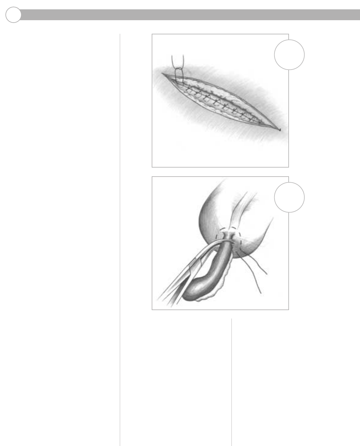
may be used on peritoneum and/or
fascial layers of the abdominal wall
to provide a temporary seal during
the healing process.
INTERRUPTED SUTURES
Interrupted sutures use a number
of strands to close the wound.
Each strand is tied and cut after
insertion. This provides a more
secure closure, because if one
suture breaks, the remaining
sutures will hold the wound edges
in approximation.
Interrupted sutures may be
used if a wound is infected,
because microorganisms may be
less likely to travel along a series
of interrupted stitches.
DEEP SUTURES
Deep sutures are placed completely
under the epidermal skin layer.
They may be placed as continuous
or interrupted sutures and are not
removed postoperatively.
BURIED SUTURES
Buried sutures are placed so that the
knot protrudes to the inside, under
the layer to be closed. This tech-
nique is useful when using large
diameter permanent sutures on
deeper layers in thin patients who
may be able to feel large knots that
are not buried.
PURSE-STRING SUTURES
Purse-string sutures are continuous
sutures placed around a lumen and
tightened like a drawstring to
invert the opening. They may be
placed around the stump of the
appendix, in the bowel to secure an
intestinal stapling device, or in an
organ prior to insertion of a tube
(such as the aorta, to hold the
cannulation tube in place during
an open heart procedure).
SUBCUTICULAR SUTURES
Subcuticular sutures are continuous
or interrupted sutures placed in the
dermis, beneath the epithelial layer.
Continuous subcuticular sutures are
placed in a line parallel to the
wound. This technique involves
taking short, lateral stitches the full
length of the wound. After the
suture has been drawn taut, the
distal end is anchored in the same
manner as the proximal end. This
may involve tying or any of a
variety of anchoring devices.
Subcuticular suturing may be
performed with absorbable suture
which does not require removal, or
with monofilament nonabsorbable
suture that is later removed by
simply removing the anchoring
device at one end and pulling the
opposite end.
THE SUTURE
22
DEEP
SUTURES
FIGURE
4
PURSE-STRING
SUTURES
FIGURE
5
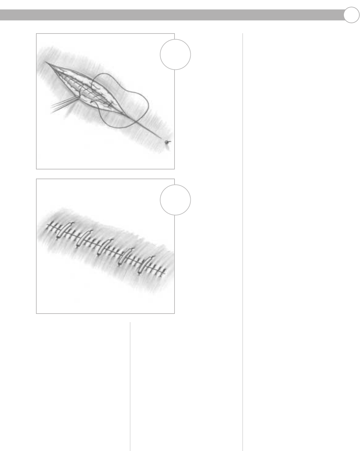
THE SECONDARY
SUTURE LINE
A secondary line of sutures may
be used:
• To reinforce and support the
primary suture line, eliminate
dead space, and prevent fluid
accumulation in an abdominal
wound during healing by first
intention. When used for this
purpose, they may also be called
retention, stay, or tension sutures.
• To support wounds for healing
by second intention.
• For secondary closure following
wound disruption when healing
by third intention.
NOTE: If secondary sutures are
used in cases of nonhealing, they
should be placed in opposite fashion
from the primary sutures
(i.e., interrupted if the primary
sutures were continuous,
continuous if the primary sutures
were interrupted).
Retention sutures are placed approxi-
mately 2 inches from each edge of
the wound. The tension exerted
lateral to the primary suture line
contributes to the tensile strength of
the wound. Through-and-through
sutures are placed from inside the
peritoneal cavity through all layers
of the abdominal wall, including the
peritoneum. They should be insert-
ed before the peritoneum is closed
using a simple interrupted stitch.
The wound may be closed in layers
for a distance of approximately
three-fourths its length. Then the
retention sutures in this area may be
drawn together and tied. It is
important that a finger be placed
within the abdominal cavity to
prevent strangulation of the viscera
in the closure. The remainder of the
wound may then be closed. Prior
to tightening and tying the final
retention sutures, it is important
to explore the abdomen again with
a finger to prevent strangulation
of viscera in the closure. The
remainder of the wound may then
be closed.
Retention sutures utilize
nonabsorbable suture material.
They should therefore be removed
as soon as the danger of sudden
increases in intra-abdominal
pressure is over— usually 2 to 6
weeks, with an average of 3 weeks.
STITCH PLACEMENT
Many types of stitches are used
for both continuous and interrupted
suturing. In every case, equal
"bites" of tissue should be taken
on each side of the wound. The
needle should be inserted from
1 to 3 centimeters from the edge
of the wound, depending upon the
type and condition of the tissue
being sutured.
CHAPTER 2 23
* Trademark
SUBCUTANEOUS
SUTURES
FIGURE
6
RETENTION
SUTURE
BOLSTER
FIGURE
7
KNOT TYING
Of the more than 1,400 different
types of knots described in THE
ENCYLCOPEDIA OF KNOTS,
only a few are used in modern
surgery. It is of paramount
importance that each knot placed
for approximation of tissues or
ligation of vessels be tied with
precision and each must hold with
proper tension.
KNOT SECURITY
The construction of ETHICON*
sutures has been carefully
designed to produce the optimum
combination of strength,
uniformity, and hand for each
material. The term hand is the
most subtle of all suture quality
aspects. It relates to the feel of the
suture in the surgeon’s hands, the
smoothness with which it passes
through tissue and ties down, the
way in which knots can be set and
snugged down, and most of all, to
the firmness or body of the suture.
Extensibility relates to the way in
which the suture will stretch slightly
during knot tying and then recover.
The stretching characteristics
provide the signal that alerts the
surgeon to the precise moment
when the suture knot is snug.
The type of knot tied will depend
upon the material used, the depth
and location of the incision, and the
amount of stress that will be placed
upon the wound postoperatively.
Multifilament sutures are generally
easier to handle and tie than
monofilament sutures, however, all
the synthetic materials require a
specific knotting technique. With
multifilament sutures, the nature of
the material and the braided or
twisted construction provide a high
coefficient of friction and the knots
remain as they are laid down. In
monofilament sutures, on the other
hand, the coefficient of friction is
relatively low, resulting in a greater
tendency for the knot to loosen
after it has been tied. In addition,
monofilament synthetic polymeric
materials possess the property of
memory. Memory is the tendency
not to lie flat, but to return to a
given shape set by the material’s
extrusion process or the suture’s
packaging. The RELAY* suture
delivery system delivers sutures with
minimal package memory due to its
unique package design.
Suture knots must be properly
placed to be secure. Speed in knot
tying frequently results in less than
perfect placement of the strands.
In addition to variables inherent in
the suture materials, considerable
variation can be found between
knots tied by different surgeons
and even between knots tied by
the same individual on different
occasions. The general principles
of knot tying which apply to all
suture materials are:
1. The completed knot must be
firm, and so tied that slipping is
virtually impossible. The
simplest knot for the material is
the most desirable.
2. The knot must be as small as
possible to prevent an excessive
amount of tissue reaction when
absorbable sutures are used, or to
minimize foreign body reaction
to nonabsorbable sutures. Ends
should be cut as short as possible.
3. In tying any knot, friction
between strands ("sawing") must
be avoided as this can weaken the
integrity of the suture.
4. Care should be taken to avoid
damage to the suture material
when handling. Avoid the
crushing or crimping application
of surgical instruments, such as
needleholders and forceps, to the
strand except when grasping the
free end of the suture during an
instrument tie.
5. Excessive tension applied by the
surgeon will cause breaking of
the suture and may cut tissue.
Practice in avoiding excessive
tension leads to successful use of
finer gauge materials.
6. Sutures used for approximation
should not be tied too tightly,
because this may contribute to
tissue strangulation.
THE SUTURE
24
COMMONLY
USED TYPES
OF STITCHES
To appose skin and other tissue
Over-and-over Over-and-over
Subcuticular Vertical mattress
Horizontal mattress
To invert tissue
Lembert Lembert
Cushing Halsted
Connell Purse-string
To evert tissue
Horizontal mattress Horizontal mattress
CONTINUOUS SUTURE INTERRUPTED SUTURES
TABLE
6
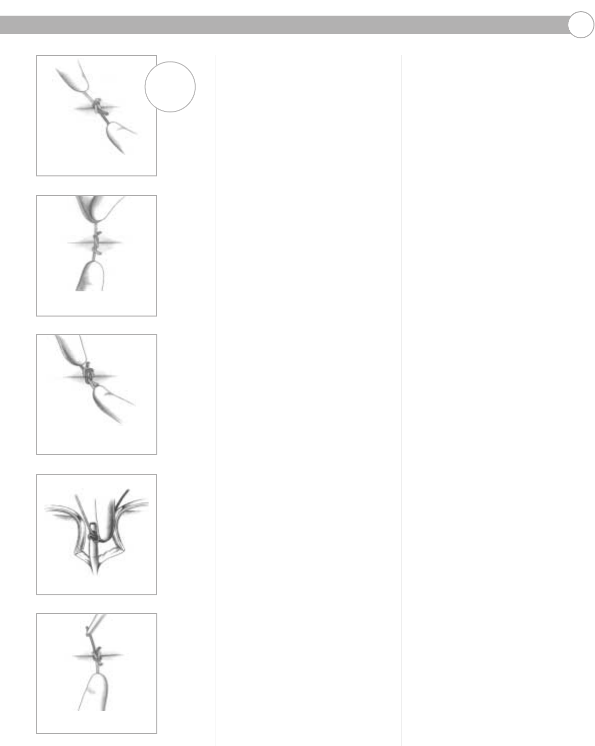
7. After the first loop is tied, it is
necessary to maintain traction
on one end of the strand to
avoid loosening of the throw if
being tied under any tension.
8. Final tension on final throw
should be as nearly horizontal
as possible.
9. The surgeon should not hesitate
to change stance or position in
relation to the patient in order
to place a knot securely and flat.
10. Extra ties do not add to the
strength of a properly tied and
squared knot. They only
contribute to its bulk. With
some synthetic materials, knot
security requires the standard
surgical technique to flat and
square ties with additional
throws if indicated by surgical
circumstance and the experience
of the surgeon.
KNOT TYING TECHNIQUES
MOST OFTEN USED
An important part of good suturing
technique is correct method in
knot tying. A seesaw motion, or
the sawing of one strand down over
another until the knot is formed,
may materially weaken sutures to
the point that they may break when
the second throw is made, or even
worse, in the postoperative period
when the suture is further weakened
by increased tension or motion. If
the two ends of the suture are
pulled in opposite directions with
uniform rate and tension, the knot
may be tied more securely.
Some procedures involve tying
knots with the fingers, using one or
two hands; others involve tying with
the help of instruments. Perhaps
the most complex method of knot
tying is done during endoscopic
procedures, when the surgeon must
manipulate instruments from well
outside the body cavity.
Following are the most frequently
used knot tying techniques with
accompanying illustrations of
finished knots.
SQUARE KNOT
The two-hand square knot is the
easiest and most reliable for tying
most suture materials. It may be
used to tie surgical gut, virgin silk,
surgical cotton, and surgical stain-
less steel. Standard technique of flat
and square ties with additional
throws if indicated by the surgical
circumstance and the experience of
the operator should be used to tie
MONOCRYL* (poliglecaprone 25)
suture, coated VICRYL*
(polyglactin 910) suture, coated
VICRYL* RAPIDE (polyglactin
910) suture, PDS* II (polydiox-
anone) suture, ETHILON* nylon
suture, ETHIBOND* EXCEL
polyester suture, PERMA-HAND*
silk suture, PRONOVA* poly
(hexafluoropropylene-VDF)
suture, and PROLENE*
polypropylene suture.
Wherever possible, the square
knot is tied using the two-hand
technique. On some occasions it
will be necessary to use one hand,
either the left or the right, to tie a
square knot.
CAUTION: If the strands of a
square knot are inadvertently
incorrectly crossed, a granny knot
will result. Granny knots are not
recommended because they have a
tendency to slip when subjected to
increased stress.
CHAPTER 2 25
* Trademark
FIGURE
8
FINISHED
SUTURE
TIES
Square knot
Surgeon’s knot–first
throw
Surgeon’s knot–second
throw
Deep tie
Instrument tie

SURGEON’S OR
FRICTION KNOT
The surgeon's or friction knot is
recommended for tying Coated
VICRYL* (polyglactin 910) suture,
ETHIBOND* EXCEL polyester
suture, ETHILON* nylon suture,
MERSILENE* polyester fiber
suture, NUROLON* nylon suture,
PRONOVA* poly (hexafluoro-
propylene-VDF) suture, and
PROLENE* polypropylene
suture. The surgeon’s knot also
may be performed using a
one-hand technique.
DEEP TIE
Tying deep in a body cavity can be
difficult. The square knot must be
firmly snugged down as in all
situations. However, the operator
must avoid upward tension which
may tear or avulse the tissue.
LIGATION USING A
HEMOSTATIC CLAMP
Frequently it is necessary to ligate
a blood vessel or tissue grasped in
a hemostatic clamp to achieve
hemostasis in the operative field.
INSTRUMENT TIE
The instrument tie is useful when
one or both ends of the suture
material are short. For best results,
exercise caution when using a
needleholder with any monofila-
ment suture, as repeated bending
may cause these sutures to break.
ENDOSCOPIC KNOT
TYING TECHNIQUES
During an endoscopic procedure,
a square knot or surgeon's knot may
be tied either outside the abdomen
and pushed down into the body
through a trocar (extracorporeal)
or directly within the abdominal
cavity (intracorporeal).
In extracorporeal knot tying, the
suture appropriately penetrates the
tissue, and both needle and suture
are removed from the body cavity,
bringing both suture ends outside
of the trocar. Then a series of
half-hitches are tied, each one
being pushed down into the cavity
and tightened with an endoscopic
knot pusher.
Intracorporeal knot tying is
performed totally within the
abdominal cavity. After the suture
has penetrated the tissue, the needle
is cut from the suture and removed.
Several loops are made with the
suture around the needleholder,
and the end of the suture is pulled
through the loops. This technique
is then repeated to form a surgeon's
knot, which is tightened by the
knot pusher.
In both extracorporeal and intracor-
poreal knot tying, the following
principles of suture manipulation
on tissue should be observed:
1. Handle tissue as gently as
possible to avoid tissue trauma.
2. Grasp as little tissue as possible.
3. Use the smallest suture possible
for the task.
4. Exercise care in approximating
the knot so that the tissue being
approximated is not strangulated.
5. Suture must be handled with care
to avoid damage.
CUTTING THE
SECURED SUTURES
Once the knot has been securely
tied, the ends must be cut. Before
cutting, make sure both tips of
the scissors are visible to avoid
inadvertently cutting tissue beyond
the suture.
Cutting sutures entails running
the tip of the scissors lightly down
the suture strand to the knot.
The ends of surgical gut are left
relatively long, approximately 1/4"
(6mm) from the knot. Other
materials are cut closer to the
knot, approximately 1/8" (3mm),
to decrease tissue reaction and
minimize the amount of foreign
material left in the wound. To
ensure that the actual knot is not
cut, twist or angle the blades of the
scissors prior to cutting.
Make certain to remove the
cut ends of the suture from the
operative site.
SUTURE REMOVAL
When the external wound has
healed so that it no longer needs the
support of nonabsorbable suture
material, skin sutures must be
removed. The length of time the
sutures remain in place depends
upon the rate of healing and the
nature of the wound. General rules
are as follows.
THE SUTURE
26
SUTURE
REMOVAL
Skin on the face and neck 2 to 5 days
Other skin sutures 5 to 8 days
Retention sutures 2 to 6 weeks
SUTURE LOCATION TIME FOR SUTURE REMOVAL TABLE
7
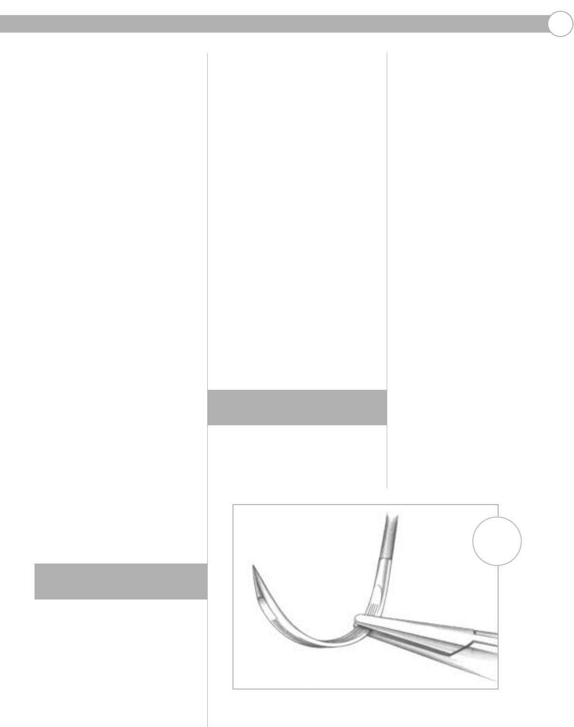
Sutures should be removed using
aseptic and sterile technique. The
surgeon uses a sterile suture removal
tray prepared for the procedure.
The following steps are taken:
• STEP 1—Cleanse the area with
an antiseptic. Hydrogen peroxide
can be used to remove dried serum
encrusted around the sutures.
•STEP 2—Pick up one end of the
suture with thumb forceps, and
cut as close to the skin as possible
where the suture enters the skin.
•STEP 3—Gently pull the suture
strand out through the side oppo-
site the knot with the forceps. To
prevent risk of infection, the
suture should be removed without
pulling any portion that has been
outside the skin back through
the skin.
NOTE: Fast absorbing synthetic or
gut suture material tend to lose all
tensile strength in 5 to 7 days and
can be removed easily without
cutting. A common practice is
to cover the skin sutures with
PROXI-STRIP* skin closures
during the required healing period.
After the wound edges have
regained sufficient tensile strength,
the sutures may be removed by
simply removing the PROXI-STRIP
skin closures.
SUTURE
HANDLING TIPS
These guidelines will help the surgi-
cal team keep their suture inventory
up to date and their sutures in the
best possible condition.
1. Read labels.
2. Heed expiration dates and
rotate stock.
3. Open only those sutures needed
for the procedure at hand.
4. Straighten sutures with a gentle
pull. Never crush or rub them.
5. Don't pull on needles.
6. Avoid crushing or crimping
suture strands with surgical
instruments.
7. Don't store surgical gut
near heat.
8. Moisten—but never soak—
surgical gut.
9. Do not wet rapidly absorbing
sutures.
10. Keep silk dry.
11. Wet linen and cotton to increase
their strength.
12. Don't bend stainless steel wire.
13. Draw nylon between gloved
fingers to remove the packaging
"memory."
14. Arm a needleholder properly.
SUTURE SELECTION
PROCEDURE
PRINCIPLES OF
SUTURE SELECTION
The surgeon has a choice of suture
materials from which to select for
use in body tissues. Adequate
strength of the suture material will
prevent suture breakage. Secure
knots will prevent knot slippage.
But the surgeon must understand
the nature of the suture material,
the biologic forces in the healing
wound, and the interaction of
the suture and the tissues. The
following principles should guide
the surgeon in suture selection.
1. When a wound has reached
maximal strength, sutures are no
longer needed. Therefore:
a. Tissues that ordinarily heal
slowly such as skin, fascia,
and tendons should usually
be closed with nonabsorbable
sutures. An absorbable suture
with extended (up to 6
months) wound support may
also be used.
b. Tissues that heal rapidly
such as stomach, colon and
bladder may be closed with
absorbable sutures
2. Foreign bodies in potentially
contaminated tissues may convert
contamination into infection.
CHAPTER 2 27
* Trademark
ARMING
A NEEDLE-
HOLDER
PROPERLY
FIGURE
9
Grasp the needle one-third to one-half of the distance from
the swaged end to the point.

3. Where cosmetic results are
important, close and prolonged
apposition of wounds and
avoidance of irritants will
produce the best results.
Therefore:
a. Use the smallest inert
monofilament suture
materials such as nylon
or polypropylene.
b. Avoid skin sutures and close
subcuticularly whenever
possible.
c. Under certain circumstances,
to secure close apposition of
skin edges, a topical skin
adhesive or skin closure
tape may be used.
4. Foreign bodies in the presence
of fluids containing high concen-
trations of crystalloids may act
as a nidus for precipitation and
stone formation.
Therefore:
a. In the urinary and biliary
tracts, use rapidly absorbed
sutures.
5. Regarding suture size:
a. Use the finest size suture
commensurate with the
natural strength of the tissue.
b. If the postoperative course of
the patient may produce
sudden strains on the suture
line, reinforce it with
retention sutures. Remove
them as soon as the patient’s
condition is stabilized.
SURGERY WITHIN THE
ABDOMINAL WALL CAVITY
Entering the abdomen, the surgeon
will need to seal or tie off
subcutaneous blood vessels
immediately after the incision is
made, using either an electrosurgical
unit designed for this purpose or
free ties (ligatures). If ligatures are
used, an absorbable suture material
is generally preferred. When
preparing the ties, the scrub person
often prepares one strand on a
needle for use as a suture ligature
should the surgeon wish to
transfix a large blood vessel.
Once inside, the type of suture
selected will depend upon the
nature of the operation and the
surgeon's technique.
THE GASTROINTESTINAL TRACT
Leakage from an anastomosis
or suture site is the principal
problem encountered performing
a procedure involving the
gastrointestinal tract. This problem
can lead to localized or generalized
peritonitis. Sutures should not be
tied too tightly in an anastomotic
closure. Wounds of the stomach and
intestine are rich in blood supply
and may become edematous and
hardened. Tight sutures may cut
through the tissue and cause
leakage. A leak-proof anastomosis
can be achieved with either a
single or double-layer closure.
For a single-layer closure, interrupt-
ed sutures should be placed
approximately 1/4" (6mm) apart.
Suture is placed through the
submucosa, into the muscularis and
through the serosa. Because the
submucosa provides strength in
the gastrointestinal tract, effective
closure involves suturing the
submucosal layers in apposition
without penetrating the mucosa.
A continuous suture line provides a
tighter seal than interrupted sutures.
However, if a continuous suture
breaks, the entire line may separate.
Many surgeons prefer to use a
double-layer closure, placing a
second layer of interrupted sutures
through the serosa for insurance.
Absorbable VICRYL* sutures, or
chromic gut sutures may be used
in either a single or double-layer
closure. Surgical silk may also be
used for the second layer of a
double-layer closure.
Inverted, everted, or end-to-end
closure techniques have all been
used successfully in this area, but
they all have drawbacks. The
surgeon must take meticulous care
in placing the sutures in the submu-
cosa. Even with the best technique,
some leakage may occur.
Fortunately, the omentum usually
confines the area, and natural body
defenses handle the problem.
THE STOMACH
For an organ that contains free
hydrochloric acid and potent
proteolytic enzymes, the stomach
heals surprisingly quickly.
Stomach wounds attain maximum
strength within 14 to 21 days
postoperatively, and have a peak
rate of collagen synthesis at 5 days.
Absorbable sutures are usually
acceptable in the stomach, although
they may produce a moderate
reaction in both the wound and
normal tissue. Coated VICRYL*
sutures are most commonly used.
PROLENE* sutures may also be
used for stomach closure.
THE SMALL INTESTINE
Closure of the small intestine
presents the same considerations
as the stomach. Proximal intestinal
contents, primarily bile or
pancreatic juices, may cause a
THE SUTURE
28
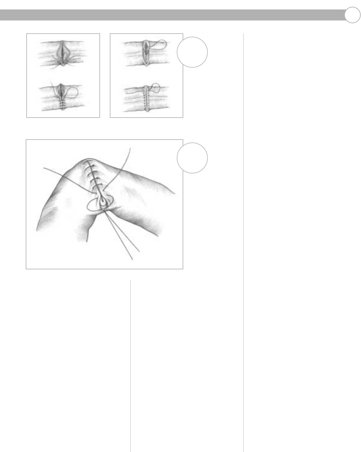
severe chemical (rather than
bacterial) peritonitis.
If using an inverted closure
technique, care must be taken to
minimize the cuff of tissue which
protrudes into the small sized
intestinal lumen in order to avoid
partial or complete obstruction.
Absorbable sutures are usually
preferred, particularly because they
will not permanently limit the
lumen diameter. A nonabsorbable
suture may be used in the serosal
layer for added assurance.
The small intestine heals very
rapidly, reaching maximal strength
in approximately 14 days.
THE COLON
The high microbial content of
the colon once made contamination
a major concern. But absorbable
sutures, once absorbed, leave no
channel for microbial migration.
Still, leakage of large bowel
contents is of great concern as it
is potentially more serious than
leakage in other areas of the
gastrointestinal tract.
The colon is a strong organ—
approximately twice as strong in the
sigmoid region as in the cecum. Yet,
wounds of the colon gain strength
at the same rate regardless of their
location. This permits the same
suture size to be used at either end
of the colon. The colon heals at a
rate similar to that of the stomach
and small intestine. A high rate of
collagen synthesis is maintained for
a prolonged period (over 120 days).
The entire gastrointestinal tract
exhibits a loss of collagen and
increased collagenous activity
immediately following colon
anastomosis. Both absorbable and
nonabsorbable sutures may be used
for closure of the colon. Placement
of sutures in the submucosa,
avoiding penetration of the mucosa,
will help prevent complications.
THE RECTUM
The rectum heals very slowly.
Because the lower portion is below
the pelvic peritoneum, it has no
serosa. A large bite of muscle should
be included in an anastomosis, and
the sutures should be tied carefully
to avoid cutting through the tissues.
Monofilament sutures reduce the
risk of bacterial proliferation in
the rectum.
THE BILIARY TRACT
THE GALLBLADDER
Within the gallbladder, the cystic
and common bile ducts heal rapidly.
Their contents present special
considerations for suture selection.
The presence of a foreign body such
as a suture in an organ that is prone
to crystal formation may precipitate
the formation of "stones."
Multifilament sutures should
probably not be used because it
is not always possible to prevent
exposure of a suture in the ducts.
The surgeon should choose an
absorbable suture in the finest size
possible that leaves the least surface
area exposed.
CHAPTER 2 29
* Trademark
ANASTOMOTIC
CLOSURE
TECHNIQUE
INVERTED
CLOSURE
TECHNIQUE
FIGURE
10
FIGURE
11
Single layer Double layer
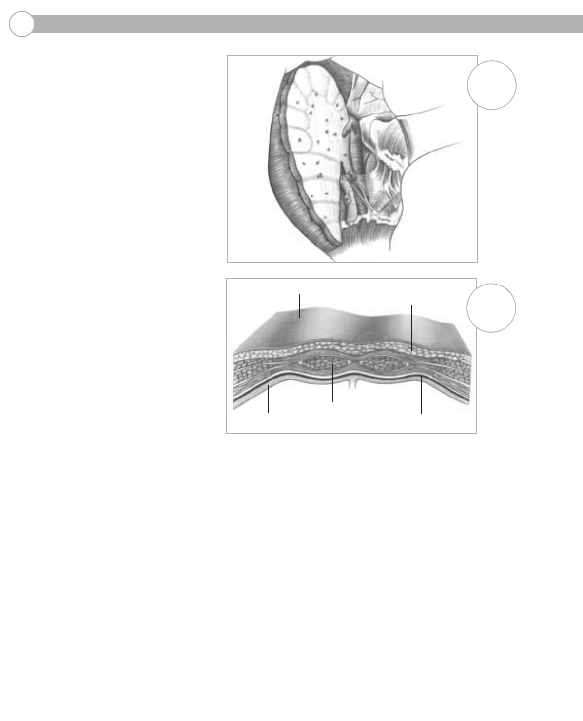
PARENCHYMATOUS ORGANS
THE SPLEEN, LIVER AND KIDNEY
On occasion, a surgeon may be
called upon to repair a laceration
of one of these vital organs. If
large vessels, particularly arteries,
within these organs have been
severed, they must be located
and ligated before attempting
to close the defect. Otherwise,
hematomas or secondary hemor-
rhage may occur.
Because these organs are composed
chiefly of cells with little connective
tissue for support, attempts must
be made to coapt the outer fibrous
capsule of the torn tissue. In
the absence of hemorrhage, little
tension is placed on the suture line
and only small size sutures need to
be used. If the tissue cannot be
approximated, tacking a piece of
omentum over the defect will
usually suffice to provide closure.
Sutures do not need to be placed
close together or deeply into
the organ.
Lacerations in this area tend to
heal rapidly. New fibrous tissue will
usually form over the wound with
7 to 10 days.
In a liver resection, suturing of the
wedges in a horizontal through-
and-through fashion should hold
the tissue securely. Large vessels
should be tied using VICRYL*
sutures or silk. Raw surfaces can be
closed or repaired using VICRYL
(polyglactin 910) mesh.
CLOSING THE ABDOMEN
When closing the abdomen, the
closure technique may be more
important than the type of suture
material used.
THE PERITONEUM
The peritoneum, the thin membra-
nous lining of the abdominal cavity,
lies beneath the posterior fascia. It
heals quickly. Some believe that the
peritoneum does not require sutur-
ing, while others disagree. If the
posterior fascia is securely closed,
suturing the peritoneum may not
contribute to the prevention of an
incisional hernia. Among surgeons
who choose to close the peri-
toneum, a continuous suture line
with absorbable suture material
is usually preferred. Interrupted
sutures can also be used for
this procedure.
FASCIA
This layer of firm, strong connective
tissue covering the muscles is the
main supportive structure of the
body. In closing an abdominal
incision, the fascial sutures must
hold the wound closed and
also help to resist changes in intra-
abdominal pressure. Occasionally,
synthetic graft material may be
used when fascia is absent or weak.
PROLENE* polypropylene mesh may
be used to replace abdominal wall
or repair hernias when a great deal
of stress will be placed on the suture
line during healing. Nonabsorbable
sutures such as PROLENE suture
may be used to suture the graft to
the tissue.
THE SUTURE
30
LIVER
RESECTION
FIGURE
12
THE
ABDOMINAL
WALL
FIGURE
13
Skin
Subcutaneous fat
Peritoneum
Muscle tissue
Transversalis fascia
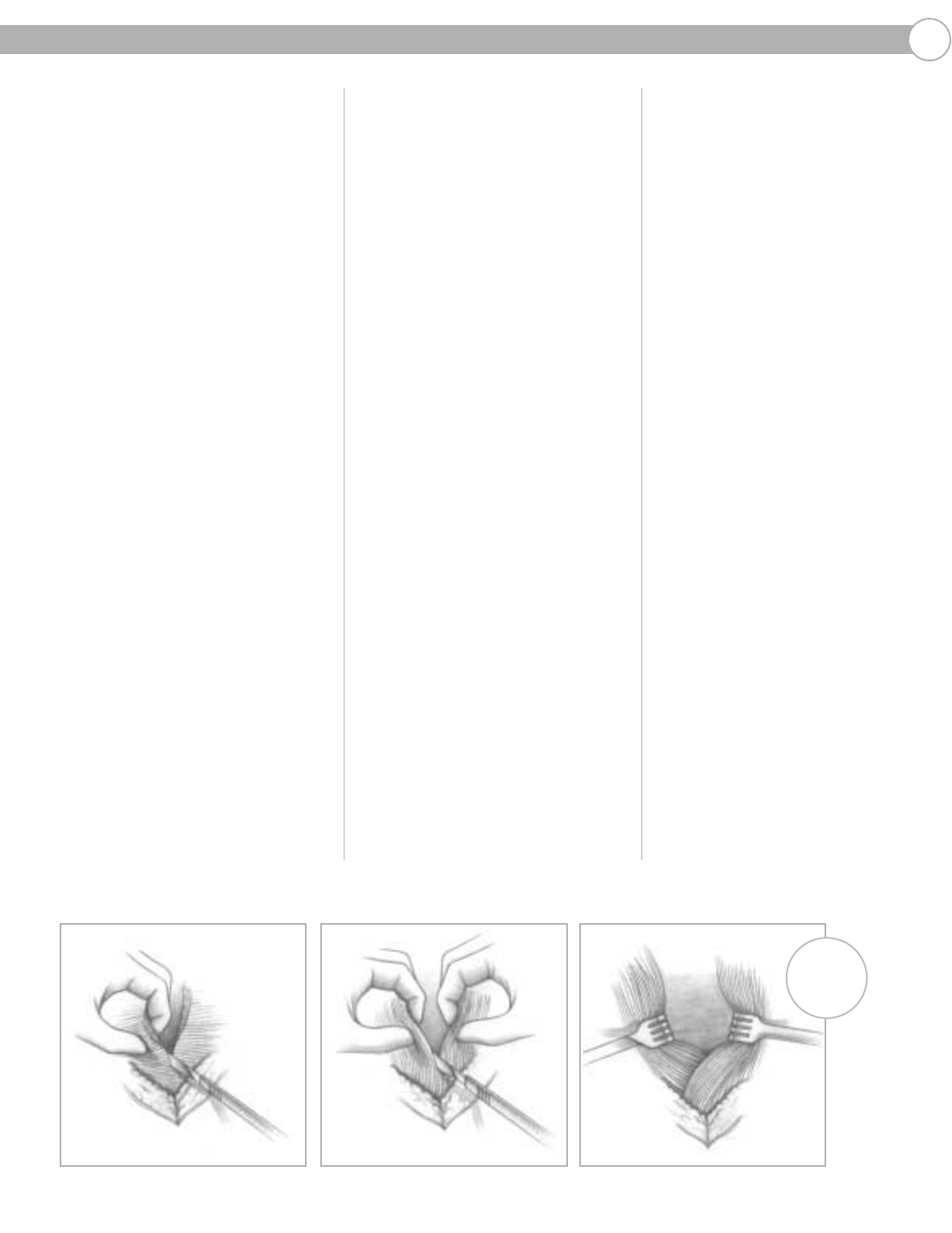
Fascia regains approximately 40%
of its original strength in 2 months.
It may take up to a year or longer to
regain maximum strength. Full
original strength is never regained.
The anatomic location and type of
abdominal incision will influence
how may layers of fascia will be
sutured. The posterior fascial layer
is always closed. The anterior layer
may be cut and may also require
suturing. Mass closure techniques
are becoming the most popular.
Most suture materials have some
inherent degree of elasticity. If not
tied too tightly, the suture will
"give" to accommodate postopera-
tive swelling that occurs. Stainless
steel sutures, if tied too tightly, will
cut like a knife as the tissue swells
or as tension is placed upon the
suture line. Because of the slow
healing time and because the fascial
suture must bear the maximum
stress of the wound, a moderate size
nonabsorbable suture may be used.
An absorbable suture with longer
lasting tensile strength, such as
PDS* II sutures, may also provide
adequate support. PDS II sutures
are especially well-suited for use in
younger, healthy patients.
Many surgeons prefer the use of
interrupted simple or figure-of eight
sutures to close fascia, while others
employ running suture or a
combination of these techniques.
In the absence of infection or gross
contamination, the surgeon may
choose either monofilament or
multifilament sutures. In the
presence of infection, a
monofilament absorbable material
like PDS II sutures or inert nonab-
sorbable sutures like stainless steel or
PROLENE* sutures may be used.
MUSCLE
Muscle does not tolerate suturing
well. However, there are several
options in this area.
Abdominal muscles may be either
cut, split (separated), or retracted,
depending upon the location and
type of the incision chosen. Where
possible, the surgeon prefers to
avoid interfering with the blood
supply and nerve function by
making a muscle-splitting incision
or retracting the entire muscle
toward its nerve supply. During
closure, muscles handled in this
manner do not need to be sutured.
The fascia is sutured rather than
the muscle.
The Smead-Jones far-and-near-
technique for abdominal wound
closure is strong and rapid, provides
good support during early healing
with a low incidence of wound
disruption, and has a low incidence
of late incisional problems. This is
a single-layer closure through both
layers of the abdominal wall fascia,
abdominal muscles, peritoneum,
and the anterior fascial layer. The
interrupted sutures resemble a
"figure of eight" when placed.
Absorbable PDS II sutures or
VICRYL* sutures are usually used.
Stainless steel sutures may also be
used. Monofilament PROLENE
sutures also provide all the
advantages of steel sutures: strength,
minimal tissue reactivity, and resist-
ance to bacterial contamination.
They are better tolerated than steel
sutures by patients in the late post-
operative months and are easier for
the surgeon to handle and tie.
However, both stainless steel and
PROLENE sutures may be
detectable under the skin of thin
patients. To avoid this problem,
knots should be buried in fascia
instead of in the subcutaneous
space.
CHAPTER 2 31
SURGICAL
OPTIONS IN
MUSCLE
FIGURE
14
Cutting Splitting Retracting
* Trademark

SUBCUTANEOUS FAT
Neither fat nor muscle tolerate
suturing well. Some surgeons
question the advisability of placing
sutures in fatty tissue because it
has little tensile strength due to
its composition, which is mostly
water. However, others believe it
is necessary to place at least a
few sutures in a thick layer of
subcutaneous fat to prevent dead
space, especially in obese patients.
Dead spaces are most likely to
occur in this type of tissue, so
the edges of the wound must be
carefully approximated. Tissue
fluids can accumulate in these
pocket-like spaces, delaying healing
and predisposing infection.
Absorbable sutures are usually
selected for the subcutaneous layer.
VICRYL* suture is especially suited
for use in fatty, avascular tissue since
it is absorbed by hydrolysis. The
surgeon may use the same type and
size of material used earlier to ligate
blood vessels in this layer.
SUBCUTICULAR TISSUE
To minimize scarring, suturing
the subcuticular layer of tough
connective tissue will hold the skin
edges in close approximation. In a
single-layer subcuticular closure, less
evidence of scar gaping or expansion
may be seen after a period of 6 to 9
months than is evident with simple
skin closure. The surgeon takes
continuous short lateral stitches
beneath the epithelial layer of skin.
Either absorbable or nonabsorbable
sutures may be used. If nonab-
sorbable material is chosen, one end
of the suture strand will protrude
from each end of the incision, and
the surgeon may tie them together
to form a "loop" or knot the ends
outside of the incision.
To produce only a hair-line scar
(on the face, for example), the
skin can be held in very close
approximation with skin closure
tapes in addition to subcuticular
sutures. Tapes may be left on the
wound for an extended period of
time depending upon their location
on the body.
When great tension is not placed
upon the wound, as in facial or
neck surgery, very fine sizes of
subcuticular sutures may be used.
Abdominal wounds that must
withstand more stress call for larger
suture sizes.
Some surgeons choose to close
both the subcuticular and epidermal
layers to achieve minimal scarring.
Chromic surgical gut and polymeric
materials, such as MONOCRYL*
suture, are acceptable for placement
within the dermis. They are capable
of maintaining sufficient tensile
strength through the collagen
synthesis stage of healing which
lasts approximately 6 weeks. The
sutures must not be placed too
close to the epidermal surface to
reduce extrusion. If the skin is
nonpigmented and thin, a clear or
white monofilament suture such
as MONOCRYL suture will be
invisible to the eye. MONOCRYL
suture is particularly well-suited for
this closure because, as a monofila-
ment, it does not harbor infection
and, as a synthetic absorbable
suture, tissue reaction is minimized.
After this layer is closed, the skin
edges may then be approximated.
SKIN
Skin is composed of the epithelium
and the underlying dermis. It is so
tough that a very sharp needle is
essential for every stitch to minimize
tissue trauma. (See Chapter 3: The
Surgical Needle.)
Skin wounds regain tensile strength
slowly. If a nonabsorbable suture
material is used, it is typically
removed between 3 and 10 days
postoperatively, when the wound
has only regained approximately
5% to 10% of its strength. This is
possible because most of the stress
placed upon the healing wound is
absorbed by the fascia, which the
surgeon relies upon to hold the
wound closed. The skin or
subcuticular sutures need only be
strong enough to withstand natural
skin tension and hold the wound
edges in apposition.
The use of coated VICRYL*
RAPIDE suture, a rapidly absorbed
synthetic suture, eliminates the
need for suture removal. Coated
VICRYL RAPIDE suture, which
is indicated for superficial closure
of skin and mucosa, provides
short-term wound support
consistent with the rapid healing
characteristics of skin. The sutures
begin to fall off in 7 to 10 days,
with absorption essentially
complete at 42 days.
Suturing technique for skin
closure may be either continuous
or interrupted. Skin edges should
be everted. Preferably, each suture
strand is passed through the skin
only once, reducing the chance
of cross-contamination across the
entire suture line. Interrupted
technique is usually preferred.
THE SUTURE
32
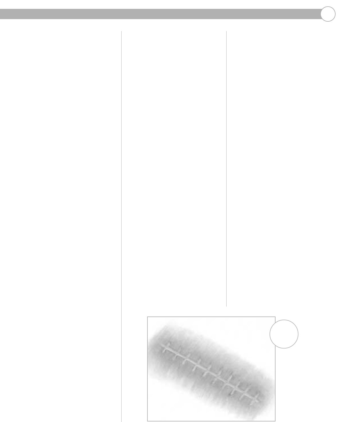
If surgeon preference indicates
the use of a nonabsorbable suture
material, several issues must be
considered. Skin sutures are exposed
to the external environment,
making them a serious threat
to wound contamination and
stitch abscess. The interstices of
multifilament sutures may provide
a haven for microorganisms.
Therefore, monofilament nonab-
sorbable sutures may be preferred for
skin closure. Monofilament sutures
also induce significantly less tissue
reaction than multifilament sutures.
For cosmetic reasons, nylon or
polypropylene monofilament sutures
may be preferred. Many skin
wounds are successfully closed with
silk and polyester multifilaments
as well. Tissue reaction to nonab-
sorbable sutures subsides and
remains relatively acellular as
fibrous tissue matures and forms a
dense capsule around the suture.
(Note, surgical gut has been known
to produce tissue reaction. Coated
VICRYL* RAPIDE suture elicits
a lower tissue reaction than chromic
gut suture due to its accelerated
absorption profile.) The key to suc-
cess is early suture removal before
epithelialization of the suture tract
occurs and before contamination is
converted into infection.
A WORD ABOUT SCARRING
(EPITHELIALIZATION)
When a wound is sustained in
the skin—whether accidentally or
during a surgical procedure—the
epithelial cells in the basal layer at
the margins of the wound flatten
and move into the wound area.
They move down the wound edge
until they find living, undamaged
tissue at the base of the wound.
Then they move across the wound
bed to make contact with similar
cells migrating from the opposite
side of the wound. They move
down the suture tract after if has
been embedded in the skin. When
the suture is removed, the tract of
the epithelial cells remains.
Eventually, it may disappear, but
some may remain and form keratin.
A punctate scar is usually seen on
the skin surface and a "railroad
track" or "crosshatch" appearance
on the wound may result. This is
relatively rare if the skin sutures are
not placed with excessive tension
and are removed by the seventh
postoperative day.
The forces that create the distance
between the edges of the wound
will remain long after the sutures
have been removed. Significant
collagen synthesis will occur from
5 to 42 days postoperatively. After
this time, any additional gain in
tensile will be due to remodeling,
or crosslinking, of collagen fibers
rather than to collagen synthesis.
Increases in tensile strength will
continue for as long as 2 years, but
the tissue will never quite regain its
original strength.
CLOSURE WITH
RETENTION SUTURES
We have already discussed the
techniques involved with placing
retention sutures, and using them
in a secondary suture line. (See the
section on Suturing Techniques.)
Heavy sizes (0 to 5) of nonabsorbable
materials are usually used for
retention sutures, not for strength,
but because larger sizes are less likely
to cut through tissue when a sudden
rise in intra-abdominal pressure
occurs from vomiting, coughing,
straining, or distention. To prevent
the heavy suture material from
cutting into the skin under stress,
one end of the retention suture may
be threaded through a short length
of plastic or rubber tubing called a
bolster or bumper before it is tied. A
plastic bridge with adjustable
features may also be used to protect
the skin and primary suture line
and permit postoperative wound
management for patient comfort.
Properly placed retention sutures
provide strong reinforcement for
abdominal wounds, but also cause
the patient more postoperative pain
than does a layered closure. The
best technique is to use a material
with needles swaged on each end
CHAPTER 2 33
* Trademark
THE RAILROAD
TRACK SCAR
CONFIGURATION
FIGURE
15
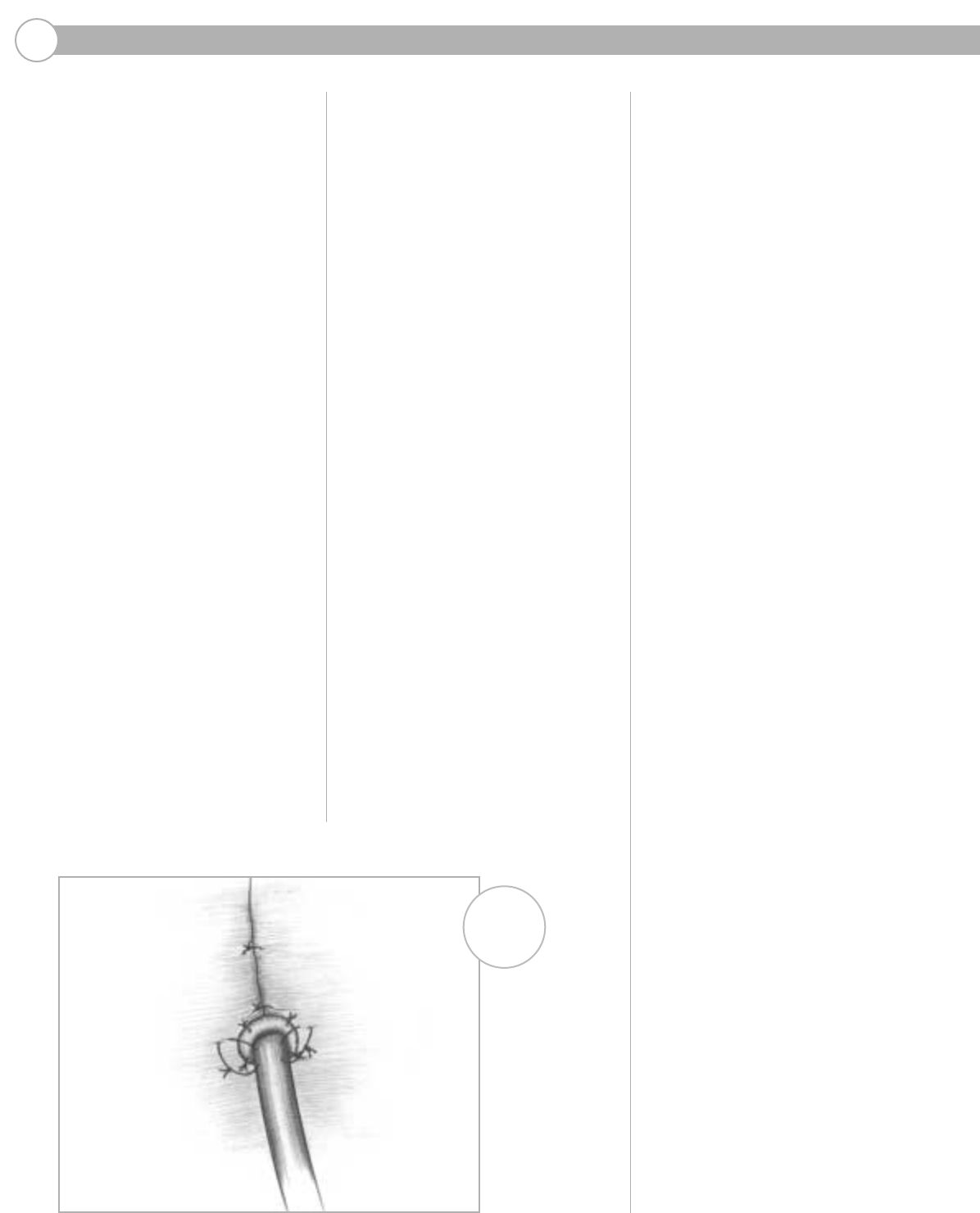
(double-armed). They should be
placed from the inside of the wound
toward the outside skin to avoid
pulling potentially contaminated
epithelial cells through the entire
abdominal wall.
The ETHICON retention
suture line includes ETHILON*
sutures, MERSILENE* sutures,
ETHIBOND* EXCEL sutures, and
PERMA-HAND* sutures. Surgical
steel sutures may also be used.
Retention sutures may be left in
place for 14 to 24 days postopera-
tively. Three weeks is an average
length of time. Assessment of the
patient's condition is the controlling
factor in deciding when to remove
retention sutures.
SUTURE FOR DRAINS
If a drainage tube is placed in a
hollow organ or a bladder drain is
inserted, it may be secured to the
wall of the organ being drained
with absorbable sutures. The surgeon
may also choose to minimize the
distance between the organ and the
abdominal wall by using sutures to
tack the organ being drained to the
peritoneum and fascia.
Sutures may be placed around the
circumference of the drain, either
two sutures at 12 and 6 o'clock
positions, or four sutures at 12, 3,
6, and 9 o'clock positions, and
secured to the skin with temporary
loops. When the drain is no longer
needed, the skin sutures may be
easily removed to remove the drain.
The opening can be left open to
permit additional drainage until it
closes naturally.
A drainage tube inserted into the
peritoneal cavity through a stab
wound in the abdominal wall
usually is anchored to the skin with
one or two nonabsorbable sutures.
This prevents the drain from
slipping into or out of the wound.
SUTURE NEEDS IN OTHER
BODY TISSUES NEUROSURGERY
Surgeons have traditionally used an
interrupted technique to close the
galea and dura mater.
The tissue of the galea, similar to
the fascia of the abdominal cavity,
is very vascular and hemostatic.
Therefore, scalp hematoma is a
potential problem, and the surgeon
must be certain to close well.
The dura mater is the outermost of
the three meninges that protects the
brain and spinal cord. It tears with
ease and cannot withstand too
much tension. The surgeon may
drain some of the cerebrospinal
fluid to decrease volume, easing the
tension on the dura before closing.
If it is too damaged to close, a
patch must be inserted and sutured
in place.
Surgical silk is appropriate in this
area for its pliability and easy knot
tying properties. Unfortunately,
it elicits a significant foreign body
tissue reaction. Most surgeons have
switched to NUROLON* sutures
or coated VICRYL* sutures because
they tie easily, offer greater strength
than surgical silk, and cause less
tissue reaction. PROLENE* sutures
have also been accepted by surgeons
who prefer a continuous closure
technique, who must repair
potentially infected wounds, or
who must repair dural tears.
In peripheral nerve repair, precise
suturing often requires the aid of
an operating microscope. Suture
gauge and needle fineness must be
consistent with nerve size. After the
motor and sensory fibers are proper-
ly realigned, the epineurium (the
outer sheath of the nerve) is
sutured. The strength of sutures in
this area is less of a consideration
than the degree of inflammatory
and fibroplastic tissue reaction. Fine
sizes of nylon, polyester, and poly-
propylene are preferred.
MICROSURGERY
The introduction of fine sizes of
sutures and needles has increased
the use of the operating microscope.
ETHICON introduced the first
THE SUTURE
34
PLACEMENT
OF SUTURES
AROUND
A DRAIN
FIGURE
16
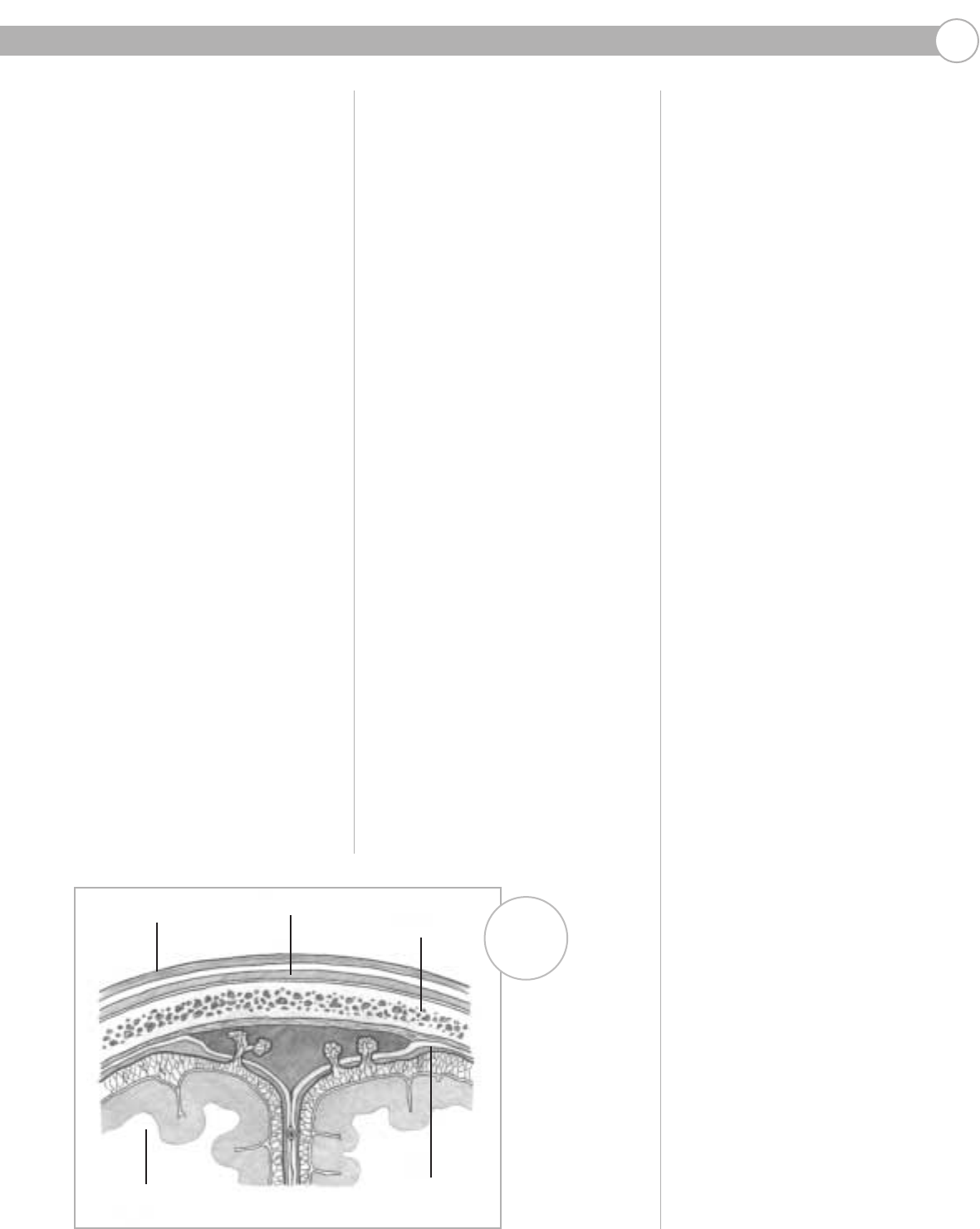
* Trademark
microsurgery sutures—ETHILON*
sutures—in sizes 8-0 through
11-0. Since then, the microsurgery
line has expanded to include
PROLENE* sutures and coated
VICRYL* sutures. Literally all
surgical specialties perform some
procedures under the operating
microscope, especially vascular and
nerve anastomosis.
OPHTHALMIC SURGERY
The eye presents special healing
challenges. The ocular muscles, the
conjunctiva, and the sclera have
good blood supplies; but the cornea
is an avascular structure. While
epithelialization of the cornea
occurs rapidly in the absence of
infection, full thickness cornea
wounds heal slowly. Therefore, in
closing wounds such as cataract
incisions, sutures should remain in
place for approximately 21 days.
Muscle recession, which involves
suturing muscle to sclera, only
requires sutures for approximately
7 days.
Nylon was the preferred suture
material for ophthalmic surgery.
While nylon is not absorbed,
progressive hydrolysis of nylon
in vivo may result in gradual loss of
tensile strength over time. Fine sizes
of absorbable sutures are currently
used for many ocular procedures.
Occasionally, the sutures are
absorbed too slowly in muscle
recessions and produce granulomas
to the sclera. Too rapid absorption
has, at times, been a problem in
cataract surgery. Because they
induce less cellular reaction than
surgical gut and behave dependably,
VICRYL sutures have proven useful
in muscle and cataract surgery.
While some ophthalmic surgeons
promote the use of a "no-stitch"
surgical technique, 10-0 coated
VICRYL (polyglactin 910) violet
monofilament sutures offer distinct
advantages. They provide the securi-
ty of suturing immediately follow-
ing surgery but eliminate the risks
of suture removal and related
endophthalmitis.
The ophthalmologist has many
fine size suture materials to choose
from for keratoplasty, cataract,
and vitreous retinal microsurgical
procedures. In addition to
VICRYL* sutures, other monofila-
ment suture materials including
ETHILON sutures, PROLENE
sutures, and PDS* II sutures may
be used. Braided material such as
virgin silk, black braided silk,
MERSILENE* sutures, and coated
VICRYL sutures are also available
for ophthalmic procedures.
UPPER ALIMENTARY
TRACT PROCEDURES
The surgeon must consider the
upper alimentary tract from the
mouth down to the lower
esophageal sphincter to be a
potentially contaminated area.
The gut is a musculomembranous
canal lined with mucus membranes.
Final healing of mucosal wounds
appears to be less dependent upon
suture material than on the wound
closure technique.
The oral cavity and pharynx
generally heal quickly if not
infected. Fine size sutures are
adequate in this area as the wound
is under little tension. Absorbable
sutures may be preferred. Patients,
especially children, usually find
them more comfortable. However,
the surgeon may prefer a monofila-
ment nonabsorbable suture under
certain circumstances. This option
causes less severe tissue reaction
than multifilament materials in
buccal mucosa, but also requires
suture removal following healing.
In cases involving severe periodonti-
tis, VICRYL periodontal mesh
may be used to promote tissue
regeneration, a technique that
enhances the regeneration and
attachment of tissue lost due to
periodontitis. VICRYL periodontal
mesh, available in several shapes and
sizes with a preattached VICRYL
CHAPTER 2 35
LAYERS
OF SUTURES
SURROUNDING
A DRAIN
FIGURE
17
Skin Galea
Skull
Brain Dura mater
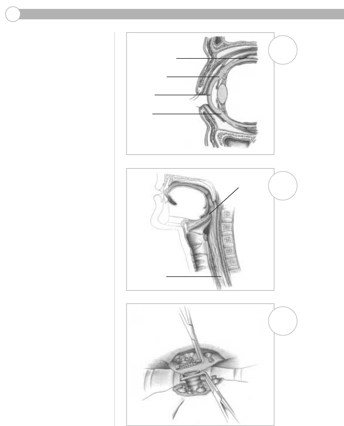
ligature, is woven from the same
copolymer used to produce
absorbable VICRYL* suture. As a
synthetic absorbable, VICRYL*
periodontal mesh eliminates the
trauma associated with a second
surgical procedure and reduces the
risk of infection or inflammation
associated with this procedure.
The esophagus is a difficult organ
to suture. It lacks a serosal layer.
The mucosa heals slowly. The thick
muscular layer does not hold sutures
well. If multifilament sutures are
used, penetration through the
mucosa into the lumen should be
avoided to prevent infection.
RESPIRATORY
TRACT SURGERY
Relatively few studies have been
done on healing in the respiratory
tract. Bronchial stump closure
following lobectomy or pneumonec-
tomy presents a particular challenge.
Infection, long stumps, poor
approximation of the transected
bronchus, and incomplete closure
(i.e., air leaks) may lead to
bronchopleural fistula. Avoidance
of tissue trauma and maintenance
of the blood supply to the area of
closure are critical to healing. The
bronchial stump heals slowly, and
sometimes not at all. Unless it is
closed tightly with strong, closely
spaced sutures, air may leak into
the thoracic cavity.
Closure is usually achieved with
mechanical devices, particularly
staples. When sutures are used,
polypropylene monofilament nonab-
sorbable sutures are less likely to
cause tissue reaction or harbor
infection. Silk suture is also
commonly used. Surgeons usually
THE SUTURE
36
THE EYE
FIGURE
18
BRONCHIAL
STUMP
CLOSURE
FIGURE
20
THE UPPER
ALIMENTARY
CANAL
FIGURE
19
Ocular muscles
Conjunctiva
Cornea
Sclera
Esophagus
Oral cavity
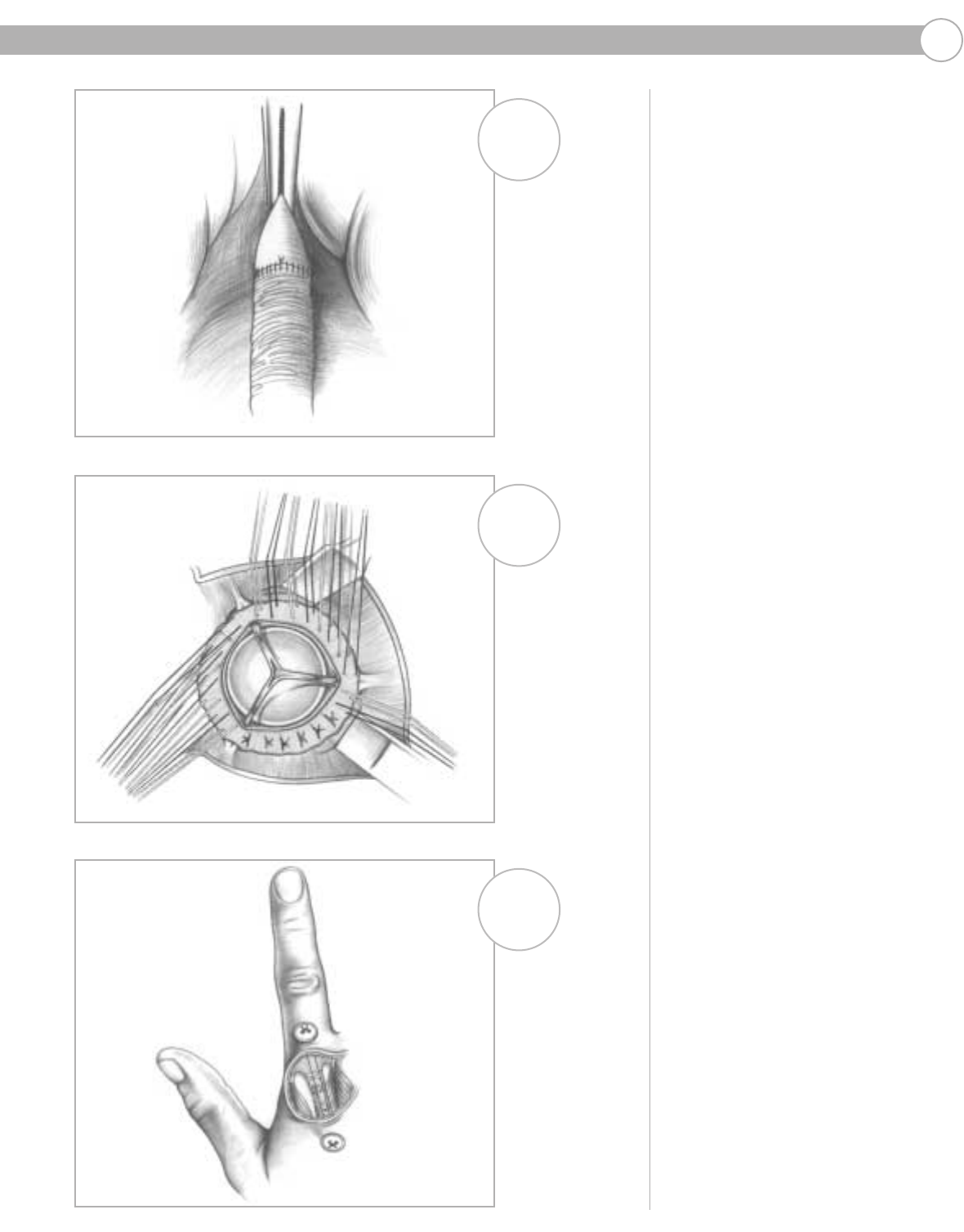
avoid absorbable sutures because
they may permit secondary leakage
as they lose strength.
Monofilament nylon suture should
also be avoided because of its
potential for knot loosening.
CARDIOVASCULAR SURGERY
Although definitive studies are few,
blood vessels appear to heal rapidly.
Most cardiovascular surgeons prefer
to use synthetic nonabsorbable
sutures for cardiac and peripheral
vascular procedures. Lasting
strength and leakproof anastomoses
are essential. Wire sutures are used
on the sternum unless it is fragile,
in which case absorbable sutures
can be used.
VESSELS
Excessive tissue reaction to suture
material may lead to decreased
luminal diameter or to thrombus
formation in a vessel. Therefore,
the more inert synthetics including
nylon and polypropylene are
the materials of choice for vessel
anastomoses. Multifilament
polyester sutures allow clotting to
occur within the interstices which
helps to prevent leakage at the
suture line. The advantages of a
material such as ETHIBOND*
EXCEL sutures are its strength,
durability, and slippery surface
which causes less friction when
drawn through a vessel. Many
surgeons find that PROLENE*
sutures, PRONOVA* sutures, or
silk are ideal for coronary artery
procedures because they do not
"saw" through vessels.
Continuous sutures provide a more
leakproof closure than interrupted
sutures in large vessel anastomoses
CHAPTER 2 37
* Trademark
CONTINUOUS
STRAND
SUTURING
IN VASCULAR
SURGERY
FIGURE
21
THE BUNNELL
TECHNIQUE
FIGURE
23
SEATING A
HEART VALVE
WITH ETHIBOND
EXCEL SUTURE
FIGURE
22

because the tension along the
suture strand is distributed evenly
around the vessel's circumference.
Interrupted monofilament sutures
such as ETHILON* sutures,
PROLENE* sutures, or
PRONOVA* sutures are used for
microvascular anastomoses.
When anastomosing major vessels
in young children, special care
must be taken to anticipate the
future growth of the patient.
Here, the surgeon may use silk to
its best advantage, because it loses
much of its tensile strength after
approximately 1 year, and is usually
completely absorbed after 2 or more
years. Continuous polypropylene
sutures have been used in children
without adverse effects. The
continuous suture, when placed,
is a coil which stretches as the
child grows to accommodate the
changing dimensions of the blood
vessel. However, reports of stricture
following vessel growth have
stimulated interest in use of a suture
line which is one-half continuous,
one-half interrupted. Clinical
studies suggest that a prolonged
absorbable suture, such as PDS* II
suture, may be ideal, giving
adequate short-term support while
permitting future growth.
Following vascular trauma, mycotic
aneurysms from infection are
extremely serious complications.
A suture may act as a nidus for
an infection. In the presence of
infection, the chemical properties
of suture material can cause
extensive tissue damage which may
reduce the tissue's natural ability
to combat infection. Localized
sepsis can also spread to adjacent
vascular structures, causing necrosis
of the arterial wall. Therefore, the
surgeon may choose a monofilament
suture material that causes only a
mild tissue reaction and resists
bacterial growth.
VASCULAR PROSTHESES
The fixation of vascular prostheses
and artificial heart valves presents an
entirely different suturing challenge
than vessel anastomosis. The sutures
must retain their original physical
properties and strength throughout
the life of the patient. A prosthesis
never becomes completely incorpo-
rated into the tissue and constant
movement of the suture line occurs.
Coated polyester sutures are the
choice for fixation of vascular
prostheses and heart valves because
they retain their strength and
integrity indefinitely.
Either a continuous or interrupted
technique may be used for vessel to
graft anastomoses.
To assist in proper strand identifica-
tion, many surgeons alternate green
and white strands of ETHIBOND*
EXCEL suture around the cuff of
the valve before tying the knots.
Some surgeons routinely use
pledgets to buttress sutures in valve
surgery. They are used most
commonly in valve replacement
procedures to prevent the annulus
from tearing when the prosthetic
valve is seated and the sutures are
tied. They may also be used in heart
wall closure of penetrating injuries,
excising aneurysms, vascular graft
surgery, and to add support when
the surgeon encounters extreme
deformity, distortion, or tissue
destruction at the annulus.
URINARY TRACT SURGERY
Closure of tissues in the urinary
tract must be leakproof to prevent
escape of urine into surrounding
tissues. The same considerations
that affect the choice of sutures for
the biliary tract affect the choice of
sutures for this area. Nonabsorbable
sutures incite the formation of cal-
culi, and therefore cannot be used.
Surgeons use absorbable sutures as a
rule, especially MONOCRYL*
sutures, PDS II sutures, Coated
VICRYL* sutures, and chromic
gut sutures.
The urinary tract heals rapidly. The
transitional cell epithelium migrates
over the denuded surfaces quickly.
Unlike other epithelium, the
migrating cells in the urinary tract
undergo mitosis and cell division.
Epithelial migration may be found
along suture tracts in the body of
the bladder. The bladder wall
regains 100% of its original tensile
strength within 14 days. The rate of
collagen synthesis peaks at 5 days
and declines rapidly thereafter.
Thus, sutures are needed for only
7 to 10 days.
THE FEMALE GENITAL TRACT
Surgery within this area presents
certain challenges. First, it is
usually regarded as a potentially
contaminated area. Second, the
surgeon must frequently work
within a very restricted field.
Endoscopic technique is frequently
used in this area.
Most gynecological surgeons prefer
to use absorbable sutures for repair of
incisions and defects. Some prefer
using heavy, size 1 surgical gut
sutures, MONOCRYL sutures, or
VICRYL sutures. However, the
THE SUTURE
38

stresses on the reproductive organs
and the rate of healing indicate that
these larger-sized sutures may only
be required for abdominal closure.
Handling properties, especially
pliability of the sutures used for
internal use, are extremely
important. Synthetic absorbable
sutures such as VICRYL* sutures in
size 0 may be used for the tough,
muscular, highly vascular tissues
in the pelvis and vagina. These
tissues demand strength during
approximation and healing. Coated
VICRYL* RAPIDE suture, for
example, is an excellent choice for
episiotomy repair.
TENDON SURGERY
Tendon surgery presents several
challenges. Most tendon injuries are
due to trauma, and the wound may
be dirty. Tendons heal slowly. The
striated nature of the tissue makes
suturing difficult.
Tendon repair fibroblasts are
derived from the peritendonous
tissue and migrate into the wound.
The junction heals first with scar
tissue, then by replacement with
new tendon fibers. Close apposition
of the cut ends of the tendon
(especially extensor tendons)
must be maintained to achieve
good functional results. Both the
suture material and the closure
technique are critical for successful
tendon repair.
The suture material the surgeon
chooses must be inert and strong.
Because tendon ends can separate
due to muscle pull, sutures with a
great degree of elasticity should be
avoided. Surgical steel is widely used
because of its durability and lack of
elasticity. Synthetic nonabsorbable
materials including polyester fibers,
polypropylene, and nylon may be
used. In the presence of potential
infection, the most inert monofila-
ment suture materials are preferred.
The suture should be placed to
cause the least possible interference
with the surface of the tendon, as
this is the gliding mechanism. It
should also not interfere with the
blood supply reaching the wound.
Maintenance of closed apposition
of the cut ends of the tendons,
particularly extensor tendons, is
critical for good functional results.
The parallel arrangement of tendon
fibers in a longitudinal direction
makes permanent and secure place-
ment of sutures difficult. Various
figure-of-eight and other types of
suturing have been used successfully
to prevent suture slippage and the
formation of gaps between the cut
ends of the tendon.
Many surgeons use the Bunnell
Technique. The suture is placed to
be withdrawn when its function as a
holding structure is no longer neces-
sary. Referred to as a pull-out suture,
it is brought out through the skin
and fastened over a polypropylene
button. The Bunnell Technique
suture can also be left in place.
NUROLON* sutures, PROLENE*
sutures, PRONOVA* sutures and
ETHIBOND* EXCEL sutures may
be used for connecting tendon to
bone. Permanent wire sutures also
yield good results because healing is
slow. In periosteum, which heals
fairly rapidly, surgical gut or coated
VICRYL sutures may be used. In
fact, virtually any suture may be
used satisfactorily in the periosteum.
SUTURES FOR BONE
In repairing facial fractures,
monofilament surgical steel has
proven ideal for its lack of elasticity.
Facial bones do not heal by callus
formation, but more commonly by
fibrous union. The suture material
must remain in place for a long
period of time—perhaps months—
until the fibrous tissue is laid down
and remodeled. Steel sutures
immobilize the fracture line and
keep the tissues in good apposition.
Following median stemotomy,
surgeons prefer interrupted steel
sutures to close. Sternum closure
may be difficult. Appropriate
tension must be maintained, and
the surgeon must guard against
weakening the wire. Asymmetrical
twisting of the wire may cause it to
buckle, fatiguing the metal, and
ultimately causing the wire to
break. Motion between the sides
of the sternum will result, causing
postoperative pain and possibly
dehiscence. Painful nonunion is
another possible complication.
(In osteoporotic patients, very
heavy VICRYL sutures may be
used to close the sternum securely.)
The surgeon may use a bone anchor
to hold one end of a suture in place
when needed (e.g., shoulder repair
surgery). This involves drilling a
hole in the bone and inserting
the anchor, which expands once
completely inside the bone to keep
it from being pulled out.
OTHER PROSTHETIC DEVICES
Often, it is necessary for the sur-
geon to implant a prosthetic device
such as an automatic defibrillator or
drug delivery system into a patient.
To prevent such a device from
CHAPTER 2 39
* Trademark
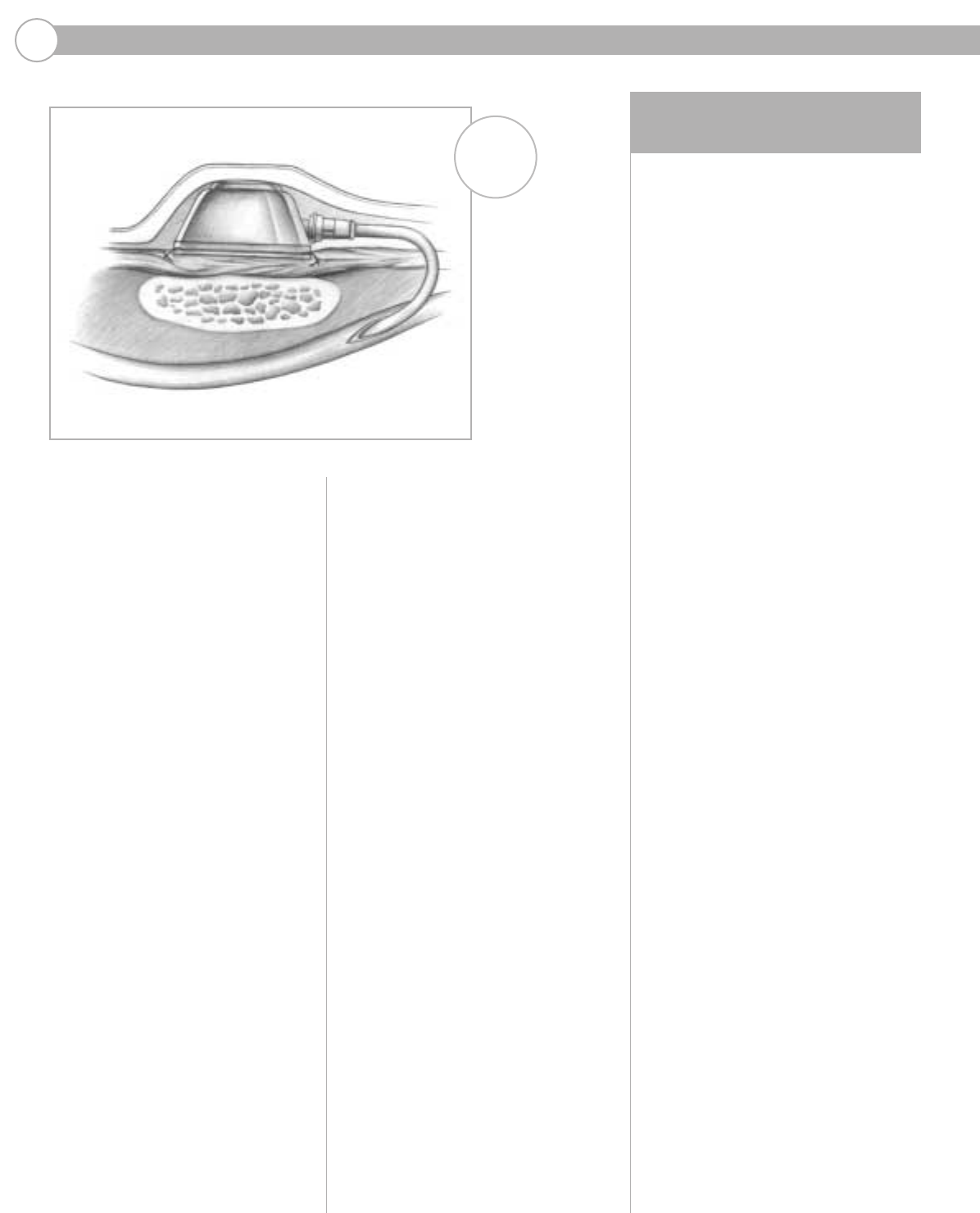
migrating out of position, it may be
tacked to the fascia or chest wall
with nonabsorbable sutures.
CLOSING CONTAMINATED
OR INFECTED WOUNDS
Contamination exists when
microorganisms are present, but in
insufficient numbers to overcome
the body's natural defenses.
Infection exists when the level of
contamination exceeds the
tissue's ability to defend against the
invading microorganisms. Generally,
contamination becomes infection
when it reaches approximately 106
bacteria per gram of tissue in an
immunologically normal host.
Inflammation without discharge
and/or the presence of culture-posi-
tive serous fluid indicate possible
infection. Presence of purulent dis-
charge indicates positive infection.
Contaminated wounds can
become infected when hematomas,
necrotic tissue, devascularized tissue,
or large amounts of devitalized
tissue (especially in fascia, muscle,
and bone) are present. Micro-
organisms multiply rapidly under
these conditions, where they are
safe from cells that provide local
tissue defenses.
In general, contaminated wounds
should not be closed but should be
left open to heal by secondary
intention because of the risk of
infection. Foreign bodies, including
sutures, perpetuate localized
infection. Therefore, the
surgeon's technique and choice of
suture is critical.
Nonabsorbable monofilament nylon
sutures are commonly used in
anticipation of delayed closure of
dirty and infected wounds. The
sutures are laid in but not tied.
Instead, the loose suture ends are
held in place with PROXI-STRIP*
skin closures (sterile tape). The
wound should be packed to
maintain a moist environment.
When the infection has subsided,
the surgeon can easily reopen the
wound, remove the packing and
any tissue debris, and then close
using the previously inserted
monofilament nylon suture.
IN THE
NEXT SECTION
The surgeon depends as much upon
the quality and configuration of the
needle used as on the suturing
material itself to achieve a successful
closure. The relationship between
needles and sutures will be explored
on the pages that follow.
THE SUTURE
40
TACKING A
PROSTHETIC
DEVICE IN
POSITION TO
PREVENT
MIGRATION
FIGURE
24
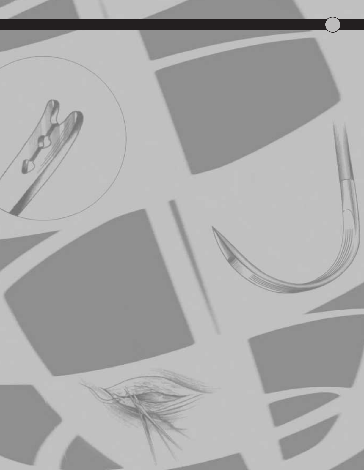
THE SURGICAL NEEDLE
CHAPTER 3

Necessary for the placement of
sutures in tissue, surgical needles
must be designed to carry suture
material through tissue with
minimal trauma. They must be
sharp enough to penetrate tissue
with minimal resistance. They
should be rigid enough to resist
bending, yet flexible enough to
bend before breaking. They must
be sterile and corrosion-resistant
to prevent introduction of
microorganisms or foreign bodies
into the wound.
Comfort with needle security in the
needleholder, the ease of passage
through tissue, and the degree of
trauma that it causes all have an
impact upon the overall results of
surgical needle performance. This is
especially true when precise cosmet-
ic results are desired.
The best surgical needles are:
• Made of high quality stainless
steel.
• As slim as possible without
compromising strength.
• Stable in the grasp of a
needleholder.
• Able to carry suture material
through tissue with minimal
trauma.
• Sharp enough to penetrate tissue
with minimal resistance.
• Rigid enough to resist bending,
yet ductile enough to resist
breaking during surgery.
• Sterile and corrosion-resistant
to prevent introduction of
microorganisms or foreign
materials into the wound.
Variations in needle geometries are
just as important as variations in
suture sizes. Needle dimensions
must be compatible with suture
sizes, allowing the two to work
in tandem.
ELEMENTS OF
NEEDLE DESIGN
Needle design involves analyzing a
surgical procedure and the density
of the tissue involved in great
detail. ETHICON engineers work
continuously to improve upon their
needle line, sometimes making sub-
tle alterations resulting in a positive
impact upon the procedure itself.
The various metal alloys used in the
manufacture of surgical needles
determine their basic characteristics
to a great degree. ETHICON*
stainless steel alloy needles are heat-
treated to give them the maximum
possible strength and ductility.
ETHALLOY* needle alloy (Patent
No. 5,000,912) was developed for
unsurpassed strength in precision
needles used in cardiovascular,
ophthalmic, plastic, and microsurgi-
cal procedures. It is produced
economically without sacrificing
ductility or corrosion resistance.
A needle's strength is determined
by how it resists deformation
during repeated passes through
tissue. Tissue trauma can be
induced if a needle bends during
penetration and compromises tissue
apposition. Therefore, greater needle
strength equals less tissue trauma.
A weak needle that bends too easily
can compromise the surgeon's
control and damage surrounding
tissue during the procedure. In
addition, loss of control in needle
placement could result in an
inadvertent needlestick.
Manufacturers measure needle
strength in the laboratory by
bending them 90° to determine the
needle's maximum strength. This is
referred to as the needle's "ultimate
moment," and is more important to
the needle manufacturer than to the
surgeon. The most critical aspect of
needle strength to the surgeon is
the "surgical yield" point. Surgical
yield indicates the amount of
angular deformation the needle
can with-stand before becoming
permanently deformed. This point
is usually 10° to 30° depending
upon the material and the manufac-
turing process. Any angle beyond
that point renders the needle
useless. Reshaping a bent needle
may cause it to lose strength
and be less resistant to bending
and breaking.
At ETHICON, the combination
of alloy selection and the needle
manufacturing process are carefully
selected to achieve the highest
possible surgical yield, which also
optimizes needle strength.
Ductility refers to the needle's
resistance to breaking under a given
amount of bending. If too great a
force is applied to a needle it may
break, but a ductile needle will
bend before breaking. Needle
breakage during surgery can prevent
apposition of the wound edges as
the broken portion passes through
tissue. In addition, searching for
part of a broken needle can cause
added tissue trauma and add to the
time the patient is anesthetized. A
piece that cannot be retrieved will
remain as a constant reminder to
THE SURGICAL NEEDLE
42
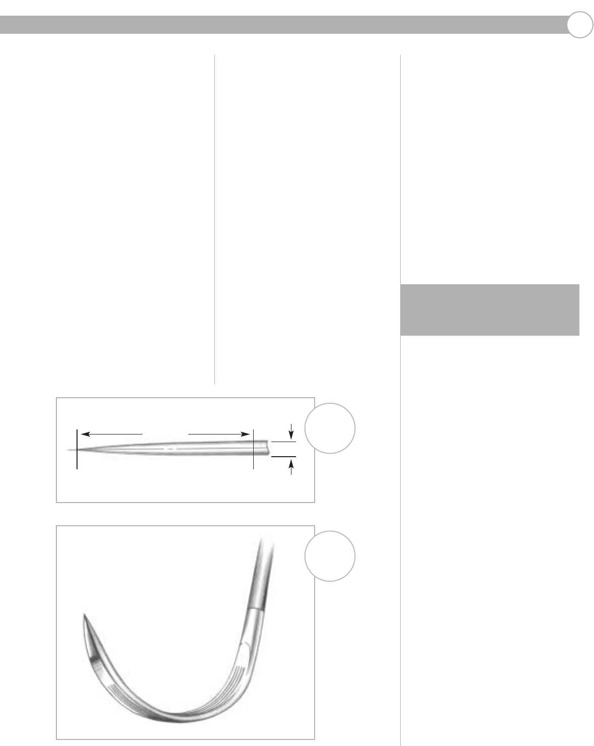
* Trademark
both the patient and surgeon.
Needle bending and breakage can
be minimized by carefully passing
needles through tissue in the
direction of the needle body.
Needles are not designed to be
used as retractors to lift tissue.
Needle sharpness is especially
important in delicate or cosmetic
surgery. The sharper the needle,
the less scarring that will result.
However, the right balance must be
found. If a needle is too sharp, a
surgeon may not feel he or she has
adequate control of needle passage
through tissue.
Sharpness is related to the angle of
the point as well as the taper ratio
of the needle. The ETHICON
sharpness tester incorporates a thin,
laminated, synthetic membrane
that simulates the density of human
tissue, allowing engineers to gauge
exactly how much force is required
for penetration.
Most ETHICON needles have
a micro-thin coating comprised
of silicone or similar lubricants
which significantly and measurably
improves ease of needle penetration.
According to laboratory tests, this
coating serves several important
functions:
✦It reduces the force needed to
make the initial penetration
through tissue.
✦It reduces the drag force on
the needle body as it passes
repeatedly through tissue.
Needle performance is also
influenced by the stability of the
needle in the grasp of a needlehold-
er. Most curved needles are
flattened in the grasping area to
enhance control. All ETHICON
curved needles of 22 mil wire
or heavier are ribbed as well as
flattened. Longitudinal ribbing
or grooves on the inside or outside
curvatures of curved needles
provides a crosslocking action in
the needleholder for added needle
control. This reduces undesirable
rocking, twisting, and turning in
the needleholder.
PRINCIPLES OF
CHOOSING A
SURGICAL NEEDLE
While there are no hard and fast
rules governing needle selection, the
following principles should be kept
in mind. (Specific types of needles
mentioned here will be described in
full detail later on in this section.)
1. Consider the tissue in which the
surgeon will introduce the
needle. Generally speaking, taper
point needles are most often used
to suture tissues that are easy
to penetrate. Cutting or
TAPERCUT* needles are
more often used in tough, hard-
to-penetrate tissues. When in
doubt about whether to choose
a taper point or cutting needle,
choose the taper point for
everything except skin sutures.
2. Watch the surgeon's technique
closely. Select the length,
diameter, and curvature of the
needle according to the desired
placement of the suture and the
space in which the surgeon
is working.
CHAPTER 3 43
TAPER
RATIO
FIGURE
1
ETHICON
RIBBED
NEEDLE
FIGURE
2
12:1 ratio
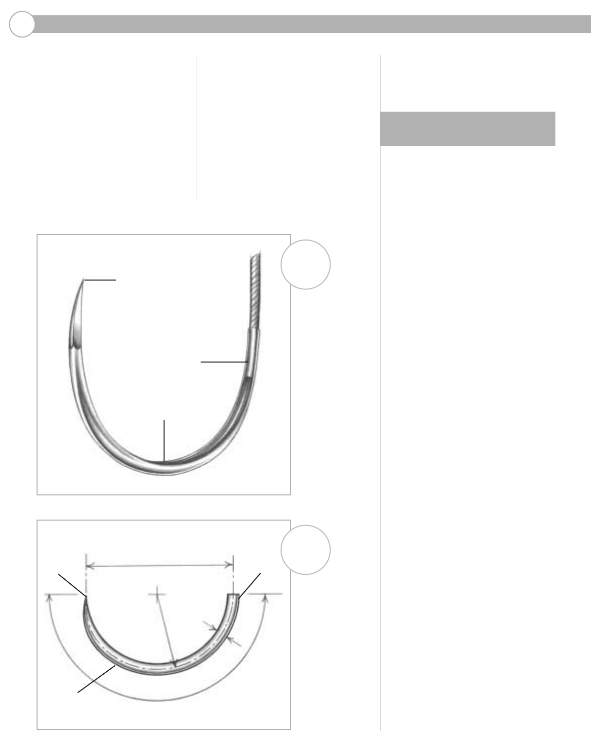
3. Consult frequently with the
surgeon. Working with the same
surgeon repeatedly leads to
familiarity with his or her
individual routine. However,
even the same surgeon may need
to change needle type or size to
meet specific requirements,
even during a single operative
procedure.
4. When using eyed needles, try to
match needle diameter to suture
size. Swaged needles, where the
needle is already attached to the
suture strand, eliminate this
concern.
5. The best general rule of thumb
for the scrub person to follow is
pay attention and remain alert to
the progress of the operation.
Observation is the best guide to
needle selection if the surgeon
has no preference.
THE ANATOMY
OF A NEEDLE
Regardless of its intended use,
every surgical needle has three basic
components:
✦The eye.
✦The body.
✦The point.
The measurements of these
specific components determine,
in part, how they will be used
most efficiently.
Needle size may be measured in
inches or in metric units. The
following measurements determine
the size of a needle.
✦CHORD LENGTH—The
straight line distance from the
point of a curved needle to
the swage.
✦NEEDLE LENGTH—The
distance measured along the
needle itself from point to end.
✦RADIUS—The distance from
the center of the circle to the
body of the needle if the
curvature of the needle were
continued to make a full circle.
✦DIAMETER—The gauge or
thickness of the needle wire.
Very small needles of fine gauge
are needed for microsurgery.
Large, heavy gauge needles are
used to penetrate the sternum
and to place retention sutures
in the abdominal wall. A broad
spectrum of sizes are available
between the two extremes.
THE SURGICAL NEEDLE
44
NEEDLE
COMPONENTS
FIGURE
3
ANATOMY
OF A NEEDLE
FIGURE
4
Point
Eye
(Swaged end)
Body
Needle
point
Chord length
Swage
Needle
radius
Needle length
Needle body
Needle
diameter

THE NEEDLE EYE
The eye falls into one of three cate-
gories: closed eye, French (split or
spring) eye, or swaged (eyeless).
The closed eye is similar to a
household sewing needle. The shape
of the eye may be round, oblong,
or square. French eye needles have a
slit from inside the eye to the end
of the needle with ridges that catch
and hold the suture in place.
Eyed needles must be threaded, a
time-consuming procedure for the
scrub person. This presents the
disadvantage of having to pull a
double strand of suture material
through tissue, creating a larger hole
with additional tissue disruption.
In addition, the suture may still
become unthreaded while the
surgeon is using it. While tying the
suture to the eye may minimize this
possibility, it also adds to the bulk
of the suture. Another disadvantage
of eyed needles is that repeated use
of these needles with more than
one suture strand causes the needle
to become dull, thereby making
suturing more difficult.
Virtually all needles used today are
swaged. This configuration joins
the needle and suture together as
a continuous unit—one that is
convenient to use and minimizes
trauma. The method of attaching
the suture to the needle varies with
the needle diameter. In larger
diameter needles, a hole is drilled
in the needle end. In smaller
diameter needles, a channel is
made by forming a "U" at the
swage end or a hole is drilled in
the wire with a laser. Each hole or
channel is specifically engineered
for the type and size of suture
material it will hold, and crimped
or closed around the suture to hold
it securely. When the surgeon has
finished placing the suture line in
the patient's tissue, the suture may
be cut, or easily released from the
needle as is the case when using
CONTROL RELEASE* needles
(Patent No. 3,980,177).
The diameter of a needle swaged
to suture material is no larger
than necessary to accommodate
the diameter of the suture strand
itself. Swaged sutures offer several
advantages to the surgeon, nurse,
and patient.
1. The scrub person does not have
to select a needle when the
surgeon requests a specific suture
material since it is already
attached.
2. Handling and preparation are
minimized. The strand with
needle attached may be used
directly from the packet. This
helps maintain the integrity of
the suture strand.
3. Tissues are subjected to minimal
trauma.
4. Tissue trauma is further reduced
because a new, sharp, undamaged
needle is provided with each
suture strand.
5. Swaged sutures do not unthread
prematurely.
6. If a needle is accidentally
dropped into a body cavity, the
attached suture strand makes it
easier to find.
7. Inventory and time spent
cleaning, sharpening, handling,
and sterilizing reusable eyed
needles is eliminated, thereby
reducing cost as well as risk of
needle punctures.
8. CONTROL RELEASE needles
allow placement of many sutures
rapidly. This may reduce
operating time and, ultimately,
the length of time the patient
is anesthetized.
9. The ATRALOC* surgical needle
and CONTROL RELEASE
needle ensure consistent quality
and performance.
10. Swaged sutures eliminate suture
fraying or damage due to sharp
comers in the eye of eyed
needles.
11. Needles are corrosion-free.
Small diameter ETHICON taper
point needles commonly used
in cardiovascular surgery were
compared in laboratory tests—
some with "split" channels and
some with laser-drilled holes. The
needles with laser-drilled holes
produced less drag force as they
passed through a membrane that
simulated vascular tissue. This
could be associated with less
trauma to the vessel walls.
The swaged ATRALOC surgical
needles made by ETHICON
are supplied in a variety of sizes,
shapes, and strengths. Some of
them incorporate the CONTROL
RELEASE needle suture principle
which facilitates fast separation
of the needle from the suture
when desired by the surgeon.
This feature allows rapid placement
of many sutures, as in interrupted
suturing techniques. Even though
the suture is securely fastened
to the needle, a slight, straight tug
will release it. This needle/suture
configuration was created originally
* Trademark
CHAPTER 3 45
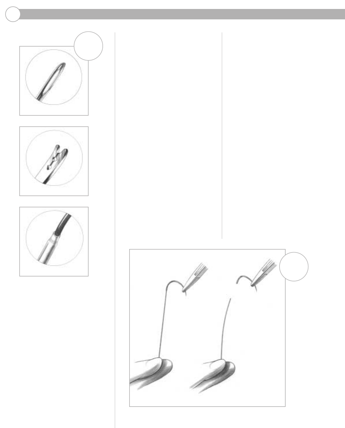
for abdominal closure and
hysterectomies, but is now used
in a wide variety of procedures.
THE NEEDLE BODY
The body of the needle is the
portion which is grasped by the
needleholder during the surgical
procedure. The body of the needle
should be as close as possible to the
diameter of the suture material to
minimize bleeding and leakage.
This is especially true for
cardiovascular, gastrointestinal,
and bladder procedures.
The curvature of the needle body
may come in a variety of different
shapes. Each shape gives the needle
different characteristics.
STRAIGHT NEEDLE
This shape may be preferred when
suturing easily accessible tissue.
Most of these needles are designed
to be used in places where direct
finger-held manipulation can easily
be performed.
The Keith needle is a straight
cutting needle. It is used primarily
for skin closure of abdominal
wounds. Varying lengths are also
used for arthroscopic suturing of
the meniscus in the knee.
Bunnell (BN) needles are used for
tendon repair. Taper point needle
variations may also be used for
suturing the gastrointestinal tract.
Some microsurgeons prefer straight
needles for nerve and vessel repair.
In ophthalmology, the straight tran-
schamber needle protects endothe-
lial cells and facilitates placement of
intraocular lenses.
HALF-CURVED NEEDLE
The half-curved or "ski" needle
may be used for skin closure or in
laparoscopy. Its low profile allows
easy passage down laparoscopic
trocars. Its use in skin closure is
limited because, while the curved
portion passes through tissue easily,
the remaining straight portion of
the body is unable to follow the
curved path of the needle without
bending or enlarging its path in
the tissue.
CURVED NEEDLE
Curved needles allow predictable
needle turnout from tissue, and
are therefore used most often.
THE SURGICAL NEEDLE
46
FIGURE
5
THE NEEDLE EYE
Closed eye
French eye
Swaged
CONTROL
RELEASE
NEEDLE
SUTURE
FIGURE
6
Holding the needle securely in the needleholder, the suture
should be grasped securely and pulled straight and taut. The
needle will be released with a straight tug of the needleholder.
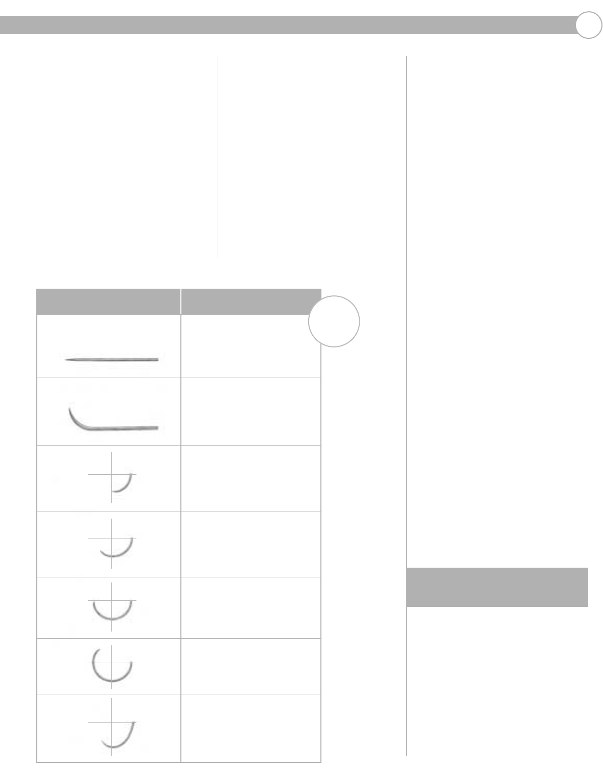
* Trademark
This needle shape requires less
space for maneuvering than a
straight needle, but the curve
necessitates manipulation with a
needleholder. The curvature may
be 1/4, 3/8, 1/2, or 5/8circle.
The most common use for the 3/8
circle is skin closure. The surgeon
can easily manipulate this curvature
with slight pronation of the wrist in
a relatively large and superficial
wound. It is very difficult to use
this needle in a deep body cavity or
restricted area because a larger arc of
manipulation is required.
The 1/2circle needle was designed
for use in a confined space,
although it requires more pronation
and supination of the wrist. But
even the tip of this needle may
be obscured by tissue deep in
the pelvic cavity. A 5/8circle
needle may be more useful in
this situation, especially in some
anal, urogenital, intraoral, and
cardiovascular procedures.
COMPOUND
CURVED NEEDLE
The compound curved needle
(Patent No. 4,524,771) was
originally developed for anterior
segment ophthalmic surgery. It
allows the surgeon to take precise,
uniform bites of tissue. The tight
80° curvature of the tip follows into
a 45° curvature throughout the
remainder of the body. The initial
curve allows reproducible, short,
deep bites into the tissue. The
curvature of the remaining portion
of the body forces the needle out of
the tissue, everting the wound edges
and permitting a view into the
wound. This ensures equidistance of
the suture material on both sides of
the incision. Equalized pressure on
both sides of the comeal-scleral
junction minimizes the possibility
of astigmatism following anterior
segment surgery.
THE NEEDLE POINT
The point extends from the extreme
tip of the needle to the maximum
cross-section of the body. Each nee-
dle point is designed and produced
to the required degree of sharpness
to smoothly penetrate specific types
of tissue.
TYPES OF
NEEDLES
CUTTING NEEDLES
Cutting needles have at least two
opposing cutting edges. They are
sharpened to cut through tough,
difficult-to-penetrate tissue.
Cutting needles are ideal for skin
sutures that must pass through
dense, irregular, and relatively
thick connective dermal tissue.
CHAPTER 3 47
NEEDLE
SHAPES
AND TYPICAL
APPLICATIONS
FIGURE
7
Straight
Half-curved
1/4Circle
3/8Circle
1/2Circle
5/8Circle
Compound
Curved
gastrointestinal tract, nasal
cavity, nerve, oral cavity,
pharynx, skin, tendon, vessels
skin (rarely used)
laparoscopy
eye (primary application)
microsurgery
aponeurosis, biliary tract, cardiovascular
system, dura, eye, gastrointestinal tract,
muscle, myocardium, nerve, perichon-
drium, periosteum, pleura, skin, tendon,
urogenital tract, vessels
biliary tract, cardiovascular system, eye,
fascia, gastrointestinal tract, muscle,
nasal cavity, oral cavity, pelvis, peri-
toneum, pharynx, pleura, resporatory
tract, skin, tendon, subcutaneous fat,
urogenital tract
anal (hemorrhoidectomy), nasal
cavity, pelvis, urogenital tract (primary
application)
eye (anterior segment)
laparoscopy
SHAPE APPLICATION
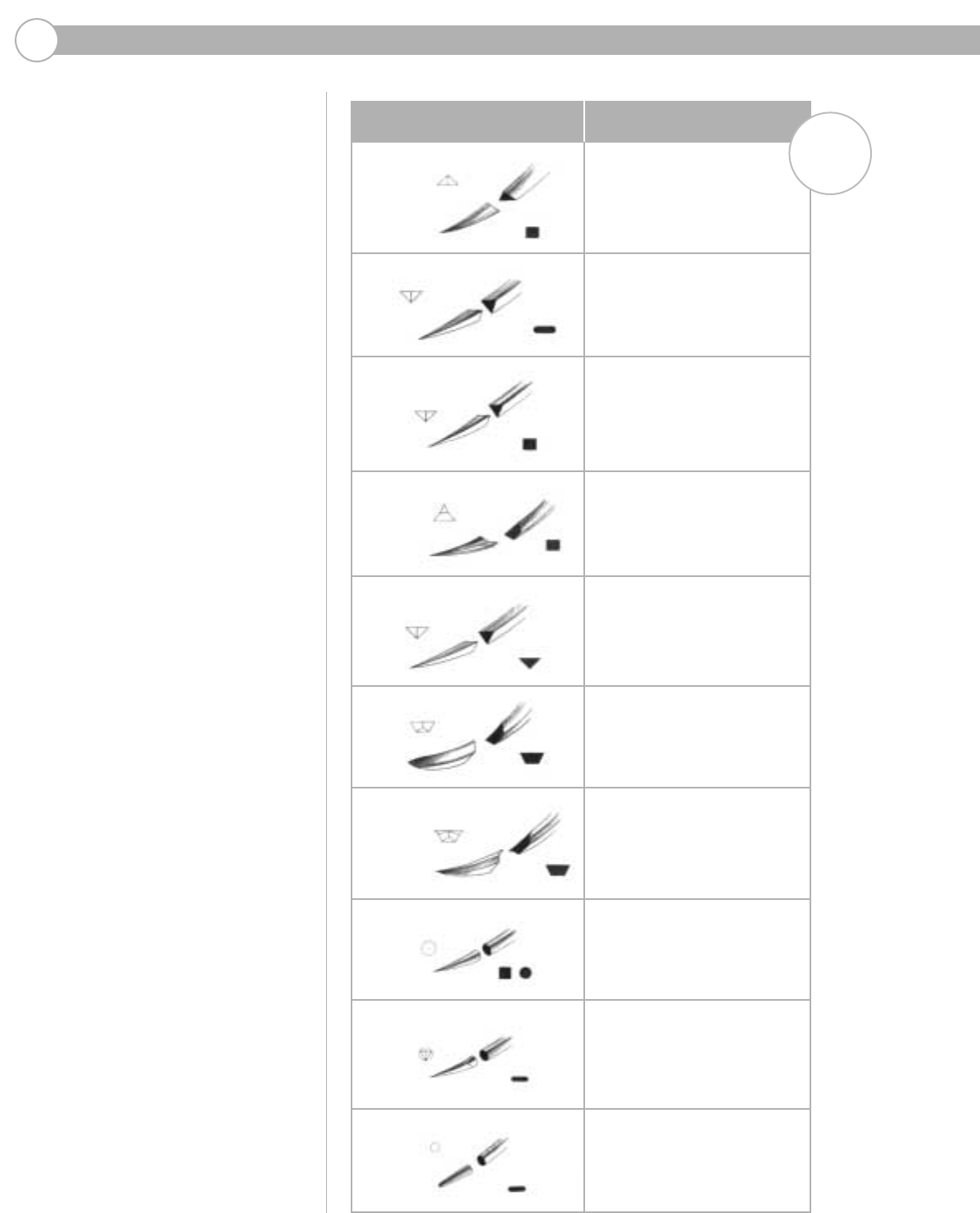
Because of the sharpness of the
cutting edge, care must be taken
in some tissue (tendon sheath
or oral mucous membrane) to
avoid cutting through more tissue
than desired.
CONVENTIONAL
CUTTING NEEDLES
In addition to the two cutting edges,
conventional cutting needles have a
third cutting edge on the inside
concave curvature of the needle.
The shape changes from a triangular
cutting blade to that of a flattened
body on both straight and curved
needles. This needle type may be
prone to cutout of tissue because
the inside cutting edge cuts toward
the edges of the incision or wound.
The PC PRIME* needle (Precision
Cosmetic, Patent No. 5,030,228)
is designed specifically for aesthetic
plastic surgery, and has conventional
cutting edges. Where cosmetic
results are important, the PC
PRIME needle is superior to any
other for more delicate surgery,
especially facial surgery. The
narrow point, fine wire diameter,
and fine taper ratio allow superior
penetration of soft tissue. The inside
and outside curvatures of the body
are flattened in the needle grasping
area for greater stability in the
needleholder. Fattened sides reduce
bending that might occur due to
the fine wire diameter.
The tip configuration of the conven-
tional cutting sternotomy needle is
slightly altered to resist bending as it
penetrates the sternum. The alloy
used for this needle provides the
increased strength and ductility need-
ed for its function. The cutting edges
of the point extend approximately
THE SURGICAL NEEDLE
48
NEEDLE
POINTSAND
BODY SHAPES
AND TYPICAL
APPLICATIONS
FIGURE
8
Conventional Cutting
Reverse Cutting
Precision Point Cutting
PC PRIME* Needle
MICRO-POINT* Reverse Cutting Needle
Side-Cutting Spatula
CS ULTIMA* Ophthalmic Needle
Taper
TAPERCUT* Surgical Needle
Blunt
skin, sternum
fascia, ligament, nasal cavity, oral
mucosa, pharynx, skin, tendon sheath
skin (plastic or cosmetic)
skin (plastic or cosmetic)
eye
eye (primary application),
microsurgery, ophthalmic
(reconstructive)
eye (primary application)
aponeurosis, biliary tract, dura, fascia,
gastrointestinal tract, laparoscopy,
muscle, myocardium, nerve, peritoneum,
pleura, subcutaneous fat, urogenital
tract, vessels, valve
bronchus, calcified tissue, fascia,
laparoscopy, ligament, nasal cavity,
oral cavity, ovary, perichondrium,
periosteum, pharynx, sternum, tendon,
trachea, uterus, valve, vessels (sclerotic)
Blunt dissection (friable tissue),
cervix (ligating incompetent cervix),
fascia, intestine, kidney, liver, spleen
SHAPE APPLICATION
Point
Point
Point
Body
Body
Body
Body
Body
Body
Body
Point
Point
Point
Point
Point
Point
Point
Body
Body
Body
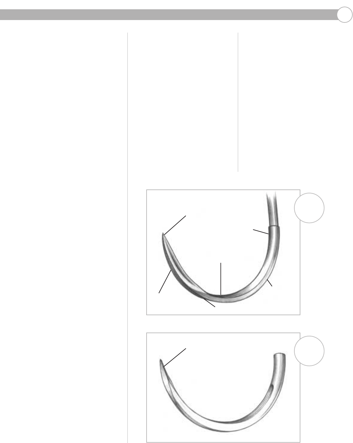
1/4" (6mm) from the round body
and terminate in a triangular-shaped
tip. This particular sternotomy needle
maximizes cutting efficiency and con-
trol in the needleholder. TAPERCUT
surgical needles may also be used for
this procedure.
REVERSE CUTTING
NEEDLES
These needles were created
specifically for tough, difficult-to-
penetrate tissue such as skin, tendon
sheath, or oral mucosa. Reverse
cutting needles are used in
ophthalmic and cosmetic surgery
where minimal trauma, early
regeneration of tissue, and little scar
formation are primary concerns.
The reverse cutting needle is as
sharp as the conventional cutting
needle, but its design is distinctively
different. The third cutting edge
is located on the outer convex
curvature of the needle. This offers
several advantages:
✦Reverse cutting needles have
more strength than similar-sized
conventional cutting needles.
✦The danger of tissue cutout is
greatly reduced.
✦The hole left by the needle leaves
a wide wall of tissue against
which the suture is to be tied.
The MICRO-POINT* surgical
needle for ophthalmic procedures
has a smooth surface and is honed
to extreme sharpness. This allows
the surgeon to suture the extremely
tough tissues of the eye with
optimum precision and ease.
A needle manufactured by the
exclusive ETHICON* Precision
Point Process may be used for
plastic or cosmetic surgery, and
passes smoothly through tissue
creating a minute needle path.
This results in superior apposition.
The bottom third cutting edge
on the Precision Point needle
flattens out as it transitions to the
needle body for greater security in
the needleholder.
The OS (Orthopaedic Surgery)
needles are curved, heavy bodied,
reverse-cutting needles. The
orthopaedic surgeon may use the
OS needle for extremely tough
tissue, such as cartilage, where force
is required for penetration.
SIDE CUTTING NEEDLES
Also referred to as spatula needles,
they feature a unique design which
is flat on both the top and bottom,
eliminating the undesirable tissue
cutout of other cutting needles.
The side-cutting edges are designed
for ophthalmic procedures. They
permit the needle to separate or
split through the thin layers of
scleral or comeal tissue and travel
within the plane between them.
The optimal width, shape, and
precision sharpness of this needle
ensure maximum ease of penetra-
tion, and gives the surgeon greater
* Trademark
CHAPTER 3 49
THE PC
PRIME
NEEDLE
FIGURE
9
STERNOTOMY
NEEDLE
FIGURE
10
Narrow point
Fine wire diameter
Flattened inside curvature
Flattened outside curvature
Conventional
cutting tip
Flat sides
Conventional cutting edges (1/4 inch or 6mm)
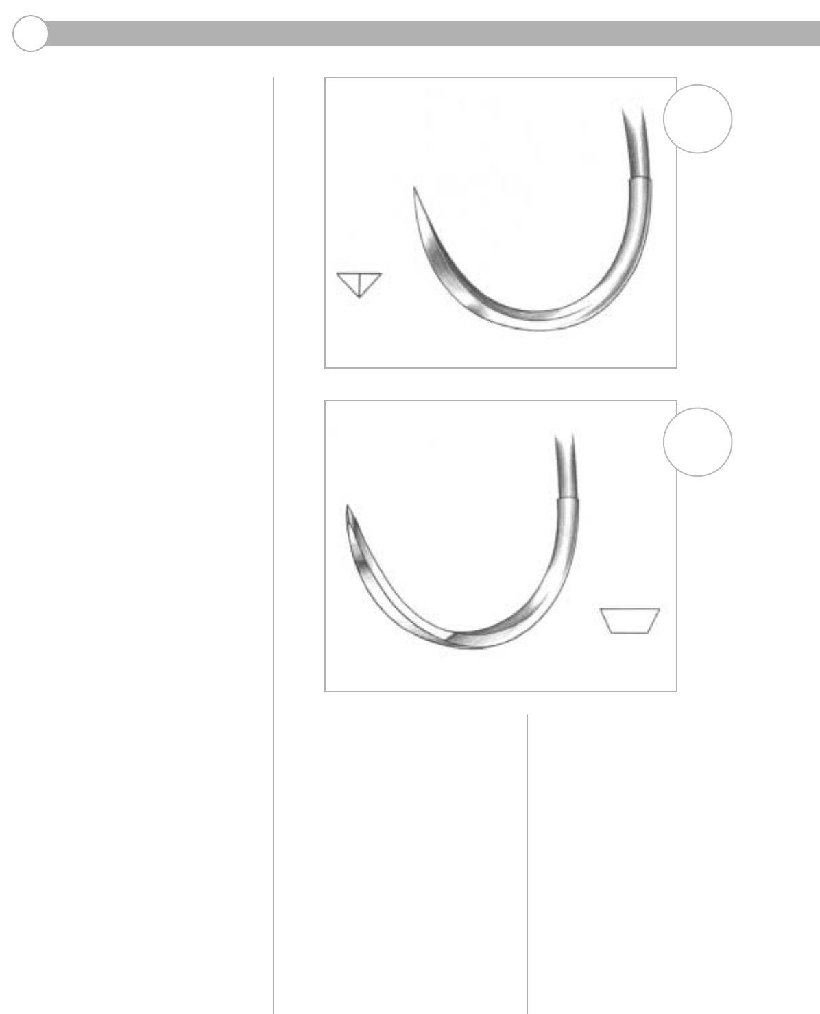
control of the needle as it passes
between or through tissue layers.
The position of the point varies
with the design of each specific
type of spatulated needle.
The SABRELOC* spatula needle
has two cutting edges and a
trapezoidal-shaped body.
The SABRELOC* needle with
the cobra-shaped tip has four
equidistant defined edges.
The CS ULTIMA* ophthalmic
needle (Corneal-Scleral, Patent No.
5,002,564) is the sharpest needle
in its category and is used for
corneal scleral closure. The smaller
angles and increased cutting-edge
length result in superior sharpness
facilitating easy tissue penetration.
The TG PLUS* needle (Transverse
Ground) has a long, ultra-sharp,
slim tip. This needle undergoes a
unique honing process which results
in a sharper needle. The surgeon
encounters low penetration resist-
ance with the TG PLUS needle, and
gets excellent tactile feedback.
TAPER POINT NEEDLES
Also referred to as round needles,
taper point needles pierce and
spread tissue without cutting it. The
needle point tapers to a sharp tip.
The needle body then flattens to an
oval or rectangular shape. This
increases the width of the body to
help prevent twisting or turning in
the needleholder.
Taper point needles are usually
used in easily penetrated tissue
such as the peritoneum, abdominal
viscera, myocardium, dura, and
subcutaneous layers. They are
preferred when the smallest possible
hole in the tissue and minimum
tissue cutting are desired. They are
also used in internal anastomoses
to prevent leakage which can
subsequently lead to contamination
of the abdominal cavity. In the
fascia, taper point needles minimize
the potential for tearing the thin
connective tissue lying between
parallel and interlacing bands of
denser, connective tissue.
The Mayo (MO) needle has a taper
point, but a heavier and more
flattened body than conventional
taper needles. This needle was
designed for use in dense tissue;
particularly for gynecological
procedures, general closure, and
hernia repair.
TAPERCUT SURGICAL
NEEDLES
ETHICON* manufactures TAPER-
CUT* needles which combine the
features of the reverse cutting edge
tip and taper point needles. Three
cutting edges extend approximately
1/32" back from the point. These
blend into a round taper body. All
three edges are sharpened to provide
THE SURGICAL NEEDLE
50
REVERSE
CUTTING
NEEDLE
FIGURE
11
SPATULA
NEEDLE
CROSS-
SECTION
FIGURE
12
* Trademark
uniform cutting action. The point,
sometimes referred to as a trocar
point, readily penetrates dense,
tough tissue. The objective should
be for the point itself not to exceed
the diameter of the suture material.
The taper body portion provides
smooth passage through tissue and
eliminates the danger of cutting
into the surrounding tissue.
Although initially designed for
use in cardiovascular surgery on
sclerotic or calcified tissue, the
TAPERCUT* needle is widely
used for suturing dense, fibrous
connective tissue— especially in
fascia, periosteum, and tendon
where separation of parallel
connective tissue fibers could occur
with a conventional cutting needle.
ETHICON* developed a modified
TAPERCUT CC needle (Calcified
Coronary) for anastomosis of small
fibrotic and calcified blood vessels.
The calcified portion of an artery
requires a cutting tip only for initial
penetration to avoid tearing the
vessel. This needle configuration has
a slimmer geometry than other
TAPERCUT needles from the body
through the point which facilitates
penetration. It also minimizes the
risk of leakage from friable vessels
or vascular graft material.
BLUNT POINT NEEDLES
Blunt point (BP) needles can
literally dissect friable tissue rather
than cutting it. They have a taper
body with a rounded, blunt point
that will not cut through tissue.
They may be used for suturing the
liver and kidney. Due to safety
considerations, surgeons also use
blunt point needles in obstetric
and gynecological procedures when
working in deep cavities which
are prone to space and visibility
limitations. In addition, blunt
point needles for general closure are
especially helpful when performing
procedures on at-risk patients.
The ETHIGUARD* blunt point
needle combines the safety of the
blunt point with the security of a
ribbed and flattened design, and the
convenience of a swaged needle.
NEEDLEHOLDERS
The surgeon uses the needleholder
to pass a curved needle through
tissue. It must be made of noncor-
rosive, high strength, good quality
steel alloy with jaws designed for
holding the surgical needle securely.
CHAPTER 3 51
ETHICON
NEEDLE
CODES
& OTHER
MEANING
BB Blue Baby
BIF Intraocular Fixation
BN Bunnell
BP Blunt Point
BV Blood Vessel
BVH Blood Vessel Half
CCardiovascular
CC Calcified Cornary
CCS Conventional Cutting Sternotomy
CE Cutting Edge
CFS Conventional for Skin
CIF Cutting Intraocular Fixation
CP Cutting Point
CPS Conventional Plastic Surgery
CPX Cutting Point Extra Large
CS Corneal-Scleral
CSB Corneal-Scleral Bi-Curve
CSC Corneal-Scleral Compound Curve
CT Circle Taper
CTB Circle Taper Blunt
CTX Circle Taper Extra Large
CTXB Circle Taper Extra Large Blunt
CV Cardiovascular
DC Dura Closure
DP Double Point
EN Endoscopic Needle
EST Eyed Straight Taper
FN For Tonsil
FS For Skin
FSL For Skin Large
CODE MEANING
FSLX For Skin Extra Large
G Greishaber
GS Greishaber Spatula
JConjunctive
KS Keith Straight
LH Large Half
LR Larger Retention
LS Large Sternotomy
MMuscle
MF Modified Fergusan
MH Medium Half (circle)
MO Mayo
MOB Mayo Blunt
OPS Ocular Plastic Surgery
OS Orthopaedic Surgery
PPlastic
PC Precision Cosmetic
PS Plastic Surgery
RB Renal (artery) Bypass
RD Retinal Detachment
RH Round Half (circle)
RV Retinal-Vitreous
SSpatula
SC Straight Cutting
SFS Spatulated for Skin
SH Small Half (circle)
SIF Ski Intraocular Fixation
SKS Sternotomy Keith Straight
SM Spatulated Module
ST Straight Taper
CODE MEANING
STB Straight Blunt
STC Straight Cutting
STP Straight Taper Point
TE Three-Eighths
TF Tetralogy of Fallot
TG Transverse Ground
TGW Transverse Ground Wide
TN Trocar Needle
TP Taper Pericostal / Point
TPB Taper Pericostal /Point Blunt
TS Tendon Straight
TQ Twisty Q
UCL 5/8 Circle Colateral Ligament
UR Urology
URB Urology Blunt
VTAPERCUT Surgical Needle
VAS Vas Deferens
X or P Exodontal (dental)
XLH Extra Large Half (circle)
XXLH Extra Extra Large Half (circle)
CODE MEANING
TABLE
1
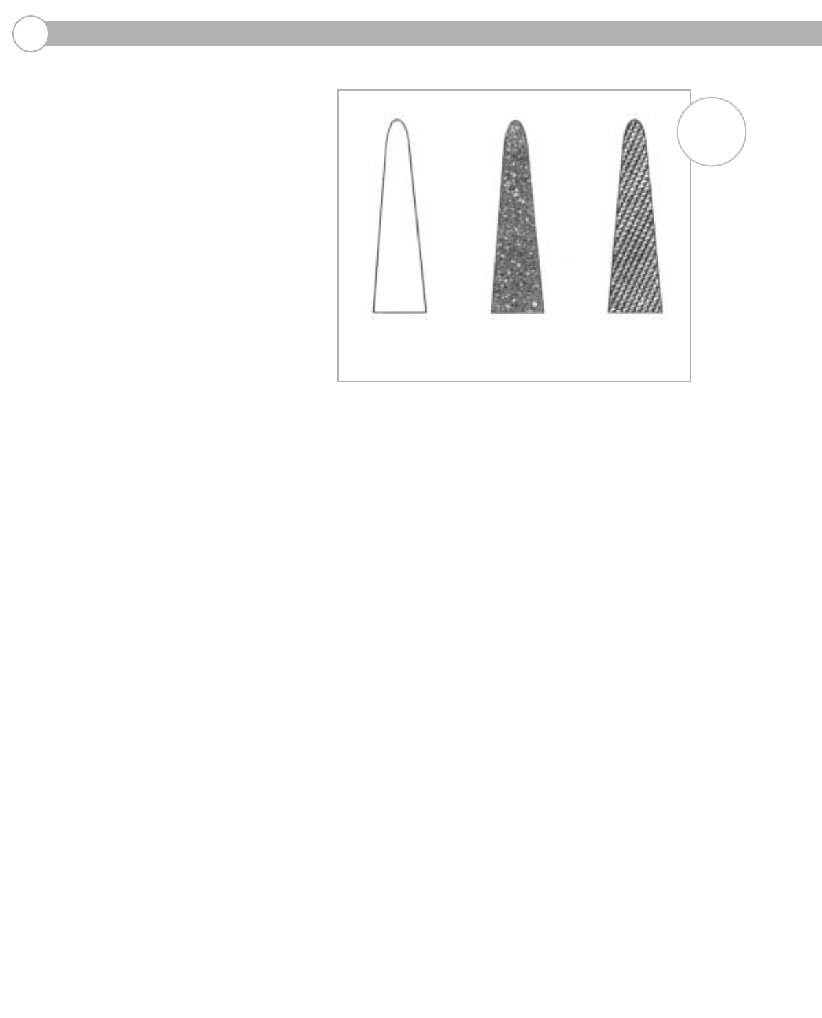
Needleholder jaws may be short or
flat, concave or convex, smooth or
serrated. Smooth jaws may allow the
needle to wobble or twist. Jaws with
teeth hold most securely but may
damage the suture or needle if too
much pressure is applied. Most, but
not all, needleholders have a ratchet
lock near to thumb and finger rings.
Surgical needles are designed for
optimum needleholder stability.
Because this tool actually drives the
needle, its performance will have an
impact upon the entire suturing
procedure. The surgeon has maxi-
mum control only when the needle
sits well in the holder without wob-
bling as it is passed through tissue.
Needleholders, like pliers, weaken
with repeated use. Therefore, the
scrub person should check before
each procedure to make sure that
the needleholder jaws align properly
and grasp securely.
When selecting a needleholder,
the following should be taken
into consideration:
✦It must be the appropriate size
for the needle selected. A very
small needle should be held with
small, fine jaws. The larger and
heavier the needle, the wider and
heavier the jaws of the needle-
holder should be.
✦It should be an appropriate size
for the procedure. If the surgeon
is working deep inside the body
cavity, a longer needleholder is
in order.
NEEDLEHOLDER USE
The following guidelines are offered
to the scrub person for needleholder
use:
1. Grasp the needle with the tip of
the needleholder jaws in an area
approximately one-third to
one-half of the distance from the
swaged end to the point. Avoid
placing the holder on or near the
swaged area which is the weakest
part of the needle.
2. Do not grasp the needle too
tightly as the jaws of the needle-
holder may deform, damage, or
bend it irreversibly.
3. Always check alignment of the
needleholder jaw to make certain
the needle does not rock, twist,
or turn.
4. Handle the needle and needle-
holder as a unit.
5. Pass the needleholder to the
surgeon so that he or she will not
have to readjust it before placing
the suture in tissue. Make sure
the needle is pointing in the
direction in which it will be
used and that the suture strand is
not entangled.
6. Always provide a needleholder—
never a hemostat—to pull the
needle out through tissue. A
hemostat or other clamp can
damage the needle.
7. Immediately after use, every
needle should be returned to the
scrub person while clamped in a
needleholder. Needles are less
likely to be lost if they are passed
one-for-one (one returned for
each one received).
PLACING THE NEEDLE
IN TISSUE
The actual placement of the
needle in the patient's tissue can
cause unnecessary trauma if done
incorrectly. Keep the following in
mind during suturing:
1. Apply force in the tissue to be
sutured in the same direction as
the curve of the needle.
2. Do not take excessively large bites
of tissue with a small needle.
3. Do not force a dull needle
through tissue. Take a new
needle.
THE SURGICAL NEEDLE
52
REVERSE
CUTTING
NEEDLE
FIGURE
11
Smooth Jaws Jaws with tungsten
carbide particles
Jaws with
teeth
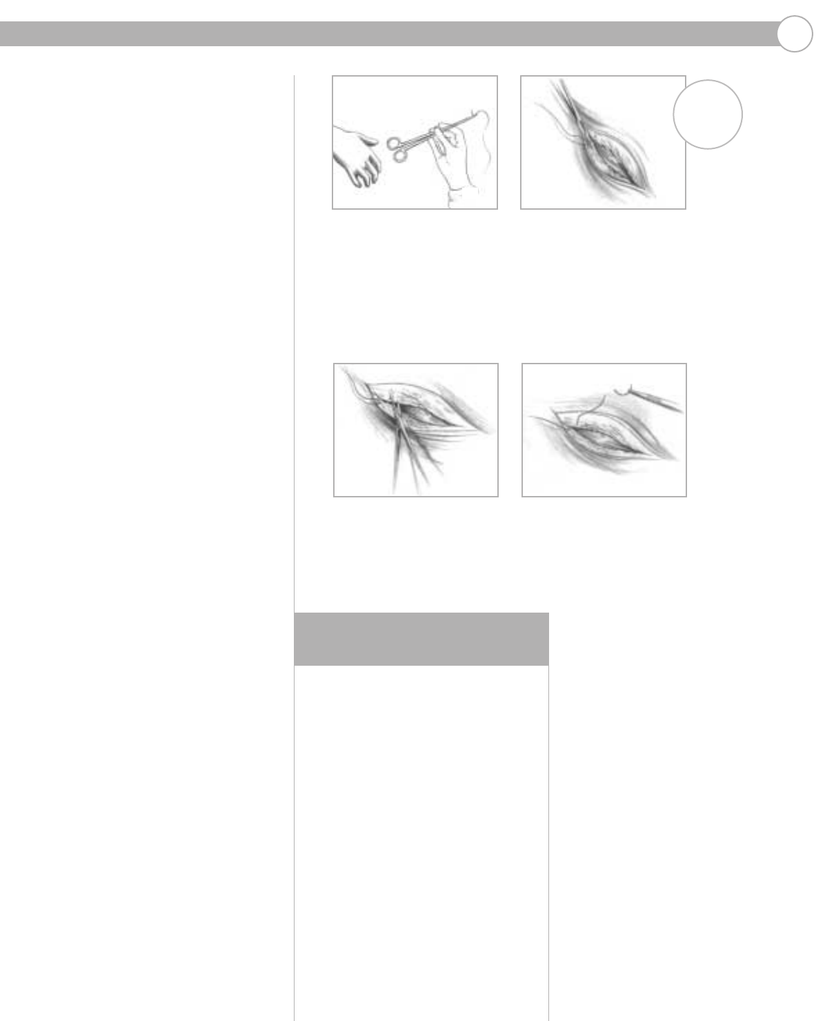
* Trademark
4. Do not force or twist the needle
in an effort to bring the point
out through the tissue. Withdraw
the needle completely and then
replace it in the tissue, or use a
larger needle.
5. Avoid using the needle to bridge
or approximate tissues for
suturing.
6. Do not damage taper points or
cutting edges when using the
needleholder to pull the needle
through tissue. Grasp as far back
on the body as possible.
7. Depending upon the patient, the
tissue may be tougher or more
fibrous than anticipated and
require the use of a heavier gauge
needle. Conversely, a smaller
needle may be required when
tissue is more friable than usual.
8. In a deep, confined area, ideal
positioning of the needle may
not be possible. Under these
circumstances, proceed with
caution. A heavier gauge needle
or a different curvature may help
and a second needleholder
should be used to locate a needle
in a confined body cavity.
9. If a glove is punctured by a
needle, the needle must be
discarded immediately and the
glove must be changed for the
safety of the patient, as well as
the surgical team. Appropriate
serological testing of the patient
should be undertaken for
transmissable agents such as
hepatitis B and C and HIV.
NEEDLE
HANDLING TIPS
Needles should be protected from
bacterial contamination and damage
during handling by adhering to the
following guidelines:
1. Open needle packets and prepare
sutures carefully, protecting
needle sharpness.
2. Make sure the needle is free
of corrosion.
3. If using eyed needles, make sure
they do not have rough or sharp
edges inside the eye to fray or
break suture strands. Also check
the eyes for burrs or bluntness to
ensure easy penetration and
passage through tissue.
4. If a needle is defective, discard it.
5. Pass needles on an exchange
basis; one is passed to the
surgeon for one returned.
6. Employ the nontransfer
technique to avoid inadvertent
needlesticks: the surgeon places
the needle and needleholder
down in a neutral area of the
sterile field; the scrub person
then picks up the needleholder.
7. Secure each needle as soon as it is
used. Do not allow needles to lie
loose on the sterile field or Mayo
stand. Keep them away from
sponges and tapes so they will
not inadvertently be dragged into
the wound.
CHAPTER 3 53
PLACEMENT
OF THE
NEEDLE IN
TISSUE
1. The surgeon receives the
needleholder with the needle
point toward the thumb to
prevent unnecessary wrist
motion. The scrub person
controls the free end of the
suture to prevent dragging it
across the sterile field, and to
keep the suture from entering
the surgeon’s hand along
with the needleholder.
2. The surgeon begins closure
with theswaged suture.
3. The needle is passed into the tissue. The surgeon releases the
needle from the holder and reclamps the holder onto the body of the
needle near the point end to pull the needle and strand through
tissue. The needle is released or cut from the suture strand. The
surgeon leaves the needle clamped in the same position and returns
it to the scrub person. The scrub person immediately passes another
prepared suture to the surgeon, one-for-one.
FIGURE
14

THE SURGICAL NEEDLE
54
8. If a needle breaks, all pieces must
be accounted for.
9. Count all needles before and after
use according to hospital
procedure. Retain the packets
containing descriptive informa-
tion on quantity and needle type
for swaged needles to help
determine if all are accounted for.
Follow these steps for safe needle
handling:
1. Use sterile adhesive pads with or
without magnets or disposable
magnetic pads to facilitate
counting and safe disposal.
2. Swaged needles can be inserted
through or into their original
packet after use. An empty
packet indicates a missing needle.
If using an E-PACK* procedure
kit, compare the count of needles
used to the number preprinted
on the kit label.
3. Return eyed needles to the needle
rack. If eyed needles are to be
reused, they must be cleaned and
reprocessed at the end of the
operation.
4. Do not collect used needles in a
medicine cup or other container
since they must then be handled
individually to count them. This
can potentially contaminate
gloves and increase the risk of an
accidental puncture.
5. Discard used needles in a
"sharps" container.
IN THE
NEXT SECTION
In the section that follows, the
dual role that suture and needle
packaging plays will be covered.
Packaging does much more than
keep the needle and suture sterile.
Package design can help or
seriously hinder the efficiency of
the surgical procedure.
PACKAGING
CHAPTER 4
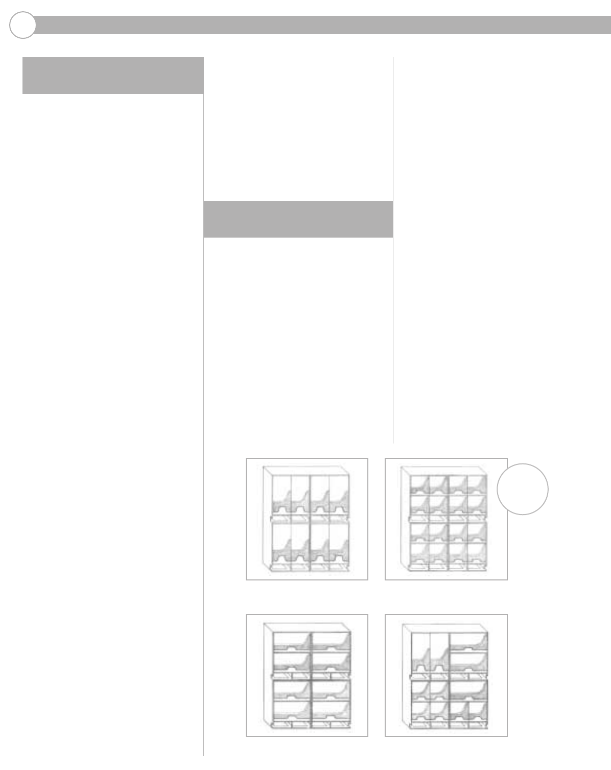
AN INTEGRAL PART
OF THE PRODUCT
The purpose of a package is to
protect its contents and provide
convenience to the user.
ETHICON* wound closure
packaging is an integral part of
each product. Over the past half a
century, packaging has evolved
from glass tubes packed in jars,
to multi-layered foil and paper
packages, to new materials that
reflect concern for both the
environment and the individuals
who must maintain operative
sterility and efficiency. Packaging
has kept pace with the technological
developments of wound closure
products themselves. Several factors
have influenced these developments:
✦Increasing product diversity
✦Technological advances in
packaging materials
✦Stringent regulatory requirements
To prevent infection in an operative
wound, all instruments and supplies
that come in contact with the
wound must be sterile (free of
living microorganisms and spores)
including sutures, needles, ligating
clips, stapling instruments, adhesive
tapes and topical skin adhesives.
High standards and criteria are set
for all components in the packaging
of sterile products:
1. Protect and preserve product
stability and sterility from
potential deterioration from
outside forces such as oxygen,
moisture, light, temperature,
dust, and vermin.
2. Prevent product damage or
microbial contamination in
transit and storage.
3. Provide identifiable product
information.
4. Permit convenient, safe, and
sterile transfer of the product
from the package to the
sterile field.
5. Meet the functional needs of all
members of the surgical team.
RELAY* SUTURE
DELIVERY SYSTEM
Most suture materials are packaged
and sterilized by the manufacturer.
They arrive ready for use in boxes
which can be stored until needed.
The RELAY* suture delivery system,
developed by ETHICON with
human, clinical, and environmental
factors in mind, stores and delivers
sutures in a time-efficient manner
and reduces unnecessary handling
to access sutures. The system
also provides control over suture
storage, usage, inventory rotation,
needle counting, and cost contain-
ment. The RELAY suture system
consists of three basic, interrelated
components: modular suture
storage racks, dispenser boxes,
and primary packets.
MODULAR STORAGE RACKS
The modular storage racks are
designed for maximum convenience
and versatility to meet the individ-
ual needs of a particular specialty,
nurse, surgeon, or department.
Modules can be easily assembled
to accommodate both vertical and
horizontal suture dispenser boxes.
Any number of modules can be
fastened together to meet both
small and large storage needs.
Once assembled, the racks may be
used on shelves, mounted on walls,
placed on mobile carts, or
connected to IV poles. Racks can
also be fitted with a rotating base
for more convenient access, as well
as with a handle for easy carrying.
PACKAGING
56
ETHICON
MODULAR
STORAGE
RACKS
FIGURE
1
Full-vertical boxes Half-vertical boxes
Horizontal boxes Combination of boxes

Each module has a built-in
inventory control area to facilitate
restocking. This feature enables
unused suture packets to be
systematically fed back into the
proper rotational flow without
mixing lots within the boxes.
Sutures may be grouped within
the modular system by material
type or size, or by use (i.e., general
closure, gastrointestinal surgery,
plastic surgery, etc.).
DISPENSER BOXES
Gravity-fed dispenser boxes dispense
suture packets from the opening at
the bottom of the box. The opening
can accommodate the removal of
several suture packets at one time.
All ETHICON* dispenser boxes
are made of recyclable paper
and printed with either water or
soy-based inks. Each box provides
clear product identification through
streamlined graphics, product
color coding, bold label copy,
and descriptive symbols. The
information required for quick
reference and easy selection of
suture materials is highlighted in a
logical sequence. The three most
important criteria necessary for
proper identification and suture
selection are:
1. Suture size
2. Suture material
3. Type and size of needle
Other important product
information found on all suture
boxes includes:
1. Surgical application
2. Product code number
3. Suture length and color
4. Metric diameter equivalent of
suture size and length
5. Shape and quantity of needles
(single- or double-armed, shown
by silhouette)
6. Needle point geometry
7. Lot number
8. Expiration date
A package insert with detailed infor-
mation about the suture material is
inserted in every dispenser box. Users
should be familiar with this informa-
tion as it contains FDA-approved
indications, contraindications, and
all appropriate warnings and precau-
tionary statements for each product.
Dispenser boxes should be restocked
when the last few suture packets
appear in the box opening, before
the box is completely empty. The
unused packets from the previous
box should be used before a new
dispenser box is opened. This will
help to avoid mixing lot numbers
and ensure proper stock rotation.
ETHICON advocates rotation
of the entire dispenser box. In
addition to ensuring the use of the
oldest suture materials first, this
helps to maintain a fresh stock of
dispenser boxes.
Most dispenser boxes contain
three dozen suture packets. Others
may contain one or two-dozen
packets. The product code number
suffix and a statement on the box
indicate the quantity of suture
packets in the box (product code
suffix G = 1 dozen, D = 1 dozen,
T = 2 dozen, H = 3 dozen). The
dispenser boxes are held securely for
easy dispensing by firmly pushing
the box into a "lock" in the back of
the rack module.
PRIMARY PACKETS
Individual sutures and multiple
suture strands are supplied sterile
within a primary packet. The
exterior surfaces of the overwrap are
not sterile. ETHICON primary
packaging is designed to permit fast
and easy opening in one peelable
motion. The single layer overwrap
of primary packaging is made of
either foil or coated Tyvek®on one
side heat-sealed to polyethylene film
on the other. Absorbable sutures are
always encased in foil to provide a
safe and durable moisture barrier
and to withstand sterilization in
the manufacturing process. Most
nonabsorbable sutures are encased
in coated Tyvek®overwraps.
In a continuous effort to be more
environmentally conscious,
ETHICON has chosen materials
in the manufacture of primary
packets which generate minimal
negative impact to the environment
upon incineration or disposal.
Furthermore, wherever possible,
the number of primary packaging
layers has been reduced by as
much as 50 percent, thus reducing
the volume of environmental waste
per OR procedure.
Each primary packet provides
critical product information and
the same color-coding as its
dispenser box. The packet also
identifies the product code number,
material, size, needle type, and the
number of needles per packet to
simplify needle counts.
Primary packets of suture material
may contain sutures in one of
five styles:
1. Standard lengths of non-needled
material: 54 inches (135cm) of
absorbable or 60 inches (150cm)
of nonabsorbable suture, which
may be cut in half, third, or
quarter lengths for ligating
or threading.
* Trademark
CHAPTER 4 57
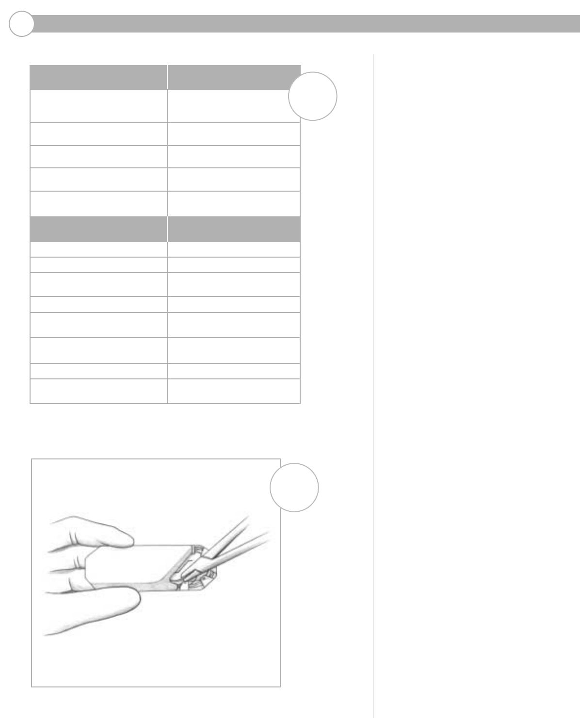
2. SUTUPAK* pre-cut sterile suture
is nonneedled material for
ligating or threading. These
lengths may be supplied in a
multistrand labyrinth packet or
in a folder packet, both of which
are designed to deliver one strand
at a time. SUTUPAK sutures
may be removed from the packet
and placed in the suture book.
3. One single strand of material
with single- or double-armed
swaged needle(s). Needles for
one-step RELAY suture packets,
micro-surgery, and some
ophthalmic needles are secured in
a "needle park." The needle park
is designed to provide a standard
location for, and easy access to,
the needle. All other needles are
protected within an inner
folder or other specific channel
within a paper folder.
Most single strand needled
sutures are sealed in convenient
one-step RELAY delivery
packages. One-step RELAY pack-
ages allow the needle to be armed
in the needleholder from any
angle without touching the
needle. This increases the safety
of handling needles intraopera-
tively. If it is preferred to locate
the needle by hand, this can be
accomplished with the one-step
RELAY package by pushing up
the flap behind the needle park,
thereby elevating the needle so it
can be grasped by hand.
4. Multiple suture strands, either
swaged to a single needle or
double-armed. This type is
appropriate for procedures
requiring numerous interrupted
sutures of the same type. It saves
valuable operative time by
PACKAGING
58
MOST
COMMON
ETHICON
SUTURE
PACKAGING
TABLE
1
Surgical Gut Suture
Coated VICRYL* RAPIDE
(polyglactin 910) suture
Coated VICRYL*
(polyglactin 910) suture
MONOCRYL*
(poliglecaprone 25) suture
PDS* II
(polydioxanone) suture
PERMA-HAND* Silk Suture
Stainless Steel Suture
ETHILON* nylon suture
NUROLON* nylon suture
MERSILENE*
polyester fiber suture
ETHIBOND* EXCEL
polyester fiber suture
PROLENE* polypropylene suture
PRONOVA*
poly (hexafluoropropylene-VDF) suture
ABSORBABLE SUTURE LAYERS
Tyv ek ®overwrap, foil primary
package containing one-step
RELAY* suture delivery system tray
Tyv ek ®overwrap, foil primary package,
paper folder
Peelable foil overwrap, one-step
RELAY tray
Peelable foil overwrap, one-step
RELAY tray
Peelable foil overwrap, one-step
RELAY tray
Tyv ek ®overwrap, one-step RELAY tray
Tyv ek ®overwrap, paper folder
Peelable foil overwrap, one-step
RELAY tray
Tyv ek ®overwrap, one-step RELAY tray
Tyv ek ®overwrap, one-step RELAY tray
Tyv ek ®overwrap, one-step RELAY tray
Tyv ek ®overwrap, one-step RELAY tray
Tyv ek ®overwrap, one-step RELAY tray
NONABSORBABLE SUTURE LAYERS
Tyvek ®is a registered trademark of E.I. du Pont de Nemours and Company
METHOD FOR
PREPARING
ONE-STEP
RELAY
PACKAGE
SUTURES
FIGURE
2
Arm the needle directly from the one-step RELAY tray and
deliver the single suture to the surgeon.

* Trademark
enabling the surgeon to use one
suture while the next is being
armed—without delay of open-
ing packets or threading needles.
Multistrand packets are labeled
with the symbol MS/ that
denotes multiple strands/number
of strands of surgical needles per
packet. Multistrand packets may
contain 3 to 10 swaged sutures.
The inner folder for these
products is white.
All packets containing
CONTROL RELEASE* needle
sutures have multiple strands
(8, 5, 4, 3, or 1) and are
designated CR/8, CR/5, CR/4,
CR/3, or CR/1. CONTROL
RELEASE sutures may be
available in foil or Tyvek®
overwrap packets for single
strand delivery. The single strand
delivery folder is used for some
coated VICRYL* (polyglactin
910) sutures, MONOCRYL*
(poliglecaprone 25) sutures,
PDS* II (polydioxanone) sutures,
ETHIBOND* EXCEL polyester
sutures, NUROLON* nylon
sutures, MERSILENE* polyester
sutures, and PERMA-HAND*
silk sutures. The suture material
straightens as it is delivered from
the folder. Each suture may be
delivered to the surgeon individ-
ually from the opening packet or
removed from the folder and
placed in the suture book. The
inner folder for these products is
either red with a black C/R
symbol or white with red
lettering. The safety organizer
tray is used for coated VICRYL
sutures, MONOCRYL sutures,
PDS II sutures, ETHIBOND
EXCEL sutures, PERMA-HAND
silk sutures, NUROLON nylon
sutures, MERSILENE sutures,
and surgical gut sutures. The
safety organizer tray allows for
single strand arming and
dispensing. The needles are
situated in individually num-
bered needle parks and may be
armed and dispensed with little
or no hand-to-needle contact.
5. Ligating material used as either
single strand (free or freehand)
ties, or as continuous ties
unwound from a reel or other
device. The length of single
strand ties is determined by the
depth of the wound. In subcuta-
neous tissue, quarter lengths
(approximately 14 inches) are
usually long enough for ligating.
Single strand ligating material is
available in pre-cut lengths or 18,
24, and 30 inch strands.
Many surgeons prefer continuous
ties. Some prefer LIGAPAK*
ligature, which is supplied on
disc like plastic radiopaque
dispensing reels that are color
coded by material. The size of
the ligature material is indicated
by the number of holes visible on
the side of the reel (e.g., 3 holes=
3-0 suture). The reel is held in
the palm of the hand as blood
vessels are ligated. Other
surgeons may prefer the ligating
material rewound onto a rubber
reel, gauze sponge, metal bobbin,
or other device.
The number of packets of
ligating material required to tie
off subcutaneous vessels
(bleeders) will vary with patient
size and age, the amount of
bleeding, the type of operation,
the length of the incision, and
the surgical technique. An
abdominal incision 8 to 12
inches long might require
one to three packets to
ligate the subcutaneous
blood vessels.
All suture material is packaged
dry with the exception of surgical
gut and pliabilized ETHILON
sutures. Natural absorbable suture
materials are packaged with a
small amount of sterile fluid,
usually alcohol with water, to
maintain pliability. They should
therefore be opened over a basin to
prevent any solution from spilling
onto the sterile field.
All needles should be counted
after packets of swaged sutures
are opened, according to
established hospital procedure.
The packets should be retained
to facilitate verification of the
final needle count after the
surgical procedure.
E-PACK*
PROCEDURE KIT
The E-PACK procedure kit
contains numerous sutures and
other products for a specific
procedure, surgeon, or surgical
specialty. The packaging concept
saves valuable time in the OR by
eliminating the need to open and
coordinate multiple individual
suture packages. The E-PACK
procedure kit is also an effective
means of reducing inventory
levels of individual product
codes, and providing a record
for determining the suture
costs associated with a given
surgical procedure.
CHAPTER 4 59

PACKAGING
60
The suture packages are secured
in an organizer sleeve to facilitate
sterile transfer to the sterile field.
The procedure kit label provides all
the pertinent information regarding
the number and types of needles, as
well as sizes and types of suture.
Suture quantities are listed on the
label, making it easy to quickly
determine how many needles have
been used and thus simplifying
needle accountability at the end of
the procedure. The organizer sleeve
is delivered in a Tyvek®pouch.
EXPIRATION DATE
The expiration date of a product is
determined by product stability
studies. The Food and Drug
Administration (FDA) requires
that all synthetic absorbable suture
products have an expiration date
stamped on each dispenser box
and primary packet to indicate
the known shelf life of the material,
provided the physical integrity
of the package is maintained.
Tests conducted by ETHICON*
show conclusively that synthetic
absorbable suture products such
as Coated VICRYL* suture and
PDS* II suture continue to meet
all product requirements even at
five years of storage.
In addition, all ETHICON
nonabsorbable suture products
contain a five-year expiry dating
on each dispenser box and
primary packet. This expiry
dating is necessary to comply
with various international
regulatory guidelines and is an aid
in inventory management.
The RELAY suture delivery system
is designed as a "first-in, first-out"
inventory control system. Dispenser
boxes are rotated, permitting the
oldest sutures to be used first. The
expiration date stamped on the
outside of each box and every
packet clearly indicates the month
and year of product expiration.
SUTURE
STERII IZATION
Sutures sterilized by ETHICON are
either irradiated with cobalt 60 or
exposed to ethylene oxide gas. Both
processes alter proteins, enzymes,
and other cellular components to
the extent that microorganisms are
unable to survive or cause infection.
Irradiation and ethylene oxide gas
are considered cold sterilization
processes because radiation sterilizes
at room temperature and ethylene
oxide gas sterilizes at much lower
temperatures than other sterilization
methods such as dry heat or steam
under pressure.
Irradiation sterilization exposes
products to ionizing radiation—
either beta rays produced by high
energy electron accelerators or
gamma rays from radioisotopes—
until absorbed in appropriate
sterilizing dose. ETHICON was
a pioneer in both beta and
gamma irradiation and routinely
sterilizes products with cobalt
60 which emits gamma rays.
Cobalt 60 irradiation is the simplest
of all sterilization processes.
Some suture materials cannot
withstand the effects of irradiation
sterilization, becoming unusable.
Instead, they are gas sterilized.
Gas sterilization uses
ethylene oxide gas. As an environ-
mental measure, ETHICON
replaced chlorofluorocarbons
(CFCs) with more environmentally
friendly compounds in all gas
sterilization processes. The
combination of ethylene oxide
gas concentration, temperature,
humidity, and exposure time must
be carefully controlled to ensure
reliable sterilization.
WARNING: Surgical sutures are
labeled as disposable, single-use
medical devices. Suture products
manufactured by ETHICON are
provided in easy-to-use packages
designed to maintain the stability
and sterility of the suture and
needle materials. The component
layers of packaging materials do
not permit exposure to high
temperatures or extremes of pres-
sure without affecting package and
product integrity. For this reason,
all sterile products manufactured
by ETHICON are clearly labeled,
"DO NOT RESTERILIZE."
Manufacturers cannot be held
responsible for the quality,
effectiveness, or integrity of
suture materials resterilized in
the hospital, office, or by outside
vendors. Therefore, if customers
utilize the services of a sterilization
reprocessor for suture, ETHICON
will disclaim any responsibility for
sterilization and/or other product
failures resulting from the resteril-
ization process. The practice of
resterilization is not recommended,
except for ETHI-PACK* pre-cut
steel sutures and spools or cardreels
of nonabsorbable materials
supplied nonsterile.

* Trademark
CHAPTER 4 61
ANTICIPATING
SUTURE NEEDS
Today's healthcare environment
dictates that hospitals continue to
maintain quality standards while
lowering costs to remain financially
viable. Through total quality man-
agement initiatives, many hospitals
have identified material use as an
opportunity to lower cost. To
increase the efficiency of suture
utilization during a surgical
procedure, it is important to
determine and anticipate the
surgeon's needs more precisely.
For this reason, a file system of
preference cards for each surgeon
on staff is usually maintained in the
operating suite. The cards contain
such information as the surgeon's
"suture routine," suture materials,
sizes, needles, and/or product
code numbers customarily used in
specific procedures.
Becoming more aware of each
surgeon's routine through good
communication and regularly
updated preference cards can help
reduce preparation time, minimize
waste, and assure cost effectiveness.
Prior to dispensing suture packets,
the circulating nurse should have a
brief discussion with the surgeon to
ascertain whether a change in suture
routine is anticipated due to a
specific patient's needs.
While it is difficult to say precisely
how many suture packets are
enough, three major factors should
be considered in deciding how
many packets to open:
1. Fewer packets will be needed if
products with multiple strands of
suture material are used.
2. Opening sufficient suture packets
to prevent prolonging operative
time and causing surgeon
inconvenience.
3. Leftover suture on the surgical
field must be discarded.
Therefore, opening too many
suture packets should be
avoided to reduce waste and to
lower cost.
Although it is important to be
prepared to answer requests at a
moment's notice, it is not necessary
to overload the table with sutures.
The introduction of single-layer
peelable packaging, such as one-step
RELAY* packaging, helps encourage
less handling to access the suture,
enhancing quick delivery of suture
materials to the surgeon in the
sterile field. Unexpected suture
needs can also be obtained rapidly
from the storage racks.
STERILE TRANSFER OF
SUTURE PACKETS
At some point, suture packets
must cross the sterile barrier—the
invisible line of demarcation
between the sterile and the nonster-
ile. In all settings (e.g., operating
room, delivery room, emergency
department, or physician's office),
the individual who removes
the nonsterile overwrap must
remember these three points about
sterile transfer:
1. Outer surfaces of the overwrap
are not sterile and may be
handled with nonsterile hands.
2. The sterile inner packet or tray
must be transferred to the sterile
field without being touched or
contacting any nonsterile object
or surface.
3. Nonsterile hands over the sterile
field violate aseptic technique.
There are two methods commonly
used for achieving sterile transfer
of suture packets: handing-off the
sterile inner one-step RELAY tray
directly to the scrub person or
"flipping" the inner contents of the
primary packet onto the sterile field.
Regardless of the aseptic technique
performed, all items introduced
onto the sterile field should be
opened, dispensed, and transferred
by methods that maintain product
sterility and integrity. AORN
Guidelines recommend the
“hand-off” method, since items
tossed or flipped have a greater
potential to roll off the edge of the
sterile field, causing contamination
or other items to be displaced.
METHOD I: STERILE TRANSFER TO
THE SCRUB PERSON
Grasp the two flaps of the peelable
overwrap between the knuckles of
the thumbs and forefingers. With
a rolling-outward motion, peel
the flaps apart to approximately
one- third of the way down the
sealed edges. Keeping pressure
between the knuckles for control,
offer the sterile inner packet or
tray to the scrub person, who takes
it with a gloved hand or sterile
instrument. Care must be taken to
avoid contact with the nonsterile
overwrap as the packet or tray
is withdrawn.
This method must be used to
remove paper folder packets of sur-
gical steel and PROLENE* sutures
from long straight overwraps, and to
remove the organizer sleeves from
E-PACK* procedure kits. It should
also be used for transfer of flexible,
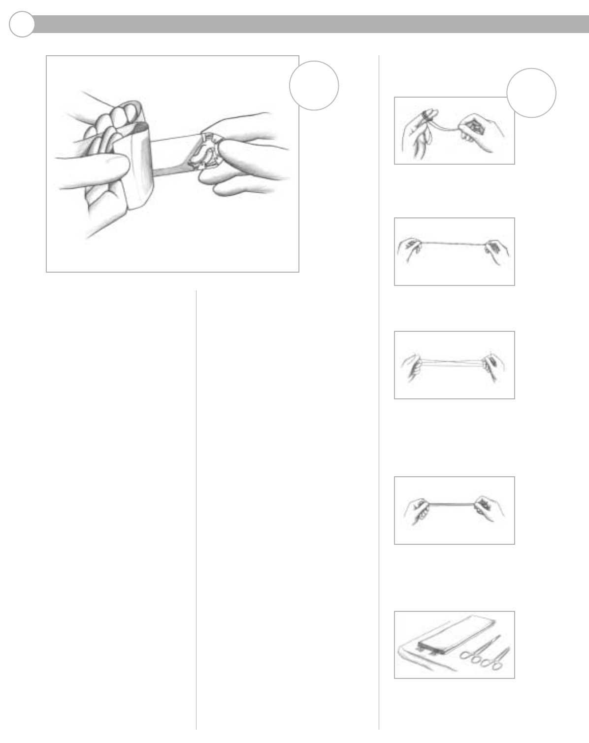
lightweight, transparent packets
containing microsurgery and oph-
thalmic products.
METHOD II: STERILE TRANSFER
TO THE STERILE FIELD
"Flipping" is a rapid and efficient
method of ejecting sterile product
from its overwrap onto the sterile
field without contacting the
unsterile outer packet or reaching
over the field. However, skill must
be acquired to ensure its effective
use. The circulating nurse must
stand near enough to the sterile
table to project the suture packet
or tray onto it, but not too close
as to risk contaminating the table
by touching it or extending
nonsterile hands over it. To
accomplish this, grasp the flaps
of the overwrap as described in
Method I and peel the flaps apart
with the same rolling-outward
motion. The sterile packet or tray
is projected onto the sterile table
as the overwrap is completely
peeled apart.
NOTE: DO NOT attempt to proj-
ect the inner folder of long straight
packets onto the sterile table.
Instead, present them to the scrub
person as outlined in Method I.
SUTURE PREPARATION
IN THE STERILE FIELD
Suture preparation may be more
confusing than virtually any other
aspect of case preparation.
Familiarity and understanding of
the sequence in which tissue layers
are handled by the surgeon will help
to eliminate this confusion. (See the
Suturing Section, Chapter 2.)
Once the suture packets are opened
and prepared according to the
surgeon's preference card, sutures
can be organized in the sequence in
which the surgeon will use them.
Ligatures (ties) are often used first
in subcutaneous tissue shortly after
the incision is made, unless ligating
clips or an electrosurgical cautery
device is used to coagulate severed
blood vessels.
After the ligating materials have
been prepared, the suturing
(sewing) materials can be prepared
in the same manner. Preparing large
amounts of suture material in
PACKAGING
62
STERILE
TRANSFER
TO THE
SCRUB
PERSON
FIGURE
3FIGURE
4
PREPARATION OF
STANDARD LENGTH
LIGATURE STRANDS
1. Prepare cut lengths of ligature
material, coil around fingers of left
hand, grasp free ends with right
hand, and unwind to full length.
2. Maintain loop in left hand and two
free ends in right hand. Gently pull
the strand to straighten.
3. To make 1/3lengths: Pass one free
end of strand from right to left hand.
Simultaneously catch a loop around
third finger of right hand. Make
strands equal in thirds and cut the
loops with scissors.
4. To make 1/4lengths: Pass both free
ends from right to left hand.
Simultaneously catch a double loop
around third finger of right hand.
Cut the loops.
5. Place packets or strands in suture
book (folded towel)—or under Mayo
tray—with ends extended far
enough to permit rapid extraction.
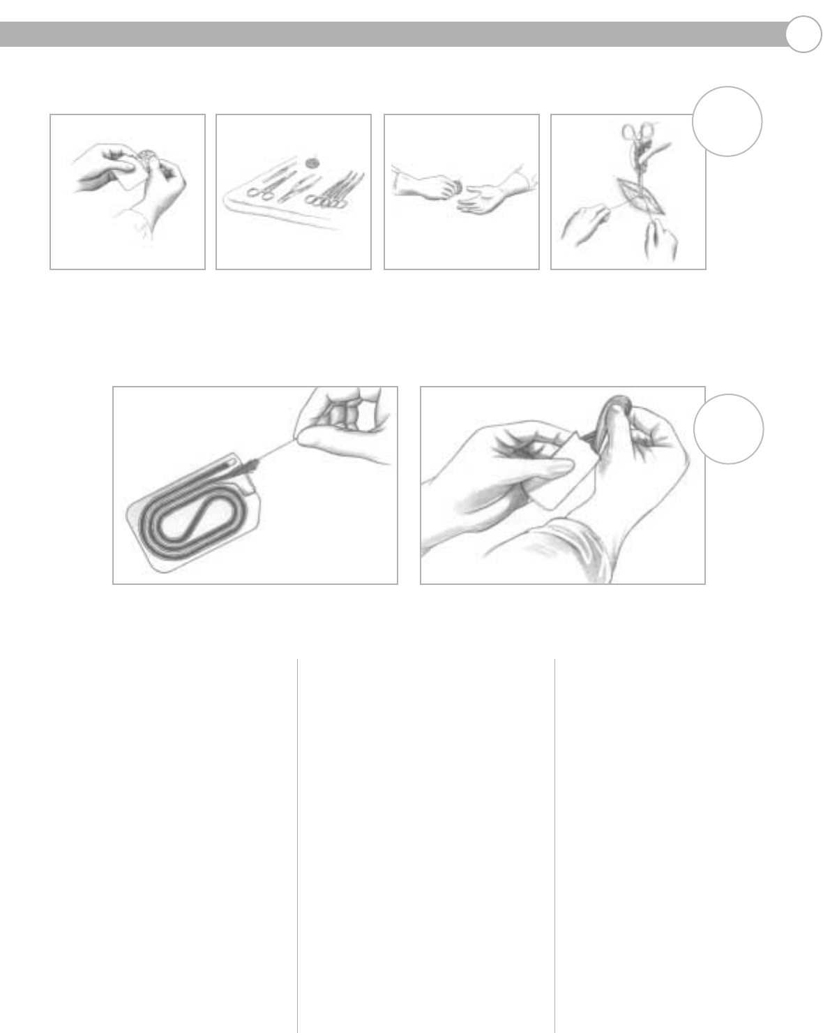
advance should be avoided. For
example, if the surgeon opens the
peritoneum (the lining of the
abdominal cavity) and discovers
disease or a condition that alters
plans for the surgical procedure
and anticipated use of sutures,
opened packets would be wasted.
At closure following abdominal
surgery, remembering the letters
PFS (peritoneum, fascia, skin) will
be helpful for organizing sutures.
By watching the progress of the
procedure closely, listening to
comments between the surgeon
and assistants, and evaluating
the situation; suture needs can
be anticipated. Free moments can
be used to prepare sufficient suture
material to stay one step ahead of
the surgeon. The goal should be to
have no unused strands at the end
of the procedure.
Ligature material which remains
toward the end of the procedure
may be the same material and size
specified by the surgeon for sutures
in the subcutaneous layer of wound
closure. In this case, the remaining
ligating material should be used
rather than opening an additional
suture packet.
If the surgeon requires "only
one more suture," and strands of
suitable material remain which
are shorter than those prepared
originally, do not be reluctant to
ask the surgeon if one of the
strands will serve the purpose
before opening a new packet.
Most surgeons are cooperative in
efforts to conserve valuable supplies.
SUTURE HANDLING
TECHNIQUE
During the first postoperative week,
the patient's wound has little or no
strength. The sutures or mechanical
devices must bear the responsibility
of holding the tissues together
during this period. They can only
perform this function reliably if the
quality and integrity of the wound
* Trademark
CHAPTER 4 63
FIGURE
5
PEPARATION
OF PRE-CUT
SUTURES
FOR TIES OR
LIGATURE
SUTURES
PREPARATION OF CONTINUOUS TIES
ON A LIGAPAK DISPENSING REEL
3. Hand reel to surgeon as
needed, being certain that
the end of the ligating
material is free to grasp.
4. Surgeon holds reel in
palm, feeds strand
beween fingers, and
places around tip of
hemostat.
1. Open the packet contain-
ing the appropriate
material on a reel. Transfer
the inner contents of the
primary packet to the
sterile field using aseptic
technique.
2. Extend the strand end
slightly for easy grasp-
ing. Place reel conviently
on the Mayo tray.
FIGURE
6
1. Remove one pre-cut length from nonabsorbable
suture at a time from the labrinth packet as it is
needed by the surgeon.
2. Extract pre-cut strands of SUTUPAK* sterile
absorbable or nonabsorbable suture. Straighten
surgical gut with a gentle pull. Place strands in
the suture book or under Mayo tray.
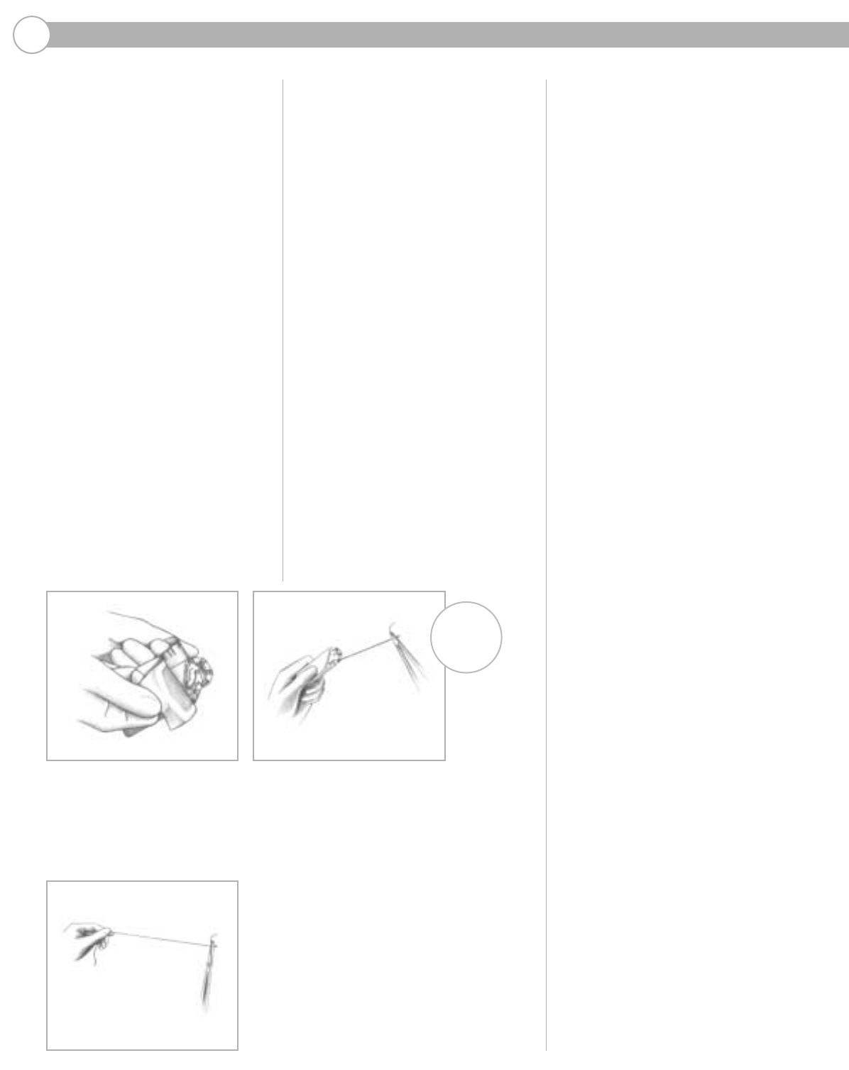
closure materials are preserved
during handling and preparation
prior to use. It is therefore essential
for everyone who will handle the
suture materials to understand
proper procedure to preserve
suture tensile strength.
In general, avoid crushing or
crimping sutures with surgical
instruments such as needleholders
and forceps, except as necessary
to grasp the free end of a suture
during an instrument tie. There
are also specific procedures to
follow to preserve suture tensile
strength which depend upon
whether the material is absorbable
or nonabsorbable. The following
summarizes the most important
points for each member of the
surgical team to remember and
observe in handling suture materials
and surgical needles.
FOR THE CIRCULATING NURSE
1. Consult the surgeon's preference
card for suture routine.
2. Check the label on the dispenser
box for type and size of suture
material and needle(s). Note the
number of strands per packet.
Fewer packets will be needed if
multistrand or CONTROL
RELEASE* sutures are used.
3. Estimate suture requirements
accurately and dispense only the
type and number of sutures
required for the procedure.
4. Read the label on the primary
packet or overwrap before using
to avoid opening the wrong
packet.
5. Use aseptic technique when
peeling the overwrap. Transfer
the inner contents of the primary
packet to the sterile field by
offering it to the scrub person or
by projecting (flipping) it onto
the sterile table, avoiding
contamination.
6. To open long straight packets,
peel overwrap down 6 to 8 inch-
es and present to the scrub
person. Do not attempt to
project the inner folder of long
straight packets onto the
sterile table.
7. Maintain an adequate supply of
the most frequently used sutures
readily accessible.
8. Rotate stock using the "first-in,
first-out" rule to avoid expira-
tion of dated products and keep
inventories current.
9. Suture packets identify the
number of needles per packet to
simplify needle counts. Retain
this information during the
procedure and/or until final
needle counts are completed.
10. Count needles with the scrub
person, per hospital procedure.
FOR THE SCRUB PERSON
1. If appropriate, remove the inner
one-step RELAY* tray or folder
containing suture materials from
the primary packet being offered
from the circulating nurse.
2. Hold the one-step RELAY tray or
folder in gloved hand and arm
the needle using the "no-touch"
technique. Gently dispense the
suture.
3. Leave pre-cut suture lengths in
labyrinth packet on the Mayo
tray. Strands can then be
removed one at a time as needed.
4. Surgical gut and collagen sutures
for ophthalmic use must first be
rinsed briefly in tepid water to
avoid irritating sensitive tissues.
PACKAGING
64
ETHICON
MODULAR
STORAGE
RACKS
FIGURE
7
1. With a rolling-outward motion,
peel the flaps apart to approxi-
mately ont-third the way down
the sealed edges. Keeping pres-
sure beween the knuckles for
control, offer the sterile inner
RELAY tray to the scrub person.
2. Clamp the needleholder approx-
imately one-third to one-half of
the distance from the swage
area to the needle point. Do not
clamp the swaged area. Gently
pull the suture to the right in a
straight line.
3. Additional suture straightening
should be minimal. If the strand
must be straightened, hold the
armed needleholder and gently
pull the strand making certain
not to disarm the needle from
the suture.

If the surgeon prefers to use
sutures wet, dip only momentar-
ily. Do not soak. Silk sutures
should be used dry.
5. Do not pull or stretch surgical
gut or collagen. Excessive
handling with rubber gloves can
weaken and fray these sutures.
6. Count needles with the
circulating nurse, per hospital
procedure.
7. Hold single strands taut for
surgeon to grasp and use as a
freehand tie.
8. Do not pull on needles to
straighten as this may cause
premature separation of
CONTROL RELEASE needle
suture.
9. Always protect the needle to
prevent dulling points and
cutting edges. Clamp the needle-
holder forward of the swaged
area, approximately one-third to
one-half the distance from the
swage to the point.
10. Microsurgery sutures and
needles are so fine that they may
be difficult to see and handle.
They are packaged with the
needles parked in foam to
protect delicate points and
edges. The needles may be
armed directly from the foam
needle park. If the microsurgeon
prefers to arm the needle, the
removable orange colored tab
may be used to transport the
needle into the microscopic
field.
11. Handle all sutures and needles
as little as possible. Sutures
should be handled without using
instruments unless absolutely
necessary. Clamping instruments
on strands can crush, cut, and
weaken them.
12. Cut sutures only with suture
scissors. Cut surgical steel with
wire scissors.
13. When requesting additional
suture material from the
circulating nurse, estimate usage
as accurately as possible to
avoid waste.
FOR THE SURGEON
1. Avoid damage to the suture
strand when handling. This is
particularly critical when
handling fine sizes of monofila-
ment material. Touch strands
only with gloved hand or closed
blunt instrument. Do not crush
or crimp sutures with instru-
ments, such as needleholders or
forceps, except when grasping the
free end of the suture during an
instrument tie.
2. Clamp a rubber shod hemostat
onto the suture to anchor the
free needle on a double-armed
strand until the second needle is
used. Never clamp the portion of
suture that will be incorporated
into the closure or the knot.
3. Use a closed needleholder or
nerve hook to distribute tension
along a continuous suture line.
Be careful not to damage the
suture.
4. Use knot tying techniques that
are appropriate for the suture
material being used.
* Trademark
CHAPTER 4 65

PACKAGING
66
PRESERVATION
OF TENSILE
STRENGTH:
ABSORBABLE
SUTURES
TABLE
2
1. Protect absorbable sutures from heat and moisture.
a. Store suture packets at room temperature. Avoid prolonged storage in hot
areas such as near steam pipes or sterilizers.
b. Do not soak absorbable sutures. Also avoid prolonged placement of sutures
in a moist suture book.
c. Surgical gut can be dipped momentarily in tepid (room temperature) water
or saline to restore pliability if strands dry out before use. Surgical gut or
collagen for use in ophthalmic surgery should be rinsed briefly in tepid
water before use, as they are packed in a solution usually consisting of
alcohol and water to maintain pliability.
d. Synthetic absorbable sutures must be kept dry. Use strands directly from
packet when possible. Store sutures in a dry suture book if necessary.
2. Straighten strands with a gently, steady, even pull. Jerking and tugging can
weaken sutures.
3. Do not "test" suture strength.
4. Do not resterilize.
PRESERVATION
OF TENSILE
STRENGTH:
NONABSORBABLE
SUTURES
TABLE
3
SILK – Store strands in a dry towel. Dry strands are stronger than wet strands. Wet silk
loses up to 20% in strength. Handle carefully to avoid abrasion, kinking, nicking, or
instrument damage. Do not resterilize.
SURGICAL STAINLESS STEEL – Handle carefully to avoid kinks and bends. Repeated
bending can cause breakage. Stainless steel suture can be steam sterilized without any
loss of tensile strength. However, DO NOT steam sterilize on spool or in contact with
wood. Lignin is leached from wood subjected to high temperature and may cling to
suture material. Handle carefully to avoid abrasion, kinking, nicking, or instrument dam-
age.
POLYESTER FIBER – Unaffected by moisture. May be used wet or dry. Handle carefully
to avoid abrasion, kinking, nicking, or instrument damage. Do not resterilize.
NYLON – Straighten kinks or bends by "caressing" strand between gloved fingers a few
times. Handle carefully to avoid abrasion, kinking, nicking, or instrument damage.
POLYPROPYLENE – Unaffected by moisture. May be used wet or dry. Straighten
strands with a gentle, steady, even pull. Handle with special care to avoid abrasion,
kinking, nicking, or instrument damage. Do not resterilize.
TOPICAL SKIN ADHESIVES
CHAPTER 5

Low tension wounds (those where
the skin edges lie close together
without significant tension) can be
closed by gluing the skin edges
together with a skin adhesive.
Butylcyanoacrylate adhesives have
been available in Europe, Israel, and
Canada for decades.1They have
been used successfully for the
closure of traumatic lacerations and
surgical incisions. Application of
butylcyanoacrylate was found to be
more rapid and cost effective than
suturing, but only recently has it
been evaluated in well-designed
clinical trials for wound closure.1
The most significant advance in
the field of topical skin adhesives
has been the development of
2-octyl cyanoacrylate, marketed as
DERMABOND* Topical Skin
Adhesive by ETHICON Products.
This topical skin adhesive forms a
transparent and flexible bond,
unlike the opaque and brittle bond
formed by butylcyanoacrylate
adhesives. The flexibility of
octyl cyanoacrylate allows it to be
applied over nonuniform surfaces.
This flexibility also combats the
topical shear forces exerted on the
adhesive, reducing the risk of
premature sloughing and wound
dehiscence. Additionally, octyl
cyanoacrylate adhesive has been
found to have three times the
breaking strength of butylcyano-
acrylate, so it can be used on longer
incisions and lacerations.1
DERMABOND*
TOPICAL SKIN ADHESIVE
(2-OCTYL CYANOACRYLATE)
is a sterile, liquid topical adhesive
designed to repair low-tension
lacerations and to close wounds
and surgical incisions. It utilizes the
moisture on the skin's surface to
form a strong, flexible bond and can
be used in many instances where
sutures, staples or skin strips
have been traditionally used.
DERMABOND adhesive is ideally
suited for wounds on the face, torso
and limbs and offers cosmetic
advantages over other forms of
skin closure. It can be used in
conjunction with, but not in place
of, deep dermal sutures.
Approved by the FDA in 1998,
DERMABOND adhesive has
been used extensively by health
professionals in the fields of trauma,
plastic and other surgeries,
emergency medicine, and pediatrics.
Since its approval, DERMABOND
adhesive has proven useful in
closing surgical incisions where
suture removal can cause significant
discomfort. The adhesive does not
produce suture or "track" marks
along the healed incision and unlike
stitches, a patient can shower right
away without fear of compromising
the incision.
STRENGTH AND SECURITY
In less than three minutes,
DERMABOND adhesive provides
the strength of healed tissue at
7 days after traditional closure with
sutures.2A strong, flexible
3-dimensional bond makes it
suitable for use in closing easily
approximated incisions
(example—deep, short, long).3
SEALS OUT BACTERIA
DERMABOND adhesive is the first
wound-closure technology approved to
protect wounds & incisions from
common microbes. For trauma and
post-surgical patients, infections are
often the most common, and in
some cases, the most serious
complications. DERMABOND
adhesive helps protect against the
penetration of bacteria commonly
associated with surgical site
infections.2In vitro studies
demonstrated that DERMABOND
adhesive provides 99% efficacy in
protection against Staphylococcus
epidermidis, Staphylococcus aureus,
Escherichia coli, Pseudomonas
aeruginosa and Enterococcus faecium.2
DERMABOND adhesive acts as a
barrier to microbial penetration as
long as the adhesive film remains
intact.
PROMOTES A MOIST,
WOUND-HEALING ENVIRONMENT
DERMABOND adhesive creates a
protective seal that helps the wound
stay moist.2Maintaining a moist
wound-healing environment around
the wound has been shown to speed
the rate of epithelialization.4As the
wound heals (generally between 5 to
10 days), DERMABOND adhesive
will gradually peel off.2
PROVIDES EXCELLENT
COSMETIC RESULTS
In a prospective, randomized,
controlled, unmasked study of 818
patients, DERMABOND adhesive
provided cosmesis equivalent to that
of sutures. At 3 months, it produced
optimal cosmesis in 80% of
patients, using the Modified
Hollander Cosmesis Scale.2
TOPICAL SKIN ADHESIVES
68

MORE COST EFFECTIVE
THAN SUTURES
In most cases, DERMABOND
adhesive allows for significantly
faster closure than sutures.3,5,6
DERMABOND adhesive applica-
tion requires fewer surgical supplies,
reduced equipment needs, and
eliminates the need for suture
removal.6,7
DERMABOND adhesive is also
more convenient and comfortable
for the patient because it often does
not require anesthetic, is gentler to
the skin than sutures or staples, and
does not require suture removal.
DERMABOND adhesive also
reduces the risk of needle stick
injury to both physician and
patient.
INDICATIONS AND
CONTRAINDICATIONS
DERMABOND adhesive is intended
for topical application only to hold
closed easily approximated skin edges
of wounds from surgical incisions,
including punctures from minimally
invasive surgery, and simple,
thoroughly cleansed, trauma-induced
lacerations. DERMABOND adhesive
may be used in conjunction with, but
not in place of, subcuticular sutures.
Topical skin adhesives are not
appropriate for closing wounds that
are subject to significant static or
dynamic tensions (e.g., over joints)
unless deep sutures, immobilization,
or both are also used.
DERMABOND adhesive is contra-
indicated for use on any wounds
with evidence of active infection or
gangrene. It should also not be used
on mucosal surfaces or across
mucocutaneous junctions (e.g., lips,
oral cavity), or on skin that is
regularly exposed to body fluids or
with dense hair (e.g., scalp).
DERMABOND adhesive should
not be used on patients with a
known hypersensitivity to
cyanoacrylate or formaldehyde.
APPLICATION
Mastery of tissue adhesive use
is generally quite rapid. Proper
wound selection, evaluation and
preparation before closure is
important. Wounds must be
thoroughly cleansed and debrided
in accordance with standard practice
before using adhesives. The wound
edges must be tightly apposed so
that the adhesive is not placed
into the wound. Patient positioning
is also important to reduce runoff
of tissue adhesive. The patient
should be positioned so that the
wound surface is parallel to the
floor, taking special care that
any runoff does not flow in the
direction of vital structures such
as the eye.
* Trademark
CHAPTER 5 69
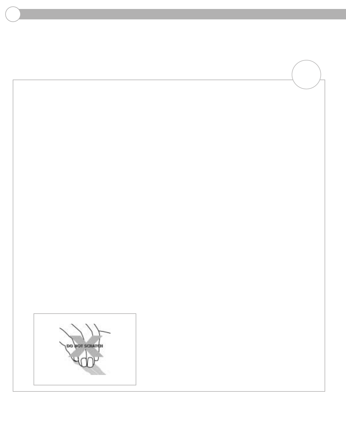
TOPICAL SKIN ADHESIVES
70
DERMABOND Topical Skin Adhesive (2-octyl
cyanoacrylate) is a sterile, liquid skin adhesive that
holds wound edges together. The film will usually
remain in place for 5 to 10 days, then naturally falls
off the skin.
The following provides instructions for proper care of
the wound while it is healing:
CHECK WOUND APPEARANCE
Some swelling, redness and pain are common with all
wounds and normally will go away as the wound
heals. If swelling, redness, or pain increases or if the
wound feels warm to the touch, instruct patients to
contact a doctor. A doctor should also be contacted if
the wound edges reopen or separate.
REPLACE BANDAGES
If the wound is bandaged, the patient should be
instructed to keep the bandage dry. The dressing
should be replaced daily until the adhesive film has
fallen off or if the bandage should become wet, unless
otherwise instructed by the physician.
When changing the dressing, tape should not be
placed directly over the DERMABOND adhesive
film, because removing the tape later may also
remove the film.
AVOID TOPICAL MEDICATIONS
Instruct patients not to apply liquid or ointment
medications or any other product to the wound while
the DERMABOND adhesive film is in place. These
may loosen the film before the wound is healed.
KEEP WOUND DRY AND PROTECTED
Patients may occasionally and briefly wet the wound
in the shower or bath. They should not soak or scrub
the wound. They should not swim and should
avoid periods of heavy perspiration until the
DERMABOND adhesive has naturally fallen off.
After showering or bathing, the patient should blot
the wound dry with a soft towel. If a protective
dressing is being used, a fresh, dry bandage should
be applied, being sure to keep the tape off the
DERMABOND adhesive film.
Additional instructions for patients include:
• Apply a clean, dry bandage over the wound if
necessary to protect it.
• Protect the wound from injury until the skin has
had sufficient time to heal.
• Do not scratch, rub or pick at the DERMABOND
adhesive film. This may loosen the film before the
wound is healed.
• Protect the wound from prolonged exposure to
sunlight or tanning lamps while the film is in place.
HOW TO CARE FOR A WOUND
AFTER IT’S TREATED WITH
DERMABOND* TOPICAL SKIN ADHESIVE TABLE
1

* Trademark
CHAPTER 5 71
REFERENCES
1. Singer, Adam J., Lacerations and
Acute Wounds, An Evidence-
Based Guide; F.A. Davis
Company, 2003, p. 83-97.
2. Data on file, ETHICON, INC.
3. Quinn, G Wells, T Sutcliffe,
et al. A Randomized Trial
Comparing Octyl Cyanoacrylate
Tissue Adhesive and Sutures in
the Management of Lacerations.
JAMA 1997;227(19):1527-1530.
4. Singer AJ, Hollander JE, Quinn
JV. Evaluation and management
of traumatic lacerations.
N Engl J Med.
1997;337(16):1142-1148.
5. Bruns TB, Robinson BS, Smith
RJ et al. A new tissue adhesive
for laceration repair in children.
J Pediatr. 1998;132:1067-1070.
6. Theodore N, et al. The
Economics of DERMABOND
in Neurosurgical Wound
Closure. Phoenix, Ariz:
Neuroscience Publications,
Barrow Neurological Institute.
March 2001:2-10.
7. Osmond MH, Klassen TP,
Quinn JV. Economic comparison
of a tissue adhesive and suturing
in the repair of pediatric facial
lacerations. J Pediatr. 1995;
126:892-895.
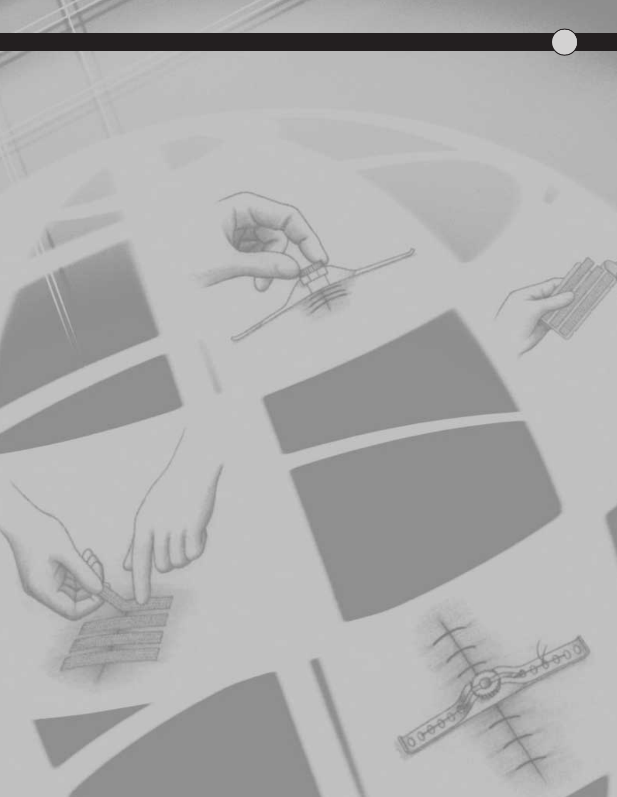
OTHER SURGICAL
PRODUCTS
CHAPTER 6

ADHESIVE TAPES
There are many surgical products
available which may be used during
wound closure and other operative
procedures which involve suturing.
Each of these products has specific
indications for use. Adhesive tapes
are used for approximating the
edges of lacerations, skin closures,
repair and/or support in selected
operative procedures.
Adhesive tapes are associated with
minimal tissue reactivity and yield
the lowest rate of infection, but
they tend to slough off in the
presence of tension or moisture.1
Advantages of adhesive tapes
include rapid application, little or
no patient discomfort, low cost, and
no risk of needle-stick injuries. They
are associated with minimal tissue
reactivity and yield the lowest infec-
tion rates of any wound closure
method. They may be left on for
long periods without resulting in
suture hatch marks.
Surgical tapes are not commonly
recommended as the sole modality
for primary wound closure due
to the high probability of
dislodgement and dehiscence,
the inability to use them in
hair-bearing areas, and the need
to keep them dry. Their use is
typically reserved for linear lacera-
tions under minimal tension.
Furthermore, surgical tapes do not
approximate deeper tissues and do
not control bleeding.
INDICATIONS AND USAGE
Skin closure tapes are an effective
alternative to sutures or staples
when tensile strength and resistance
to infection are not critical factors.
Skin closure tapes can also be
used to complement suture or
staple closures. Stress is applied
uniformly to the collagen fibers,
aiding in rapid fiber orientation
and increased tensile strength.
APPLICATION
Skin closure tapes may be applied to
the skin over a subcuticular closure
in lieu of skin sutures or used as a
primary closure in conjunction with
sutures in an alternating pattern.
The tape is placed on one side of
the wound at its midpoint, while
grasping it with forceps in the
dominant hand. The opposite
wound edge is then gently apposed
by pushing with a finger of the
nondominant hand. The wound
edges should not be apposed by
pulling on the free end of the
tape. This can result in unequal
distribution of skin tensions,
causing erythema or even blistering
of the skin. Additional strips are
then placed perpendicular to the
laceration on either side of the
original tape, bisecting the
remaining open wound with each
strip until the space between tapes
is no more than about 2 to 5 mm.
Additional strips are then placed
over the ends of the other strips,
parallel to the laceration.
Skin closure tapes may also be used
as a replacement for sutures or
staples which are removed on the
first to fourth postoperative day.
Effective skin closure tapes provide
good porosity in terms of air inflow
to the wound and water vapor
transmission escaping from the
wound during the healing process.
AFTER CARE AND REMOVAL
Adhesive tapes should be left in
place as long as possible, at least as
long as sutures would be left before
removal. To prevent the tapes from
prematurely coming loose, patients
must be warned to keep them as
dry as possible and not to cover
them with ointments. Showering
is permitted and during the
first week many surgeons
recommend that patients cover
their wounds and tape with a
nonadherent dressing.
OTHER SURGICAL PRODUCTS
74
ADVANTAGES
AND
DISADVANTAGES
OF ADHESIVE
TAPES FOR
SKIN CLOSURE1
TABLE
1
ADVANTAGES DISADVANTAGES
• Rapid and simple
closure
• Least damage to
host defenses
• No risk of needle-stick
injuries
• Do not cause tissue
ischemia or necrosis
• Relatively poor
adherence
• Easily removed by
patient
• Must be kept dry
• Cannot be used over
oily or hair-bearing
areas
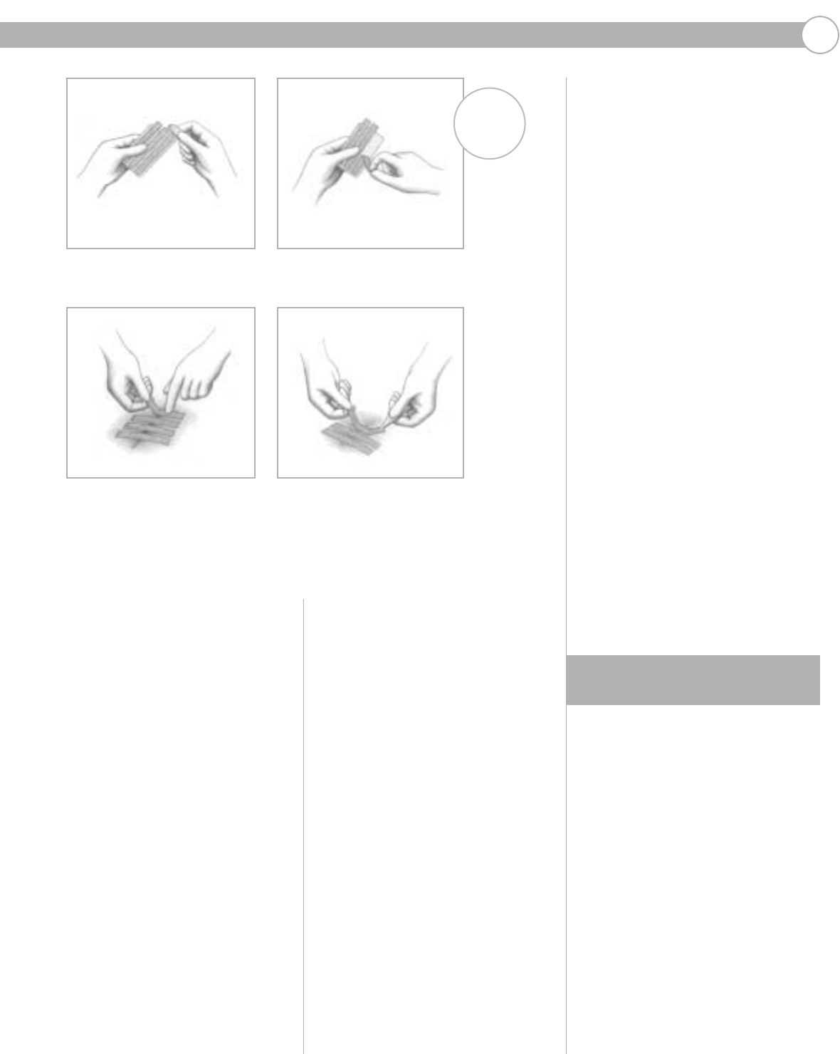
* Trademark
SKIN CLOSURE TAPES
PROXI-STRIP* skin closures are
long, narrow, sterile strips of tape
with an adhesive backing. They are
used for approximating the edges of
lacerations and for closing skin fol-
lowing many operative procedures.
PROXI-STRIP skin closures have a
high degree of porosity to allow the
wound to breathe, but have suffi-
cient adhesive strength to negate the
use of adjunct applications, such as
tincture of benzoin. Their antistatic
properties minimize the tendency of
the tape strips to curl up.
POLYESTER FIBER STRIP
MERSILENE* polyester fiber strip
is comprised of a double thickness
of MERSILENE polyester fiber
that is 5mm wide. The strips are
available with and without
needles and may be used instead
of large-sized suture for ligation,
repair, and/or support in selected
operative procedures.
Incompetence of the cervix is a
condition characterized by the
habitual premature, spontaneous
abortion of the fetus. A ligature is
placed around the cervix in a
collar-like fashion, drawn tight,
and either sutured together or tied
closed. A MERSILENE strip is
then woven carefully with a swage
blunt needle in and out of the
mucosa. When placed properly,
the flatness of the ligature will not
cut or damage the wall of the cervix.
MERSILENE strip attached to
a heavy reverse cutting needle
provides a wide band of strong
material for orthopaedic procedures
such as rotator cuff repair and
support. The blunt needles used for
the incompetent cervix ligation may
also be used for this purpose.
UMBILICAL TAPE
Umbilical tape is a white woven
cotton ligature, 1/8or 1/4inch
(0.32 or 0.64cm) wide that is strong
enough to tie off the umbilical cord
of the newborn infant. While this
was its original use, umbilical
tape is also used in pediatric and
cardiovascular procedures to
suspend small structures and vessels
during the operation, but is not left
in place.
Umbilical tape easily absorbs
blood when used in an area of
gross bleeding. The 1/8-inch
(0.32cm) tape is available with a
radiopaque thread woven into the
length of the fabric to facilitate
x-ray identification.
SURGICAL
STAPLES
The staple closure is mainly used for
large wounds that are not on the
face. Stapling is especially useful
for closing scalp wounds. Staples
are also used for linear lacerations
of the torso and extremities,
especially if they are relatively long.
Many surgeons routinely use skin
staples for closure of standard
abdominal, thorax and extremity
incisions. Advantages of stapling
include ease of use, rapidity, cost
effectiveness, and minimal damage
to host defenses.1
CHAPTER 6 75
SKIN
CLOSURE
TAPES
FIGURE
1
1. Using sterile technique,
remove card from sleeve and
tear off tab.
2. Peel off tapes as needed in
diagonal direction.
3. Apply tapes at 1/8-inch
intervals as needed to
complete wound apposition.
Make sure the skin surface is
dry before applying each tape.
4. When healing is judged to be
adequate, remove each tape
by peeling off each half from
the outside toward the wound
margin. Then, gently lift
the tape away from the
wound surface.

A variety of stapling devices is
available for wound closure. With
all devices, the staple creates an
incomplete rectangle: the legs of the
staple extend into the skin, and the
cross-limb lies on the skin surface
across the wound. Each device may
differ in its handling characteristics,
visual access, the angle at which
the staples enter tissues, the ease
of position and the pre-cocking
mechanism. Optimal visibility as
the staple is placed in the skin is
important, as is the angle at which
the staple enters the skin because
insertion of the staple perpendicular
to the surface of the skin results in
deep penetration that increases the
likelihood of tissue strangulation
and permanent cross-hatching of
the wound. The ability of the
staple end to swivel allows the head
to be adjusted for use in deep
recesses. Finally, the presence of a
pre-cocking mechanism allows the
practitioner to maintain constant
control while stapling the skin.1
Before inserting staples, it is impor-
tant to line up the wound edges
with the centerline indicator on the
head of the stapler to make sure that
the legs of the staple will enter the
skin at equal distances on either side
of the wound edge. Each edge is
typically picked up with a forceps,
everted and precisely lined up.
The surgeon then places the staples
to close the wound while the first
assistant advances the forceps,
everting the edges of the wound.
This technique is continued until
the entire wound is everted and
closed with staples.2
INDICATIONS AND USAGE
Wound closure with staples is
indicated for scalp lacerations
that do not require extensive
hemostasis and do not involve tears
in the underlying frontooccipital
aponeurosis (galea). They are
also indicated for linear nonfacial
lacerations caused by shear forces
(e.g., sharp objects).
AFTERCARE AND REMOVAL
Skin staples should be removed
at the same time that sutures
would be removed, based on wound
location and tension. For scalp
wounds, staples should be removed
on day 7 after insertion. For trunk
and extremity wounds, staples
should be removed between days 7
and 14. Wounds closed with staples
may be covered with a topical
antibiotic cream or ointment.
Patients may bathe or shower
the next day, but should avoid
prolonged exposure to moisture.
When used on the scalp, patients
should be very careful about
combing or brushing their hair.
A specially designed, single-handed,
disposable staple remover should be
used to remove the staples by a
health care provider.
PROXIMATE* Skin Staplers
PROXIMATE Skin Staplers place
single staples to close surgical
incisions. Staples are made of
lubricant-coated stainless steel;
OTHER SURGICAL PRODUCTS
76
PROXIMATE*
SKIN STAPLERS TABLE
2
PROXIMATE* RH Skin Staplers
(Rotating Head Skin Staplers)
Rectangular
staples
Head rotates 360°;
cartridge is clear
Staples are coated
with lubricant
Pistol-grip handle
PROXIMATE* PX Skin Staplers PROXIMATE* PLUS MD Skin Staplers
(Multi-Directional Skin Staplers)
Features Benefits Features Benefits Features Benefits
Minimizes staple
rotation
Improves visibility
and access
Easy staple
extraction
Comfortable
to use
Ergonomic pistol
grip
Positive ratchet
mechanism
Staples are coated
with lubricant
Intuitive and
comfortable to use
Easy staple
placement
Easy staple
extraction
Improved kick-off
spring design
Ergonomic design
Alignment indicator
Staples are coated
with lubricant
Multi-direction
release
Comfortable for
smaller hands
Improves visibility
Easy staple extraction
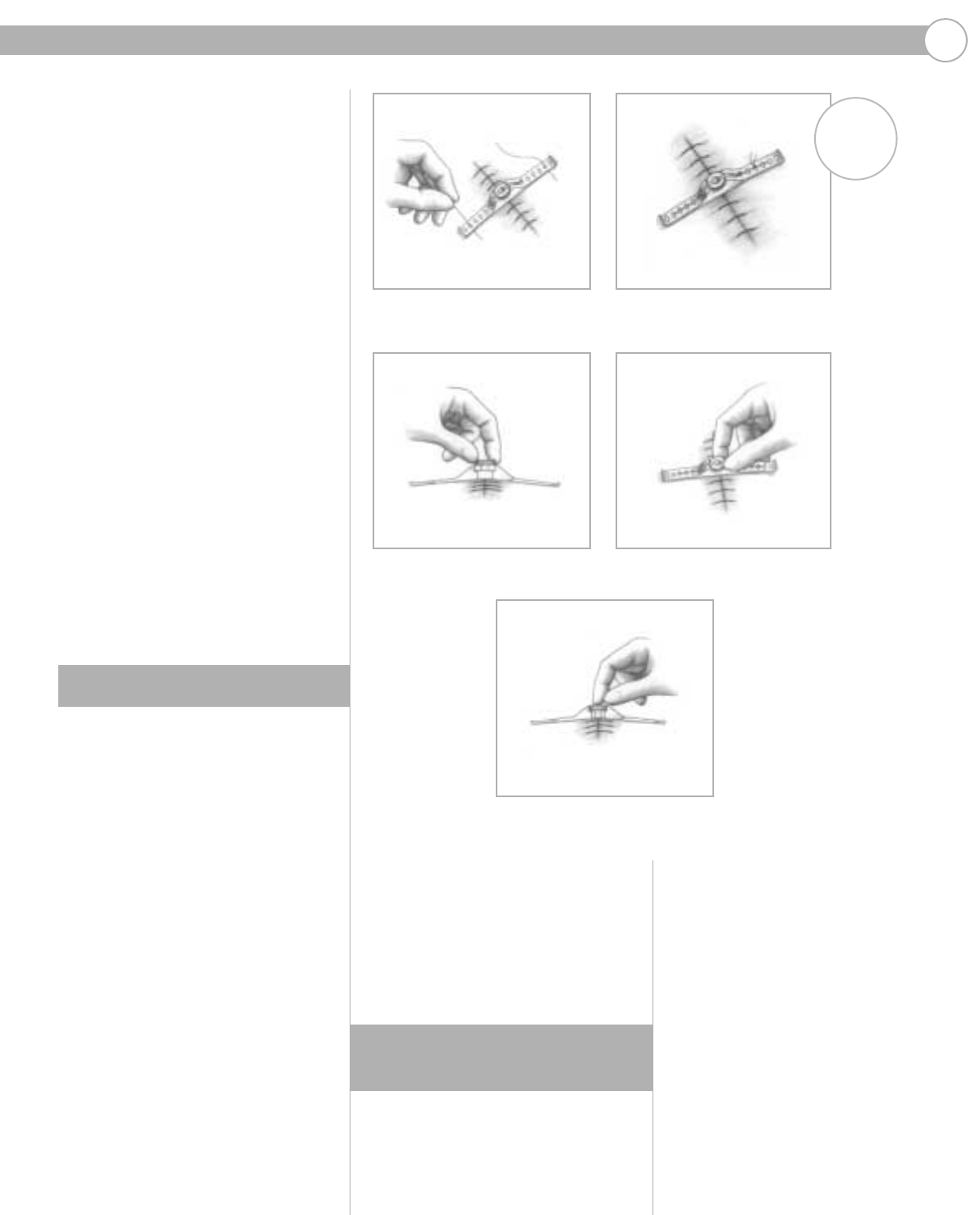
* Trademark
the staplers are not reloadable.
Ethicon Endo-Surgery makes
three different skins staplers to
meet surgeons’ needs.
PROXIMATE* RH is a full-feature
skin stapler with a 360° rotating
head assembly.
PROXIMATE RH has improved
visibility over existing fixed-head
skin stapling products along with
a ratchet mechanism for easier
staple placement.
PROXIMATE* PX skin stapler
provides many of the same features
as the PROXIMATE RH skin
stapler but in a fixed-head format.
PROXIMATE* PLUS MD is a
high-value, low-cost skin stapler
that permits multi-directional
release in an ergonomic design.
LOOPED SUTURE
ETHICON looped sutures range in
length up to a 60-inch strand with
both ends swaged to a single taper
point needle. Available in various
materials and suture sizes, they
provide a simple, reliable technique
for continuous closure of the fascia
of the abdominal wall. The needle
of the looped suture is passed
through the fascia from inside out
at one end of the incision, then
through the opposite wound edge
from outside in, and then passed
through the loop. The locking stitch
lies beneath the wound edge. The
double strand is run over and over
to the other end of the incision.
The final stitch is completed by
passing the needle from the outside
in, cutting one strand, and passing
the needle through the opposite
wound edge from the outside in.
The needle is then cut off and
the loose suture ends tied together,
leaving the knot inverted under
the fascia.
RETENTION
SUTURE DEVICES
Retention sutures, if not placed
carefully without excessive tension,
can cut the skin. Devices such as
bolsters and bridges are used to
prevent such complications and
eliminate pressure. However, care
should also be taken in the use of
these devices.
Retention suture bolsters are sterile
21/2-inch (6cm) lengths of 3/16-inch
(0.48cm) diameter surgical latex
tubing with a 1/32-inch (0.08cm)
wall. The suture is threaded through
the bolster and tied. Sutures
sheathed in this manner can cause
an inflammatory response with
reaction both at the site of the
suture exit from the skin and along
CHAPTER 6 77
ADJUSTMENT
OF
RETENTION
SUTURE
BRIDGE
FIGURE
1
1. Pass the retention suture
through appropriate holes in
the bridge.
2. Place the suture with tension
over the slit in the capstan,
and tie.
3. To adjust tension, lift capstan. 4. Rotate capstan until desired
tension is attained.
5. To lock, press capstan down
into bridge.

the entire length of the suture itself.
Also, the skin may become necrotic
beneath the bolsters if the sutures
are too tight. This invariably occurs
if the sutures are tightly tied at the
time of the operation, as subsequent
tissue edema ensues.
The retention suture bridge is a
strong plastic truss that can be
adjusted to relieve the pressure of
the retention suture on the skin
during, and subsequent to, initial
suture placement. After the desired
number of sutures is placed in the
wound, a sterile bridge is positioned
over each retention suture. Each
side of the bridge has six holes
spaced 1/4inch (0.64cm) apart to
accommodate many patient sizes.
The ends of the sutures are passed
through the appropriate holes and
tied loosely over the bridge. The
suture strand is then slipped into
the capstan located in the middle
of the bridge, and the capstan is
rotated to apply the desired
tension before locking into place.
The bridge permits easy tension
readjustment by raising and rotating
the capstan to compensate for
postoperative wound edema, and
again when the edema subsides.
The suture remains elevated away
from the skin while the bridge has
contact along its entire 4 3/8-inch
(11cm) length. Pressure is evenly
distributed over the area, and the
transparent bridge facilitates com-
plete visualization of the wound.
REFERENCES
1. Singer, Adam J., Lacerations and
Acute Wounds, An Evidence-Based
Guide; F.A. Davis Company,
©2003, p. 64-71, 73-82.
2. Skerris, David A., Mayo Clinic
Basic Surgery Skills, Mayo Clinic
Scientific Press, 1999; p.117.
OTHER SURGICAL PRODUCTS
78
PRODUCT TERMS
AND TRADEMARKS
CHAPTER 7

ABSORBABLE SUTURE
Sutures which are broken down and
eventually absorbed by either
hydrolysis (synthetic absorbable
sutures) or digestion by lysosomal
enzymes elicited by white blood
cells (surgical gut and collagen).
APPROXIMATE
Bring together sides or edges.
ATRALOC*
SURGICAL NEEDLES
ETHICON* trademark for eyeless
needles permanently attached
(swaged) to suture strands.
B & S GAUGE
Brown and Sharpe gauge commonly
used in hospitals to identify wire
diameter. ETHICON stainless steel
suture products are labeled with
both B & S gauge and U.S.P. size.
BURIED SUTURE
Any stitch made and tied so that
it remains completely under
the surface.
CALCIFIED CORONARY
NEEDLE (CC)
A TAPERCUT* surgical needle
with a 1/16" cutting tip and slim
taper ratio for significant ease of
penetration when suturing tough
valve cuffs or atherosclerotic vessels.
CARDIOVASCULAR SUTURES
Swaged sutures designed to meet the
specific needs of heart and blood
vessel surgery.
CATGUT
Outmoded term for surgical gut
suture.
CHROMIC SURGICAL GUT
Gut suture which has been treated
by chromium salts to resist
digestion by lysosomal enzymes.
CHROMICIZING
ETHICON process for producing
chromic gut. Each ribbon of
surgical gut is bathed in a
chromium salt solution before
spinning into strands to provide
uniform controlled absorption.
COATED VICRYL*
(POLYGLACTIN 910) SUTURE
ETHICON trademark for synthetic
absorbable suture extruded from a
copolymer of glycolide and lactide
and coated with a mixture of
polyglactin 370 and calcium
stearate.
COATED VICRYL* RAPIDE
(POLYGLACTIN 910) SUTURE
ETHICON trademark for braided,
rapidly absorbing synthetic suture
extruded from a copolymer of
glycolide and lactide and coated
with a mixture of polyglactin 370
and calcium stearate.
COBALT 60
Source of irradiation used by
ETHICON, INC., to sterilize some
suture materials. Also used in
hospitals to treat some cancer
patients.
COMPOUND CURVED NEEDLE
Needle that incorporates two
curvatures in one needle: a tight
curve at the tip, and a more gradual
curve through the body. Used for
precise positioning of sutures for
comeal/scleral closure and for
skin suturing.
CONTAMINATE
To cause a sterile object or surface
to become unsterile.
CONTINUOUS SUTURE
TECHNIQUE
Single suture strand passed back and
forth between the two edges of the
wound to close a tissue layer; tied
only at each end of the suture line.
CONTROL RELEASE* NEEDLE
ETHICON trademark for swaging
method which permits fast and
controlled separation of the needle
from the suture material.
CONVENTIONAL
CUTTING NEEDLE
Needle with triangular point and
cutting edge along inner curvature
of needle body.
CORNEAL BEADED
RETRACTION SUTURE
Swaged suture strand with a small
bead of epoxy used to elevate cornea
for placement of intraocular lens.
CS ULTIMA*
OPHTHALMIC NEEDLE (CS)
ETHICON trademark for needle
with reduced side edge angles
providing excellent penetration
necessary for ophthalmic surgery.
Design facilitates knot rotation
during surgery.
CUTICULAR SUTURES
Sutures designed for skin closure.
DEAD SPACE
Pockets left in a tissue layer when
tissues are not in close
approximation.
PRODUCT TERMS AND TRADEMARKS
80

DECONTAMINATION
Process used to destroy
microorganisms known or thought
to be present on a surface or object.
DEHISCENCE
Total or partial separation of
wound edges.
DERMABOND*
TOPICAL SKIN ADHESIVE
(2-OCTYL CYANOACRYLATE)
ETHICON trademark for sterile,
liquid topical skin adhesive for
approximation of wound edges of
trauma-induced lacerations or
surgical incisions.
DISPENSER BOXES
Gravity-fed vertical or horizontal
boxes that readily dispense wound
closure products. Labels on boxes
include product information.
DOUBLE-ARMED SUTURE
Suture strand with a needle swaged
at each end.
EASY ACCESS* PACKAGING
ETHICON trademark for patented
delivery system that presents the
needle in position for immediate
arming in the needleholder as soon
as the primary packet is opened.
E-PACK* PROCEDURE KIT
ETHICON trademark for single
overwrapped organizer tray
containing multiple ETHICON
suture products. Each E-PACK kit
may be customized with choice of
sutures for specific procedures or
surgeon preferences.
ETHALLOY* NEEDLE ALLOY
ETHICON trademark for exclusive
patented stainless steel alloy that is
40 percent stronger than needles
made of 300 Series stainless
steel. Provides improved tissue
penetration, smoother needle-to-
suture transition, and better flow
through tissue.
ETHIBOND* EXCEL
POLYESTER SUTURE
ETHICON trademark for braided
polyester suture coated with
polybutilate coating.
ETHIGUARD* BLUNT
POINT NEEDLE
ETHICON trademark for specially
designed needle which has a
rounded tip.
ETHILON* NYLON SUTURE
ETHICON trademark for sutures
made of monofilament nylon.
ETHI-PACK* PRE-CUT SUTURE
ETHICON trademark for
pre-cut strands of nonabsorbable
sutures without needles, sterile
and nonsterile.
ETHYLENE OXIDE GAS
Chemical agent used to sterilize
some suture materials.
EVISCERATION
Protrusion of bowel through
separated edges of abdominal
wound closure.
EXPIRATION DATE
Date on a suture product
representing the time through
which satisfactory stability studies
have been carried out.
EXTRUSION OF KNOTS, KNOT
EXTRUSION, OR "SPITTING"
Attempt by the human body to rid
itself of nonabsorbable sutures or
absorbable sutures which are not
completely absorbed (“foreign
bodies”). Suture knots encapsulated
by cells may work their way to the
skin surface months or even years
after surgery.
FASCIA
Areolar tissue layers under the skin
(superficial fascia) or fibrous tissue
between muscles and forming the
sheaths of muscles or investing
other structures such as nerves or
blood vessels (deep fascia).
FDA
Abbreviation for federal Food and
Drug Administration.
GASTROINTESTINAL SUTURES
Sutures designed for use in
anastomosis of bowel and stomach
surgery.
GAUGE
Term used to express diameter of
suture strand.
GENERAL CLOSURE SUTURES
Sutures used in closing fascia,
particularly in the abdominal wall.
Also for hernia repair and other
fascial defects.
* Trademark
CHAPTER 7 81

GENTLE BEND* PACKAGE
ETHICON trademark for
packaging designed to deliver
monofilament PROLENE*
polypropylene suture to the surgical
field in a straight usable form.
HEMO-SEAL* NEEDLE SUTURE
ETHICON trademark for a
needle/suture combination
manufactured using a swaging
method that provides a smoother
needle-to-suture transition.
Beneficial in reducing leakage
from the suture line, especially in
cardiovascular procedures.
HYDROLYSIS
Chemical process whereby a
compound or polymer reacts with
water to cause an alteration or
breakdown of the molecular
structure. Synthetic absorbable
sutures are degraded in vivo by
this mechanism.
INFECTION
Invasion of body tissue by a
pathogen.
INTERRUPTED SUTURE
TECHNIQUE
Single stitches separately placed,
tied, and cut.
KEITH NEEDLE (KS)
Straight needle with cutting edges,
used primarily for abdominal skin
closure. Named for a Scottish
surgeon, Dr. Thomas Keith, who
made the needle popular.
KINK
Undesirable deformation of a
strand, such as a sharp bend in wire.
LABYRINTH* PACKAGE
ETHICON trademark for unique
package that dispenses straight,
kink-free, pre-cut nonabsorbable
sutures.
LIGAPAK* DISPENSING REEL
ETHICON trademark for disc-like
plastic reel that contains and
dispenses suture for ligation.
LIGAPAK LIGATURE
ETHICON trademark for a length
of suture material wound on a reel,
primarily used for ligating.
LIGATING REEL
Tube, plastic disc, or other device
from which continuous ligating
material is unwound as blood vessels
are tied.
LIGATURE
Strand of material used to tie off a
blood vessel.
LOOPED SUTURE
Single strand of suture material
with both ends swaged onto a
single needle.
MERSILENE* POLYESTER
FIBER MESH
ETHICON trademark for machine-
knitted fabric which is used in
hernia repair and other fascial
deficiencies that require addition of
a reinforcing or bridging material.
MERSILENE* POLYESTER FIBER
STRIP/TAPE
ETHICON trademark for a flat
band 5mm wide. Useful as a
cerclage ligature in patients with an
incompetent cervix. Also used for
bladder support or repair and
support of the rotator cuff in
the shoulder.
MERSILENE* POLYESTER
FIBER SUTURE
ETHICON trademark for uncoated
braided nonabsorbable suture
material made of polyester polymer.
MICRO-POINT*
SPATULA NEEDLE
ETHICON trademark for
side-cutting ophthalmic needles
which are thin and flat in profile
and specially honed for exceptional
sharpness.
MICRO-POINT*
SURGICAL NEEDLE
ETHICON trademark for
ophthalmic needles which are
honed and polished to an extremely
fine finish and sharpness.
MICROSURGERY SUTURES
Sutures for surgeries in which an
operating microscope may be used
to visualize the very small structures
involved, e.g., blood vessels
and nerves.
MIL
Unit of linear measurement,
equivalent to 0.001 inch. Frequently
used to express wire diameter of
surgical needles.
PRODUCT TERMS AND TRADEMARKS
82

MODULAR SUTURE
STORAGE RACK
Plastic modules of expandable
interlocking units that provide neat,
convenient storage of ETHICON
suture dispenser boxes.
MONOCRYL*
(POLIGLCAPRONE 25) SUTURE
ETHICON trademark for
monofilament synthetic absorbable
suture prepared from a copolymer
of glycolide and e-caprolactone.
MONOFILAMENT
A single filament strand.
MULTIFILAMENT
Strand made of more than one
twisted or braided filament.
MULTI-STRAND PACKAGE
Multiple swaged sutures of one type
supplied in a single packet.
NEEDLEHOLDER
Surgical instrument used to hold
and drive a surgical needle during
suturing.
NEEDLE/SUTURE
JUNCTION (SWAGE)
Point at which eyeless needles and
suture strands are joined.
NUROLON* BRAIDED
NYLON SUTURE
ETHICON trademark for
multifilament braided nylon suture.
NONABSORBABLE SUTURE
Material which tissue enzymes can-
not dissolve. Remains encapsulated
when buried in tissues. Removed
postoperatively when used as skin
suture.
NYLON
Synthetic suture material made of
polyamide polymer.
OB-GYN SUTURES
Needle/suture combinations
particularly useful in obstetric and
gynecological operations.
OPHTHALMIC SUTURES
Small gauge sutures attached to
ultrafine needles that meet exacting
needs in ophthalmic surgery.
OVERWRAP
Exterior packet which protects the
sterility of inner suture packet.
PACKAGE INSERT
Complete product information
inserted in every box of wound
closure products, as required by
the FDA.
PC PRIME* NEEDLE (PC)
ETHICON trademark for a
conventional cutting needle with a
geometry that reduces the angle of
the cutting edge. Requires less force
to penetrate tissue, minimizing
tissue trauma in precision
cosmetic surgery.
PDS* II (POLYDIOXANONE)
SUTURE
ETHICON trademark for
monofilament synthetic absorbable
suture prepared from the polyester
poly (p-dioxanone).
PERMA-HAND* SILK SUTURE
ETHICON trademark for sutures
specially processed to remove gum
and impurities from raw silk before
braiding selected sizes into strands.
Also treated with mismo beeswax to
reduce capillarity.
PLAIN SURGICAL GUT
Untreated absorbable suture with
short-term absorption profile.
PLASTIC SURGERY SUTURES
Sutures specifically designed to
assist the surgeon in obtaining
excellent cosmetic results in plastic
and reconstructive surgery.
PLEDGETS
Small pieces of TFE polymer felt
used as a buttress under sutures in
cardiovascular surgery.
POLYBUTILATE
A nonabsorbable nonreactive
polyester lubricant developed by
ETHICON, INC., as a coating for
ETHIBOND EXCEL sutures.
POLYESTER FIBER
Synthetic material made of a
polyester polymer of polyethylene
terephthalate.
POLYPROPYLENE
Synthetic material of an isotactic
crystalline stereoisomer of a linear
hydrocarbon polymer which will
not absorb fluids.
POLYPROPYLENE BUTTONS
Synthetic material made into
buttons. Useful in orthopaedic
procedures such as tendon repair.
Sutures are tied over buttons to
relieve underlying skin of excessive
pressure.
PRECISION COSMETIC
NEEDLE (PC)
Conventional cutting needles
specially polished and carefully
honed for aesthetic plastic surgery.
* Trademark
CHAPTER 7 83

PRECISION POINT NEEDLE
Reverse-cutting needles specially
polished and carefully honed for
plastic surgery.
PRE-CUT SUTURES
Strands of suture material packaged
pre-cut into various lengths.
PRIMARY PACKET
Suture packet which contains the
sterile suture.
PRIMARY WOUND CLOSURE
The approximation of wound edges
to facilitate rapid healing.
PRODUCT CODE
Numbers or combination of letters
and numbers which identify a
specific product.
PROLENE* POLYPROPYLENE
HERNIA SYSTEM
ETHICON trademark for a sterile,
pre-shaped, three-dimensional
device constructed of an onlay patch
connected by a mesh cylinder to a
circular underlay patch. Used for
the repair of indirect and direct
inguinal hernia defects.
PROLENE* POLYPROPYLENE
MESH
ETHICON trademark for mesh
made of polypropylene which is
knitted by a process which inter-
links each fiber juncture. Used for
the repair of abdominal wall defects
and tissue deficiencies.
PROLENE* POLYPROPYLENE
SUTURE
ETHICON trademark for synthetic
nonabsorbable suture material made
of monofilament polypropylene.
PRONOVA* POLY
(HEXAFLUOROPROPYLENE-VDF)
SUTURE
ETHICON trademark for synthetic
nonabsorbable suture material made
of a polymer blend of poly
(vinylidene fluoride) and poly
(vinylidene fluoride-cohexafluoro-
propylene).
PROXI-STRIP* SKIN CLOSURES
ETHICON trademark for adhesive
strips used for skin closure.
RELAY* SUTURE
DELIVERY SYSTEM
ETHICON trademark for the
packaging of single strand and
multistrand sutures. Provides
delivery of one suture at a time,
one-step arming, individual needle
parks, and straight, tangle-free
sutures ready for use.
RETENTION SUTURE
BOLSTERS
Surgical tubing used to sheath
retention sutures to prevent cutting
the skin. Also known as "Booties."
RETENTION SUTURE BRIDGE
Clear plastic device designed with a
capstan to permit postoperative
wound management by adjusting
the tension of retention sutures,
preventing suture crosshatching on
the skin.
REVERSE CUTTING NEEDLE
Needles produced by ETHICON
Products, which have triangular
shapethroughout their entire length
and cutting edge along the outside
needle curvature to prevent
tissue cutout. Needles with
longitudinal grooves on the inner
and outer flattened curvatures. Ribs
engage the needleholder jaw and
help to minimize movement of the
needle in the needleholder.
SABRELOC* SPATULA NEEDLE
ETHICON trademark for
ophthalmic needles. Side-cutting
spatula-shaped edges separate the
ultrathin layers of scleral or comeal
tissue without cutting through.
SAFETY ORGANIZER TRAY
ETHICON design for a suture tray
which delivers multistrand products.
Offers single strand delivery, and a
singulated needle park which
permits one-step arming and
tanglefree straight suture strands.
SECONDARY CLOSURE
Retention sutures placed
approximately 2 inches from wound
edges to reinforce primary closure
and protect it from stress.
SIDE-FLATTENED NEEDLES
Configuration of stainless steel alloy
needles designed to increase
strength and reduce bending when
penetrating vascular prostheses or
calcified tissues.
SINGLE STRAND DELIVERY
Terminology used to describe the
delivery of one straight suture at a
time from the RELAY suture
delivery system.
PRODUCT TERMS AND TRADEMARKS
84

STERILE
Free of living microorganisms
(bacteria and their spores,
viruses, etc.).
STERILE TECHNIQUE
Collectively, all the efforts made and
procedures followed to exclude
microorganisms from the operative
wound and field.
STERILIZATION
Process by which all living
microorganisms on an object are
destroyed.
SUPER-SMOOTH FINISH
An exclusive process that provides a
finish on most ETHICON needles,
enabling the needles to penetrate
and pass through the toughest tissue
with minimal resistance.
SURGICAL GUT
Absorbable suture made from
serosal layer of beef intestine or
submucosal layer of sheep intestine.
SURGICAL STAINLESS
STEEL SUTURE
Nonabsorbable suture made of
316L steel alloy.
SUTUPAK* PRE-CUT
STERILE SUTURES
ETHICON trademark for packet
containing multiple pre-cut lengths
of suture material without needles,
sterile and ready for immediate use.
SUTURE
Material used to approximate
(sew) tissues or tie off (ligate)
blood vessels.
SUTURE BOOK
Sterile towel folded by the scrub
person and used to contain multiple
sutures.
SWAGED SUTURE
Strand of material with eyeless nee-
dle attached by the manufacturer.
TAPERCUT*
SURGICAL NEEDLE
ETHICON trademark for a needle
which has a 1/16" triangular tip with
three cutting edges. Remainder of
needle has a gradually tapered body.
TAPER POINT NEEDLE (TP)
Needle with a body that gradually
tapers to a sharp point, making the
smallest possible hole in tissue.
TENSILE STRENGTH
Amount of tension or pull,
expressed in pounds, which a
suture strand will withstand before
it breaks.
TIES (LIGATURES)
Strands of suture used to tie off the
ends of severed blood vessels: free
or freehand—single strands used as
individual ties; continuous—long
strands unwound from a reel or
other device as blood vessels are
tied; suture ligature—strand on a
needle used to transfix (suture) a
large blood vessel to ensure security
against knot slippage; stick tie—a
suture ligature or a single strand
handed to surgeon for ligating with
a hemostat clamped on one suture
end; transfixion suture—suture
ligature.
TRANSVERSE GROUND
NEEDLES (TG)
Spatulated ophthalmic needles
specially honed to a long, sharp,
slim tip.
TRU-GAUGING
ETHICON process which ensures
uniform diameter and uniformly
higher tensile strength of
surgical gut.
TRU-PERMANIZING
ETHICON process of treating silk
for noncapillarity.
TUBING FLUID
Solution inside packets of surgical
gut and collagen. Purpose is to
maintain material (and needle, if
attached) in optimum condition for
immediate use upon withdrawal
from the packet.
UMBILICAL TAPE
Woven cotton tape, classified as a
ligature, used as a gentle means of
retracting vessels in cardiovascular
and pediatric surgery and for tying
off the umbilicus of the newbom.
UROLOGICAL SUTURES
Sutures designed to meet the needs
of surgery performed by urologists.
Features 5/8circle needles which
turn out of tissue quickly.
VICRYL* (POLYGLACTIN 910)
MESH
ETHICON trademark for mesh
prepared from a copolymer of
glycolide and lactide. An absorbable
material used as a buttress to
provide temporary support during
healing.
* Trademark
CHAPTER 7 85

VICRYL* (POLYGLACTIN 910)
PERIODONTAL MESH
ETHICON trademark for mesh
prepared from a copolymer of gly-
colide and lactide. An absorbable
material used in periodontal surgery
for guided tissue regeneration.
VISI-BLACK* SURGICAL
NEEDLES
ETHICON trademark for surgical
needles with a black surface
finish to enhance visibility in the
operative site.
WOUND DISRUPTION
Separation of wound edges.
PRODUCT TERMS AND TRADEMARKS
86
* Trademark
PRODUCT INFORMATION
CHAPTER 8

PRODUCT INFORMATION
88
Coated VICRYL*
(Polyglactin 910) Suture
U.S.P., EXCEPT FOR DIAMETER
DESCRIPTION
Coated VICRYL* (polyglactin 910) suture is a synthetic absorbable sterile
surgical suture composed of a copolymer made from 90% glycolide and 10%
L-lactide. Coated VICRYL suture is prepared by coating VICRYL suture material
with a mixture composed of equal parts of copolymer of glycolide and lactide
(polyglactin 370) and calcium stearate. Polyglactin 910 copolymer and
polyglactin 370 with calcium stearate have been found to be nonantigenic,
nonpyrogenic and elicit only a mild tissue reaction during absorption.
The sutures are available dyed and undyed (natural).
Coated VICRYL sutures are U.S.P. except for diameters in the following sizes:
MAXIMUM SUTURE OVERSIZE IN DIAMETER (mm) FROM U.S.P.
U.S.P. SUTURE SIZE DESIGNATION MAXIMUM OVERSIZE (mm)
6-0 .008
5-0 .016
4-0 .017
3-0 .018
2-0 .004
0 .022
INDICATIONS
Coated VICRYL suture is indicated for use in general soft tissue approximation
and/or ligation, including use in ophthalmic procedures, but not for use in
cardiovascular and neurological tissues.
ACTIONS
Coated VICRYL suture elicits a minimal acute inflammatory reaction in tissue
and ingrowth of fibrous connective tissue. Progressive loss of tensile strength
and eventual absorption of coated VICRYL suture occurs by means of
hydrolysis, where the copolymer degrades to glycolic and lactic acids which
are subsequently absorbed and metabolized in the body. Absorption begins as
a loss of tensile strength followed by a loss of mass. Implantation studies in
rats indicate that coated VICRYL suture retains approximately 75% of the
original tensile strength at two weeks post implantation. At three weeks,
approximately 50% of the original strength is retained for sizes 6-0 and larger
and approximately 40% of its original strength is retained for sizes 7-0 and
smaller. At four weeks, approximately 25% of the original strength is retained
for sizes 6-0 and larger. All of the original tensile strength is lost by five weeks
post implantation. Absorption of coated VICRYL suture is essentially complete
between 56 and 70 days.
APPROXIMATE % ORIGINAL
DAYS IMPLANTATION STRENGTH REMAINING
14 Days 75%
21 Days (6-0 and larger) 50%
21 Days (7-0 and smaller) 40%
28 Days 25%
CONTRAINDICATIONS
This suture, being absorbable, should not be used where extended
approximation of tissue is required.
WARNINGS
Users should be familiar with surgical procedures and techniques involving
absorbable sutures before employing coated VICRYL suture for wound
closure, as risk of wound dehiscence may vary with the site of application and
the suture material used. Physicians should consider the in vivo performance
(under ACTIONS section) when selecting a suture. The use of this suture may
be inappropriate in elderly, malnourished, or debilitated patients, or in patients
suffering from conditions which may delay wound healing. As this is an
absorbable suture material, the use of supplemental nonabsorbable sutures
should be considered by the surgeon in the closure of the sites which
may undergo expansion, stretching or distention, or which may require
additional support.
Do not resterilize. Discard opened packages and unused sutures.
As with any foreign body, prolonged contact of any suture with salt solutions,
such as those found in the urinary or biliary tracts, may result in calculus
formation. As an absorbable suture, coated VICRYL suture may act
transiently as a foreign body. Acceptable surgical practice should be followed
for the management of contaminated or infected wounds.
PRECAUTIONS
Skin sutures which must remain in place longer than 7 days may cause
localized irritation and should be snipped off or removed as indicated.
Under some circumstances, notably orthopaedic procedures, immobilization of
joints by external support may be employed at the discretion of the surgeon.
Consideration should be taken in the use of absorbable sutures in tissues with
poor blood supply as suture extrusion and delayed absorption may occur.
In handling this or any other suture material, care should be taken to avoid
damage from handling. Avoid crushing or crimping damage due to application
of surgical instruments such as forceps or needle holders. Coated VICRYL
sutures, which are treated to enhance handling characteristics, require the
accepted surgical technique of flat and square ties with additional throws as
warranted by surgical circumstance and the experience of the surgeon.
Avoid prolonged exposure to elevated temperatures.
To avoid damaging needle points and swage areas, grasp the needle in an area
one-third (1/3) to one-half (1/2) of the distance from the swaged end to the
point. Reshaping needles may cause them to lose strength and be less resistent
to bending and breaking. Users should exercise caution when handling
surgical needles to avoid inadvertent needle sticks. Discard used needles in
"sharps" container.
ADVERSE REACTIONS
Adverse effects associated with the use of this device include wound
dehiscence, failure to provide adequate wound support in closure of the sites
where expansion, stretching, or distension occur, failure to provide adequate
wound support in elderly, malnourished or debilitated patients or in patients
suffering from conditions which may delay wound healing, infection, minimal
acute inflammatory tissue reaction, localized irritation when skin sutures are
left in place for greater than 7 days, suture extrusion and delayed absorption in
tissue with poor blood supply, calculi formation in urinary and biliary tracts
when prolonged contact with salt solutions such as urine and bile occurs, and
transitory local irritation at the wound site. Broken needles may result in
extended or additional surgeries or residual foreign bodies. Inadvertent needle
sticks with contaminated surgical needles may result in the transmission of
bloodborne pathogens.
HOW SUPPLIED
Coated VICRYL sutures are available sterile, as braided dyed (violet) and
undyed (natural) strands in sizes 8-0 through 3 (metric sizes 0.4-6), in a variety
of lengths, with or without needles, and on LIGAPAK*dispensing reels.
Coated VICRYL sutures are also available in size 8-0 with attached beads for
use in ophthalmic procedures. Coated VICRYL sutures are also available in
sizes 4-0 through 2 (metric sizes 1.5-5.0) attached to CONTROL RELEASE*
removable needles. Coated VICRYL sutures are available in one, two, and three
dozen boxes.
389389 *Trademark ©ETHICON,INC. 1996

CHAPTER 8 89
Coated VICRYL* RAPIDE (Polyglactin 910)
Braided Coated Synthetic Absorbable
Suture, Undyed
Non-U.S.P.
DESCRIPTION
Coated VICRYL* RAPIDE (polyglactin 910) suture is a synthetic absorbable ster-
ile surgical suture composed of a copolymer made from 90% glycolide and 10%
L-lactide. The empirical formula of the copolymer is (C2H2O2)m(C3H4O2)n.
The characteristic of rapid loss of strength is achieved by use of a polymer
material with a lower molecular weight than coated VICRYL* (polyglactin 910)
suture.
Coated VICRYL RAPIDE sutures are obtained by coating the braided suture
material with a mixture composed of equal parts of copolymer of glycolide and
lactide (polyglactin 370) and calcium stearate. Polyglactin 910 copolymer and
polyglactin 370 with calcium stearate have been found to be nonantigenic,
nonpyrogenic and elicit only a mild tissue reaction during absorption.
Coated VICRYL RAPIDE sutures are only available undyed.
Although this suture is a synthetic absorbable suture, its performance charac-
teristics are intended to model the performance of collagen (surgical gut)
suture. The knot tensile strength of coated VICRYL RAPIDE suture meets U.S.P.
knot tensile strength requirements for collagen sutures, however, Coated
VICRYL RAPIDE suture strength is up to 26% less than knot tensile strength
requirements for synthetic absorbable sutures.
MAXIMUM SUTURE OVERSIZE IN DIAMETER (mm) FROM U.S.P.
U.S.P. SUTURE SIZE DESIGNATION MAXIMUM OVERSIZE(mm)
5-0 .016
4-0 .017
3-0 .018
2-0 .010
0 .022
INDICATIONS
Coated VICRYL RAPIDE synthetic absorbable suture is indicated only for use in
superficial soft tissue approximation of the skin and mucosa, where only short
term wound support (7-10 days) is required. Coated VICRYL RAPIDE suture is
not intended for use in ligation, ophthalmic, cardiovascular or neurological
procedures.
ACTIONS
Coated VICRYL RAPIDE suture, when used in closure of skin and mucous
membranes, typically begins to fall off 7-10 days post-operatively and can be
wiped off subsequently with sterile gauze. Natural mechanical abrasion of the
sutures while in situ may also accelerate this disappearance rate. Rapid loss of
tensile strength may preclude the need for stitch removal.
Coated VICRYL RAPIDE elicits a minimal to moderate acute inflammatory
reaction in tissue. Progressive loss of tensile strength and eventual absorption
of coated VICRYL RAPIDE occurs by means of hydrolysis, where the copolymer
degrades to glycolic and lactic acids which are subsequently absorbed and
metabolized in the body. Absorption begins as a loss of tensile strength
followed by a loss of mass.
Subcutaneous tissue implantation studies of coated VICRYL RAPIDE sutures in
rats show that 5 days post-implantation approximately 50% of the original
tensile strength remains. All of the original tensile strength is lost by approxi-
mately 10 to 14 days post-implantation. Intramuscular implantation studies in
rats show that the absorption of these sutures occurs thereafter and is
essentially complete by 42 days.
CONTRAINDICATIONS
Due to the rapid loss of tensile strength, this suture should not be used where
extended approximation of tissues under stress is required or where wound
support beyond 7 days is required.
WARNINGS
Users should be familiar with surgical procedures and techniques involving
absorbable sutures before employing coated VICRYL RAPIDE suture for wound
closure, as a risk of wound dehiscence may vary with the site of application and
the suture material used. Physicians should consider the in vivo performance
when selecting a suture. The use of this suture may be inappropriate in
elderly, malnourished, or debilitated patients, or in patients suffering from
conditions which may delay wound healing.
Do not resterilize. Discard opened packages and unused sutures.
As with any foreign body, prolonged contact of any suture with salt solutions,
such as those found in the urinary or biliary tracts, may result in calculus
formation. As an absorbable suture, coated VICRYL RAPIDE suture may act
transiently as a foreign body.
Acceptable surgical practice should be followed for the management of
contaminated or infected wounds.
As this is an absorbable suture material, the use of supplemental nonab-
sorbable sutures should be considered by the surgeon in the closure of sites
which may undergo expansion, stretching or distention, or which may require
additional support.
PRECAUTIONS
Skin sutures which remain in place longer than 7 days may cause localized
irritation and should be snipped off or removed as indicated.
Under some circumstances, notably orthopaedic procedures, immobilization of
joints by external support may be employed at the discretion of the surgeon.
Consideration should be taken in the use of absorbable sutures in tissues with
poor blood supply as suture extrusion and delayed absorption may occur.
In handling this or any other suture material, care should be taken to avoid
damage from handling. Avoid crushing or crimping damage due to application
of surgical instruments such as forceps or needle holders.
Coated VICRYL RAPIDE suture, which is treated with coating to enhance
handling characteristics, requires the accepted surgical technique of flat and
square ties with additional throws as warranted by surgical circumstance and
the experience of the surgeon.
Avoid prolonged exposure to elevated temperatures.
To avoid damaging needle points and swage areas, grasp the needle in an area
one-third (1/3) to one-half (1/2) of the distance from the swaged end to the
point. Reshaping needles may cause them to lose strength and be less
resistant to bending and breaking.
Users should exercise caution when handling surgical needles to avoid in
advertent needle sticks. Discard used needles in “sharps” containers.
ADVERSE REACTIONS
Adverse effects associated with the use of this device include wound dehis-
cence, failure to provide adequate wound support in closure of the sites where
expansion, stretching, or distension occur, failure to provide adequate wound
support in elderly, malnourished or debilitated patients or in patients suffering
from conditions which may delay wound healing, infection, minimal acute
inflammatory tissue reaction, localized irritation when skin sutures are left in
place for greater than 7 days, suture extrusion and delayed absorption in tissue
with poor blood supply, calculi formation in urinary and biliary tracts when pro-
longed contact with salt solutions such as urine and bile occurs, and transitory
local irritation at the wound site. Broken needles may result in extended or
additional surgeries or residual foreign bodies. Inadvertent needle sticks with
contaminated surgical needles may result in the transmission of bloodborne
pathogens.
HOW SUPPLIED
Coated VICRYL RAPIDE sutures are available sterile, undyed and attached to
stainless steel needles of varying types and sizes.
Coated VICRYL RAPIDE sutures are available in various lengths in sizes 5-0 to
1 (1.0 to 4.0 metric) in one and three dozen boxes.
389307 *Trademark ©ETHICON, INC. 1994

PRODUCT INFORMATION
90
DERMABOND* Topical Skin Adhesive
(2-Octyl Cyanoacrylate)
DESCRIPTION
DERMABOND topical skin adhesive is a sterile, liquid topical skin adhesive
containing a monomeric (2-octyl cyanoacrylate) formulation and the colorant
D & C Violet #2. It is provided in a single use applicator packaged in a blister pouch.
The applicator is comprised of a crushable glass ampule contained within a
plastic vial with attached applicator tip. As applied to the skin, the liquid adhesive
is slightly more viscous than water and polymerizes within minutes.
INDICATIONS
DERMABOND Topical Skin Adhesive is intended for topical application only to
hold closed easily approximated skin edges of wounds from surgical incisions,
including punctures from minimally invasive surgery, and simple, thoroughly
cleansed, trauma-induced lacerations. DERMABOND adhesive may be used in
conjunction with, but not in place of, subcuticular sutures.
CONTRAINDICATIONS
• Do not use on any wound with evidence of active infection, gangrene, or
wounds of decubitus etiology.
• Do not use on mucosal surfaces or across mucocutaneous junctions (e.g., oral
cavity, lips), or on skin which may be regularly exposed to body fluids or with
dense natural hair, (e.g., scalp).
• Do not use on patients with a known hypersensitivity to cyanoacrylate or
formaldehyde.
WARNINGS
• DERMABOND adhesive is a fast setting adhesive capable of adhering to most
body tissue and many other materials, such as latex gloves and stainless steel.
Inadvertent contact with any body tissue, and any surfaces or equipment that
are not disposable or that cannot be readily cleaned with a solvent such as
acetone should be avoided.
• Polymerization of DERMABOND adhesive may be accelerated by water or
fluids containing alcohol: DERMABOND adhesive should not be applied to
wet wounds.
• DERMABOND adhesive should not be applied to the eye. If contact with the
eye occurs, flush the eye copiously with saline or water. If residual adhesive
remains, apply topical ophthalmic ointment to help loosen the bond and contact
an ophthalmologist.
• When closing facial wounds near the eye with DERMABOND adhesive, position
the patient so that any run-off of adhesive is away from the eye. The eye should
be closed and protected with gauze. Prophylactic placement of petroleum jelly
around the eye, to act as a mechanical barrier or dam, can be effective in
preventing inadvertent flow of adhesive into the eye. DERMABOND adhesive
will not adhere to skin pre-coated with petroleum jelly. Therefore, avoid using
petroleum jelly on any skin area where DERMABOND adhesive is intended
to adhere. Use of DERMABOND adhesive near the eye has inadvertently
caused some patient’s eyelids to be sealed shut. In some of these cases,
general anesthesia and surgical removal has been required to open the eyelid.
• DERMABOND adhesive should not be used below the skin because the poly-
merized material is not absorbed by tissue and can elicit a foreign body reaction.
• DERMABOND adhesive should not be used in high skin tension areas or across
areas of increased skin tension, such as knuckles, elbows, or knees, unless the
joint will be immobilized during the skin healing period.
• DERMABOND adhesive treated wounds should be monitored for signs of
infection. Wounds with signs of infection, such as erythema, edema, warmth,
pain and pus, should be evaluated and treated according to standard practice
for infection.
• DERMABOND adhesive should not be used on wound sites that will be subject-
ed to repeated or prolonged moisture or friction.
• DERMABOND adhesive should only be used after wounds have been
cleaned and debrided in accordance with standard surgical practice. Local
anesthetic should be used when necessary to assure adequate cleansing
and debridement.
• Excessive pressure of the applicator tip against wound edges or surrounding
skin can force the wound edges apart and allow adhesive into the wound.
Adhesive within the wound could delay wound healing and/or result in adverse
cosmetic outcome. Therefore, DERMABOND adhesive should be applied with
a very light brushing motion of the applicator tip over easily approximated
wound edges.
• DERMABOND adhesive polymerizes through an exothermic reaction in which
a small amount of heat is released. With the proper technique of applying
DERMABOND adhesive in multiple thin layers (at least three) onto a dry wound
and allowing time for polymerization between applications, heat is released
slowly and the sensation of heat or pain experienced by the patient is minimized.
However, if DERMABOND adhesive is applied so that large droplets of liquid are
allowed to remain unspread, the patient may experience a sensation of heat or
discomfort.
• DERMABOND adhesive is packaged for single patient use. Discard remaining
opened material after each wound closure procedure.
• Do not resterilize DERMABOND adhesive.
• Do not place DERMABOND adhesive in a procedure pack /tray that is to be ster-
ilized prior to use. Exposure of DERMABOND adhesive, after its final manufac-
ture, to excessive heat (as in autoclaves or ethylene oxide sterilization) or radia-
tion (such as gamma or electron beam), is known to increase its viscosity and
may render the product unusable.
PRECAUTIONS
• Do not apply liquid or ointment medications or other substances to the wound
after closure with DERMABOND adhesive, as these substances can weaken the
polymerized film and allow for wound dehiscence. DERMABOND adhesive
permeability by topical medications has not been studied.
• DERMABOND adhesive permeability by fluids is not known and has not
been studied.
• DERMABOND adhesive is a free flowing liquid slightly more viscous than water.
To prevent inadvertent flow of liquid DERMABOND adhesive to unintended
areas: (1) the wound should be held in a horizontal position, with DERMABOND
adhesive applied from above, and (2) DERMABOND adhesive should be
applied in multiple (at least 3), thin layers rather than in a few large droplets.
• Hold applicator away from yourself and the patient and break ampule close
to its center one time only. Do not crush the contents of the applicator tube
repeatedly as further manipulation of the applicator may cause glass shard
penetration of the outer tube.
• DERMABOND adhesive should be used immediately after crushing the glass
ampule as the liquid adhesive will not flow freely from the applicator tip after a
few minutes.
• If unintended bonding of intact skin occurs, peel, but do not pull the skin apart.
Petroleum jelly or acetone may help loosen the bond. Other agents such as
water, saline, Betadine® Antibiotics, HIBICLENS†(chlorhexidene gluconate), or
soap, are not expected to immediately loosen the bond.
• Safety and effectiveness of DERMABOND adhesive on wounds of patients with
peripheral vascular disease, insulin dependent diabetes mellitus, blood clotting
disorders, personal or family history of keloid formation or hypertrophy, or burst
stellate lacerations, have not been studied.
• Safety and effectiveness of DERMABOND adhesive on the following wounds
have not been studied: animal or human bites, puncture or stab wounds.
• Safety and effectiveness on wounds that have been treated with DERMABOND
adhesive and then exposed for prolonged periods to direct sunlight or tanning
lamps have not been studied.
• Safety and effectiveness of DERMABOND adhesive on wounds in vermilion
surfaces has not been studied.
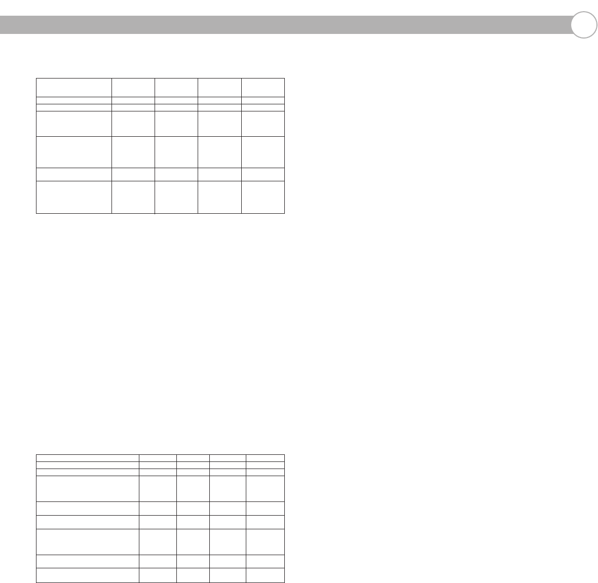
CHAPTER 8 91
ADVERSE REACTIONS
• Adverse reactions encountered during clinical study:
*In the clinical study, presence of infection was to be identified by observation
of redness more than 3–5 mm from the repaired wound, swelling, purulent
discharge, pain, increased skin temperature, fever, or other systemic signs of
infection. (See clinical study). Confirmatory culture was not routinely obtained.
Among cases of suspected infection for DERMABOND adhesive, 7/14 (50%) were
in patients less than 12 years old with traumatic lacerations; overall, 8 of the 14
(approximately 60%) DERMABOND adhesive wounds with suspected infections
were associated with sub-optimal cosmetic outcome.
• Reactions may occur in patients who are hypersensitive to cyanoacrylate or
formaldehyde. See CONTRAINDICATIONS.
• The polymerization of DERMABOND adhesive on the skin releases small
amounts of heat which may cause a sensation of heat or discomfort in some
patients.
• Adverse reactions may be experienced following DERMABOND adhesive
contact with the eye.
CLINICAL STUDY
Description: A prospective, randomized, controlled, unmasked study was
conducted to evaluate the safety and effectiveness of closing the approximated
skin edges of surgical incisions, including punctures from minimally invasive
surgery, and trauma-induced lacerations using DERMABOND adhesive in
comparison to U.S.P. size 5-0 or smaller suture, adhesive strips or staples, with or
without dermal closure (subcuticular stitch) as per investigator judgment.
Summary of Effectiveness Results Comparing Dermabond adhesive to Sutures
(U.S.P. size 5-0 and smaller diameter), Staples, and Adhesive Strips
* Cosmesis: Modified Hollander Cosmesis Scale
The study population included patients at least one year of age, in good general
health, who signed informed consent and agreed to follow-up visits. Patients were
excluded if presenting with: significant multiple trauma, peripheral vascular
disease, insulin dependent diabetes mellitus, blood clotting disorder, keloid for-
mation or hypertrophy history (patient or family), cyanoacrylate or formaldehyde
allergy, burst or stellate lacerations due to crush or hard blow, animal or human
bite, and decubitus ulcer.
Follow-up was at 5–10 days and at 3 months. All wounds were assessed by
visual inspection at 5–10 days after wound closure. The total kinds of wounds
treated in the study were 46.1% lacerations and 53.9% incisions. The incisions
were comprised of 47.8% excisions of skin lesions, 27.4% minimally invasive
surgery punctures, and 24.8% general surgery incisions.
For wounds closed without subcuticular stitches, mean wound length was 1.5cm,
mean wound width was 2.5mm, and mean wound depth was 5.8mm. For wounds
closed with subcuticular stitches, mean wound length was 3.2cm, mean wound
width was 5.3mm, and mean wound depth was 3.8mm.
If the primary method of closure was insufficient for closure, an additional secur-
ing device was placed. The time to perform treatment included the time required
later to remove the closure device when applicable.
The Modified Hollander Cosmesis Scale (MHCS), a validated scale, was used to
evaluate cosmesis at three months: step-off borders, edge inversion, contour
irregularities, excess inflammation, wound margin separation, and overall
appearance.
HOW SUPPLIED
DERMABOND adhesive is supplied sterile, in a pre-filled, single-use applicator.
The applicator is comprised of a crushable glass ampule contained within a plas-
tic vial with attached applicator tip. The applicator contains 0.5ml of liquid adhe-
sive. The applicator is packaged in a blister pouch to maintain the device sterile
until opened or damaged.
DERMABOND adhesive may be available in boxes of 6 or 12 applicators.
STORAGE
Recommended storage conditions: below 30°C, 86°F, away from moisture and
direct heat. Do not use after expiry date.
STERILITY
DERMABOND adhesive is originally sterilized by dry heat and ethylene oxide gas.
Do not resterilize. Do not use if package is opened or damaged. Discard any
unused material following completion of medical procedure.
STERILE SINGLE USE ONLY
REPORTING
Physicians should use the following toll free number 1-800-255-2500 (valid in
U.S.A. only), when reporting adverse reactions or potentially threatening
complications involving DERMABOND adhesive.
CAUTION
Federal (U.S.A.) Law restricts this device to sale by or on the order of a physician.
® Trademark of Purdue Fredericks
† Registered Trademark of Zeneca Pharmaceuticals
389529.R02 *Trademark ©ETHICON,INC. 2001
Clinical Study Outcomes No With
Subcuticular Subcuticular
Sutures Sutures
DERMABOND Control DERMABOND Control
N (%) N (%) N (%) N (%)
Accounting
N, patients enrolled 240 243 167 168
N, patients treated 239 242 167 166
Patients completed 228 (95%) 215 (88%) 164 (98%) 162 (96%)
Adverse Reactions
Suspected Infection* 8 (3.6%) 2 (0.9%) 6 (3.6%) 2 (1.2%)
Wound type
# Lacerations 8 2 1 0
# Incisions 0 0 5 2
Dehiscence with Need 6 (2.5%) 5 (2.1%) 3 (1.8%) 0
for Retreatment
Acute Inflammation
Erythema 26 (11.5%) 74 (33.0%) 52 (31.3%) 75 (45.1%)
Edema 22 (9.7%) 28 (12.5%) 62 (37.3%) 71 (42.8%)
Pain 14 (6.1%) 13 (5.8%) 56 (33.7%) 57 (34.3%)
Warmth 3 (1.3%) 6 (2.6%) 3 (1.8%) 4 (2.4%)
Clinical Study Outcomes NSS WSS
DERMABOND Control DERMABOND Control
N (%) N (%) N (%) N (%)
Accounting
N, patients enrolled 240 243 167 168
N, patients treated 239 242 167 166
Patients completed 228 (95%) 215 (88%) 164 (98%) 162 (96%)
N, control:
suture/strips/staples/missing 194/46/1/1 116/45/5/0
Wound Closure Assessment
Immediate: Additional Devices 18 (7.5%) 13 (5.4%) 2 (1.2%) 11 (6.6%)
@ 5–10 days:
100% epidermal
apposition 169 (75.1%) 199 (88.8%) 140 (84.3%) 160 (96.4%)
>50% epidermal apposition 205 (91.1%) 214 (95.5%) 163 (98.2%) 165 (99.4%)
@ 3 months:
Cosmesis Score*= 0 (optimal) 188 (82.5%) 180 (83.7%) 128 (78.0%) 128 (79.0%)
Median Time for
Treatment (Minutes) 1.5 6.0 1.3 2.9

ETHIBOND* EXCEL POLYESTER SUTURE
NONABSORBABLE SURGICAL SUTURE, U.S.P.
Except for size 6-0 diameter
DESCRIPTION
ETHIBOND* EXCEL polyester suture is a nonabsorbable, braided, sterile,
surgical suture composed of Poly (ethylene terephthalate). It is prepared from
fibers of high molecular weight, long-chain, linear polyesters having recurrent
aromatic rings as an integral component. ETHIBOND EXCEL suture is uniform-
ly coated with polybutilate or poly {oxy-1, 4 butanediyloxy (1, 6-dioxo-1,
6 hexanediyl)}. The highly adherent coating is a relatively nonreactive
nonabsorbable compound which acts as a lubricant to mechanically improve
the physical properties of the uncoated suture by improving ease of passage
through tissues and by providing overall improved handling qualities as
contrasted to the braided, uncoated fiber.
ETHIBOND EXCEL sutures are braided for optimal handling properties, and for
good visibility in the surgical field, are dyed green.
Size 6-0 ETHIBOND EXCEL sutures are U.S.P., except for diameter.
MAXIMUM SUTURE OVERSIZE IN DIAMETER (mm) FROM U.S.P.
U.S.P. SUTURE SIZE DESIGNATION MAXIMUM OVERSIZE (mm)
6-0 0.024
INDICATIONS
ETHIBOND EXCEL suture is indicated for use in general soft tissue
approximation and/or ligation, including use in cardiovascular, ophthalmic and
neurological procedures.
ACTIONS
ETHIBOND EXCEL suture elicits a minimal acute inflammatory reaction in
tissue, followed by a gradual encapsulation of the suture by fibrous connective
tissue. Implantation studies in animals show no meaningful decline in
polyester suture strength over time. Both polyester fiber suture material and
the polybutilate coating are pharmacologically inactive.
CONTRAINDICATIONS
None known.
WARNINGS
Users should be familiar with surgical procedures and techniques involving
nonabsorbable sutures before employing ETHIBOND EXCEL suture for wound
closure, as risk of wound dehiscence may vary with the site of application and
the suture material used.
Do not resterilize. Discard opened packages and unused sutures.
As with any foreign body, prolonged contact of any suture with salt solutions,
such as those found in the urinary or biliary tracts, may result in calculus
formation. Acceptable surgical practice should be followed for the
management of infected or contaminated wounds.
PRECAUTIONS
In handling this or any other suture material, care should be taken to avoid
damage from handling. Avoid crushing or crimping damage due to application
of surgical instruments such as forceps or needle holders.
As with any suture material, adequate knot security requires the accepted
surgical technique of flat and square ties with additional throws as warranted
by surgical circumstance and the experience of the surgeon.
To avoid damaging needle points and swage areas, grasp the needle in an area
one-third (1/3) to one-half (1/2) of the distance from the swaged end to the
point. Reshaping needles may cause them to lose strength and be less
resistant to bending and breaking. Users should exercise caution when
handling surgical needles to avoid inadvertent needle sticks. Discard used
needles in "sharps" containers.
ADVERSE REACTIONS
Adverse effects associated with the use of this device include wound
dehiscence, calculi formation in urinary and biliary tracts when prolonged
contact with salt solutions such as urine and bile occurs, infection, minimal
acute inflammatory tissue reaction and transitory local irritation at the wound
site. Broken needles may result in extended or additional surgeries or residual
foreign bodies. Inadvertent needle sticks with contaminated surgical needles
may result in the transmission of bloodborne pathogens.
HOW SUPPLIED
ETHIBOND EXCEL sutures are available as sterile, braided, green and undyed
(white) strands in sizes 7-0 through 5 (metric sizes 0.5-7) in a variety of lengths,
with and without permanently attached needles.
ETHIBOND EXCEL sutures, green, braided, in sizes 4-0 through 1 (metric sizes
1.5-4) are also available attached to CONTROL RELEASE* removable needles.
ETHIBOND EXCEL sutures, green and undyed, are also available attached to
TFE polymer pledgets measuring 1/8" x 1/8" x 1/16" (3.0mm x 3.0mm x 1.5mm),
1/4" x 1/8" x 1/16" (7.0mm x 3.0mm x 1.5mm).
ETHIBOND EXCEL sutures are available in one, two and three dozen boxes.
389397 *Trademark ©ETHICON,INC. 1996
PRODUCT INFORMATION
92

CHAPTER 8 93
ETHILON* NYLON SUTURE
NONABSORBABLE SURGICAL SUTURES, U.S.P.
DESCRIPTION
ETHILON* nylon suture is a nonabsorbable, sterile surgical monofilament
suture composed of the long-chain aliphatic polymers Nylon 6 and Nylon 6,6.
ETHILON sutures are dyed black or green to enhance visibility in tissue. The
suture is also available undyed (clear).
ETHILON suture meets all requirements established by the United States
Pharmacopoeia (U.S.P.) for nonabsorbable surgical suture.
INDICATIONS
ETHILON suture is indicated for use in general soft tissue
approximation and/or ligation, including use in cardiovascular,
ophthalmic and neurological procedures.
ACTIONS
ETHILON suture elicits a minimal acute inflammatory reaction in tissue, which
is followed by gradual encapsulation of the suture by fibrous connective tissue.
While nylon is not absorbed, progressive hydrolysis of the nylon in vivo may
result in gradual loss over time of tensile strength.
CONTRAINDICATIONS
Due to the gradual loss of tensile strength which may occur over prolonged
periods in vivo, nylon suture should not be used where permanent retention of
tensile strength is required.
WARNINGS
Users should be familiar with surgical procedures and techniques involving
nonabsorbable sutures before employing ETHILON suture for wound closure,
as the risk of wound dehiscence may vary with the site of application and the
suture material used.
As with any foreign body, prolonged contact of any suture with salt solutions,
such as those found in the urinary or biliary tracts, may result in calculus
formation. Acceptable surgical practice should be followed for the management
of contaminated or infected wounds.
Do not resterilize. Discard open packages and unused sutures.
PRECAUTIONS
In handling this or any other suture material, care should be taken to avoid
damage from handling. Avoid crushing or crimping damage due to application
of surgical instruments such as forceps or needle holders.
As with any suture material, adequate knot security requires the accepted sur-
gical technique of flat and square ties, with additional throws as warranted by
surgical circumstance and the experience of the surgeon. The use of addition-
al throws may be particularly appropriate when knotting monofilaments.
To avoid damaging needle points and swage areas, grasp the needle in an area
one-third (1/3) to one-half (1/2) of the distance from the swaged end to the point.
Reshaping needles may cause them to lose strength and be less resistant to
bending and breaking. Users should exercise caution when handling surgical
needles to avoid inadvertent needle sticks. Discard used needles in "sharps"
containers.
ADVERSE REACTIONS
Adverse effects associated with the use of this device include wound dehis-
cence, gradual loss of tensile strength over time, calculi formation in urinary
and biliary tracts when prolonged contact with salt solutions such as urine and
bile occurs, infection, minimal acute inflammatory tissue reaction, and transi-
tory local irritation at the wound site. Broken needles may result in extended or
additional surgeries or residual foreign bodies. Inadvertent needle sticks with
contaminated surgical needles may result in the transmission of bloodborne
pathogens.
HOW SUPPLIED
ETHILON sutures are available as sterile monofilament strands in U.S.P. sizes
11-0 through 2 (metric sizes 0.1-5.0) in a variety of lengths, with and without
permanently attached needles. ETHILON sutures are available in one, two and
three dozen boxes.
389355 *Trademark ©ETHICON, INC. 1995

PRODUCT INFORMATION
94
FAST ABSORBING
SURGICAL GUT (PLAIN)
ABSORBABLE SURGICAL SUTURES
Non-U.S.P.
DESCRIPTION
Fast absorbing surgical gut suture is a strand of collagenous material prepared
from the submucosal layers of the small intestine of healthy sheep, or from the
serosal layers of the small intestine of healthy cattle.
Fast absorbing surgical gut sutures are sterile and elicit only a slight to
minimal tissue reaction during absorption.
Fast absorbing surgical gut sutures differ from U.S.P. minimum strength
requirements by less than 30%.
INDICATIONS
Fast absorbing surgical gut sutures are intended for dermal (skin) suturing
only. They should be utilized only for external knot tying procedures.
ACTIONS
The results of implantation studies of fast absorbing surgical gut sutures in the
skin of animals indicate that nearly all of its original strength is lost within
approximately seven (7) days of implantation.
When surgical gut suture is placed in tissue, a moderate tissue inflammation
occurs which is characteristic of foreign body response to a substance. This is
followed by a loss of tensile strength followed by a loss of suture mass, as the
proteolytic enzymatic digestive process dissolves the surgical gut. This process
continues until the suture is completely absorbed. Many variable factors may
affect the rate of absorption. Some of the major factors which can affect tensile
strength loss and absorption rates are:
1. Type of suture - plain gut generally absorbs more rapidly than chromic gut.
2. Infection - surgical gut is absorbed more rapidly in infected tissue than in
non-infected tissue.
3. Tissue sites - surgical gut will absorb more rapidly in tissue where increased
levels of proteolytic enzymes are present, as in the secretions exhibited in
the stomach, cervix and vagina.
Data obtained from implantation studies in rats show that the absorption of
these sutures is essentially complete by the twenty-first (21st) to forty-second
(42nd) post implantation day.
CONTRAINDICATIONS
These sutures, being absorbable, should not be used where prolonged
approximation of tissue under stress is required. These sutures have been
designed to absorb at a rapid rate and must be used on dermal tissue only.
These sutures should never be used on internal tissue. The use of this suture
is contraindicated in patients with known sensitivities or allergies to collagen,
as gut is a collagen based material.
WARNINGS
Users should be familiar with surgical procedures and techniques involving gut
suture before using fast absorbing surgical gut suture for wound closure, as
the risk of wound dehiscence may vary with the site of application and the
suture material used. Physicians should consider the in vivo performance when
selecting a suture for use in patients.
The use of this suture may be inappropriate in elderly, malnourished, or debil-
itated patients, or in patients suffering from conditions which may delay
wound healing. As this is an absorbable material, the use of supplemental non-
absorbable sutures should be considered by the surgeon in the closure of sites
which may undergo expansion, stretching or distention or which may require
additional support.
As with any foreign body, prolonged contact of any suture with salt solutions,
such as those found in the urinary or biliary tracts, may result in calculus for-
mation. As an absorbable suture, fast absorbing surgical gut may act tran-
siently as a foreign body. Acceptable surgical practice should be followed for
the management of contaminated or infected wounds.
Do not resterilize. Discard open packages and unused sutures. Store at room
temperature.
Certain patients may be hypersensitive to collagen and might exhibit an
immunological reaction resulting in inflammation, tissue granulation or fibro-
sis, wound suppuration and bleeding, as well as sinus formation.
PRECAUTIONS
In handling this or any other suture material, care should be taken to avoid
damage from handling. Avoid crushing or crimping damage due to application
of surgical instruments such as forceps or needle holders. Surgical gut sutures
require the accepted surgical technique of flat and square ties with additional
throws as warranted by surgical circumstance and the experience of the
surgeon.
Under some circumstances, notably orthopaedic procedures, immobilization of
joints by external support may be employed at the discretion of the surgeon.
The surgeon should avoid unnecessary tension when running down knots, to
reduce the occurrence of surface fraying and weakening of the strand.
Avoid prolonged exposure to elevated temperatures.
To avoid damaging needle points and swage areas, grasp the needle in an area
one-third (1/3) to one-half (1/2) of the distance from the swaged end to
the point. Reshaping needles may cause them to lose strength and be less
resistant to bending and breaking. Users should exercise caution when
handling surgical needles to avoid inadvertent needle sticks. Discard used
needles in "sharps" containers.
ADVERSE REACTIONS
Adverse effects associated with the use of this device include wound
dehiscence, variable rates of absorption over time (depending on such factors
as the type of suture used, the presence of infection and the tissue site), failure
to provide adequate wound support in closure of sites where expansion,
stretching or distention occur, etc., unless additional support is supplied
through the use of nonabsorbable suture material, failure to provide adequate
wound support in elderly, malnourished or debilitated patients or in patients
suffering from cancer, anemia, obesity, diabetes, infection or other conditions
which may delay wound healing, allergic response in patients with known
sensitivities to collagen which may result in an immunological reaction result-
ing in inflammation, tissue granulation or fibrosis, wound suppuration and
bleeding, as well as sinus formation, infection, moderate tissue inflammatory
response characteristic of foreign body response, calculi formation in urinary
and biliary tracts when prolonged contact with salt solutions such as urine and
bile occurs, and transitory local irritation at the wound site. Broken needles
may result in extended or additional surgeries or residual foreign bodies.
Inadvertent needle sticks with contaminated surgical needles may result in the
transmission of bloodborne pathogens.
HOW SUPPLIED
Fast absorbing surgical gut sutures are available in sizes 5-0 (metric size 1.5)
and 6-0 (metric size 1.0) with needles attached in one, two and three dozen
boxes.
389359 *Trademark ©ETHICON, INC. 1995

CHAPTER 8 95
MERSILENE* POLYESTER FIBER SUTURE
NONABSORBABLE SURGICAL SUTURE, U.S.P.
Except for size 6-0 diameter
DESCRIPTION
MERSILENE* polyester suture is a nonabsorbable, braided, sterile, surgical
suture composed of Poly (ethylene terephthalate). It is prepared from fibers of
high molecular weight, long-chain, linear polyesters having recurrent aromatic
rings as an integral component. MERSILENE sutures are braided for optimal
handling properties, and for good visibility in the surgical field, are dyed green.
Size 6-0 MERSILENE sutures are U.S.P., except for diameter.
MAXIMUM SUTURE OVERSIZE IN DIAMETER (mm) FROM U.S.P.
U.S.P. SUTURE SIZE DESIGNATION MAXIMUM OVERSIZE (mm)
6-0 0.024
INDICATIONS
MERSILENE suture is indicated for use in general soft tissue approximation
and/or ligation, including use in cardiovascular, ophthalmic and neurological
procedures.
ACTIONS
MERSILENE suture elicits a minimal acute inflammatory reaction in tissue,
followed by a gradual encapsulation of the suture by fibrous connective
tissue. Implantation studies in animals show no meaningful decline in
polyester suture strength over time. The polyester fiber suture material is
pharmacologically inactive.
CONTRAINDICATIONS
None known.
WARNINGS
Users should be familiar with surgical procedures and techniques involving
nonabsorbable sutures before employing MERSILENE suture for wound
closure, as risk of wound dehiscence may vary with the site of application and
the suture material used.
Do not resterilize. Discard opened packages and unused sutures.
As with any foreign body, prolonged contact of any suture with salt solutions,
such as those found in the urinary or biliary tracts, may result in calculus for-
mation. Acceptable surgical practice should be followed for the management
of infected or contaminated wounds.
PRECAUTIONS
In handling this or any other suture material, care should be taken to avoid
damage from handling. Avoid crushing or crimping damage due to application
of surgical instruments such as forceps or needle holders. The use of addition-
al throws is particularly appropriate when knotting monofilament sutures.
As with any suture material, adequate knot security requires the accepted
surgical technique of flat and square ties with additional throws as warranted
by surgical circumstance and the experience of the surgeon.
To avoid damaging needle points and swage areas, grasp the needle in an area
one-third (1/3) to one-half (1/2) of the distance from the swaged end to
the point. Reshaping needles may cause them to lose strength and be less
resistant to bending and breaking. Users should exercise caution when
handling surgical needles to avoid inadvertent needle sticks. Discard used
needles in "sharps" containers.
ADVERSE REACTIONS
Adverse effects associated with the use of this device include wound
dehiscence, calculi formation in urinary and biliary tracts when prolonged
contact with salt solutions such as urine and bile occurs, infection, minimal
acute inflammatory tissue reaction and transitory local irritation at the wound
site. Broken needles may result in extended or additional surgeries or residual
foreign bodies. Inadvertent needle sticks with contaminated surgical needles
may result in the transmission of bloodborne pathogens.
HOW SUPPLIED
MERSILENE sutures are available as sterile, braided, green and undyed (white)
strands in sizes 6-0 through 5 (metric sizes 0.7-7) in a variety of lengths, with
and without permanently attached needles.
MERSILENE sutures are also available in green monofilament in sizes 10-0 and
11-0 (metric sizes 0.2-0.1).
MERSILENE sutures, green, braided in U.S.P. size 0 (metric size 3.5) are also
available attached to CONTROL RELEASE* removable needles.
MERSILENE sutures are available in one, two and three dozen boxes.
389395 *Trademark ©ETHICON,INC. 1996

PRODUCT INFORMATION
96
MERSILENE* POLYESTER FIBER MESH
Nonabsorbable Synthetic Surgical Mesh
STERILE
DESCRIPTION
MERSILENE* Polyester Fiber Mesh is constructed from polyethylene
terephthalate, the same material used to make MERSILENE* Polyester Fiber
Suture, Nonabsorbable Surgical Suture, U.S.P. (ETHICON, INC.) MERSILENE
Polyester Fiber Mesh affords excellent strength, durability and surgical
adaptability, along with maximal porosity for necessary tissue ingrowth.
The mesh is approximately 0.010 inches thick and is a highly flexible and
compliant material.
MERSILENE mesh is knitted by a process which interlinks each fiber junction
and which provides for elasticity in both directions. This construction permits
the mesh to be cut into any desired shape or size without unraveling. The fiber
junctions are not subject to the same work fatigue exhibited by more rigid
metallic meshes. This bi-directional elastic property allows adaption to various
stresses encountered in the body.
ACTIONS
MERSILENE mesh is a nonabsorbable mesh used to span and reinforce
traumatic or surgical wounds to provide extended support during and
following wound healing. Animal studies show that implantation of
MERSILENE mesh elicits a minimum to slight inflammatory reaction, which is
transient and is followed by the deposition of a thin fibrous layer of tissue
which can grow through the interstices of the mesh, thus incorporating the
mesh into adjacent tissue. The mesh remains soft and pliable, and normal
wound healing is not noticeably impaired. The material is not absorbed nor is
it subject to degradation or weakening by the action of tissue enzymes.
INDICATIONS
This mesh may be used for the repair of hernia and other fascial deficiencies
that require the addition of a reinforcing or bridging material to obtain the
desired surgical result.
CONTRAINDICATIONS
When this mesh is used in infants or children with future growth potential, the
surgeon should be aware that this product will not stretch significantly as the
patient grows.
MERSILENE polyester fiber mesh in contaminated wounds should be used
with the understanding that subsequent infection may require removal of
the material.
WARNINGS
MERSILENE mesh is provided by ETHICON, INC. as a sterile product. Unused
MERSILENE Mesh which has been removed from the package may be resteril-
ized not more than one time by a conventional stream autoclaving process
at conditions of 250˚F (121˚C) for 20 minutes. MERSILENE mesh may also be
flash autoclaved not more than one time at conditions of 270˚F (132˚C) for
10 minutes. Resterilization under any other conditions or by any other means
is neither recommended nor endorsed by ETHICON, INC.
If this product should become stained with blood or soiled, it should not be
resterilized for reuse.
PRECAUTIONS
A minimum of 6.5mm (1/4 inch) of mesh should extend beyond the suture line.
ADVERSE REACTIONS
No significant adverse clinical reactions to MERSILENE mesh have been
reported. The use of nonabsorbable MERSILENE mesh in a wound that is
contaminated or infected could lead to fistula formation and/or extrusion of
the mesh.
INDICATIONS FOR USE
It is recommended that nonabsorbable sutures be placed 6.5 to 12.5mm (1/4 to
1/2 inch) apart at a distance approximately 6.5mm (1/4 inch) from edge of the
mesh. Some surgeons prefer to suture and uncut section of mesh that is
considerably large than the defect into position over the wound. The opposite
sides are then sutured to assure proper closure under correct tension. When
the margin sutures have all been placed, the extra mesh is trimmed away.
HOW SUPPLIED
MERSILENE mesh is available in single packets as sterile, undyed (white)
sheets in two sizes. The sizes available are 6 x 11cm (2.5 x 4.5 inches) and 30
x 30cm (12 x 12 inches). Each sheet is 0.25mm (0.010 inch) thick.
389131 *Trademark ©ETHICON, INC. 1987

CHAPTER 8 97
MONOCRYL* (Poliglecaprone 25)Suture
SYNTHETIC ABSORBABLE SUTURE,
U.S.P., EXCEPT FOR DIAMETER
DESCRIPTION
MONOCRYL* (poliglecaprone 25) suture is a monofilament synthetic
absorbable surgical suture prepared from a copolymer of glycolide and
epsilon-caprolactone. Poliglecaprone 25 copolymer has been found to be
nonantigenic, nonpyrogenic and elicits only a slight tissue reaction during
absorption.
MONOCRYL sutures are U.S.P. except for diameters in the following sizes:
MAXIMUM SUTURE OVERSIZE IN DIAMETER (mm) FROM U.S.P.
U.S.P. SUTURE SIZE DESIGNATION MAXIMUM OVERSIZE (mm)
6-0 0.049
5-0 0.033
4-0 0.045
3-0 0.067
2-0 0.055
0 0.088
1 0.066
2 0.099
INDICATIONS
MONOCRYL sutures are indicated for use in general soft tissue approximation
and/or ligation, but not for use in cardiovascular or neurological tissues,
microsurgery or ophthalmic surgery.
ACTIONS
MONOCRYL suture is a monofilament which elicits a minimal acute inflamma-
tory reaction in tissues and ingrowth of fibrous connective tissue. Progressive
loss of tensile strength and eventual absorption of MONOCRYL sutures occurs
by means of hydrolysis. Absorption begins as a loss of tensile strength
followed by a loss of mass. Implantation studies in rats indicate that
MONOCRYL suture retains approximately 50 to 60% of its original strength
7 days post implantation, and approximately 20 to 30% of its original tensile
strength at 14 days post implantation. All of the original tensile strength is lost
by 21 days post implantation. The absolute strength remaining 14 days post
implantation meets or exceeds that historically observed with plain or chromic
surgical gut sutures. Absorption of MONOCRYL absorbable synthetic suture is
essentially complete between 91 and 119 days.
APPROXIMATE % ORIGINAL
DAYS IMPLANTATION STRENGTH REMAINING
7 DAYS 50 TO 60%
14 DAYS 20 TO 30%
CONTRAINDICATIONS
This suture, being absorbable, should not be used where extended approxi-
mation of tissue under stress is required, such as in fascia.
WARNINGS
Users should be familiar with surgical procedures and techniques involving
absorbable sutures before employing MONOCRYL suture for wound closure,
as risk of wound dehiscence may vary with the site of application and the
suture material used. Physicians should consider the in vivo performance
(under ACTIONS section) when selecting a suture for use in patients. The use
of this suture may be inappropriate in elderly, malnourished, or debilitated
patients, or in patients suffering from conditions which may delay wound
healing.
Do not resterilize. Discard opened packages and unused sutures.
As with any foreign body, prolonged contact of any suture with salt solutions,
such as those found in the urinary or biliary tracts, may result in calculus
formation. As an absorbable suture, MONOCRYL suture may act transiently as
a foreign body. Acceptable surgical practice should be followed for the
management of contaminated or infected wounds.
As this is an absorbable suture material, the use of supplemental
nonabsorbable sutures should be considered by the surgeon in the closure of
the sites which may undergo expansion, stretching or distention, or which may
require additional support.
PRECAUTIONS
Skin sutures which must remain in place longer than 7 days may cause local-
ized irritation and should be snipped off or removed as indicated. Subcuticular
sutures should be placed as deeply as possible to minimize the erythema and
induration normally associated with absorption.
Under some circumstances, notably orthopaedic procedures, immobilization of
joints by external support may be employed at the discretion of the surgeon.
Consideration should be taken in the use of absorbable sutures in tissue with
poor blood supply as suture extrusion and delayed absorption may occur.
In handling this or any other suture material, care should be taken to avoid
damage from handling. Avoid crushing or crimping damage due to application
of surgical instruments such as forceps or needle holders.
MONOCRYL suture knots must be properly placed to be secure. Adequate knot
security requires the accepted surgical technique of flat and square ties with
additional throws as warranted by surgical circumstance and the experience of
the surgeon. The use of additional throws may be particularly appropriate
when knotting monofilaments.
Avoid prolonged exposure to elevated temperature.
To avoid damaging needle points and swage areas, grasp the needle in an area
one-third (1/3) to one-half (1/2) of the distance from the swaged end to
the point. Reshaping needles may cause them to lose strength and be less
resistant to bending and breaking. Users should exercise caution when
handling surgical needles to avoid inadvertent needle sticks. Discard used
needles in "sharps" containers.
ADVERSE REACTIONS
Adverse effects associated with the use of synthetic absorbable sutures include
wound dehiscence, failure to provide adequate wound support in closure of the
sites where expansion, stretching, or distension occur, failure to provide ade-
quate wound support in elderly, malnourished or debilitated patients or in
patients suffering from conditions which may delay wound healing, infection,
minimal acute inflammatory tissue reaction, localized irritation when skin
sutures are left in place for greater than 7 days, suture extrusion and delayed
absorption in tissue with poor blood supply, calculi formation in urinary and
biliary tracts when prolonged contact with salt solutions such as urine and bile
occurs, and transitory local irritation at the wound site. Broken needles may
result in extended or additional surgeries or residual foreign bodies.
Inadvertent needle sticks with contaminated surgical needles may result in the
transmission of bloodborne pathogens.
HOW SUPPLIED
MONOCRYL sutures are available as sterile, monofilament, undyed (natural)
strands in sizes 6-0 through 2 (metric sizes 0.7-5), in a variety of lengths, with
or without needles.
MONOCRYL sutures are also available in sizes 3-0 through 1 (metricsizes 2-4)
attached to CONTROL RELEASE* removable needles.
MONOCRYL sutures are available in one and three dozen boxes.
389385 *Trademark ©ETHICON, INC. 1995

PRODUCT INFORMATION
98
MONOCRYL* VIOLET MONOFILAMENT
(Poliglecaprone 25)Suture
SYNTHETIC ABSORBABLE SUTURE, U.S.P.,
EXCEPT FOR DIAMETER
DESCRIPTION
MONOCRYL* (poliglecaprone 25)suture is a monofilament synthetic
absorbable surgical suture prepared from a copolymer of glycolide and
epsilon-caprolactone. Poliglecaprone 25 copolymer has been found to be
nonantigenic, nonpyrogenic and elicits only a slight tissue reaction during
absorption.
MONOCRYL sutures are U.S.P. except for diameters in the following sizes:
MAXIMUM SUTURE OVERSIZE IN DIAMETER (mm) FROM U.S.P.
U.S.P. SUTURE SIZE DESIGNATION MAXIMUM OVERSIZE (mm)
6-0 0.049
5-0 0.033
4-0 0.045
3-0 0.067
2-0 0.055
0 0.088
1 0.066
2 0.099
INDICATIONS
MONOCRYL sutures are indicated for use in general soft tissue approximation
and/or ligation, but not for use in cardiovascular or neurological tissues,
microsurgery or ophthalmic surgery.
ACTIONS
MONOCRYL suture is a monofilament which elicits a minimal acute inflamma-
tory reaction in tissues and ingrowth of fibrous connective tissue. Progressive
loss of tensile strength and eventual absorption of MONOCRYL sutures occurs
by means of hydrolysis. Absorption begins as a loss of tensile strength
followed by a loss of mass. Implantation studies in rats indicate that
MONOCRYL suture retains approximately 60 to 70% of its original strength
7 days post implantation, and approximately 30 to 40% of its original tensile
strength at 14 days post implantation. Essentially all of the original tensile
strength is lost by 28 days post implantation. Absorption of MONOCRYL
absorbable synthetic suture is essentially complete between 91 and 119 days.
APPROXIMATE % ORIGINAL
DAYS IMPLANTATION STRENGTH REMAINING
7 DAYS 60 TO 70%
14 DAYS 30 TO 40%
CONTRAINDICATIONS
This suture, being absorbable, should not be used where extended approxi-
mation of tissue under stress is required.
WARNINGS
Users should be familiar with surgical procedures and techniques involving
absorbable sutures before employing MONOCRYL suture for wound closure,
as risk of wound dehiscence may vary with the site of application and the
suture material used. Physicians should consider the in vivo performance
(under ACTIONS section) when selecting a suture for use in patients. The
use of this suture may be inappropriate in elderly, malnourished, or debilitated
patients, or in patients suffering from conditions which may delay wound
healing.
Do not resterilize. Discard opened packages and unused sutures.
As with any foreign body, prolonged contact of any suture with salt solutions,
such as those found in the urinary or biliary tracts, may result in calculus
formation. As an absorbable suture, MONOCRYL suture may act transiently as
a foreign body. Acceptable surgical practice should be followed for the
management of contaminated or infected wounds.
As this is an absorbable suture material, the use of supplemental
nonabsorbable sutures should be considered by the surgeon in the closure of
the sites which may undergo expansion, stretching or distention, or which may
require additional support.
PRECAUTIONS
Skin sutures which must remain in place longer than 7 days may cause local-
ized irritation and should be snipped off or removed as indicated. Subcuticular
sutures should be placed as deeply as possible to minimize the erythema and
induration normally associated with absorption.
Under some circumstances, notably orthopaedic procedures, immobilization of
joints by external support may be employed at the discretion of the surgeon.
Consideration should be taken in the use of absorbable sutures in tissue with
poor blood supply as suture extrusion and delayed absorption may occur.
In handling this or any other suture material, care should be taken to avoid
damage from handling. Avoid crushing or crimping damage due to application
of surgical instruments such as forceps or needle holders.
MONOCRYL suture knots must be properly placed to be secure. Adequate knot
security requires the accepted surgical technique of flat and square ties with
additional throws as warranted by surgical circumstance and the experience of
the surgeon. The use of additional throws may be particularly appropriate
when knotting monofilaments.
Avoid prolonged exposure to elevated temperature.
To avoid damaging needle points and swage areas, grasp the needle in an area
one-third (1/3) to one-half (1/2) of the distance from the swaged end to the
point. Reshaping needles may cause them to lose strength and be less
resistant to bending and breaking. Users should exercise caution when
handling surgical needles to avoid inadvertent needle sticks. Discard used
needles in "sharps" containers.
ADVERSE REACTIONS
Adverse effects associated with the use of synthetic absorbable sutures include
wound dehiscence, failure to provide adequate wound support in closure of the
sites where expansion, stretching, or distension occur, failure to provide
adequate wound support in elderly, malnourished or debilitated patients or in
patients suffering from conditions which may delay wound healing, infection,
minimal acute inflammatory tissue reaction, localized irritation when skin
sutures are left in place for greater than 7 days, suture extrusion and delayed
absorption in tissue with poor blood supply, calculi formation in urinary and
biliary tracts when prolonged contact with salt solutions such as urine and bile
occurs, and transitory local irritation at the wound site. Broken needles may
result in extended or additional surgeries or residual foreign bodies.
Inadvertent needle sticks with contaminated surgical needles may result in the
transmission of bloodborne pathogens.
HOW SUPPLIED
MONOCRYL sutures are available as sterile, monofilament, dyed (violet)
strands in sizes 6-0 through 2 (metric sizes 0.7-5), in a variety of lengths, with
or without needles. MONOCRYL sutures are also available in sizes 3-0 through
1 (metric sizes 2-4) attached to CONTROL RELEASE* removable needles.
MONOCRYL sutures are available in one and three dozen boxes.
389310 *Trademark © ETHICON, INC. 1996

CHAPTER 8 99
NUROLON* NYLON SUTURE
NONABSORBABLE SURGICAL SUTURE, U.S.P.
DESCRIPTION
NUROLON* nylon suture is a nonabsorbable sterile surgical braided suture
composed of the long-chain aliphatic polymers Nylon 6 or Nylon 6,6.
NUROLON sutures are dyed black to enhance visibility in tissue. The suture is
also available undyed (clear).
NUROLON suture meets all requirements established by the United States
Pharmacopoeia (U.S.P.) for nonabsorbable surgical suture.
INDICATIONS
NUROLON suture is indicated for use in general soft tissue
approximation and/or ligation, including use in cardiovascular,
ophthalmic and neurological procedures.
ACTIONS
NUROLON suture elicits a minimal acute inflammatory reaction in
tissue, which is followed by a gradual encapsulation of the suture by fibrous
connective tissue. While nylon is not absorbed, progressive hydrolysis of the
nylon in vivo may result in gradual loss of tensile strength over time.
CONTRAINDICATIONS
Due to the gradual loss of tensile strength which may occur over prolonged
periods in vivo, nylon suture should not be used where permanent retention of
tensile strength is required.
WARNINGS
Users should be familiar with surgical procedures and techniques involving
nonabsorbable sutures before employing NUROLON suture for wound closure,
as risk of wound dehiscence may vary with the site of application and the
suture material used.
As with any foreign body, prolonged contact of any suture with salt solutions,
such as those found in the urinary or biliary tracts, may result in calculus for-
mation. Acceptable surgical practices should be followed for the management
of infected or contaminated wounds.
Do not resterilize. Discard opened packages and unused sutures.
PRECAUTIONS
In handling this or any other suture material, care should be taken to avoid
damage from handling. Avoid crushing or crimping damage due to application
of surgical instruments such as forceps or needle holders.
As with any suture material, adequate knot security requires the accepted
surgical technique of flat and square ties with additional throws as warranted
by surgical circumstance and the experience of the surgeon.
To avoid damaging needle points and swage areas, grasp the needle in an area
one-third (1/3) to one-half (1/2) of the distance from the swaged end to the
point. Reshaping needles may cause them to lose strength and be less
resistant to bending and breaking. Users should exercise caution when
handling surgical needles to avoid inadvertent needle sticks. Discard used
needles in "sharps" containers.
ADVERSE REACTIONS
Adverse effects associated with the use of this device include wound
dehiscence, gradual loss of tensile strength over time, calculi formation in
urinary and biliary tracts when prolonged contact with salt solutions such as
urine and bile occurs, infection, minimal acute inflammatory tissue reaction,
and transitory local irritation at the wound site. Broken needles may result in
extended or additional surgeries or residual foreign bodies. Inadvertent needle
sticks with contaminated surgical needles may result in the transmission of
bloodborne pathogens.
HOW SUPPLIED
NUROLON sutures are available in U.S.P. sizes 6-0 through 1 (metric sizes
0.7-4.0) in a variety of lengths with and without permanently attached needles.
NUROLON sutures are available in U.S.P. sizes 4-0 through 1 (metric sizes
1.5-4.0) attached to CONTROL RELEASE* removable needles.
NUROLON sutures are available in one, two and three dozen boxes.
389354 *Trademark ©ETHICON, INC. 1995

PRODUCT INFORMATION
100
PDS* II (POLYDIOXANONE) Suture
DYED and CLEAR MONOFILAMENT
SYNTHETIC ABSORBABLE SUTURES, U.S.P.,
EXCEPT FOR DIAMETER.
DESCRIPTION
PDS* II (polydioxanone) monofilament synthetic absorbable suture is prepared
from the polyester, poly (p-dioxanone). The empirical molecular formula of the
polymer is (C4H603)x.
Polydioxanone polymer has been found to be nonantigenic, nonpyrogenic and
elicits only a slight tissue reaction during absorption.
PDS II sutures are U.S.P., except for diameter.
MAXIMUM SUTURE OVERSIZE IN DIAMETER (mm) FROM U.S.P.
U.S.P. SUTURE
SIZE DESIGNATION MAXIMUM OVERSIZE (mm)
9-0 .005
8-0 .008
7-0 .020
6-0 .015
5-0 .029
4-0 .029
3-0 .056
2-0 .029
0 .071
1 .047
2 .023
ACTIONS
Two important characteristics describe the in vivo performance of absorbable
sutures: first, tensile strength retention, and second, the absorption rate (loss
of mass). PDS II synthetic absorbable suture has been formulated to minimize
the variability of these characteristics and to provide wound support through
an extended healing period.
The results of implantation studies of PDS II monofilament suture in animals
indicate that approximately 70% of its original strength remains two weeks
after implantation. At four weeks post-implantation, approximately 50% of its
original strength is retained, and at six weeks, approximately 25% of the origi-
nal strength is retained.
Data obtained from implantation studies in rats show that the absorption of
these sutures is minimal until about the 90th post-implantation day. Absorption
is essentially complete within six months.
INDICATIONS
PDS II monofilament synthetic absorbable sutures are indicated for use in all
types of soft tissue approximation, including use in pediatric cardiovascular tis-
sue where growth is expected to occur and opthalmic surgery. PDS II suture is
not indicated in adult cardiovascular tissue, microsurgery and neural tissue.
These sutures are particularly useful where the combination of an absorbable
suture and extended wound support (up to six weeks) is desirable.
CONTRAINDICATIONS
These sutures, being absorbable, are not to be used where prolonged (beyond
six weeks) approximation of tissues under stress is required are not to be used
in conjunction with prosthetic devices, i.e., heart valves or synthetic grafts.
WARNINGS
The safety and effectiveness of PDS II (polydioxanone) sutures have not been
established in neural tissue, adult cardiovascular tissue or for use in micro-
surgery.
Under certain circumstances, notably orthopaedic procedures, immobilization
by external support may be employed at the discretion of the surgeon.
Do not resterilize.
PRECAUTIONS
The PDS II suture knots must be properly placed to be secure. As with other
synthetic sutures, knot security requires the standard surgical technique of flat
and square ties with additional throws if indicated by surgical circumstance
and the experience of the operator.
As with any suture, care should be taken to avoid damage when handling.
Avoid the crushing or crimping application of surgical instruments, such as
needle holders and forceps, to the strand except when grasping the free end of
the suture during an instrument tie.
Conjunctival and vaginal mucosal sutures remaining in place for extended
periods may be associated with localized irritation and should be removed as
indicated.
Subcuticular sutures should be placed as deeply as possible in order to
minimize the erythema and induration normally associated with absorption.
Acceptable surgical practice should be followed with respect to drainage and
closure of infected wounds.
ADVERSE REACTIONS
Due to prolonged suture absorption, some irritation and bleeding has been
observed in the conjunctiva and mild irritation has been observed in the
vaginal mucosa.
DOSAGE AND ADMINISTRATION
Use as required per surgical procedure.
HOW SUPPLIED
PDS II sutures are available as sterile, monofilament dyed (violet) strands in
sizes 9-0 thru 2 (metric sizes 0.3-5), and sterile, monofilament dyed (blue)
strands in size 9-0 thru 7-0 (metric size 0.3-0.5) in a variety of lengths, with a
variety of needles.
PDS II monofilament dyed (violet) sutures, sizes 4-0 thru 1 (metric size 1.5-4) are
also available attached to CONTROL RELEASE* removable needles.
PDS II Clear suture strands are available in sizes 7-0 thru 1 (metric size 0.5-4) in
a variety of lengths with permanently attached needles.
388W91 *Trademark ©ETHICON, INC. 1992

CHAPTER 8 101
PERMA-HAND* SILK SUTURE
NONABSORBABLE SURGICAL SUTURE, U.S.P.
DESCRIPTION
PERMA-HAND* silk suture is a nonabsorbable, sterile, surgical suture
composed of an organic protein call fibroin. This protein is derived from the
domesticated species Bombyx mori (b. More) of the family Bombycidae.
PERMA-HAND sutures are processed to remove the natural waxes and gums.
PERMA-HAND suture is dyed black and coated with a special was mixture.
PERMA-HAND suture is also available in its natural color. PERMA-HAND Virgin
silk suture is available in which the sericin gum is not removed and serves to
hold the filaments together.
PERMA-HAND suture meets requirements established by the United States
Pharmacopoeia (U.S.P.) for nonabsorbable surgical suture.
INDICATIONS
PERMA-HAND suture is indicated for use in general soft tissue approximation
and/or ligation, including use in cardiovascular, ophthalmic and neurological
procedures.
ACTIONS
PERMA-HAND suture elicits an acute inflammatory reaction in tissue, which is
followed by a gradual encapsulation of the suture by fibrous connective tissue.
While silk sutures are not absorbed, progressive degradation of the proteina-
ceous silk fiber in vivo may result in gradual loss of all of the suture’s tensile
strength over time.
CONTRAINDICATIONS
The use of this suture is contraindicated in patients with known sensitivities or
allergies to silk.
Due to the gradual loss of tensile strength which may occur over prolonged
periods in vivo, silk should not be used where permanent retention of tensile
strength is required.
WARNINGS
Users should be familiar with surgical procedures and techniques involving
nonabsorbable sutures before employing PERMA-HAND suture for wound
closure, as risk of wound dehiscence may vary with the site of application and
the suture material used.
As with any foreign body, prolonged contact of any suture with salt solutions,
such as those found in the urinary or biliary tracts, may result in calculus for-
mation. Acceptable surgical practice should be followed for the management
of infected or contaminated wounds.
Do not sterilize. Discard opened packages and unused sutures.
PRECAUTIONS
In handling this or any other suture material, care should be taken to avoid
damage from handling. Avoid crushing or crimping damage due to application
of surgical instruments such as forceps or needle holders.
As with any suture material, adequate knot security requires the accepted
surgical technique of flat and square ties with additional throws as warranted
by surgical circumstance and the experience of the surgeon.
To avoid damaging needles points and swage areas, grasp the needle in an
area one-third (1/3) to one-half (1/2) of the distance from the swaged end to the
point. Reshaping needles may cause them to lose strength and be less
resistant to bending and breaking g. Users should exercise caution when
handling surgical needles to avoid inadvertent needle sticks. Discard used
needles in "sharps" containers.
ADVERSE REACTIONS
Adverse effects associated with the use of this device include wound
dehiscence, gradual loss of all tensile strength over time, allergic response in
patients that are know to be sensitive to silk, calculi formation in urinary and
biliary tracts when prolonged contact with salt solutions such as urine and bile
occurs, infection, acute inflammatory tissue reaction, and transitory local
irritation at the wound site. Broken needles may result in extended or
additional surgeries or residual foreign bodies. Inadvertent needle sticks with
contaminated surgical needles may result in the transmission of bloodborne
pathogens.
HOW SUPPLIED
PERMA-HAND sutures are available in U.S.P. sizes 9-0 through 5 (metric sizes
0.3-7.0) in a variety of lengths with and without permanently attached needles
and on LIGAPAK* dispensing reels.
PERMA-HAND sutures are also available in U.S.P. sizes 4-0 through 1 (metric
sizes 1.5-4.0) attached to CONTROL RELEASE* removable needles.
PERMA-HAND sutures are available in one, two, and three dozen boxes.
389353 *Trademark ©ETHICON, INC. 1995

PRODUCT INFORMATION
102
PROLENE* POLYPROPYLENE SUTURE
NONABSORBABLE SURGICAL SUTURE, U.S.P.
Except for size 7-0 diameter
and HEMO-SEAL* Needle Suture Attachment
DESCRIPTION
PROLENE* polypropylene suture (clear or pigmented) is a nonabsorbable,
sterile surgical suture composed of an isotactic crystalline stereoisomer of
polypropylene, a synthetic linear polyolefin. The suture is pigmented blue to
enhance visibility.
Size 7-0 PROLENE sutures are U.S.P., except for diameter.
MAXIMUM SUTURE OVERSIZE IN DIAMETER (mm) FROM U.S.P.
U.S.P. SUTURE SIZE DESIGNATION MAXIMUM OVERSIZE (mm)
7-0 .007
PROLENE suture, available as HEMO-SEAL* needle suture, is a needle suture
combination in which the diameter of the needle swage area has been reduced
to facilitate attachment of finer wire diameter needles. The diameter of the
suture strand and the needle wire have been more closely aligned to reduce the
degree of needle hole bleeding. HEMO-SEAL needle suture differs from U.S.P.
in needle attachment requirements only.
INDICATIONS
PROLENE suture is indicated for use in general soft tissue approximation
and/or ligation, including use in cardiovascular, ophthalmic and neurological
procedures.
ACTIONS
PROLENE suture elicits a minimal acute inflammatory reaction in tissue, which
is followed by gradual encapsulation of the suture by fibrous connective tissue.
PROLENE suture is not absorbed, nor is it subject to degradation or weakening
by the action of tissue enzymes. As a monofilament, PROLENE suture, U.S.P.
resists involvement in infection and has been successfully employed in con-
taminated and infected wounds to eliminate or minimize later sinus formation
and suture extrusion. The lack of adherence to tissues has facilitated the use of
PROLENE suture as a pull-out suture.
CONTRAINDICATIONS
None known.
WARNINGS
Users should be familiar with surgical procedures and techniques involving
nonabsorbable sutures before employing PROLENE suture for wound closure,
as risk of wound dehiscence may vary with the site of application and the
suture material used.
Do not resterilize. Discard opened packages and unused sutures.
As with any foreign body, prolonged contact of any suture with salt solutions,
such as those found in the urinary or biliary tracts, may result in calculus for-
mation. Acceptable surgical practice must be followed for the management of
infected or contaminated wounds.
PRECAUTIONS
In handling this suture material, care should be taken to avoid damage from
handling. Avoid crushing or crimping damage due to application of surgical
instruments such as forceps or needle holders.
Adequate knot security requires the accepted surgical technique of flat, square
ties of single suture strands. The use of additional throws is particularly appro-
priate when knotting polypropylene sutures.
To avoid damaging needle points and swage areas, grasp the needle in an area
one-third (1/3) to one-half (1/2) of the distance from the swaged end to the
point. Reshaping needles may cause them to lose strength and be less resist-
ant to bending and breaking. Users should exercise caution when handling sur-
gical needles to avoid inadvertent needle sticks. Discard used needles in
"sharps" containers.
ADVERSE REACTIONS
Adverse effects associated with the use of this device include wound
dehiscence, calculi formation in urinary and biliary tracts when prolonged
contact with salt solutions such as urine and bile occurs, infection, minimal
acute inflammatory tissue reaction, and transitory local irritation at the wound
site. Broken needles may result in extended or additional surgeries or residual
foreign bodies. Inadvertent needle sticks with contaminated surgical needles
may result in the transmission of bloodborne pathogens.
HOW SUPPLIED
PROLENE sutures, pigmented, are available as sterile strands in U.S.P. sizes
10-0 through 8-0 (metric sizes 0.2-0.4) and 6-0 through 2 (metric sizes 0.7-5.0).
PROLENE sutures, clear, are available as sterile strands in U.S.P. sizes 6-0
through 2 (metric sizes 0.7-5.0). Size 7-0 (metric size 0.5) PROLENE sutures, pig-
mented and clear are U.S.P. except for diameter. All PROLENE sutures are avail-
able in a variety of lengths, with permanently attached needles.
PROLENE sutures, pigmented and clear are also available as sterile strands in
U.S.P. sizes 0 through 2 (metric sizes 3.5-5.0) attached to CONTROL RELEASE*
removable needles.
PROLENE sutures, pigmented and clear are also available as sterile strands in
U.S.P. sizes 0 through 5-0, attached to TFE pledgets measuring 1/4" x 1/8" x
1/16" (7.0mm x 3.0mm x 1.5mm).
PROLENE sutures, pigmented and clear are also available in sterile strands as
HEMO-SEAL* needle sutures in the following sizes:
PROLENE Suture HEMO-SEAL HEMO-SEAL Needle Suture Limits
U.S.P. Size Needle Suture on Needle Attachment
Avg. (Kgf) Individual (Kgf)
Min. Min.
5-0 HS-7 0.17 0.08
4-0 HS-6 0.23 0.11
3-0 HS-5 0.45 0.23
USP Limits on Needle Attachment
Avg. (Kgf) Individual (Kgf)
U.S.P. Size Min. Min.
5-0 0.23 0.11
4-0 0.45 0.23
3-0 0.68 0.34
PROLENE sutures are available in one, two, and three dozen boxes.
389361 *Trademark ©ETHICON, INC. 1995

CHAPTER 8 103
PROLENE* (POLYPROPYLENE)
HERNIA SYSTEM
Nonabsorbable Synthetic Surgical Mesh
DESCRIPTION
The PROLENE* (Polypropylene) Hernia System is a sterile, pre-shaped, three
dimensional device constructed of an onlay patch connected by a mesh
cylinder to a circular underlay patch. The material is undyed PROLENE*
(Polypropylene) MESH constructed of knitted nonabsorbable polypropylene
filaments.
ACTIONS/PERFORMANCE
The PROLENE Hernia System is a nonabsorbable mesh used to reinforce or
bridge inguinal hernia deficiencies to provide extended support during and
following wound healing. Animal studies show that implantation of PROLENE
Mesh elicits a minimum to slight inflammatory reaction, which is transient and is
followed by the deposition of a thin fibrous layer of tissue which can grow
through the interstices of the mesh, thus incorporating the mesh into adjacent
tissue. The mesh remains soft and pliable, and normal wound healing is not
noticeably impaired. The material is neither absorbed nor is it subject to
degradation or weakening by the action of tissue enzymes.
INDICATIONS
This product is indicated for the repair of indirect and direct inguinal hernia
defects.
WARNINGS
The PROLENE Hernia System is provided by ETHICON, INC. as a sterile
product. This device is for single use only. Do not resterilize. Discard opened
packages and unused product.
When this device is used in infants or children with future growth potential, the
surgeon should be aware that this product will not stretch significantly as the
patient grows.
The PROLENE Hernia System should only be used in contaminated wounds
with the understanding that subsequent infection may require removal of the
device.
PRECAUTIONS
Sutures or clips, if necessary, should be placed such that a minimum of 6.5 mm
(1/4") of mesh should extend beyond the suture line.
ADVERSE REACTIONS
Potential adverse reactions are those typically associated with surgically
implantable materials which include infection potentiation, inflammation,
adhesion formation, fistula formation and extrusion.
INSTRUCTIONS FOR USE
For indirect hernia, a high dissection of the neck of the hernia sac to utilize the
potential of the preperitoneal space can be performed to insert the PROLENE
Hernia System. The circular or bottom underlay portion of the PROLENE Hernia
System is folded and is inserted through the internal ring allowing the mesh to
expand to the underlay position. Surgical manipulation may be used to
facilitate the expansion of the device to the underlay position. No sutures are
necessary in the bottom underlay patch. The top onlay patch, which is
designed to cover the posterior wall (floor of the canal), is then modified as
needed to accommodate the cord structures. If one end of the oval onlay patch
is longer than the other, the PROLENE Hernia System is positioned so that the
longer end covers the posterior wall (floor of the canal) and overlaps the
public tubercle.
For direct hernia, the defect is circumscribed at its base, the contents fully
reduced, and the preperitoneal space is actualized prior to the insertion of the
PROLENE Hernia System. The circular or bottom underlay portion of the
PROLENE Hernia System is folded and is inserted through the defect or the
internal ring allowing the mesh to expand to the underlay position. The
underlay portion should expand under the defect in the floor of the canal.
Surgical manipulation may be used to facilitate the expansion of the device to
the underlay position. Sutures or clips may be used to secure the top onlay
patch in place.
STERILITY
The PROLENE Hernia System is sterilized by Ethylene Oxide. Do not resterilize.
Do not use if package is opened or damaged. Discard open, unused product.
STORAGE
Recommended storage conditions: below 25°C, 77°F, away from moisture and
direct heat. Do not use after expiry date.
HOW SUPPLIED
The PROLENE Hernia System is available sterile, undyed in several sizes.
CAUTION
Federal (U.S.A.) Law restricts this device to sale by or on the order of a
physician.
* Trademark ©ETHICON, INC. 1997

PRODUCT INFORMATION
104
PROLENE* POLYPROPYLENE MESH
NONABSORBABLE SYNTHETIC SURGICAL MESH
STERILE
DESCRIPTION
PROLENE* polypropylene mesh is constructed of knitted filaments of extruded
polypropylene identical in composition to that used in PROLENE*
Polypropylene Suture, Nonabsorbable Surgical Sutures, U.S.P. (ETHICON,
INC.). The mesh is approximately 0.020 inches thick. This material, when used
as a suture, has been reported to be non-reactive and to retain its strength
indefinitely in clinical use.
PROLENE mesh is knitted by a process which interlinks each fiber junction and
which provides for elasticity in both directions. This construction permits the
mesh to be cut into any desired shape or size without unraveling. The fiber
junctions are not subject to the same work fatigue exhibited by more rigid
metallic meshes. This bi-directional elastic property allows adaption to various
stresses encountered in the body.
ACTIONS
PROLENE mesh is a nonabsorbable mesh used to span and reinforce traumat-
ic or surgical wounds to provide extended support during and following wound
healing. Animal studies show that implantation of PROLENE mesh elicits a
minimum to slight inflammatory reaction, which is transient and is followed by
the deposition of a thin fibrous layer of tissue which can grow through the
interstices of the mesh, thus incorporating the mesh into adjacent tissue. The
mesh remains soft and pliable, and normal wound healing is not noticeably
impaired. The material is not absorbed nor is it subject to degradation or
weakening by the action of tissue enzymes.
INDICATIONS
This mesh may be used for the repair of hernia and other fascial deficiencies
that require the addition of a reinforcing or bridging material to obtain the
desired surgical result.
CONTRAINDICATIONS
When this mesh is used in infants or children with future growth potential, the
surgeon should be aware that this product will not stretch significantly as the
patient grows.
PROLENE mesh in contaminated wounds should be used with the understand-
ing that subsequent infection may require removal of the material.
WARNINGS
PROLENE mesh is provided by ETHICON, INC. as a sterile product.
Resterilization of the device is NOT recommended. However, testing has
demonstrated that reprocessing of unused PROLENE mesh which has been
removed from the package will not be adversely affected when exposed not
more than one time to conventional steam autoclave conditions of 250° F
(121° C) for 20 minutes. Reprocessing under any other condition or by any
other means is neither recommended nor endorsed by ETHICON, INC.
PROLENE mesh should not be flash autoclaved.
If this product should become stained with blood or soiled, it should not be
resterilized for reuse.
When reprocessed as outlined above, it is the responsibility of the end-user to
assure sterility of the product via a validated sterilization process as ETHICON,
INC. has no control over environmental conditions the product may encounter
prior to - during - or after reprocessing.
PRECAUTIONS
A minimum of 6.5mm (1/4") of mesh should extend beyond the suture line.
ADVERSE REACTIONS
Potential adverse reactions are those typically associated with surgically
implantable materials which include infection potentiation, inflammation,
adhesion formation, fistula formation and extrusion.
INSTRUCTIONS FOR USE
It is recommended that nonabsorbable sutures be placed 6.5mm to 12.5mm
(1/4" to 1/2") apart at a distance approximately 6.5mm (1/4") from edge of the
mesh. Some surgeons prefer to suture an uncut section of mesh that is
considerably larger than the defect into position over the wound. The opposite
sides are then sutured to assure proper closure under correct tension. When
the margin sutures have all been placed, the extra mesh is trimmed away.
HOW SUPPLIED
PROLENE mesh is available in single packets as sterile, undyed (clear) sheets
in seven sizes. The sizes available are 2.5cm x 10cm (1" x 4"), 4.6cm x 10.2cm
(1.8" x 4"), 6cm x 11cm (2.5" x 4.5"), 6.1cm x 13.7cm (2.4" x 5.4"), 7.6cm x 12.7cm
(3" x 5"), 15cm x 15cm (6" x 6") and 30cm x 30cm (12" x 12"). Each sheet is
approximately 0.5mm (0.020") thick.
389392.R01 *Trademark ©ETHICON, INC. 1996

CHAPTER 8 105
PRONOVA* POLY (HEXAFLUOROPROPY-
LENE-VDF)SUTURE
NONABSORBABLE SURGICAL SUTURE, U.S.P.
EXCEPT FOR SIZE 7-0 DIAMETER
DESCRIPTION
PRONOVA* suture (clear or pigmented) is a nonabsorbable, sterile surgical
suture made from a polymer blend of poly(vinylidene fluoride) and poly
(vinylidene fluoride-co-hexafluoropropylene). The suture is pigmented blue to
enhance visibility.
Size 7-0 PRONOVA sutures are U.S.P., except for diameter.
MAXIMUM SUTURE OVERSIZE IN DIAMETER (mm) FROM U.S.P.
U.S.P. SUTURE SIZE DESIGNATION MAXIMUM OVERSIZE (mm)
7-0 .007
INDICATIONS
PRONOVA suture is indicated for use in general soft tissue approximation
and/or ligation, including use in cardiovascular, ophthalmic and neurological
procedures.
ACTIONS
PRONOVA suture elicits a minimal to mild inflammatory reaction in tissue,
which is followed by gradual encapsulation of the suture by fibrous connective
tissue. PRONOVA suture is not absorbed, nor is it subject to degradation or
weakening by the action of tissue enzymes. As a monofilament, PRONOVA
suture, U.S.P. resists involvement in infection and has been successfully
employed in contaminated and infected wounds to eliminate or minimize later
sinus formation and suture extrusion. Furthermore, the lack of adherence to
tissues has facilitated the use of PRONOVA suture as a pull-out suture.
CONTRAINDICATIONS
None known.
WARNINGS
Users should be familiar with surgical procedures and techniques involving
nonabsorbable sutures before employing PRONOVA suture for wound closure,
as risk of wound dehiscence may vary with the site of application and the
suture material used.
Do not resterilize. Discard opened packages and unused sutures.
As with any foreign body, prolonged contact of any suture with salt solutions,
such as those found in the urinary or biliary tracts, may result in calculus for-
mation. Acceptable surgical practice must be followed for the management of
infected or contaminated wounds.
PRECAUTIONS
In handling this suture material, care should be taken to avoid damage from
handling. Avoid crushing or crimping damage due to application of surgical
instruments such as forceps or needle holders.
Adequate knot security requires the accepted surgical technique of flat, square
ties of single suture strands. The use of additional throws is particularly
appropriate when knotting monofilament sutures.
To avoid damaging needle points and swage areas, grasp the needle in an area
one-third (1/3) to one-half (1/2) of the distance from the swaged end to the
point. Reshaping needles may cause them to lose strength and be less
resistant to bending and breaking. Users should exercise caution when
handling surgical needles to avoid inadvertent needle sticks. Discard used
needles in "sharps" containers.
ADVERSE REACTIONS
Adverse effects associated with the use of this device include wound dehis-
cence, calculi formation in urinary and biliary tracts when prolonged contact
with salt solutions such as urine and bile occurs, infection, minimal to mild
inflammatory tissue reaction, and transitory local irritation at the wound site.
Broken needles may result in extended or additional surgeries or residual for-
eign bodies. Inadvertent needle sticks with contaminated surgical needles may
result in the transmission of bloodborne pathogens.
HOW SUPPLIED
PRONOVA sutures, pigmented, are available as sterile strands in U.S.P. sizes
10-0 through 8-0 (metric sizes 0.2-0.4) and 6-0 through 2 (metric sizes 0.7-5.0).
PRONOVA sutures, clear, are available as sterile strands in U.S.P. sizes 6-0
through 2 (metric sizes 0.7-5.0). Size 7-0 (metric size 0.5) PRONOVA sutures,
pigmented and clear are U.S.P. except for diameter. All PRONOVA sutures are
available in a variety of lengths, with permanently attached needles.
PRONOVA sutures, pigmented and clear are also available as sterile strands in
U.S.P. sizes 0 through 2 (metric sizes 3.5-5.0) attached to CONTROL RELEASE*
removable needles.
PRONOVA sutures are available in one, two, and three dozen boxes.
389556 *Trademark ©ETHICON, INC. 1998
PRODUCT INFORMATION
106
PROXIMATE®
SKIN STAPLER
INDICATIONS
The Skin Stapler has application for routine skin closure in a wide variety of
surgical procedures.
CONTRAINDICATIONS
When it is not possible to maintain at least a 5 mm distance from the stapled
skin to underlying bones, vessels, or internal organs, the use of staples for skin
closure is contraindicated.
DEVICE DESCRIPTION
The Skin Stapler is a sterile, single patient use instrument designed to deliver
rectangular, stainless steel staples for routine wound closure.
INSTRUCTIONS FOR USE
Verify compatibility of all instruments and accessories prior to using the
instrument.
1 Using sterile technique, remove the instrument from the package.
To avoid damage, do not flip the instrument into the sterile field.
2 Suggested Eversion Techniques: With two tissue forceps, pick up each
wound edge individually and approximate the edges (Illustration 1).
Or, with one tissue forceps, pull skin edges together until edges evert
(Illustration 2).
Or, apply tension to either end of the incision, such that the tissue edges
begin to approximate themselves. One forceps can be used to ensure that
the edges are everted (Illustration 3).
3 Position the instrument with moderate pressure over the everted skin
edges. The instrument should be held at a 60° angle to the skin
(Illustration 4).
4 Squeeze the trigger until the trigger motion is halted, then release the
trigger and move the instrument off the incision in any direction
(Illustration 5).
Alternate Release: If desired, before releasing the trigger lift up on the
instrument. This will help to evert the skin edges, which can then be more
easily grasped with the tissue forceps. Release the trigger after the
forceps are in place, and repeat the sequence to fire the next staple.
(Illustration 6)
Alternate Technique: The instrument can also be precocked (partially
fired) so that the staple points are visible at the nose of the instrument.
This feature, in conjunction with the clear nose and alignment arrow,
ensures precise staple placement in the skin (Illustration 7).
Note: If desired, after the instrument has been precocked, one leg of the
staple can be hooked onto one side of the tissue. This will aid in drawing
the tissue together. This technique may be suitable for attaching skin
grafts under moderate tension (Illustration 8).
WARNINGS AND PRECAUTIONS
• Dispose of all opened products whether used or unused. Do Not
Resterilize the instrument. Resterilization may compromise the integrity of
the instrument which may result in unintended injury.
HOW SUPPLIED
The PROXIMATE Skin Stapler is supplied sterile and preloaded for single
patient use. Discard after use.
FORMED STAPLE DIMENSIONS
Regular staples have an approximate diameter of 0.53 mm, span of 5.7 mm,
and leg length of 3.9 mm.
Wide staples have an approximate diameter of 0.58 mm, span of 6.9 mm, and
leg length of 3.9 mm.
P40334P02 *Trademark ©ETHICON ENDO-SURGERY, INC. 1999
CHAPTER 8 107
PROXIMATE®
SKIN STAPLER EXTRACTOR
INDICATIONS
The PROXIMATE Skin Staple Extractor has application for routine skin closure
in a wide variety of surgical procedures.
CONTRAINDICATIONS
When it is not possible to maintain at least a 5 mm distance from the stapled
skin to underlying bones, vessels, or internal organs, the use of staples for skin
closure is contraindicated.
DEVICE DESCRIPTION
The PROXIMATE Skin Staple Extractor is a sterile, single patient use, stainless
steel device specifically designed to completely open skin staples for removal.
The function of the Skin Staple Extractor is to remove Proximate Regular or
Wide Skin Staples from skin wounds.
Illustration and Nomenclature
1. Safety Cap
2. Jaws
3. Upper Handle
4. Lower Handle
INSTRUCTIONS FOR USE
Verify compatibility of all instruments and accessories prior to using the device.
1 Using sterile technique, remove the device from the package. To avoid
damage, do not flip the device into the sterile field.
2 Remove the safety cap from the device.
3 Slide lower jaw of extractor under regular or wide staple until staple is
secured in slot in lower jaw. (Illustration 1)
4 Squeeze down with thumb to open staple until handles are firmly
touching. (Illustration 2)
5 Ensure staple is completely opened before lifting extractor from skin.
Never pull up before extractor is fully closed. (Illustration 3)
Warnings and Precautions
• Dispose of all opened products whether used or unused. Do Not
Resterilize the device. Resterilization may compromise the integrity of the
device which may result in unintended injury.
• Instruments or devices which come into contact with bodily fluids may
require special disposal handling to prevent biological contamination.
HOW SUPPLIED
The PROXIMATE Skin Staple Extractor is supplied sterile for single patient use.
Discard after use.
P40184P05 *Trademark ©ETHICON ENDO-SURGERY, INC. 2001
PRODUCT INFORMATION
108
PROXIMATE®PLUS MD
MULTI-DIMENTIONAL RELEASE SKIN STAPLER
INDICATIONS
The PROXIMATE PLUS MD Skin Stapler has application for routine skin closure
in a wide variety of surgical procedures.
CONTRAINDICATIONS
When it is not possible to maintain at least a 5 mm distance from the stapled
skin to underlying bones, vessels, or internal organs, the use of staples for skin
closure is contraindicated.
DEVICE DESCRIPTION
The PROXIMATE PLUS MD Skin Stapler is a sterile, single patient use
instrument designed to deliver rectangular, stainless steel staples for routine
wound closure.
INSTRUCTIONS FOR USE
Verify compatibility of all instruments and accessories prior to using the instru-
ment (refer to Warnings and Precautions).
1 Using sterile technique, remove the instrument from the package.
To avoid damage, do not flip the instrument into the sterile field.
2 Evert and approximate skin edges as desired. Several techniques are
suggested:
a) With one tissue forcep, pull skin edges together until edges evert.
(Illustration 1)
OR
b) With two tissue forceps, pick up each wound edge individually and
approximate the edges. (Illustration 2)
OR
c) Apply tension to either end of the incision, such that the tissue edges
begin to approximate themselves. One forcep can be used to ensure that
the edges are everted.
3 Position the instrument over the everted skin edges, aligning the
instrument arrow with the incision. (Illustration 3)
4 Squeeze the trigger until the trigger motion is halted. Release the trigger
and remove the instrument from the fired staple. (Illustration 4)
WARNINGS AND PRECAUTIONS
• Dispose of all opened instruments, whether used or unused. Do Not
Resterilize the instrument. Resterilization may compromise the integrity of
the stapler which may result in unintended injury.
HOW SUPPLIED
The PROXIMATE PLUS MD Skin Stapler is supplied sterile and preloaded for
single patient use. Discard after use.
FORMED STAPLE DIMENSIONS
Regular staples have a diameter of 0.53 mm, a span of 5.7 mm, and a leg length
of 3.9 mm.
Wide staples have a diameter of 0.58 mm, a span of 6.9 mm, and a leg length
of 3.9 mm.
Caution: Federal (USA) law restricts this device to sale by or on the order
of a physician.
P40225P04 *Trademark ©ETHICON ENDO-SURGERY, INC. 1996
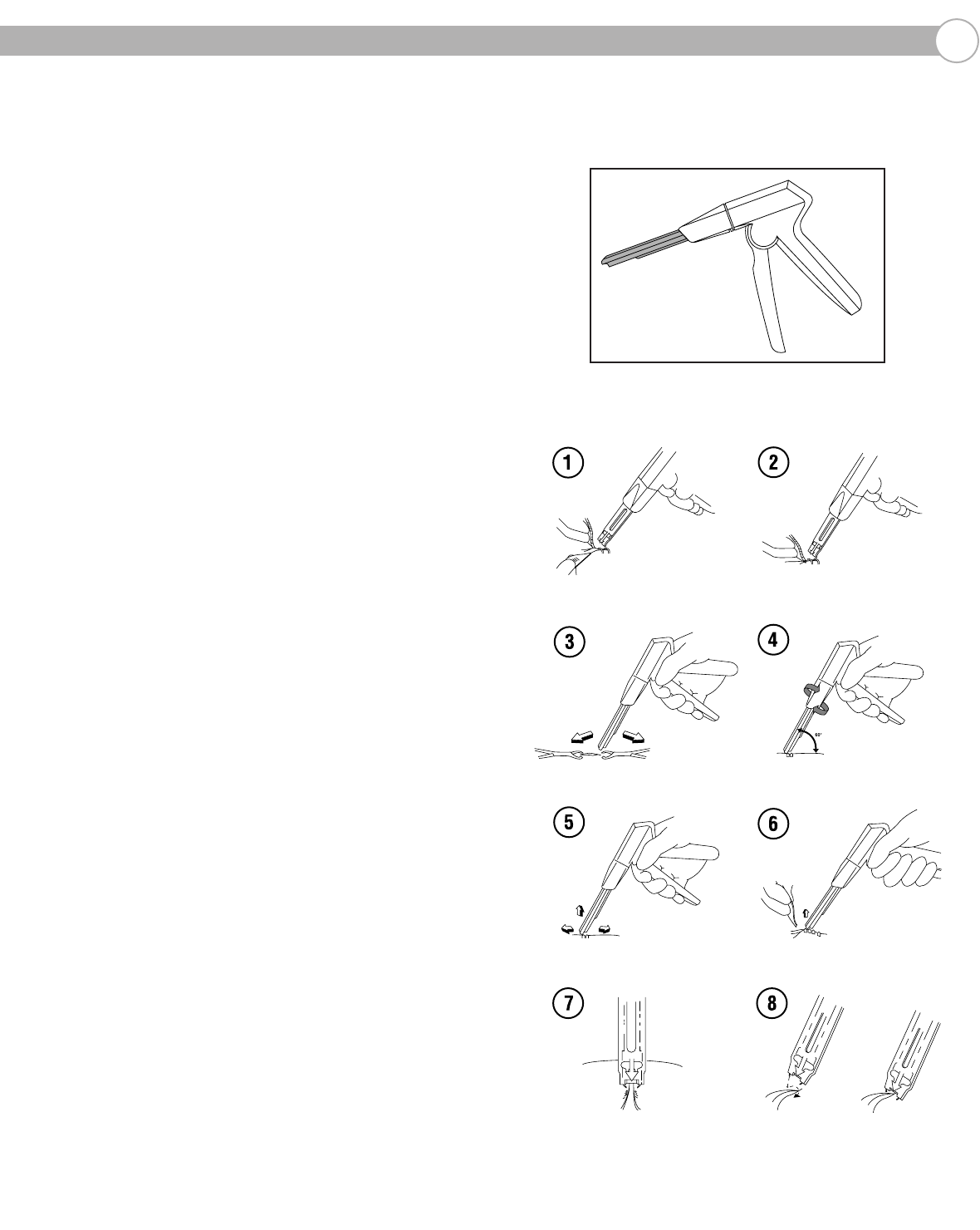
CHAPTER 8 109
PROXIMATE®RH
ROTATING HEAD SKIN STAPLER
INDICATIONS
The PROXIMATE RH Rotating Head Skin Stapler has application for routine skin
closure in a wide variety of surgical procedures.
CONTRAINDICATIONS
When it is not possible to maintain at least a 5 mm distance from the stapled
skin to underlying bones, vessels, or internal organs, the use of staples for skin
closure is contraindicated.
DEVICE DESCRIPTION
The PROXIMATE RH Rotating Head Skin Stapler is a sterile, single patient use
instrument designed to deliver rectangular, stainless steel staples for routine
wound closure. The head rotates 360° to facilitate placement of the staples.
INSTRUCTIONS FOR USE
Verify compatibility of all instruments and accessories prior to using the
instrument.
1 Using sterile technique, remove the instrument from the package. To
avoid damage, do not flip the instrument into the sterile field.
2 Suggested Eversion Techniques: With two tissue forceps, pick up each
wound edge individually and approximate the edges (Illustration 1).
Or, with one tissue forceps, pull skin edges together until edges evert
(Illustration 2).
Or, apply tension to either end of the incision, such that the tissue edges
begin to approximate themselves. One forceps can be used to ensure that
the edges are everted (Illustration 3).
3 Position the instrument with moderate pressure over the everted skin
edges. The instrument should be held at a 60° angle to the skin
(Illustration 4).
4 Squeeze the trigger until the trigger motion is halted, then release the
trigger and move the instrument off the incision in any direction
(Illustration 5).
Alternate Release: If desired, before releasing the trigger lift up on the
instrument. This will help to evert the skin edges, which can then be more
easily grasped with the tissue forceps. Release the trigger after the for-
ceps are in place, and repeat the sequence to fire the next staple.
(Illustration 6).
Alternate Technique: The instrument can also be precocked (partially
fired) so that the staple points are visible at the nose of the instrument.
This feature, in conjunction with the clear nose and alignment arrow,
ensures precise staple placement in the skin (Illustration 7).
Note: If desired, after the instrument has been precocked one leg of the
staple can be hooked onto one side of the tissue. This will aid in drawing
the tissue together. This technique may be suitable for attaching skin
grafts under moderate tension (Illustration 8).
WARNINGS AND PRECAUTIONS
• Dispose of all opened products whether used or unused. Do Not
Resterilize the instrument. Resterilization may compromise the integrity of
the instrument which may result in unintended injury.
HOW SUPPLIED
The PROXIMATE RH Rotating Head Skin Stapler is supplied sterile and
preloaded for single patient use. Discard after use.
Caution: Federal (USA) law restricts this device to sale by or on the order
of a physician.
FORMED STAPLE DIMENSIONS
Regular staples have an approximate diameter of 0.53 mm, span of 5.7 mm,
and leg length of 3.9 mm.
Wide staples have an approximate diameter of 0.58 mm, span of 6.9 mm, and
leg length of 3.9 mm.
P40105P04 *Trademark ©ETHICON ENDO-SURGERY, INC. 1994

PRODUCT INFORMATION
110
SURGICAL GUT SUTURE
ABSORBABLE SURGICAL SUTURES, U.S.P.
DESCRIPTION
Surgical gut suture is an absorbable, sterile surgical suture composed of
purified connective tissue (mostly collagen) derived from either the serosal
layer of beef (bovine) or the submucosal fibrous layer of sheep (ovine)
intestines. Surgical gut sutures are available in plain or chromic. Chromic gut
is processed to provide greater resistance to absorption. Surgical gut is
packaged in tubing fluid. Blue dyed chromic gut suture is also available.
Surgical gut suture meets all requirements established by the United States
Pharmacopoeia (U.S.P.) for absorbable surgical sutures.
INDICATIONS
Surgical gut suture is indicated for use in general soft tissue approximation
and/or ligation, including use in ophthalmic procedures, but not for use in
cardiovascular and neurological tissues.
ACTIONS
When surgical gut suture is placed in tissue, a moderate tissue inflammation
occurs which is characteristic of foreign body response to a substance. This is
followed by a loss of tensile strength and a loss of suture mass, as the prote-
olytic enzymatic digestive process dissolves the surgical gut. This process
continues until the suture is completely absorbed. Many variable factors may
affect the rate of absorption. Some of the major factors which can affect tensile
strength loss and absorption rates are:
1. Type of suture - plain gut generally absorbs more rapidly than chromic gut.
2. Infection - surgical gut is absorbed more rapidly in infected tissue
than in non-infected tissue.
3. Tissue sites - surgical gut will absorb more rapidly in tissue where
increased levels of proteolytic enzymes are present, as in the secre-
tions exhibited in the stomach, cervix and vagina.
CONTRAINDICATIONS
This suture, being absorbable, should not be used where extended approxi-
mation of tissue is required.
The use of this suture is contraindicated in patients with known sensitivities or
allergies to collagen or chromium, as gut is a collagen based material, and
chromic gut is treated with chromic salt solutions.
WARNINGS
Users should be familiar with surgical procedures and techniques involving gut
suture before using surgical gut suture for wound closure, as the risk of wound
dehiscence may vary with the site of application and the suture material used.
Physicians should consider the in vivo performance when selecting a suture.
The use of this suture may be inappropriate in elderly, malnourished, or debil-
itated patients, or in patients suffering from conditions which may delay
wound healing. As this is an absorbable material, the use of supplemental non-
absorbable sutures should be considered by the surgeon in the closure of sites
which may undergo expansion, stretching or distention or which may require
additional support.
As with any foreign body, prolonged contact of any suture with salt solutions,
such as those found in the urinary or biliary tracts, may result in calculus for-
mation. As an absorbable suture, surgical gut may act transiently as a foreign
body. Acceptable surgical practice should be followed for the management of
contaminated or infected wounds.
Do not resterilize. Discard open packages and unused sutures.
Certain patients may be hypersensitive to collagen or chromium and might
exhibit an immunological reaction resulting in inflammation, tissue granulation or
fibrosis, wound suppuration and bleeding, as well as sinus formation.
PRECAUTIONS
In handling this or any other suture material, care should be taken to avoid
damage from handling. Avoid crushing or crimping damage due to application
of surgical instruments such as forceps or needle holders. Surgical gut sutures
require the accepted surgical technique of flat and square ties with
additional throws as warranted by surgical circumstance and the experience of
the surgeon.
Under some circumstances, notably orthopaedic procedures, immobilization of
joints by external support may be employed at the discretion of the surgeon.
The surgeon should avoid unnecessary tension when running down knots, to
reduce the occurrence of surface fraying and weakening of the strand.
Avoid prolonged exposure to elevated temperatures.
To avoid damaging needle points and swage areas, grasp the needle in an area
one-third (1/3) to one-half (1/2) of the distance from the swaged end to the
point. Reshaping needles may cause them to lose strength and be less
resistant to bending and breaking. Users should exercise caution when
handling surgical needles to avoid inadvertent needle sticks. Discard used
needles in "sharps" containers.
ADVERSE REACTIONS
Adverse effects associated with the use of this device include wound dehis-
cence, variable rates of absorption over time (depending on such factors as the
type of suture used, the presence of infection and the tissue site), failure to pro-
vide adequate wound support in closure of sites where expansion, stretching
or distension occur, etc., unless additional support is supplied through the use
of nonabsorbable suture material, failure to provide adequate wound support
in elderly, malnourished or debilitated patients or in patients suffering from
cancer, anemia, obesity, diabetes, infection or other conditions which may
delay wound healing, allergic response in patients with known sensitivities to
collagen or chromium which may result in an immunological reaction resulting
in inflammation, tissue granulation or fibrosis, wound suppuration and bleed-
ing, as well as sinus formation, infection, moderate tissue inflammatory
response characteristic of foreign body response, calculi formation in urinary
and biliary tracts when prolonged contact with salt solutions such as urine and
bile occurs, and transitory local irritation at the wound site. Broken needles
may result in extended or additional surgeries or residual foreign bodies.
Inadvertent needle sticks with contaminated surgical needles may result in the
transmission of bloodborne pathogens.
HOW SUPPLIED
Surgical gut sutures are available in U.S.P. sizes 7-0 through 3 (metric sizes
0.7-7.0) in a variety of lengths with and without permanently attached needles
and on LIGAPAK* dispensing reels. Surgical gut sutures are also available in
U.S.P. sizes 0 through 1 (metric sizes 4.0-5.0) attached to CONTROL RELEASE*
removable needles. The suture is supplied sterile in one, two and three
dozen boxes.
389360 *Trademark ©ETHICON, INC. 1995

CHAPTER 8 111
SURGICAL STAINLESS STEEL SUTURE
NONABSORBABLE SURGICAL SUTURES, U.S.P.
DESCRIPTION
Surgical stainless steel suture is a nonabsorbable, sterile surgical suture
composed of 316L stainless steel. Surgical stainless steel suture is available as
a monofilament and multifilament suture.
Surgical stainless steel suture meets all requirements established by the United
States Pharmacopoeia (U.S.P.) for nonabsorbable surgical sutures. Surgical
stainless steel suture is also labeled with the B&S gauge classifications.
INDICATIONS
Surgical stainless steel suture is indicated for use in abdominal wound closure,
hernia repair, sternal closure and orthopaedic procedures including cerclage
and tendon repair.
ACTIONS
Surgical stainless steel suture elicits a minimal acute inflammatory reaction in
tissue and is not absorbed.
CONTRAINDICATIONS
The use of this suture is contraindicated in patients with known sensitivities or
allergies to 316L stainless steel, or constituent metals such as chromium and
nickel.
WARNINGS
Users should be familiar with surgical procedures and techniques involving
nonabsorbable, stainless steel sutures before employing for wound closure, as
the risk of wound dehiscence may vary with the site of application and the
suture material used.
Acceptable surgical practice must be followed for the management of contam-
inated or infected wounds.
PRECAUTIONS
In handling this or any other suture material, care should be taken to avoid
damage from handling, such as kinking or excessive twisting.
To avoid damaging needle points and swage areas, grasp the needle in an area
one-third (1/3) to one-half (1/2) of the distance from the swaged end to the
point. Reshaping needles may cause them to lose strength and be less
resistant to bending and breaking. Users should exercise caution when
handling surgical needles to avoid inadvertent needle sticks. Discard used
needles in "sharps" containers.
ADVERSE REACTIONS
Adverse effects associated with the use of this device include wound dehis-
cence, allergic response in patients with known sensitivities to 316L stainless
steel, or constituent metals such as chromium and nickel, infection, minimal
acute inflammatory tissue reaction, pain, edema and local irritation at the
wound site. Broken needles may result in extended or additional surgeries or
residual foreign bodies. Inadvertent needle sticks with contaminated surgical
needles may result in the transmission of bloodborne pathogens.
HOW SUPPLIED
Surgical stainless steel sutures are available in sizes 7 through 10-0 (metric
sizes 9.0-0.2) in a variety of lengths with and without permanently attached
needles in one, two and three dozen boxes.
389357 *Trademark ©ETHICON, INC. 1995

PRODUCT INFORMATION
112
VICRYL* Knitted Mesh
DESCRIPTION
VICRYL* (polyglactin 910) knitted mesh is prepared from a synthetic absorbable
copolymer of glycolide and lactide, derived respectively from glycolic and lactic
acids. This knitted mesh is prepared from uncoated, undyed fiber identical in
composition to that used in VICRYL (polyglactin 910) synthetic absorbable
suture, which has been found to be inert, nonantigenic, nonpyrogenic and to
elicit only a mild tissue reaction during absorption.
VICRYL knitted mesh is intended for use as a buttress to provide temporary sup-
port during the healing process.
ACTIONS
Two important characteristics describe the in vivo function and behavior of
VICRYL knitted mesh: reinforced wound strength and the rate of absorption
(loss of mass).
The dehiscence force of healing abdominal wounds in rats closed with size 4-0
absorbable sutures was compared with corresponding wounds closed with size
4-0 absorbable sutures and reinforced with VICRYL knitted mesh. In this animal
model, the strength of the incision, when supported by the mesh, was signifi-
cantly greater than the sutured incisional wound. Explanted VICRYL knitted
mesh, which, before implantation had an initial average burst strength of 63
lbs., was found to have 80% of its original burst strength remaining after
fourteen days in vivo.
Subcutaneous implantation studies in rats indicate that the absorption of
VICRYL mesh material is minimal until about six weeks post implantation and
essentially complete between 60 and 90 days.
INDICATIONS
VICRYL knitted mesh may be used wherever temporary wound or organ sup-
port is required, particularly in instances in which compliant and stretchable
support material is desired and containment of wound transudate is not
required. VICRYL knitted mesh may be cut to the shape or size desired for each
specific application.
CONTRAINDICATIONS
Because VICRYL knitted mesh is absorbable, it should not be used where
extended wound or organ support is required.
WARNINGS
DO NOT RESTERILIZE.
The safety and effectiveness of VICRYL knitted mesh in neural tissue and in car-
diovascular tissue has not been established.
PRECAUTIONS
None.
ADVERSE REACTIONS
No significant clinical adverse reactions to the mesh have been reported.
DIRECTIONS FOR USE
It is recommended that absorbable or nonabsorbable sutures by placed 1/4 to
1/2 inch (6 to 12mm) apart at a distance approximately 1/4 inch (6mm) form the
edge of the mesh. Some surgeons prefer to suture a mesh larger than the
defect into position over the defect. The edges are then sutured to assure a
proper closure under correct tension. When all margin sutures have been
placed, the excess mesh is trimmed away, leaving at least 1/4 inch of mesh
extending beyond the suture line.
HOW SUPPLIED
VICRYL knitted mesh is available in single packets as a sterile, undyed fabric
mesh in single sheet sizes of approximately 6 x 6 inches and 12 x 12 inches
(15 x 15 centimeters and 30 x 30 centimeters).
389096 *Trademark ©ETHICON, INC. 1986

CHAPTER 8 113
VICRYL* Woven Mesh
DESCRIPTION
VICRYL* (polyglactin 910) woven mesh is prepared from a synthetic absorbable
copolymer of glycolide and lactide, derived respectively from glycolic and lactic
acids. This tightly woven mesh is prepared from uncoated, undyed fiber
identical in composition to that used in VICRYL* (polyglactin 910) synthetic
absorbable suture, which has been found to be inert, nonantigenic, nonpyro-
genic and to elicit only a mild tissue reaction during absorption.
VICRYL woven mesh is intended for use as a buttress to provide temporary sup-
port during the healing process.
ACTIONS
Two important characteristics describe the in vivo function and behavior of
VICRYL woven mesh: reinforced wound strength and the rate of absorption
(loss of mass).
The dehiscence force of healing abdominal wounds in rats closed with size 4-0
absorbable sutures was compared with corresponding wounds closed with size
4-0 absorbable sutures and reinforced with VICRYL woven mesh. In this animal
model, the strength of the incision, when supported by the mesh, was signifi-
cantly greater than the sutured incisional wound. Explanted VICRYL woven
mesh, which, before implantation had an initial average burst strength of
approximately 121 lbs., was found to have approximately 23% of its original
burst strength remaining after fourteen days in vivo.
Subcutaneous implantation studies in rats indicate that the absorption of
VICRYL mesh material is minimal until about six weeks post implantation and
essentially complete between 60 and 90 days.
INDICATIONS
VICRYL woven mesh may be used wherever temporary wound or organ support
is required. The woven mesh structure is less porous than VICRYL knitted
mesh. It is indicated in instances in which containment of wound transudate is
desirable. VICRYL woven mesh may be cut to the shape or size desired for each
specific application.
CONTRAINDICATIONS
Because VICRYL woven mesh is absorbable, it should not be used where
extended wound or organ support is required.
WARNINGS
DO NOT RESTERILIZE.
The safety and effectiveness of VICRYL woven mesh in neural tissue and in car-
diovascular tissue has not been established.
PRECAUTIONS
None.
ADVERSE REACTIONS
None known.
DIRECTIONS FOR USE
It is recommended that absorbable or nonabsorbable sutures be placed 1/4 to
1/2 inch (6 to 12mm) apart at a distance at least 1/4 inch (6mm) from the edge
of the mesh. Some surgeons prefer to suture a mesh larger than the defect into
position over the defect. The edges are then sutured to assure proper closure
under correct tension. When all margin sutures have been placed, the excess
mesh is trimmed away, leaving at least 1/4 inch of mesh extending beyond the
suture line.
HOW SUPPLIED
VICRYL woven mesh is available in single packets as a sterile, undyed, fabric
mesh in single sheet sizes of approximately 6 x 6 inches and 12 x 12 inches
(15 x 15 centimeters and 30 x 30 centimeters).
389065 *Trademark ©ETHICON, INC. 1986

INDEX
CHAPTER 9

INDEX
116
ABDOMEN, 23, 26, 28, 30
abdominal cavity, 23, 26, 30, 34,
50, 63
abdominal wall, 16, 19, 23, 28,
30-31, 33-34, 44, 77, 82, 84
fascia, 2, 5, 21, 30-32, 34, 39-40,
47-48, 50-51, 63, 77, 82, 96
muscle tissue, 2, 30
peritoneum, 19, 23, 29-31, 34,
47-48, 50, 63
skin, 2, 31-33
subcutaneous fat, 30-32, 47-48
subcuticular tissue, 32
transversalis fascia, 30
ALIMENTARY TRACT, 35
esophagus, 36
oral cavity, 35-36, 47-48, 69, 89
pharynx, 35, 47-48
upper alimentary tract, 35
BILIARY TRACT, 6, 29, 38, 47-48
gallbladder, 29
BONE, 2, 16, 39-40, 80
anchor, 18, 39, 65
sternum, 39
BRAIN, 34
cerebrospinal fluid, 34
dura mater, 34-35
galea, 34, 76
peripheral nerve repair, 34
skull, 35
CARDIOVASCULAR SURGERY, 18, 37,
45, 51, 83
heart valves, 21, 38, 99
pledgets, 18, 21, 38, 91, 101
sternum, 16, 37, 39, 44, 48
COATED VICRYL*
(POLYGLACTIN 910) SUTURE, 15, 25,
88, 112
COATED VICRYL RAPIDE*
(POLYGLACTIN 910) SUTURE, 14,
32-33, 39, 89
A
B
C
DEAD SPACE, 5, 22, 32, 81
DERMABOND*
TOPICAL SKIN ADHESIVE
(2-OCTYL CYANOACRYLATE), 68-71,
81, 90
DISSECTION, 4, 48, 103
ETHIBOND* EXCEL POLYESTER
SUTURE, 10, 17-18, 20, 25, 33, 37-38,
58-59, 81, 83, 92
ETHILON* NYLON SUTURE, 10, 17, 20,
25, 33-36, 58-59, 81, 93
EYE, 35-36
conjunctiva, 35-36, 100
cornea, 35-36, 80
ocular muscles, 35-36
sclera, 35-36
FEMALE GENITAL TRACT, 38
GASTROINTESTINAL TRACT, 6, 28-29,
46-48
colon, 2, 27, 29
rectum, 29
small intestine, 2, 28-29, 94
stomach, 2-3, 27-29, 81, 94, 110
INCISION, 4-6, 24, 28, 30-32, 48, 59, 62,
68
INFLAMMATORY RESPONSE, 6-7, 77,
94, 110
D
E
F
I
G

CHAPTER 9 117
* Trademark
KNOT SECURITY, 11, 15, 24, 92-93, 95,
97-103, 105
KNOT TENSILE STRENGTH, 11, 89
KNOT TYING, 17, 23-26, 34, 65, 94
endoscopic, 25-26, 38, 51
monofilament sutures, 11, 18,
24, 29, 32-33, 35, 37, 95,
105
multifilament sutures, 11, 24, 29,
31-33, 36
techniques, 18, 25-26
deep tie, 25-26
instrument tie, 24-26
square knot, 25-26
surgeon’s knot, 25-26
LIGATURES, 10, 18-19, 28, 62, 85
free tie, 18-19, 28
LIGAPAK* dispensing reel, 21,
59, 63, 82, 101, 110
stick tie, 18-19, 85
MERSILENE*
POLYESTER FIBER MESH, 82, 96
MERSILENE*
POLYESTER FIBER STRIP, 75, 82
MERSILENE*
POLYESTER FIBER SUTURE, 13, 17, 20,
25, 33, 35, 58-59, 82, 95
MESH, 30, 35, 82, 84, 86, 96
See also MERSILENE* polyester
fiber mesh; PROLENE*
Polypropylene Hernia System;
PROLENE* polypropylene mesh;
VICRYL* (polyglactin 910)
knitted mesh; VICRYL*
(polyglactin 910) periodontal
mesh; VICRYL* (polyglactin 910)
woven mesh
MICROSURGERY, 15, 21, 34, 44, 47-48,
62, 65, 82, 97-98, 100
MONOCRYL* (POLIGLECAPRONE 25)
SUTURE, 10, 12, 14-15, 20, 25, 32, 38,
58-59, 70-71, 83, 97
K
L
M
NECROTIC TISSUE, 4, 40
NEEDLE, 10, 42-54
anatomy, 44
body, 43-54
eye, 44-46
point, 42-44, 46, 48-49
applications, 47-48
shape, 45-50
compound curved, 47
curved, 46-47
half-curved, 46-47
ski, 46
straight, 46-47
Bunnell, 46, 51
Keith, 46, 51
sharpness, 43, 47-50, 53, 82
stability, 43, 48
strength, 42
ETHALLOY* needle alloy,
42, 81
swage, 44-45, 65, 83
CONTROL RELEASE*
needle, 21, 45, 59, 80
types, 47-51
blunt point, 51
ETHIGUARD* blunt
point needle, 51, 81
cutting, 43, 46-49, 51
conventional cutting,
47-49, 83
PC PRIME* needle,
48, 83
sternotomy, 48-49
reverse cutting, 48-50, 75,
84
MICRO-POINT*
needle, 48-49, 82
OS, 49, 51
side-cutting, 48-49, 82
CS ULTIMA*
needle, 48, 50, 80
SABRELOC*
needle, 50, 84
spatula, 48-51, 82
TG PLUS* needle,
50
N

INDEX
118
taper point, 21, 43, 45-46,
50-51, 77, 85
MAYO, 50-51
TAPERCUT, 43, 48, 50-51
TAPERCUT needle,
43, 48, 50-51,
85
trocar point, 51
NEEDLEHOLDER, 26-27, 42-43, 46-53
arming, 27, 59
jaws, 51-52
NEUROSURGERY, 16, 34
NUROLON* BRAIDED NYLON
SUTURE, 10, 13, 17, 20, 25, 34, 39,
58-59, 83
OPHTHALMIC SURGERY, 15, 35, 47,
66, 80, 83
ORTHOPAEDIC SURGERY, 18, 49, 51
PARENCHYMATOUS ORGANS, 29
kidney, 3, 29, 51
liver, 29-30, 51
spleen, 29
PDS* II (POLYDIOXANONE) SUTURE,
10, 15-16, 25, 31, 35, 38,
59-60, 83, 100
PERMA-HAND* SILK SUTURE, 16, 25,
33, 59, 83, 101
PLASTIC SURGERY, 14, 17, 48, 51, 57,
83-84
PRIMARY SUTURE LINE, 18, 22-23, 33
buried sutures, 22, 80
continuous sutures, 19, 22, 37
Connell Technique, 24
Cushing Technique, 24
Lembert Technique, 24
deep sutures, 19, 22, 69
interrupted sutures, 19, 22, 28,
30-31, 38
interrupted horizontal
mattress suture, 19, 24
interrupted vertical
mattress suture, 19, 24
O
P
purse-string sutures, 19, 22
running stitches, 19
subcuticular sutures, 19, 22,
32, 90
PROLENE* POLYPROPYLENE HERNIA
SYSTEM, 30, 70, 84
PROLENE* POLYPROPYLENE MESH,
30, 70, 84
PROLENE* POLYPROPYLENE SUTURE,
18, 20, 25-26, 28, 30-31, 34-35,
37, 39, 61, 84
PRONOVA* POLY
(HEXAFLUOROPROPYLENE-VDF)
SUTURE, 18, 20, 25-26, 37, 39, 84
PROXI-STRIP* SKIN CLOSURES, 27,
40, 75, 84
RESPIRATORY TRACT, 36
bronchial stump closure, 36
thoracic cavity, 36
RETENTION SUTURE DEVICES, 33,
77-78, 84
SECONDARY SUTURE LINE, 33
continuous sutures, 19, 22, 24,
37
interrupted sutures, 19, 22, 24,
28, 30-31, 37
railroad track scar, 33
retention sutures, 33, 77-78, 84
SKIN, 2-5, 8, 10, 13-14, 16-17, 22,
27-28, 31-34, 46-47
STITCH PLACEMENT, 23
STITCHES AND TYPES, 18-23
Connell Technique, 24
Cushing Technique, 24
Halsted Technique, 24
horizontal mattress technique,
19, 24
Lembert Technique, 24
over-and-over technique, 19, 24
purse-string technique, 19, 22
running technique, 19
R
S

CHAPTER 9 119
* Trademark
subcuticular technique, 19, 22
vertical mattress technique,
19, 24
SURGICAL GUT SUTURE, 14, 20, 58,
94, 110
chromic, 13-14, 20, 28, 32-33, 38,
80, 94, 97, 110
CHROMICIZING process,
14
collagen pure, 12-14
fast absorbing, 14, 27, 94
plain, 12-14, 20, 83, 94, 110
TRU-GAUGING process, 13, 85
tubing fluid, 85
SURGICAL SILK SUTURE, 16, 28, 34
See also PERMA-HAND* silk suture
SURGICAL STAINLESS STEEL
SUTURE, 16-17, 20, 25, 66, 111
SUTURE, 10-40
absorbable suture, 15, 22, 27-32,
38, 58-60, 80, 83,
85, 89
See also Coated VICRYL*
(polyglactin 910) suture;
Coated VICRYL RAPIDE*
(polyglactin 910) suture;
MONOCRYL*
(poliglecaprone 25)
suture; PANACRYL*
suture; PDS* II
(polydioxanone) suture;
surgical gut suture
alloy suture, 16, 42, 48, 51, 81
wire gauge equivalents,
17
See also surgical stain-
less steel suture;
temporary cardiac
pacing wire
characteristics, 10-11, 14, 16-17,
24, 32
absorption profile, 20-21
hydrolysis, 15, 17,
32, 35
absorption rate, 14, 20-21
braided strands, 18
breaking strength, 2,
20-21
in vivo strength,
20-21
burst strength, 2
monofilament strands, 11,
15, 17, 24, 31-32, 35
multifilament strands, 11,
16-17, 24, 29, 31-33
size, 10-11, 13-16, 28
tensile strength, 2, 6,
10-17, 23, 27, 31-33,
35, 38, 66, 85
tissue reaction, 12, 14-15,
17-18, 26, 32-38
labeling, 56-60
expiration date, 57, 60, 81
Food and Drug
Administration (FDA), 12,
57, 60, 68, 81
lot number, 57
package insert, 57, 83
product code, 57, 84
manufacture, 13, 16, 42, 57, 90
nonabsorbable suture, 16, 18,
22-23, 26, 29, 31-32, 35,
57, 60, 63, 82-83
See also ETHIBOND* EXCEL
polyester suture;
ETHILON* nylon suture;
MERSILENE* polyester
fiber suture; NUROLON*
braided nylon suture;
PROLENE* polypropylene
suture; PRONOVA*
poly(hexafluoropropy-
lene-VDF) suture; surgical
silk suture
nylon suture, 10, 13, 17, 20, 25,
36, 40, 81, 83, 93, 99
See also ETHILON* nylon
suture; NUROLON*
braided nylon suture
packaging, 10, 16, 56-65
dispenser boxes, 56-57,
60, 81, 83
environmentally conscious, 57

INDEX
120
primary packets, 56-57
overwrap, 57, 59, 61-62,
64, 83
peelable foil, 58
single strand and multi-
strand packaging packets,
58, 82-83
E-PACK* procedure
kit, 54, 59, 61, 81
EASY ACCESS*
packaging, 81
ETHI-PACK* pre-cut
suture, 81
GENTLE BEND*
package, 82
LABYRINTH*
package, 82
looped suture, 19,
77, 82
RELAY* suture
delivery system,
56-58, 60-61,
64, 84
SUTUPAK* pre-cut
sterile suture, 58,
63, 85
storage racks, 56
IV pole racks, 56
modular storage
racks, 56-57
polyester fiber suture, 13, 17, 20,
25, 58, 92, 95, 96
See also ETHIBOND* EXCEL
polyester suture
polypropylene suture, 18, 25-26,
28, 30, 32, 36-39, 66,
82-84, 102-103
See also PROLENE*
polypropylene suture
sterilization, 57, 60, 85, 90, 104
SUTURE CUTTING, 26
SUTURE HANDLING, 10-11, 26-27, 63
sterile barrer, 61
sterile field, 53, 56, 59-62, 64
sterile technique, 26, 75, 85
sterile transfer, 56, 60-62
sterile sterilization, 60
T
SUTURE PREPARATION, 62
SUTURE REMOVAL, 11, 14, 26, 32-33,
35, 68-69
SUTURE TECHNIQUES, 28-34, 45
double-layer closure, 28
inverted closure technique, 28
single-layer closure, 28, 31
Smaed-Jones far-and-
near technique, 31
See also ligatures; primary suture
line; secondary suture line
TAPES, 4, 32, 52, 56, 74-75
skin closure tapes, 32, 74-75
umbilical tape, 75, 85
See also MERSILENE* polyester
fiber strip; PROXI-STRIP* skin
closures
TENDON SURGERY, 39
Bunnell Technique, 37, 39
periosteum, 39, 51
TISSUE ADHESIVES, 68-71
See also DERMABOND* Topical
Skin Adhesive (2-Octyl
Cyanoacrylate)
TISSUE STRENGTH, 2
breaking strength, 2
burst strength, 2
tensile strength, 2, 6, 11-17, 23
URINARY TRACT SURGERY, 38
U

CHAPTER 9 121
* Trademark
VASCULAR SURGERY, 11
heart valves, 21, 38, 99
VESSELS, 4, 6, 10, 14, 18, 24, 32,
37-38, 51, 59, 62, 75
pediatric, 15, 21, 75, 85
VICRYL* (POLYGLACTIN 910)
KNITTED MESH, 30, 86
VICRYL* (POLYGLACTIN 910)
PERIODONTAL MESH, 35-36, 86
VICRYL* (POLYGLACTIN 910)
SUTURE, 10, 14-15, 25, 34-35
VICRYL* (POLYGLACTIN 910)
WOVEN MESH, 30, 86
WOUND CLASSIFICATION, 5-6
clean, 4-5
clean-contaminated, 5
contaminated, 6-7, 18
dirty and infected, 5-7, 18
WOUND COMPLICATIONS, 3
dehiscence, 16, 38, 68, 74, 81
dehydration, 3
edema, 2, 5-6, 11, 78
epithelialization, 7, 33, 35, 68
infection, 2-7, 10-12, 27, 31-32,
35, 35-36, 38-40
wound disruption, 23, 86
WOUND HEALING, 2-7, 10
collagen formation, 7
dehydration, 3
primary intention, 6
second intention, 6
delayed primary closure, 6
V
W

www.jnjgateway.com
ETHICON, INC. • P.O. Box 151 • Somerville, NJ 08876-0151
