CV 180 EVIS EXERA II Video System Center Instruction Manual Olympus CV180 User
User Manual: Olympus CV180 User Manual
Open the PDF directly: View PDF ![]() .
.
Page Count: 296 [warning: Documents this large are best viewed by clicking the View PDF Link!]
- INSTRUCTIONS
- EVIS EXERA II VIDEO SYSTEM CENTER
- OLYMPUS CV-180
- Contents
- Labels and Symbols 1
- Important Information - Please Read Before Use 3
- Summary of Equipment Functions 12
- Chapter 1 Checking the Package Contents 14
- Chapter 2 Nomenclature and Functions 15
- Chapter 3 Inspection 34
- Chapter 4 Operation 41
- Chapter 5 Functions 62
- Chapter 6 Fuse replacement 153
- Chapter 7 Care, Storage and Disposal 155
- Chapter 8 Installation and Connection 157
- Chapter 9 Function setup 193
- Chapter 10 Troubleshooting 259
- Appendix 269
- Labels and Symbols
- Important Information - Please Read Before Use
- Summary of Equipment Functions
- Displaying the endoscopic images on the monitor
- Special light observation
- Adjusting the endoscopic images
- Entering patient data
- Customizing the operations
- Recording images
- Operation of ancillary equipment
- Chapter 1 Checking the Package Contents
- Chapter 2 Nomenclature and Functions
- 2.1 Front panel
- 1. Power switch
- 2. Power indicator
- 3. Video connector socket
- 4. Locking lever
- 5. Image source buttons
- 6. Picture in picture (PinP) button
- 7. White balance (Wh/B) button
- 8. White balance (Wh/B OK) indicator
- 9. HDTV indicator
- 10. NBI indicator
- 11. Exposure adjustment (EXPOSURE) buttons
- 12. Exposure level (EXPOSURE) indicator
- 13. PinP composite terminal
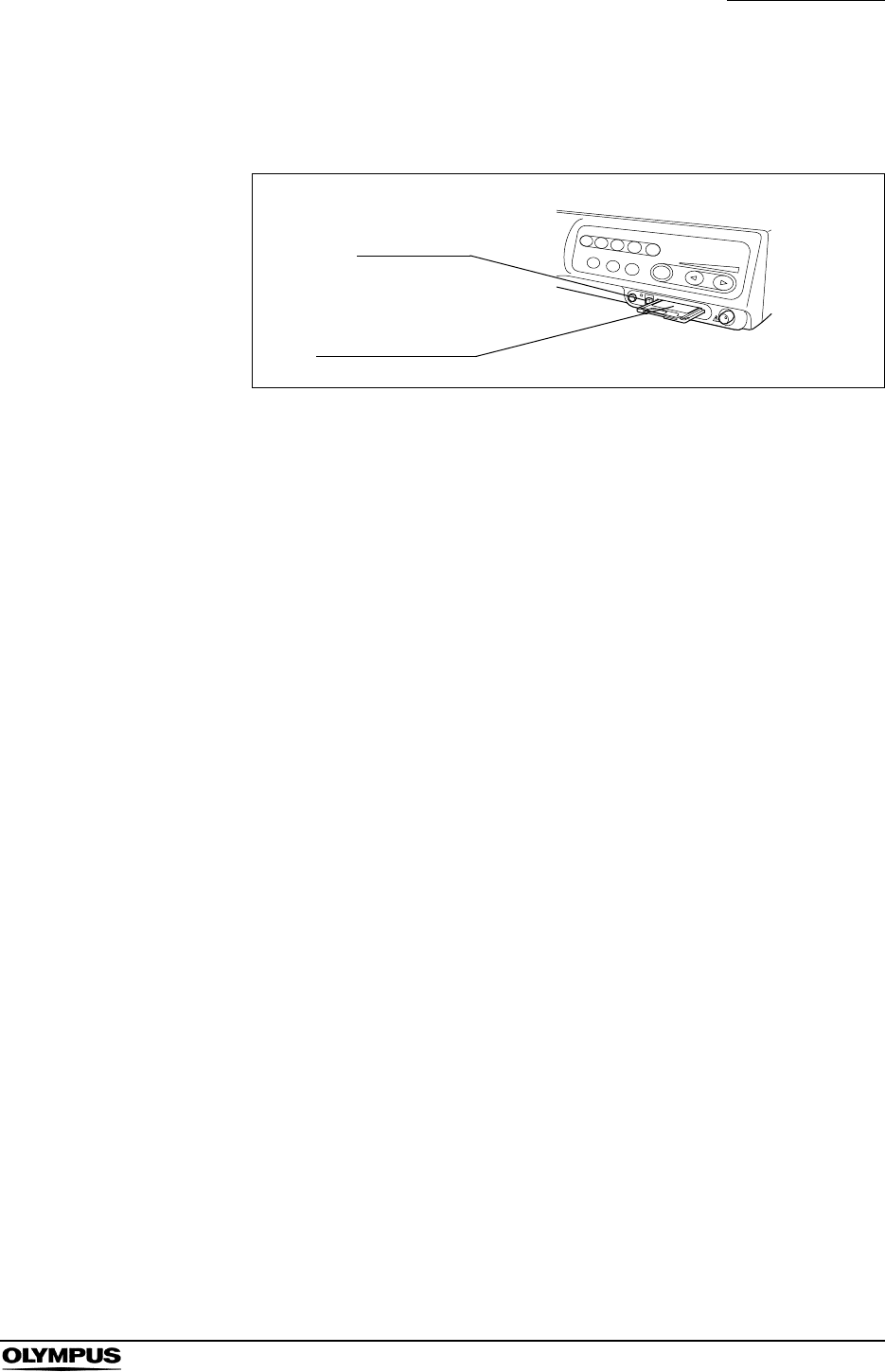
- 14. PC card slot
- 15. Eject button
- 16. PC card status indicator
- 17. STOP button
- 18. Iris mode (IRIS) button
- 19. Iris mode indicators
- 20. Image enhancement mode (ENH.) button
- 21. Image enhancement mode indicators
- 22. RESET button
- 2.2 Rear panel
- 1. Keyboard terminal
- 2. Y/C OUT terminal
- 3. Printer IN terminal
- 4. Printer OUT terminal
- 5. Printer remote terminal
- 6. PC OUT terminal
- 7. PC IN terminal
- 8. PC remote terminal
- 9. Remote terminal
- 10. Foot switch terminal
- 11. VCR remote terminal
- 12. Fuse box
- 13. AC power inlet
- 14. Light control terminal
- 15. Light source terminal
- 16. PinP Y/C terminal
- 17. HD/SD SDI OUT terminal
- 18. Digital OUT terminal
- 19. PC OUT2 terminal
- 20. Monitor remote terminal
- 21. Composite OUT terminal
- 22. Monitor OUT terminal
- 23. Potential equalization terminal
- 2.3 Keyboard
- 1. F1 key
- 2. F2 key
- 3. F3 key
- 4. F4 key
- 5. F5 key
- 6. F6 key
- 7. F7 key
- 8. F8 key
- 9. F9 key
- 10. F10 key
- 11. F11 key
- 12. F12 key
- 13. Print screen key
- 14. Scroll lock key
- 15. Pause key
- 16. #PER PAGE key
- 17. CAPTURE key
- 18. DEL IMAGE key
- 19. PRINT key
- 20. PRINT QTY. key
- 21. COLOR key
- 22. FREEZE key
- 23. RELEASE key
- 24. EXAM END key
- 25. Click key
- 26. Domepoint
- 27. Arrow keys
- 28. Other keyboard keys
- 2.4 Side panels
- 2.6 Set-up of screen options
- 2.7 Monitor
- 2.8 Pointer
- 2.1 Front panel
- Chapter 3 Inspection
- 3.1 Inspection of the power supply
- 3.2 Inspection of the examination light
- 3.3 Inspection of the automatic brightness control function
- 1. Confirm that this instrument is connected to the light source using the light source cable or light control cable (see Section 8.4, “Light source” on page 164).
- 2. According to the directions given in the light source's instruction manual, confirm that the light source's brightness control is set to “AUTO” and that the brightness level is in the center of the adjustable range.
- 3. Move the distal end of the endoscope between 1 and 3 cm from your palm. Confirm that the brightness of the image on the monitor remains constant. Confirm that the light emitted from the distal end of the endoscope changes in your palm.
- 4. Hold the distal end of the endoscope 3 cm from your palm. Use a piece of gauze, etc. to prevent the endoscope's distal end and your palm from being exposed to extraneous light. View the image on the monitor.
- 5. Confirm that the brightness of the image on the monitor changes when the light source's brightness level is changed.
- 3.4 Inspection of the monitor display
- 1. Turn the instrument ON. Then the endoscopic image appears on the screen (see Figure 3.3).
- 2. Confirm that the endoscopic image is normal by observing any object such as the palm of your hand.
- 3. Confirm that the date and time are correct.
- 4. Confirm that the “CVP” counter and “D.F” counter are displayed on the screen when the video printer and digital filing system are connected.
- 5. Confirm that enough space is available on the PC card to store endoscopic images.
- 3.5 Inspection of the freeze function
- 1. Press the “FREEZE” key on the keyboard, and confirm that the live endoscopic image freezes and a short beep is heard.
- 2. Press the “FREEZE” key again and confirm that the frozen image returns to the live image.
- 3. Confirm the function of the scope switches and/or foot switches, when the freeze function is assigned to these switches.
- 3.6 Inspection of the release function
- 1. Press the “RELEASE” key on the keyboard.
- 2. Confirm that the live image freezes for a short time and a beep is heard.
- 3. Confirm that the selected recording device is activated.
- 4. Confirm that the counter for the recording devices, which are displayed on the monitor, increments by one.
- 5. Confirm the function of the scope switches and/or foot switches, when the release function is assigned to these switches.
- 3.7 Inspection of the PinP (picture in picture) function
- 3.8 Inspection of the orientation function
- 3.9 Inspection of the special light observation function
- Chapter 4 Operation
- 4.1 Operation flow
- 4.2 Connection of an endoscope
- Figure 4.2
- VISERA series videoscope
- 1. Ensure that this instrument and all connected devices are turned OFF.
- 2. Connect the endoscope connector of the videoscope to the light source, referring to the instruction manual for the light source.
- 3. Push the video plug into the video connector socket of the instrument all the way until it clicks, holding this instrument with a hand so that it will not move. Confirm that the “UP” mark points upwards (see Figure 4.3).
- EVIS series videoscope and ultrasonic videoscope
- 1. Ensure that this instrument and all connected devices are turned OFF.
- 2. Connect the endoscope connector of the fiberscope to the light source referring to the instruction manual for the light source.
- 3. Push the video plug of the scope cable EXERA II into the video connector socket of the instrument all the way until it clicks, holding this instrument with a hand so that it will not move. Confirm that the “UP” mark points upwards (see Figure 4.3).
- 4. Connect the scope side connector of the scope cable EXERA II to the endoscope, referring to the instruction manual of the endoscope.
- Fiberscope and camera head
- 1. Ensure that this instrument and all connected devices are turned OFF.
- 2. Connect the endoscope connector of the fiberscope to the light source, referring to the instruction manual for the light source.
- 3. Push the video plug of the camera head into the video connector socket of the instrument all the way until it clicks, holding this instrument with a hand so that it will not move. Confirm that the “UP” mark points upwards (see Figure 4.3).
- 4. Connect the video adapter and camera head to the eyepiece section of the fiberscope, referring to the instruction manuals for the video adapter and camera head.
- Rigidscope and camera head
- 1. Ensure that this instrument and all connected devices are turned OFF.
- 2. Connect the light guide cable to the light source, referring to the instruction manual for the light source.
- 3. Push the video plug of the camera head into the video connector socket of the instrument all the way until it clicks, holding this instrument with a hand so that it will not move. Confirm that the “UP” mark points upwards (see Figure 4.3).
- 4. Attach the light guide cable, video adapter and camera head to the rigidscope, referring to the instruction manuals for the light guide cable, video adapter and camera head.
- Summary of connection
- 4.3 Turning the video system center ON
- 4.4 Recall of user preset data
- 1. Press the “Shift” and “F2” keys together. The “User preset” menu appears on the monitor (see Figure 4.7).
- 2. Click the desired user name in the user name dialog box. The selected user name is highlighted.
- 3. Check if the correct user name is selected, then click “Select”. The endoscopic image appears on the monitor and the user preset data is loaded to the video system center.
- 4.5 White balance adjustment
- For normal light observation
- 1. Confirm the lighting status of the “Wh/B OK” (Wh/B = White balance) indicator on the front panel (see Figure 4.9 and Table 4.1).
- 2. Proceed as follows according to the use of the endoscope.
- 1. Insert the endoscope's distal end into the white cap and hold the white cap and endoscope stable to avoid wash-out of the monitor image (see Figure 4.10).
- 2. Maintaining the stable condition in step 1., press the “Wh/B” button until a short beep is generated. The result of the adjustment will be displayed on the monitor for a few seconds (see Table 4.2).
- 3. Confirm that the “Wh/B OK” indicator is ON (see Table 4.1). If it is OFF, go back to step 1. again.
- For normal light observation
- When using an endoscope for the sterilized zone
- 1. Hold the endoscope stable to avoid wash-out of the monitor image, and enlarge the image to the full monitor, monitoring a white object such as a piece of gauze in such a way that it does not contact the endoscope.
- 2. Maintaining the stable condition in step 1., press the “Wh/B” button until a short beep is generated. The result of white balance adjustment will be displayed on the monitor for a few seconds (see Table 4.3).
- 3. Confirm that the “Wh/B OK” indicator is ON (see Table 4.1). If it is OFF, go back to step 1. again.
- For NBI observation
- 1. Adjust the white balance for normal light observation according to “For normal light observation” on page 53.
- 2. Switch to the NBI mode by operating the mode button of the light source. Refer to the instruction manual of the light source.
- 3. Confirm that “NBI” appears on the monitor, and that the “NBI” indicator on the front panel of the video system center turns from green to white.
- 4. Hold the endoscope as shown in step 1. of “When using an endoscope for the non-sterilized zone” on page 54 or step 1. of “When using an endoscope for the sterilized zone” on page 55 according to the use of it.
- 5. Maintaining the stable condition in step 4., press the “Wh/B” button until a short beep is generated. The result of the adjustment will be displayed on the monitor for a few seconds (see Table 4.4).
- 6. If the adjustment failed, perform the operation of steps 4. and 5. again.
- 7. Switch to the normal mode by operating the mode button of the light source. Refer to the instruction manual of the light source.
- 8. Confirm that “NBI” disappears on the monitor, and that the “NBI” indicator on the front panel of the video system center turns from green to white.
- 4.6 Patient data
- 1. Press the “F1” key to change the monitor display full-patient-data display.
- 2. Press the “EXAM END” key to clear the previous patient data.
- 3. Enter data.
- 4. When modifying data, press the arrow key to move the cursor to the input position and edit the data.
- 5. When deleting all patient data displayed on the monitor, press the “EXAM END” key on the keyboard (see Figure 4.13).
- 4.7 Observation of the endoscopic image
- 4.9 Termination of the operation
- 1. Press the “EXAM END” key (see Figure 4.14) to execute the following process;
- 2. Turn the instrument and ancillary equipment OFF.
- 3. When an EVIS series endoscope is used: Disconnect the scope side connector of the videoscope cable, and place it on the scope cable holder (A in Figure 4.15). For disconnecting the video plug, see B in Figure 4.15.
- 4. When a VISERA series endoscope or camera head is used: Disconnect the video plug of the videoscope cable from the instrument, holding the instrument with a hand so that it will not move and push the locking lever down (see Figure 4.16).
- Chapter 5 Functions
- 5.1 Front panel
- PinP (picture in picture) display
- 1. Confirm that the external device is connected to either the PinP composite terminal on the front panel or the PinP Y/C terminal on the rear panel (see Figure 5.2).
- 2. Press the “SCOPE” button to display the endoscopic live image.
- 3. Press the “PinP” button. The sub image appears on the monitor (see Figure 5.3).
- 4. Press the “PinP” button to change the display. The transitions of the displays are different according to the settings in “Us...
- Image enhancement mode (ENH.)
- 1. Press the “ENH.” button to change the enhancement mode (see Figure 5.6). The indicator above the button lights up and the selected mode is displayed on the monitor for a few seconds.
- 2. To switch OFF the image enhancement at any mode, press and hold the “ENH.” button. The indicator above the button goes OFF.
- Iris mode
- White balance
- Brightness adjustment (Exposure)
- STOP button and PC card indicator
- PC card slot and eject button
- Figure 5.12
- 1. Insert the xD picture card into the PC card adapter.
- 2. Push the PC card adapter all the way into the PC card slot.
- 3. The video system center recognizes the PC card, and the PC card status indicator lights up green.
- 1. Press the “STOP” button. The PC card status indicator goes OFF (see Figure 5.14).
- 2. Press the eject button.
- 3. Press the eject button again. The PC card adapter comes out slightly (see Figure 5.15).
- 4. Pull the PC card adapter straight out.
- RESET button
- 5.2 Keyboard
- Domepoint
- 1. Press the domepoint using your finger tip. The arrow pointer on the screen moves to the direction corresponding to the pressed part of the domepoint.
- 2. Move the arrow pointer to a text box area. Click the text box to place the cursor.
- 3. Click a button on the menu to move the highlight, or perform the function of the button.
- Clearing characters from the screen (“F1”)
- System setup (“Shift” + “F1”)
- Scope information (“F2”)
- User preset (“Shift” + “F2”)
- Patient data (“Shift” + F3)
- Browse (“Shift” + “F4”)
- Stopwatch (“F5”)
- Automatic gain control (AGC) (“F6”)
- Contrast mode (“Shift” + “F6”)
- Color bar (“Shift” + “F7”)
- 1. Turn the video system center and the monitor ON (see Figure 5.38).
- 2. Press the “Shift” and “F7” keys together (see Figure 5.39). The color bar appears on the monitor (see Figure 5.40).
- 3. Confirm that all colors of the color chart are displayed properly.
- 4. If the colors do not appear properly, adjust them according to the instruction manual of the monitor.
- 5. Press the “Shift” and “F7” keys together (see Figure 5.39) to return to the endoscopic image screen.
- Image size (“F8”)
- Printer lock (“Shift” + “F8”)
- Image enhancement (“F9”)
- Color tone adjustment (“COLOR”)
- Freeze (“FREEZE”)
- Release (“RELEASE”)
- Figure 5.49
- 1. Press the “RELEASE” key to record the endoscopic image on the recording devices assigned. The live image pauses for a few seconds.
- 2. The counter of the recording devices on the monitor changes.
- Arrow pointer (“Shift” + arrow keys and domepoint)
- Color mode (“Shift” + “Alt” + “1”, “2”, “3”, “4”)
- Ending examination (“EXAM END”)
- Domepoint
- 5.3 Image recording and playback (PC card)
- Storage level of the PC card
- Recording the frozen image on a PC card
- 1. Insert the PC card into the PC card slot. The PC card status indicator lights up green (see Figure 5.56).
- 2. Press the “FREEZE” key to pause the endoscopic image (see Figure 5.57).
- 3. Check the frozen image if it is suitable to record. If not, press the “FREEZE” key again to return to the live image and repeat steps 2. and 3.
- 4. Press the “RELEASE” key (see Figure 5.57) to record the image. It may take around several seconds to record the images.
- 5. During recording, the PC card status indicator blinks orange, and the endoscopic image returns to the live image.
- PC card menu
- Basic operation of the PC card menu
- 1. Insert the PC card into the PC card slot. The PC card status indicator lights up green (see Figure 5.59).
- 2. Press the “Shift” and “F4” keys together (see Figure 5.60). The message “Please wait” is displayed on the monitor, then the folder list screen appears (see Figure 5.61).
- 3. Click an image folder in the folder list. The patient data is displayed on the right side of the window.
- 4. Click “Load” or press the “Enter” key. The message “Please wait” is displayed on the monitor, then the thumbnail screen appears (see Figure 5.62). The thumbnail-sized images for the annotation image is not an image but an icon.
- 5. Perform file operation such as playback and deletion of images, etc.
- 6. Click “Back” or press the “Esc” key to return to the previous screen. Or press the “Shift” and “F4” keys together to return to the endoscopic image.
- Formatting of the PC card
- 1. Display the folder list screen (refer to “Basic operation of the PC card menu” on page 111).
- 2. Click “Format” (see Figure 5.63). A confirmation message is displayed to ask if the card can be formatted.
- 3. Click “No” to go back to the folder list screen instead of formatting. Click “Yes” to start formatting. During formatting, the message “Please wait” appears on the screen and the PC card status indicator of the video system center blinks orange.
- 4. The PC card status indicator lights up green after formatting has been completed and the message “Complete” appears on screen. A notification of normal or abnormal completion appears on the monitor.
- Playback images from the PC card
- 1. Display the thumbnail screen (refer to “Basic operation of the PC card menu” on page 111).
- 2. Click a thumbnail image to playback. The selected image should be edged with a thick frame. The shooting date and time of the image are displayed on the right side of the window.
- 3. Click “View” or press the “Enter” key. The message “Please wait” is displayed on the monitor, then the normal screen image selected appears (see Figure 5.65).
- 4. Click “<” or “>” to playback the images before and after the image currently being played back.
- 5. Click “Zoom”. The message “Please wait” is displayed on the monitor, then the full screen image appears (see Figure 5.66).
- 6. Press the “Esc” key to return to the normal sized playback screen. Or press the “Shift” and “F4” keys together to return to the endoscopic image screen.
- Deleting images from a PC card
- 1. Display the thumbnail screen (refer to “Basic operation of the PC card menu” on page 111).
- 2. Click a thumbnail image to be deleted. The selected thumbnail is edged with a thick frame. The shooting date and time of the image are displayed on the right side of the window.
- 3. Click “Delete”. A confirmation message appears on the monitor.
- 4. Click “No” to go back to the thumbnail screen instead of deleting. Click “Yes” to delete the selected image.
- 5. Click “Back” or press the “Esc” key to return to the folder list screen. Or press the “Shift” and “F4” keys together to return to the endoscopic image.
- Deleting folder from PC card
- 1. Display the folder list screen (refer to “Basic operation of the PC card menu” on page 111).
- 2. Click the image folder to be deleted. The patient data is displayed on the right side of the window (see Figure 5.68).
- 3. Click “Delete”. A confirmation message appears on the monitor.
- 4. Click “No” to go back to the folder list screen instead of deleting. Click “Yes” to delete the selected folder.
- 5. Click “Back”, press the “Esc” key, or press the “Shift” and “F4” keys together to return to the endoscopic image.
- Annotation of images
- 1. Display the thumbnail screen (refer to “Basic operation of the PC card menu” on page 111).
- 2. Click “Annotate” (see Figure 5.69).
- 3. The annotation selection screen appears (see Figure 5.70).
- 4. Click “<” or “>” to scroll the thumbnail images if necessary.
- 5. Click the required thumbnail image. The selected image is edged with a thick frame. The shooting date and time of the image are displayed on the right side of the window (see Figure 5.70).
- 6. Click one of the position buttons on the screen. The number of the selected image appears at the position button and the selected image is edged with a thick frame in the color of the position button (see Figure 5.71).
- 7. Select up to 4 images repeating steps 4. to 6., and then proceed with “Annotating” on page 121.
- Annotating
- 1. Click “Preview” on the annotation selection screen after selecting the images (see Figure 5.71). The message “Please wait” is displayed on the monitor, then the annotation preview screen appears (see Figure 5.72).
- 2. Click the title input area to place the cursor, and enter the title.
- 3. Click the comment input area under each image to place the cursor, and enter the title or comments.
- 4. Click “Save”. The selected images, title and comments are recorded as an annotation image file on the PC card.
- 5. Click “Back” or press the “Esc” key to return to the annotation selection screen. Or press the “Shift” and “F4” keys together to return to the endoscopic image.
- Playback image annotation
- 1. Display the thumbnail screen (refer to “Basic operation of the PC card menu” on page 111).
- 2. Click an annotation icon to be played back. The selected icon is edged with a thick frame (see Figure 5.73).
- 3. Click “View” or press the “Enter” key. The annotation screen appears on the monitor (see Figure 5.74).
- 4. Click “Print” to print out the images on the video printer. A confirmation message appears on the monitor.
- 5. Click “No” to go back to the annotation screen instead of printing. Click “Yes” to print the images with the patient data and the comment.
- 6. Click “Back” or press the “Esc” key to return to the thumbnail screen. Or press the “Shift” and “F4” keys together to return to the endoscopic image.
- Playback the images using the personal computer
- 1. Insert the PC card into the PC card slot of the personal computer. Refer to the instruction manual of the personal computer.
- 2. Select the drive in which the PC card is inserted. Figure 5.75 shows an example of the image files/folders structure on the PC card.
- 3. Open the desired folder.
- 4. Open the file “FILES.xml”. The list of image files saved in the folder is shown in an Internet Explorer window.
- 5. Click the desired file of the list to display the image.
- Image files and folders
- 5.4 Image recording and playback (other than PC card)
- Image filing system
- 1. Press the “FREEZE” key (see Figure 5.76) to freeze the endoscopic live image.
- 2. Check the frozen image if it is suitable for recording. If not, press the “FREEZE” key again to return to the live image and repeat steps 1. and 2.
- 3. Press the “RELEASE” key (see Figure 5.76) to record the image. The frozen endoscopic image returns to the live image. The D.F...
- Videocassette recorder (VCR)
- Image filing system
- 5.5 Printing images
- Video printer
- °
- °
- °
- -
- Table 5.14
- 1. Press the “#PER PAGE” key to display the number of images to be printed on the print sheet.
- 2. While the window opens, press the “#PER PAGE” key to change the number of images to be printed on the print sheet. The indicator above the “#PER PAGE” key indicates the number.
- 1. Press the “PRINT QTY.” key to select the number of print sheets to be printed. The indicator above the “PRINT QTY.” key indicates the number.
- 2. Press the “FREEZE” key to freeze the endoscopic live image.
- 3. Check the frozen image if it is suitable for recording. If not, press the “FREEZE” key again to return to the live image and repeat steps 2. and 3.
- 4. Press the “CAPTURE” key or “RELEASE” key to capture the endoscopic image into the memory of the video printer. The CVP counter on the monitor increments by one (see Figure 5.81).
- 5. When the number of images captured reaches the number of images to print on a print sheet, printing starts automatically. The indicator above the “PRINT” key lights up during printing.
- 6. To start printing before the number of images captured reaches the number of images to be printed on a print sheet, press the “PRINT” key anytime. The indicator above the “PRINT” key lights up during printing.
- 1. Press the “DEL IMAGE” key a number of times until the CVP counter shows the image number to be overwritten.
- 2. Press the “CAPTURE” key or “RELEASE” key to overwrite the new image on the previous image. The CVP counter on the monitor increments by one (see Figure 5.81).
- [1]
- [2]
- [4]
- [N]
- [N]
- Full size
- 2
- 4
- 8
- 16
- [1]
- [2]
- [4]
- [N]
- -
- -
- Full size
- 2
- 4
- 8
- -
- [1]
- [4]
- [N]
- -
- -
- -
- -
- Full size
- 4
- 16
- -
- -
- [1]
- [2]
- [4]
- [N]
- -
- -
- Full size
- 2
- 4
- 16
- -
- [1]
- [4]
- [N]
- -
- -
- -
- -
- Full size
- 4
- 9
- -
- -
- [1]
- [2]
- [4]
- -
- -
- -
- -
- Full size
- 2
- 4
- -
- -
- 5.6 Pre-entry of patient data
- Table 5.16
- Basic operation in the patient menu
- Entering new patient data
- 1. Press the “Shift” and “F3” keys together to display “Patient Data” menu (see Figure 5.85).
- 2. Click “No data” in the patient name list (see Figure 5.85).
- 3. Click “Edit”. The patient data input screen appears on the monitor (see Figure 5.86).
- 4. Click each text box to place the cursor.
- 5. Enter the data in the text boxes (see Figure 5.87).
- 6. Click “OK” to register the entered data. The next patient data input screen appears.
- 7. After finishing entering the data, click “Back” to return to the patient name list (see Figure 5.88).
- 8. Click again “Back” to return to the endoscopic image.
- Displaying patient data
- Editing previously entered patient data
- 1. Press the “Shift” and “F3” keys together to display the “Patient Data” menu (see Figure 5.90).
- 2. Click the desired patient name in the patient name list. The selected patient name is highlighted.
- 3. Click “Edit”. The patient data input screen appears (see Figure 5.91).
- 4. Click the data to be modified and place the cursor in the data area.
- 5. Enter the data in the text boxes.
- 6. Click “OK” to register the entered data.
- 7. Click “Back” to return to the endoscopic image.
- Deleting previously entered patient data
- 1. Press the “Shift” and “F3” keys together to display the “Patient Data” menu (see Figure 5.92).
- 2. Click the desired patient name in the patient name list. The selected patient name is highlighted.
- 3. Click “Delete”. A confirmation message appears on the monitor.
- 4. Click “No” to return to step 1. Click “Yes” to delete the selected patient data. The patient name selected in the patient name dialog box changes to “No data” (see Figure 5.93).
- 5. Click “Back” to return to the endoscopic image.
- Clearing all patient data previously entered
- 1. Press the “Shift” and “F3” keys together to display the “Patient Data” menu (see Figure 5.94).
- 2. Click “Clear”. A confirmation message appears on the monitor.
- 3. Click “No” to go back to step1. Click “Yes” to delete all patient data, and all names in the patient name dialog box changes to “No data” (see Figure 5.95).
- 4. Click “Back” to return to the endoscopic image.
- Recording patient data into PC card
- 1. Insert the PC card into the PC card slot. The PC card status indicator lights up in green.
- 2. Press the “Shift” and “F3” keys together to display “Patient Data” menu (see Figure 5.97).
- 3. Click “Save” to record all patient data on the PC card.
- 4. When patient data is stored on the PC card, a confirmation message appears on the monitor. Click “Yes” to overwrite the data....
- 5. Click “Back” to return to the endoscopic image.
- Loading patient data from PC card
- 1. Insert the PC card into the PC card slot. The PC card status indicator lights up green (see Figure 5.98).
- 2. Press the “Shift” and “F3” keys together to display the PC card menu on the monitor (see Figure 5.99).
- 3. Click “Load” (see Figure 5.99). A confirmation message appears on the monitor.
- 4. Click “No” to return to step 2. Click “Yes” to register the patient data into the video system center. The patient name in th...
- 5. Click “Back” to return to the endoscopic image.
- 5.7 Scope information
- No
- No
- Yes
- No
- Yes
- No
- No
- Yes
- Yes
- No
- No
- No
- No
- No
- Displaying and entering scope information
- 1. Press the “F2” key (see Figure 5.100). The scope information window appears on the monitor for about 6 seconds (see Figure 5.101).
- 2. While the window opens, press the “F2” key again to show the scope information screen on the monitor (see Figure 5.102).
- 3. Click each input area to modify.
- 4. Modify the data using the keyboard.
- 5. Click “OK” to store the entered data into the scope ID chip in the endoscope. While the data is being stored, the message “Please wait” appears on the monitor.
- 6. Click “Back” to return to the endoscopic image.
- 5.8 Special light observation
- NBI (narrow band imaging)
- 1. Select the NBI mode on the light source using the mode button, and confirm the NBI indicator of the light source, referring to the instruction manual of the light source.
- 2. Confirm that the NBI indicator on the front panel of the video system center turns from green to white (see Figure 5.103).
- 3. Perform the white balance adjustment (see page 52).
- 4. Perform the NBI observation.
- 5. Return to the normal observation using the mode button on the light source, referring to the instruction manual of the light source.
- 6. Confirm that the NBI indicator on the front panel of the video system center lights up green, and that “NBI” disappears on the monitor.
- NBI (narrow band imaging)
- Chapter 6 Fuse replacement
- 1. Turn the video system center OFF and disconnect the power cord from the wall mains outlet.
- 2. Pull the fuse box straight out, squeezing the tabs projected on both sides of the fuse box using a pair of tweezers (see Figure 6.1).
- 3. Replace both fuses (see Figure 6.2).
- 4. Insert the fuse box into the video system center until it clicks into position.
- 5. Plug the power cord and turn the video system center ON and confirm the power output.
- Chapter 7 Care, Storage and Disposal
- 7.1 Care
- 1. Turn the video system center OFF and disconnect the power cord from the wall mains outlet.
- 2. If the video system center is soiled with blood or other potentially infectious materials, wipe off all debris using a piece of gauze moistened with neutral detergent.
- 3. Remove dust, dirt and other stains on the surface by wiping with a piece of gauze moistened with 70% ethyl or isopropyl alcohol.
- 4. Make sure to dry the video system center after wiping with 70% ethyl or isopropyl alcohol.
- 7.2 Storage
- 7.3 Disposal
- 7.1 Care
- Chapter 8 Installation and Connection
- 8.1 Installation work flow
- 8.2 Installation of the equipment
- Installation on the mobile workstation (WM-NP1, WM-WP1)
- 1. Place the mobile workstation on a level and flat floor. Lock the caster brakes by pushing them down (see Figure 8.2).
- 2. Install the mobile shelf of the mobile workstation according to the configuration of the equipment installed on it as described in the mobile workstation's instruction manual.
- 3. Place the pattern sheet on the light source. The pattern sheet is packed with the foot holder (MAJ-1433).
- 4. Peel the paper from the back of the foot holders and attach them firmly at the right position on the light source using the pattern sheet (see Figure 8.3).
- 5. Remove the pattern sheet.
- 6. Place the light source on the mobile shelf of the mobile workstation as described in the light source's instruction manual.
- 7. Place the video system center on the light source so that the feet of the video system center fit into the foot holders.
- Installation in another location
- Installation on the mobile workstation (WM-NP1, WM-WP1)
- 8.3 Fitting of accessories
- 8.4 Light source
- 8.5 Monitor
- 8.6 Keyboard
- 8.7 Videocassette recorder (VCR)
- 8.8 Video printer
- 8.9 OLYMPUS flushing pump (OFP)
- 8.10 Foot switch
- 8.11 Ultrasound center
- 8.12 Connection to the AC mains power supply
- 1. Confirm that the video system center is OFF.
- 2. Connect the power cord provided with the mobile workstation to the AC power inlet of the video system center and the AC mains outlet of the mobile workstation (see Figure 8.27).
- 3. Connect the power cords provided with the mobile workstation to the AC power inlets of the ancillary equipment and the AC mains outlets of the mobile workstation.
- 4. Connect the power cord of the mobile workstation to the wall mains outlet.
- When a mobile workstation other than the WM-NP1 and WM-WP1 is used or when no mobile workstation is used
- 1. Confirm that the video system center is OFF.
- 2. Connect the power cord provided with the video system center first to its AC power inlet, then to the wall mains outlet.
- 3. Connect the instruments listed in Table 8.25 to the wall mains outlet.
- 4. Connect the instruments listed in Table 8.26 to the isolation transformer.
- 5. Connect the power cord of the isolation transformer to the wall mains outlet.
- Chapter 9 Function setup
- Table 9.1
- 9.1 Turning power ON
- 9.2 System setup
- Basic operation of the system setup
- 1. Press the “Shift” and “F1” keys together (see Figure 9.2). The “System setup” menu appears on the monitor (see Figure 9.3).
- 2. Click “System”, “Printer”, etc., on the left side of the menu. The applicable setting items appear on the right side of the window.
- 3. Click the desired text box to place the cursor and enter the data.
- 4. Click “” of the list box to open the pull down menu, and click the data in the pull down menu.
- 5. Click “Back” or “Esc” to cancel the input data and to go back to the endoscopic image.
- 6. Click “OK” after completing input operation. A confirmation message appears on the monitor.
- 7. Click “Y” or press “Enter” to store the input data and to go back to the endoscopic image. Click “N” to go back to step 2.
- System
- Figure 9.5
- 1. Click the text box of “Date” and enter numeric characters for month, date and year.
- 2. Click the text box of “Time” and enter hour, minute and second.
- 3. Click “” of “Format”. The date formats appear in the pull-down menu.
- 4. Click the format to use. The format is selected and displayed in the dialog box.
- Light sources
- Release Time of PC card
- Display of Patient data
- Printer
- Figure 9.6
- Printer type
- 1. Click “” of the “Type”. The video printer types appear in the pull-down menu.
- 2. Click the video printer type to be used. The selected printer type is displayed.
- 1. Click “” of the “Qty.“N””. The numbers of the print sheet (4, 5, 6, 7, 8 and 9) appear in the pull-down menu (see Figure 9.6). These numbers are assigned to “N” of the “PRINT QTY.” key on the keyboard.
- 2. Click the desired number. The selected number is displayed.
- Caption
- Release time
- 1. Click “” of the “Release time (see Figure 9.6 on page 201). The release times appear in the pull-down menu.
- 2. Click the desired release time. The selected time is displayed.
- 1. Click “” of the “OEP4 mode” (see Figure 9.6 on page 201). The output signal appear in the pull-down menu.
- 2. Click the desired mode. The selected mode is displayed.
- Image filing system
- Monitor
- Figure 9.9
- 1. Click “” of “Monitor”. The monitor types appear in the pull-down menu.
- 2. Click the desired monitor type. The selected monitor type is displayed.
- Video signal format
- 1. Click “” of the “Video signal”. The video signals appear in the pull-down menu.
- 2. Click the desired video signal. The selected video signal is displayed.
- 1. Click “” of “HDTV format”. The aspect ratios appear in the pull-down menu.
- 2. Click the desired aspect ratio. The selected aspect ratio is displayed.
- The video signal output
- Videocassette recorder
- Saving the system setup
- Summary of settings
- Basic operation of the system setup
- 9.3 User preset
- Basic operation of the user preset
- 1. Press the “Shift” and “F2” key together (see Figure 9.12). The “User preset” menu appears on the monitor (see Figure 9.13).
- 2. Click “User Data No. #” in the user name list, in which no user is registered. The background of the selected user name is blue.
- 3. Click “EDIT” on the right side of the screen (see Figure 9.13).The edit screen of the user preset menu appears (see Figure 9.14).
- 4. Enter the user name in the text box of “User name”. Otherwise, entered data cannot be saved.
- 5. Click “” of the dialog box of the setting values to open the pull down menu, and click the data on the pull down.
- 6. To cancel the input, click “Back” or “Esc”. The input values are canceled and the display returns to the user list menu.
- 7. After completing data input, click “OK”. A confirmation message appears on the monitor.
- 8. Click “N” to return to step 2. Click “Y” or press “Enter” to save the input values and return to the endoscopic image.
- Remote switch and foot switch (EXERA and VISERA)
- Figure 9.16
- 1. Click “EXERA” or “VISERA” on the user preset menu. The setting items appear on the right side of the window.
- 2. Click “” of “Scope switch” or “Foot switch” (see Figure 9.16). The available functions appear in the pull-down menu.
- 3. Click the desired function. The selected function is displayed.
- 4. Follow steps 1. to 3. to assign the functions to all the scope and foot switches in the same way.
- Release function
- 1. Click “Release” on the system setup menu. The setting items appear on the right side of the window (see Figure 9.18).
- 2. Click the check box of the device to be used. The selected check box is highlighted (see Figure 9.18). It is possible to select multiple devices.
- 3. Follow steps 1. and 2. to assign the instruments to “Release 2” in the same way.
- Recording format for PC card
- Freeze function
- Image enhancement (normal observation)
- Table 9.26
- 1. Click “Adjustment” on the system setup menu. The setting items appear on the right side of the window (see Figure 9.21).
- Initial enhancement mode
- 1. Click “” of “Mode position” (see Figure 9.21). The numbers of enhancement mode appear in the pull-down menu.
- 2. Click the enhancement mode number to use. The selected number is displayed.
- 1. Click “” of “Mode 1” (see Figure 9.21).The enhancement types appear in the pull-down menu.
- 2. Click the desired enhancement type. The selected type is displayed.
- 3. Follow steps 1. and 2. to assign the enhancement type to “Mode 2” and “Mode 3” in the same way.
- Color mode
- Image size
- -
- °
- -
- °
- -
- °
- °
- -
- -
- -
- -
- °
- -
- °
- -
- -
- °
- -
- °
- -
- -
- -
- -
- -
- °
- Table 9.29
- Table 9.30
- 1. Click “Image size” on the system setup menu. The setting items appear on the right side of the window (see Figure 9.23).
- 2. Click “” of the scope type to use (see Figure 9.23).The image sizes appear in the pull-down menu.
- 3. Click the desired image size. The selected image size is displayed.
- 4. Follow steps 1. to 3. to set the image size of all endoscopes to be used.
- Iris
- Table 9.31 Method of photometry for endoscopes of “Scope A” (see Table 9.33)
- Table 9.32 Method of photometry for endoscopes of “Scope B”, “Scope C”, and “Scope D” (see Table 9.33)
- Table 9.33 Types of the endoscope
- 1. Click “Brightness” on the system setup menu. The “Brightness setting” appears on the right side of the window (see Figure 9.25).
- 2. Click “” of “Iris” (see Figure 9.25). The measuring methods appear in the pull-down menu.
- 3. Click the desired method. The selected mode is displayed.
- 4. Click “” of “Iris auto” (see Figure 9.25). The measuring methods of “Auto” appear in the pull-down menu.
- 5. Click the desired method. The selected method is displayed.
- Iris speed
- Figure 9.26
- 1. Click “” of “Iris speed” (see Figure 9.26). The iris speeds appear in the pull-down menu.
- 2. Click the desired iris speed. The selected iris speed is displayed.
- 3. Click “” of “Iris L (Iris D)” (see Figure 9.26). The fine-tuning speeds appear in the pull-down menu.
- 4. Click the desired speed. The selected speed is displayed.
- Auto gain control (AGC)
- Contrast
- Exposure area
- Electronic shutter
- Patient data display
- Figure 9.31
- 1. Click “Display” on the system setup menu. The setting items appear on the right side of the window (see Figure 9.31).
- 2. Click “” of “Save data” (see Figure 9.31). The setting values of “Yes” and “No” appear in the pull-down menu.
- 3. Click “Yes” or “No”. The selected option is displayed.
- Scope nickname
- Release index time
- Figure 9.34
- Figure 9.35
- 1. Click “” of “Release index” (see Figure 9.35). The display times appear in the pull-down menu.
- 2. Click the desired display time. The selected time is displayed.
- Indication of the special light observation
- Monitor orientation function
- PinP (picture in picture) function
- Position of the PinP sub image
- Figure 9.43
- 1. Click “” of “Movement” (see Figure 9.41).The PinP modes appear in the pull-down menu.
- 2. Click the desired PinP mode. The selected PinP mode is displayed.
- 3. Click “” of “Mode” if “ON/OFF” of “Movement” is selected (see Figure 9.41). The display modes appear in the pull-down menu.
- 4. Click the desired display mode. This selected mode is displayed.
- Special light observation
- Saving the user preset
- Resetting the user preset data to the factory defaults
- 1. Press the “Shift” and “F2” keys together (see Figure 9.48). The user name list of the user preset menu appears on the monitor (see Figure 9.49).
- 2. Click the user name in the user name dialog box to be reset to the factory default setting. The user name is highlighted.
- 3. Click “Edit”. The user preset menu appears.
- 4. Click “Default” on top of the menu. A confirmation message appears on the monitor.
- 5. Click “No” to return to the user preset menu. Click “Yes” to reset the settings of the selected user to the factory default setting.
- 6. Click “Back” to return to the user name list.
- 7. To reset the settings of other registered users, repeat steps 2. to 6.
- 8. Click “Back” to return to the endoscopic image.
- Deleting user preset data
- 1. Press the “Shift” and “F2” keys together (see Figure 9.50). The user name list of the user preset menu appears on the monitor (see Figure 9.51).
- 2. Click the user name in the user name dialog box to be deleted. The user name is highlighted.
- 3. Click “Delete” to delete the user preset data. A confirmation message appears on the monitor.
- 4. Click “No” to return to step 2. Click “Yes” to delete the user being selected. The user name changes to “User Data No. #”.
- 5. To delete the settings of other registered users, repeat steps 2. to 4.
- 6. Click “Back” to return to the endoscopic image.
- Summary of settings
- Basic operation of the user preset
- Chapter 10 Troubleshooting
- Appendix
- System chart
- System chart for non-ultrasonic endoscopes
- System chart for ultrasonic endoscopes
- Water container
- Connection cable (OEV143, OEV203, OEV191, OEV181H, OEV191H)
- Transportation, storage, and operation environment
- EMC information
- Electromagnetic immunity compliance information and recommended electromagnetic environments
- Cautions and recommended electromagnetic environment regarding portable and mobile RF communications equipment such as a cellular phones
- Recommended separation distance between portable and mobile RF communications equipment and this instrument

USA: CAUTION: Federal law restricts this device to sale by or on the order of a physician.
INSTRUCTIONS
EVIS EXERA II VIDEO SYSTEM CENTER
OLYMPUS CV-180

Contents
i
EVIS EXERA II VIDEO SYSTEM CENTER CV-180
Contents
Labels and Symbols..................................................................... 1
Important Information — Please Read Before Use.................... 3
Intended use ............................................................................................ 3
Instruction manual .................................................................................... 3
User qualifications .................................................................................... 5
Instrument compatibility ........................................................................... 5
Repair and modification ............................................................................ 6
Signal words ............................................................................................. 6
Dangers, warnings and cautions............................................................... 7
Cardiac applications.................................................................................. 11
Summary of Equipment Functions ............................................. 12
Chapter 1 Checking the Package Contents............................ 14
Chapter 2 Nomenclature and Functions................................. 15
2.1 Front panel...................................................................................... 15
2.2 Rear panel ...................................................................................... 19
2.3 Keyboard......................................................................................... 22
2.4 Side panels ..................................................................................... 27
2.5 Videoscope cable EXERA II (MAJ-1430)........................................ 27
2.6 Set-up of screen options ................................................................. 28
2.7 Monitor ............................................................................................ 29
2.8 Pointer............................................................................................. 33
Chapter 3 Inspection ................................................................ 34
3.1 Inspection of the power supply ....................................................... 35
3.2 Inspection of the examination light.................................................. 36
3.3 Inspection of the automatic brightness control function .................. 37
3.4 Inspection of the monitor display .................................................... 38
3.5 Inspection of the freeze function ..................................................... 39
3.6 Inspection of the release function ................................................... 39
3.7 Inspection of the PinP (picture in picture) function.......................... 39
3.8 Inspection of the orientation function .............................................. 39
3.9 Inspection of the special light observation function......................... 40
3.10 Inspection of the scope switches and foot switches ....................... 40

Contents
ii EVIS EXERA II VIDEO SYSTEM CENTER CV-180
3.11 Power OFF...................................................................................... 40
Chapter 4 Operation.................................................................. 41
4.1 Operation flow................................................................................. 44
4.2 Connection of an endoscope .......................................................... 46
4.3 Turning the video system center ON............................................... 50
4.4 Recall of user preset data ............................................................... 51
4.5 White balance adjustment............................................................... 52
4.6 Patient data..................................................................................... 57
4.7 Observation of the endoscopic image............................................. 59
4.8 Recording of the observation image ............................................... 59
4.9 Termination of the operation ........................................................... 60
Chapter 5 Functions.................................................................. 62
5.1 Front panel...................................................................................... 62
Image source buttons............................................................................ 62
PinP (picture in picture) display............................................................. 64
Image enhancement mode (ENH.)........................................................ 67
Iris mode................................................................................................ 69
White balance........................................................................................ 70
Brightness adjustment (Exposure) ........................................................ 71
STOP button and PC card indicator ...................................................... 75
PC card slot and eject button ................................................................ 76
RESET button ....................................................................................... 80
5.2 Keyboard......................................................................................... 81
Domepoint............................................................................................. 81
Clearing characters from the screen (“F1”) ........................................... 82
System setup (“Shift” + “F1”)................................................................. 84
Scope information (“F2”) ....................................................................... 84
User preset (“Shift” + “F2”) .................................................................... 85
Cursor (“F3”).......................................................................................... 85
Patient data (“Shift” + F3)...................................................................... 86
Freeze mode (“F4”) ............................................................................... 86
Browse (“Shift” + “F4”)........................................................................... 87
Stopwatch (“F5”).................................................................................... 88
Automatic gain control (AGC) (“F6”)...................................................... 89
Contrast mode (“Shift” + “F6”) ............................................................... 90
Image zooming (“F7”)............................................................................ 91
Color bar (“Shift” + “F7”)........................................................................ 93
Image size (“F8”) ................................................................................... 94
Printer lock (“Shift” + “F8”)..................................................................... 95
Image enhancement (“F9”).................................................................... 96
White balance adjustment (“Shift” + “F9”) ............................................. 97
Color tone adjustment (“COLOR”)......................................................... 98
Freeze (“FREEZE”) ............................................................................... 99
Release (“RELEASE”)........................................................................... 101
Arrow pointer (“Shift” + arrow keys and domepoint).............................. 102

Contents
iii
EVIS EXERA II VIDEO SYSTEM CENTER CV-180
Color mode (“Shift” + “Alt” + “1”, “2”, “3”, “4”)........................................ 104
Ending examination (“EXAM END”)...................................................... 105
5.3 Image recording and playback (PC card) ....................................... 106
Storage level of the PC card................................................................. 106
Recording the frozen image on a PC card............................................ 108
PC card menu....................................................................................... 110
Basic operation of the PC card menu................................................... 111
Formatting of the PC card..................................................................... 114
Playback images from the PC card ...................................................... 115
Deleting images from a PC card........................................................... 117
Deleting folder from PC card ................................................................ 118
Annotation of images............................................................................ 119
Playback image annotation................................................................... 122
Playback the images using the personal computer .............................. 124
Image files and folders.......................................................................... 125
5.4 Image recording and playback (other than PC card) ...................... 127
Image filing system............................................................................... 127
Videocassette recorder (VCR).............................................................. 129
5.5 Printing images ............................................................................... 131
Video printer ......................................................................................... 131
5.6 Pre-entry of patient data ................................................................. 136
Basic operation in the patient menu ..................................................... 136
Entering new patient data..................................................................... 137
Displaying patient data ......................................................................... 140
Editing previously entered patient data................................................. 141
Deleting previously entered patient data .............................................. 142
Clearing all patient data previously entered.......................................... 143
Recording patient data into PC card..................................................... 144
Loading patient data from PC card....................................................... 146
5.7 Scope information ........................................................................... 148
Displaying and entering scope information........................................... 149
5.8 Special light observation ................................................................. 151
NBI (narrow band imaging)................................................................... 151
Chapter 6 Fuse replacement.................................................... 153
Chapter 7 Care, Storage and Disposal.................................... 155
7.1 Care ................................................................................................ 155
7.2 Storage ........................................................................................... 156
7.3 Disposal .......................................................................................... 156

Contents
iv EVIS EXERA II VIDEO SYSTEM CENTER CV-180
Chapter 8 Installation and Connection.................................... 157
8.1 Installation work flow....................................................................... 158
8.2 Installation of the equipment ........................................................... 159
8.3 Fitting of accessories ...................................................................... 162
8.4 Light source .................................................................................... 164
8.5 Monitor ............................................................................................ 170
8.6 Keyboard......................................................................................... 178
8.7 Videocassette recorder (VCR) ........................................................ 179
8.8 Video printer.................................................................................... 181
8.9 OLYMPUS flushing pump (OFP) .................................................... 183
8.10 Foot switch...................................................................................... 184
8.11 Ultrasound center............................................................................ 185
8.12 Connection to the AC mains power supply ..................................... 189
Chapter 9 Function setup......................................................... 193
9.1 Turning power ON........................................................................... 193
9.2 System setup .................................................................................. 194
Basic operation of the system setup ..................................................... 194
System .................................................................................................. 197
Printer.................................................................................................... 201
Image filing system................................................................................ 206
Monitor .................................................................................................. 208
Videocassette recorder ......................................................................... 211
Saving the system setup ....................................................................... 213
Summary of settings.............................................................................. 214
9.3 User preset ..................................................................................... 216
Basic operation of the user preset......................................................... 216
Remote switch and foot switch (EXERA and VISERA)......................... 219
Release function.................................................................................... 223
Recording format for PC card................................................................ 224
Freeze function...................................................................................... 225
Image enhancement (normal observation)............................................ 226
Color mode............................................................................................ 228
Image size............................................................................................. 229
Iris.......................................................................................................... 232
Iris speed............................................................................................... 235
Auto gain control (AGC) ........................................................................ 237
Contrast................................................................................................. 238
Exposure area....................................................................................... 239
Electronic shutter................................................................................... 240
Patient data display............................................................................... 241
Scope nickname.................................................................................... 242
Release index time................................................................................ 243
Indication of the special light observation.............................................. 244
Monitor orientation function................................................................... 245

Contents
v
EVIS EXERA II VIDEO SYSTEM CENTER CV-180
PinP (picture in picture) function........................................................... 246
Special light observation....................................................................... 250
Image enhancement (NBI observation)................................................ 250
Saving the user preset.......................................................................... 251
Resetting the user preset data to the factory defaults .......................... 252
Deleting user preset data...................................................................... 253
Summary of settings............................................................................. 255
Chapter 10 Troubleshooting ...................................................... 259
10.1 Troubleshooting guide .................................................................... 259
10.2 Returning the video system center for repair .................................. 268
Appendix ....................................................................................... 269
System chart ............................................................................................ 269
Transportation, storage, and operation environment ................................ 276
Specifications ........................................................................................... 276
EMC information ....................................................................................... 282

Contents
vi EVIS EXERA II VIDEO SYSTEM CENTER CV-180
Labels and Symbols
1
EVIS EXERA II VIDEO SYSTEM CENTER CV-180
Labels and Symbols
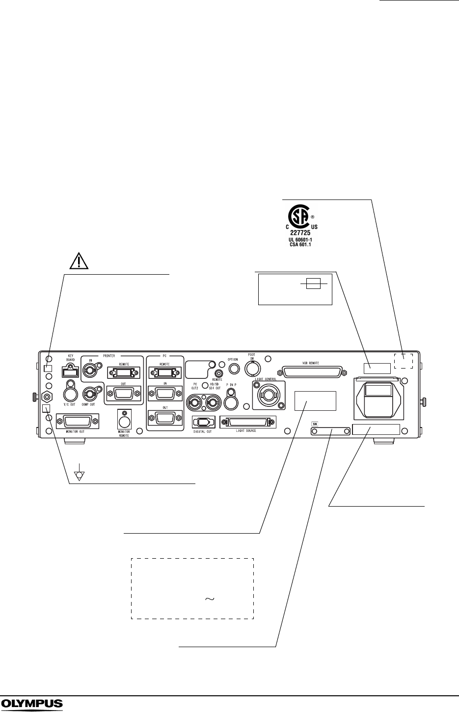
Safety-related labels and symbols are attached on the locations shown below. If
labels or symbols are missing or illegible, contact OLYMPUS.
Rear panel
Fuse rating
FUSES
T5AL250V
CSA/UL marking
Caution that only the
exclusive cable can be
connected.
Manufacturer name
Serial number plate
Electric rating
The product name, rated voltage
and frequency are shown.
EVIS EXERA II
VIDEO SYSTEM CENTER
MODEL OLYMPUS CV-180
INPUT 100-240V
50/60Hz 150VA
Potential equalization terminal
2
Labels and Symbols
EVIS EXERA II VIDEO SYSTEM CENTER CV-180
Back cover of this instruction manual
Manufacturer
Authorized representative in the European Community

Important Information — Please Read Before Use
3
EVIS EXERA II VIDEO SYSTEM CENTER CV-180
Important Information — Please Read
Before Use
Intended use
This video system center has been designed to be used with OLYMPUS camera
heads, endoscopes, light sources, monitors, endo-therapy accessories and
other ancillary equipment for endoscopic diagnosis, treatment and video
observation. Do not use this video system center for any purpose other than its
intended use.
Instruction manual
This instruction manual contains essential information on using this video
system center safely and effectively. Before use, thoroughly review this manual
and the manuals of all equipment which will be used during the procedure and
use the equipment as instructed.
Keep this and all related instruction manuals in a safe, accessible location. If you
have any questions or comments about any information in this manual, please
contact Olympus.
Terms used in this manual
Light source:
The light source provides light and electrical signals to the endoscope. It
also provides electrical signals to the video system center.
Video printer:
The video printer is a device that prints the frozen video image.
Wall mains outlet:
The wall mains outlet is a wall AC mains power outlet socket having the
exclusive terminal for grounding.
Isolation transformer:
The isolation transformer is a safety device that is used to isolate non-
insulated equipment with potentially high leakage currents to decrease the
possibility of electric shock.
Image sensor (CCD):
Image sensor (CCD) is a device that converts light into electrical signals.

4
Important Information — Please Read Before Use
EVIS EXERA II VIDEO SYSTEM CENTER CV-180
Automatic brightness control:
The automatic brightness control automatically adjusts the intensity of the
light emitted from the light source so that the endoscopic image will be
maintained at constant brightness even if the distance between the distal
end of the endoscope's insertion tube and the subject changes.
Color adjustment:
Color adjustment adjusts the color balance on the video monitor.
Iris:
The iris function is used to electrically measure the brightness of an
endoscopic image to obtain a control signal for the purpose of automatic
light adjustment.
Freeze:
The freeze function creates a stationary view of the moving image.
Release:
The release function is used to capture and record an endoscopic image.
Edge enhancement:
Edge enhancement is an image processing technique that electronically
sharpens the edges of an image.
Structure enhancement:
Structure enhancement is an image processing technique that
electronically emphasizes the detailed patterns and edges of an image to
increase sharpness.
PinP (Picture in picture):
PinP function displays both the image of the endoscopic live image and
the image of an external device on the monitor simultaneously.
PC card:
A digital medium for storage of images, etc.
Wash out:
Wash out is the inability to see details in the endoscopic image due to
excessive brightness.
HDTV:
High Definition Television. This is a format for high resolution video
transmission featuring higher definition than the standard SDTV format.

Important Information — Please Read Before Use
5
EVIS EXERA II VIDEO SYSTEM CENTER CV-180
User qualifications
If there is an official standard on user qualifications to perform endoscopy and
endoscopic treatment that is defined by the medical administration or other
official institutions, such as academic societies on endoscopy, follow that
standard. If there is no official qualification standard, the operator of this
instrument must be a physician approved by the medical safety manager of the
hospital or person in charge of the department (department of internal medicine,
etc.).
The physician should be capable of safely performing the planned endoscopy
and endoscopic treatment following guidelines set by the academic societies on
endoscopy, etc., and considering the difficulty of endoscopy and endoscopic
treatment. This manual does not explain or discuss endoscopic procedures.
Instrument compatibility
Refer to the “System chart” in the Appendix to confirm that this video system
center is compatible with the ancillary equipment being used. Using incompatible
equipment can result in patient injury or equipment damage and makes it
impossible to obtain the expected functionality.
This instrument complies with the EMC standard for medical electrical
equipment; edition 2 (IEC 60601-1-2: 2001). However, when connecting to an
instrument that complies with the EMC standard for medical electrical
equipment; edition 1 (IEC 60601-1-2: 1993), the whole system complies with
edition 1.
SDTV:
Standard Definition Television. It is the format used in standard video
systems.
Special light observation:
This is a observation using filtered light.
NBI (narrow band imaging):
This is one of the special light observations using the narrow band
observation light.
6
Important Information — Please Read Before Use
EVIS EXERA II VIDEO SYSTEM CENTER CV-180
Repair and modification
This video system center does not contain any user-serviceable parts. Do not
disassemble, modify or attempt to repair it; patient or operator injury, equipment
damage and/or the impossibility to obtain the expected functionality can result.
Some problems that appear to be malfunctions may be correctable by referring
to Chapter 10, “Troubleshooting”. If the problem cannot be resolved using the
information in Chapter 10, contact Olympus. This instrument is to be repaired by
Olympus technicians only.
Signal words
The following signal words are used throughout this manual:
Indicates an imminently hazardous situation which, if not
avoided, will result in death or serious injury.
Indicates a potentially hazardous situation which, if not
avoided, could result in death or serious injury.
Indicates a potentially hazardous situation which, if not
avoided, may result in minor or moderate injury. It may also
be used to alert against unsafe practices or potential
equipment damage.
Indicates additional helpful information.

Important Information — Please Read Before Use
7
EVIS EXERA II VIDEO SYSTEM CENTER CV-180
Dangers, warnings and cautions
Follow the dangers and cautions given below when handling this video system
center. This information is to be supplemented by the dangers and cautions
given in each chapter.
• Strictly observe the following precautions. Failure to do so
may place the patient and medical personnel in danger of
electric shock.
When this video system center is used to examine a
patient, do not allow metal parts of the endoscope or its
accessories to touch metal parts of other system
components. Such contact may cause unintended current
flow to the patient.
Keep fluids away from all electrical equipment. If fluids are
spilled on or into the unit, stop operation of the video
system center immediately and contact Olympus.
Do not prepare, inspect or use this video system center
with wet hands.
• Never install and operate the video system center in
locations where:
the concentration of oxygen is high;
oxidizing agents (such as nitrous oxide (N2O)) are present
in the atmosphere;
flammable gases are present in the atmosphere;
flammable liquids are near.
Otherwise, explosion or fire may result because this video
system center is not explosion-proof.

8
Important Information — Please Read Before Use
EVIS EXERA II VIDEO SYSTEM CENTER CV-180
• In case of instrument failure or malfunction, always keep
another video system center in the room ready for use.
• Never insert anything into the ventilation grills of the video
system center. It can cause an electric shock and/or fire.
• Although the illumination light emitted from the endoscope's
distal end is required for endoscopic observation and
treatment, it may also cause alteration of living tissues such
as protein denaturation of liver tissue and perforation of the
intestines by inappropriate use.
Observe the following warnings on the illumination.
Always set the minimum required brightness. The
brightness of the image on a video monitor may differ
from the actual brightness at the distal end of an
endoscope. Especially in combination with endoscopes
using an electrical shutter function, pay attention to the
brightness level setting of the light source. When this
instrument is used with a light source compatible with
automatic brightness control function, be sure to use this
function. The automatic brightness control function can
keep the illumination light properly. Refer to the instruction
manual of the light source for details.
Do not continue observation in the proximity to tissue or
keep the distal end of the endoscope in contact with living
tissue for a long time. It may cause patient burns.
When discontinuing the use of the endoscope, be sure to
turn the light source OFF so that the endoscope does not
irradiate unnecessary light.
• This product may interfere with other medical electronic
equipment used in combination with it. Before use, refer to
the Appendix to confirm the compatibility of this instrument
with all equipment to be used.
• Do not use this product in any place where it may be subject
to strong electromagnetic radiation (for example, in the
vicinity of a microwave therapeutic device, MRI, wireless set,
short-wave therapeutic device, cellular/portable phone, etc.).
This may impair the performance of the product.

Important Information — Please Read Before Use
9
EVIS EXERA II VIDEO SYSTEM CENTER CV-180
• If the endoscopic image dims during use, blood, mucus or
debris may adhere to the light guide on the distal end of the
endoscope. Carefully withdraw the endoscope from the
patient and remove the blood or mucus in order to obtain
optimum illumination and to ensure the safety of the
examination. If you continue to use the endoscope in such a
condition, the distal end temperature may rise and cause
mucosal burns. It may also cause patient and/or operator
injury.
• Do not rely on the special light observation method alone for
primary detection of lesions or for a decision regarding any
potential diagnostic or therapeutic intervention.
• For reasons described below, do not rely on the NBI imaging
modality alone for primary detection of lesions or to make a
decision regarding any potential diagnostic or therapeutic
intervention.
It has not been demonstrated to increase the yield or
sensitivity of finding any specific mucosal lesion including
colonic polyps or Barrett’s esophagus.
• To display observation images, connect the output terminal
of the video system center directly to the monitor. Do not
make the connection via any ancillary equipment. Images
may disappear during observation depending on the
condition of ancillary equipment.
• Do not use a pointed or hard object to press the buttons on
the front panel and/or keyboard. This may damage the
buttons.
• Do not touch the electrical contacts inside the video system
center's connectors.
• Do not apply excessive force to this video system center
and/or other instruments connected. Otherwise, damage
and/or malfunction can occur.
• Do not connect or disconnect the endoscope connector while
this video system center is turned ON. Connecting or
disconnecting the endoscope while this video system center
is ON may destroy the CCD. Turn the video system center
OFF before connecting or disconnecting the endoscope.
• Clean and vacuum dust the ventilation grills using a vacuum
cleaner, when necessary. Otherwise, the video system
center may break down and gets damaged from overheating.
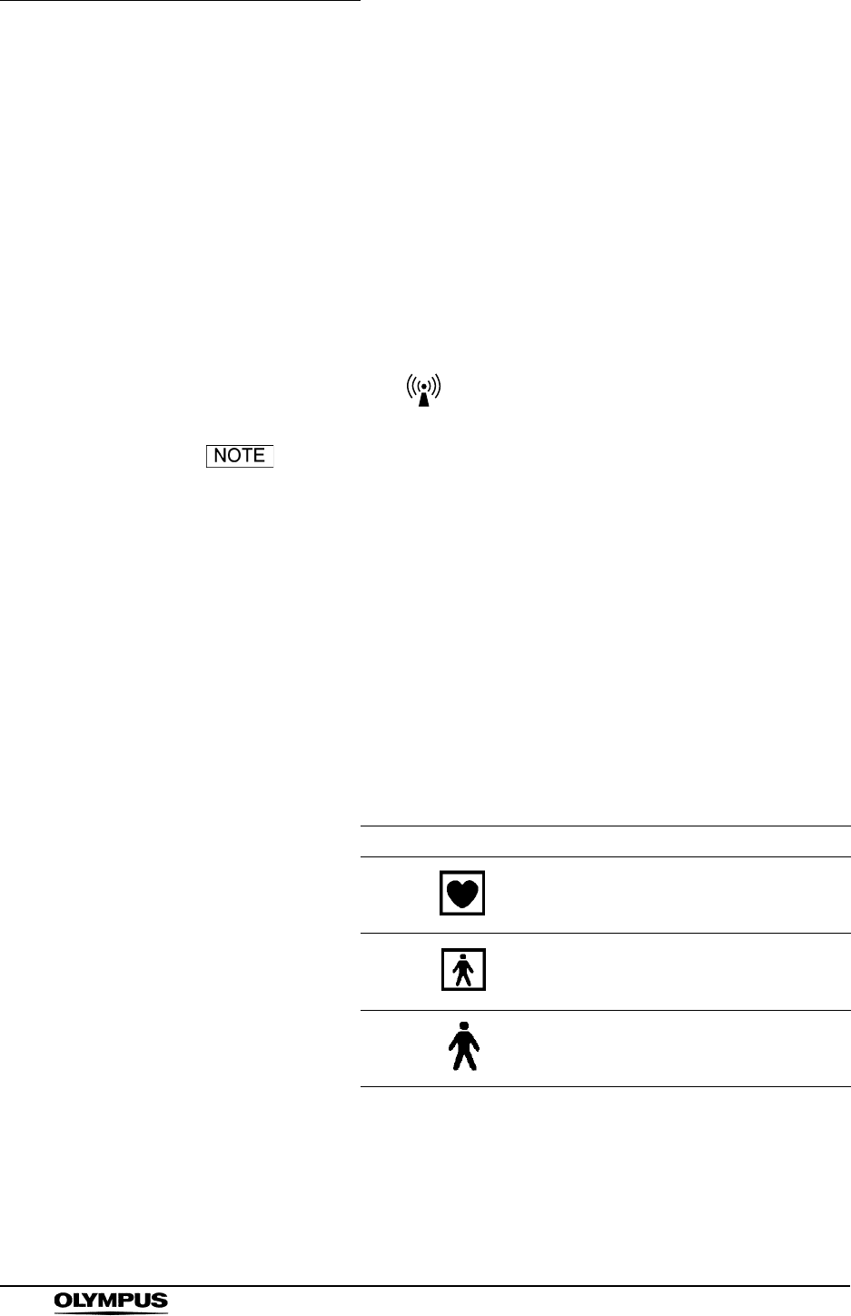
10
Important Information — Please Read Before Use
EVIS EXERA II VIDEO SYSTEM CENTER CV-180
• Be sure that this instrument is not used adjacent to or
stacked with other equipment (other than the components of
this instrument or system) to avoid electromagnetic
interference.
• Electromagnetic interference may occur to this instrument
when it is placed near equipment marked with the following
symbol or other portable and mobile RF communications
equipment such as cellular phones. If radio interference
occurs, mitigation measures may be necessary, such as
reorienting or relocating this instrument or shielding the
location.
As defined by the international safety standard (IEC 60601-
1), medical electrical equipment is classified into the
following types: TYPE CF applied part (the instrument can
safely be applied to any part of the body, including the heart),
and TYPE B/BF applied part (the instrument can safely be
applied to any organ except the heart). The part of the body
that an endoscope or electrosurgical accessory can safely be
applied to depends on the classification of the equipment to
which the instruments are connected. Before beginning the
procedure, check the current leakage classification type of
each instrument to be used for the procedure. Classification
types are clearly specified in the instruments' instruction
manuals.
Symbol Classification
TYPE CF applied part
TYPE BF applied part
TYPE B applied part
Important Information — Please Read Before Use
11
EVIS EXERA II VIDEO SYSTEM CENTER CV-180
Cardiac applications
• Use only the devices listed in the “System chart” in the
Appendix for endoscopic observation or treatment of the
heart or areas near the heart. Other combinations of
equipment may cause ventricular fibrillation or seriously
affect the cardiac function of the patient.
• For cardiac applications, never support the endoscope with a
metal surgical arm which is not electrically isolated from the
ground. If not isolated, the endoscope will be connected to
the ground through the surgical arm and bed, and will
conduct unexpected leakage current which may seriously
affect the cardiac function of the patient.
• The use of medical devices not specifically designed for
cardiac applications may cause ventricular fibrillation or
seriously affect the cardiac function of the patient. As
specified by the international standard IEC 60601-1, any
applied part used for observation or treatment of the heart or
areas near the heart must meet “TYPE CF applied part”
requirements for low electrical leakage current. When using
endoscopes for endoscopic cardiac applications, the applied
part requirements include all devices directly connected to
the endoscope, such as the light guide cable, camera head
and telescope holder. Each of these devices must
individually meet the “TYPE CF applied part” requirements
for leakage current limits if they are to be used for cardiac
applications.
The OLYMPUS light guide cables and camera heads listed in
the “System chart” in the Appendix (TYPE CF applied part)
which are suitable for cardiac applications bear a mark.

12
Summary of Equipment Functions
EVIS EXERA II VIDEO SYSTEM CENTER CV-180
Summary of Equipment Functions
This instrument is a system controller of the endoscopic image observation
system that displays, records and prints the endoscopic images. Some of the
functions of this instrument described below are enabled only when the required
equipment are connected to this instrument. For more details, refer to the
instruction manuals for this instrument and the other instruments connected.
Displaying the endoscopic images on the monitor
• The endoscopic live image and the other images of, for example, the
ultrasonic endoscope connected to this instrument can be displayed on
the monitor.
• The endoscopic image and other external images can be displayed on
the same monitor at the same time (PinP function).
“PinP (picture in picture) display” on page 64
• Either a standard-definition (SDTV) monitor or high-definition (HDTV)
monitor can be used.
Special light observation
Endoscopic observation using filtered light is available.
Section 5.8, “Special light observation” on page 151
Adjusting the endoscopic images
Images can be adjusted to enable clear and convenient observation.
• Adjustment of the image color
“Color tone adjustment (“COLOR”)” on page 98
• Adjustment of the image brightness
“Brightness adjustment (Exposure)” on page 71
• Changing the iris mode
“Iris mode” on page 69
• Changing the contrast mode
“Contrast mode (“Shift” + “F6”)” on page 90
• Enhancement of edge lines and patterns of the images
“Image enhancement mode (ENH.)” on page 67
• Changing the image size
“Image size (“F8”)” on page 94

Summary of Equipment Functions
13
EVIS EXERA II VIDEO SYSTEM CENTER CV-180
• Enlargement of the images
“Image zooming (“F7”)” on page 91
Entering patient data
• The patient data such as name, sex, etc. can be entered and displayed
on the monitor with the endoscopic live image.
Section 4.6, “Patient data” on page 57 and Section 5.6, “Pre-entry of
patient data” on page 136.)
• Up to 40 sets of patients data can be stored on the PC card. These
patient data can be copied to the other CV-180.
“Recording patient data into PC card” on page 144
Customizing the operations
Up to 20 remote switch settings and other functions such as iris mode, image
enhancement, etc., can be stored.
Section 9.3, “User preset” on page 216
Recording images
• The endoscopic image can be recorded on the PC card.
Section 5.3, “Image recording and playback (PC card)” on page 106
• The endoscopic image can be recorded on the image-recording device
connected to this instrument, and the recorded images can be played
back.
Section 5.4, “Image recording and playback (other than PC card)” on
page 127
• The endoscopic image can be printed from the printer connected to this
instrument.
Section 5.5, “Printing images” on page 131
Operation of ancillary equipment
• Video casette recorder
“Videocassette recorder (VCR)” on page 129
• Video printer
Section 5.5, “Printing images” on page 131
• Image filing system
“Image filing system” on page 127
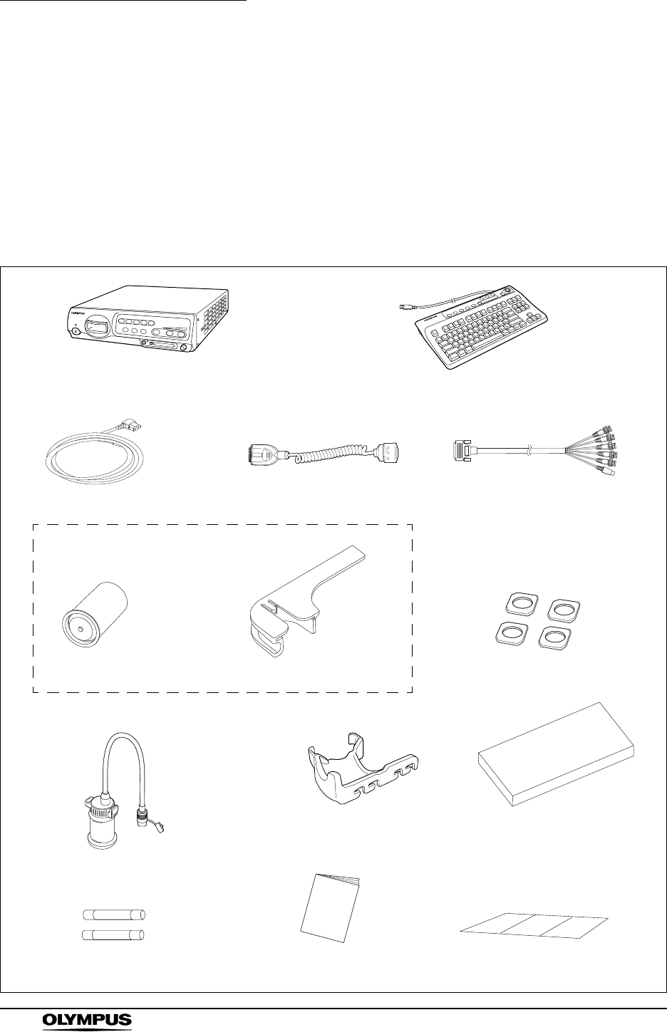
14
Chapter 1 Checking the Package Contents
EVIS EXERA II VIDEO SYSTEM CENTER CV-180
Chapter 1 Checking the Package
Contents
Match all items in the package with the components shown below. Inspect each
item for damage. If the instrument is damaged, a component is missing or you
have any questions, do not use the instrument; immediately contact Olympus.
White cap holder (MAJ-960)
White cap (MH-155)
Video system center (CV-180) Keyboard (MAJ-1428)
Videoscope cable EXERA II
(MAJ-1430)
Power cord HDTV/SDTV monitor cable
(MAJ-1462)
Foot holder (MAJ-1433, 4 pcs.)
Spare fuse (MAJ-1432, 2 pcs.) Cable color sheet
Scope cable holder (MAJ-1466)
Instruction manual
Water container (MAJ-901)
Keyboard cover
(MAJ-1557)
White cap set (MAJ-941)
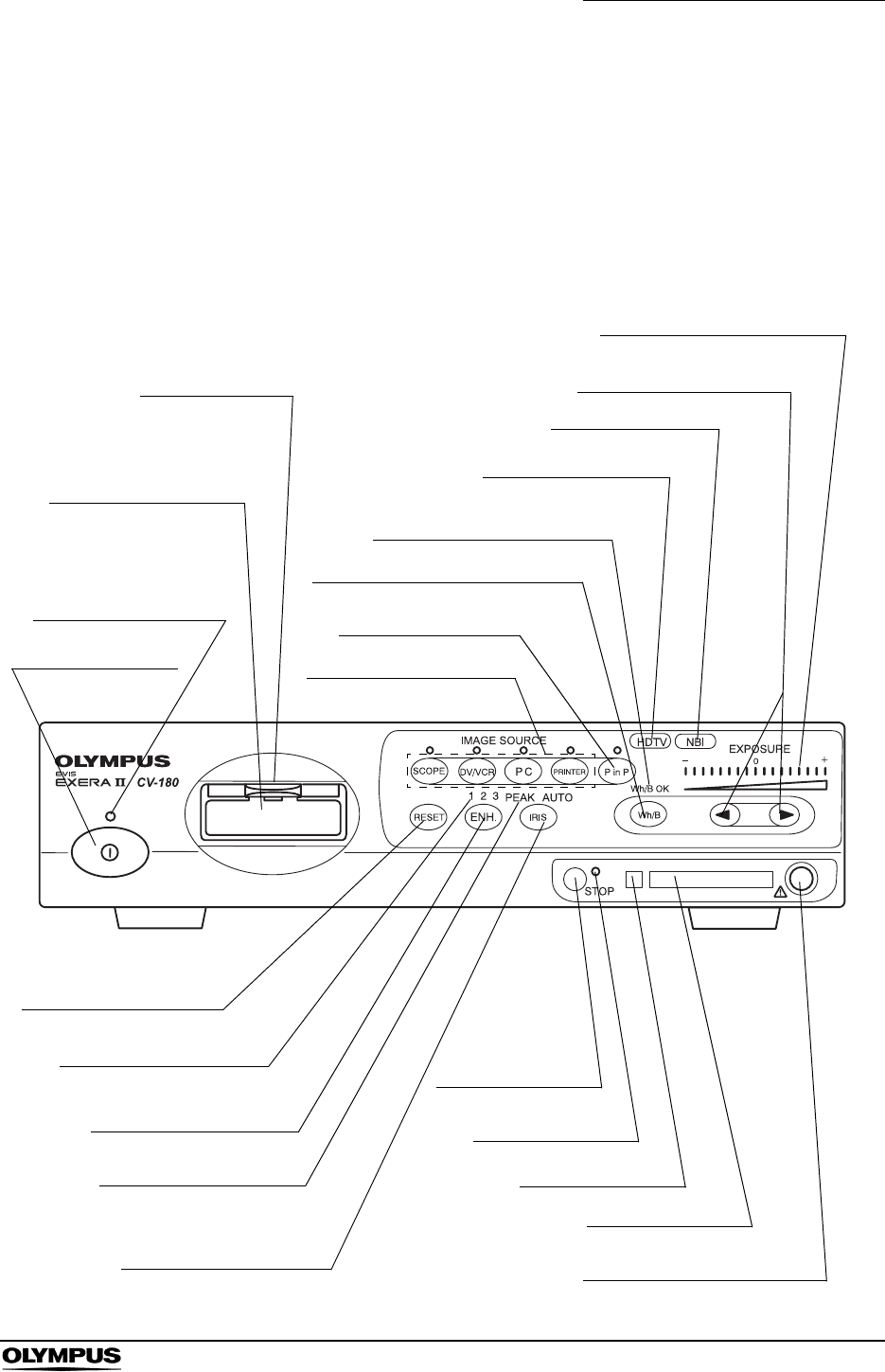
Chapter 2 Nomenclature and Functions
15
EVIS EXERA II VIDEO SYSTEM CENTER CV-180
Chapter 2 Nomenclature and Functions
2.1 Front panel
4. Locking lever
3. Video connector
socket
2. Power indicator
1. Power switch 5. Image source buttons
7. White balance (Wh/B) button
9. HDTV indicator
12. Exposure level
(EXPOSURE) indicator
11. Exposure adjustment
(EXPOSURE) buttons
10. NBI indicator
22. RESET button
21. Image enhancement
mode indicators
20. Image enhancement
mode (ENH.) button
19. Iris mode indicators
18. Iris mode (IRIS)
button
17. STOP button
16. PC card status
indicator
15. Eject button
14. PC card slot
13. PinP composite terminal
6. Picture in picture
(PinP) button
8. White balance (Wh/B OK)
indicator

16
Chapter 2 Nomenclature and Functions
EVIS EXERA II VIDEO SYSTEM CENTER CV-180
1. Power switch
Press to turn the video system center ON or OFF.
2. Power indicator
Lights up when the video system center is ON.
3. Video connector socket
The video plug of the videoscope cable, videoscope or camera head are
connected to this socket.
4. Locking lever
Press down to disconnect the video plug of the videoscope cable,
videoscope or camera head.
5. Image source buttons
Press these buttons to select the image sources to be displayed on the
monitor. Press and hold the buttons to change (except “SCOPE”).
“Image source buttons” on page 62
6. Picture in picture (PinP) button
Press to display an image of the connected ancillary equipment and the
endoscopic live image together on the monitor.
• Setting of the PinP function
“PinP (picture in picture) function” on page 246
• Operation of the PinP function
“PinP (picture in picture) display” on page 64
7. White balance (Wh/B) button
Press to perform the white balance adjustment.
Section 4.5, “White balance adjustment” on page 52
8. White balance (Wh/B OK) indicator
The indicator lights up when the white balance adjustment is completed.
9. HDTV indicator
Lights up green when the instrument is turned ON, and turns white when the
HDTV compatible endoscope is connected to this instrument.
10. NBI indicator
Lights up green when the NBI compatible endoscope is connected to this
instrument, and turns white during NBI observation. This indicator works
only when the light source CLV-180 is used.
“NBI (narrow band imaging)” on page 151
Button The image on the monitor
SCOPE The endoscopic live image
DV/VCR The image of the videocassette recorder, etc.
PC The image of the image filing system
PRINTER The image of the video printer

Chapter 2 Nomenclature and Functions
17
EVIS EXERA II VIDEO SYSTEM CENTER CV-180
11. Exposure adjustment (EXPOSURE) buttons
Press to adjust the brightness of the observation light. When CLV-180 is
used, this button is interlocked with the “BRIGHTNESS” on CLV-180.
“Brightness adjustment (Exposure)” on page 71
12. Exposure level (EXPOSURE) indicator
Indicates the brightness level of the observation light.
“Brightness adjustment (Exposure)” on page 71
13. PinP composite terminal
The ultrasound center (EUS), endoscope position detecting unit (UPD) etc.
can be connected to this connector to input the images to be displayed
together with the endoscopic observation image. The PinP function can also
be used with the PinP Y/C terminal on the rear panel. However, the PinP
composite terminal takes priority over the PinP Y/C terminal.
14. PC card slot
Insert the PC card adapter (optional) in this slot. The xD picture card can be
used as the storage media.
“PC card slot and eject button” on page 76
15. Eject button
Press to remove the PC card from the PC card slot.
“PC card slot and eject button” on page 76
16. PC card status indicator
This indicator lights up green when the PC card is inserted into the PC card
slot, and blinks orange while accessing the PC card.
“PC card slot and eject button” on page 76
17. STOP button
Press to stop accessing the PC card. Be sure to press this button before
removing the PC card from the PC card slot.
“PC card slot and eject button” on page 76
18. Iris mode (IRIS) button
Press to switch the iris mode (brightness adjustment method) of the
endoscopic image. Either “AUTO” or “PEAK” mode can be selected.
•Presetting
“Iris” on page 232
• Switching operation
“Iris mode” on page 69
19. Iris mode indicators
Indicates the iris mode being selected.

18
Chapter 2 Nomenclature and Functions
EVIS EXERA II VIDEO SYSTEM CENTER CV-180
20. Image enhancement mode (ENH.) button
“Image enhancement” refers to facilitate observation of edges and patterns
of the endoscopic image by electronic treatment. Press this button to
change the modes of the enhancement methods.
• Presetting
“Image enhancement (normal observation)” on page 226
or “Image enhancement (NBI observation)” on page 250
• Operation
“Image enhancement mode (ENH.)” on page 67
21. Image enhancement mode indicators
One of these indicators light up and indicates the image enhancement mode
being selected. The indicator goes off when the image enhancement is not
used.
22. RESET button
Press and hold the switch to return the settings changed during operation to
the default settings.
“RESET button” on page 80
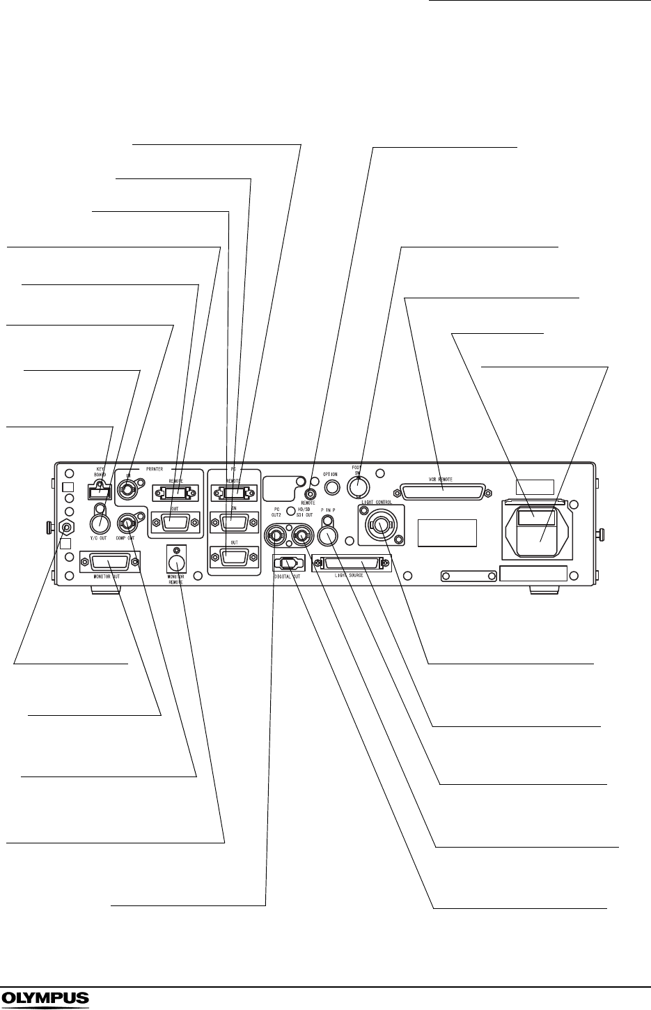
Chapter 2 Nomenclature and Functions
19
EVIS EXERA II VIDEO SYSTEM CENTER CV-180
2.2 Rear panel
8. PC remote terminal
12. Fuse box
10. Foot switch terminal
9. Remote terminal
13. AC power inlet
7. PC IN terminal
6. PC OUT terminal
5. Printer remote terminal
4. Printer OUT terminal
3. Printer IN terminal
2. Y/C OUT
terminal
1. Keyboard
terminal
23. Potential
equalization
terminal
22. Monitor OUT
terminal
21. Composite OUT
terminal
20. Monitor remote terminal
19. PC OUT2 terminal
14. Light control terminal
15. Light source terminal
16. PinP Y/C terminal
17. HD/SD SDI OUT terminal
18. Digital OUT terminal
11. VCR remote terminal

20
Chapter 2 Nomenclature and Functions
EVIS EXERA II VIDEO SYSTEM CENTER CV-180
1. Keyboard terminal
Connect the keyboard.
2. Y/C OUT terminal
Outputs a Y/C video signals.
3. Printer IN terminal
Connect the video printer. Inputs the analog video signal from the video
printer.
4. Printer OUT terminal
Connect the video printer. Outputs the analog video signal to the video
printer.
5. Printer remote terminal
Connect the video printer. Establishes communication with the video printer.
6. PC OUT terminal
Connect the image filing system. Outputs the analog video signal to the
image filing system.
7. PC IN terminal
Connect the image filing system. Inputs the analog video signal from the
image filing system.
8. PC remote terminal
Connect the image filing system. Establishes communication with the image
filing system.
9. Remote terminal
Outputs the signal synchronizing the release and VCR (Rec/Pause)
operation.
10. Foot switch terminal
Connect the foot switch.
11. VCR remote terminal
Connect an Olympus-recommended VCR. Outputs the analog video signal
and the remote signals to the VCR.
12. Fuse box
Stores the fuses that protect the instrument from electrical surges.
13. AC power inlet
Connect the provided power cord to supply the AC power via this inlet.
14. Light control terminal
Connect a light source that supports the analog interface.
15. Light source terminal
Connect a light source CLV-180 that supports the digital interface.

Chapter 2 Nomenclature and Functions
21
EVIS EXERA II VIDEO SYSTEM CENTER CV-180
16. PinP Y/C terminal
The ultrasound center (EUS), endoscope position detecting unit (UPD) etc.
can be connected to this connector to input the image to be displayed
together with the endoscopic observation image. The PinP function can also
be used with the PinP composite terminal on the front panel. However, the
PinP composite terminal takes priority over the PinP Y/C terminal.
17. HD/SD SDI OUT terminal
Connect a monitor compatible with the serial digital interface (SDI). Outputs
the SDI signal.
18. Digital OUT terminal
Connect an Olympus-recommended digital video recorder to output and
input the digital video signal to the digital video recorder, using IEEE1394
cable.
19. PC OUT2 terminal
Connect the image filing system. Outputs an SDI signal to the image filing
system.
20. Monitor remote terminal
Connect the monitor. Outputs the monitor control signal to the monitor.
21. Composite OUT terminal
Outputs the composite video signal.
22. Monitor OUT terminal
Connect the monitor. Outputs analog video signals to the monitor. HDTV
signal is output when the HDTV compatible endoscope is connected. This
connector can output a 180 rotated image (see “Monitor orientation
function” on page 245).
23. Potential equalization terminal
This terminal is connected to a potential equalization terminal of the other
equipment connected to this instrument. The electric potential of their
equipment are made equal.
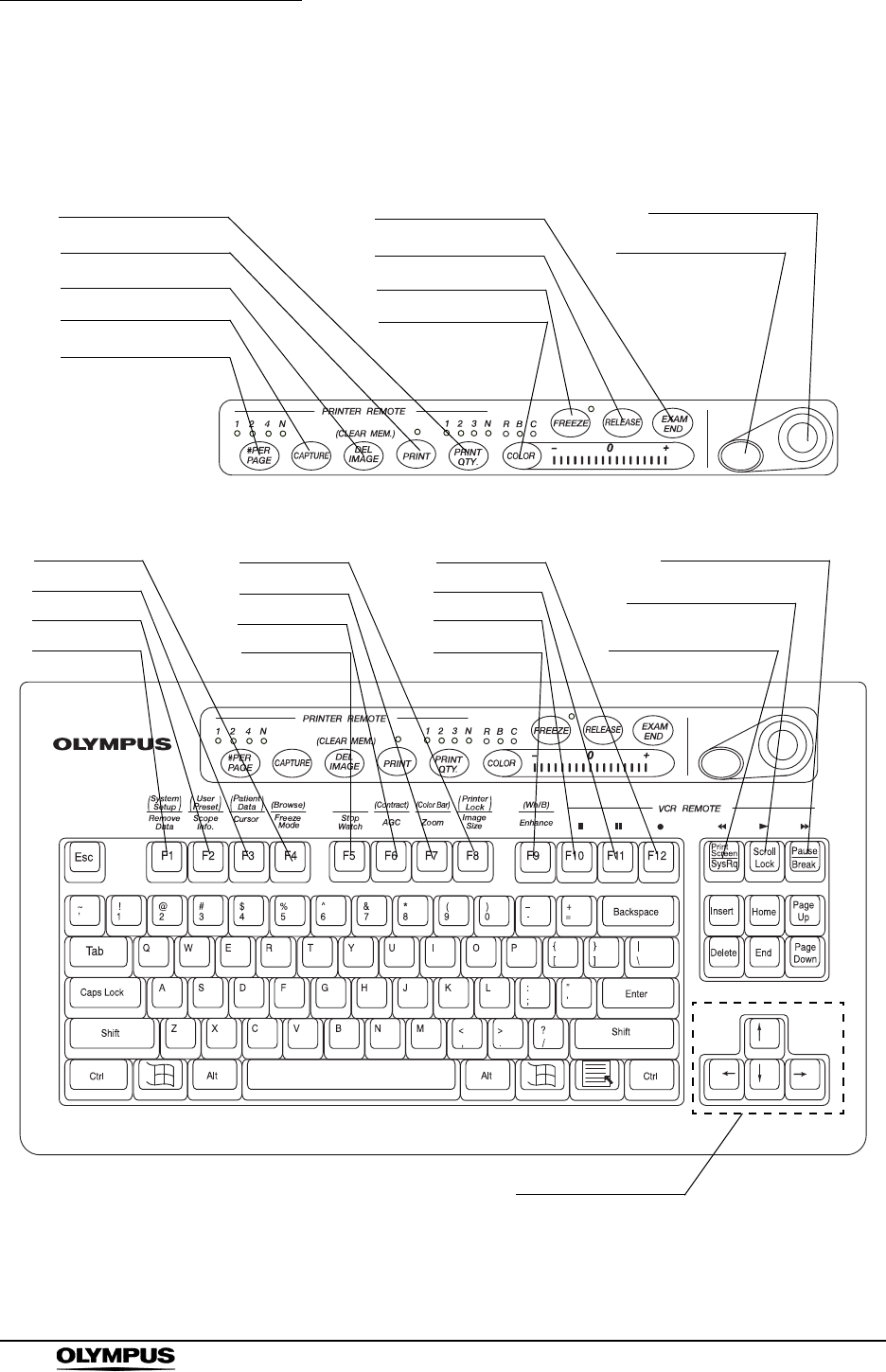
22
Chapter 2 Nomenclature and Functions
EVIS EXERA II VIDEO SYSTEM CENTER CV-180
2.3 Keyboard
20. PRINT QTY. key
19. PRINT key
18. DEL IMAGE key
17. CAPTURE key
24. EXAM END key
23. RELEASE key
22. FREEZE key
21. COLOR key
26. Domepoint
25. Click key
4. F4 key
3. F3 key
2. F2 key
1. F1 key
8. F8 key
7. F7 key
6. F6 key
5. F5 key
12. F12 key
11. F11 key
10. F10 key
9. F9 key
16. #PER PAGE key
15. Pause key
14. Scroll lock key
13. Print screen key
27. Arrow keys

Chapter 2 Nomenclature and Functions
23
EVIS EXERA II VIDEO SYSTEM CENTER CV-180
1. F1 key
Press to clear or re-display the patient data on the monitor step by step.
“Clearing characters from the screen (“F1”)” on page 82
Press together with the “Shift” key to display the system setup menu to set
the basic functions of this instrument.
“System setup (“Shift” + “F1”)” on page 84
2. F2 key
Press to display the scope information window when using an endoscope
with the endoscope information memory function. While the window is still
open, press this key again to display the scope information menu that shows
the information about the connected endoscope.
“Scope information (“F2”)” on page 84
Press together with the “Shift” key to display the user preset menu to set
and call up the observation condition of the endoscopic image.
See “User preset (“Shift” + “F2”)” on page 85
3. F3 key
Press to switch the display of the cursor on screen ON and OFF.
“Cursor (“F3”)” on page 85
Press together with the “Shift” key to display the patient data menu to enter
or call up the patient data onto the monitor.
“Patient data (“Shift” + F3)” on page 86
4. F4 key
Press to change the freeze mode.
“Freeze mode (“F4”)” on page 86
Press together with the “Shift” key to display the PC card menu to store or
call up the image data or the patient data from the PC card.
“Browse (“Shift” + “F4”)” on page 87
5. F5 key
Press to change the clock on the monitor to a stopwatch, and to start time
counting.
“Stopwatch (“F5”)” on page 88
6. F6 key
Press to switch the auto gain control (AGC) function ON and OFF.
“Automatic gain control (AGC) (“F6”)” on page 89
Press together with the “Shift” key to switch between the three steps of the
observation image contrast.
“Contrast mode (“Shift” + “F6”)” on page 90
7. F7 key
Press to change the zoom ratio of the observation image in three steps (x1;
x1.2; x1.5).
“Image zooming (“F7”)” on page 91
Press together with the “Shift” key to display the color bar for checking the
color of the monitor.
“Color bar (“Shift” + “F7”)” on page 93

24
Chapter 2 Nomenclature and Functions
EVIS EXERA II VIDEO SYSTEM CENTER CV-180
8. F8 key
Press to change the image area size on the monitor.
“Image size (“F8”)” on page 94
Press together with the “Shift” key to disable the functions of the following 5
keys for video printer operation: #PER Page, CAPTURE, DEL IMAGE,
PRINT, PRINT QTY.
“Printer lock (“Shift” + “F8”)” on page 95
9. F9 key
Press to switch the image enhancement mode.
“Image enhancement (“F9”)” on page 96
Press together with the “Shift” key to perform the white balance adjustment.
“White balance adjustment (“Shift” + “F9”)” on page 97
10. F10 key
Press to stop ( ) the VCR.
“Videocassette recorder (VCR)” on page 129
11. F11 key
Press to pause ( ) the VCR. Press the Scroll ( ) key to resume play.
“Videocassette recorder (VCR)” on page 129
12. F12 key
Press to start ( ) VCR recording.
“Videocassette recorder (VCR)” on page 129
13. Print screen key
Press to fast-rewind ( ) the VCR.
“Videocassette recorder (VCR)” on page 129
14. Scroll lock key
Press to playback ( ) the VCR.
“Videocassette recorder (VCR)” on page 129
15. Pause key
Press to fast-forward ( ) the VCR.
“Videocassette recorder (VCR)” on page 129
16. #PER PAGE key
Press to set the number of images per video print sheet. The indicator
corresponding to the number lights up. An arbitrary number “N” on the
keyboard depends on the printer.
Section5.5, “Printing images” on page 131
17. CAPTURE key
Press to capture the image in the video printer.
Section5.5, “Printing images” on page 131

Chapter 2 Nomenclature and Functions
25
EVIS EXERA II VIDEO SYSTEM CENTER CV-180
18. DEL IMAGE key
Press to set the printer cursor back on the print sheet by one. Press together
with the “Shift” key to delete a captured image at the position of the cursor.
Section5.5, “Printing images” on page 131
19. PRINT key
Press to print the images captured in the video printer.
Section5.5, “Printing images” on page 131
20. PRINT QTY. key
Press to specify the number of video print sheets to print simultaneously.
The indicator corresponding to the number lights up. An arbitrary number
“N” on the keyboard can be specified in the system setup menu.
Section5.5, “Printing images” on page 131
21. COLOR key
Press to select R (Red), B (Blue) or C (Chroma) to adjust the color of the
endoscopic image. The lamp corresponding to the selected tone above the
key lights up. Adjust the selected color tone using the “right” or “left” arrow
keys. The indicator on the right side of the “COLOR” key shows the
adjustment status.
“Color tone adjustment (“COLOR”)” on page 98
22. FREEZE key
Press to freeze the live endoscopic image. Press the key again to return to
the live image.
“Freeze (“FREEZE”)” on page 99
23. RELEASE key
Press to record the image into the video printer, image filing system and PC
card. The recording devices to operate should be set in advance.
“Release (“RELEASE”)” on page 101
24. EXAM END key
Press to execute the examination end processing.
“Ending examination (“EXAM END”)” on page 105
25. Click key
Press this key to enter an item after selecting the item using the domepoint.
“Domepoint” on page 81
26. Domepoint
Moves the arrow pointer. Selects an item in the menu or puts a marking in
the endoscopic image.
“Domepoint” on page 81

26
Chapter 2 Nomenclature and Functions
EVIS EXERA II VIDEO SYSTEM CENTER CV-180
27. Arrow keys
Moves the cursor.
Press one of these keys together with the “Shift” key to display the arrow
pointer on the endoscopic image.
“Arrow pointer (“Shift” + arrow keys and domepoint)” on page 102
28. Other keyboard keys
•Esc
Cancels the selection or returns to the previous screen.
•Tab
Goes to the next input area, or returns to the previous input area.
• Enter
Fixes entry and goes to the next text box or screen.
• Shift, Alt
Executes functions together with other keys.
• Back space
Clears the character left of the cursor.
•Delete
Clears the character right of the cursor.
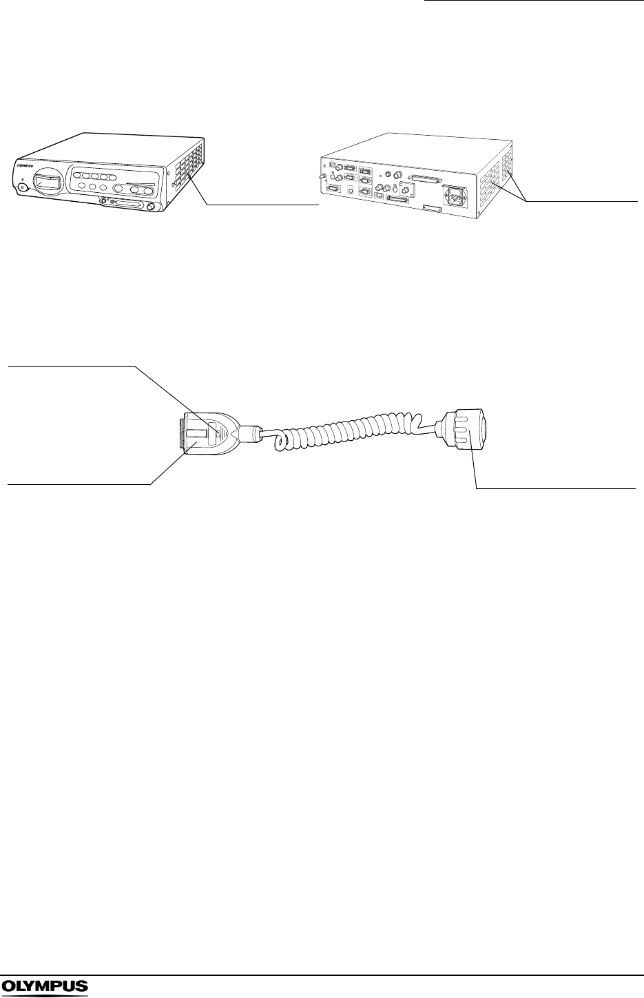
Chapter 2 Nomenclature and Functions
27
EVIS EXERA II VIDEO SYSTEM CENTER CV-180
2.4 Side panels
2.5 Videoscope cable EXERA II (MAJ-1430)
Front side Rear side
Ventilation grills Ventilation grills
Scope side connector
Connect to the scope
connector of the endoscope.
“UP” mark
Video plug
Connect to the video
system center “UP”
side up.
Connect to the video
connector socket of the
video system center.

28
Chapter 2 Nomenclature and Functions
EVIS EXERA II VIDEO SYSTEM CENTER CV-180
2.6 Set-up of screen options
The software of this instrument has the following functions.
• The screen of the endoscopic live image and the images of the external
instruments connected to this instrument
This is the basic screen of the instrument. This instrument starts the
endoscopic live image when it is turned ON.
Section 3.4, “Inspection of the monitor display” on page 38
• System setup screen
This screen is used for setting the basic functions to operate this instrument
and the other instruments connected to it correctly.
Section 9.2, “System setup” on page 194
• User preset screen
Up to 20 user presets are available to save individual user settings. The
factory default settings are set before shipment.
Section 9.3, “User preset” on page 216
• Patient data screen
Patient name, sex, age, etc. can be entered for up to 40 patients in advance.
Existing patient data can be accessed and displayed on the monitor together
with the endoscopic image, and can be stored on the PC card.
Section 5.6, “Pre-entry of patient data” on page 136
• PC card menu screen
This screen is used to browse endoscopic images on the PC card.
“PC card menu” on page 110
• Scope information screen
This screen is used to display and/or enter endoscope information such as
the type of endoscope, etc.
Section 5.7, “Scope information” on page 148
• Color bar screen
This screen is used to check the display color.
“Color bar (“Shift” + “F7”)” on page 93
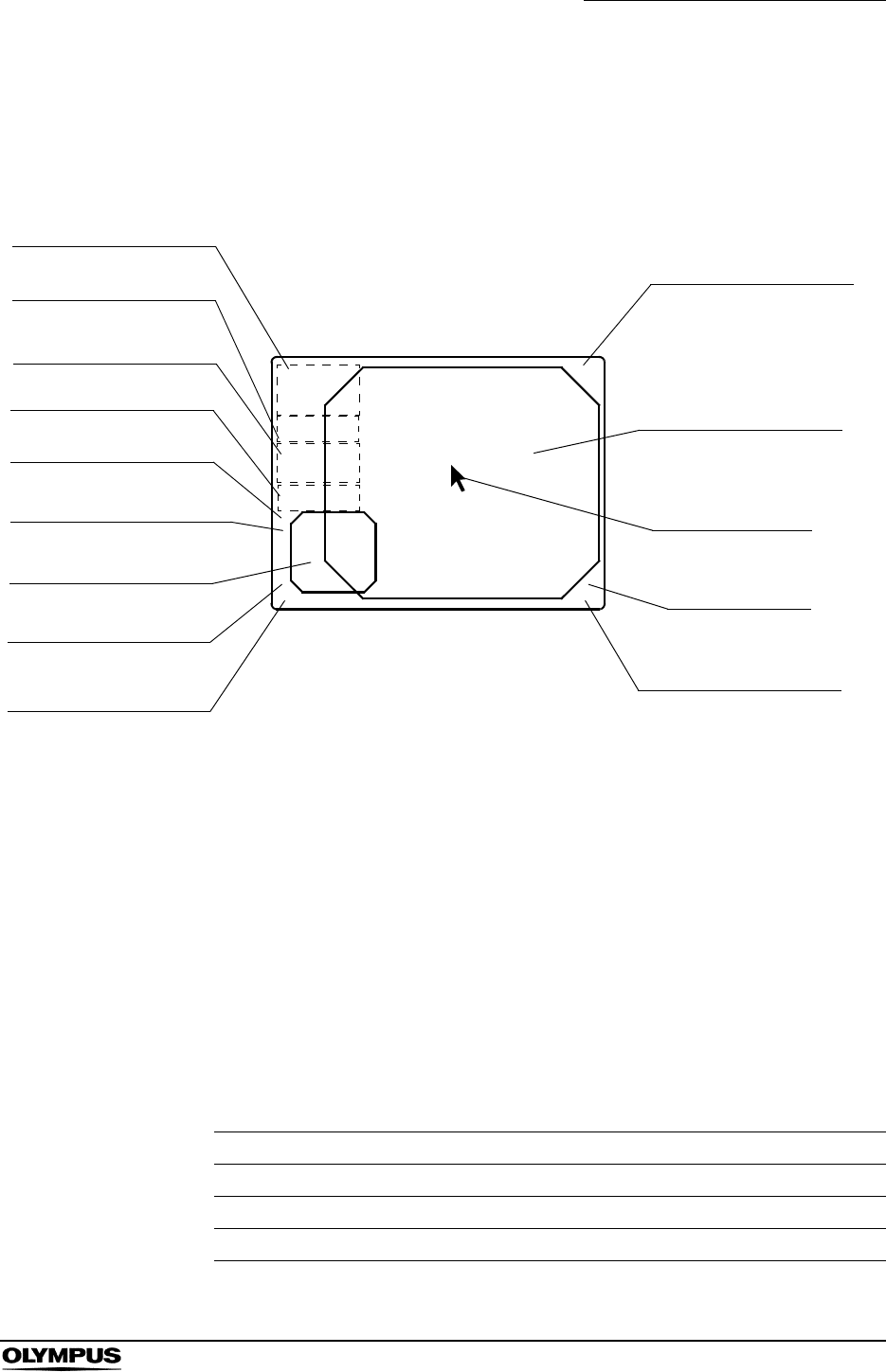
Chapter 2 Nomenclature and Functions
29
EVIS EXERA II VIDEO SYSTEM CENTER CV-180
2.7 Monitor
Endoscopic image display
1. Patient data
Patient data such as name sex, etc. can be entered and displayed in this
area.
Section 4.6, “Patient data” on page 57
2. System clock
Date and time are displayed. The date format can be set.
“Date and time” on page 197
The clock has the stopwatch function.
“Stopwatch (“F5”)” on page 88
3. Image recording device display
The status of the image recording devices that record and print the image
are displayed only when the recording devices are activated.
Indication Device Details
CVP Video printer page 131
D.F Digital filing system page 127
VCR Videocassette recorder page 129
1. Patient data
2. System clock
3. Image recording
device display
4. Image information
5. Flushing pump
6. PC card capacity
7. Index image
8. Attending physician
10. Special light
observation display
11. Endoscopic image
12. Arrow pointer
14. Scope nickname
9. Comments
13. Orientation
ABC123
Mike Johnson
M 51
03/03/1954
12/12/2005
12:12:12
CVP: A4/4
D.F: 99
VCR
Ct: N Eh: A8
Z: x1.5
Pump
Media:
John Smith
Cardiac end of the stomach R
V
NBI

30
Chapter 2 Nomenclature and Functions
EVIS EXERA II VIDEO SYSTEM CENTER CV-180
4. Image information
Displays image information on the monitor. The indications are displayed
only when these function are operated.
5. Flushing pump
Displayed only when the Olympus flushing pump (OFP) is processing.
6. PC card capacity
Indicates the remaining memory level of the PC card when the PC card is
inserted in the PC card slot.
“Storage level of the PC card” on page 106
7. Index image
Displays the reference image of the image taken by “RELEASE”.
“Release index time” on page 243
8. Attending physician
The physician’s name can be entered and displayed together with the
patient data.
9. Comments
Comments can be entered and displayed together with the patient data.
10. Special light observation display
Indicates the name of the special observation function during the
observation.
Section 5.8, “Special light observation” on page 151
11. Endoscopic image
The live endoscopic image is displayed in this area. The size and shape of
the image depends on the type of endoscope used.
“Image size” on page 229
12. Arrow pointer
The arrow pointer is used for pointing out a part of the endoscopic image
and for entering data in the menus.
• Displaying
“Arrow pointer (“Shift” + arrow keys and domepoint)” on
page 102
• Operation
“Domepoint” on page 81
13. Orientation
“R” mark appears when a 180 reverse image is displayed.
“Monitor orientation function” on page 245
Indication Meaning Details
Ct Contrast page 90
Eh Enhancement mode page 67
Z Zoom ratio page 91
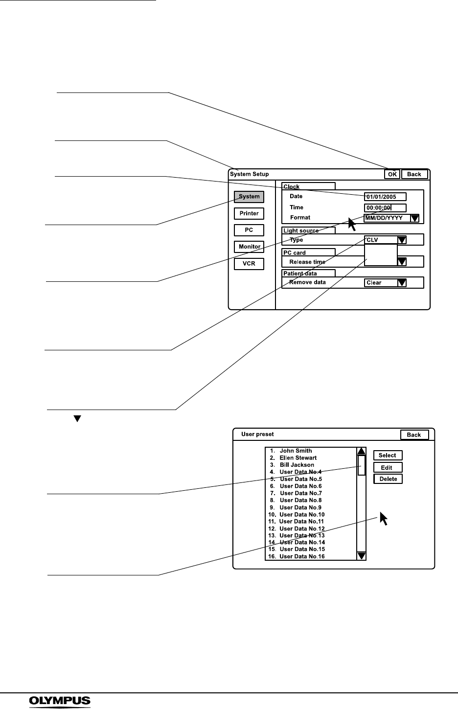
32
Chapter 2 Nomenclature and Functions
EVIS EXERA II VIDEO SYSTEM CENTER CV-180
Data input menu
CLV
CLV-S
CLE
Function button
Menu name
Text box
Cursor
Arrow pointer
List box
Pull down menu
Scroll bar
Highlight
Saves data, terminates the
menu, etc.
For entering and displaying
data.
Used for entering data.
Moves the cursor, selects the
function buttons, displays the
pull down menus.
Opens the pull down menu
and selects the setting values.
Click “ ” to show the setting
values in the pull down menu.
Shows all setting values not
being displayed in the list box.
Indicates in blue that the
function button is selected.
Chapter 2 Nomenclature and Functions
33
EVIS EXERA II VIDEO SYSTEM CENTER CV-180
2.8 Pointer
Highlight, cursor and arrow pointer are available as pointing devices for the
endoscopic image and menus on the monitor.
Pointer Function Screen Displaying and operation
Highlight Indicates the button
selected.
• System setup menu
• User preset menu
• Patient data menu
• Scope information menu
• PC card information
menu
Always displayed.
Movable by pressing the arrow, “Tab”,
or “Shift” + “Tab” keys.
Cursor Indicates the position to
enter data.
• System setup menu
• User preset menu
• Patient data menu
• Scope information menu
• PC card information
menu
• Endoscopic image
screen
Always displayed.
Movable by pressing the arrow,
“Home”, or “End” keys.
Arrow pointer Moves the cursor and
focus or points out a
specific portion of the
image.
• System setup menu
• User preset menu
• Patient data menu
• Scope information menu
Always displayed.
Movable by the domepoint.
• PC card menu Always displayed on image screen
(unless full image screen selected).
Press “Shift” and any arrow key in the
full image screen to display/remove the
arrow pointer.
• Endoscopic image
screen
Press “Shift” and any arrow key.

34
Chapter 3 Inspection
EVIS EXERA II VIDEO SYSTEM CENTER CV-180
Chapter 3 Inspection
• Review Chapter 8, “Installation and Connection” thoroughly,
and prepare the instruments properly before inspection. If the
equipment is not properly prepared before each use,
equipment damage, patient and operator injury and/or fire
can occur.
• Before each procedure, inspect the video system center as
instructed below. Inspect other equipment to be used with
this video system center as instructed in their respective
instruction manuals. Should any irregularity be observed, do
not use the video system center and see Chapter 10,
“Troubleshooting”. If the irregularity is still observed after
consulting Chapter 10, contact Olympus. Damage or
irregularity may compromise patient or user safety and may
result in more severe equipment damage.
Prepare the video system center and other ancillary equipment before each
particular case. Refer to the respective instruction manual for each piece of
equipment.
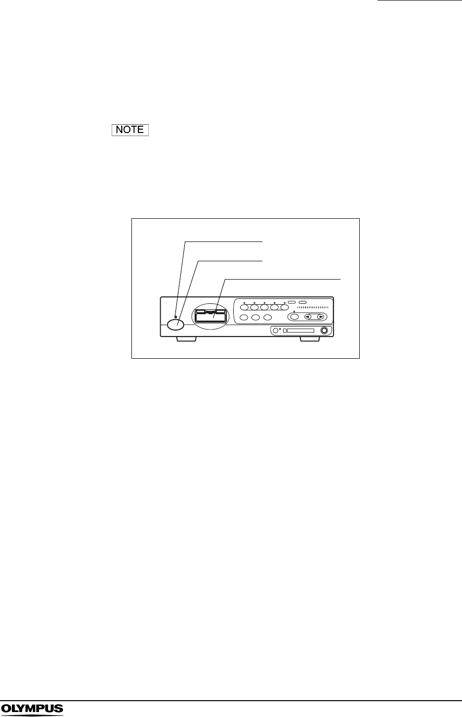
Chapter 3 Inspection
35
EVIS EXERA II VIDEO SYSTEM CENTER CV-180
3.1 Inspection of the power supply
1. Confirm that the videoscope cable, camera head and/or endoscope is
connected to the videoscope cable socket of the instrument.
For the connection of the endoscope or scope cable, refer to
Section 4.2, “Connection of an endoscope” on page 46.
2. Press the power switch of the instrument (see Figure 3.1). The indicator
lamp above the power switch lights up.
Figure 3.1
If the power fails to come ON
When the power fails to come ON, turn the video system center OFF. Then
check the video system center referring to Chapter 10, “Troubleshooting”. If the
power still fails to come ON, contact Olympus.
Video connector socket
Power indicator
Power switch
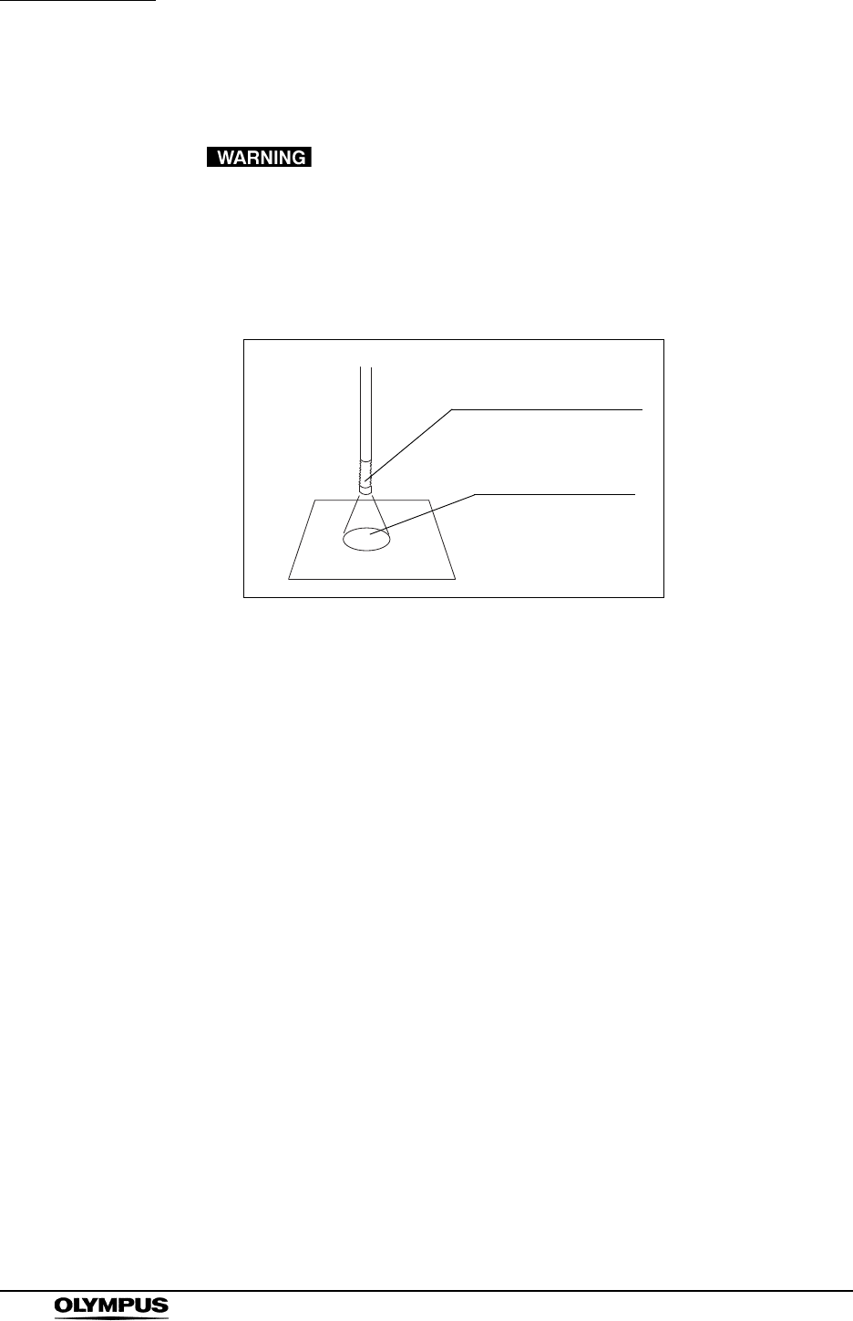
36
Chapter 3 Inspection
EVIS EXERA II VIDEO SYSTEM CENTER CV-180
3.2 Inspection of the examination light
Do not stare directly into the light beam. This may result in
eye injury.
Turn ON the light source and confirm that examination light is emitted from the
distal end of the endoscope (see Figure 3.2). For operation of the light source,
refer to its instruction manual.
Figure 3.2
Examination light
Endoscope’s distal end

Chapter 3 Inspection
37
EVIS EXERA II VIDEO SYSTEM CENTER CV-180
3.3 Inspection of the automatic brightness control
function
1. Confirm that this instrument is connected to the light source using the light
source cable or light control cable (see Section 8.4, “Light source” on
page 164).
2. According to the directions given in the light source's instruction manual,
confirm that the light source's brightness control is set to “AUTO” and that
the brightness level is in the center of the adjustable range.
3. Move the distal end of the endoscope between 1 and 3 cm from your palm.
Confirm that the brightness of the image on the monitor remains constant.
Confirm that the light emitted from the distal end of the endoscope changes
in your palm.
4. Hold the distal end of the endoscope 3 cm from your palm. Use a piece of
gauze, etc. to prevent the endoscope's distal end and your palm from being
exposed to extraneous light. View the image on the monitor.
5. Confirm that the brightness of the image on the monitor changes when the
light source's brightness level is changed.
• In combination with some endoscope models, the space
between the distal end of the endoscope and your palm in
which the automatic brightness control function is available
will be smaller than 1 - 3 cm. Please refer to the instruction
manual of the endoscope used.
• Depending on the light source connected, the exposure level
indicator on the video system center goes off. Control the
brightness on the light source referring to “Brightness
adjustment (Exposure)” on page 71.
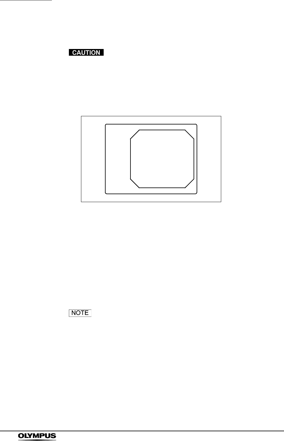
38
Chapter 3 Inspection
EVIS EXERA II VIDEO SYSTEM CENTER CV-180
3.4 Inspection of the monitor display
Be sure to perform white balance adjustment before
inspecting the color on the monitor display. See Section 4.5,
“White balance adjustment” on page 52.
1. Turn the instrument ON. Then the endoscopic image appears on the screen
(see Figure 3.3).
Figure 3.3
2. Confirm that the endoscopic image is normal by observing any object such
as the palm of your hand.
3. Confirm that the date and time are correct.
4. Confirm that the “CVP” counter and “D.F” counter are displayed on the
screen when the video printer and digital filing system are connected.
5. Confirm that enough space is available on the PC card to store endoscopic
images.
• The display layout is variable according to the connected
endoscope and user preset.
• For setting the date or time, refer to “Date and time” on
page 197.
ID:
Name:
Sex: Age:
D.O.B.
12/12/2005
12:12:12
CVP: A4/4
D.F: 99
VCR
Ct: N Eh: A8
Z: x1.5
Pump
Media:
Physician:
Comment:

Chapter 3 Inspection
39
EVIS EXERA II VIDEO SYSTEM CENTER CV-180
3.5 Inspection of the freeze function
Do not use this instrument when the live image cannot be
observed. Otherwise, patient injury may occur.
1. Press the “FREEZE” key on the keyboard, and confirm that the live
endoscopic image freezes and a short beep is heard.
2. Press the “FREEZE” key again and confirm that the frozen image returns to
the live image.
3. Confirm the function of the scope switches and/or foot switches, when the
freeze function is assigned to these switches.
3.6 Inspection of the release function
1. Press the “RELEASE” key on the keyboard.
2. Confirm that the live image freezes for a short time and a beep is heard.
3. Confirm that the selected recording device is activated.
4. Confirm that the counter for the recording devices, which are displayed on
the monitor, increments by one.
5. Confirm the function of the scope switches and/or foot switches, when the
release function is assigned to these switches.
3.7 Inspection of the PinP (picture in picture)
function
According to the “PinP (picture in picture) display” on page 64, confirm that the
PinP indication can be performed correctly.
3.8 Inspection of the orientation function
If the orientation function is activated, confirm that the indication on the monitor
is an endoscopic image rotated by 180 (refer to “Monitor orientation function” on
page 245).

40
Chapter 3 Inspection
EVIS EXERA II VIDEO SYSTEM CENTER CV-180
3.9 Inspection of the special light observation
function
According to Section 5.8, “Special light observation” on page 151, confirm that
the image of the special light observation can be displayed correctly.
3.10 Inspection of the scope switches and foot
switches
If any function is assigned to the scope's remote switches and/or foot switches,
confirm the proper function of these switches.
3.11 Power OFF
Press the power switch of the instrument (see Figure 3.1) to turn the instrument
OFF. The indicator above the switch goes off.

Chapter 4 Operation
41
EVIS EXERA II VIDEO SYSTEM CENTER CV-180
Chapter 4 Operation
This chapter explains the work flow of endoscopic observation using the video
system center. For information on how to use the functions that are not
explained in this chapter, refer to the reference pages.
The operator of the video system center must be a physician or medical
personnel under the supervision of a physician and must have received sufficient
training in clinical endoscopic techniques. This manual, therefore, does not
explain or discuss clinical endoscopic procedures. It only describes basic
operation and precautions related to the operation of the video system center.
• Be sure to wear protective equipment such as eye wear, face
mask, moisture-resistant clothing and chemical-resistant
gloves that fit properly and are long enough so that your skin
is not exposed. Otherwise, dangerous chemicals and/or
potentially infectious material such as blood and/or mucus of
the patient may cause an infection.
• Should any irregularity is observed, do not use the video
system center. Damage or irregularity may compromise
patient or user safety and may result in more severe
equipment damage.
• Anytime you observe an abnormality in a video system
center function, stop the examination immediately and take
action according to the following procedures. Using a
defective video system center may cause patient and/or
operator injury.
If the endoscopic image disappears or if the image
freezes and cannot be restored, press the “RESET”
button or temporarily turn the video system center OFF
and wait for about 10 seconds. Then turn it back ON
again.
For ancillary equipment used in conjunction with the video
system center, also turn the power OFF and then ON
again as directed in their respective instruction manuals. If
this fails to correct the problem, immediately stop using
the equipment and turn the video system center and light
source OFF. Then, gently withdraw the endoscope from
the patient as described in the endoscope's instruction
manual.

42
Chapter 4 Operation
EVIS EXERA II VIDEO SYSTEM CENTER CV-180
If any other abnormality occurs or is suspected,
immediately stop using the equipment, turn OFF all
equipment, and gently withdraw the endoscope from the
patient as described in the endoscope's instruction
manual. Then refer to the instructions in Chapter 10,
“Troubleshooting”. If the problems cannot be resolved by
the remedial action described in Chapter 10, do not use
the equipment and contact Olympus.
• Combination with other equipment
Do not use the video system center in locations exposed
to direct strong electromagnetic radiations (for example,
microwave treatment device, short wave treatment
device, MRI or radio equipment). Electromagnetic
radiation can interfere with the monitor display.
Use only Olympus high-frequency electrosurgical
equipment with this unit. Non-Olympus equipment can
cause interference on the monitor display or a loss of the
endoscopic image.
Before using high-frequency electrosurgical equipment,
be sure to install and connect the equipment according to
it’s instruction manual and make sure that the noise does
not affect the observation and surgical procedures. If
high-frequency electrosurgical equipment is used without
such confirmation, patient injury may result.
• To activate the auto brightness control function of the light
source, the video system center should be turned ON. If it is
not turned ON, the auto brightness control function is not
activated and the light intensity is set to maximum. In this
case, the endoscope distal end would become hot and could
cause burns to the operator and physician (if a light source
model other than CLV-180 is used).
• When using spray-type medical agents such as lubricant,
anesthetic, or alcohol, use them away from the video system
center so that the medical agents do not contact the video
system center. Medical agents might enter the video system
center through the ventilation grills and may cause
equipment damage.
• Do not use a humidifier near the video system center as dew
condensation possibly might occur and it may cause
equipment failure.

Chapter 4 Operation
43
EVIS EXERA II VIDEO SYSTEM CENTER CV-180
• High-frequency electrosurgical equipment can cause slight
interference on the monitor display.
• Sometimes horizontal line noise appears when a slim
endoscope is used. To reduce the horizontal line noise,
select “Edge enhancement” for the enhancement setting.
44
Chapter 4 Operation
EVIS EXERA II VIDEO SYSTEM CENTER CV-180
4.1 Operation flow
Please see the operation work flow in Figure 4.1 below. Follow each step of the
work flow for using the video system center.
1. Connect the endoscope to the video system center and the light source.
Section 4.2, “Connection of an endoscope” on page 46
2. Inspect the instruments before use.
Chapter 3, “Inspection” on page 34
3. Turns the instrument ON.
Section 4.3, “Turning the video system center ON” on page 50
4. Select a user name.
Section 4.4, “Recall of user preset data” on page 51
This operation can be skipped when using the user name of the last examination.
5. Adjust the white balance.
Section 4.5, “White balance adjustment” on page 52
When you are planning to use the NBI observation, perform the white balance adjustment for NBI
after performing the white balance for normal-light observation.
6. Enter the patient data.
Section 4.6, “Patient data” on page 57
It is possible to enter the patient data before the examination (see “Entering new patient data” on
page 137).
continued on next page
Chapter 4 Operation
45
EVIS EXERA II VIDEO SYSTEM CENTER CV-180
Figure 4.1
7. Perform examination.
Chapter 5, “Functions” on page 62
Changes iris mode,
Image enhancement,
Freezes image,
Zooms image,
Recording and printing of the images,
NBI observations, etc.
8. Terminate the examination.
Section 4.9, “Termination of the operation” on page 60
Press “EXAM END” key after the examination. Then turn the CV-180 and other instruments OFF.
9. Disconnect the endoscope from the video system center and the light source.
Section 4.9, “Termination of the operation” on page 60
10. Inspect the instruments after use.
Chapter 7, “Care, Storage and Disposal” on page 155
For details on endoscope and light source, see the respective instruction manuals.
11. Reprocess and store the instrument and ancillary equipment as appropriate after use.
Chapter 7, “Care, Storage and Disposal” on page 155
For details on endoscope and light source, see the respective instruction manuals.
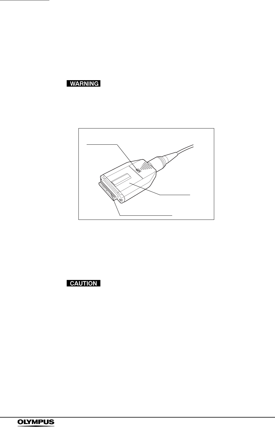
46
Chapter 4 Operation
EVIS EXERA II VIDEO SYSTEM CENTER CV-180
4.2 Connection of an endoscope
Connect the endoscope to the video system center and the light source. The
connection may require special cables. Refer to the instruction manuals of
ancillary equipment for details on the cables to be used.
• Make sure that the video plug and its electrical contacts are
completely dry before connecting the plug to the video
system center (see Figure 4.2). Wet equipment could cause
the image to flicker or disappear.
Figure 4.2
• Do not apply excessive force to the camera cable of the
camera head by bending, stretching or crushing it. Also do
not pull a bundle of camera cables, as this may cause
internal wire disconnection.
• Do not connect or disconnect the endoscope connector while
this video system center is turned ON. Connecting or
disconnecting the endoscope while this video system center
is ON may destroy the CCD. Turn the video system center
OFF before connecting or disconnecting the endoscope.
• Connect the video plug all the way into the socket. The
improper connection may increase image noise or may
cause disappearance of the endoscopic image during
operation
• Be sure to refer to the instruction manuals of the ancillary
equipment including the endoscope and camera cable.
“UP” mark
Video plug
Electrical contacts
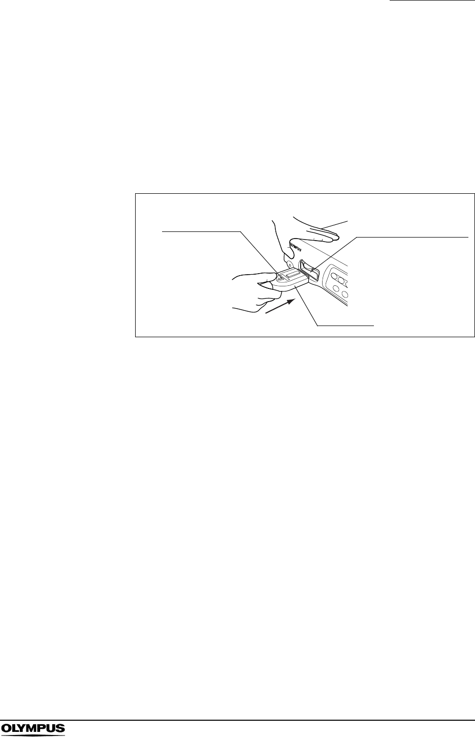
Chapter 4 Operation
47
EVIS EXERA II VIDEO SYSTEM CENTER CV-180
VISERA series videoscope
1. Ensure that this instrument and all connected devices are turned OFF.
2. Connect the endoscope connector of the videoscope to the light source,
referring to the instruction manual for the light source.
3. Push the video plug into the video connector socket of the instrument all the
way until it clicks, holding this instrument with a hand so that it will not move.
Confirm that the “UP” mark points upwards (see Figure 4.3).
Figure 4.3
EVIS series videoscope and ultrasonic videoscope
1. Ensure that this instrument and all connected devices are turned OFF.
2. Connect the endoscope connector of the fiberscope to the light source
referring to the instruction manual for the light source.
3. Push the video plug of the scope cable EXERA II into the video connector
socket of the instrument all the way until it clicks, holding this instrument
with a hand so that it will not move. Confirm that the “UP” mark points
upwards (see Figure 4.3).
4. Connect the scope side connector of the scope cable EXERA II to the
endoscope, referring to the instruction manual of the endoscope.
Video connector socket
Video plug
“UP” mark

48
Chapter 4 Operation
EVIS EXERA II VIDEO SYSTEM CENTER CV-180
Fiberscope and camera head
1. Ensure that this instrument and all connected devices are turned OFF.
2. Connect the endoscope connector of the fiberscope to the light source,
referring to the instruction manual for the light source.
3. Push the video plug of the camera head into the video connector socket of
the instrument all the way until it clicks, holding this instrument with a hand
so that it will not move. Confirm that the “UP” mark points upwards (see
Figure 4.3).
4. Connect the video adapter and camera head to the eyepiece section of the
fiberscope, referring to the instruction manuals for the video adapter and
camera head.
Rigidscope and camera head
1. Ensure that this instrument and all connected devices are turned OFF.
2. Connect the light guide cable to the light source, referring to the instruction
manual for the light source.
3. Push the video plug of the camera head into the video connector socket of
the instrument all the way until it clicks, holding this instrument with a hand
so that it will not move. Confirm that the “UP” mark points upwards (see
Figure 4.3).
4. Attach the light guide cable, video adapter and camera head to the
rigidscope, referring to the instruction manuals for the light guide cable,
video adapter and camera head.
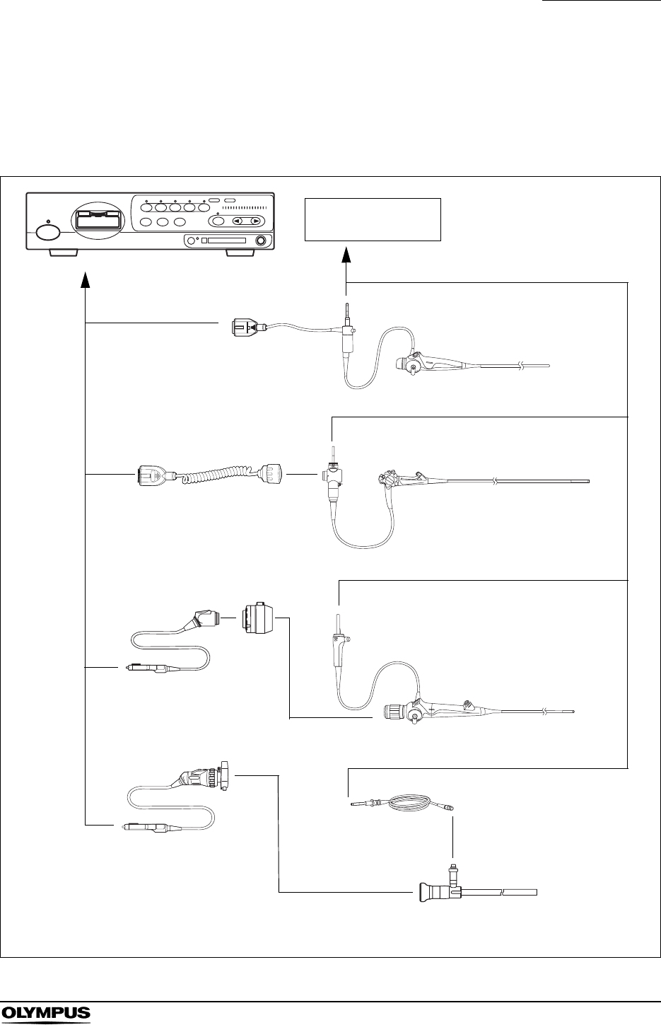
Chapter 4 Operation
49
EVIS EXERA II VIDEO SYSTEM CENTER CV-180
Summary of connection
This diagram gives an overview of all connections. For further details of the
connection, refer to the instruction manuals of the ancillary equipment being
used.
Figure 4.4
Light source
VISERA series
Fiberscopes
Rigidscope
Light guide cable
Video adapters, camera heads, etc.
Videoscope cable EXERA II
CV-180
The numbers show the order of connection.
2
2
2
3
3
4
EVIS series and
ultrasonic videoscope
2
3
Camera head
Video adapter
1
1
1
1
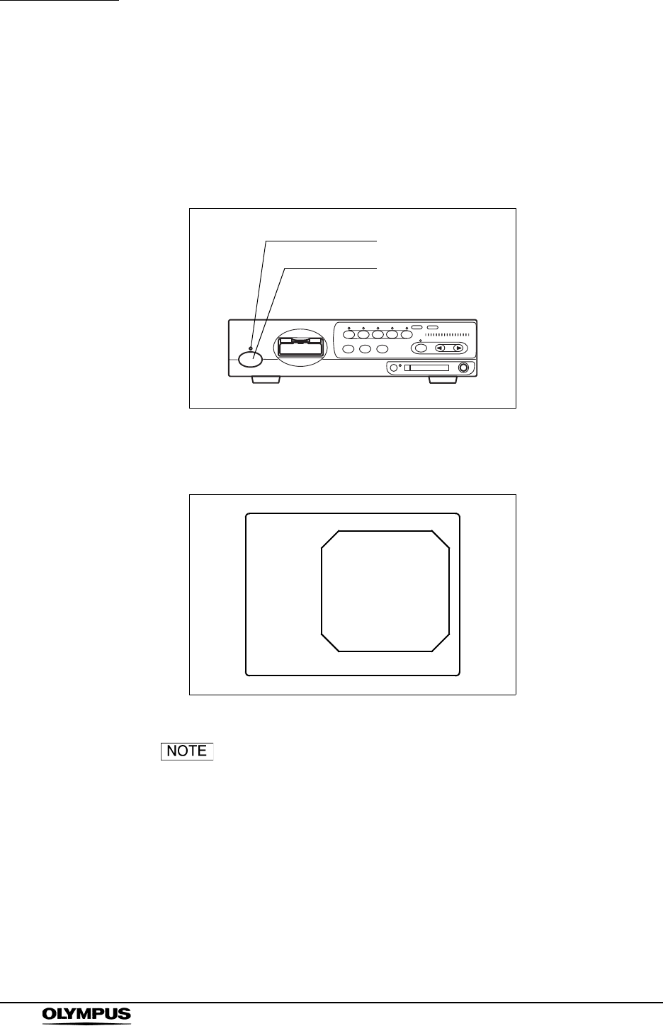
50
Chapter 4 Operation
EVIS EXERA II VIDEO SYSTEM CENTER CV-180
4.3 Turning the video system center ON
1. Turn the ancillary equipment ON.
2. Turn the instrument ON by pressing the power switch (see Figure 4.5). The
indicator above the power switch lights up.
Figure 4.5
3. The endoscopic image appears on the monitor (see Figure 4.6).
Figure 4.6
For the procedure of turning the ancillary equipment ON,
refer to each instrument's instruction manual.
Power indicator
Power switch
ID:
Name:
Sex: Age:
D.O.B.
12/12/2005
12:12:12
Physician:
Comment:
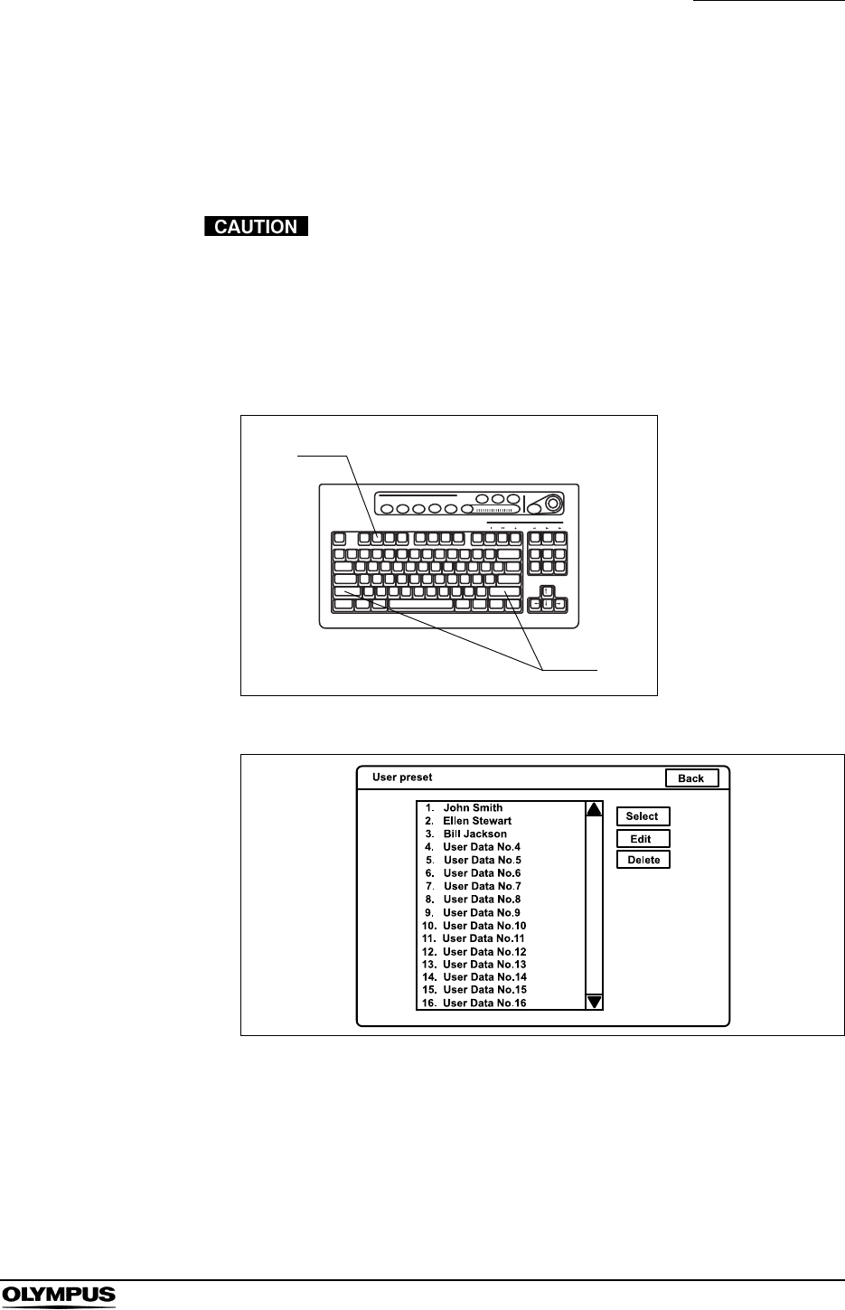
Chapter 4 Operation
51
EVIS EXERA II VIDEO SYSTEM CENTER CV-180
4.4 Recall of user preset data
The operating conditions for each user (operator) can be recalled from the “User
preset” menu (see Section 9.3, “User preset” on page 216).
Confirm that the required user preset data is selected before
starting the observation. If different user preset data is used,
unintended operations may occur.
1. Press the “Shift” and “F2” keys together. The “User preset” menu appears
on the monitor (see Figure 4.7).
Figure 4.7
Figure 4.8
2. Click the desired user name in the user name dialog box. The selected user
name is highlighted.
3. Check if the correct user name is selected, then click “Select”. The
endoscopic image appears on the monitor and the user preset data is
loaded to the video system center.
F2
Shift

52
Chapter 4 Operation
EVIS EXERA II VIDEO SYSTEM CENTER CV-180
The user preset data used in the last operation before turning
the video system center OFF comes up initially after turning
the instrument ON.
4.5 White balance adjustment
This adjustment procedure is used to display the correct image color on the
monitor. There are two white balance adjustments for the normal observation
and the NBI observation. Be sure to always adjust the white balance in the
following cases:
• Before observation.
• After exchange of the light source.
• When any abnormality can be seen on the color of the image even if
white balance adjustment has been completed.
• When adjusting the white balance of the endoscope to be
used in the sterilized zone, do not use the white cap (MH-
155) as described in this part, but use a white object such as
a piece of gauze without bringing it in contact with the
endoscope. Contact of the endoscope with a non-sterilized
object may result in cross-contamination.
• Make sure that the endoscope and white cap (MH-155) are
clean before adjusting the white balance. Otherwise, cross-
contamination may result.
• Always control the color tone and/or enhancement of the
image appropriately before observation. Setting an
inappropriate color tone or enhancement condition may
result in overlooking or wrong diagnosis.
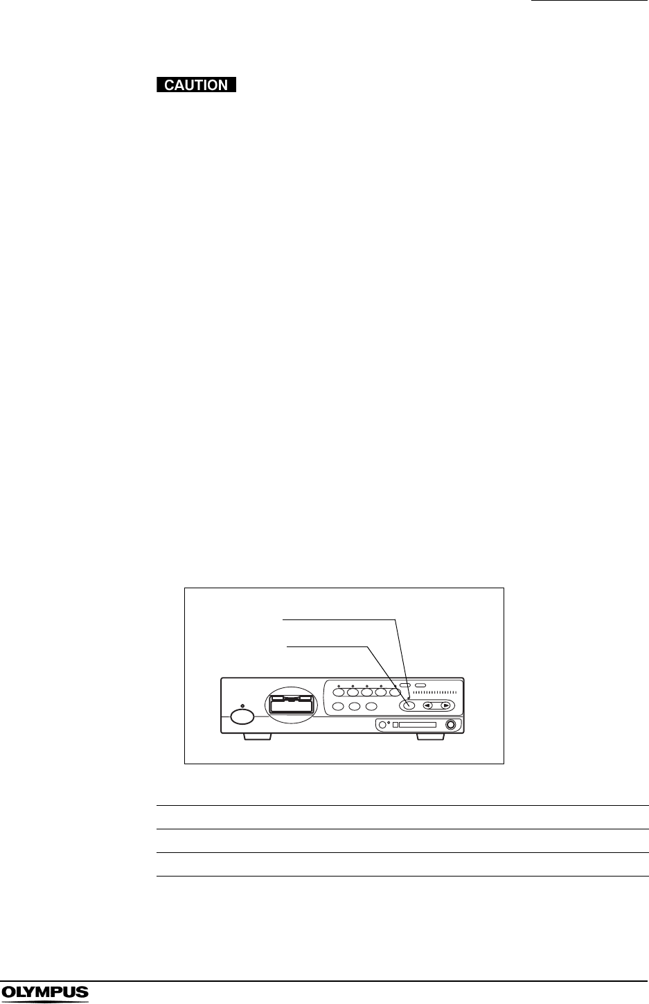
Chapter 4 Operation
53
EVIS EXERA II VIDEO SYSTEM CENTER CV-180
• Do not turn the video system center OFF or disconnect the
videoscope while the white balance button is being pressed
or the “Wh/B OK” indicator is blinking. Otherwise, the data
stored on the memory chip of the endoscope may be
destroyed.
• When adjusting the white balance, always turn the light
source ON and take care not to expose the distal end of the
endoscope to external light. Otherwise, it may cause an
incorrect white balance adjustment.
• Always use the white cap when setting the white balance for
the videoscope used in a non sterilized zone.
• Set the brightness level of the light source to “3” or the center
of the adjustment range before adjusting the white balance.
• When the color tone is not proper, white color cannot be
displayed properly on the monitor even if the white balance is
set. Adjust the color tone to the center of the adjustable
range.
For normal light observation
1. Confirm the lighting status of the “Wh/B OK” (Wh/B = White balance)
indicator on the front panel (see Figure 4.9 and Table 4.1).
Figure 4.9
2. Proceed as follows according to the use of the endoscope.
“Wh/B OK” indicator Status of white balance adjustment
OFF Before adjustment or if adjustment failed.
ON Completed.
Table 4.1
Wh/B OK indicator
Wh/B button
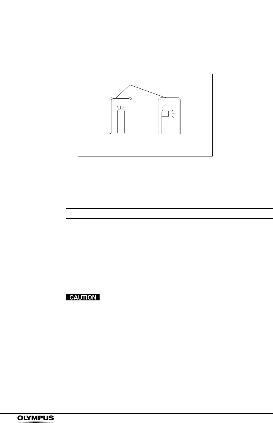
54
Chapter 4 Operation
EVIS EXERA II VIDEO SYSTEM CENTER CV-180
When using an endoscope for the non-sterilized zone
1. Insert the endoscope's distal end into the white cap and hold the white cap
and endoscope stable to avoid wash-out of the monitor image (see Figure
4.10).
Figure 4.10
2. Maintaining the stable condition in step 1., press the “Wh/B” button until a
short beep is generated. The result of the adjustment will be displayed on
the monitor for a few seconds (see Table 4.2).
3. Confirm that the “Wh/B OK” indicator is ON (see Table 4.1). If it is OFF, go
back to step 1. again.
When performing white balance adjustment keep an
appropriate distance between distal end and white cap.
Otherwise, the white balance might not be adjusted properly.
Result Message
Completed successfully “White balance complete!”
“Check NBI white balance.”
(When using NBI compatible endoscope.)
Failed “White balance incomplete! Perform again!”
Table 4.2
White cap
Direct view Side view

Chapter 4 Operation
55
EVIS EXERA II VIDEO SYSTEM CENTER CV-180
When using an endoscope for the sterilized zone
1. Hold the endoscope stable to avoid wash-out of the monitor image, and
enlarge the image to the full monitor, monitoring a white object such as a
piece of gauze in such a way that it does not contact the endoscope.
2. Maintaining the stable condition in step 1., press the “Wh/B” button until a
short beep is generated. The result of white balance adjustment will be
displayed on the monitor for a few seconds (see Table 4.3).
3. Confirm that the “Wh/B OK” indicator is ON (see Table 4.1). If it is OFF, go
back to step 1. again.
For NBI observation
When connecting an NBI compatible endoscope, perform the white balance
adjustment for NBI observation after the white balance adjustment for normal
light observation has been performed.
1. Adjust the white balance for normal light observation according to “For
normal light observation” on page 53.
2. Switch to the NBI mode by operating the mode button of the light source.
Refer to the instruction manual of the light source.
3. Confirm that “NBI” appears on the monitor, and that the “NBI” indicator on
the front panel of the video system center turns from green to white.
4. Hold the endoscope as shown in step 1. of “When using an endoscope for
the non-sterilized zone” on page 54 or step 1. of “When using an endoscope
for the sterilized zone” on page 55 according to the use of it.
Result Message
Completed successfully “White balance complete!”
“Check NBI white balance.”
(When using NBI compatible endoscope.)
Failed “White balance incomplete! Perform again!”
Table 4.3
56
Chapter 4 Operation
EVIS EXERA II VIDEO SYSTEM CENTER CV-180
5. Maintaining the stable condition in step 4., press the “Wh/B” button until a
short beep is generated. The result of the adjustment will be displayed on
the monitor for a few seconds (see Table 4.4).
6. If the adjustment failed, perform the operation of steps 4. and 5. again.
7. Switch to the normal mode by operating the mode button of the light source.
Refer to the instruction manual of the light source.
8. Confirm that “NBI” disappears on the monitor, and that the “NBI” indicator on
the front panel of the video system center turns from green to white.
• The white balance adjustment can also be initiated by
pressing the “Shift” and “F9” keys on the keyboard together
instead of the “Wh/B” button.
• When the white balance adjustment cannot be completed,
check if the color tone and/or brightness are correct and the
white cap is clean.
• Once the white balance adjustment is completed, the “Wh/B
OK” indicator keeps on lighting until the video system center
is turned OFF.
• The white balance adjustment can also be initiated from the
endoscope's remote switches and/or foot switches.
For how to set up the remote switches and foot switches, see
“Remote switch and foot switch (EXERA and VISERA)” on
page 219
Result Message
Completed successfully “NBI White Balance Complete!”
Failed “NBI White Balance Incomplete!”
Table 4.4
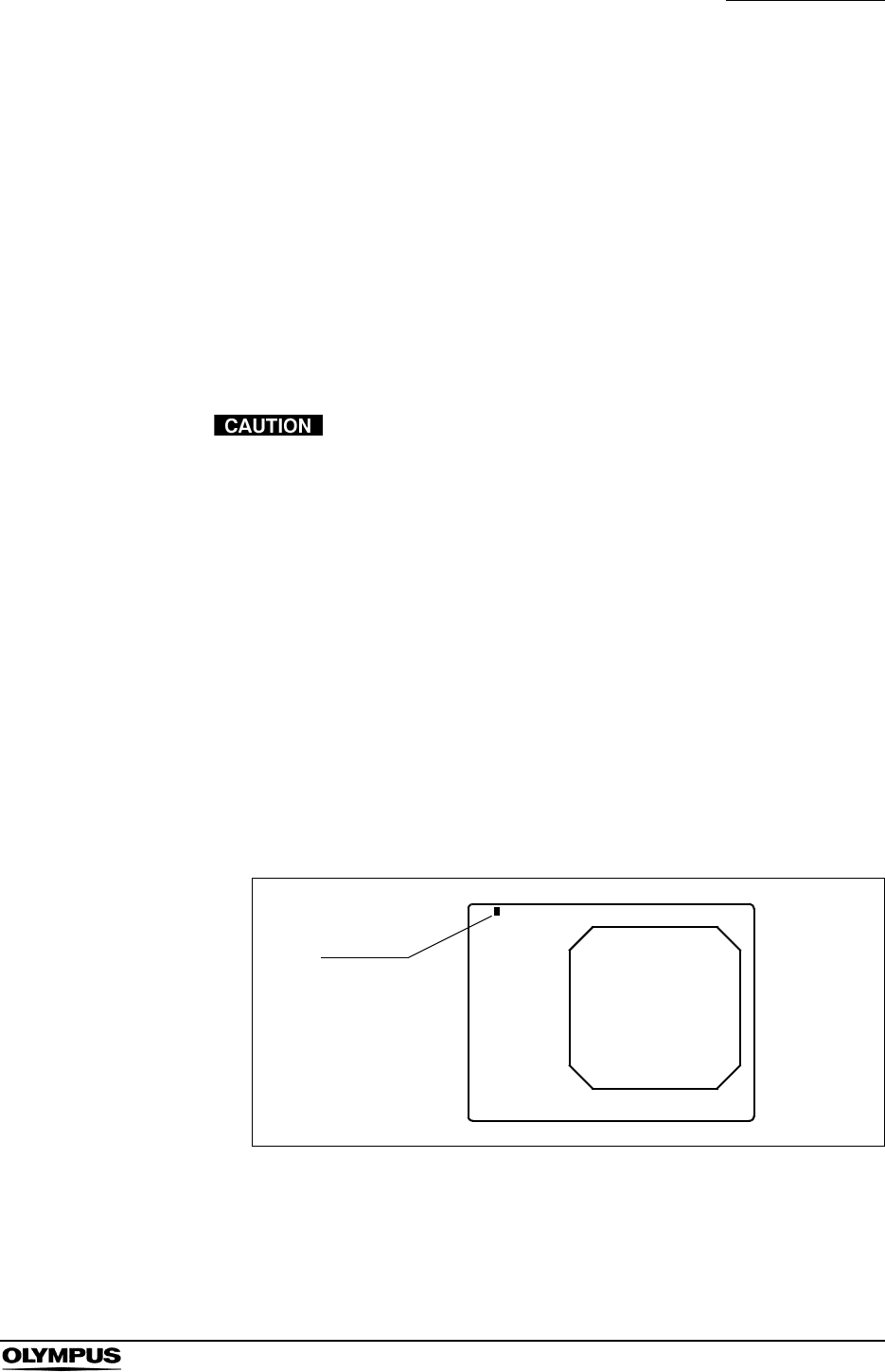
Chapter 4 Operation
57
EVIS EXERA II VIDEO SYSTEM CENTER CV-180
4.6 Patient data
Before the observation, enter the patient data into the endoscopic image.
There are two methods to enter the patient data:
• Patient data can be entered immediately before the examination.
• A list of several patient data can be entered in advance.
This section explains how to enter patient data immediately before the
examination. To enter a list of several patient data in advance, see Section 5.6,
“Pre-entry of patient data” on page 136.
• Before entering patient data, press the “EXAM END” key to
clear the previous patient data. Otherwise, different patient
data can be mixed on one print sheet and/or normal
functioning of the digital filing system cannot be guaranteed.
• When recording the images, be sure to record the images
together with the patient data. Otherwise distinction between
different observations may become difficult.
• Be sure to enter the patient ID when entering patient data.
Also be sure to enter a different patient ID for each patient.
Otherwise, the image data for some patients may mix in the
same image folder.
1. Press the “F1” key to change the monitor display full-patient-data display.
2. Press the “EXAM END” key to clear the previous patient data.
Figure 4.11
ID:
Name:
Sex: Age:
D.O.B.
12/12/2005
12:12:12
Physician:
Comment:
Cursor
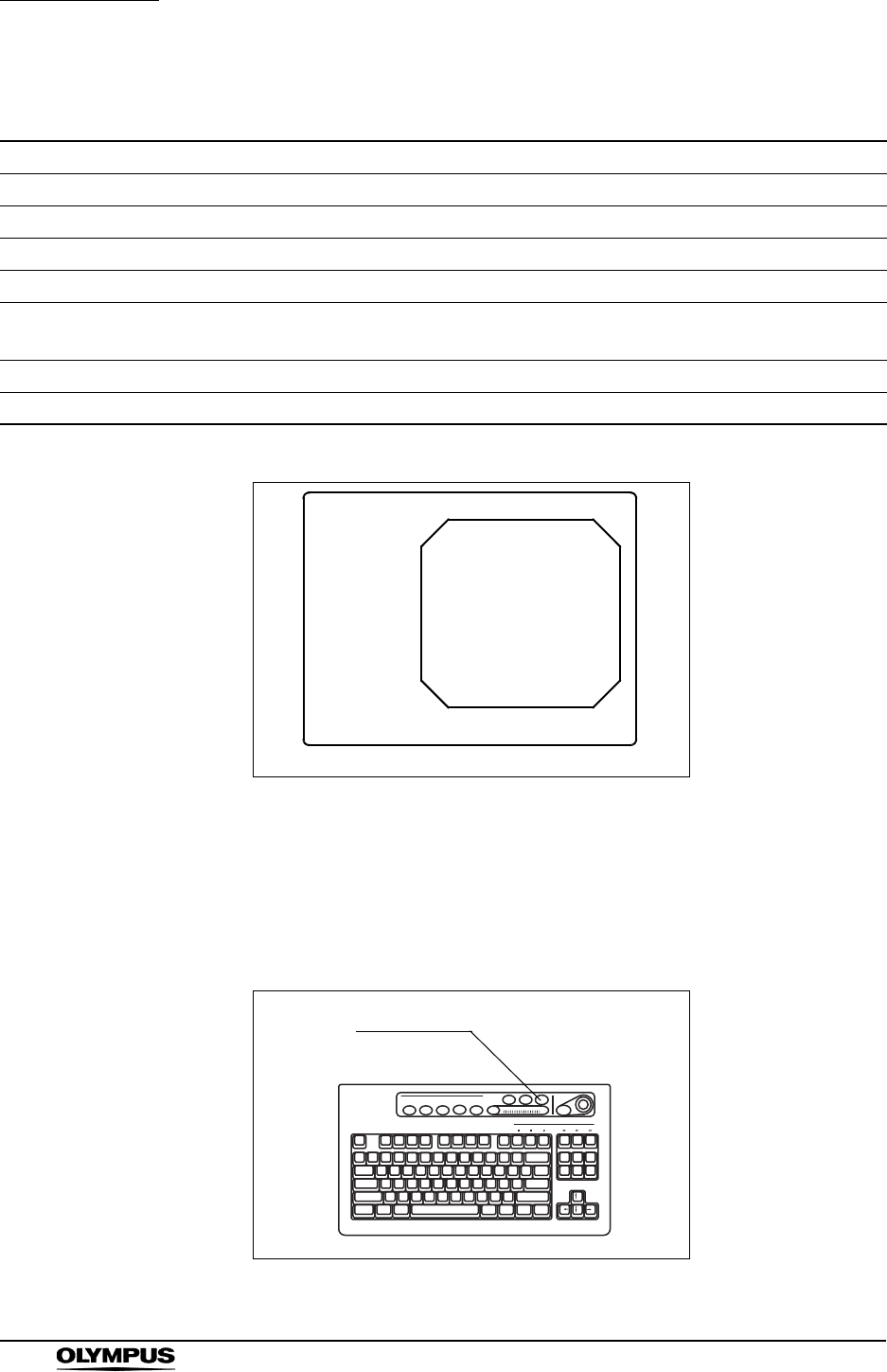
58
Chapter 4 Operation
EVIS EXERA II VIDEO SYSTEM CENTER CV-180
3. Enter data.
Figure 4.12
4. When modifying data, press the arrow key to move the cursor to the input
position and edit the data.
5. When deleting all patient data displayed on the monitor, press the “EXAM
END” key on the keyboard (see Figure 4.13).
Figure 4.13
Data data input condition Entry example
ID No. Up to 15 characters ABC123
Name Up to 20 characters Mike Johnson
Sex 1 character M
Age Up to 3 characters. Automatically calculated after entering D.O.B. 51
D.O.B. 8 characters. Enter the numbers according to the format (see
page 197).
03031954
Physician Up to 20 characters John Smith
Comment Up to 37 characters Cardiac end of the stomach
Table 4.5
ABC123
Mike Johnson
M 51
03/03/1954
12/12/2005
12:12:12
John Smith
Cardiac end of the stomach
An example of displaying
EXAM END

Chapter 4 Operation
59
EVIS EXERA II VIDEO SYSTEM CENTER CV-180
4.7 Observation of the endoscopic image
Observe the endoscopic image using the various functions provided with the
video system center. For details of the functions, see Chapter 5, “Functions”.
4.8 Recording of the observation image
Table 4.6 shows the devices that can record and/or print endoscopic images.
These devices can be controlled by the keyboard, scope switches, etc. For the
operation of these devices, see Chapter 5, “Functions”.
Recording device Details
PC card Section 5.3, “Image recording and playback (PC card)” on
page 106
Digital file system “Image filing system” on page 127
Videocassette recorder “Videocassette recorder (VCR)” on page 129
Video printer Section 5.5, “Printing images” on page 131
Table 4.6
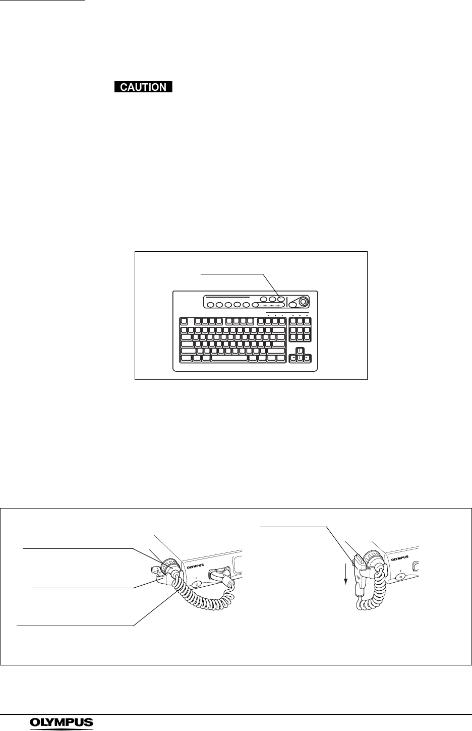
60
Chapter 4 Operation
EVIS EXERA II VIDEO SYSTEM CENTER CV-180
4.9 Termination of the operation
Do not disconnect the video connector before turning the
video system center OFF. Otherwise, the endoscope or
camera head may be damaged.
1. Press the “EXAM END” key (see Figure 4.14) to execute the following
process;
• clearing patient data on the monitor;
• completing printing of un-printed images;
• closing the digital file system.
Figure 4.14
2. Turn the instrument and ancillary equipment OFF.
3. When an EVIS series endoscope is used:
Disconnect the scope side connector of the videoscope cable, and place it
on the scope cable holder (A in Figure 4.15). For disconnecting the video
plug, see B in Figure 4.15.
Figure 4.15
EXAM END
Scope side connector
Scope cable holder
Videoscope cable
A) When the scope side connector
is not connected. B) When both connectors are
not connected.
Video plug
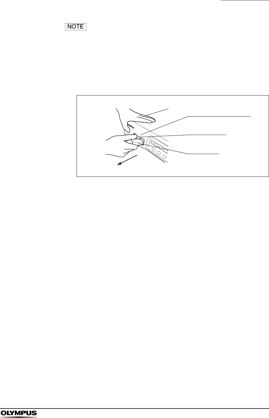
Chapter 4 Operation
61
EVIS EXERA II VIDEO SYSTEM CENTER CV-180
In B in Figure 4.15, insert the video plug to the scope cable
holder in the direction of the arrow.
4. When a VISERA series endoscope or camera head is used:
Disconnect the video plug of the videoscope cable from the instrument,
holding the instrument with a hand so that it will not move and push the
locking lever down (see Figure 4.16).
Figure 4.16
Video connector socket
Locking lever
Video plug
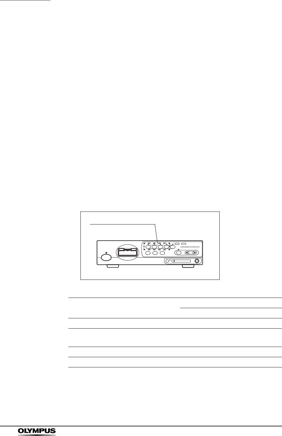
62
Chapter 5 Functions
EVIS EXERA II VIDEO SYSTEM CENTER CV-180
Chapter 5 Functions
This chapter explains the functions of the buttons and keys on the video system
center. See the “System setup” menu and “User preset” menu in Chapter 9,
“Function setup” for presetting.
5.1 Front panel
Image source buttons
Used to select the image source displayed on the monitor. The buttons other
than the “SCOPE” button has to be pressed for more than 1 second to activate.
1. The Image source buttons select the images to be displayed on the monitor
(see Figure 5.1). The indicator above the active button lights up.
2. Confirm that the desired image appears on the monitor.
Figure 5.1
Button Images on the monitor Terminal and image sources
Front panel Rear panel
SCOPE Endoscope’s live image Scope connector -
DV/VCR VCR, video recorder - VCR REMOTE or
DIGITAL OUT
PC Digital filing system - PC IN
PRINTER Video printer - PRINTER IN
Table 5.1
Image source buttons

Chapter 5 Functions
63
EVIS EXERA II VIDEO SYSTEM CENTER CV-180
When performing endoscopic observation, select the
“SCOPE” image source.Images may disappear during
observation depending on the condition of ancillary
equipment.
When pressing an image source button except PC button to
which no device is connected, the lamp above the button
blinks and the previous image appears on the monitor. When
pressing PC button, the image input from PC IN terminal is
selected, regardless of the PC’s connection.
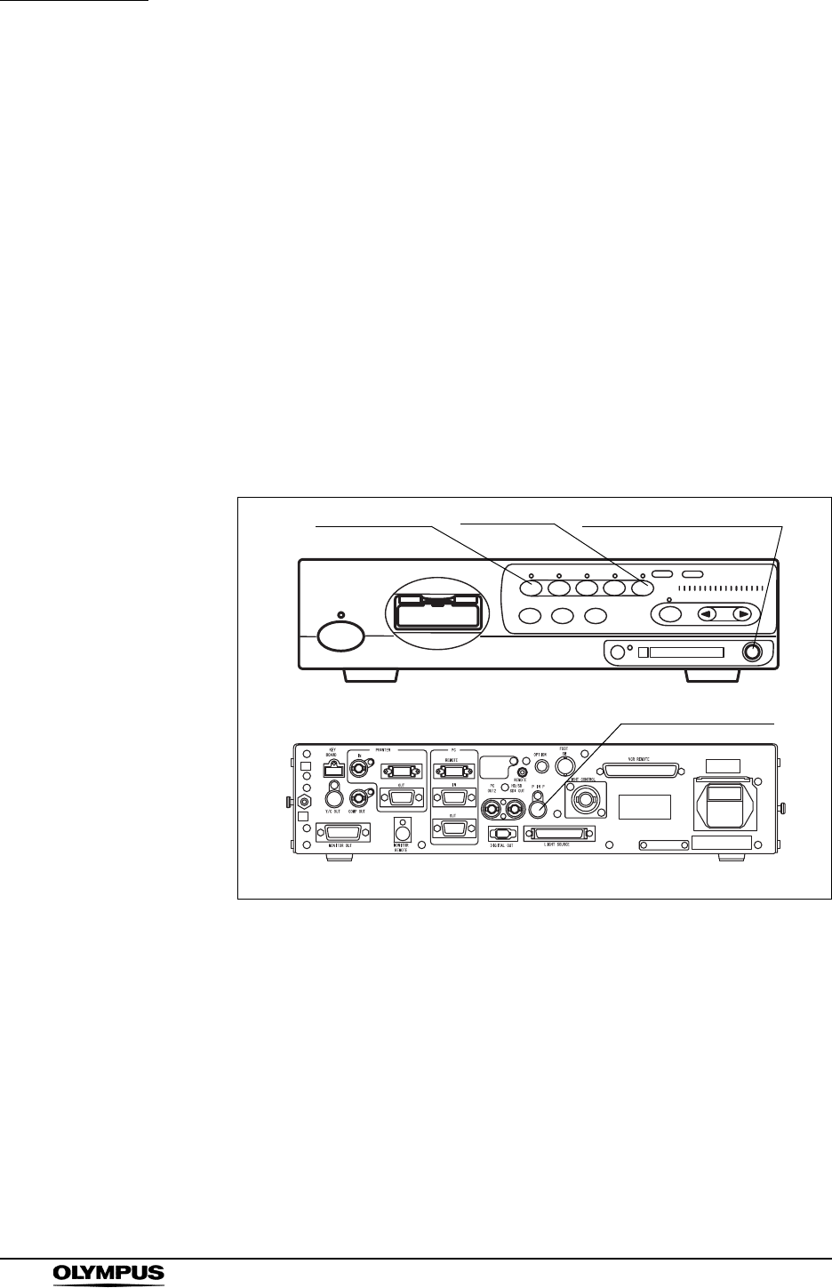
64
Chapter 5 Functions
EVIS EXERA II VIDEO SYSTEM CENTER CV-180
PinP (picture in picture) display
The image of an external device connected to the following connectors can be
displayed on the monitor together with the endoscopic live image.
• PinP composite terminal on the front panel
(prior to the PinP Y/C terminal on the rear panel.)
• PinP Y/C terminal on the rear panel
The PinP function has two modes, “ON/OFF mode” and “Mode Change mode”.
The two modes display images in different ways. Presetting of PinP in the “User
preset” menu is needed. See “PinP (picture in picture) function” on page 246.
1. Confirm that the external device is connected to either the PinP composite
terminal on the front panel or the PinP Y/C terminal on the rear panel (see
Figure 5.2).
Figure 5.2
2. Press the “SCOPE” button to display the endoscopic live image.
PinP composite terminal
PinP Y/C terminal
Front panel
Rear panel
SCOPE button PinP button
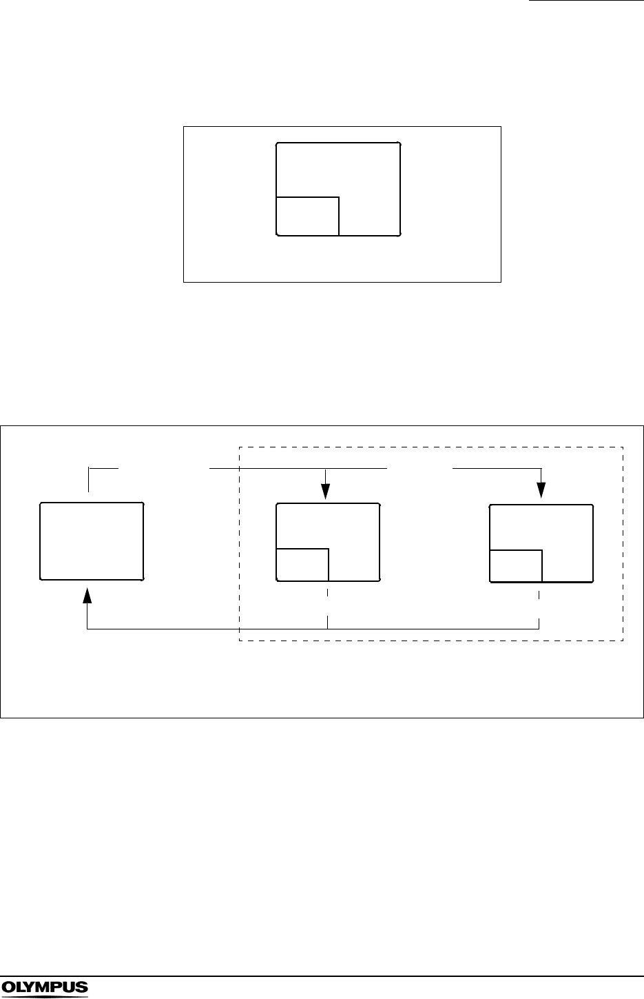
Chapter 5 Functions
65
EVIS EXERA II VIDEO SYSTEM CENTER CV-180
3. Press the “PinP” button. The sub image appears on the monitor (see Figure
5.3).
Figure 5.3
4. Press the “PinP” button to change the display. The transitions of the displays
are different according to the settings in “User preset” menu (see Figure 5.4
and Figure 5.5) (refer to “PinP (picture in picture) function” on page 246 for
the setting method).
Figure 5.4
Sub
image
Main image
PinP display
Endoscopic
image External
image
PinP button
Screen transition in the “ON/OFF mode”
PinP button
Either
(see Note)
PinP button
Note) The initial screen when pressing the PinP button depends on the user settings preset.
Endoscopic
image
Endoscopic
image
External
image
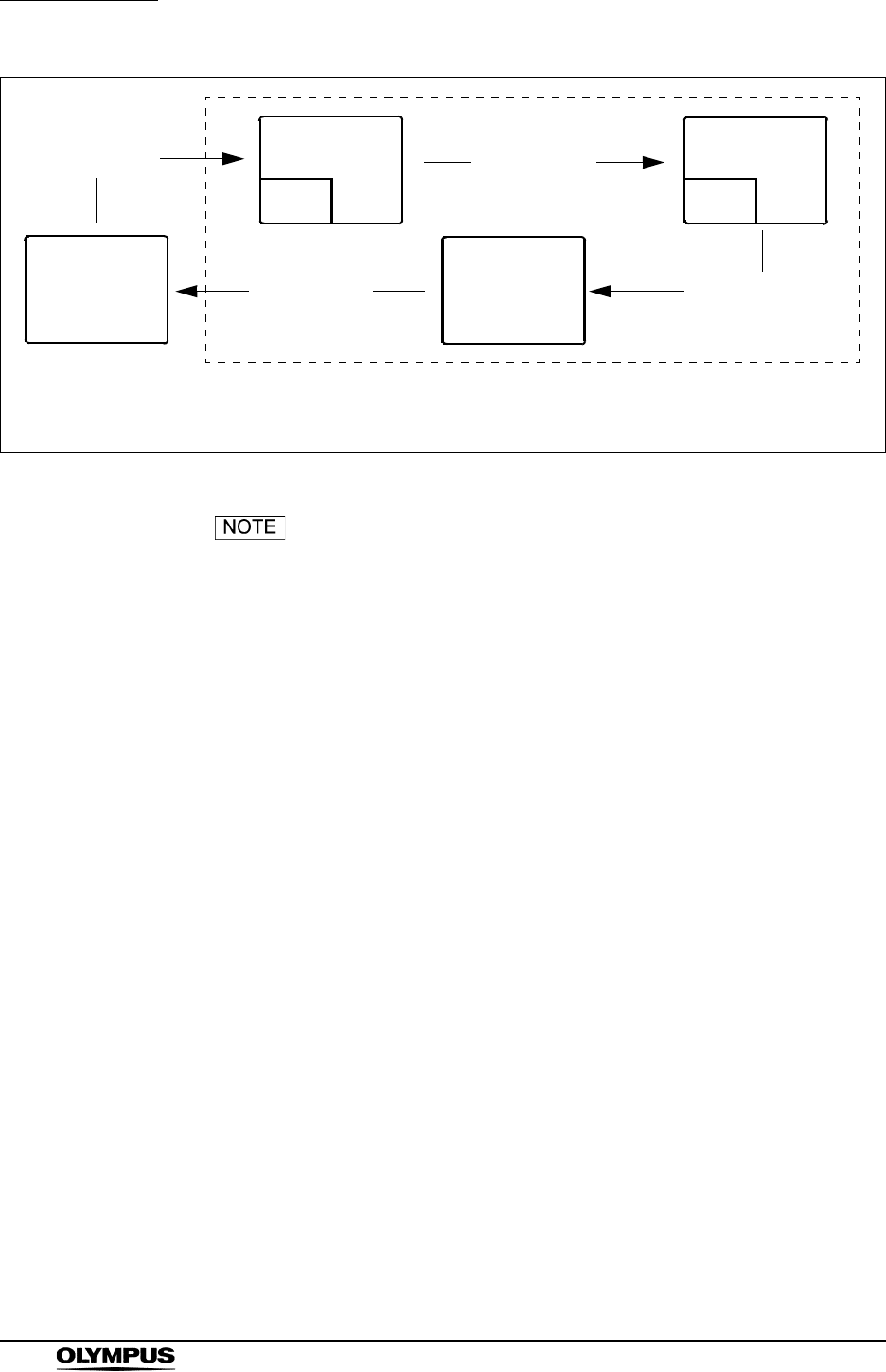
66
Chapter 5 Functions
EVIS EXERA II VIDEO SYSTEM CENTER CV-180
Figure 5.5
• All patient data disappears in the PinP display. Press the “F1”
key to display the data.
• The index image does not appear in the PinP display (see
“Release index time” on page 243).
• It takes a few seconds before the PinP display appears
depending on the devices connected.
• The PinP function is also controlled from the scope switches
and/or foot switches. For how to set up the scope switches
and foot switches. See “Remote switch and foot switch
(EXERA and VISERA)” on page 219.
• The output signal is just SDTV in PinP display.
• Before activating the PinP function make sure that an
endoscope is connected to this instrument. If no endoscope
is connected it might not be possible to use the PinP function
and image recording of an external device may not work.
Screen transition in the “Mode change mode”
Endoscopic
image External
image
PinP button PinP button
PinP buttonPinP button
External
image
Endoscopic
image
Endoscopic
image
External
image
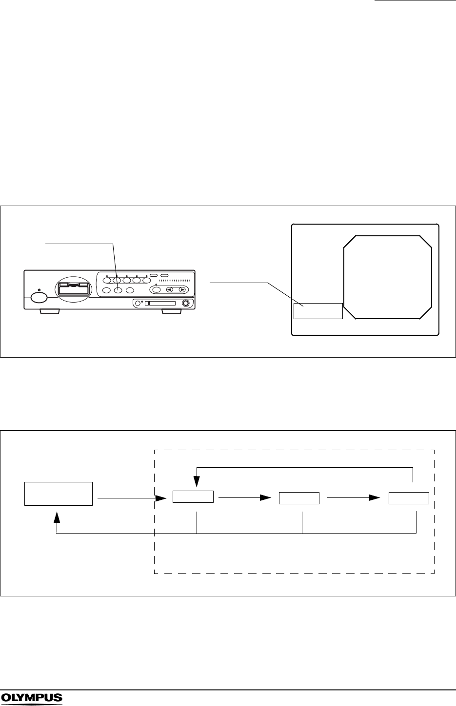
Chapter 5 Functions
67
EVIS EXERA II VIDEO SYSTEM CENTER CV-180
Image enhancement mode (ENH.)
The image enhancement function electrically increases the sharpness of the
endoscopic live image. Three enhancement modes and normal modes are
available. The type and level of image enhancement should be set in advance.
See “Image enhancement (normal observation)” on page 226 and “Image
enhancement (NBI observation)” on page 250.
1. Press the “ENH.” button to change the enhancement mode (see Figure 5.6).
The indicator above the button lights up and the selected mode is displayed
on the monitor for a few seconds.
Figure 5.6
2. To switch OFF the image enhancement at any mode, press and hold the
“ENH.” button. The indicator above the button goes OFF.
Figure 5.7
Enhancement
mode
ID:
Name:
Sex: Age:
D.O.B.
12/12/2005
12:12:12
Ct: N Eh: A8
Z: x1.5
Physician:
Comment:
Enhance:A8
ENH. button
Enhancement
OFF Mode 3
Mode 2
Mode 1
Press and hold ENH. button
ENH. button
ENH. button
ENH. button ENH. button
Enhancement ON

68
Chapter 5 Functions
EVIS EXERA II VIDEO SYSTEM CENTER CV-180
• Mesh-like noise may be observed in the image when the
image enhancement function is ON during use of a
fiberscope or hybrid endoscope. In this case, use the
recommended camera head, or switch the image
enhancement OFF.
OTV-S7H-1N, OTV-S7H-1D, OTV-S7H-1NA
• The image enhancement mode used in the last operation
before turning the video system center OFF comes up when
the instrument is turned ON.
• The image enhancement function can also be controlled from
the scope switches and/or foot switches. For how to set up
the scope switches and foot switches, see “Remote switch
and foot switch (EXERA and VISERA)” on page 219.
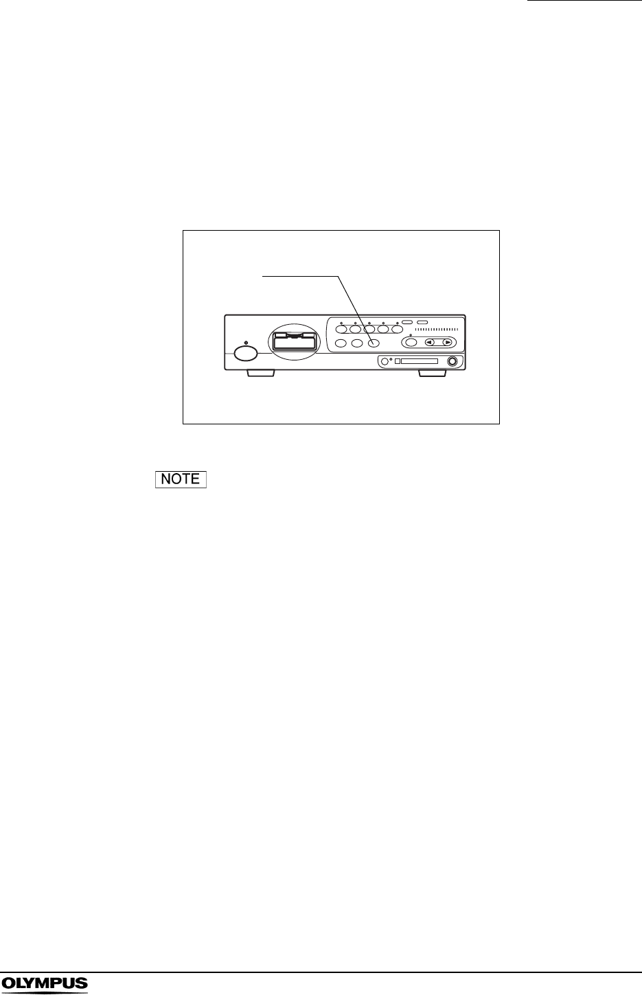
Chapter 5 Functions
69
EVIS EXERA II VIDEO SYSTEM CENTER CV-180
Iris mode
This operation selects the method to measure the brightness of the object of the
observation. Two iris modes, peak mode and auto mode, are available.
For details on the iris mode, see “Iris” on page 232.
1. Press the “IRIS” button on the front panel to switch between auto mode and
peak mode alternately. The indicator above the iris mode button lights up.
Figure 5.8
• The iris mode used in the last operation before the video
system center is turned OFF comes up when the instrument
is turned ON.
• The iris mode can also be controlled from the scope switches
and/or foot switches. For how to set up the scope switches
and foot switches, see “Remote switch and foot switch
(EXERA and VISERA)” on page 219.
IRIS button
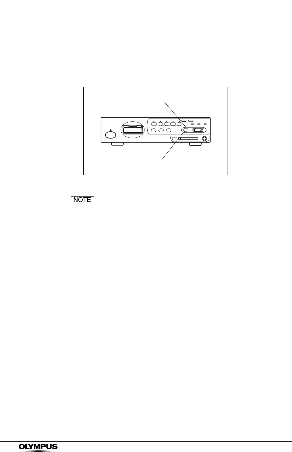
70
Chapter 5 Functions
EVIS EXERA II VIDEO SYSTEM CENTER CV-180
White balance
The “Wh/B” button performs the white balance adjustment to reproduce colors in
their original tones. The “Wh/B OK” indicator indicates if the white balance
adjustment is completed or not. For details of the operation, see Section 4.5,
“White balance adjustment” on page 52.
Figure 5.9
• The white balance adjustment can also be activated by
pressing the “Shift” and “F9” keys on the keyboard.
• The white balance adjustment can also be controlled from
the scope switches and/or foot switches. For how to set up
the scope switches and foot switches, see “Remote switch
and foot switch (EXERA and VISERA)” on page 219.
Wh/B button
Wh/B OK indicator
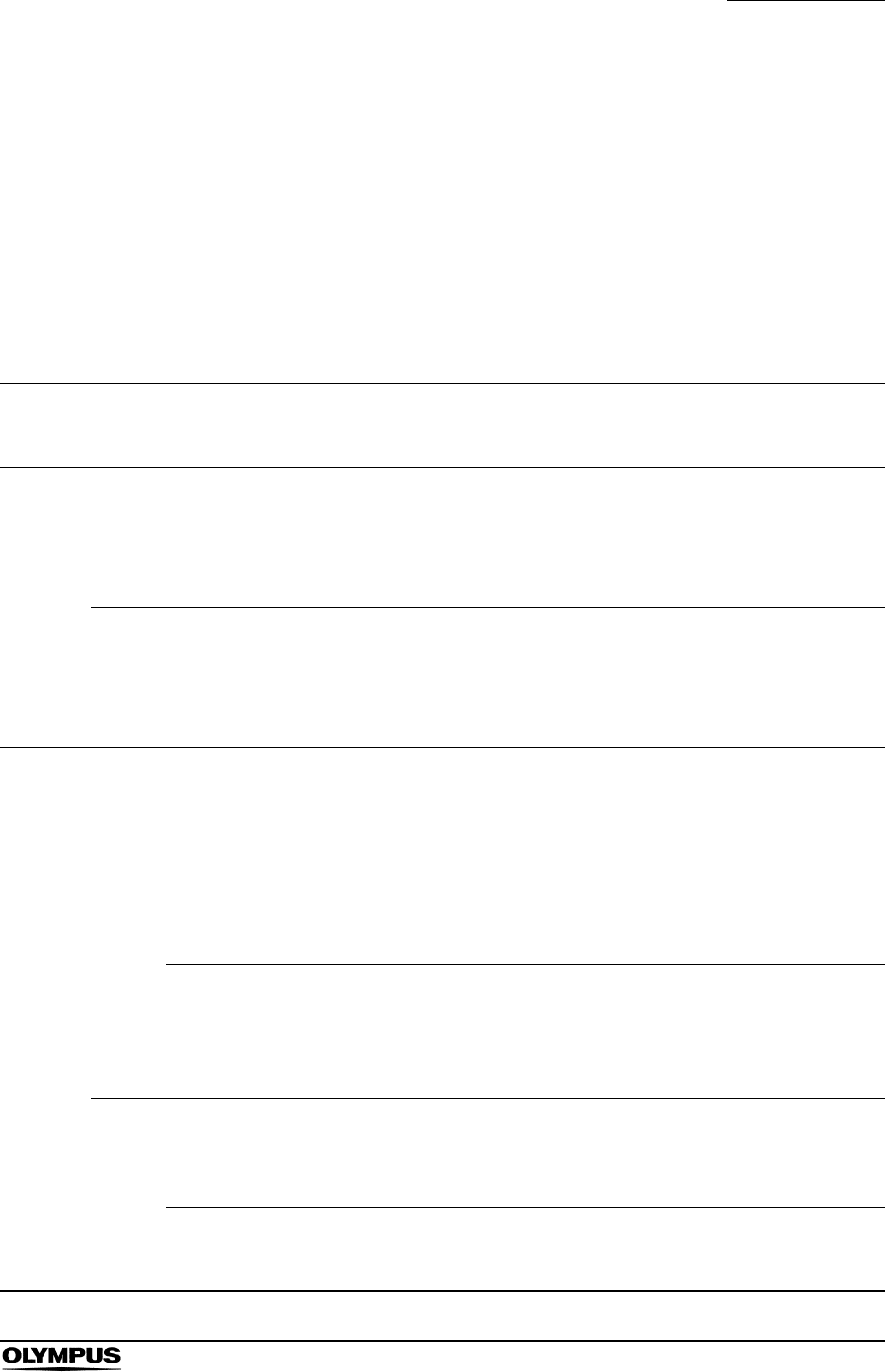
Chapter 5 Functions
71
EVIS EXERA II VIDEO SYSTEM CENTER CV-180
Brightness adjustment (Exposure)
The brightness of the live images can be adjusted. The lamp brightness is
automatically adjusted to keep the brightness of the image constant when the
light source is set to “AUTO”. The lamp brightness is manually adjusted to keep
the lamp brightness constant at the setting value when the light source is set to
“MAN.”. The function differs depending on the following conditions:
• The light source and its settings.
• The setting of the electric shutter function
(refer to “Electronic shutter” on page 240)
Light
source
Light
source
mode
Endoscope’s
electronic
shutter
Brightness adjustment Brightness level indication
CLV-180 AUTO Independent of
ON/OFF of the
electronic shutter
function.
Adjust the brightness using the
“EXPOSURE” buttons on the front panel.
The lamp brightness is automatically
adjusted to keep the image brightness
constant.
• The level indicators of the
video system center and
light source are interlocked.
• The “EXPOSURE” indicator
is a dot display.
MAN. Automatically
becomes OFF.
Adjust the brightness using the
“EXPOSURE” buttons on the front panel.
The lamp brightness remains constant.
• The level indicators of the
video system center and
light source are interlocked.
• The “EXPOSURE”
indicator is a bar display.
Other
than
above
AUTO ON • Set the light intensity of the light
source to the center of the adjustable
range.
• Adjust the brightness using the
“EXPOSURE” buttons on the front
panel. The lamp brightness is
automatically adjusted to keep the
image brightness constant.
• The level indicators of this
instrument and light source
are not interlocked.
• The “EXPOSURE” indicator
is a dot display.
OFF Adjust the brightness using the
brightness adjustment buttons on the
light source. The lamp brightness is
automatically adjusted to keep the image
brightness constant.
The “EXPOSURE” indicator
goes OFF.
MAN. ON Do not use this setting (see page 74).
The light intensity reaches its greatest
value, and excessive heat of the
endoscope's distal end may occur.
OFF Adjust the brightness using the
brightness adjustment buttons on the
light source.
The “EXPOSURE” indicator
goes OFF.
Table 5.2
72
Chapter 5 Functions
EVIS EXERA II VIDEO SYSTEM CENTER CV-180
• Always use the minimum level of illumination necessary for
adequate viewing. Whenever possible, avoid close,
stationary viewing of mucous membranes for a long time.
Intense endoscopic illumination may cause mucosal burns.
• Do not bring the metal plug of the light guide and the distal
end of the endoscope immediately after use in contact with
the body and flammable objects because the parts will be
extremely hot.
• Be sure to set the brightness control mode of the light source
to manual mode or turn the examination lamp OFF before
disconnecting the camera head from the endoscope or
disconnecting the videoscope from the video system center.
Disconnecting them can increase the light intensity to the
maximum and may cause burns or eye injury.
Refer to the instruction manual of the endoscope for details
of the electric shutter function.
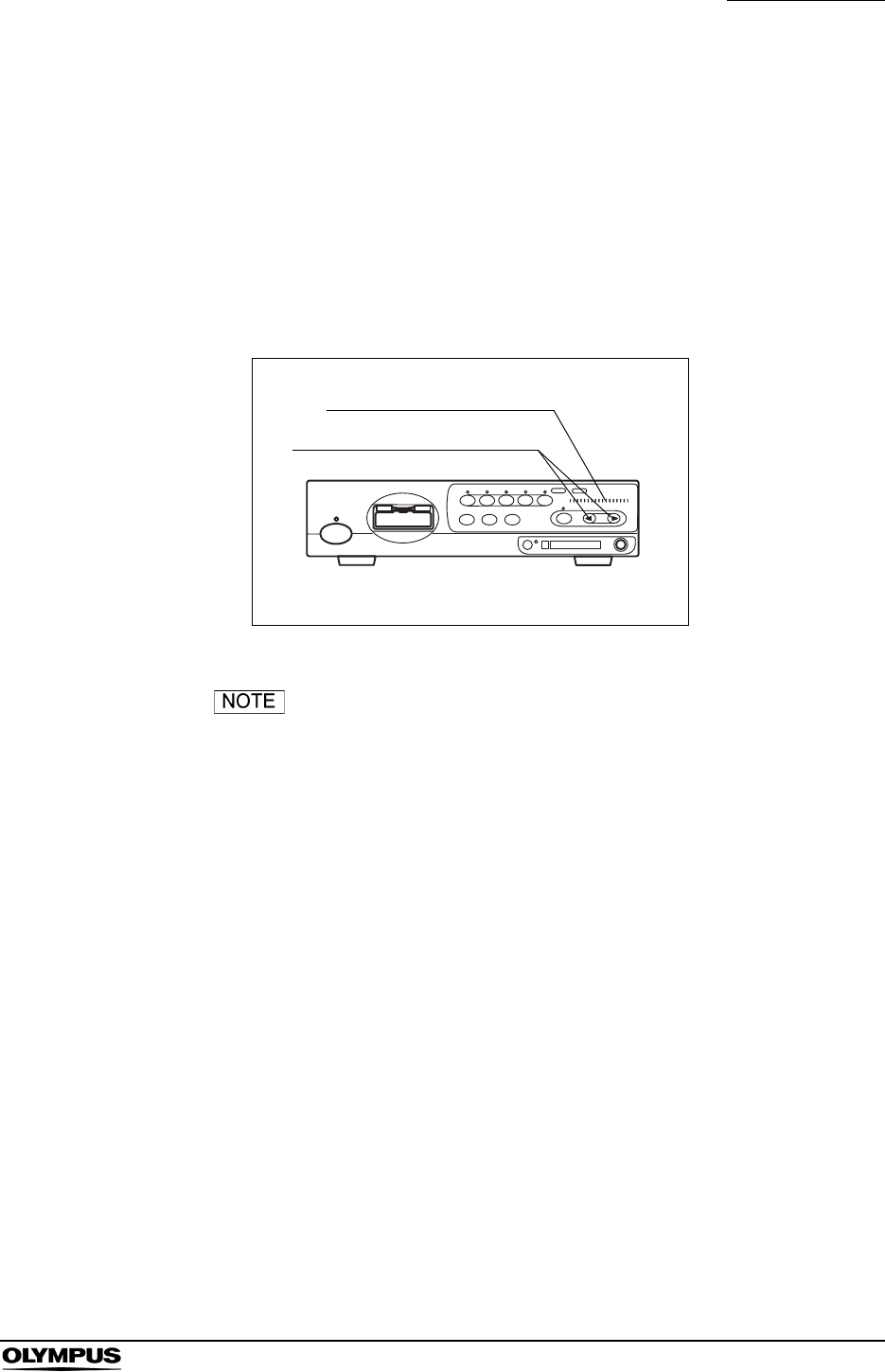
Chapter 5 Functions
73
EVIS EXERA II VIDEO SYSTEM CENTER CV-180
CLV-180
The video system center can adjust the brightness of the light source.
1. Set the brightness control of the light source to “AUTO” or “MAN.” according
to the instruction manual of the light source.
2. Set the brightness, using the “EXPOSURE” buttons on the front panel (see
Figure 5.10). The brightness level is displayed on the “EXPOSURE”
indicator.
Figure 5.10
• Each time the “EXPOSURE” button is pressed the indicator
changes by 1 level. Pressing and holding the button,
changes the indicator continuously.
• The “EXPOSURE” indication is interlocked with the
brightness level indication of the connected CLV-180. When
the brightness adjustment buttons on the CLV-180 are
pressed, the “EXPOSURE” indication on the video system
center varies in an interlocked operation.
EXPOSURE indicator
EXPOSURE buttons
74
Chapter 5 Functions
EVIS EXERA II VIDEO SYSTEM CENTER CV-180
Other than CLV-180 in AUTO mode
The operation differs depending on the setting of the electronic shutter function
of the endoscope (refer to “Electronic shutter” on page 240).
When the electronic shutter function is ON:
1. Set the brightness level of the light source to the center of the adjustable
range.
2. Set the brightness using the “EXPOSURE” buttons on the front panel of the
video system center. The brightness level is displayed on the “EXPOSURE”
indicator.
When the brightness level of the light source is not in the
center of the adjustable range, intense light, which is not
recognized clearly, may cause burns and/or appropriate
brightness may not be obtained.
When the electronic shutter function is OFF:
Set the brightness as required, using the brightness adjustment buttons of the
light source. The brightness level is displayed on the light source.
The “EXPOSURE” buttons of the video system center is
invalid, and the “EXPOSURE” indicator goes OFF.
Other than CLV-180 in MANUAL mode
Never set the electronic shutter function to ON in the user
preset menu (refer to “Electronic shutter” on page 240).
Intense light, which is not recognized clearly, may cause
burns.
Set the brightness as required, using the brightness adjustment buttons of the
light source. The brightness level is displayed on the light source.
The “EXPOSURE” buttons of the video system center is
invalid, and the “EXPOSURE” indicator goes OFF.
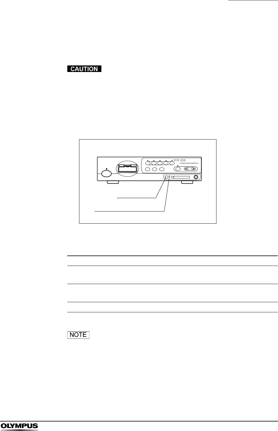
Chapter 5 Functions
75
EVIS EXERA II VIDEO SYSTEM CENTER CV-180
STOP button and PC card indicator
This button stops accessing the PC card. Press the button before ejecting the
PC card from the PC card slot.
Be sure to press the “STOP” button and confirm that the PC
card status indicator goes OFF before ejecting the PC card
from the PC card slot. Otherwise, the PC card and/or data
stored may be destroyed.
1. Press the “STOP” button before removing the PC card. The PC card status
indicator goes OFF.
Figure 5.11
The PC card indicator shows the status as shown below.
The images cannot be recorded when the PC card is not
ejected after pressing the “STOP” button. Press the eject
button to eject the PC card after a few seconds, then insert
the PC card again to let the video system center recognize
the PC card.
PC card indicator Status
OFF No PC card in the slot, or the video system center does
not recognize the PC card.
Green The PC card is in the slot, and the video system indicator
has recognized the PC card.
Orange (blinking) The video system indicator is accessing the PC card.
Table 5.3
STOP button
PC card status indicator
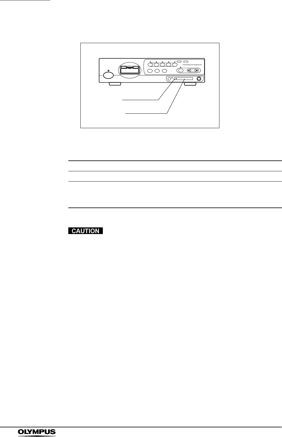
76
Chapter 5 Functions
EVIS EXERA II VIDEO SYSTEM CENTER CV-180
PC card slot and eject button
Figure 5.12
Table 5.4 shows the applicable PC card adapter and Memory Card.
• Be sure to confirm the followings. Otherwise, recording or
playing back images on the PC card may not be possible.
Format the PC card before the first use as described in
“Formatting of the PC card” on page 114.
Use the CV-180 for formatting the PC card. Do not use a
personal computer, etc.
• Be sure to confirm the followings. Otherwise, the PC card
and the data in the PC card may be destroyed.
Do not press the “STOP” button while the PC card status
indicator is blinking.
Do not press the “STOP” button while formatting the PC
card.
Press the “STOP” button and confirm if the PC card status
indicator goes OFF before ejecting the PC card from the
PC card slot.
Handle the PC card with care and avoid subjecting them
to sudden or severe impact.
Do not place the PC card in a place subject to strong
static electricity, electricity or magnetism.
Applicable device Specification
PC card adopter MAPC-10 (Olympus)
Memory Card xD picture card
M-XD32P, M-XD64P, M-XD128P, M-XD256P, M-XD512P,
M-XD1GM, M-XD1GMA, M-XD2GMA (Olympus)
Table 5.4
Eject button
PC card slot
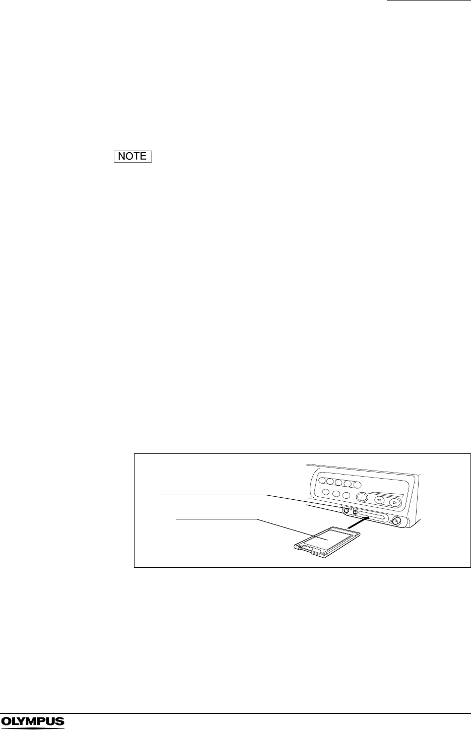
Chapter 5 Functions
77
EVIS EXERA II VIDEO SYSTEM CENTER CV-180
Do not leave the PC card under high temperatures, high
humidity or in a corrosive atmosphere.
• Do not allow a foreign object to penetrate the inside of the PC
card slot. Otherwise, the equipment may be damaged.
• Do not touch the PC card with wet hands. The video system
center and/or data stored may be destroyed.
• See the following pages for details on how to record the PC
card.
Images:
Section 5.3, “Image recording and playback (PC card)” on
page 106.
Patient data:
“Recording patient data into PC card” on page 144.
“Loading patient data from PC card” on page 146.
• Please use only the PC card adapter and Memory Card from
Olympus. Other cards might not work properly.
• The PC card of CardBus type and Smart Media are not
available.
Insertion of PC card into the PC card slot
1. Insert the xD picture card into the PC card adapter.
2. Push the PC card adapter all the way into the PC card slot.
Figure 5.13
3. The video system center recognizes the PC card, and the PC card status
indicator lights up green.
PC card adapter
PC card status indicator
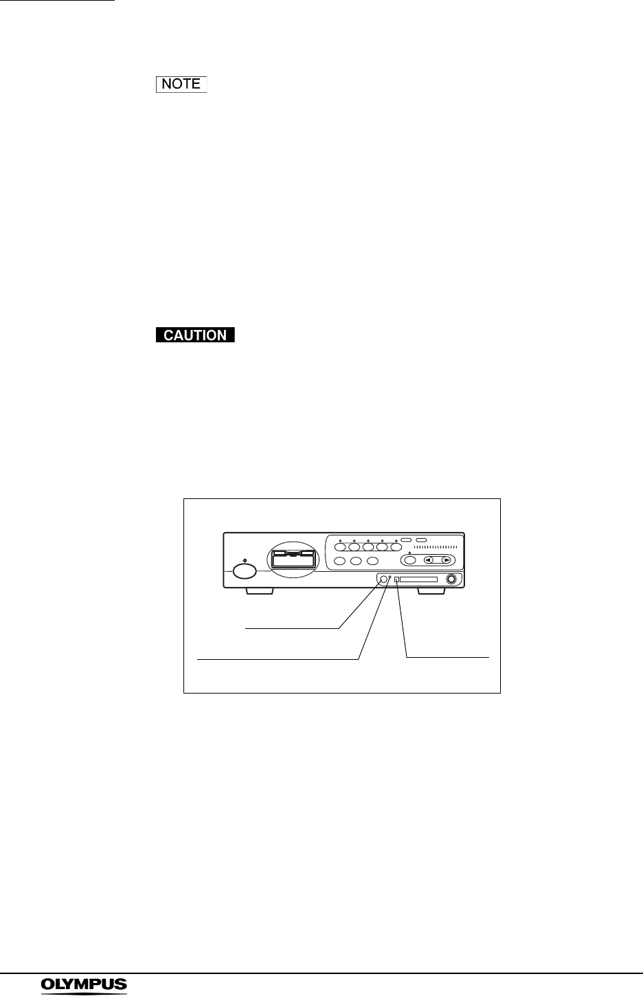
78
Chapter 5 Functions
EVIS EXERA II VIDEO SYSTEM CENTER CV-180
• If the video system center does not recognize the PC card,
eject and reinsert the PC card adapter, or turn the video
system center OFF then ON again, leaving the PC card
adapter inserted.
• Always prepare spare PC cards for use when the currently
used card becomes full.
• It is recommended to back up the image data on the PC card
into a personal computer regularly.
Ejection of PC card from the PC card slot
Be sure to press the “STOP” button and confirm that the PC
card status indicator goes OFF before ejecting the PC card
from the PC card slot. Otherwise, the PC card and/or data
stored may be destroyed.
1. Press the “STOP” button. The PC card status indicator goes OFF (see
Figure 5.14).
Figure 5.14
2. Press the eject button.
STOP button
PC card status indicator Eject button
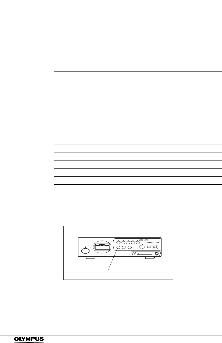
80
Chapter 5 Functions
EVIS EXERA II VIDEO SYSTEM CENTER CV-180
RESET button
The “RESET” button resets the settings modified during use of the video system
center to the original values. The settings to be reset are the following items:
• User preset data: To be reset to their original settings.
• Items shown in Table 5.5: To be reset to the factory defaults.
1. Press and hold the “RESET” button on the front panel (see Figure 5.16). All
front panel indicators should blink for about 1 second, then the settings are
reset.
Figure 5.16
Function Default setting
Image source Scope
Color tone R [0]
G[0]
C[0]
Freeze Live image
Release index 4 sec
Zoom x1.0
Special light observation Normal observation
Arrow pointer OFF
Stop watch OFF
Characters on screen Full display
Exposure Center
PinP OFF
Table 5.5
RESET button
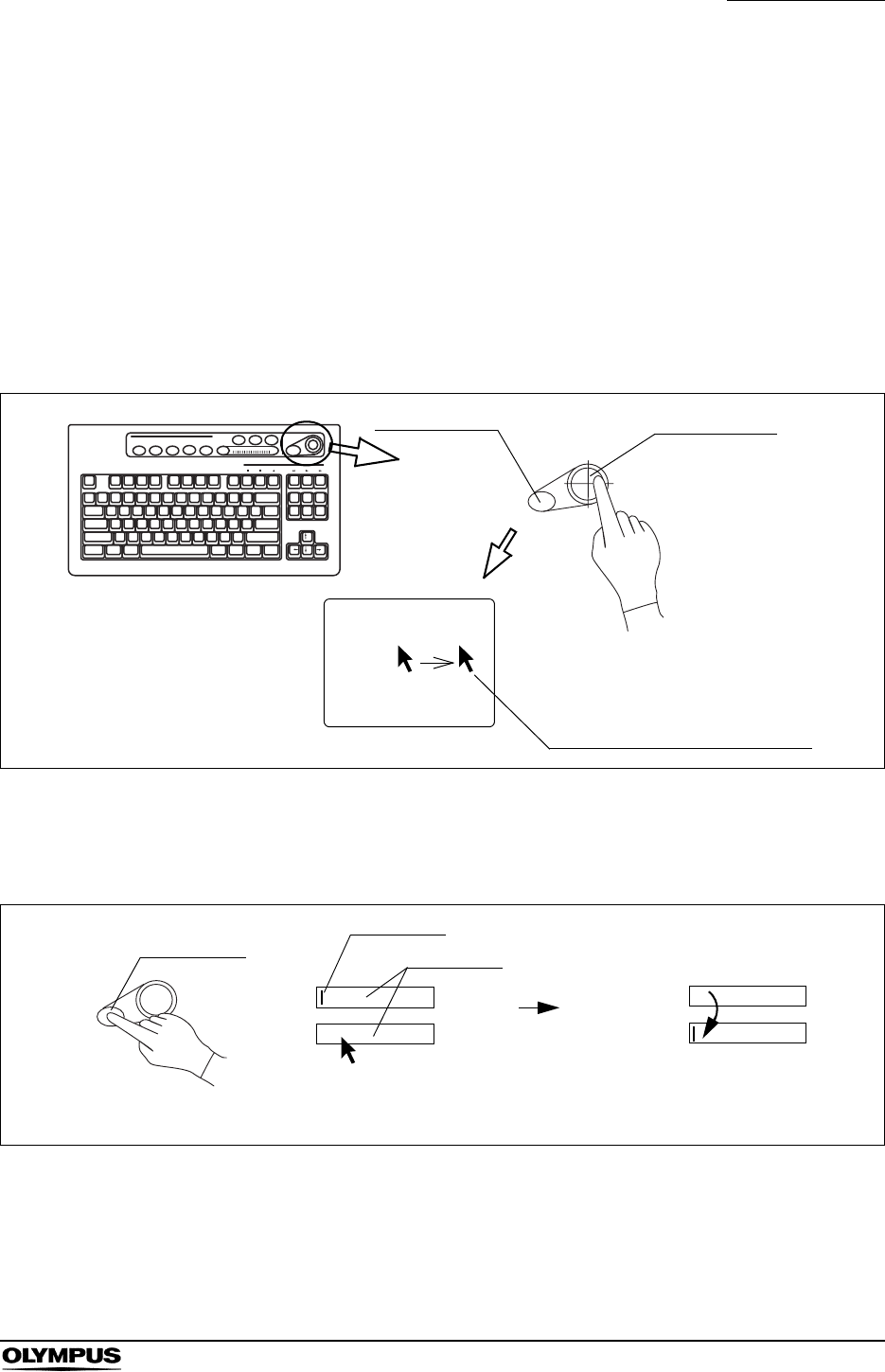
Chapter 5 Functions
81
EVIS EXERA II VIDEO SYSTEM CENTER CV-180
5.2 Keyboard
Domepoint
The domepoint is used to move the arrow pointer, and execute the function on
the screen or move the cursor on the screen. It refers as “clicking” to press the
“Click” key one time, placing the arrow pointer at the desired area.
1. Press the domepoint using your finger tip. The arrow pointer on the screen
moves to the direction corresponding to the pressed part of the domepoint.
Figure 5.17
2. Move the arrow pointer to a text box area. Click the text box to place the
cursor.
Figure 5.18
3. Click a button on the menu to move the highlight, or perform the function of
the button.
Arrow pointer on the screen
The arrow pointer moves to the right if
pressing the right side of the domepoint.
Domepoint
Click key
Monitor screen
Date
Time
Date
Time
Text box
Click key
The cursor moves.
Click the text box to place the cursor.
Cursor
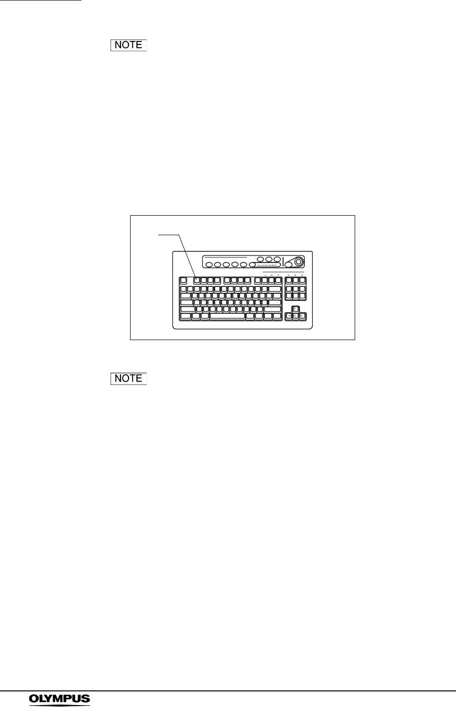
82
Chapter 5 Functions
EVIS EXERA II VIDEO SYSTEM CENTER CV-180
Also refer to Section 2.8, “Pointer” on page 33 for the
operation of cursor and highlight.
Clearing characters from the screen (“F1”)
This operation clears and redisplays the text information such as patient data on
the monitor screen.
Part of the patient data disappears from the monitor screen each time the “F1”
key is pressed. The fourth press redisplays the initial display showing all patient
data (see Figure 5.20 for the transition of the display).
Figure 5.19
• The zoom ratio of the image except “x1.0” is always
displayed on the monitor even when the text information is
cleared.
• Only when all text data are displayed on the monitor, the
patient data can be entered.
• The image size and layout may differ from those shown in
Figure 5.20 depending on the connected endoscope.
• The patient data can also be cleared using the scope
switches and/or foot switches. For how to set up the scope
switches and foot switches, see “Remote switch and foot
switch (EXERA and VISERA)” on page 219.
F1
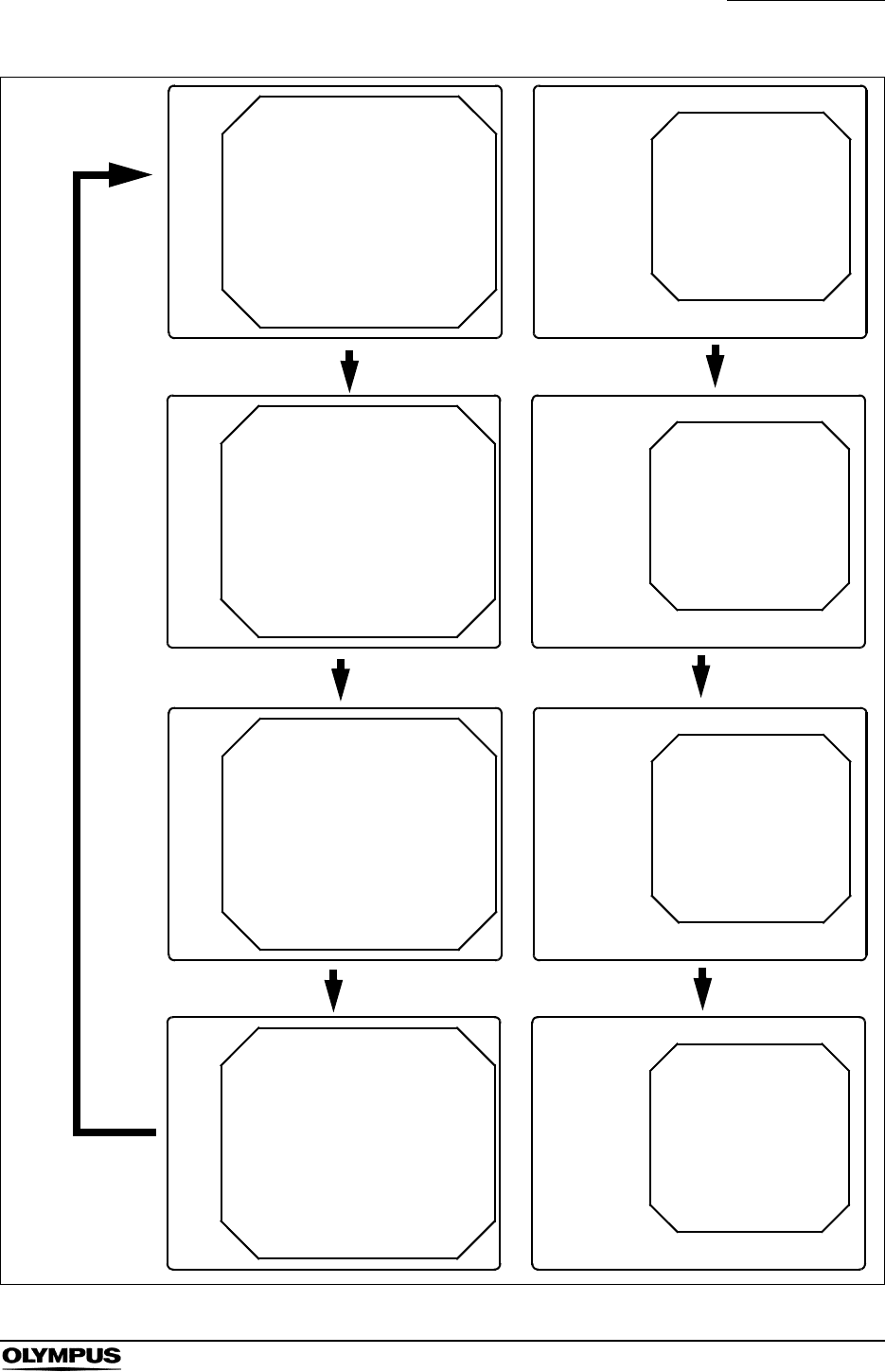
Chapter 5 Functions
83
EVIS EXERA II VIDEO SYSTEM CENTER CV-180
Figure 5.20
Z: x1.5
Z: x1.5
ABC123 Mike Johnson
12/12/2005
CVP: A4/4
D.F: 99
VCR
Ct: N Eh: A8
Z: x1.5
Pump
Media:
John Smith
Cardiac end of the stomach
ABC123
Mike Johnson
12/12/2005
CVP: A4/4
D.F: 99
VCR
Ct: N Eh: A8
Z: x1.5
Pump
Media:
John Smith
Cardiac end of the stomach
Press F1 key
Press F1 key
Press F1 key
Clear 1
All clear
Clear 2
Press F1 key
Full display
ABC123 Mike Johnson
M 51
03/03/1954
12/12/2005
12:12:12
CVP: A4/4
D.F: 99
VCR
Ct: N Eh: A8
Z: x1.5
Pump
Media:
John Smith
Cardiac end of the stomach
ABC123
Mike Johnson
M 51
03/03/1954
12/12/2005
12:12:12
CVP: A4/4
D.F: 99
VCR
Ct: N Eh: A8
Z: x1.5
Pump
Media:
John Smith
Cardiac end of the stomach
ABC123 Mike Johnson
Z: x1.5
ABC123
Mike Johnson
Z: x1.5
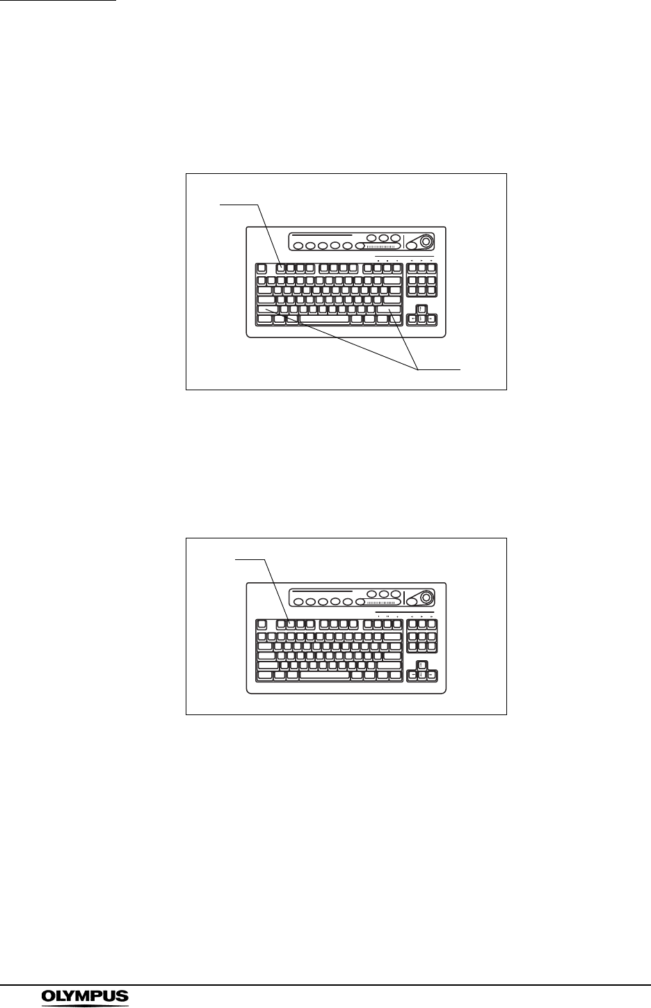
84
Chapter 5 Functions
EVIS EXERA II VIDEO SYSTEM CENTER CV-180
System setup (“Shift” + “F1”)
Press these keys to open the system setup menu that enables the proper use of
the video system center and ancillary equipment. For details on the system
setup menu, see Section 9.2, “System setup” on page 194.
Figure 5.21
Scope information (“F2”)
Press this key to open the scope information window. For details, see Section
5.7, “Scope information” on page 148.
Figure 5.22
F1
Shift
F2
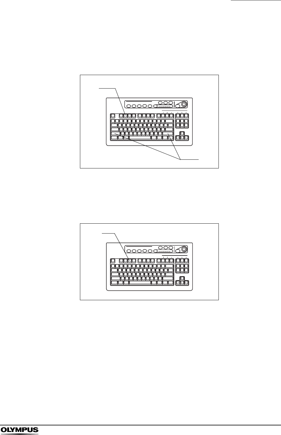
Chapter 5 Functions
85
EVIS EXERA II VIDEO SYSTEM CENTER CV-180
User preset (“Shift” + “F2”)
Press these keys to open the user preset menu that sets up the observation
conditions for each user (operator). For details on the user preset menu, see
Section 9.3, “User preset” on page 216.
Figure 5.23
Cursor (“F3”)
Press to switch the display of the cursor on the screen between ON and OFF.
Figure 5.24
F2
Shift
F3
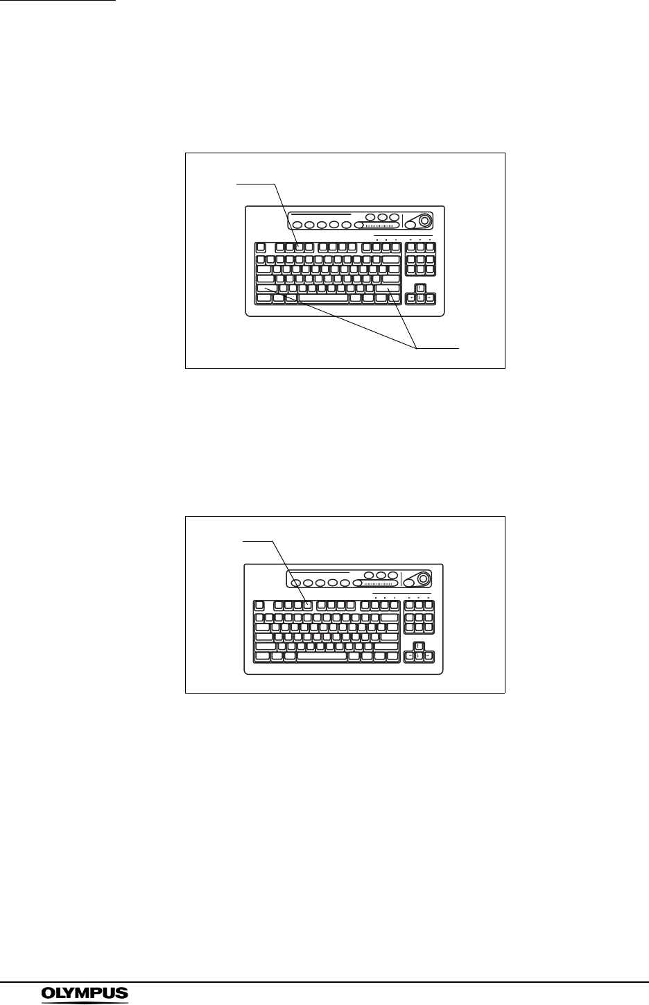
86
Chapter 5 Functions
EVIS EXERA II VIDEO SYSTEM CENTER CV-180
Patient data (“Shift” + F3)
Press these keys to open “Patient Data” menu to enter the patient data. For
details on the patient data menu, see “Entering new patient data” on page 137.
Figure 5.25
Freeze mode (“F4”)
This key switches between the two different freeze modes that pause the
endoscopic live image.
Figure 5.26
The freeze mode switches between “frame” and “field” each time the “F4” key is
pressed. For more information of the freeze mode, see “Freeze function” on
page 225.
F3
Shift
F4
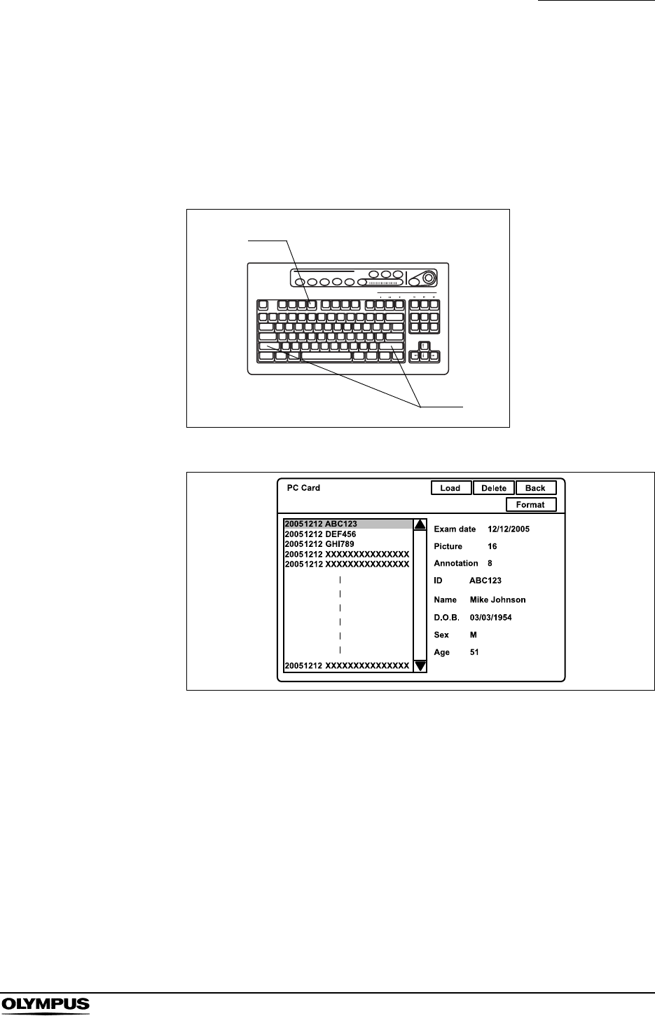
Chapter 5 Functions
87
EVIS EXERA II VIDEO SYSTEM CENTER CV-180
Browse (“Shift” + “F4”)
Press these keys to open the PC card menu. The PC card displays the
endoscopic images and patient data stored on the PC card. See “PC card menu”
on page 110.
Press the “Shift” and “F4” keys together to open the PC card menu.
Figure 5.27
Figure 5.28
F4
Shift
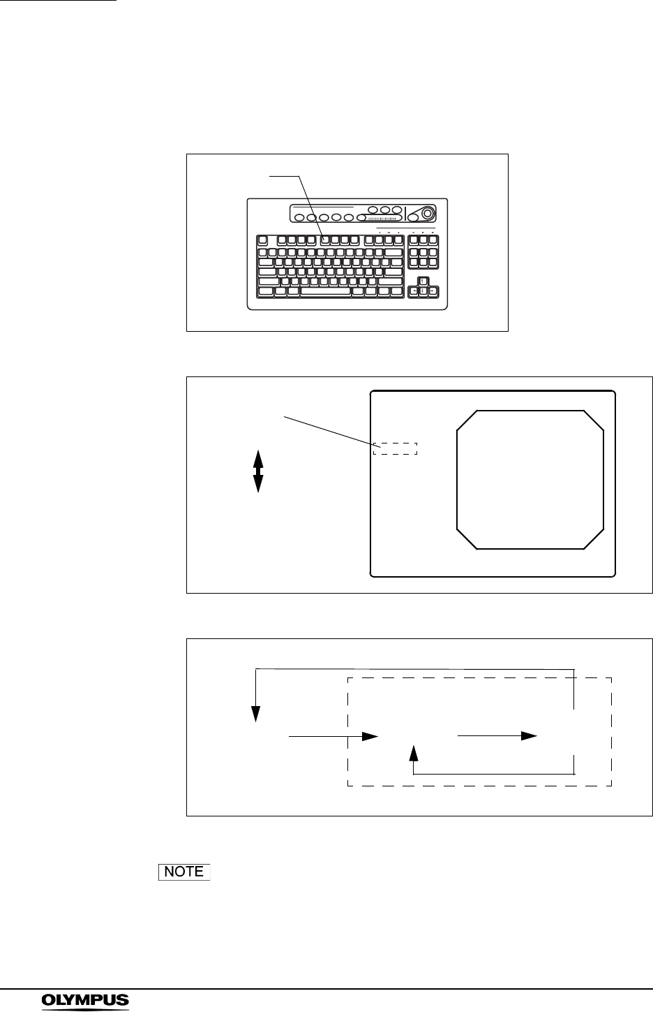
88
Chapter 5 Functions
EVIS EXERA II VIDEO SYSTEM CENTER CV-180
Stopwatch (“F5”)
Press the “F5” key to change the clock on the monitor screen to a stopwatch.
Use the “F5” key also to start and stop counting (see Figure 5.30).
Figure 5.29
Figure 5.30
Figure 5.31
The stopwatch operation can also be controlled from the
scope switches and/or foot switches. For how to set up the
scope switches and foot switches, see “Remote switch and
foot switch (EXERA and VISERA)” on page 219.
F5
Clock
12:12:12
(H : M : S)
Stop watch
<00:00:00>
(H : M : S)
ABC123
Mike Johnson
M 51
03/03/1954
12/12/2005
12:12:12
Ct: N Eh: A8
Z: x1.5
John Smith
Cardiac end of the stomach
Clock F5 F5
F5
Shift+F5
Stop watch
Timing
start
Timing
stop
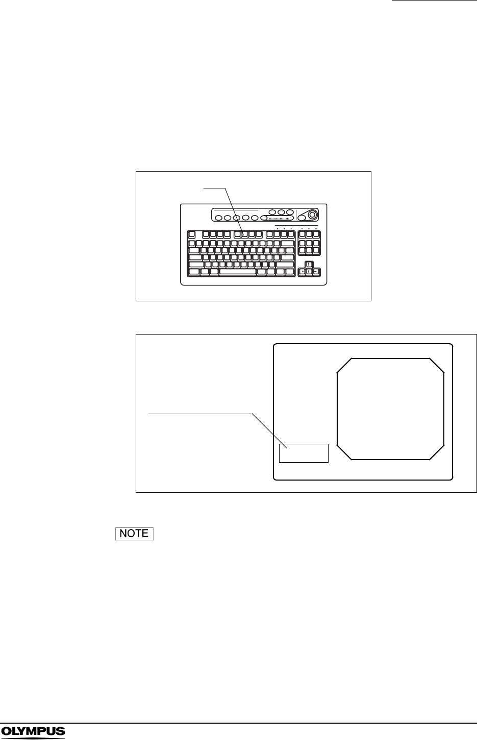
Chapter 5 Functions
89
EVIS EXERA II VIDEO SYSTEM CENTER CV-180
Automatic gain control (AGC) (“F6”)
If necessary set up the AGC function in advance. See “Auto gain control (AGC)”
on page 237.
1. The AGC function is turned ON and OFF alternately each time the “F6” key
is pressed. The AGC status, ON or OFF, is displayed on the monitor for
about 2 seconds (see Figure 5.33).
Figure 5.32
Figure 5.33
• Image noise may appear when AGC is ON.
• The AGC function cannot be switched ON or OFF while the
image is frozen.
• The AGC function used in the last operation before turning
the instrument OFF comes up, when the instrument is turned
ON.
• The AGC switching operation can also be controlled from the
scope switches and/or foot switches. For how to set up the
scope switches and foot switches, see “Remote switch and
foot switch (EXERA and VISERA)” on page 219.
F6
AGC ON/OFF window
ABC123
Mike Johnson
M 51
03/03/1954
12/12/2005
12:12:12
CVP: A4/4
D.F: 99
VCR
Ct: N Eh: A8
Z: x1.5
Pump
Media:
AGC
ON
John Smith
Cardiac end of the stomach
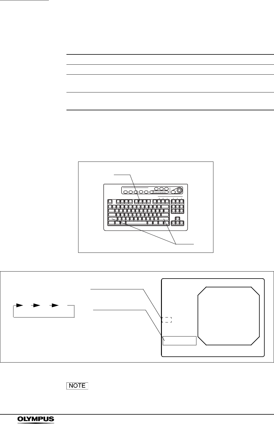
90
Chapter 5 Functions
EVIS EXERA II VIDEO SYSTEM CENTER CV-180
Contrast mode (“Shift” + “F6”)
Pressing these keys changes the contrast of the endoscopic image.
1. Press the “Shift” and “F6” keys together. Every time the two keys are
pressed, the contrast mode changes. The selected mode is displayed on
the monitor (see Figure 5.35).
Figure 5.34
Figure 5.35
• The contrast mode function does not work during the special
light observation.
Mode Explanation
Normal Standard setting
High Darkens the dark part and brightens the bright part compared to the
normal observation.
Low Brightens the dark part and darkens the bright part compared to the
normal observation.
Table 5.6
F6
Shift
Contrast indication
NHL
L: Low
H: High
N: Normal
ABC123
Mike Johnson
M 51
03/03/1954
12/12/2005
12:12:12
CVP: A4/4
D.F: 99
VCR
Ct: N Eh: A8
Z: x1.5
Pump
Media:
Contrast Normal
John Smith
Cardiac end of the stomach
Contrast window
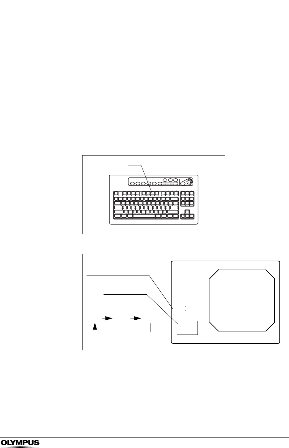
Chapter 5 Functions
91
EVIS EXERA II VIDEO SYSTEM CENTER CV-180
• The contrast mode can also be controlled from the scope
switches and foot switches. For how to set up the scope
switches and foot switches, “Remote switch and foot switch
(EXERA and VISERA)” on page 219.
Image zooming (“F7”)
The endoscopic image can electrically be magnified, when using the endoscope
compatible with the electronic zooming function (Scope 1, Scope 4, and Scope 5
in Table 9.30 on page 229). Three zoom ratios, x1, x1.2, x1.5, are available. The
image area size does not change.
Press the “F7” key to change the zoom ratio. The zoom ratio is displayed on the
monitor for about 2 seconds (see Figure 5.37).
Figure 5.36
Figure 5.37
F7
Zoom ratio indication
x1.2 x1.5
Transition of the zoom ratio
(example)
Zoom window
x1
ABC123
Mike Johnson
M 51
03/03/1954
12/12/2005
12:12:12
CVP: A4/4
D.F: 99
VCR
Ct: N Eh: A8
Z: x1.5
Pump
Media:
John Smith
Cardiac end of the stomach
Zoom
x 1.5

92
Chapter 5 Functions
EVIS EXERA II VIDEO SYSTEM CENTER CV-180
• The image area size can be changed during zooming (see
“Image size (“F8”)” on page 94). When the zoom ratio returns
to x1, the image area size also returns to the original size.
• While the image is frozen, the zoom function is not active.
• When the zoom ratio is x1, the zoom ratio is not displayed on
the monitor.
• This instrument always starts with zoom ratio x1.
• The zoom ratio is displayed at the lower left on the monitor
when the text information is cleared on the monitor.
• The zoom ratio can also be controlled from the scope
switches and/or foot switches. For how to set up the scope
switches and foot switches, see “Remote switch and foot
switch (EXERA and VISERA)” on page 219.
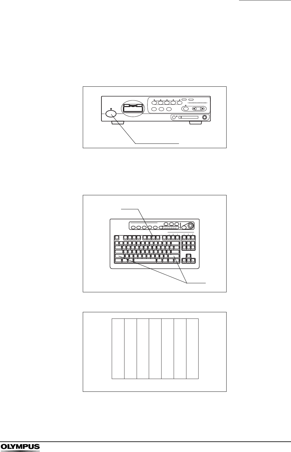
Chapter 5 Functions
93
EVIS EXERA II VIDEO SYSTEM CENTER CV-180
Color bar (“Shift” + “F7”)
The color bar is used to check the color tones of the monitor. The color bar can
be displayed when the monitor displays the endoscopic image.
1. Turn the video system center and the monitor ON (see Figure 5.38).
Figure 5.38
2. Press the “Shift” and “F7” keys together (see Figure 5.39). The color bar
appears on the monitor (see Figure 5.40).
Figure 5.39
Figure 5.40
3. Confirm that all colors of the color chart are displayed properly.
Power switch
F7
Shift
Color bar
White
Yellow
Cyan
Magenta
Red
Blue
Green
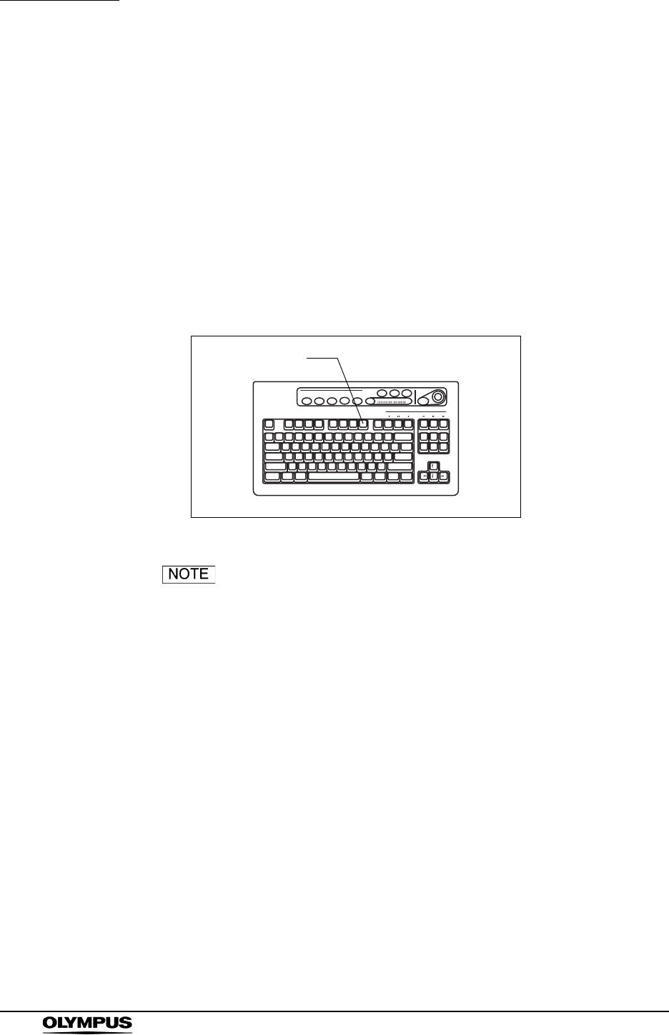
94
Chapter 5 Functions
EVIS EXERA II VIDEO SYSTEM CENTER CV-180
4. If the colors do not appear properly, adjust them according to the instruction
manual of the monitor.
5. Press the “Shift” and “F7” keys together (see Figure 5.39) to return to the
endoscopic image screen.
Image size (“F8”)
The size of the observation image on the monitor can be changed (see Table
9.31 in “Image size” on page 229). The available image sizes are variable
depending on the endoscope.
The image size changes each time the “F8” key is pressed.
Figure 5.41
• When the video system center is turned ON, the image size
used in the last operation before the instrument is turned
OFF comes up when the instrument is turned ON.
• The image size selection operation can also be controlled
from the scope switches and/or foot switches. For how to set
up the scope switches and foot switches, see “Remote switch
and foot switch (EXERA and VISERA)” on page 219.
F8
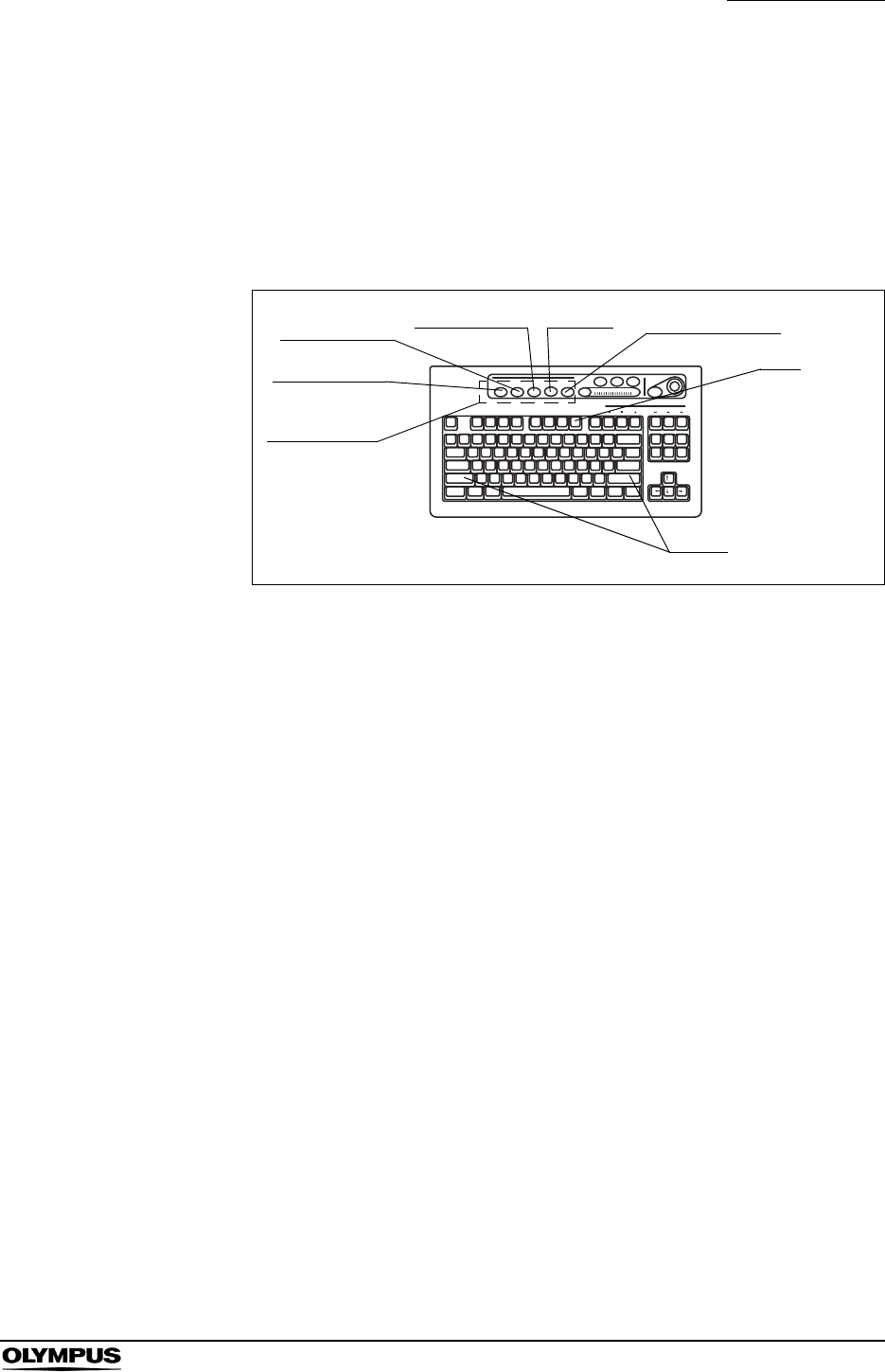
Chapter 5 Functions
95
EVIS EXERA II VIDEO SYSTEM CENTER CV-180
Printer lock (“Shift” + “F8”)
All keys of the “PRINTER REMOTE” on the keyboard can be locked. During this
time, the video printer can be operated using the keys on the printer.
1. Press the “Shift” and “F8” keys together to lock the “PRINTER REMOTE”
keys.
Figure 5.42
2. To unlock the keys, press the “Shift” and “F8” keys together again.
Shift
F8
PRINTER
REMOTE
#PER PAGE
CAPTURE DEL IMAGE PRINT PRINT QTY.
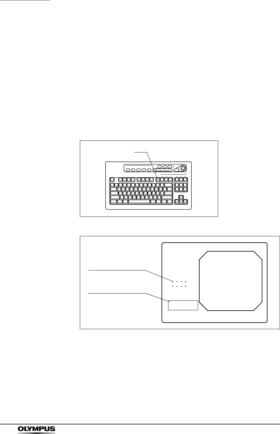
96
Chapter 5 Functions
EVIS EXERA II VIDEO SYSTEM CENTER CV-180
Image enhancement (“F9”)
This key changes the image enhancement modes. The three types and level of
the image enhancement should be set in advance. For the setup procedure, see
“Image enhancement (normal observation)” on page 226 and “Image
enhancement (NBI observation)” on page 250.
1. Press the “F9” key to display the current enhancement mode for a few
seconds.
2. During displaying the mode on the monitor, press the “F9” key to change the
enhancement mode (ex. A1, A3, etc.). The display window appears for a
few seconds.
Figure 5.43
Figure 5.44
F9
Image enhancement
indication
Image enhancement
window
ABC123
Mike Johnson
M 51
03/03/1954
12/12/2005
12:12:12
CVP: A4/4
D.F: 99
VCR
Ct: N Eh: A8
Z: x1.5
Pump
Media:
John Smith
Cardiac end of the stomach
Enhance:A1
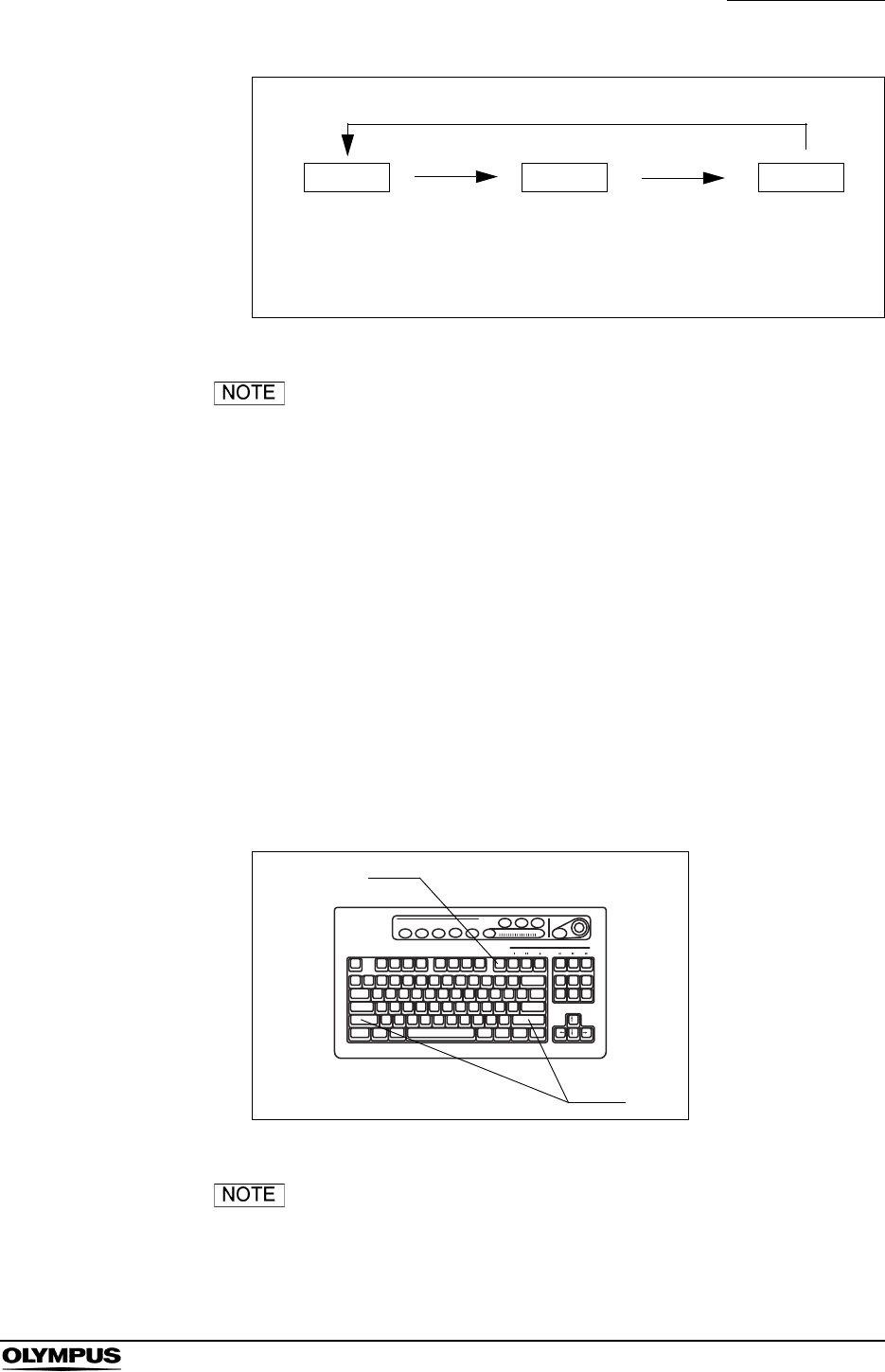
Chapter 5 Functions
97
EVIS EXERA II VIDEO SYSTEM CENTER CV-180
Figure 5.45
• When this instrument is turned ON, the enhancement mode
used in the last operation before the instrument is turned
OFF comes up when the instrument is turned ON.
• The image enhancement mode selection operation can also
be controlled from the scope switches and/or foot switches.
For how to set up the scope switches and foot switches, see
“Remote switch and foot switch (EXERA and VISERA)” on
page 219.
White balance adjustment (“Shift” + “F9”)
White balance is an adjustment function to display the appropriate color of the
image on the monitor. The operation of these two keys is the same as the “Wh/B”
button on the front panel. See Section 4.5, “White balance adjustment” on
page 52.
Figure 5.46
The white balance adjustment can also be initiated from the
scope switches and/or foot switches. For how to set up the
scope switches and foot switches, see “Remote switch and
foot switch (EXERA and VISERA)” on page 219.
Mode 3
Mode 2
F9
Mode 1 F9 F9
Transition of the image enhancement mode
F9
Shift
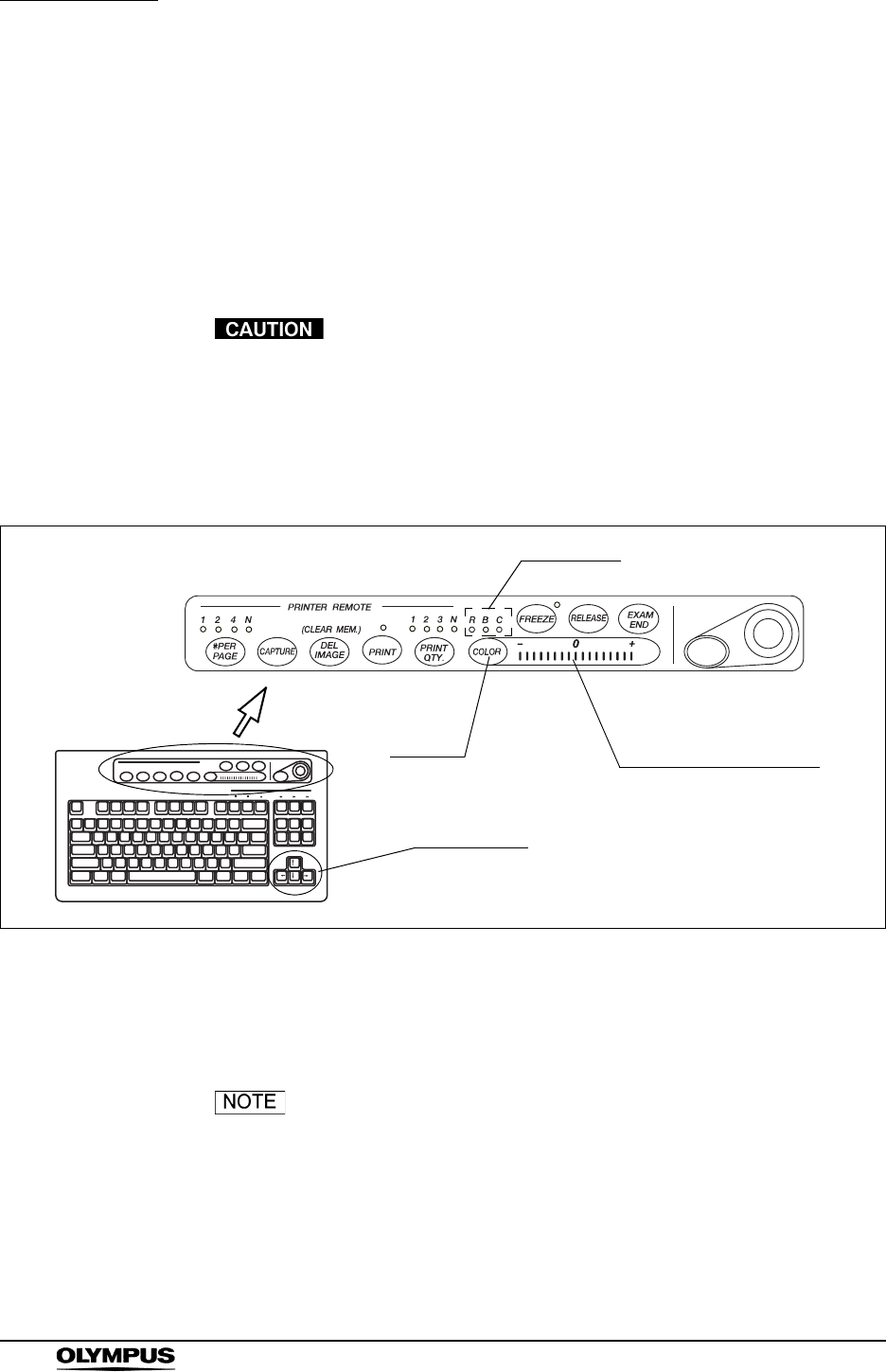
98
Chapter 5 Functions
EVIS EXERA II VIDEO SYSTEM CENTER CV-180
Color tone adjustment (“COLOR”)
This key is used for the adjustment of R (red), B (blue) and C (chroma) of the
observation image on the monitor. The adjustment level is displayed by the color
level indicator on the keyboard (see Figure 5.47).
This adjustment is effective for the normal observation and NBI observation. The
normal observation has the common setting of R, B and C. NBI observation has
its own setting.
Complete the white balance adjustment before the color tone
adjustment. Otherwise, the color tone is not adjusted
properly.
1. Press the “COLOR” key to select R (red), B (blue) or C (chroma) to adjust.
The indicator above the key lights up to show the tone selected.
Figure 5.47
2. Press the arrow keys to adjust the level of the selected tone.
3. Press the “COLOR” key to select the next tone and adjust it the same way.
• When the keyboard is left untouched for more than
10 seconds after selecting the tone, the selection is canceled
automatically and the indicator goes OFF.
• When the video system center is turned ON, the color tone
used in the last operation before the instrument is turned
OFF comes up when the instrument is turned ON.
Color tone
COLOR Color level indicator
Arrow keys
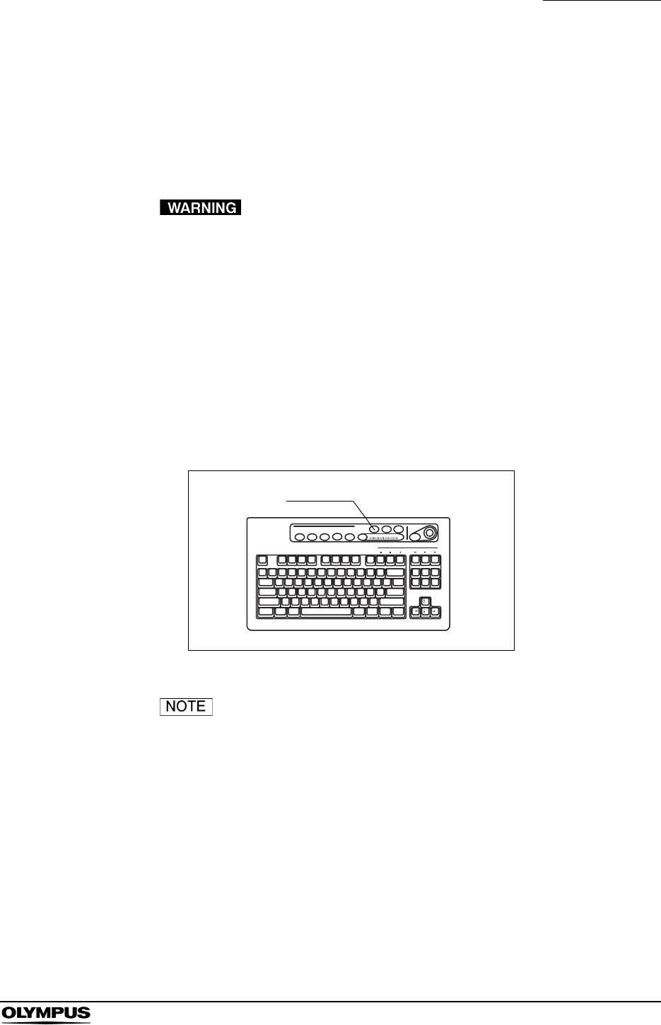
Chapter 5 Functions
99
EVIS EXERA II VIDEO SYSTEM CENTER CV-180
Freeze (“FREEZE”)
This key pauses the endoscopic live image for recording or observation.
There are two types of freeze function: “field freeze” and “frame freeze”. The
type of freeze function to start with can be set in advance in the user preset
menu. See “Freeze function” on page 225.
If the frozen image does not return to the live image, turn this
instrument OFF then ON again. Also turn the ancillary
equipment OFF then ON again by referring to the respective
instruction manual. If the image is still frozen, immediately
stop using this instrument and withdraw the endoscope
gently from the patient's body by referring to the instruction
manual of the endoscope.
1. Press the “FREEZE” key (see Figure 5.48) to freeze the endoscopic image.
2. Press the “FREEZE” key again and the frozen image returns to the live
image.
Figure 5.48
• The frozen image also returns to the live image by pressing
any key on the keyboard, except for the following keys.
“F1”, “F4”, “F5”, “F8”, “F9”, “Shift” + arrow keys.
• The frozen image might be blurred, when a fast motion has
been captured.
• A frozen image in the frame freeze mode is more easily
blurred than in the field freeze mode.
• During freezing, the size of the image area and image
enhancement can be changed.
FREEZE
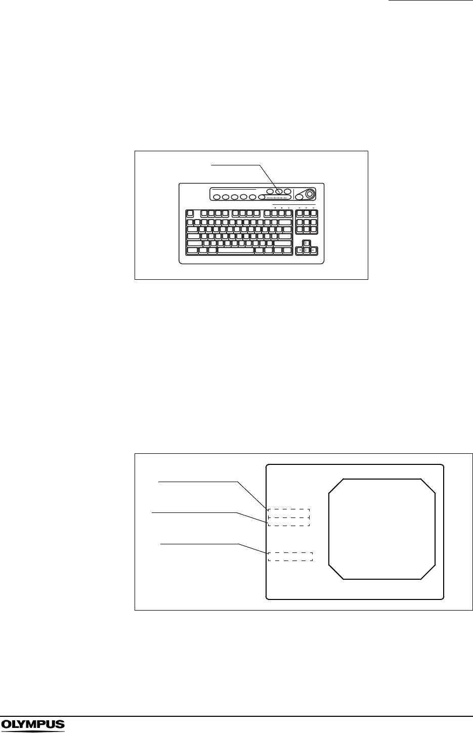
Chapter 5 Functions
101
EVIS EXERA II VIDEO SYSTEM CENTER CV-180
Release (“RELEASE”)
The key is used for image recording of the devices listed below. The device to be
controlled by this key has to be set in advance in the user preset menu
(Release1 in the user preset menu). See “Release function” on page 223.
• Video printer, PC card, Image filing system
Figure 5.49
1. Press the “RELEASE” key to record the endoscopic image on the recording
devices assigned. The live image pauses for a few seconds.
2. The counter of the recording devices on the monitor changes.
• Video printer: Increments the counter.
• Image filing system: Increments the counter.
• PC card: Displays the storage level. (see “Storage level of the PC card”
on page 106).
Figure 5.50
RELEASE
Video printer
Image filing system
PC card
ABC123
Mike Johnson
M 51
03/03/1954
12/12/2005
12:12:12
CVP: A4/4
D.F: 99
VCR
Ct: N Eh: A8
Z: x1.5
Pump
Media:
John Smith
Cardiac end of the stomach
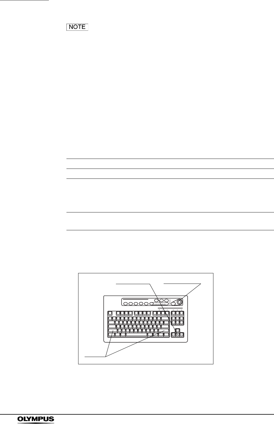
102
Chapter 5 Functions
EVIS EXERA II VIDEO SYSTEM CENTER CV-180
• The release operation can also be controlled from the scope
switches and/or foot switches. For how to set up the scope
switches and foot switches, see “Remote switch and foot
switch (EXERA and VISERA)” on page 219.
• Before activating the PinP function make sure that an
endoscope is connected to this instrument. If no endoscope
is connected it might not be possible to use the PinP function
and image recording of an external device may not work.
Arrow pointer (“Shift” + arrow keys and domepoint)
The arrow pointer can be displayed on the monitor. The arrow pointer is used to
mark a desired position in the endoscopic image, or to click the menus. The
display of the arrow pointer depends on each screen.
1. Press the “Shift” and any arrow key (see Figure 5.51). The arrow pointer
appears in the center of the endoscopic image (see Figure 5.52).
Figure 5.51
Screen Display of the arrow pointer
Endoscopic image screen Press “Shift” and any arrow key.
System setup menu
User preset menu
Patient data menu
Scope information menu
Always displayed
PC card menu Always displayed on the other than full image screen
Press “Shift” and any arrow key in the full image screen.
Table 5.7
Shift
Arrow keys Domepoint
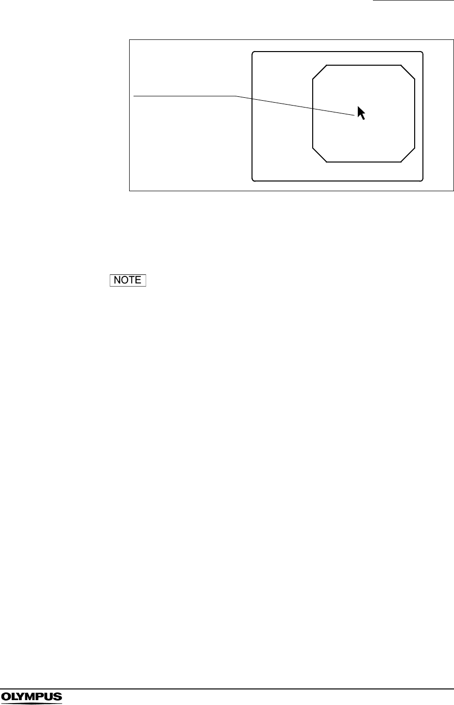
Chapter 5 Functions
103
EVIS EXERA II VIDEO SYSTEM CENTER CV-180
Figure 5.52
2. Move the arrow pointer using the domepoint on the keyboard.
3. To delete the arrow pointer, press the “Shift” and any arrow key.
• The arrow pointer disappears when the endoscopic image
screen once changes to the other menu and returns to the
endoscopic image screen.
• The arrow pointer can also be displayed and cleared from the
scope switches and/or foot switches. For how to set up the
scope switches and foot switches, see “Remote switch and
foot switch (EXERA and VISERA)” on page 219.
ABC123
Mike Johnson
M 51
03/03/1954
12/12/2005
12:12:12
John Smith
Cardiac end of the stomach
Arrow pointer
Appears at the center of
the endoscopic image.

104
Chapter 5 Functions
EVIS EXERA II VIDEO SYSTEM CENTER CV-180
Color mode (“Shift” + “Alt” + “1”, “2”, “3”, “4”)
These keys change the color tone of the monitor. This function is valid only for
Scope B, C, and D in Table 9.33 on page 233. This function is not active for NBI
observation because NBI observation uses the default setting for each
endoscope. See “Color mode” on page 228 for the initial mode.
Press “Shift”, “Alt”, and one of the “1”, “2”, “3”, or “4” keys together. The color
mode changes.
Key Color mode Color tone of the display
Shift + Alt + 1 Mode 1 Same as the color mode 1 of VISERA video
system center OTV-S7V
Shift + Alt + 2 Mode 2 Less reddish color than the Mode 1
Shift + Alt + 3 Mode 3 More yellowish color than the Mode 1
Shift + Alt + 4 Mode 4 Standard color mode
Table 5.8
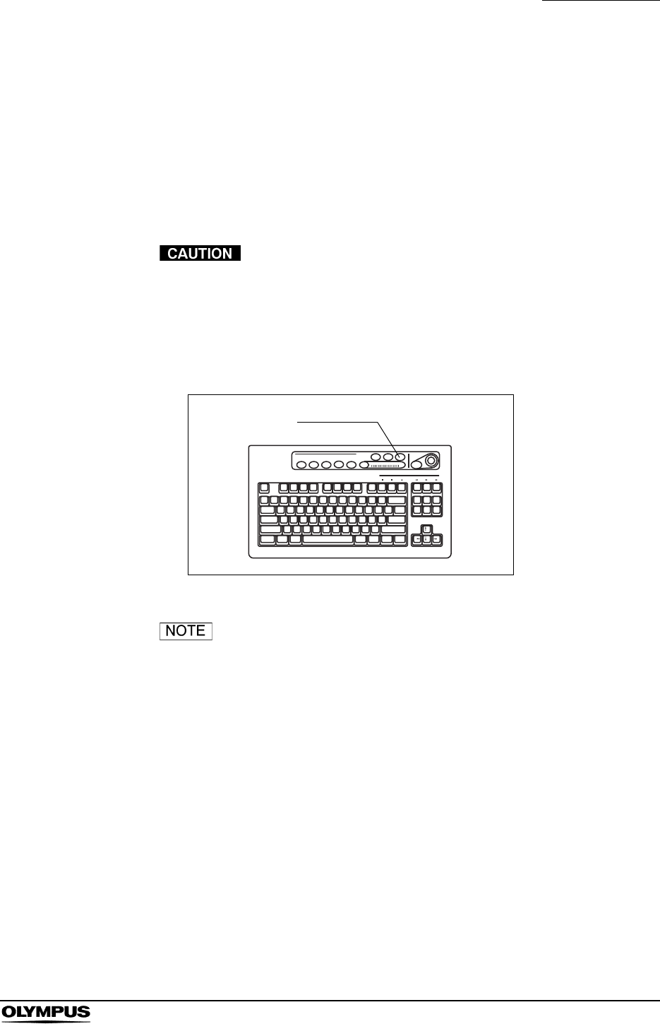
Chapter 5 Functions
105
EVIS EXERA II VIDEO SYSTEM CENTER CV-180
Ending examination (“EXAM END”)
The following steps are executed at the end of each examination procedure
using this key;
• Clearing the displayed patient data on the monitor;
• Completing printing un-printed printer image;
• Close processing of image filing system.
Do not disconnect the video connector before turning the
video system center OFF. Otherwise, the endoscope or
camera head may be damaged.
Press the “EXAM END” key. The patient data on the monitor disappears and the
data processing (e.g. to the PC card) is stopped.
Figure 5.53
The patient name that is once displayed in the endoscopic
image turns gray in the patient list after finishing the
observation by pressing the “EXAM END” key.
EXAM END
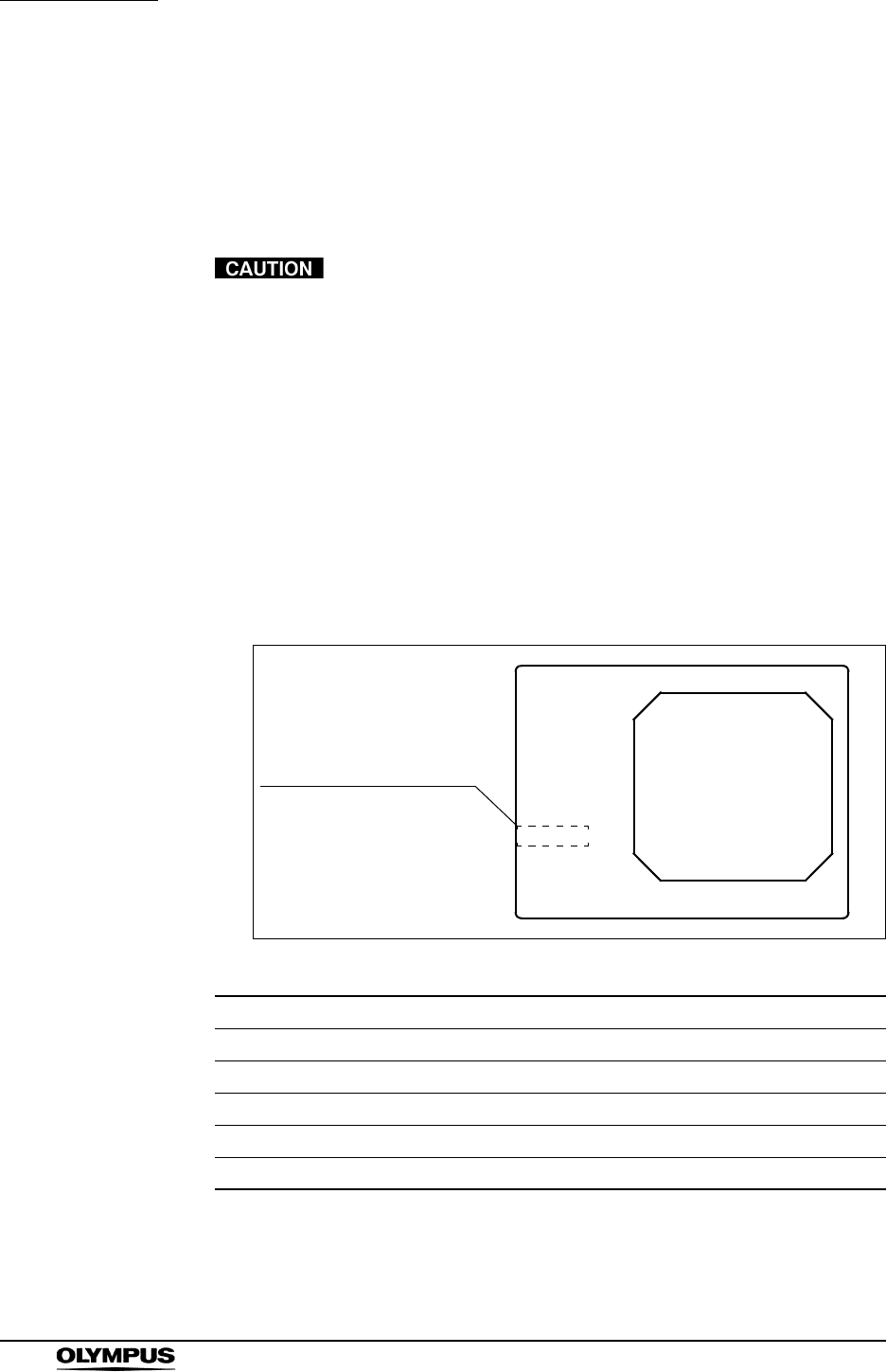
106
Chapter 5 Functions
EVIS EXERA II VIDEO SYSTEM CENTER CV-180
5.3 Image recording and playback (PC card)
This section explains how to record and playback images from the PC card. See
“STOP button and PC card indicator” on page 75 and “PC card slot and eject
button” on page 76 for the operation of the PC card slot.
• Be sure to format the PC card before recording images.
Otherwise, recording cannot be performed properly. See
“Formatting of the PC card” on page 114.
• Be sure to format the PC card using the CV-180. The PC
card that is formatted by a personal computer may cause
malfunction in recording or playback.
Storage level of the PC card
The storage level of the PC card is displayed on the monitor when the PC card is
inserted into the PC card slot. A warning message appears on the monitor when
the storage level runs out the space.
Figure 5.54
Display Rough number of recordable images
Media: 50 or more images
Media: Less than 50 images
Media: Less than 30 images
Media: Less than 20 images
Media: Full Storage device is full.
Table 5.9
ABC123
Mike Johnson
M 51
03/03/1954
12/12/2005
12:12:12
Media:
John Smith
Cardiac end of the stomach
Indication of the storage
level of the PC card
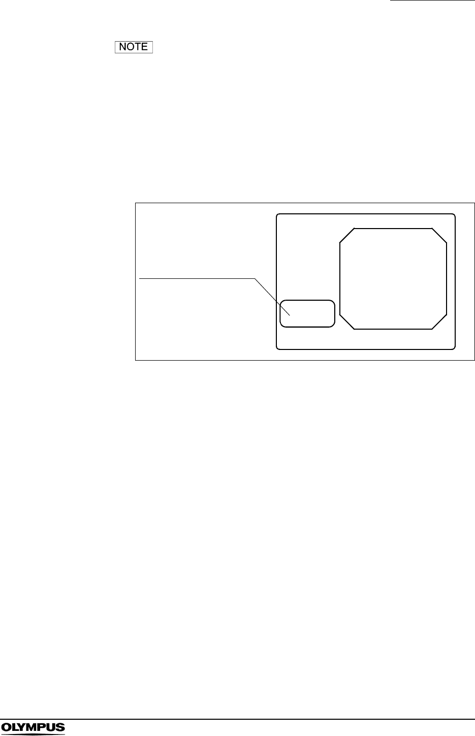
Chapter 5 Functions
107
EVIS EXERA II VIDEO SYSTEM CENTER CV-180
• The storage level is a rough estimation based on the
recording format (refer to “Recording format for PC card” on
page 224). Prepare spare PC cards before the storage
device runs out the space.
• If the storage level becomes less than 30 images during the
examination or if the first release operation is done after
turning the video system center ON when the storage level is
less than 30 images, a message appears on the screen (see
Figure 5.55).
Figure 5.55
ABC123
Mike Johnson
M 51
03/03/1954
12/12/2005
12:12:12
Media:
John Smith
Cardiac end of the stomach
Message window of the
storage level of the PC
card
Media:
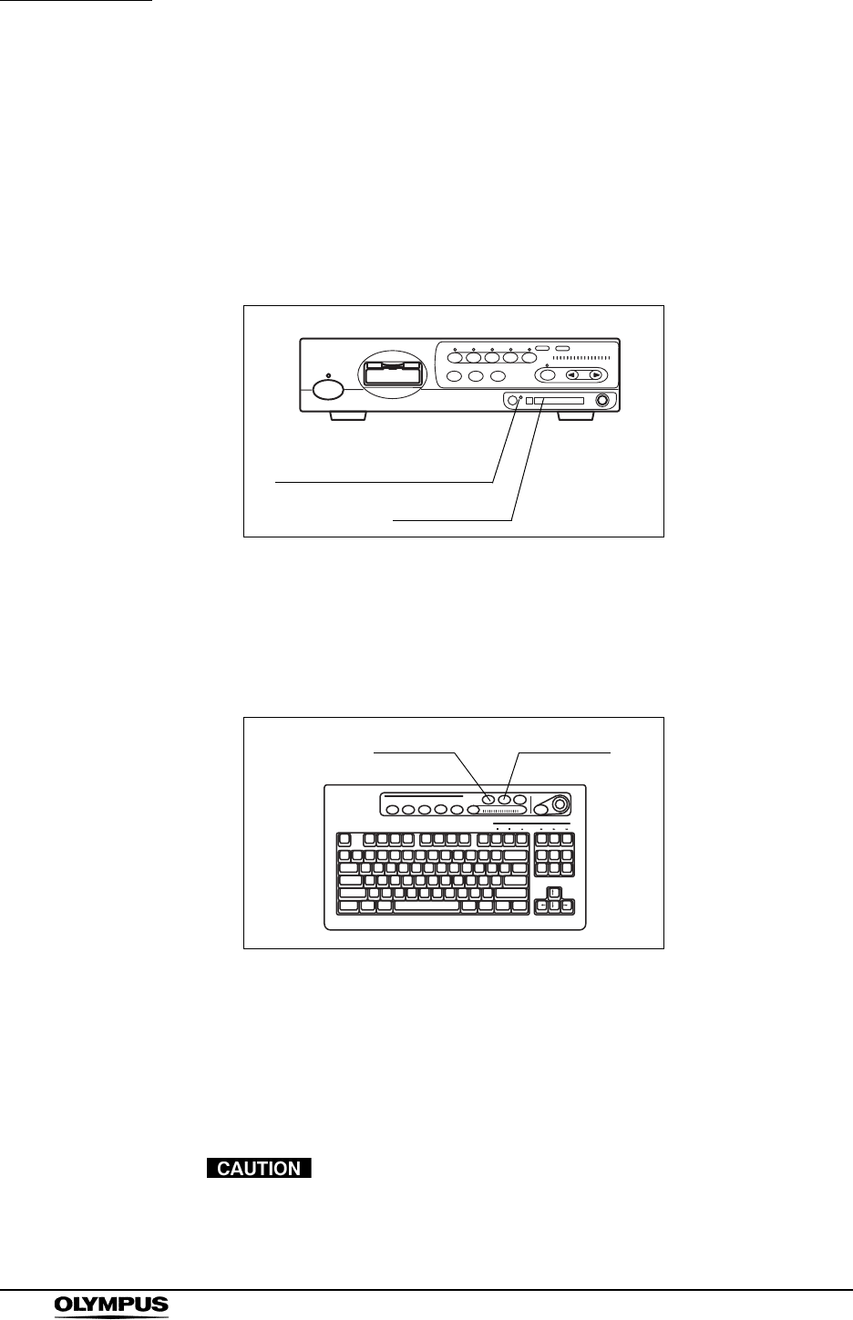
108
Chapter 5 Functions
EVIS EXERA II VIDEO SYSTEM CENTER CV-180
Recording the frozen image on a PC card
Assign the “PC Card” to “RELEASE 1” or “RELEASE 2” in the user preset menu
in advance. See “Remote switch and foot switch (EXERA and VISERA)” on
page 219.
1. Insert the PC card into the PC card slot. The PC card status indicator lights
up green (see Figure 5.56).
Figure 5.56
2. Press the “FREEZE” key to pause the endoscopic image (see Figure 5.57).
3. Check the frozen image if it is suitable to record. If not, press the “FREEZE”
key again to return to the live image and repeat steps 2. and 3.
Figure 5.57
4. Press the “RELEASE” key (see Figure 5.57) to record the image. It may
take around several seconds to record the images.
5. During recording, the PC card status indicator blinks orange, and the
endoscopic image returns to the live image.
• If the video system center is turned OFF or the “STOP”
button is pressed during recording, the images on the video
system center will be lost.
PC card status indicator
PC card slot
FREEZE RELEASE

Chapter 5 Functions
109
EVIS EXERA II VIDEO SYSTEM CENTER CV-180
• Up to 5 serial images can be recorded. The serial release
action over 5 (3 in PinP) displays the message “Please wait.”
and cannot record the images.
• It takes several seconds to record the images depending on
endoscope, recording format and object. Usually, HDTV
images take longer than SDTV images.
• The freeze and release operations can also be controlled
from the scope switches and foot switches. For how to set up
the scope switches and foot switches, see “Remote switch
and foot switch (EXERA and VISERA)” on page 219.
• The main and sub images of PinP are recorded on the PC
card as the separated files that have the same time stamp.
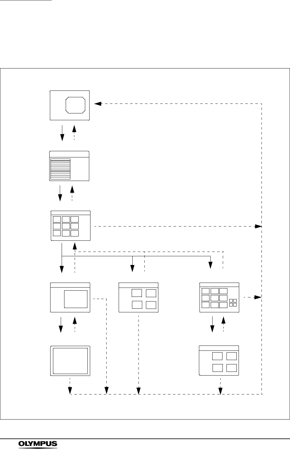
110
Chapter 5 Functions
EVIS EXERA II VIDEO SYSTEM CENTER CV-180
PC card menu
The images on the PC card can be displayed, cleared, etc., on the PC card
menu. Figure 5.58 shows the menu tree of the PC card menu.
Figure 5.58
Endoscopic image
PC Card
Title
Title
Shift + F4
Shift + F4
Back, Esc,
Shift + F4
Back,
Esc
Back,
Esc
Back,
Esc
Esc
View,
Enter
Preview
View,
Enter
Zoom
Annotate
Load,
Enter
Folder list screen
Thumbnail screen
Normal screen Annotation screen Annotation selection
screen
Full screen
Annotation preview
screen
Shift + F4
See page 111
See page 115.
See page 122.
See page 119.
See page 111.
See page 115.
See page 119.
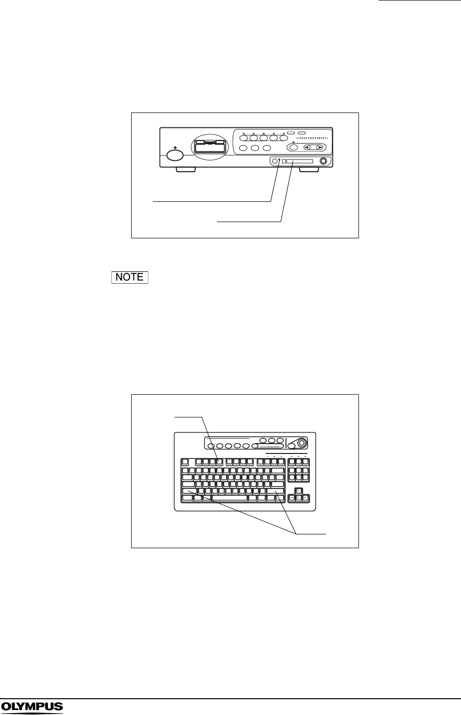
Chapter 5 Functions
111
EVIS EXERA II VIDEO SYSTEM CENTER CV-180
Basic operation of the PC card menu
1. Insert the PC card into the PC card slot. The PC card status indicator lights
up green (see Figure 5.59).
Figure 5.59
For details on the PC card and PC card slot, see “STOP
button and PC card indicator” on page 75 and “PC card slot
and eject button” on page 76.
2. Press the “Shift” and “F4” keys together (see Figure 5.60). The message
“Please wait” is displayed on the monitor, then the folder list screen appears
(see Figure 5.61).
Figure 5.60
PC card status indicator
PC card slot
F4
Shift
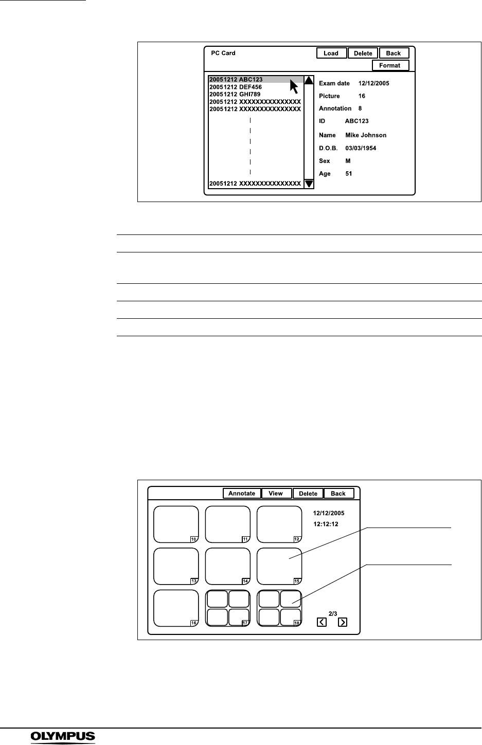
112
Chapter 5 Functions
EVIS EXERA II VIDEO SYSTEM CENTER CV-180
Figure 5.61
3. Click an image folder in the folder list. The patient data is displayed on the
right side of the window.
4. Click “Load” or press the “Enter” key. The message “Please wait” is
displayed on the monitor, then the thumbnail screen appears (see Figure
5.62). The thumbnail-sized images for the annotation image is not an image
but an icon.
Figure 5.62
5. Perform file operation such as playback and deletion of images, etc.
Button Function Details
Load Displays the thumbnail index of the data folder
selected. page 111
Delete Deletes the data folder selected. page 118
Back Returns to the endoscopic live image. page 111
Format Formats the PC card. page 114
Table 5.10
Annotation icon
Thumbnail image

Chapter 5 Functions
113
EVIS EXERA II VIDEO SYSTEM CENTER CV-180
6. Click “Back” or press the “Esc” key to return to the previous screen. Or press
the “Shift” and “F4” keys together to return to the endoscopic image.
• The aspects ratio of a thumbnail image depends on the
endoscope used.
• Refer to “Image files and folders” on page 125 for the details
of the folder names shown in the folder list screen.
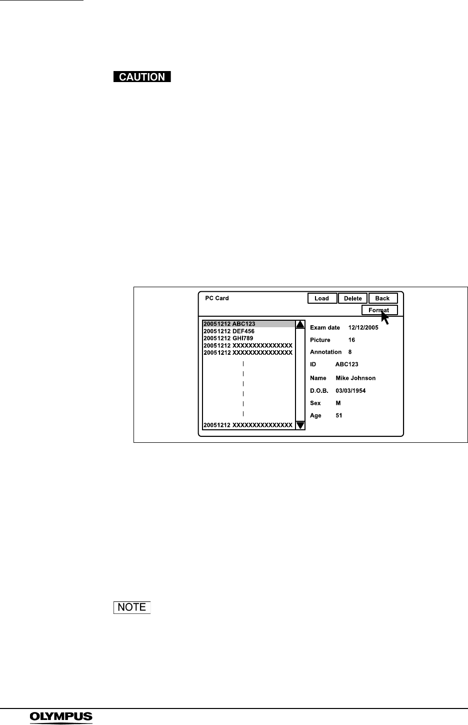
114
Chapter 5 Functions
EVIS EXERA II VIDEO SYSTEM CENTER CV-180
Formatting of the PC card
• Be sure to format the PC card using the CV-180. If it is
formatted by an other instrument such as a personal
computer, it may not be possible to record or playback
images from the PC card.
• Do not press the “STOP” button during formatting the PC
card.
• Formatting a PC card deletes all data saved on the PC card.
1. Display the folder list screen (refer to “Basic operation of the PC card menu”
on page 111).
2. Click “Format” (see Figure 5.63). A confirmation message is displayed to
ask if the card can be formatted.
Figure 5.63
3. Click “No” to go back to the folder list screen instead of formatting.
Click “Yes” to start formatting. During formatting, the message “Please wait”
appears on the screen and the PC card status indicator of the video system
center blinks orange.
4. The PC card status indicator lights up green after formatting has been
completed and the message “Complete” appears on screen.
A notification of normal or abnormal completion appears on the monitor.
If formatting failed, the message “Failed in format” appears
on screen. In this case, the PC card is not working properly.
Use a new one.
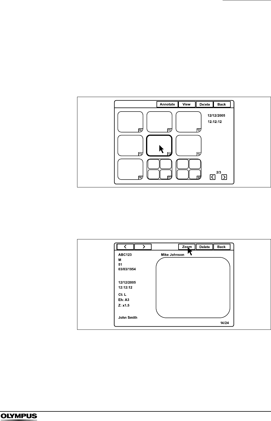
Chapter 5 Functions
115
EVIS EXERA II VIDEO SYSTEM CENTER CV-180
Playback images from the PC card
1. Display the thumbnail screen (refer to “Basic operation of the PC card
menu” on page 111).
2. Click a thumbnail image to playback. The selected image should be edged
with a thick frame. The shooting date and time of the image are displayed on
the right side of the window.
Figure 5.64
3. Click “View” or press the “Enter” key. The message “Please wait” is
displayed on the monitor, then the normal screen image selected appears
(see Figure 5.65).
Figure 5.65
4. Click “<” or “>” to playback the images before and after the image currently
being played back.
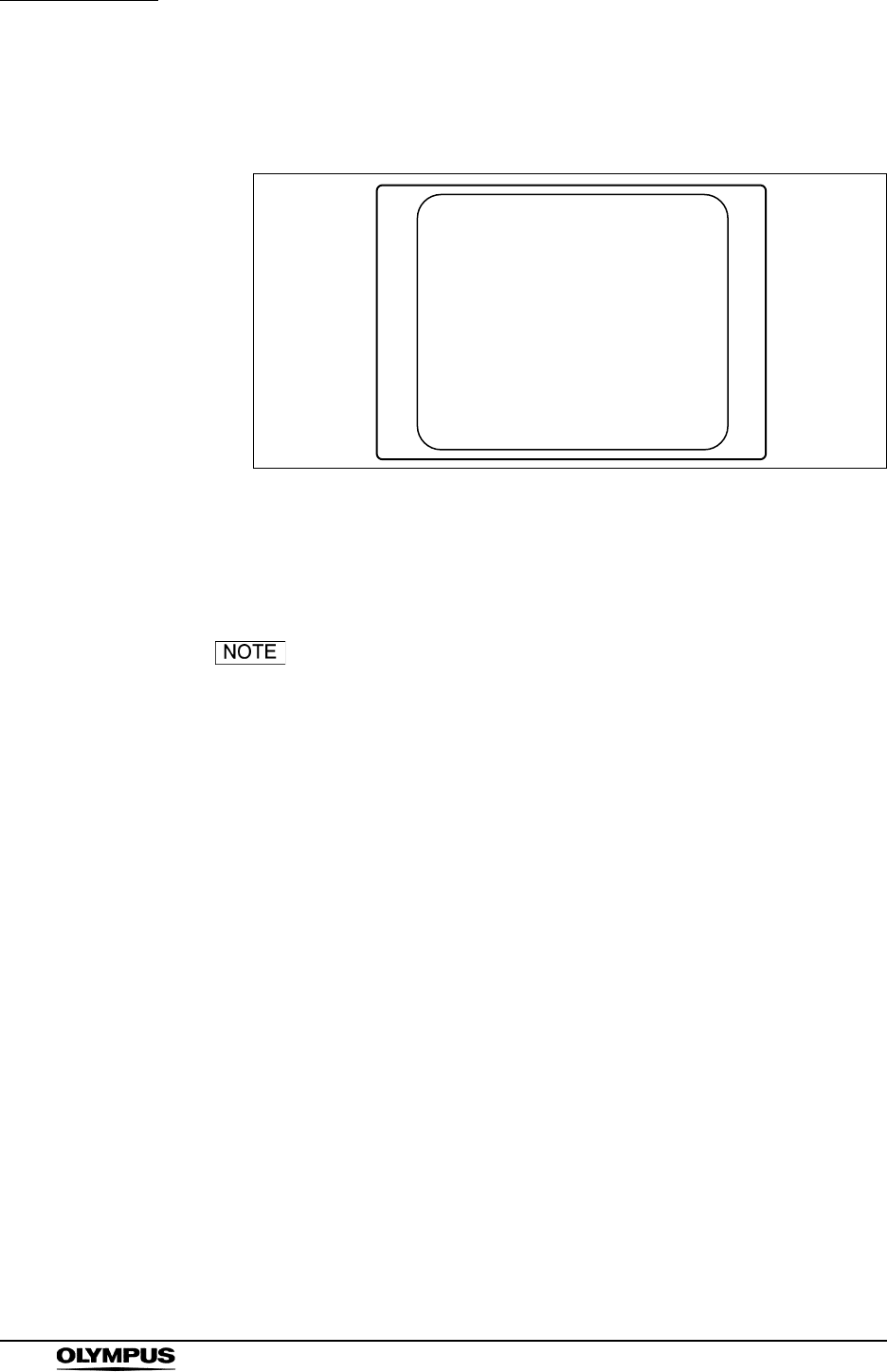
116
Chapter 5 Functions
EVIS EXERA II VIDEO SYSTEM CENTER CV-180
5. Click “Zoom”. The message “Please wait” is displayed on the monitor, then
the full screen image appears (see Figure 5.66).
Figure 5.66
6. Press the “Esc” key to return to the normal sized playback screen.
Or press the “Shift” and “F4” keys together to return to the endoscopic
image screen.
• The images recorded by an instrument other than the CV-180
cannot be played back.
• The images edited by a personal computer or an instrument
other than the CV-180 may be unable to be played back.
• The endoscopic image and the patient data is displayed
together on the same screen when using the CV-180 for
playing back, but cannot be displayed together on a personal
computer.
• In the normal screen, all images recorded in the full-height or
full size are scaled down and displayed on the constant
display area are shown in Figure 5.65.
• The two images of the PinP are displayed separately in the
thumbnail screen, which has the same time stamps but
different file names.
• The sub image of the PinP is displayed with the same patient
data as the main image.
• Playback in PinP mode is not available.
• “R” is not displayed on the monitor when the rotated image of
the orientation function is played back (refer to “Monitor
orientation function” on page 245).
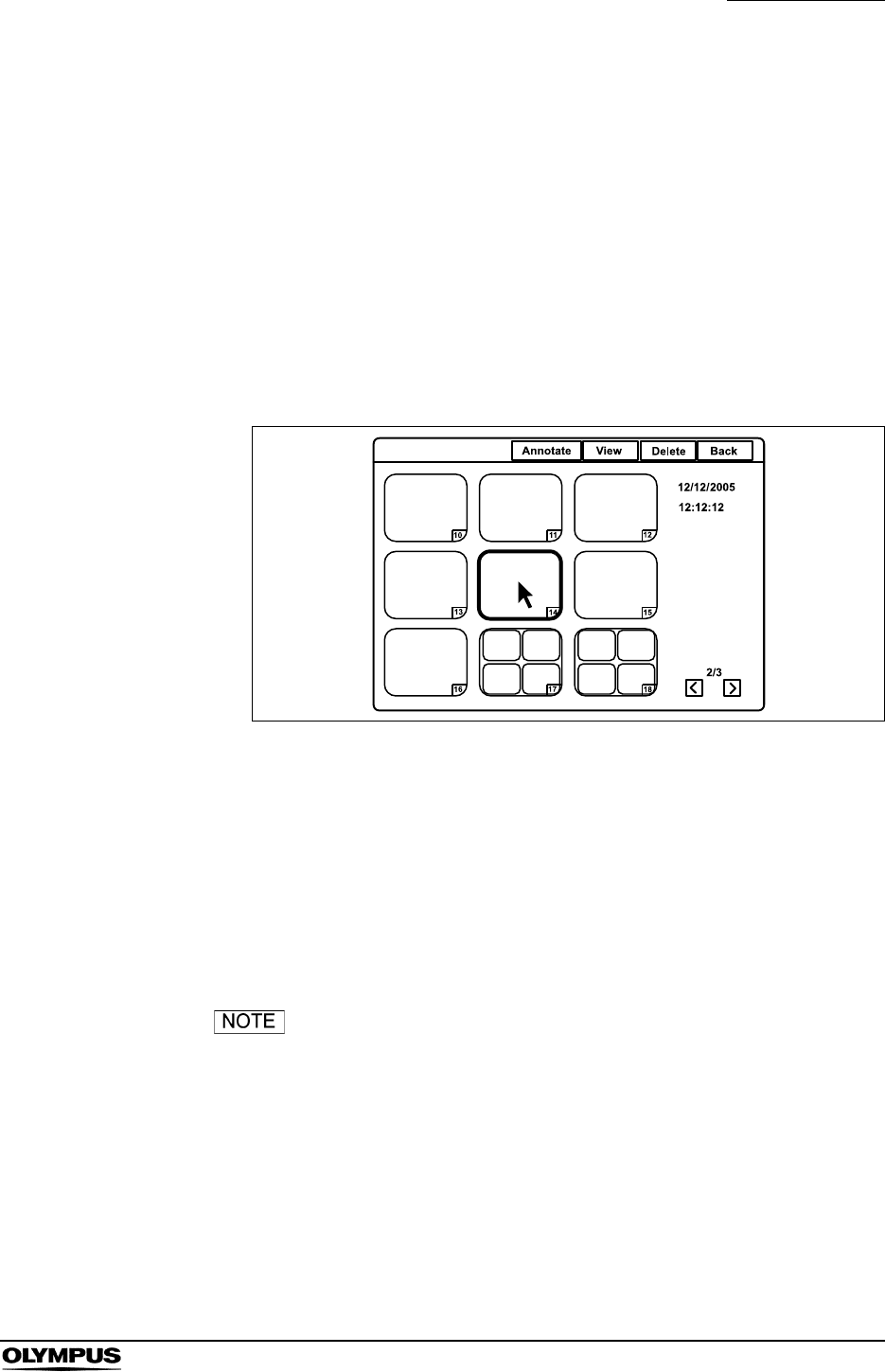
Chapter 5 Functions
117
EVIS EXERA II VIDEO SYSTEM CENTER CV-180
• The images in the thumbnail screen and the normal screen
are displayed in SDTV format. The full screen is displayed in
the original format (HDTV or SDTV).
Deleting images from a PC card
1. Display the thumbnail screen (refer to “Basic operation of the PC card
menu” on page 111).
2. Click a thumbnail image to be deleted. The selected thumbnail is edged with
a thick frame. The shooting date and time of the image are displayed on the
right side of the window.
Figure 5.67
3. Click “Delete”. A confirmation message appears on the monitor.
4. Click “No” to go back to the thumbnail screen instead of deleting.
Click “Yes” to delete the selected image.
5. Click “Back” or press the “Esc” key to return to the folder list screen.
Or press the “Shift” and “F4” keys together to return to the endoscopic
image.
It is also possible to delete the image by displaying the image
in the normal screen in step 2. and then clicking “Delete”.
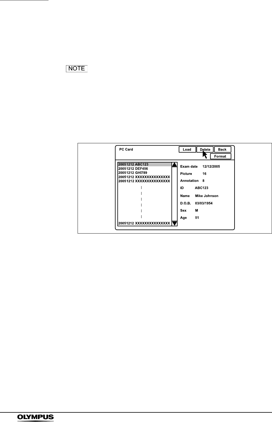
118
Chapter 5 Functions
EVIS EXERA II VIDEO SYSTEM CENTER CV-180
Deleting folder from PC card
This operation deletes the image files from the PC card. The selected folder and
all image files in this folder are deleted.
Confirm in advance if the selected folder shall be deleted.
The deleted folder cannot be restored.
1. Display the folder list screen (refer to “Basic operation of the PC card menu”
on page 111).
2. Click the image folder to be deleted. The patient data is displayed on the
right side of the window (see Figure 5.68).
Figure 5.68
3. Click “Delete”. A confirmation message appears on the monitor.
4. Click “No” to go back to the folder list screen instead of deleting.
Click “Yes” to delete the selected folder.
5. Click “Back”, press the “Esc” key, or press the “Shift” and “F4” keys together
to return to the endoscopic image.
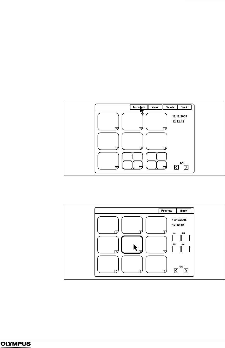
Chapter 5 Functions
119
EVIS EXERA II VIDEO SYSTEM CENTER CV-180
Annotation of images
This function selects and arranges four frozen endoscopic images from a PC
card onto a sheet with comments and stores it as a file in the same folder.
Selection of images
1. Display the thumbnail screen (refer to “Basic operation of the PC card
menu” on page 111).
2. Click “Annotate” (see Figure 5.69).
Figure 5.69
3. The annotation selection screen appears (see Figure 5.70).
Figure 5.70
4. Click “<” or “>” to scroll the thumbnail images if necessary.
5. Click the required thumbnail image. The selected image is edged with a
thick frame. The shooting date and time of the image are displayed on the
right side of the window (see Figure 5.70).
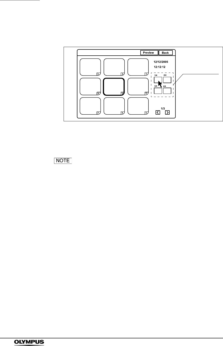
120
Chapter 5 Functions
EVIS EXERA II VIDEO SYSTEM CENTER CV-180
6. Click one of the position buttons on the screen. The number of the selected
image appears at the position button and the selected image is edged with a
thick frame in the color of the position button (see Figure 5.71).
Figure 5.71
7. Select up to 4 images repeating steps 4. to 6., and then proceed with
“Annotating” on page 121.
• If you click the position button that is already assigned with
an image, the assignation is cleared.
• If you click an image and then click a position button that is
already assigned with another image, the assigned image
changes.
• The images that are assigned with the position buttons are
edged with a thick frame in the following colors:
“1/4”: Pink
“2/4”: Blue
“3/4”: Green
“4/4”: Yellow
Position buttons
5
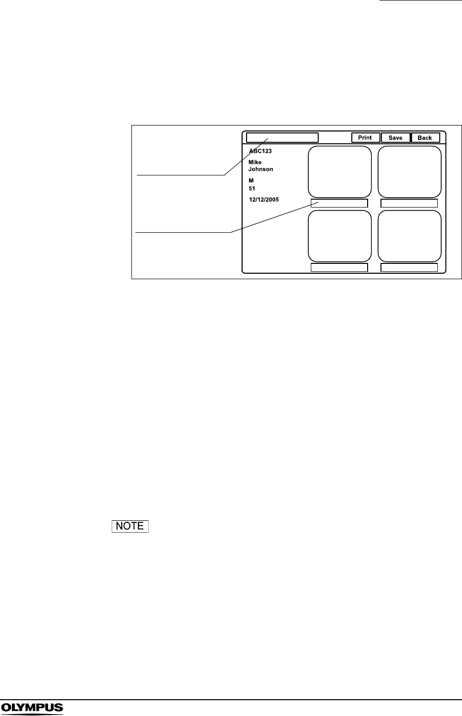
Chapter 5 Functions
121
EVIS EXERA II VIDEO SYSTEM CENTER CV-180
Annotating
1. Click “Preview” on the annotation selection screen after selecting the
images (see Figure 5.71). The message “Please wait” is displayed on the
monitor, then the annotation preview screen appears (see Figure 5.72).
Figure 5.72
2. Click the title input area to place the cursor, and enter the title.
• Up to 25 alphanumeric and symbol characters can be entered.
3. Click the comment input area under each image to place the cursor, and
enter the title or comments.
• Up to 15 alphanumeric and symbol characters can be entered.
4. Click “Save”. The selected images, title and comments are recorded as an
annotation image file on the PC card.
5. Click “Back” or press the “Esc” key to return to the annotation selection
screen.
Or press the “Shift” and “F4” keys together to return to the endoscopic
image.
All annotation images including HDTV images are displayed
and saved in SDTV format.
Title input area
Comment input area
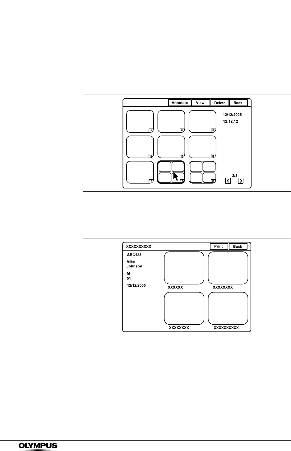
122
Chapter 5 Functions
EVIS EXERA II VIDEO SYSTEM CENTER CV-180
Playback image annotation
1. Display the thumbnail screen (refer to “Basic operation of the PC card
menu” on page 111).
2. Click an annotation icon to be played back. The selected icon is edged with
a thick frame (see Figure 5.73).
Figure 5.73
3. Click “View” or press the “Enter” key. The annotation screen appears on the
monitor (see Figure 5.74).
Figure 5.74
4. Click “Print” to print out the images on the video printer. A confirmation
message appears on the monitor.
5. Click “No” to go back to the annotation screen instead of printing.
Click “Yes” to print the images with the patient data and the comment.
6. Click “Back” or press the “Esc” key to return to the thumbnail screen.
Or press the “Shift” and “F4” keys together to return to the endoscopic
image.
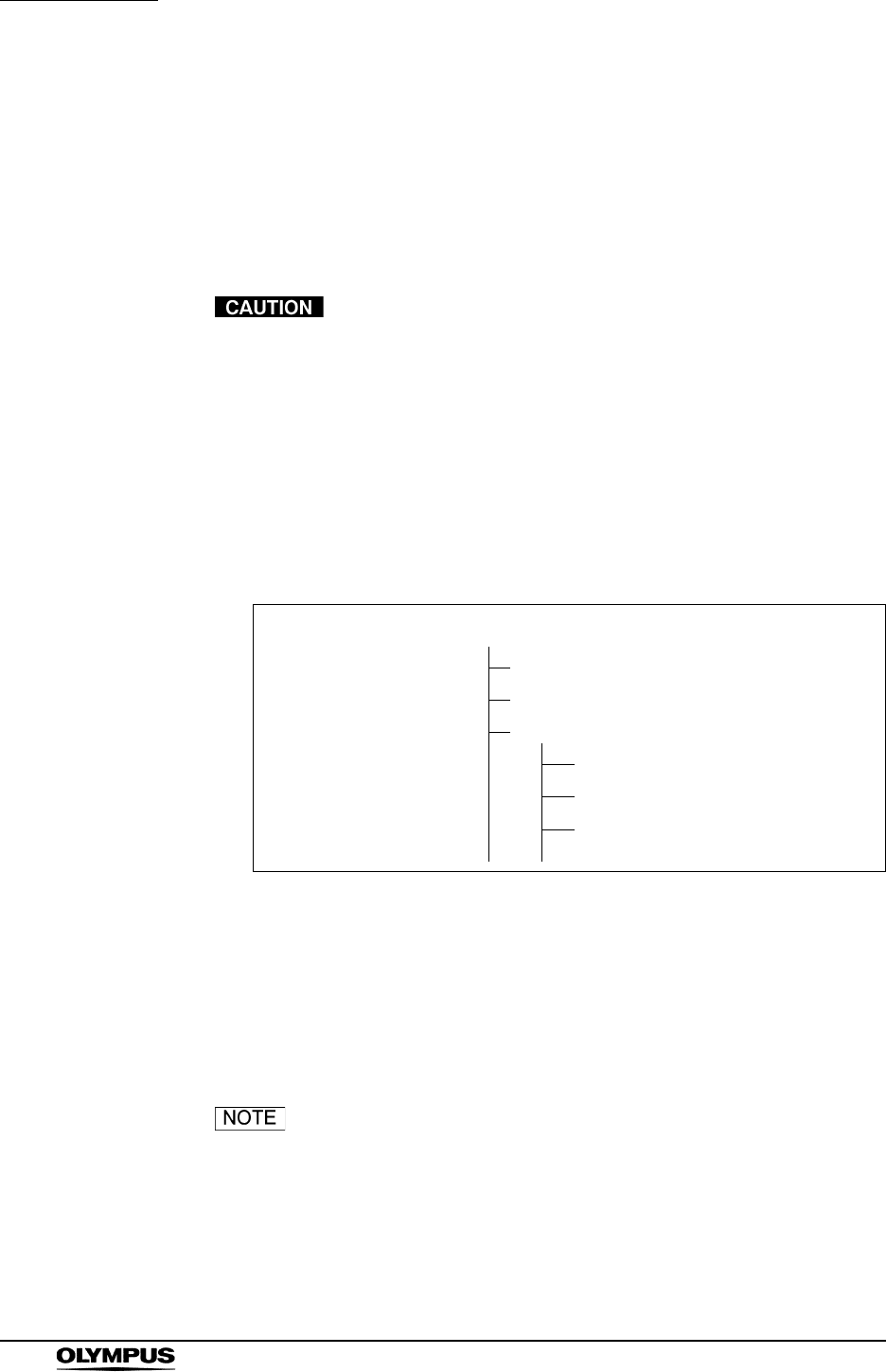
124
Chapter 5 Functions
EVIS EXERA II VIDEO SYSTEM CENTER CV-180
Playback the images using the personal computer
The images on the PC card can be played back on the personal computer.
Hardware requirements are the followings:
• Windows® 2000 or later
• Internet Explorer ver.5.0 or later
Do not delete or move data on the PC card using the
personal computer. The data may be damaged or it may not
be possible to playback images from the PC card.
1. Insert the PC card into the PC card slot of the personal computer. Refer to
the instruction manual of the personal computer.
2. Select the drive in which the PC card is inserted.
Figure 5.75 shows an example of the image files/folders structure on the PC
card.
Figure 5.75
3. Open the desired folder.
4. Open the file “FILES.xml”. The list of image files saved in the folder is shown
in an Internet Explorer window.
5. Click the desired file of the list to display the image.
• If you open the image file not using the xml. file, the
endoscopic image and the data cannot be displayed in one
image. The file names that are displayed by other than the
xml. file are different from those in Figure 5.75.
• The images cannot be displayed in the PinP style.
DCIM
100OLYMP
101OLYMP
102OLYMP
FILES.xml
OLYM0001.jpg
OLYM0002.jpg
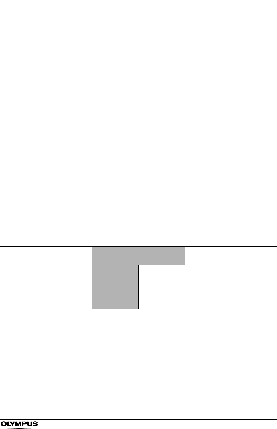
Chapter 5 Functions
125
EVIS EXERA II VIDEO SYSTEM CENTER CV-180
Image files and folders
The data files of the endoscopic images are stored in the image folder, which is
generated by the video system center, on the PC card.
Files
The video system center numbers the endoscopic image files sequentially. The
names of each image file becomes like the following, and is numbered
sequentially in order of photographing.
• OLYMnnnn.jpg:
(“nnnn” is a four-digit number.)
Folders
The folder in which image files are stored either newly generated or previously
generated ones are used according to the conditions such as turning ON or OFF
the system, with or without the patient ID, etc. (see Table 5.11). The folder
names are as follows.
• When the patient ID was entered: “8-digit date” + “max.15-digit patient
ID”
ex. 20050512 ABC123 (“2005, May, 12th” and “ABC123”)
• When no patient ID was entered: “8-digit date” + “15-digit X”
ex. 20050512 XXXXXXXXXXXXXXX
Resume function of patient data
display 1ON OFF
Patient ID in the last observation Entered Not entered Entered Not entered
Patient ID was NOT entered after
the system was turned ON.
The image files
are stored in the
previous folder.2
New folder is generated.
Folder name - 8-digit date +15-digit X
Patient ID was entered after the
system was turned ON. New folder is generated.
Folder name 8-digit date + Max.15-digit ID
1 For the resume function, see “Patient data display” on page 241.
2 The shaded region is used for recording the images into the same folder after swapping the endoscope during the
observation.
Table 5.11

126
Chapter 5 Functions
EVIS EXERA II VIDEO SYSTEM CENTER CV-180
Be sure to enter the patient ID each time when entering the
patient data. Also, be sure to enter the different patient ID for
each patient. Otherwise, the image data for some patients
may mix in the same image folder.
• When entering the same patient ID on the same date twice, a
new folder with the same ID and date will be created, if an
observation has taken place in between the two observation.
• The maximum number of image files recorded in a folder is
9999. The maximum number of folders on a PC card is 900.
The total volume of the image data cannot exceed the
amount of memory of the PC card.
• The recording speed decelerates when the number of
images in a folder exceeds 100, or when the number of
folders on a PC card exceeds 100.
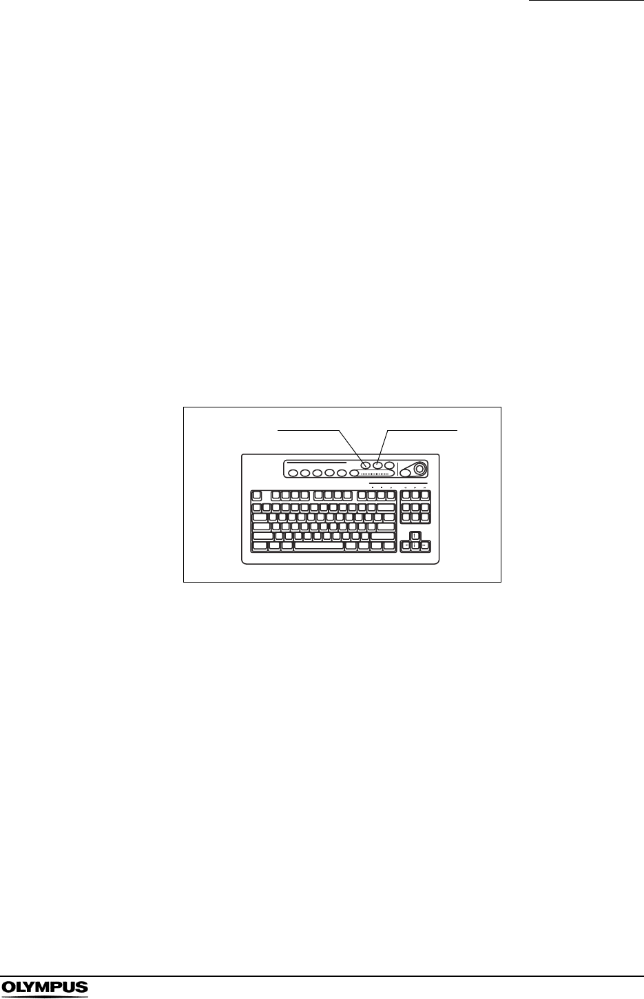
Chapter 5 Functions
127
EVIS EXERA II VIDEO SYSTEM CENTER CV-180
5.4 Image recording and playback (other than PC
card)
The recordable area of the image is variable depending on the recording
devices. Refer to the respective instruction manual of the recording device.
Image filing system
Assign the “Digital file” to “RELEASE 1” or “RELEASE 2” in the “User preset”
menu in advance. See “Release function” on page 223.
1. Press the “FREEZE” key (see Figure 5.76) to freeze the endoscopic live
image.
2. Check the frozen image if it is suitable for recording. If not, press the
“FREEZE” key again to return to the live image and repeat steps 1. and 2.
Figure 5.76
3. Press the “RELEASE” key (see Figure 5.76) to record the image. The frozen
endoscopic image returns to the live image. The D.F. counter on the monitor
increments by one (see Figure 5.77).
When the index image setting is ON, the index image is displayed at the
lower left corner on the monitor.
FREEZE RELEASE
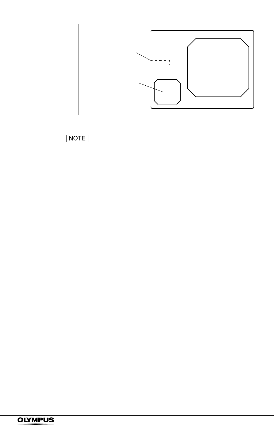
128
Chapter 5 Functions
EVIS EXERA II VIDEO SYSTEM CENTER CV-180
Figure 5.77
• Press the “RELEASE” key (see step 3.) to record the image
without checking the image. In this case, the live image is
frozen for the release time, and then returns to the live
image. See “Image filing system” on page 206 for the release
time.
• Maintain intervals of 1 second or more between each press
of the “RELEASE” key, when not using the “FREEZE” key. If
the intervals are shorter than 1 second, it may not be
possible to record images.
• Keep the endoscope as stationary as possible to record
blurless images when pressing the “RELEASE” key.
• D.F. counter does not appear on the monitor when the user
data are clear.
• The freeze and release operations can also be controlled
from the scope switches and/or foot switches. For how to set
up the scope switches and foot switches, see “Remote switch
and foot switch (EXERA and VISERA)” on page 219.
ABC123
Mike Johnson
M 51
03/03/1954
12/12/2005
12:12:12
CVP: A4/4
D.F: 99
VCR
Ct: N Eh: A8
Z: x1.5
Pump
Media:
John Smith
Cardiac end of the stomach
D.F.counter
Index image
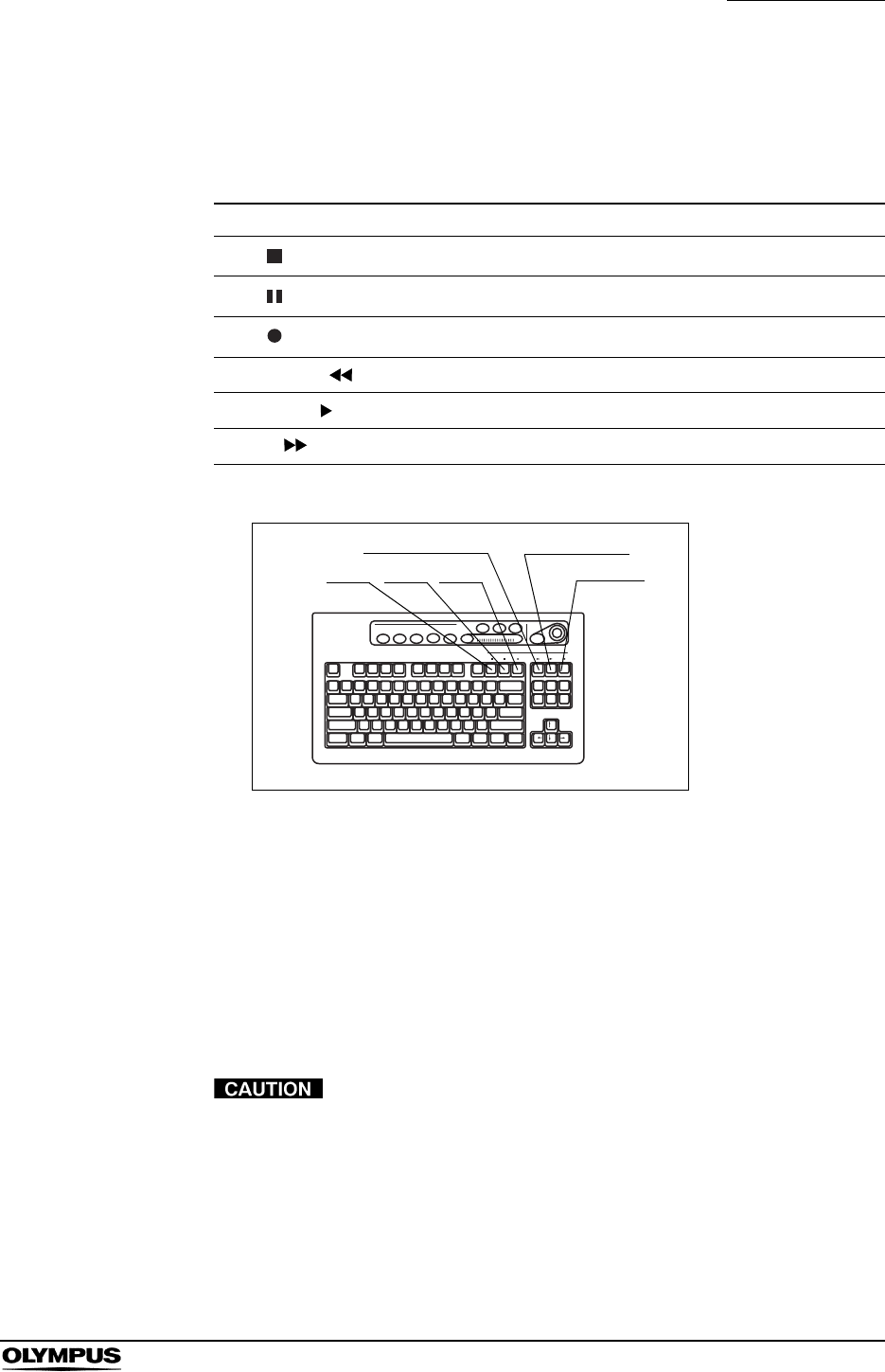
Chapter 5 Functions
129
EVIS EXERA II VIDEO SYSTEM CENTER CV-180
Videocassette recorder (VCR)
VCR can be operated using the keyboard. The following table shows the function
and the keys to operate.
Figure 5.78
1. Press the Image source button to change the image source.
• Press the “SCOPE” button to record the image.
• Press the “DV/VCR” button to playback the image.
2. Perform the VCR operation of recording or playback using the keys on the
keyboard (see Figure 5.78). “VCR” appears on the monitor during VCR
recording (see Figure 5.79).
After turning the DVO-1000MD ON and right after disk
insertion “Now Loading ...” is displayed. During this
initialization process images cannot be recorded.
Key Function
F10 ( ) Stops recording or playback.
F11 ( ) Pauses playback.
F12 ( ) Starts recording.
Print. screen ( ) Fast-rewinds the tape.
Scroll Lock ( ) Starts playback.
Pause ( ) Fast-forwards the tape.
Table 5.12
F10 F11 F12
Print screen Scroll lock
Pause
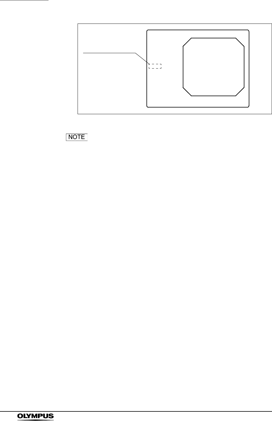
130
Chapter 5 Functions
EVIS EXERA II VIDEO SYSTEM CENTER CV-180
Figure 5.79
• When the VCR is connected using the VTR remote cable
(MH-992), only the recording and pause operations can be
operated.
• When “DVD (IEEE1394) of the VCR type is selected, only the
recording and pause operations can be operated
(“Videocassette recorder” on page 211).
• The fast forward and rewind operations can be operated only
when the VCR is set to RS-232C and IEEE1394.
• The “VCR” does not appear when the patient data are
cleared from the monitor.
• Also refer to the instruction manual for the VCR.
• The recording and pause operations can also be controlled
from the scope switches and/or foot switches. For how to set
up the scope switches and foot switches, see “Remote switch
and foot switch (EXERA and VISERA)” on page 219.
ABC123
Mike Johnson
M 51
03/03/1954
12/12/2005
12:12:12
CVP: A4/4
D.F: 99
VCR
Ct: N Eh: A8
Z: x1.5
Pump
Media:
John Smith
Cardiac end of the stomach
Indication of VCR
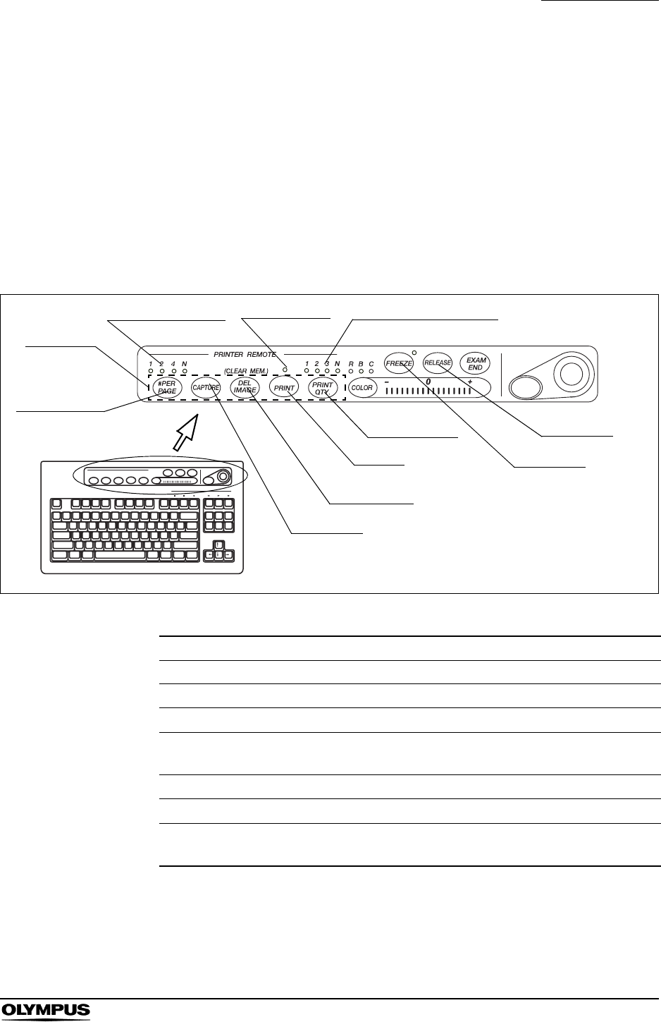
Chapter 5 Functions
131
EVIS EXERA II VIDEO SYSTEM CENTER CV-180
5.5 Printing images
The video printer captures images, and starts printing them when the number of
images reached the selected number of images to be printed per sheet.
Video printer
The video printer can be operated using the “PRINTER REMOTE” or
“RELEASE” key on the keyboard. The “RELEASE” key needs to be preset. See
“Release function” on page 223.
Figure 5.80
Key Function
#PER PAGE Sets the number of images to be printed per sheet.
CAPTURE or RELEASE Captures images into the memory of the video printer.
DEL IMAGE Deletes the last image stored in the memory of the printer.
Shift + DEL IMAGE Deletes images on the position showed by the CVP
counter.
PRINT Prints the images captured by the video printer.
PRINT QTY. Sets the number of sheets to be printed.
Shift + F8
(Printer lock)
Locks the 5 printer remote keys and allows the video printer
to be operated by using the keys on the video printer.
Table 5.13
PRINTER
REMOTE
#PER PAGE
CAPTURE
DEL IMAGE
PRINT
PRINT QTY.
FREEZE
RELEASE
Number of images Number of print sheets
Print indicator
132
Chapter 5 Functions
EVIS EXERA II VIDEO SYSTEM CENTER CV-180
The following images can be loaded using “CAPTURE” and “RELEASE”.
• Confirm that the necessary images have been printed before
deleting the images.
• Always delete all images in the video printer at the end of an
examination when the number of images per sheet is more
than one. If not, the images of a previous examination and a
new examination may be mixed on one print sheet.
• If you move to another menu before printing the images, the
images disappear.
Number of images on the print form
1. Press the “#PER PAGE” key to display the number of images to be printed
on the print sheet.
2. While the window opens, press the “#PER PAGE” key to change the
number of images to be printed on the print sheet. The indicator above the
“#PER PAGE” key indicates the number.
Printing
1. Press the “PRINT QTY.” key to select the number of print sheets to be
printed. The indicator above the “PRINT QTY.” key indicates the number.
2. Press the “FREEZE” key to freeze the endoscopic live image.
3. Check the frozen image if it is suitable for recording. If not, press the
“FREEZE” key again to return to the live image and repeat steps 2. and 3.
Image CAPTURE RELEASE
Endoscopic live image, PinP, special light observation
Normal size playback screen, full size playback screen,
annotation screen of the PC card menu,
available, not available
Table 5.14
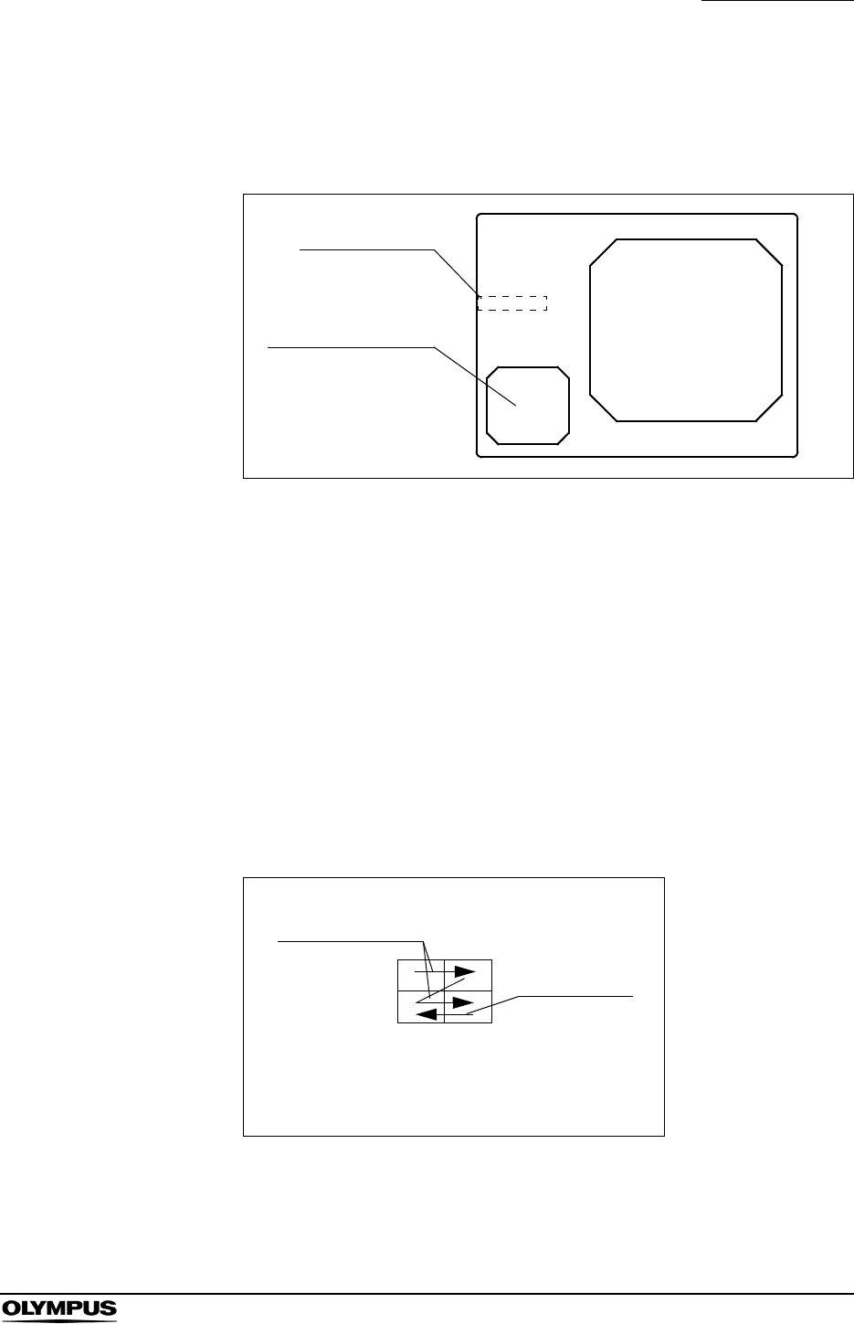
Chapter 5 Functions
133
EVIS EXERA II VIDEO SYSTEM CENTER CV-180
4. Press the “CAPTURE” key or “RELEASE” key to capture the endoscopic
image into the memory of the video printer. The CVP counter on the monitor
increments by one (see Figure 5.81).
Figure 5.81
5. When the number of images captured reaches the number of images to
print on a print sheet, printing starts automatically. The indicator above the
“PRINT” key lights up during printing.
6. To start printing before the number of images captured reaches the number
of images to be printed on a print sheet, press the “PRINT” key anytime. The
indicator above the “PRINT” key lights up during printing.
Overwriting pre-recorded images
1. Press the “DEL IMAGE” key a number of times until the CVP counter shows
the image number to be overwritten.
Figure 5.82
2. Press the “CAPTURE” key or “RELEASE” key to overwrite the new image
on the previous image. The CVP counter on the monitor increments by one
(see Figure 5.81).
ABC123
Mike Johnson
M 51
03/03/1954
12/12/2005
12:12:12
CVP: A4/4
D.F: 99
VCR
Ct: N Eh: A8
Z: x1.5
Pump
Media:
John Smith
Cardiac end of the stomach
CVP counter
Index image
Only when pressing
“RELEASE” key.
Print sheet partition and
order of image assignment
An example of 4-
partitioned sheet
DEL IMAGE
Order of image
assignment

134
Chapter 5 Functions
EVIS EXERA II VIDEO SYSTEM CENTER CV-180
• Press the “CAPTURE” key or “RELEASE” key to record the
image without checking the image. In this case, the live
image is frozen for the release time.
• Maintain the endoscope as stationary as possible when
pressing the keys to record the blurless images.
• The maximum number of images per page is variable
depending on the printer model. See Table 5.15 for details.
• Depending on the video printer or the number of images on a
print sheet, the “CAPTURE” key may not be operated during
printing.
• HDTV images can be printed only when using the OEP-4
with “Mode1”. See “Output signal to OEP-4” on page 204.
• The CVP counter does not appear when the patient data are
cleared from the monitor.
• When pressing the “RELEASE” key, the images captured are
displayed at the lower left on the monitor depending on the
settings. When pressing the “CAPTURE” key the images
taken in are displayed in full-screen on the monitor.
• A caption can be printed in the margin on the print sheet. See
“Caption” on page 203.
• The CAPTURE and RELEASE operations can also be
controlled from the scope switches and/or foot switches. For
how to set up the scope switches and foot switches, see
“Remote switch and foot switch (EXERA and VISERA)” on
page 219.
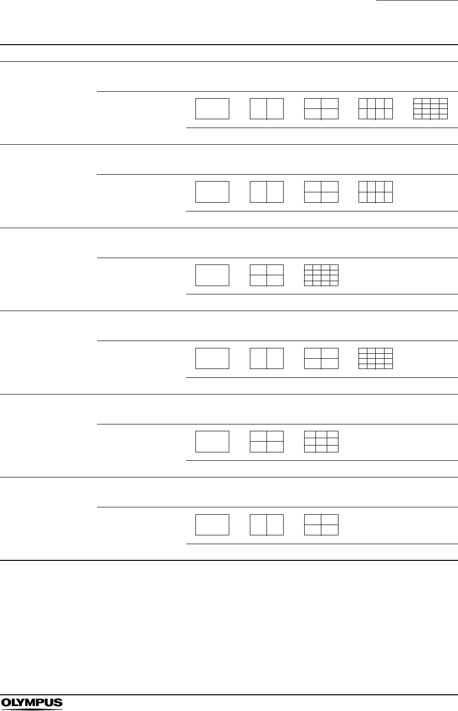
Chapter 5 Functions
135
EVIS EXERA II VIDEO SYSTEM CENTER CV-180
Printer Details
OEP (OLYMPUS)
OEP-3 (OLYMPUS)
Indication of number
of images [1] [2] [4] [N] [N]
Sheet partition
Full size 2 4 8 16
OEP-4 (OLYMPUS)
Indication of number
of images [1] [2] [4] [N] -
Sheet partition -
Full size 2 4 8 -
UP-1800 (SONY)
UP-1850 (SONY)
UP-2900MD (SONY)
Indication of number
of images [1] [4] [N] - -
Sheet partition --
Full size 4 16 - -
UP-2950MD (SONY)
Indication of number
of images [1] [2] [4] [N] -
Sheet partition -
Full size 2 4 16 -
UP-5000MD (SONY)
UP-5200MD (SONY)
UP-5250MD (SONY)
Indication of number
of images [1] [4] [N] - -
Sheet partition --
Full size 4 9 - -
UP-21MD (SONY)
Indication of number
of images [1] [2] [4] - -
Sheet partition --
Full size 2 4 - -
Table 5.15
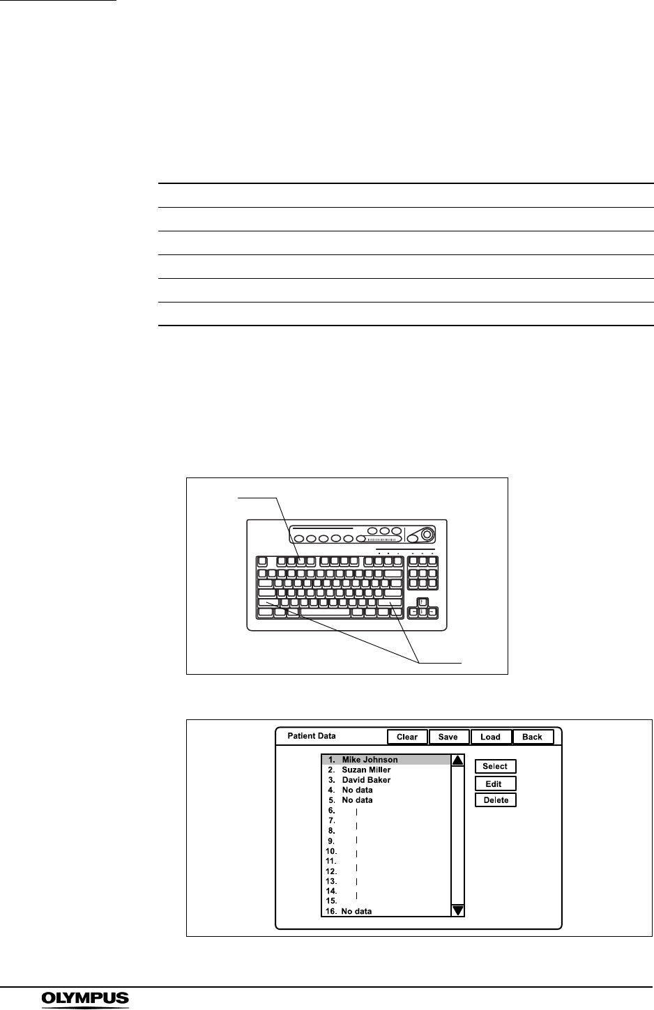
136
Chapter 5 Functions
EVIS EXERA II VIDEO SYSTEM CENTER CV-180
5.6 Pre-entry of patient data
In the patient menu, patient data can be entered in the video system center prior
to examination and called up before the examination. The following patient data
for up to 40 patients can be entered in advance.
Basic operation in the patient menu
1. Press the “Shift” and “F3” keys together. The “Patient Data” menu appears
on the monitor (see Figure 5.84).
Figure 5.83
Figure 5.84
ID Max. 15 alphanumeric and symbol characters
Name Max. 20 alphanumeric and symbol characters
Sex M or F
D.O.B. Max. 8 alphanumeric and symbol characters
Age Max. 3 numeric characters
Physician name Max. 20 alphanumeric and symbol characters
Table 5.16
F3
Shift
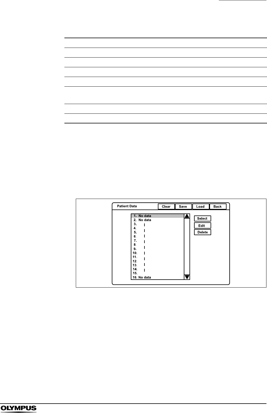
Chapter 5 Functions
137
EVIS EXERA II VIDEO SYSTEM CENTER CV-180
2. Click the buttons with the arrow pointer to operate the menu.
3. Click “Back” to return to the endoscopic image.
Entering new patient data
1. Press the “Shift” and “F3” keys together to display “Patient Data” menu (see
Figure 5.85).
Figure 5.85
2. Click “No data” in the patient name list (see Figure 5.85).
Button Function
Clear Clears all previously registered patient data.
Save Saves the registered 40 patient data on the PC card.
Load Loads the 40 patient data from the PC card onto the video system center.
Back Closes the patient menu and returns to the endoscopic image.
Call Returns to the endoscopic live image and displays the patient data
selected.
Edit Opens the patient data input screen.
Delete Deletes selected patient data.
Table 5.17
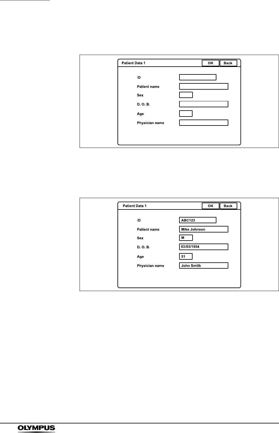
138
Chapter 5 Functions
EVIS EXERA II VIDEO SYSTEM CENTER CV-180
3. Click “Edit”. The patient data input screen appears on the monitor (see
Figure 5.86).
Figure 5.86
4. Click each text box to place the cursor.
5. Enter the data in the text boxes (see Figure 5.87).
Figure 5.87
6. Click “OK” to register the entered data. The next patient data input screen
appears.
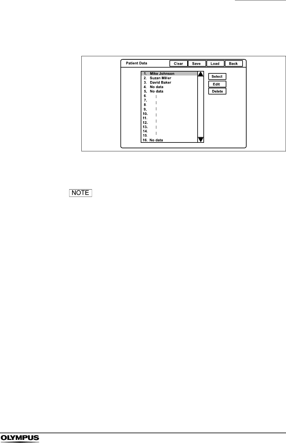
Chapter 5 Functions
139
EVIS EXERA II VIDEO SYSTEM CENTER CV-180
7. After finishing entering the data, click “Back” to return to the patient name list
(see Figure 5.88).
Figure 5.88
8. Click again “Back” to return to the endoscopic image.
• To cancel and stop the input operation, click “Back” instead of
“OK”. The entries typed in until then are canceled and the
display returns to the endoscopic image.
• If an error message is displayed, check if the entered data is
correct.
• To enter the patient data at the time of examination, see
Section 4.6, “Patient data” on page 57.
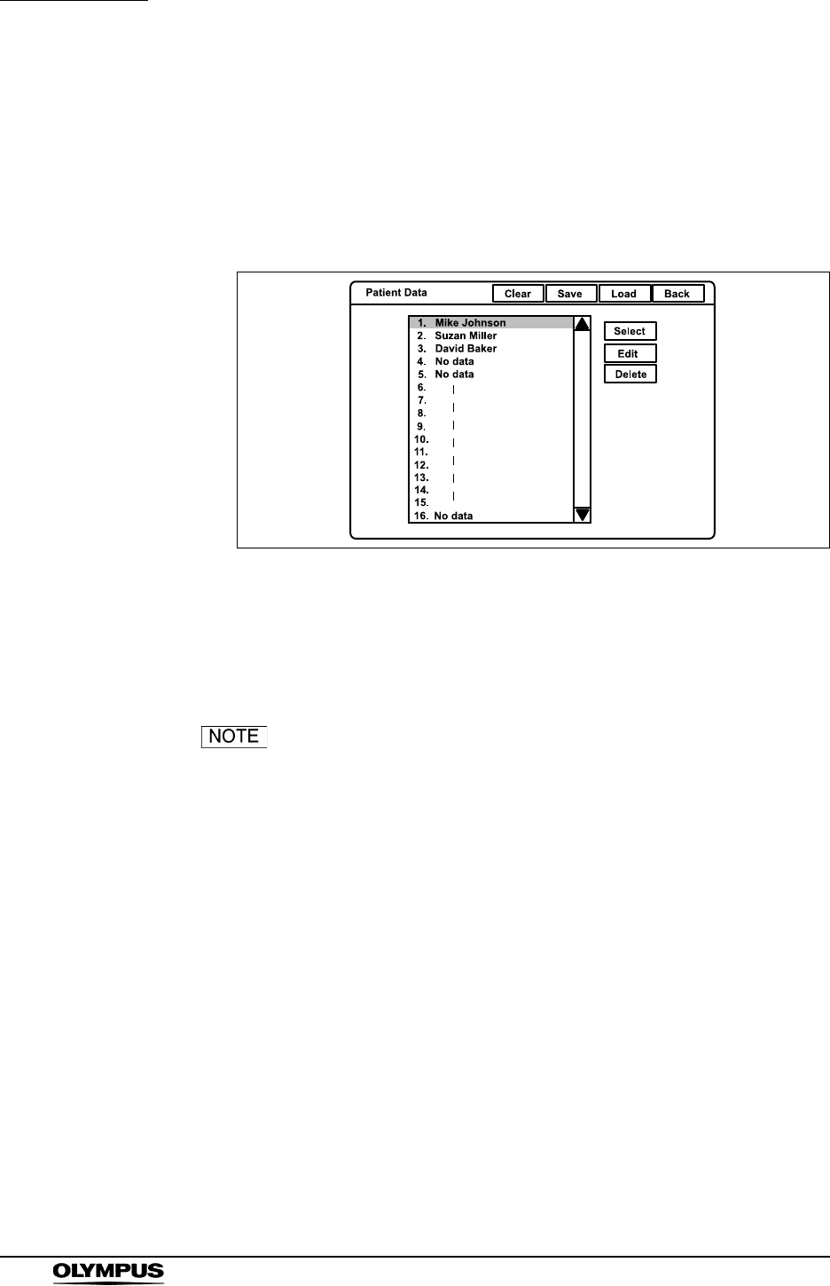
140
Chapter 5 Functions
EVIS EXERA II VIDEO SYSTEM CENTER CV-180
Displaying patient data
Call up the previously registered patient data and display it on the endoscopic
image.
1. Press the “Shift” and “F3” keys together to display the “Patient Data” menu
(see Figure 5.89).
Figure 5.89
2. Click the desired patient name in the patient name list.
3. Click “Select”. The selected patient data is displayed on the endoscopic
image.
The patient name that is once displayed in the endoscopic
image turns gray in the patient name dialog box after
finishing observation by pressing the “EXAM END” key.
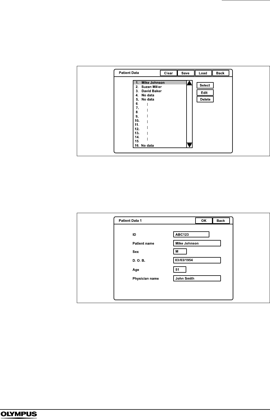
Chapter 5 Functions
141
EVIS EXERA II VIDEO SYSTEM CENTER CV-180
Editing previously entered patient data
1. Press the “Shift” and “F3” keys together to display the “Patient Data” menu
(see Figure 5.90).
Figure 5.90
2. Click the desired patient name in the patient name list. The selected patient
name is highlighted.
3. Click “Edit”. The patient data input screen appears (see Figure 5.91).
Figure 5.91
4. Click the data to be modified and place the cursor in the data area.
5. Enter the data in the text boxes.
6. Click “OK” to register the entered data.
7. Click “Back” to return to the endoscopic image.
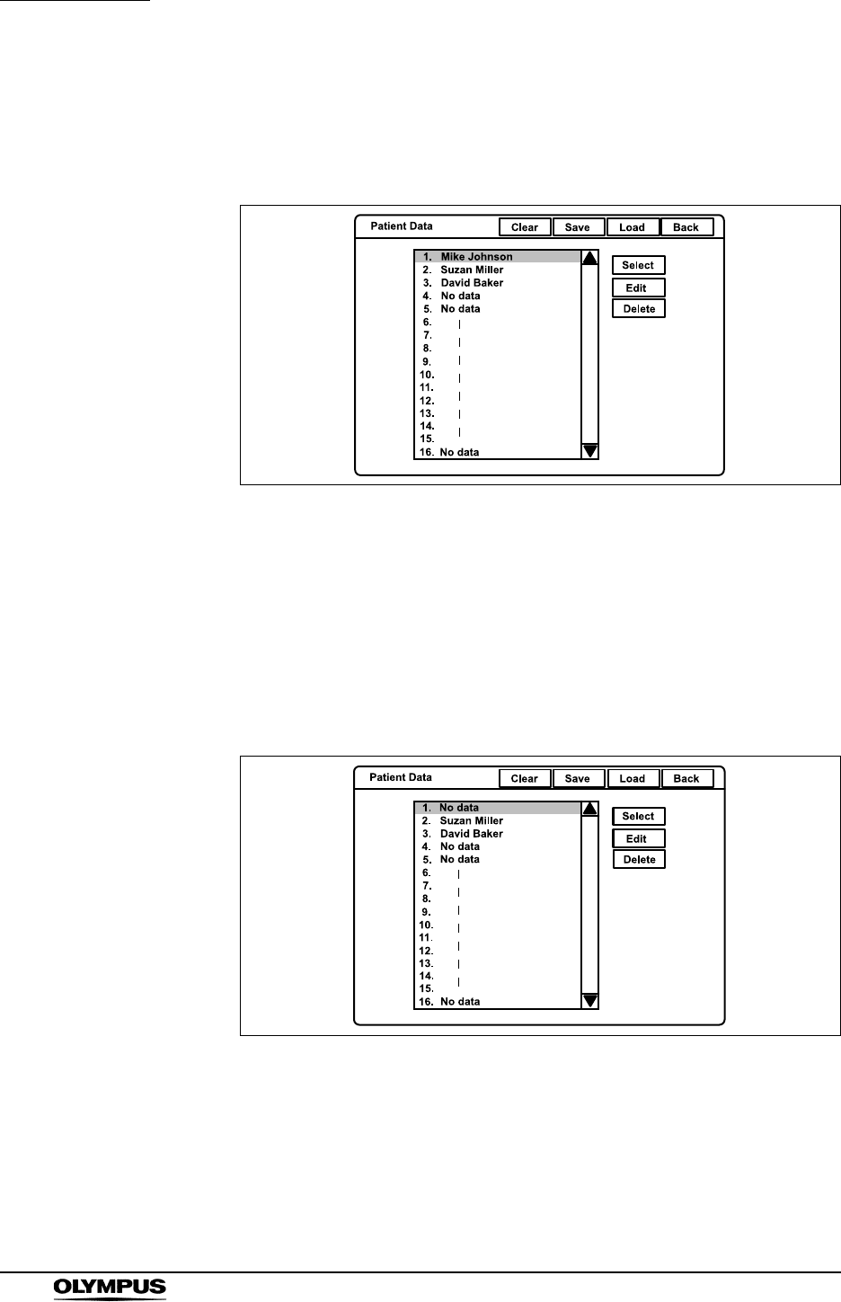
142
Chapter 5 Functions
EVIS EXERA II VIDEO SYSTEM CENTER CV-180
Deleting previously entered patient data
1. Press the “Shift” and “F3” keys together to display the “Patient Data” menu
(see Figure 5.92).
Figure 5.92
2. Click the desired patient name in the patient name list. The selected patient
name is highlighted.
3. Click “Delete”. A confirmation message appears on the monitor.
4. Click “No” to return to step 1.
Click “Yes” to delete the selected patient data. The patient name selected in
the patient name dialog box changes to “No data” (see Figure 5.93).
Figure 5.93
5. Click “Back” to return to the endoscopic image.
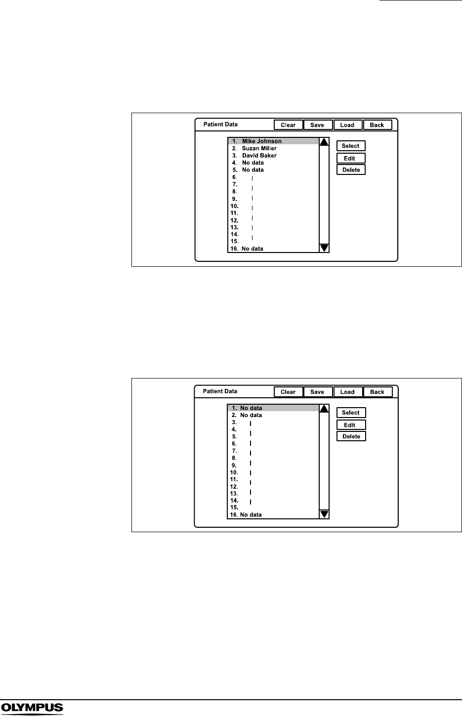
Chapter 5 Functions
143
EVIS EXERA II VIDEO SYSTEM CENTER CV-180
Clearing all patient data previously entered
1. Press the “Shift” and “F3” keys together to display the “Patient Data” menu
(see Figure 5.94).
Figure 5.94
2. Click “Clear”. A confirmation message appears on the monitor.
3. Click “No” to go back to step1.
Click “Yes” to delete all patient data, and all names in the patient name
dialog box changes to “No data” (see Figure 5.95).
Figure 5.95
4. Click “Back” to return to the endoscopic image.
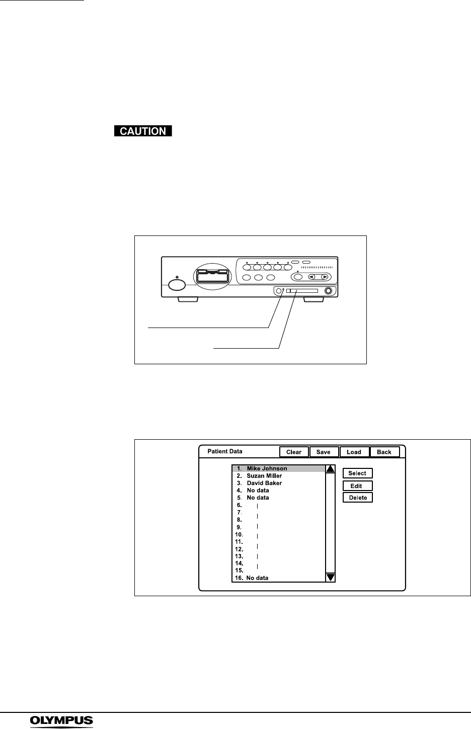
144
Chapter 5 Functions
EVIS EXERA II VIDEO SYSTEM CENTER CV-180
Recording patient data into PC card
The patient data that is entered in the video system center can be saved on the
PC card. The recorded patient data can be transferred to another CV-180 using
the PC card.
Be sure to confirm that there is no necessary patient data on
the PC card which needs to be saved. The patient data on
the PC card will be deleted when new patient data is stored.
1. Insert the PC card into the PC card slot. The PC card status indicator lights
up in green.
Figure 5.96
2. Press the “Shift” and “F3” keys together to display “Patient Data” menu (see
Figure 5.97).
Figure 5.97
3. Click “Save” to record all patient data on the PC card.
PC card status indicator
PC card slot

Chapter 5 Functions
145
EVIS EXERA II VIDEO SYSTEM CENTER CV-180
4. When patient data is stored on the PC card, a confirmation message
appears on the monitor.
Click “Yes” to overwrite the data. The PC card status indicator on the front
panel blinks in amber during patient data recording.
Click “No” to return to step 2.
5. Click “Back” to return to the endoscopic image.
All 40 patient data on the PC card are overwritten.
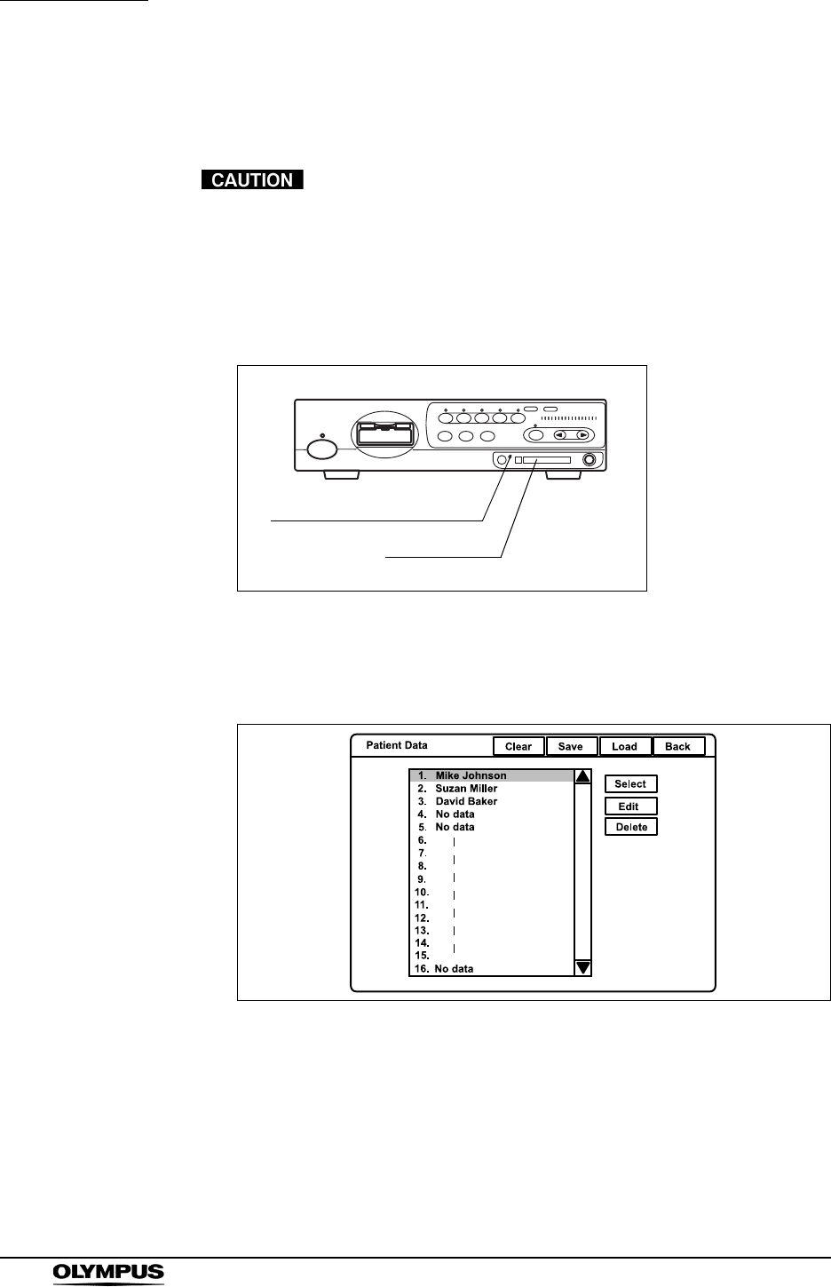
146
Chapter 5 Functions
EVIS EXERA II VIDEO SYSTEM CENTER CV-180
Loading patient data from PC card
Patient data on the PC card can be overwritten on the video system center.
Be sure to confirm that there are no necessary patient data in
the video system center. The patient data in the video system
center will be overwritten.
1. Insert the PC card into the PC card slot. The PC card status indicator lights
up green (see Figure 5.98).
Figure 5.98
2. Press the “Shift” and “F3” keys together to display the PC card menu on the
monitor (see Figure 5.99).
Figure 5.99
3. Click “Load” (see Figure 5.99). A confirmation message appears on the
monitor.
PC card status indicator
PC card slot

Chapter 5 Functions
147
EVIS EXERA II VIDEO SYSTEM CENTER CV-180
4. Click “No” to return to step 2.
Click “Yes” to register the patient data into the video system center. The
patient name in the patient name list changes to the new patient name on
the PC card. The PC card status indicator on the front panel blinks amber
during recording.
5. Click “Back” to return to the endoscopic image.
All 40 patients data are registered into the video system
center. Previous data disappear.
148
Chapter 5 Functions
EVIS EXERA II VIDEO SYSTEM CENTER CV-180
5.7 Scope information
Endoscopes equipped with the scope ID function incorporate a memory chip.
The memory chip stores data of the endoscope, and the data can be displayed
on the monitor. Some of the data can also be input from the video system center.
Table 5.18 shows the details of data stored in the memory chip.
• The memory chip is built with durability but may still be
damaged. If a memory chip is damaged, it becomes
incapable of backing up data. In such a case, please contact
Olympus.
• The following endoscopes are equipped with the scope ID
function;
EVIS EXERA II series endoscopes
EVIS EXERA 160 series or later endoscopes
(1 The items are not supported.)
Some endoscopes of the VISERA series
Data item Details Input
Scope model Model name of the endoscope No
Serial number Serial number of the endoscope No
Comment Up to 20 characters can be entered. Yes
Cumulative uses Cumulated number of uses No
Checkup period Times of inspections
User can enter a number of 0 - 4095. Yes
Service contract With or without a service contract No
Warranty date Warranty expiration date of the endoscope No
Customer name Person or hospital owning the endoscope
Up to 20 characters can be entered. Yes
Customer ID ID number of the customer
User can enter up to 20 characters. Yes
ID version The version number of the memory chip No
Channel diameter1Diameter of the endoscope channel No
Distal end diameter1Outer diameter of the endoscope's distal end No
Insertion tube
diameter1
Outer diameter of the endoscope's insertion tube No
Cumulative time1Cumulated time of uses No
Table 5.18
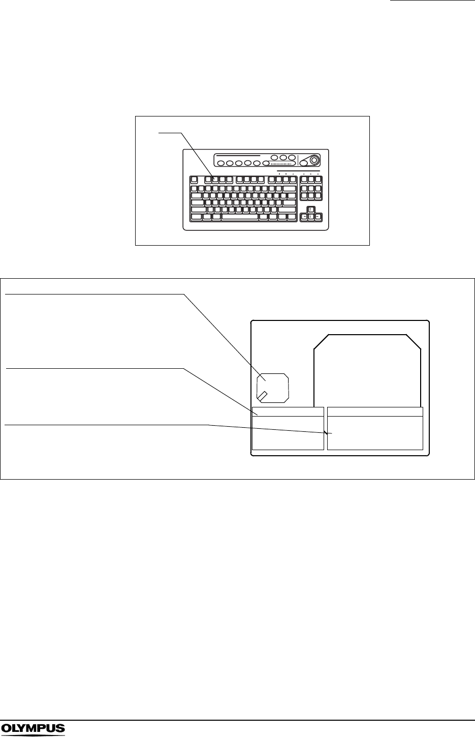
Chapter 5 Functions
149
EVIS EXERA II VIDEO SYSTEM CENTER CV-180
Displaying and entering scope information
1. Press the “F2” key (see Figure 5.100). The scope information window
appears on the monitor for about 6 seconds (see Figure 5.101).
Figure 5.100
Figure 5.101
F2
ABC123
Mike Johnson
M 51
03/03/1954
12/12/2005
12:12:12
CVP: A4/4
D.F: 99
VCR
Ct: N Eh: A8
Z: x1.5
Pump
Media:
John Smith
Cardiac end of the stomach
2.8
1:Freeze
2:Iris
3:Enhance
4:Release
Serial No.
Channel
Distal End
Insertion Tube
Scope Name
:2500001
:2.8
:9.8
:9.8
GIF-H180
Forceps channel diameter
Scope switch assignment
Endoscope’s information
Displays the position to see the forceps
and the diameter of the instrument
channel.
Displays the functions assigned to the
scope switches.
Displays serial no., forceps channel diameter,
distal end diameter and insertion part diameter.
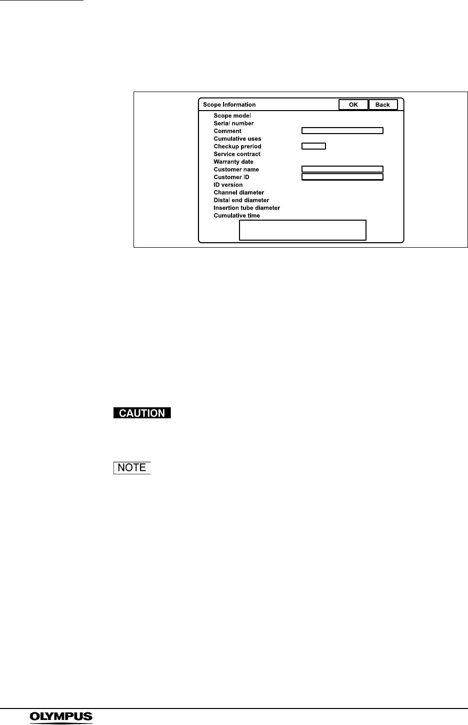
150
Chapter 5 Functions
EVIS EXERA II VIDEO SYSTEM CENTER CV-180
2. While the window opens, press the “F2” key again to show the scope
information screen on the monitor (see Figure 5.102).
Figure 5.102
3. Click each input area to modify.
4. Modify the data using the keyboard.
5. Click “OK” to store the entered data into the scope ID chip in the endoscope.
While the data is being stored, the message “Please wait” appears on the
monitor.
6. Click “Back” to return to the endoscopic image.
While the message “Please wait” is displayed, do not turn
OFF the video system center.
To cancel the input, click “Back” to return to the endoscopic
image display.
Chapter 5 Functions
151
EVIS EXERA II VIDEO SYSTEM CENTER CV-180
5.8 Special light observation
Do not rely on the special light observation method alone for
primary detection of lesions or for a decision regarding any
potential diagnostic or therapeutic intervention.
NBI (narrow band imaging)
If the endoscopic image seems to be dark in the NBI
observation, change to the normal observation. Otherwise,
the examination might not be preformed safely.
Perform the white balance adjustment for NBI observation
after completion of the white balance adjustment for the
normal observation. Otherwise, NBI observation cannot be
performed with a correct color tone.
NBI is an observation method that employs filtered light as examination light.
NBI requires the following equipment;
• CLV-180 light source
• NBI compatible endoscope:
GIF-H180, CF-H180AL/I, GIF-Q180, CF-Q180AL/I, PCF-Q180AL/I,
GIF-N180, BF-Q180, BF-P180, BF-1T180, ENF-V2/VQ, CYF-V2,
CYF-VA2, OTV-S7ProH-HD-L08E, LTF-VH, WA5001A/L
Normal observation and NBI observation can be changed during use.
All mucosal areas are to be viewed using traditional white light.
NBl imaging should not be used as a substitute for a thorough traditional
examination of the mucosa.
1. Select the NBI mode on the light source using the mode button, and confirm
the NBI indicator of the light source, referring to the instruction manual of the
light source.
2. Confirm that the NBI indicator on the front panel of the video system center
turns from green to white (see Figure 5.103).
• “NBI” may appear on the monitor by the user preset.
• During NBI observation, bile and residual debris may appear
dark and reddish.
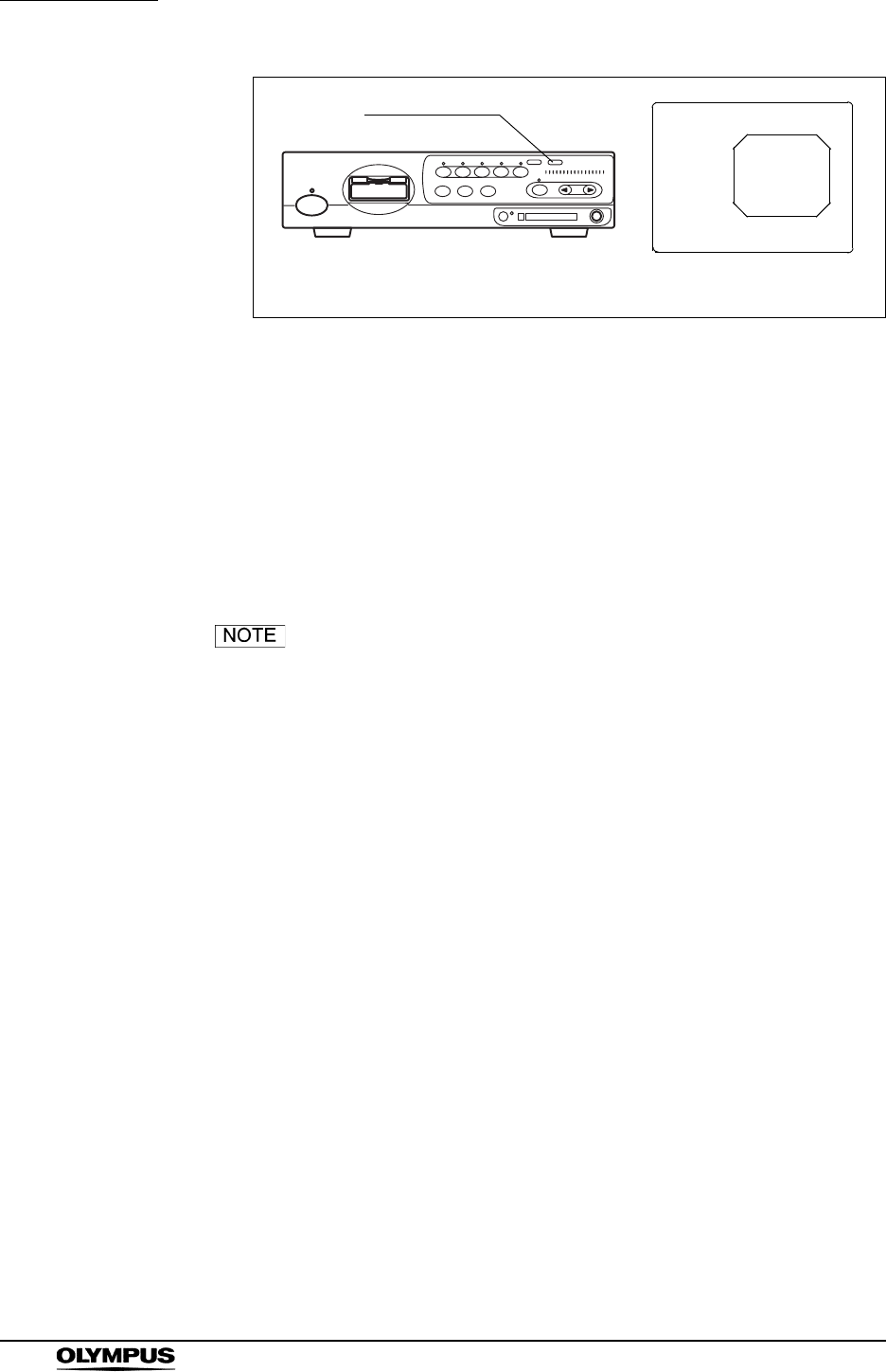
152
Chapter 5 Functions
EVIS EXERA II VIDEO SYSTEM CENTER CV-180
Figure 5.103
3. Perform the white balance adjustment (see page 52).
4. Perform the NBI observation.
5. Return to the normal observation using the mode button on the light source,
referring to the instruction manual of the light source.
6. Confirm that the NBI indicator on the front panel of the video system center
lights up green, and that “NBI” disappears on the monitor.
• If the “NBI” mode is selected when a non NBI compatible light
source and/or endoscope is used, the NBI indicator does not
change to green, and a warning message appears on the
monitor.
• Enter and button operations cannot be operated for several
seconds while the light source turns into NBI mode.
• Contrast (“Shift” + “F6”) and color mode (“Shift” + “Alt” + “1”,
“2”, “3”, “4”) cannot be operated during the NBI observation.
• The NBI mode selection operation can also be controlled
from the scope switches and/or foot switches. For how to set
up the scope switches and foot switches, see “Remote switch
and foot switch (EXERA and VISERA)” on page 219.
• The color tones of NBI observation slightly vary with
products.
• Contact Olympus for further information about the NBI
observation.
NBI
NBI indicator
NBI indication on the monitor
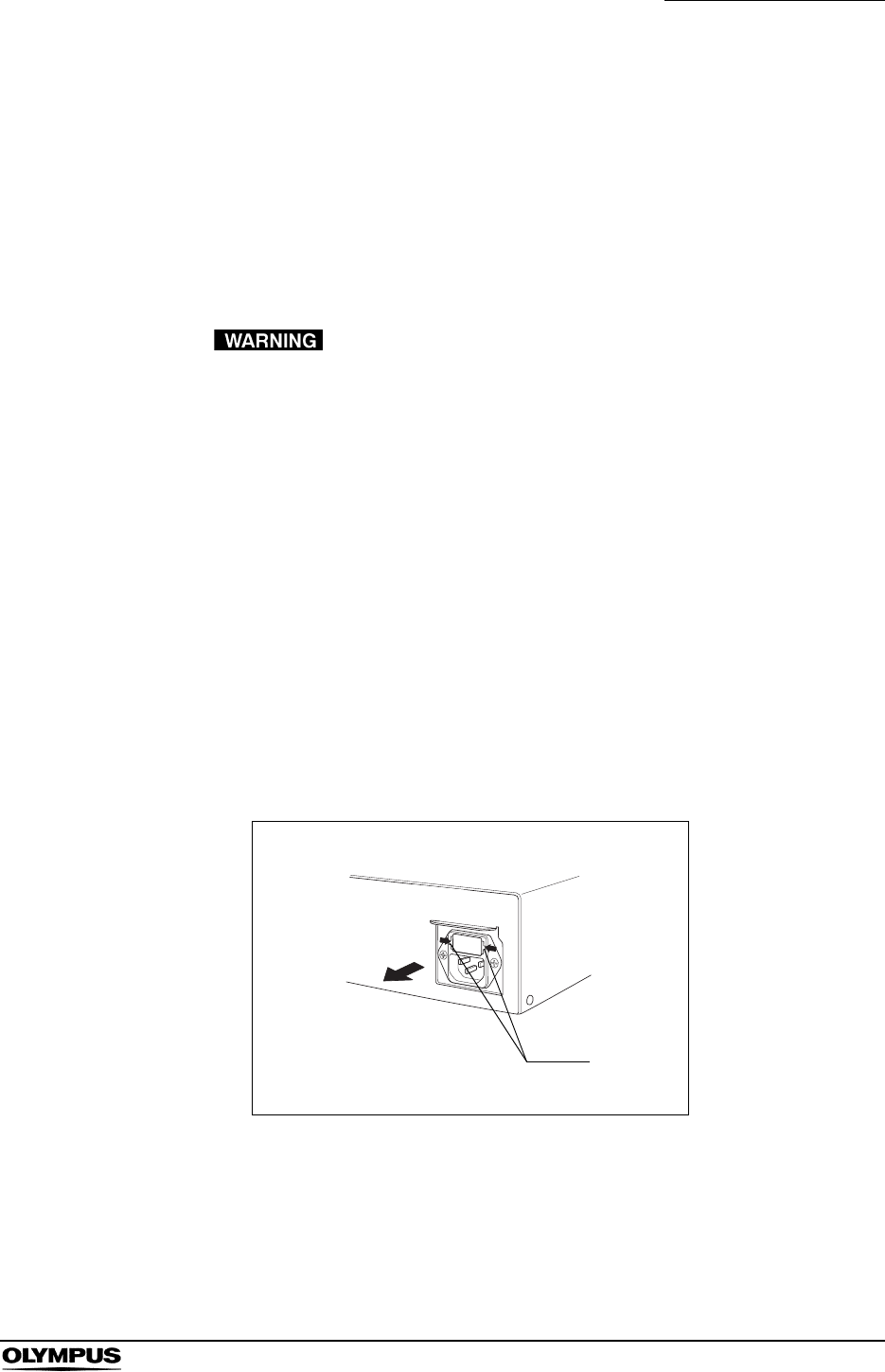
Chapter 6 Fuse replacement
153
EVIS EXERA II VIDEO SYSTEM CENTER CV-180
Chapter 6 Fuse replacement
Always use the fuses designated below. To order new fuses, please contact
Olympus.
• Never use a fuse other than the fuse model designated by
Olympus. Otherwise, malfunction or failure of the video
system center may cause a fire or electric shock hazard.
• Be sure to turn the video system center OFF and unplug the
power cord before removing the fuse box from the video
system center. Otherwise, fire or electric shock may result.
• If the power fails to come on after replacing the fuses, unplug
the power cord immediately from the AC mains power inlet
and then contact Olympus. Otherwise, electric shock may
result.
1. Turn the video system center OFF and disconnect the power cord from the
wall mains outlet.
2. Pull the fuse box straight out, squeezing the tabs projected on both sides of
the fuse box using a pair of tweezers (see Figure 6.1).
Figure 6.1
• Spare fuses MAJ-1432
Tabs
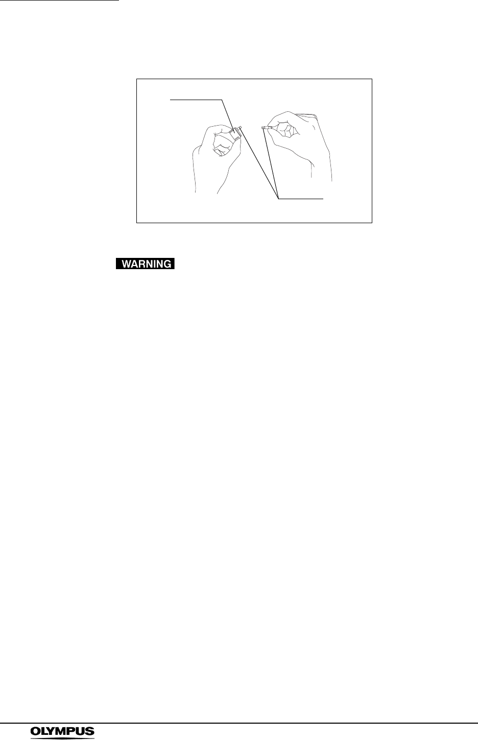
154
Chapter 6 Fuse replacement
EVIS EXERA II VIDEO SYSTEM CENTER CV-180
3. Replace both fuses (see Figure 6.2).
Figure 6.2
Insert the fuse box into this instrument until it clicks into
position. If the fuse box is inserted incompletely, the power
may fail to come ON or a power failure may occur during
operation.
4. Insert the fuse box into the video system center until it clicks into position.
5. Plug the power cord and turn the video system center ON and confirm the
power output.
Fuses
Fuse box
Chapter 7 Care, Storage and Disposal
155
EVIS EXERA II VIDEO SYSTEM CENTER CV-180
Chapter 7 Care, Storage and Disposal
7.1 Care
• After wiping with a piece of moistened gauze, dry the video
system center thoroughly before using it again. If it is used
while still wet, there is the risk of an electric shock.
• When cleaning the video system center, always wear
appropriate personal protection equipment such as eye wear,
face mask, moisture-resistant clothing and chemical-resistant
gloves that fit properly and are long enough to that your skin
is not exposed. Blood, mucus and other potentially infectious
material adhering to the video system center could pose an
infection control risk.
• Do not apply spray-type medical agents such as rubbing
alcohol directly to the video system center. Medical agents
might enter the video system center through the ventilation
grills and may cause equipment damage.
• Do not clean the videoscope cable socket, the terminals and
the AC mains power inlet. Cleaning them can deform or
corrode the contacts, which could damage the video system
center.
• Do not soak in water, autoclave or gas sterilize the video
system center. These methods will damage it.
• Do not wipe the external surface with hard or abrasive wiping
material, the surface will be scratched.
After using the video system center, immediately perform the following cleaning
procedure. If cleaning is delayed, residual organic debris will begin to solidify,
and it may be difficult to effectively clean the video system center. Always
remove debris routinely.
1. Turn the video system center OFF and disconnect the power cord from the
wall mains outlet.
2. If the video system center is soiled with blood or other potentially infectious
materials, wipe off all debris using a piece of gauze moistened with neutral
detergent.

156
Chapter 7 Care, Storage and Disposal
EVIS EXERA II VIDEO SYSTEM CENTER CV-180
3. Remove dust, dirt and other stains on the surface by wiping with a piece of
gauze moistened with 70% ethyl or isopropyl alcohol.
4. Make sure to dry the video system center after wiping with 70% ethyl or
isopropyl alcohol.
7.2 Storage
Do not store the video system center in a location exposed to
direct sunlight, X-rays, radio activity or strong
electromagnetic radiation (e.g. near microwave medical
treatment equipment, short-wave medical treatment
equipment, MRI equipment, radio or mobile phones).
Damage to the video system center may result.
1. Turn the video system center OFF and disconnect the power cord from the
wall mains outlet.
2. Disconnect the ancillary equipment connected to the video system center.
3. Store the equipment in the level position in a clean, dry and stable location.
7.3 Disposal
When disposing of this instrument or any of its components
(such as fuses), follow all applicable national and local laws
and guidelines.
1. For security reasons, clear all patient data in the instrument (see “Clearing
all patient data previously entered” on page 143).
2. Clear the image data and patient data in the PC card, or format the PC card
(see “Formatting of the PC card” on page 114).

Chapter 8 Installation and Connection
157
EVIS EXERA II VIDEO SYSTEM CENTER CV-180
Chapter 8 Installation and Connection
• Review this chapter thoroughly, and prepare the instruments
properly before use. If the equipment is not properly prepared
before each use, equipment damage, patient and operator
injury and/or fire can occur.
• When non-medical electrical ancillary equipment is used,
connect its power cord via an isolation transformer prior to
connecting it to this video system center. Failure to do so can
cause electric shock, burns and/or fire.
• Turn OFF all system components before connecting them.
Otherwise, equipment damage or malfunction can result.
• Use appropriate cables only. Otherwise, equipment damage
or malfunction can result.
• Properly and securely connect all cables. Otherwise,
equipment damage or malfunction can result.
• The cables should not be sharply bent, pulled, twisted or
crushed. Cable damage can result.
• Never apply excessive force to connectors. This could
damage the connectors.
• Use this instrument only under the conditions described in
“Transportation, storage, and operation environment” and
“Specifications” in the Appendix. Otherwise, improper
performance, compromised safety and/or equipment damage
may result.
Prepare this video system center and compatible equipment (shown in the
“System chart” in the Appendix) before each use. Referring to the instruction
manuals of each system component, install and connect the equipment
according to the procedure described in this chapter.
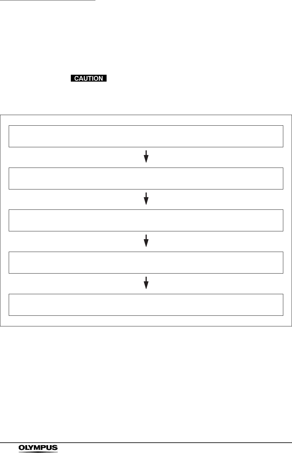
158
Chapter 8 Installation and Connection
EVIS EXERA II VIDEO SYSTEM CENTER CV-180
8.1 Installation work flow
Please see the installation work flow in Figure 8.1 below. Follow each step of the
work flow before using the video system center and the ancillary equipment.
Connect the power cord to the power source after connecting
all cables. Otherwise, equipment damage or malfunction can
result.
Figure 8.1
1. Install the video system center and the ancillary equipment to the mobile workstation, etc.
See section 8.2, “Installation of the equipment” on page 159
2. Connect the video system center and the ancillary equipment to the mobile work station, etc.
See sections 8.4, “Light source” on page 164 through 8.10, “Foot switch” on page 184
3. Connect the instruments to the power source.
See section 8.11, “Ultrasound center” on page 185
4. Set up the system setup.
See section 9.2, “System setup” on page 194
5. Set up the user preset.
See section 9.3, “User preset” on page 216

Chapter 8 Installation and Connection
159
EVIS EXERA II VIDEO SYSTEM CENTER CV-180
8.2 Installation of the equipment
• Do not place any object on the top of the video system
center. Otherwise, equipment deformation and damage can
result.
• Keep the ventilation grills of the video system center clear.
The ventilation grills are located on the side panels. Blockage
can cause overheating and equipment damage.
• Clean and vacuum dust the ventilation grills using a vacuum
cleaner. Otherwise, the video system center may break down
from over heating.
• Place the video system center on a stable, level surface
using the foot holders (MAJ-1433). Otherwise, the video
system center may topple down or drop, and user or patient
injury may occur, or equipment damage can result.
• If a trolley other than the mobile workstation (WM-NP1 or
WM-WP1) is used, confirm that the trolley can withstand the
weight of the equipment installed on it.
• Do not install the video system center near a source of strong
magnetic wave (microwave treatment device, short wave
treatment device, MRI, radio equipment, etc.). Otherwise, the
video system center may malfunction.
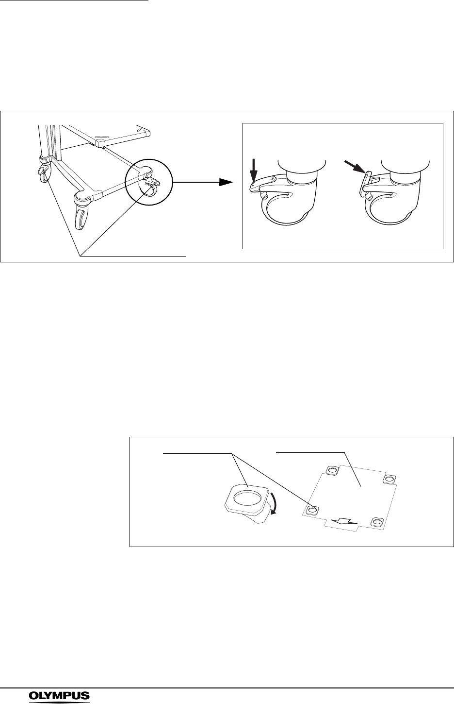
160
Chapter 8 Installation and Connection
EVIS EXERA II VIDEO SYSTEM CENTER CV-180
Installation on the mobile workstation (WM-NP1, WM-WP1)
1. Place the mobile workstation on a level and flat floor. Lock the caster brakes
by pushing them down (see Figure 8.2).
Figure 8.2
2. Install the mobile shelf of the mobile workstation according to the
configuration of the equipment installed on it as described in the mobile
workstation's instruction manual.
3. Place the pattern sheet on the light source. The pattern sheet is packed with
the foot holder (MAJ-1433).
4. Peel the paper from the back of the foot holders and attach them firmly at
the right position on the light source using the pattern sheet (see Figure
8.3).
Figure 8.3
5. Remove the pattern sheet.
6. Place the light source on the mobile shelf of the mobile workstation as
described in the light source's instruction manual.
Caster brakes
Push to lock. Push to release.
Foot holder Pattern sheet
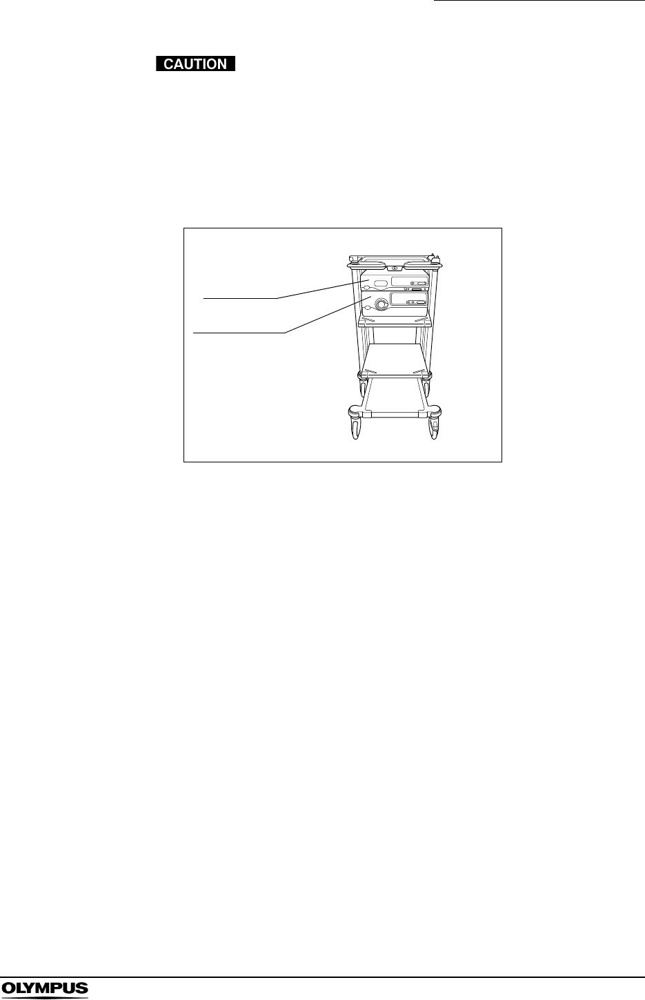
Chapter 8 Installation and Connection
161
EVIS EXERA II VIDEO SYSTEM CENTER CV-180
When using a light source that is smaller than the video
system center, such as CLV-S40, place the light source on
the video system center. Otherwise, the video system center
may not be kept level and fall down.
7. Place the video system center on the light source so that the feet of the
video system center fit into the foot holders.
Figure 8.4
Installation in another location
When installing the video system center in another location, adhere the foot
holders as described on the previous page, and place the video system center
on them.
CV-180
Light source
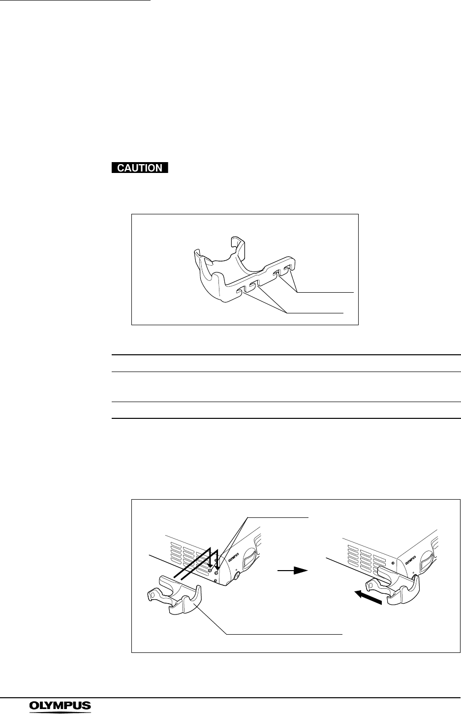
162
Chapter 8 Installation and Connection
EVIS EXERA II VIDEO SYSTEM CENTER CV-180
8.3 Fitting of accessories
Videoscope cable holder (MAJ-1466)
The videoscope cable holder (MAJ-1466) is used to temporarily hang the
connector on the endoscope end of the videoscope cable. The holder is attached
to the video system center by ditches A or ditches B (see Figure 8.5).
Do not hook anything other than the videoscope cable on the
videoscope cable holder.
Figure 8.5
1. Fit ditches A or B to the projections on the left side of the instrument.
2. Push the videoscope cable holder backward until it clicks.
Figure 8.6
Ditch Remark
AThe scope cable holder projects forward from the front panel.
(When the water container is mounted on the light source.)
B The scope cable holder does not project forward from the front panel.
Table 8.1
Scope cable holder
Ditches A
Ditches B
Scope cable holder
Projections
(a) (b)
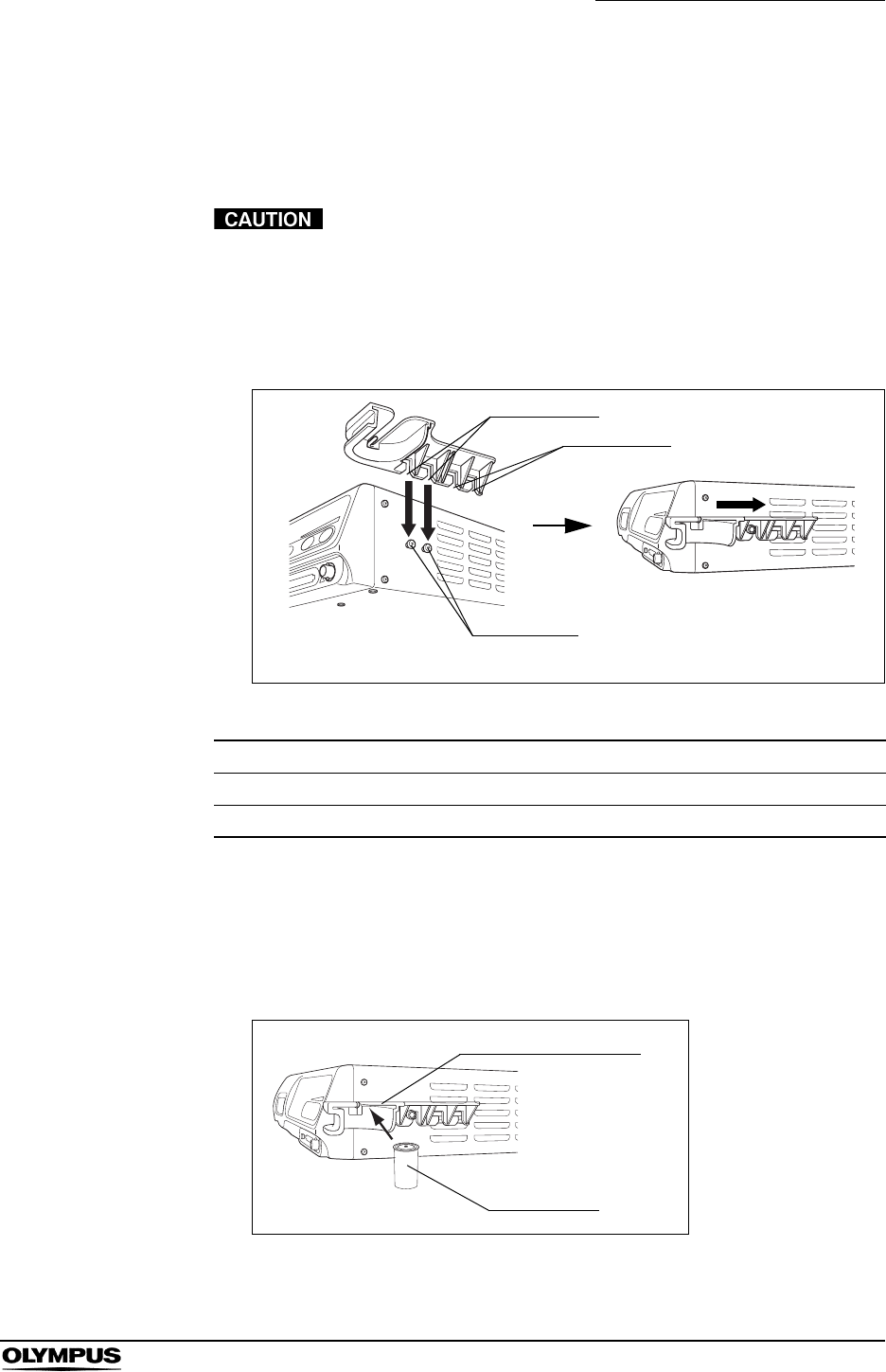
Chapter 8 Installation and Connection
163
EVIS EXERA II VIDEO SYSTEM CENTER CV-180
White cap set (MAJ-941)
The white cap (MH-155) is used for the white balance adjustment. The white cap
is attached to the video system center by the white cap holder (MAJ-960).
Do not hang any object other than the white cap on the white
cap holder.
1. Fit ditches A or B on the projections on the left side of the instrument (see
Figure 8.7).
Figure 8.7
2. Push the white cap holder backward until it clicks.
3. Insert the disk part at the top of the white cap into the ditches on the opening
of the white cap holder until it clicks (see Figure 8.8).
Figure 8.8
Ditch Remark
A The white cap projects forward from the front panel.
B The white cap does not project forward from the front panel.
Table 8.2
Projections
(a) (b)
Ditches B
Ditches A
White cap
White cap holder

164
Chapter 8 Installation and Connection
EVIS EXERA II VIDEO SYSTEM CENTER CV-180
8.4 Light source
Compatible light sources
For compatible light sources, see the table below.
When a light source other than CLV-180 is used, the operations are restricted as
follows.
Model Product name
CLV-180 EVIS EXERA II xenon light source
CLV-160 EVIS EXERA xenon light source
CLV-U40 EVIS universal light source
CLV-S40 VISERA xenon light source
Table 8.3
Brightness The brightness adjustment function interlocked with the light source
does not work.
Special light
observation
Special light observation function is not available.
Table 8.4
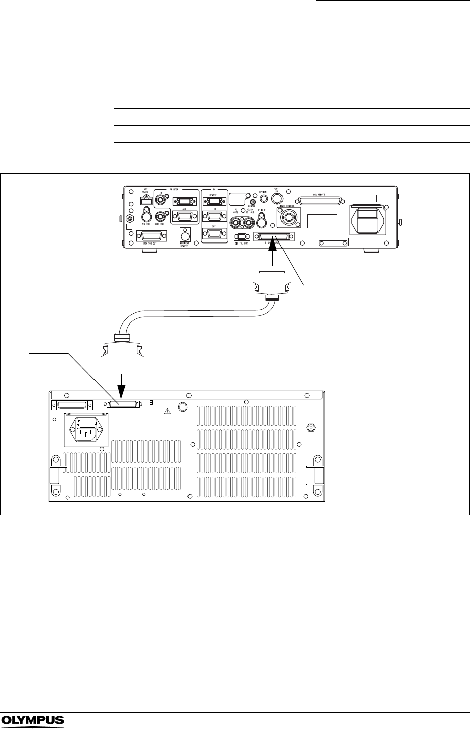
Chapter 8 Installation and Connection
165
EVIS EXERA II VIDEO SYSTEM CENTER CV-180
CLV-180
For connecting the light source (CLV-180) to the video system center, use the
cable listed below.
Figure 8.9
Model Product name note
MAJ-1411 Light source cable -
Table 8.5
MAJ-1411
LIGHT SOURCE
CLV-180
CV
CV-180
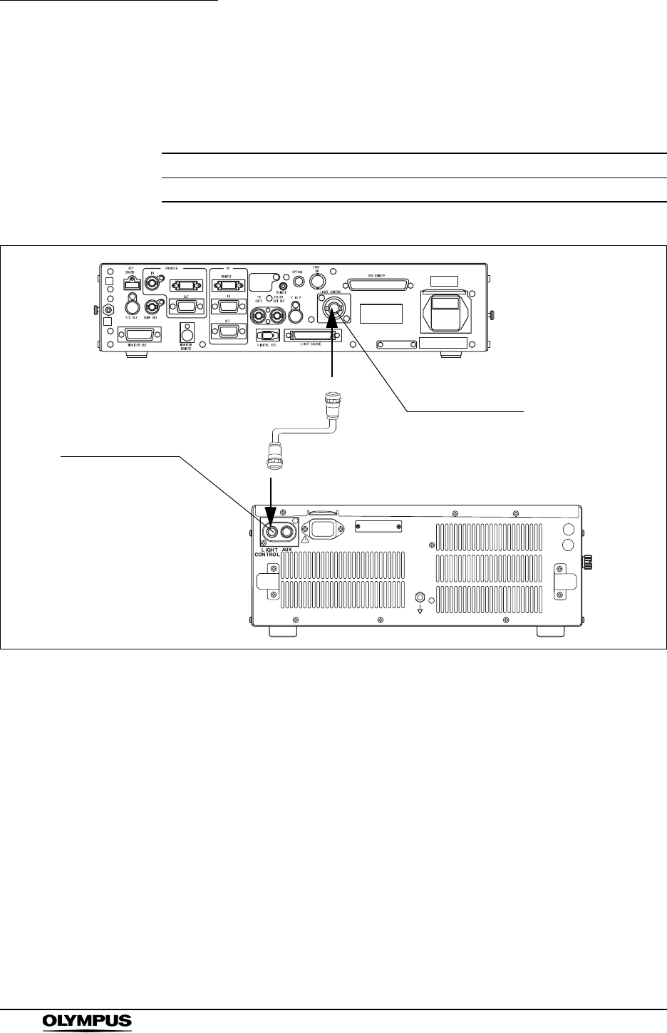
166
Chapter 8 Installation and Connection
EVIS EXERA II VIDEO SYSTEM CENTER CV-180
CLV-160
For connecting the light source (CLV-160) to the video system center, use the
cable listed below.
Figure 8.10
Model Product name note
MH-966 Light control cable -
Table 8.6
MH-966LIGHT CONTROL
CLV-160
CV-180
LIGHT CONTROL
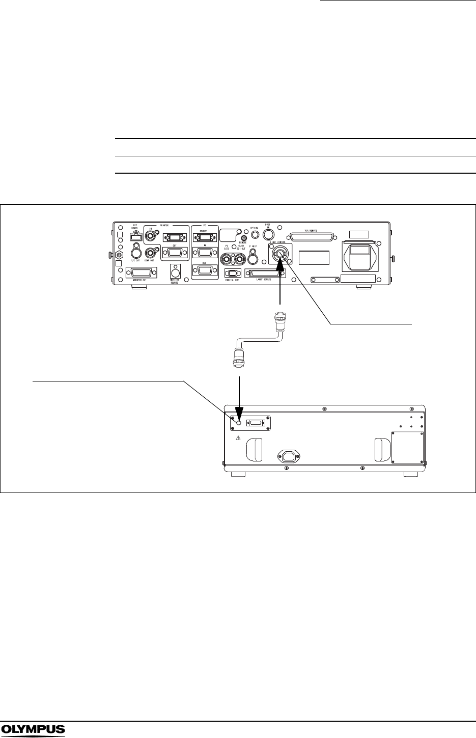
Chapter 8 Installation and Connection
167
EVIS EXERA II VIDEO SYSTEM CENTER CV-180
CLV-U40
When connecting to the rear panel of the CLV-U40
For connecting the light source (CLV-U40) to the video system center, use the
cable listed below.
Figure 8.11
Model Product name note
MH-966 Light control cable -
Table 8.7
MH-966
LIGHT CONTROL
CLV-U40
CV-180
Light control connector
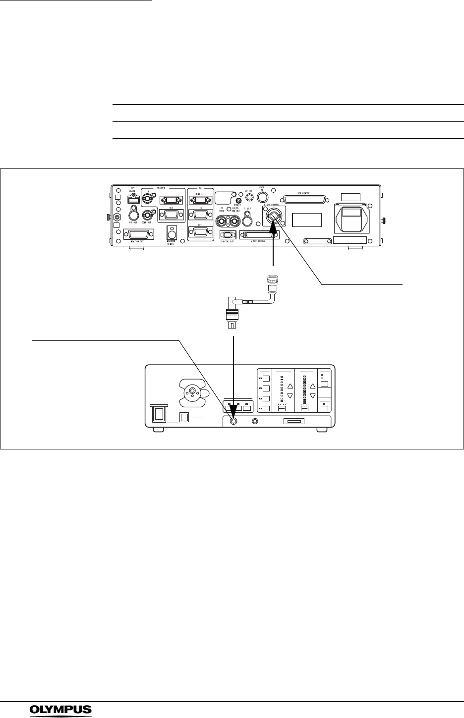
168
Chapter 8 Installation and Connection
EVIS EXERA II VIDEO SYSTEM CENTER CV-180
When connecting to the front panel of the CLV-U40
For connecting the light source (CLV-U40) to the video system center, use the
cable listed below.
Figure 8.12
Model Product name note
MH-993 or MAJ-1567 Light control cable -
Table 8.8
1
2
l
O
MH-993 or
MAJ-1567
LIGHT CONTROL
CLV-U40
Rigidscope camera connector
CV-180
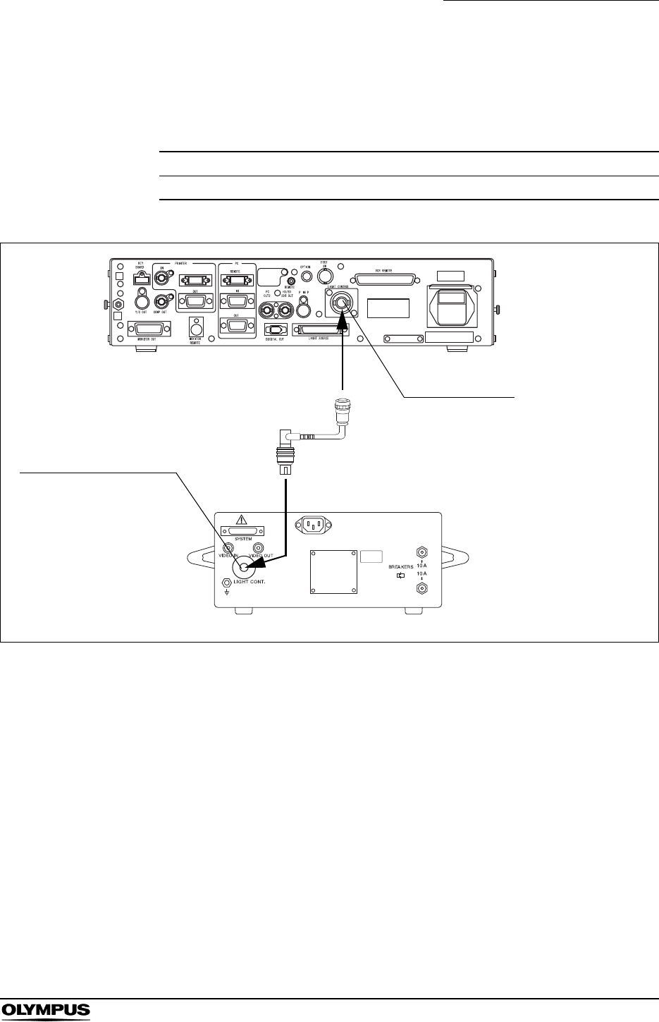
Chapter 8 Installation and Connection
169
EVIS EXERA II VIDEO SYSTEM CENTER CV-180
CLV-S40
For connecting the light source (CLV-S40) to the video system center, use the
cable listed below.
Figure 8.13
Model Product name Note
MAJ-1567 Light control cable -
Table 8.9
MAJ-1567
LIGHT CONTROL
CLV-S40
CV-180
LIGHT CONT. connector

170
Chapter 8 Installation and Connection
EVIS EXERA II VIDEO SYSTEM CENTER CV-180
8.5 Monitor
Compatible monitors
For compatible monitors, see the table below.
Model Product name
OEV143, OEV203 Color video monitor
OEV191 LCD monitor
OEV181H High definition monitor
OEV191H High definition LCD monitor
Table 8.10
Chapter 8 Installation and Connection
171
EVIS EXERA II VIDEO SYSTEM CENTER CV-180
OEV143 or OEV203
For connecting the monitors (OEV143 or OEV203) to the video system center,
use the cables listed below.
For easy cable connection, attach the cable color sheet to the cables (see Figure
8.14).
• Connect the R/G/B/S connectors of the monitor cable to the
RGB component A terminals of the monitor. If those
connectors are connected to the RGB component B
terminals, remote control of the monitor is deactivated.
• When the monitor remote cable is connected to the monitor,
the “SPLIT“, “RESET“, “UNDERSCAN“, “OVERSCAN”
switches on the front panel of the monitor are deactivated.
Model (length) Product name
Use any one of the following cables.
MAJ-846 (7 m)
MAJ-921 (1.5 m)
MAJ-970 (4 m)
MAJ-971 (15 m)
MAJ-1462 (7 m)
MAJ-1584 (15 m)
MAJ-1586 (2 m)
Monitor cable
MAJ-227 (7 m) Monitor remote cable
Table 8.11
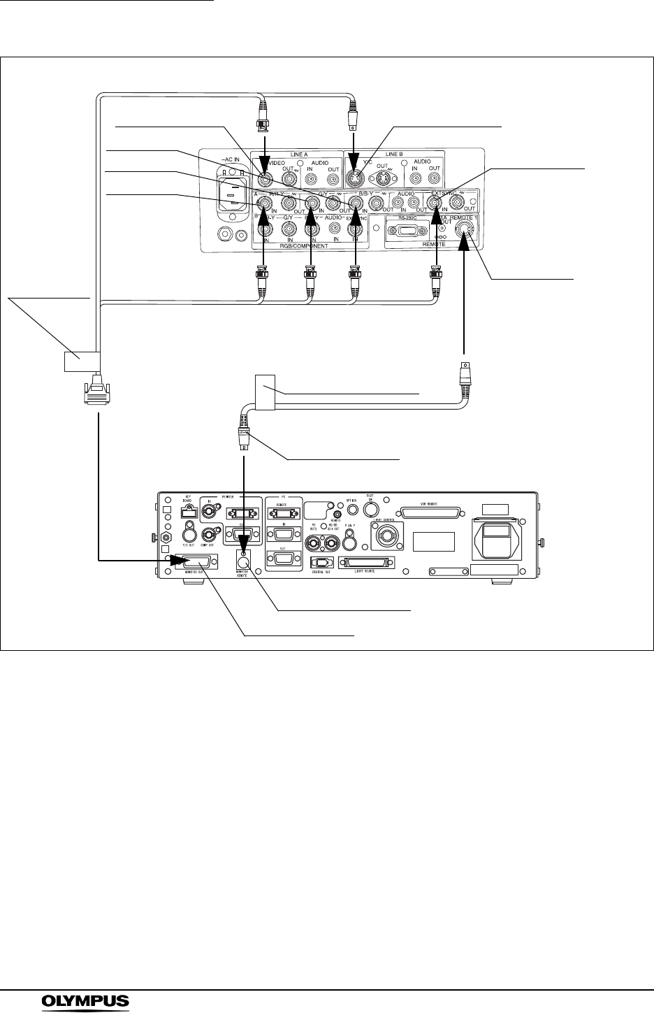
172
Chapter 8 Installation and Connection
EVIS EXERA II VIDEO SYSTEM CENTER CV-180
Figure 8.14
S-VideoV
RG
BS
MONITOR REMOTE
MONITOR OUT
MAJ-227
OEV143,
OEV203
LINE A-VIDEO-IN LINE B-Y/C-IN
A-R/R-Y-IN
A-G/Y-IN
A-B/B-Y-IN
A-EXTSYNC-IN
CV-180
“MAJ-227” mark
REMOTE 1
Cable color
sheet
“MONITOR
OUT”
(brown)
Cable color sheet
“MONITOR REMOTE”
(brown)
MAJ-846,
MAJ-921,
MAJ-970,
MAJ-971,
MAJ-1462,
MAJ-1584,
or
MAJ-1586
Chapter 8 Installation and Connection
173
EVIS EXERA II VIDEO SYSTEM CENTER CV-180
OEV181H
For connecting the monitor (OEV181H) to the video system center, use the
cables listed below.
For easy cable connection, attach the cable color sheet to the cables (see Figure
8.15).
Set the factory default “FACTORY” of the monitor to
“FACTORY1”, according to the instruction manual of the
monitor. Otherwise, the monitor remote function may not
operate correctly.
Model (length) Product name
Use any one of the following cables.
MAJ-921 (1.5 m)
MAJ-970 (4 m)
MAJ-1462 (7 m)
MAJ-1584 (15 m)
MAJ-1586 (2 m)
Monitor cable
Use either one of the following cables.
MAJ-1161 (4 m)
MAJ-1230 (7 m)
MAJ-1465 (15 m)
HDTV monitor remote cable
Table 8.12
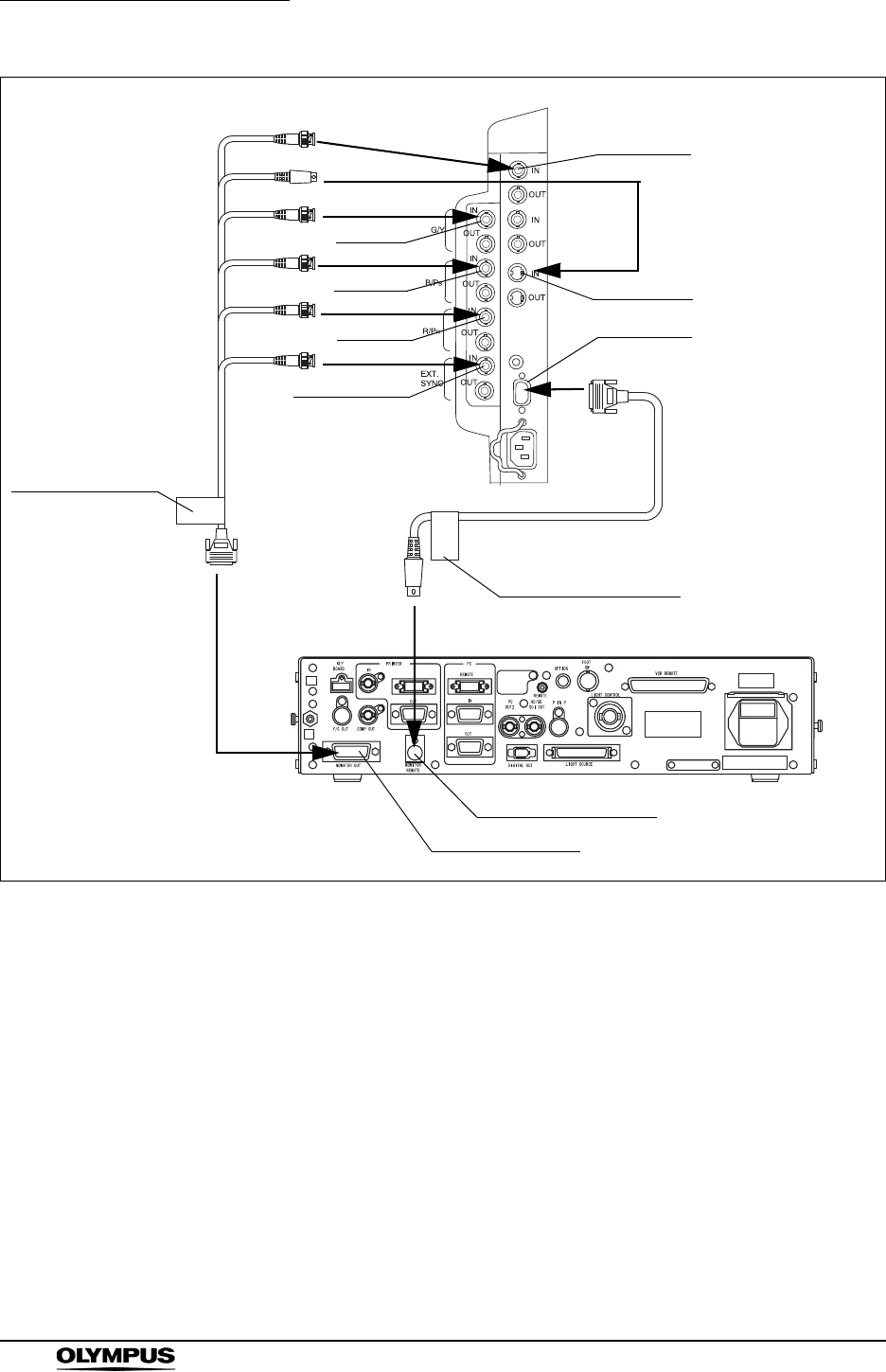
174
Chapter 8 Installation and Connection
EVIS EXERA II VIDEO SYSTEM CENTER CV-180
Figure 8.15
Y/C
V
R
G
B
S
MONITOR REMOTE
MONITOR OUT
OEV181H
G/Y-IN
B/PB-IN
R/PR-IN
EXTSYNC-IN
S-Video-IN
Line A-IN
REMOTE
CV-180
Cable color sheet
“MONITOR OUT”
(brown)
Cable color sheet
“MONITOR REMOTE”
(brown)
MAJ-921,
MAJ-970,
MAJ-1462,
MAJ-1584,
or
MAJ-1586
MAJ-1161,
MAJ-1230,
or
MAJ-1465
Chapter 8 Installation and Connection
175
EVIS EXERA II VIDEO SYSTEM CENTER CV-180
OEV191H, OEV191
When using RGB or YPbPr video signal
For connecting the monitors (OEV191H or OEV191) to the video system center,
use the cables listed below.
For easy cable connection, attach the cable color sheet to the cables (see Figure
8.16).
• To display an endoscopic image, set the factory default
“FACTORY” of the monitor to “FACTORY4”, according to the
instruction manual of the monitor. Otherwise, the monitor
remote function may not operate correctly.
• Setting the monitor to “FACTORY4” may make an ultrasound
image in the PinP display fuzzy. Set the monitor to
“FACTORY6” to sharpen it.
Model (length) Product name
Use any one of the following cables.
MAJ-921 (1.5 m)
MAJ-970 (4 m)
MAJ-1462 (7 m)
MAJ-1584 (15 m)
MAJ-1586 (2 m)
MAJ-8461 (7 m)
MAJ-9711 (15 m)
Monitor cable
Use either one of the following cables.
MAJ-1161 (4 m)
MAJ-1230 (7 m)
MAJ-1465 (15 m)
HDTV monitor remote cable
1 These cables are not compatible with the OEV191H.
Table 8.13
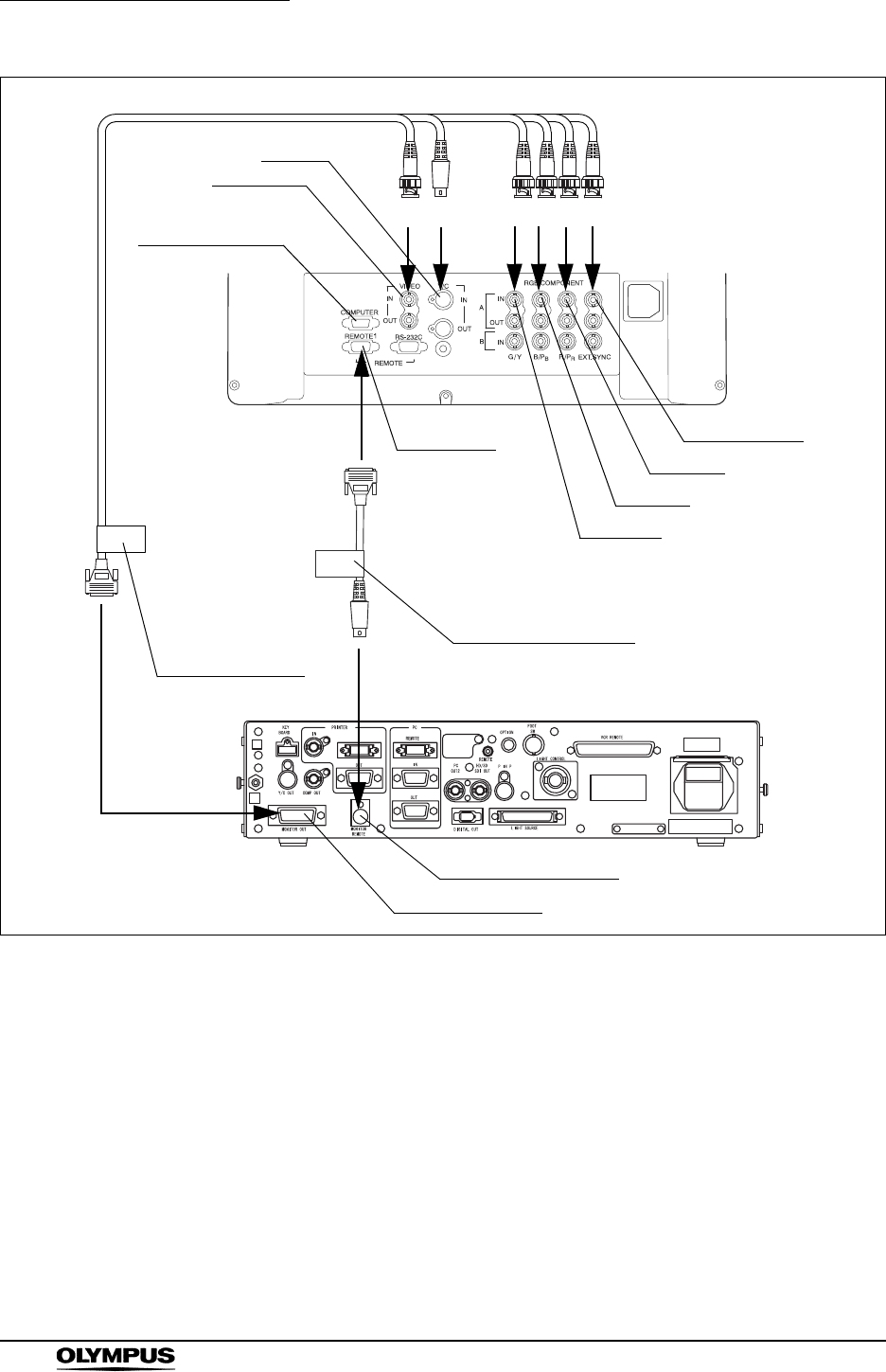
176
Chapter 8 Installation and Connection
EVIS EXERA II VIDEO SYSTEM CENTER CV-180
Figure 8.16
Y/C
VR
BS
OEV191, OEV191H
G
Y/C-IN
VIDEO-IN
G/Y-IN
B/PB-IN
R/PR-IN
EXT.SYNC-IN
REMOTE1
MONITOR REMOTE
MONITOR OUT
No PC terminal on
OEV191
CV-180
Cable color sheet
“MONITOR OUT”
(brown)
Cable color sheet
“MONITOR REMOTE”
(brown)
MAJ-921,
MAJ-970,
MAJ-1462,
MAJ-1584,
or
MAJ-1586 MAJ-1161,
MAJ-1230,
or
MAJ-1465
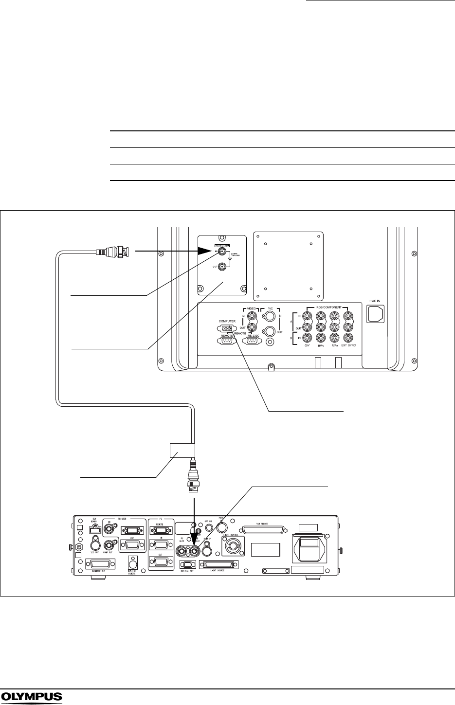
Chapter 8 Installation and Connection
177
EVIS EXERA II VIDEO SYSTEM CENTER CV-180
When using SDI video signal
For connecting the monitors (OEV191H or OEV191) to the video system center,
use the cables listed below.
For easy cable connection, attach the cable color sheet to the cable (see Figure
8.17).
Figure 8.17
Model Product name Note
MAJ-1464 SDI cable 22 m
MAJ-1431 HD/SD-SDI adapter Attach to OEV191H
Table 8.14
HD/SD SDI OUT
OEV191, OEV191H
No PC terminal on
OEV191
HD/SD SDI IN
MAJ-1464
MAJ-1431
CV-180
Cable color sheet
“HD/SD SDI OUT”
(brown)
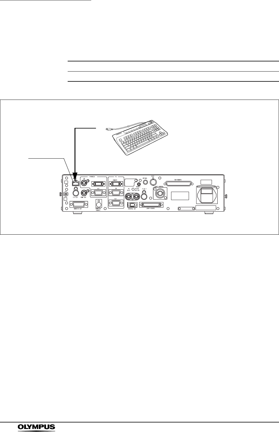
178
Chapter 8 Installation and Connection
EVIS EXERA II VIDEO SYSTEM CENTER CV-180
8.6 Keyboard
For a compatible keyboard, see the table below.
Figure 8.18
Model Product name Note
MAJ-1428 Keyboard -
Table 8.15
MAJ-1428KEYBOARD
CV-180

Chapter 8 Installation and Connection
179
EVIS EXERA II VIDEO SYSTEM CENTER CV-180
8.7 Videocassette recorder (VCR)
When using the VCR remote terminal
For compatible video cassette recorders and cables to be used, see the table
below.
• When the SVO-9500MD is used together with the VTR
remote cable (MH-989), the SVO-9500MD has to be
equipped with the RS-232C interface board (SVBK-120). For
details, refer to the instruction manual for the interface board.
• When the SVO-9500MD is used together with the VTR
remote cable (MH-992), remote control is restricted to the
[Record] and [Pause] functions.
• For details, refer to the instruction manuals for the videotape
recorder and VTR remote cable.
• When the PDW-70MD or the PDW-75MD is used, SDI image
cable (MAJ-1464) is necessary with VTR remote cable (MAJ-
1568).
VCR
Cable
MH-989 MH-992 MAJ-906 MAJ-1568
DVO-1000MD (SONY)
DSR-20MD (SONY)
SVO-9500MD (SONY)
PDW-70MD (SONY)
PDW-75MD (SONY)
compatible, incompatible
Table 8.16
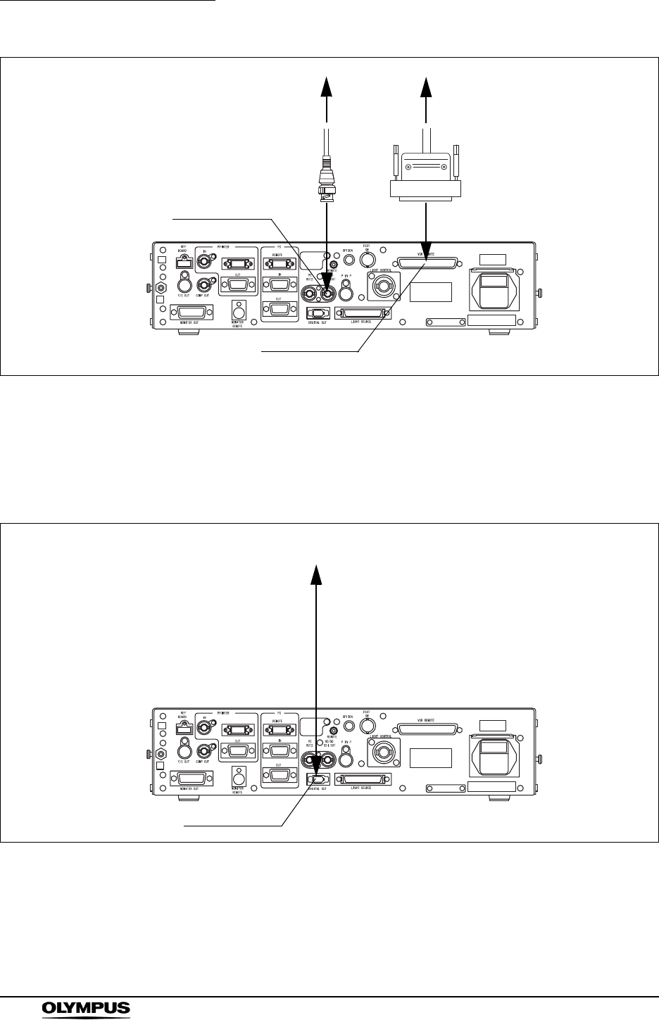
180
Chapter 8 Installation and Connection
EVIS EXERA II VIDEO SYSTEM CENTER CV-180
Figure 8.19
When using the digital out terminal
For connecting the video casette recorder to the video system center, use a
commercially available DV cable.
Figure 8.20
VCR REMOTE
to VCR
CV-180
VTR remote cable
HD/SD SDI OUT
to VCR
SDI cable
DIGITAL OUT
to VCR
CV-180

Chapter 8 Installation and Connection
181
EVIS EXERA II VIDEO SYSTEM CENTER CV-180
8.8 Video printer
For compatible video printers see the table below.
For connecting a video printer to the video system center, use the cables listed
below.
For easy cable connection, attach the cable color sheet to the cables (see Figure
8.21).
Model Product name Note
OEP, OEP-3, OEP-4 Color video printer OLYMPUS
UP-1800, UP-1850 Color video printer SONY
UP-2900MD, UP-2950MD Color video printer SONY
UP-5200MD, UP-5250MD Color video printer SONY
UP-21MD, YP-22MD Color video printer SONY
Table 8.17
Model Product name Note
MH-984, or
MD-445 and MAJ-849
Monitor cable (MH-984),
SCV cable (MD-445),
Printer adapter (MAJ-849)
-
MH-995 Remote cable -
MB-677 BNC cable -
Table 8.18
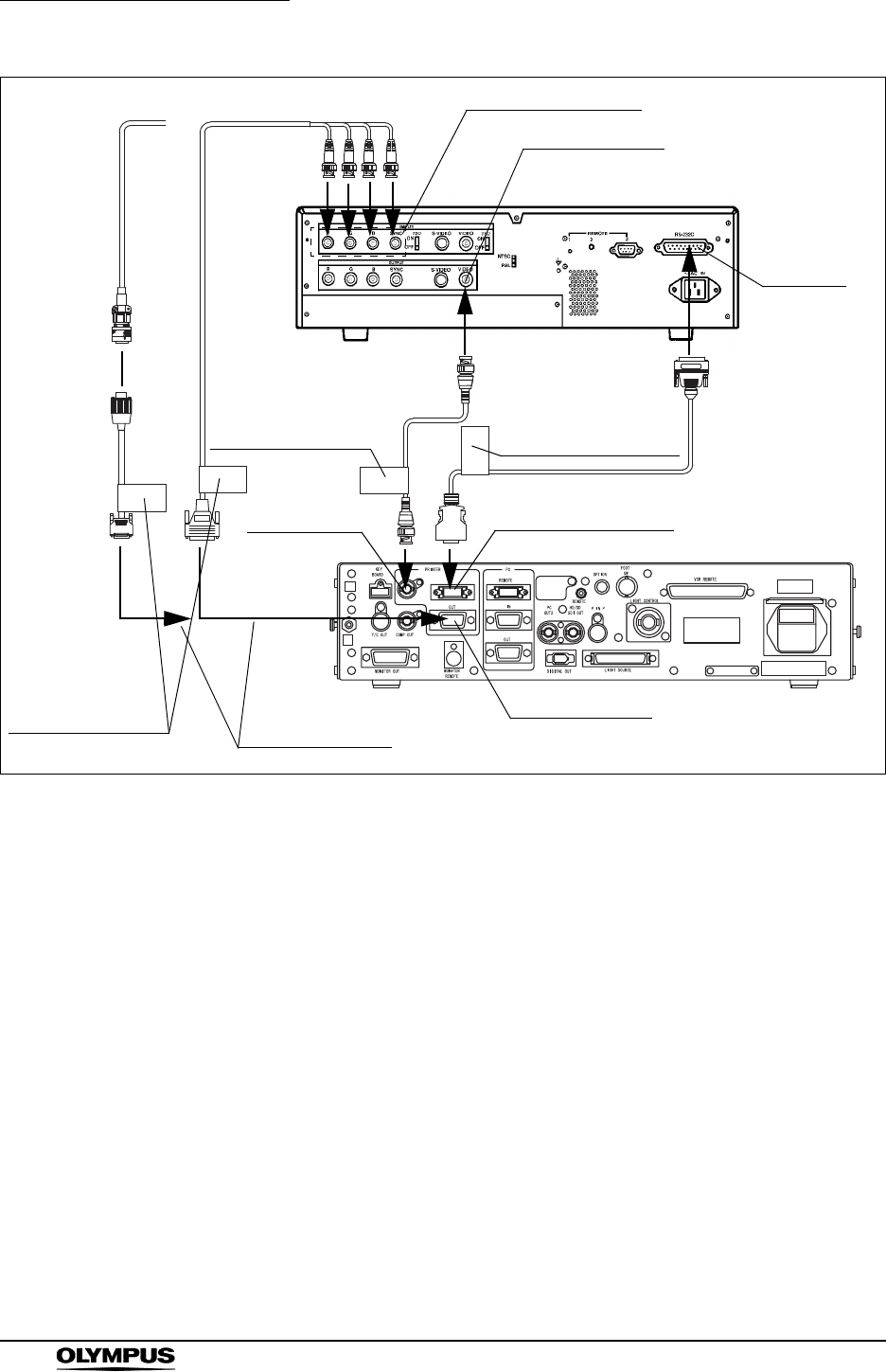
182
Chapter 8 Installation and Connection
EVIS EXERA II VIDEO SYSTEM CENTER CV-180
Figure 8.21
PRINTER REMOTEPRINTER IN
PRINTER OUT
R
GB
S
INPUT R/G/B/SYNC
MH-984
MH-995
MB-677
OUTPUT VIDEO
RS232C
Use either cable
connection.
CV-180
Video printer
Cable color sheet
“PRINTER REMOTE”
(green)
MAJ-849
MD-445
Cable color sheet
“PRINTER OUT”
(green)
Cable color sheet
“PRINTER IN”
(green)
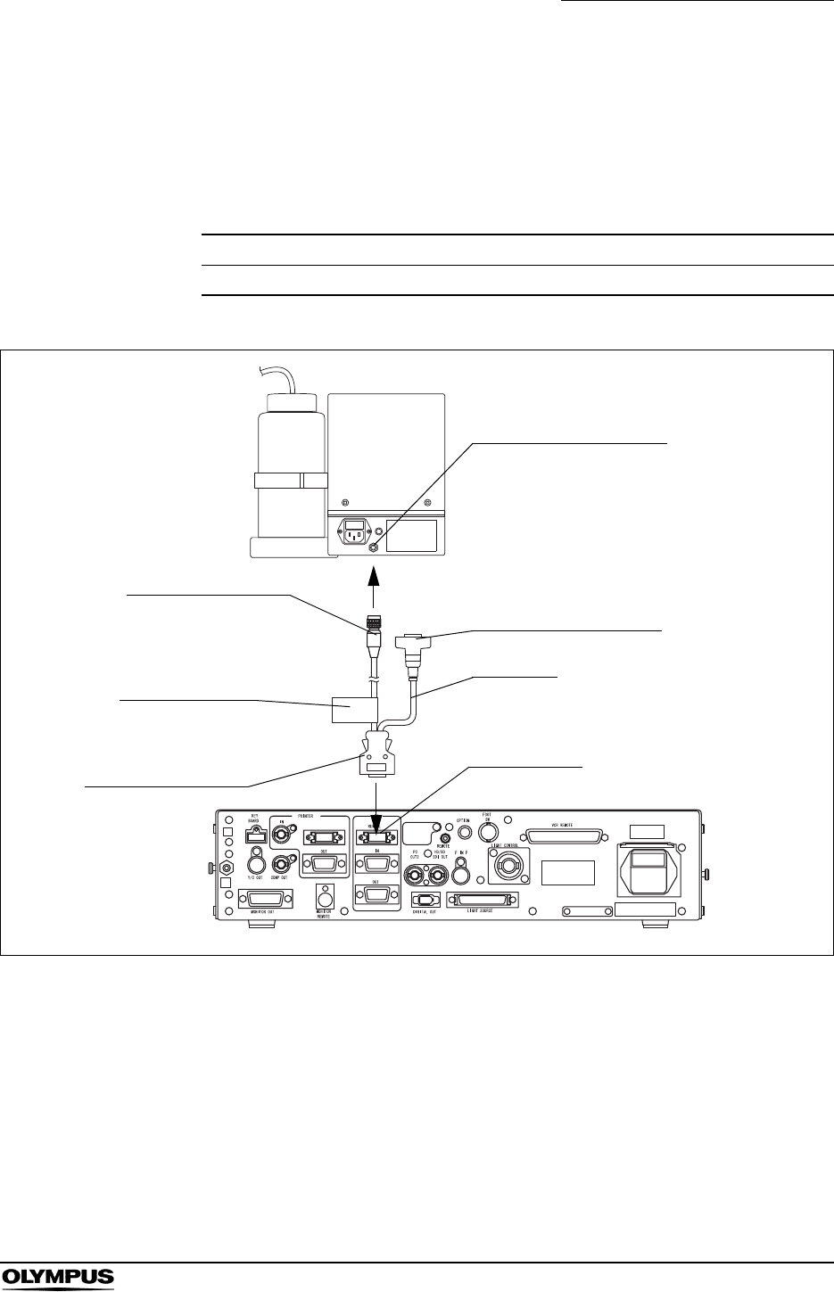
Chapter 8 Installation and Connection
183
EVIS EXERA II VIDEO SYSTEM CENTER CV-180
8.9 OLYMPUS flushing pump (OFP)
For connecting the Olympus flushing pump, use the cable listed below.
For easy cable connection, attach the cable color sheet to the cable (see Figure
8.22).
Figure 8.22
Model Product name Note
MAJ-920 Pump remote cable -
Table 8.19
PC REMOTE connector
MAJ-920
PC REMOTE
OFP
Remote control terminal
CV-180
4-pin connector
14-pin connector
Cable color sheet
“PC REMOTE”
(blue)
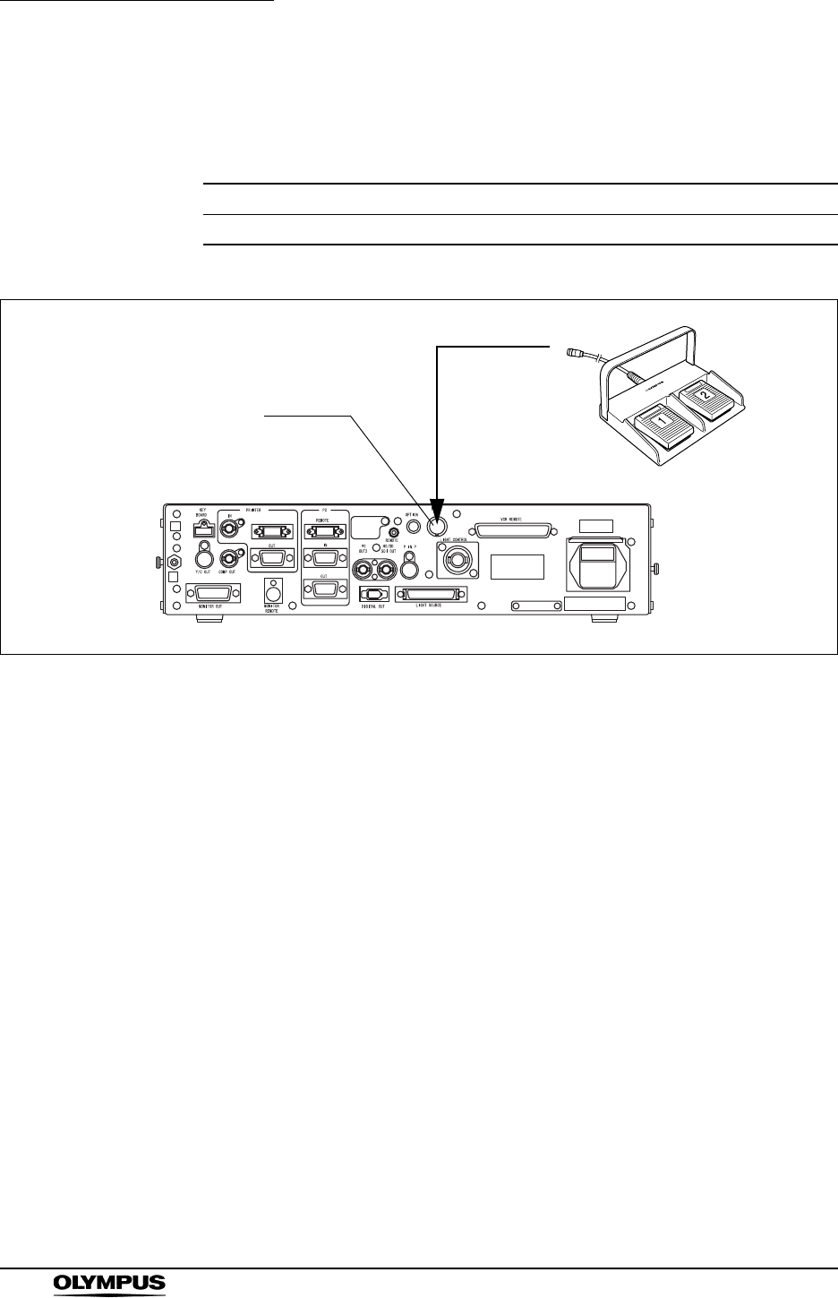
184
Chapter 8 Installation and Connection
EVIS EXERA II VIDEO SYSTEM CENTER CV-180
8.10 Foot switch
For a compatible foot switch, see the table below.
Figure 8.23
Model Product name Note
MAJ-1391 Foot switch -
Table 8.20
MAJ-1391
FOOT SW
CV-180

Chapter 8 Installation and Connection
185
EVIS EXERA II VIDEO SYSTEM CENTER CV-180
8.11 Ultrasound center
Compatible ultrasound centers
For compatible ultrasound centers, see the table below.
Model Product name
EU-C60 Compact endoscopic ultrasound center
EU-M30 Endoscopic ultrasound center
EU-M60 Endoscopic ultrasound center
EU-MA Endoscopic ultrasound center
Table 8.21
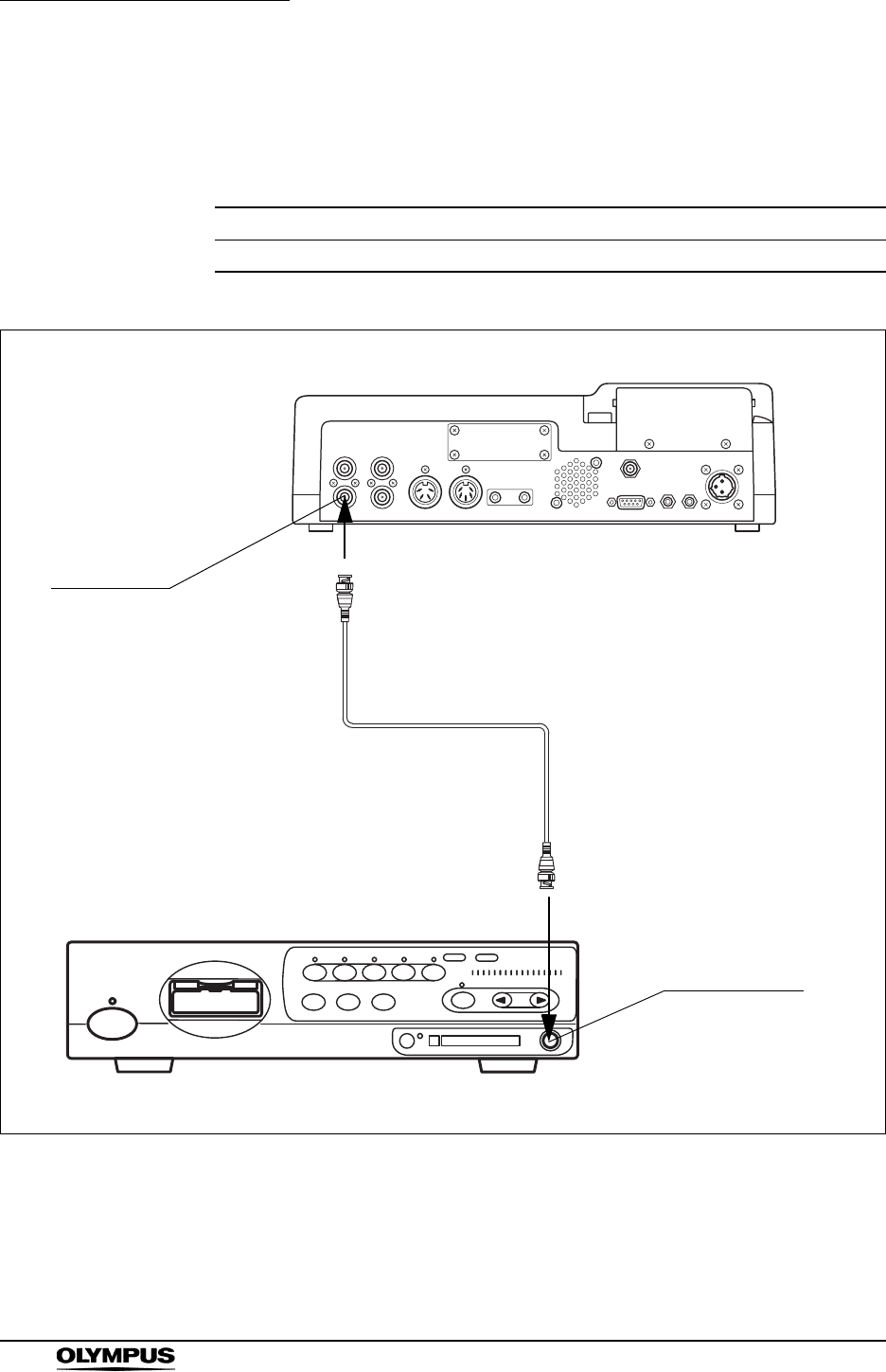
186
Chapter 8 Installation and Connection
EVIS EXERA II VIDEO SYSTEM CENTER CV-180
EU-C60
For connecting the ultrasound center (EU-C60) to the video system center, use
the cable listed below.
Figure 8.24
Model Product name Note
MB-677 BNC cable -
Table 8.22
CV-180
EU-C60
PinP composite
MB-677
VIDEO 1 OUT
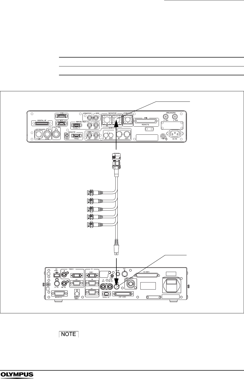
Chapter 8 Installation and Connection
187
EVIS EXERA II VIDEO SYSTEM CENTER CV-180
EU-M30
For connecting the ultrasound center (EU-M30) to the video system center, use
the cable listed below.
Figure 8.25
Do not connect the video output and B/W output of the EU-
M30 to the CV-180. An ultrasonic image in the PinP display
may be distorted.
Model Product name Note
MH-909 Monitor cable -
Table 8.23
CV-180
EU-M30
PinP Y/C
MONITOR OUT
MH-909
R
VIDEO
G
B
S
Y/C
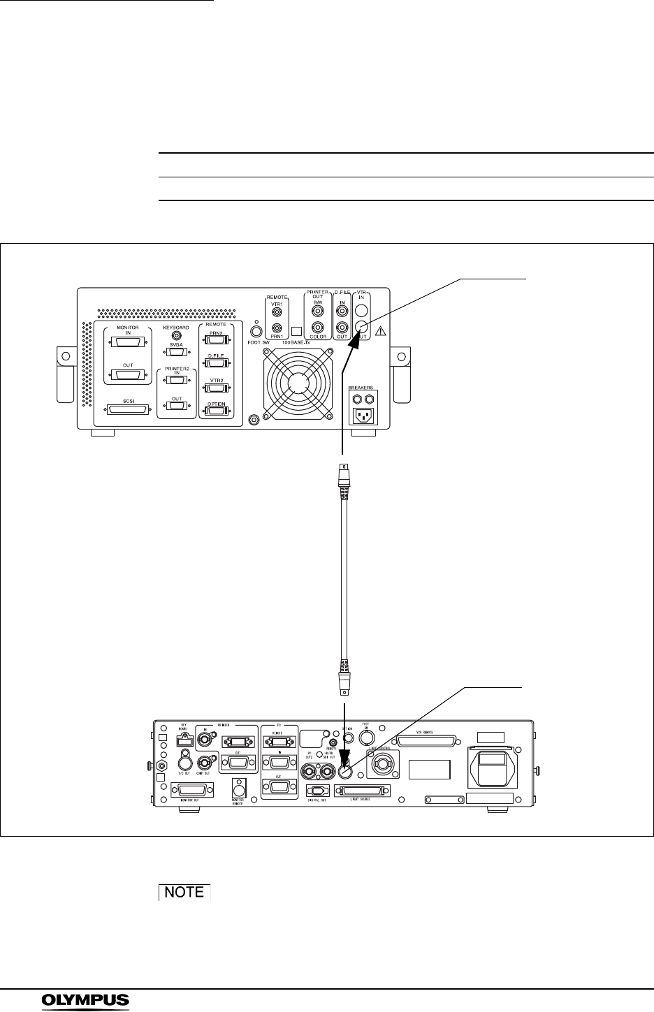
188
Chapter 8 Installation and Connection
EVIS EXERA II VIDEO SYSTEM CENTER CV-180
EU-M60, EU-MA
For connecting the ultrasound center (EU-M60 or EU-MA) to the video system
center, use the cable listed below.
Figure 8.26
Do not connect the video output and B/W output of the EU-
M60 to the CV-180. An ultrasonic image in the PinP display
may be distorted.
Model Product name Note
MH-985 or MAJ-987 S cable -
Table 8.24
CV-180
EU-M60, EU-MA1
PinP Y/C
VTR OUT
MH-985
or
MAJ-987
1 The terminal layout of EU-MA is
differ slightly from that of this
illustration.

Chapter 8 Installation and Connection
189
EVIS EXERA II VIDEO SYSTEM CENTER CV-180
8.12 Connection to the AC mains power supply
• Be sure to connect the power plug of the power cord directly
to a grounded wall mains outlet. If the video system center is
not grounded properly, it can cause an electric shock and/or
fire.
• Do not connect the power plug to the 2-pole power circuit
with a 3-pole to 2-pole adapter. It can prevent proper
grounding and cause an electric shock.
• Always keep the power plug dry. A wet power plug may
cause electric shocks.
• Confirm that the hospital-grade wall mains outlet to which this
instrument is connected has adequate electrical capacity that
is larger than the total power consumption of all connected
equipment. If the capacity is insufficient, fire can result or
circuit breaker may trip and turn OFF this instrument and all
other equipment connected to the same power circuit.
• When using the mobile workstation (WM-NP1, WM-WP1),
confirm that the mobile workstation has adequate electrical
capacity that is larger than the total power consumption of all
connected equipment. If the capacity is insufficient, drop in
the supply voltage can result or the electric protective device
may trip and turn OFF all the equipment connected to the
mobile workstation.
• When non-medical ancillary electrical equipment is used,
always connect the equipment to a wall mains outlet via an
isolation transformer. Otherwise, electric shock can result.
• The total power consumption of all connected equipment to
the isolation transformer should not exceed the rating of the
isolation transformer. If it exceeds, add another isolation
transformer. Otherwise, the equipment may not work
correctly.
• Do not bend, pull or twist the power cord. Equipment damage
including separation of the power plug and disconnection of
the cord wire as well as fire or electric shock can result.
• Be sure to connect the power plug securely to prevent
erroneous unplugging during use. Otherwise, the equipment
will not function.
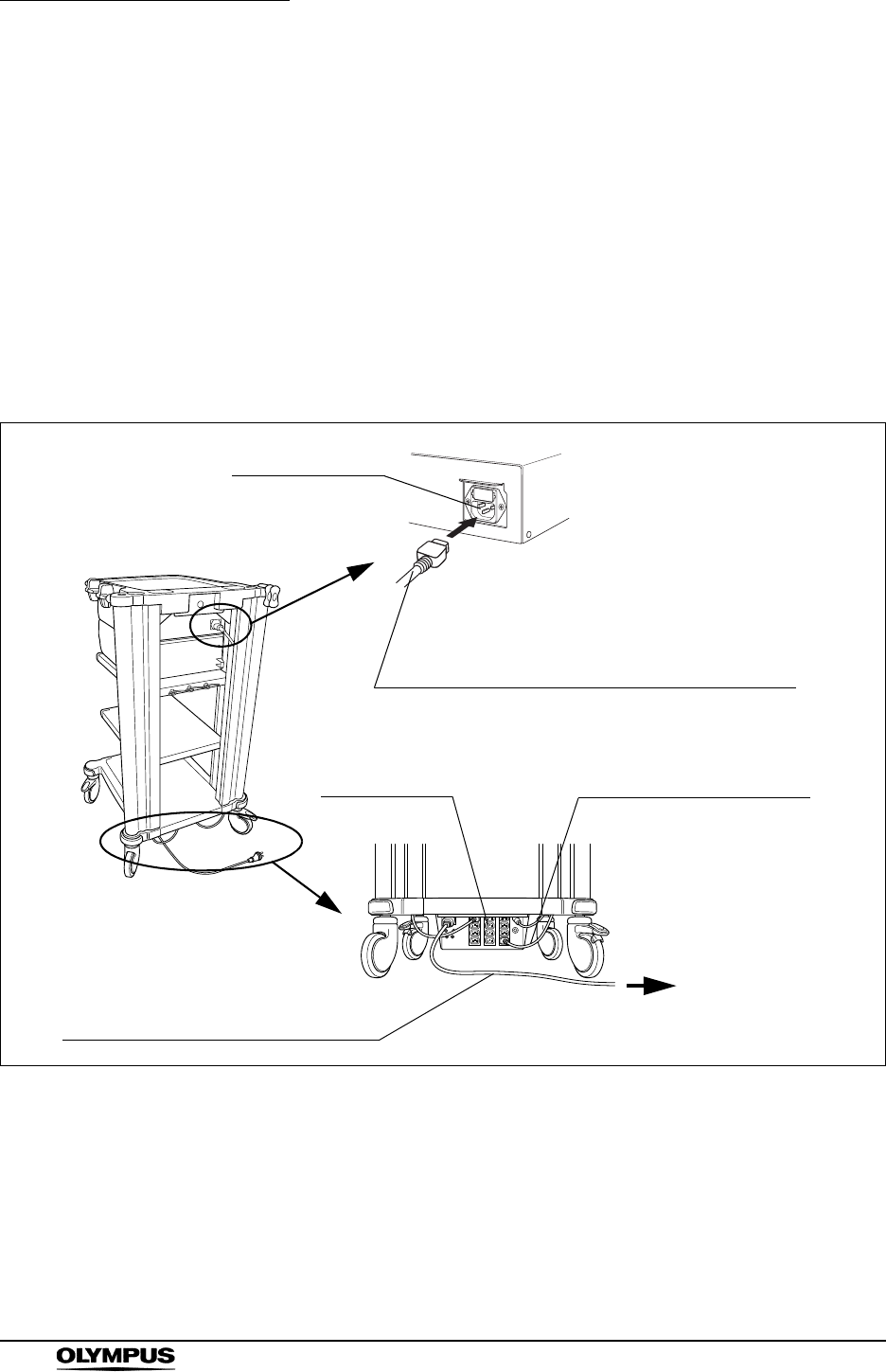
190
Chapter 8 Installation and Connection
EVIS EXERA II VIDEO SYSTEM CENTER CV-180
• Do not extend a single wall mains outlet into multiple outlets
for connecting the power cords of both the electrosurgical
unit and light source. Otherwise, malfunction of the
equipment may result.
When the mobile workstation (WM-NP1, WM-WP1) is used
1. Confirm that the video system center is OFF.
2. Connect the power cord provided with the mobile workstation to the AC
power inlet of the video system center and the AC mains outlet of the mobile
workstation (see Figure 8.27).
Figure 8.27
3. Connect the power cords provided with the mobile workstation to the AC
power inlets of the ancillary equipment and the AC mains outlets of the
mobile workstation.
4. Connect the power cord of the mobile workstation to the wall mains outlet.
AC power inlet
Mobile workstation
to the wall mains outlet
CV-180
Power socket
Power cord provided with the mobile workstation
Power cord provided with
the mobile workstation
Power cord of the mobile workstation

Chapter 8 Installation and Connection
191
EVIS EXERA II VIDEO SYSTEM CENTER CV-180
When a mobile workstation other than the WM-NP1 and
WM-WP1 is used or when no mobile workstation is used
1. Confirm that the video system center is OFF.
2. Connect the power cord provided with the video system center first to its AC
power inlet, then to the wall mains outlet.
3. Connect the instruments listed in Table 8.25 to the wall mains outlet.
4. Connect the instruments listed in Table 8.26 to the isolation transformer.
5. Connect the power cord of the isolation transformer to the wall mains outlet.
Model Product name
CLV-180, CLV-160, CLV-U40, CLV-S40, Light source
OEV monitors
(OEV143, OEV203, OEV181H, OEV191, OEV191H)
Monitor
OEP printers Video printer
Table 8.25 The devices to be connected directly to the wall mains outlet
Model Product name
DVO-1000MD, DSR-20MD, SVO-9500MD VCR
Other than OEP video printers
(UP-1800, UP-1850, UP-2900MD, UP-2950MD, UP-5200MD,
UP-5250MD, UP-21MD)
Video printer
Other than OEV video monitors Monitor
Table 8.26 The devices to be connected to the isolation transformer
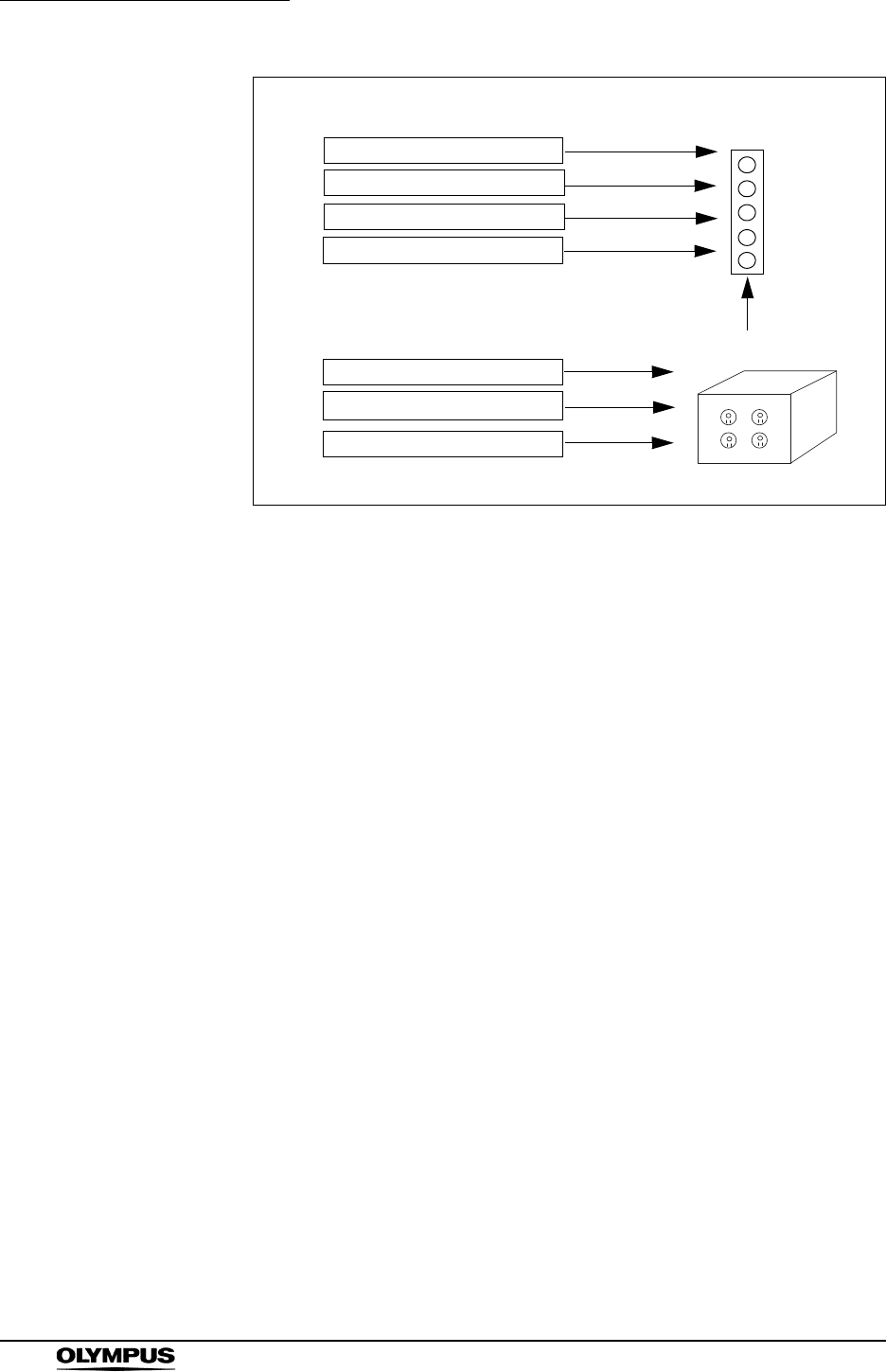
192
Chapter 8 Installation and Connection
EVIS EXERA II VIDEO SYSTEM CENTER CV-180
Figure 8.28
Video system center
Light source
OEP video printer
OEV monitor
VCR
Video printer other than OEP
Monitor other than OEV
Devices to be connected directly to the hospital grade
power outlet Hospital grade
power outlet
Isolation transformer
Devices to be connected to the isolation transformer
MB-631
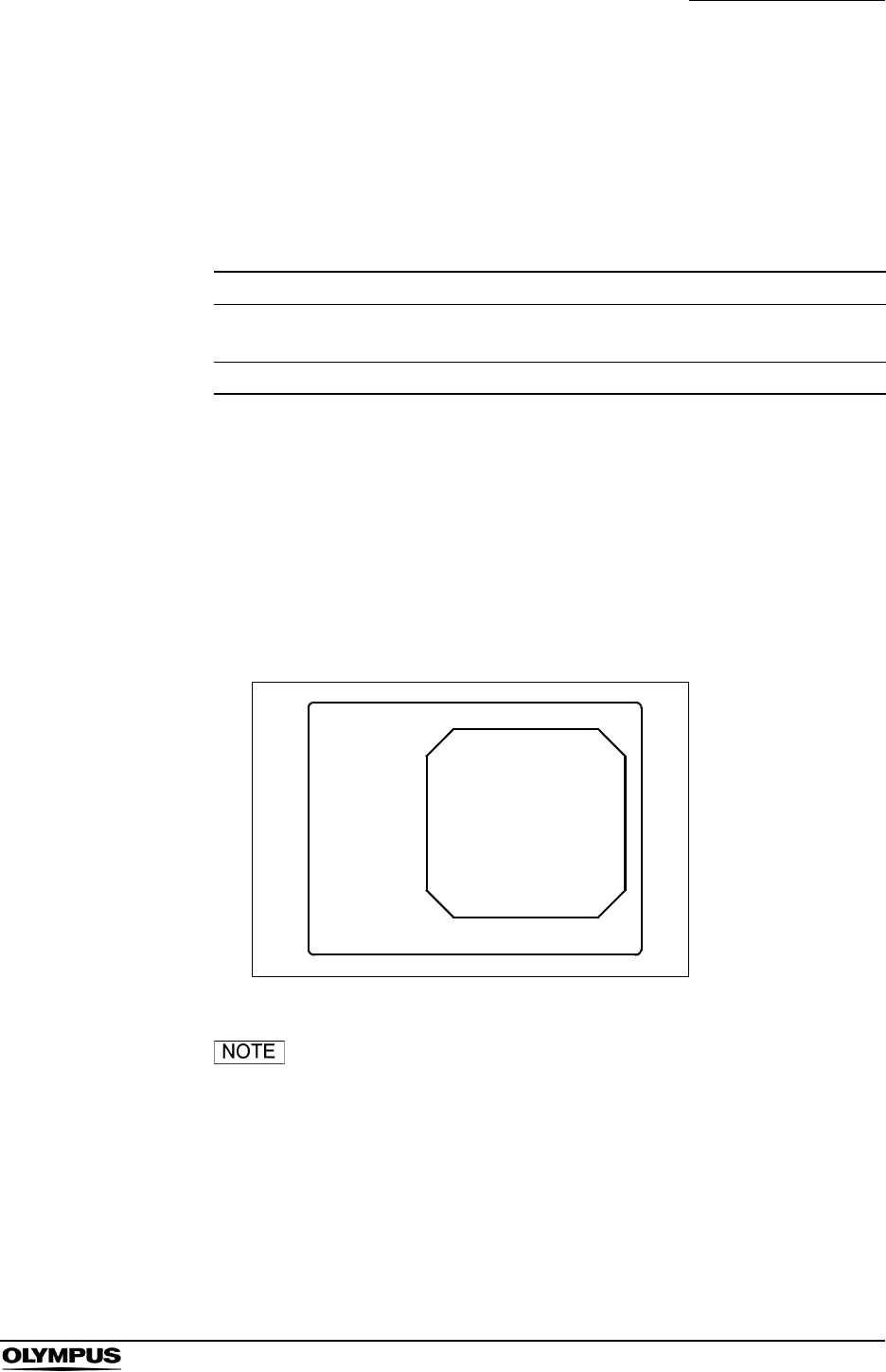
Chapter 9 Function setup
193
EVIS EXERA II VIDEO SYSTEM CENTER CV-180
Chapter 9 Function setup
This chapter shows basic settings for appropriate use of the video system center
and other equipment connected to the video system center.
9.1 Turning power ON
1. Push the power switch of the video system center. The power indicator
above the switch lights up.
2. Confirm that the endoscopic image appears on the monitor (see Figure 9.1).
Figure 9.1
No endoscope is required for the settings. If no endoscope is
connected, the color bar appears on the monitor instead of
the endoscopic image.
Menu Function
System setup Sets the basic settings of the video system center and the ancillary
instruments. See page 214 for the items to set.
User preset Sets the functions for observation. See page 255 for the items to set.
Table 9.1
ID:
Name:
Sex: Age:
D.O.B.
12/12/2005
12:12:12
Physician:
Comment:
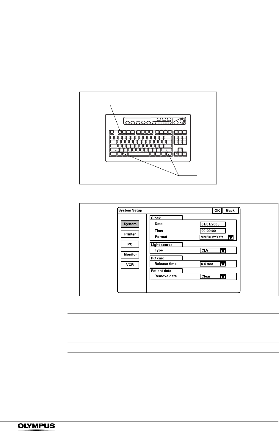
194
Chapter 9 Function setup
EVIS EXERA II VIDEO SYSTEM CENTER CV-180
9.2 System setup
Basic operation of the system setup
1. Press the “Shift” and “F1” keys together (see Figure 9.2). The “System
setup” menu appears on the monitor (see Figure 9.3).
Figure 9.2
Figure 9.3
2. Click “System”, “Printer”, etc., on the left side of the menu. The applicable
setting items appear on the right side of the window.
3. Click the desired text box to place the cursor and enter the data.
Buttons Functions
OK Determines the entered data. Used to move back to the endoscopic
image.
Back Cancels the entered data. Used to move back to the endoscopic image.
Table 9.2
F1
Shift

Chapter 9 Function setup
195
EVIS EXERA II VIDEO SYSTEM CENTER CV-180
4. Click “ ” of the list box to open the pull down menu, and click the data in
the pull down menu.
5. Click “Back” or “Esc” to cancel the input data and to go back to the
endoscopic image.
6. Click “OK” after completing input operation. A confirmation message
appears on the monitor.
7. Click “Y” or press “Enter” to store the input data and to go back to the
endoscopic image.
Click “N” to go back to step 2.
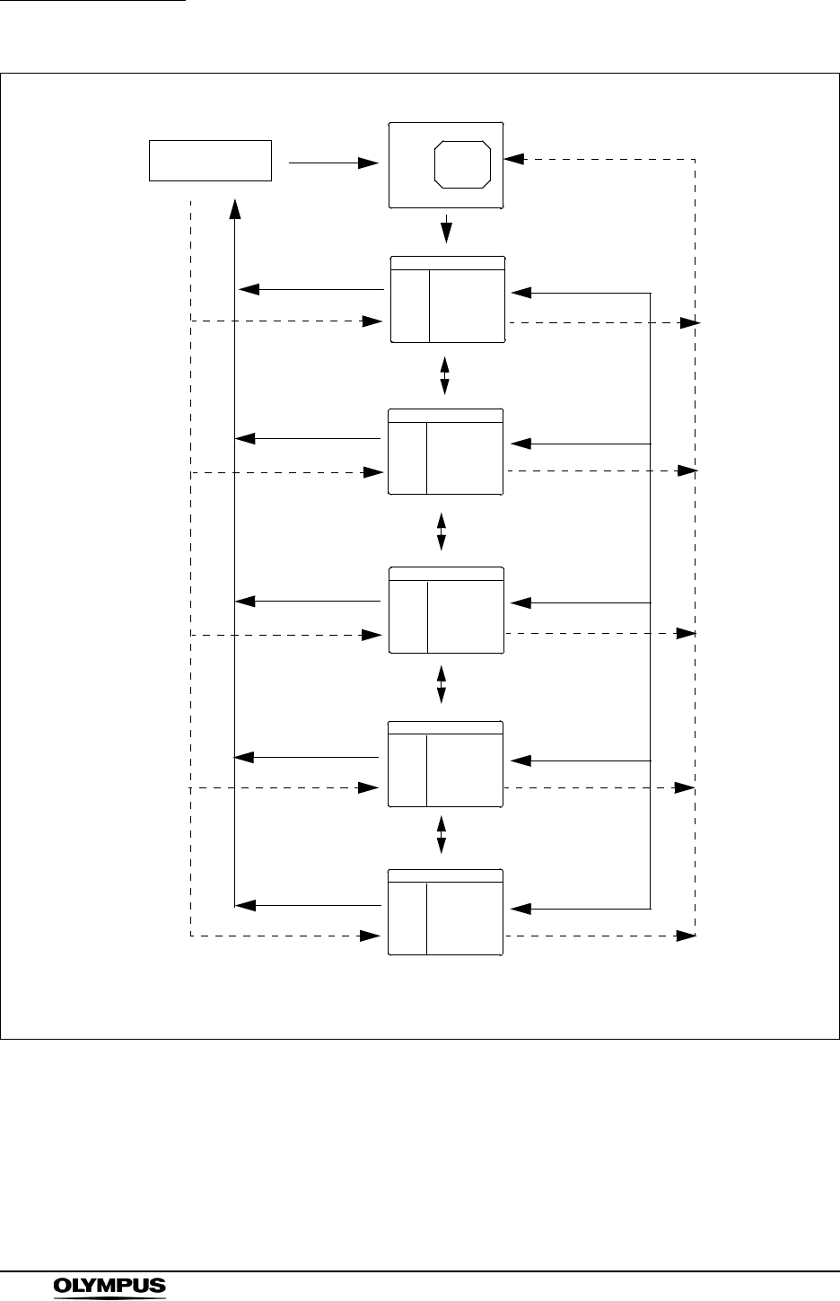
196
Chapter 9 Function setup
EVIS EXERA II VIDEO SYSTEM CENTER CV-180
Figure 9.4
Endoscopic image
System setup
Shift + F1
Are you sure?
Y/N
System
Printer
PC
Monitor
VCR
Arrow key
OK
N
System
Printer
PC
VCR
Monitor
Y
page 197 to page 200
Transition between the screens can be performed when the according box is highlighted.
System setup
System setup
System setup
System setup
page 201
page 206
page 208
page 211
Arrow key
Arrow key
Arrow key
Back, Esc,
Shift + F1
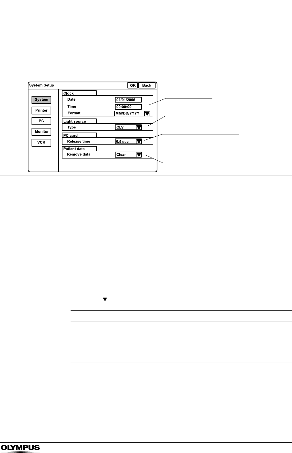
Chapter 9 Function setup
197
EVIS EXERA II VIDEO SYSTEM CENTER CV-180
System
This operation sets the current date and time, the type of the connected light
source, the release time when an endoscopic image is stored to the PC card,
and the display type of the patient data with an endoscopic image.
Figure 9.5
Click “System” on the system setup menu. The setting screen appears on the
right part of the window (see Figure 9.5).
Date and time
This operation sets the current date and time, and the display format of the date.
1. Click the text box of “Date” and enter numeric characters for month, date
and year.
2. Click the text box of “Time” and enter hour, minute and second.
3. Click “ ” of “Format”. The date formats appear in the pull-down menu.
4. Click the format to use. The format is selected and displayed in the dialog
box.
Format Explanation
• MM/DD/YYYY
• MM-DD-YYYY
• MM.DD.YYYY
• MM DD YYYY
• DD/MM/YYYY
• DD-MM-YYYY
• DD.MM.YYYY
• DD MM YYYY
• YYYY/MM/DD
• YYYY-MM-DD
• YYYY.MM.DD
• YYYY MM DD
YYYY: year
MM: month
DD: day
Table 9.3
Date and time
Light source
Release time of PC card
Resume of the patient data
198
Chapter 9 Function setup
EVIS EXERA II VIDEO SYSTEM CENTER CV-180
Light sources
This operation sets the type of light source connected.
Other settings of the light source may cause improper color
and/or brightness in the image.
1. Click “ ” of the list box “Type”. The following light sources appear in the
pull-down menu.
2. Click the setting value corresponding to the connected light source. The
selected setting value is displayed.
• When the CLV-180 is used, the setting is not necessary.
• When a light source other than CLV-180 is used, set up the
light source according to the table below. Please refer to the
instruction manual of the light source.
Setting value Connected light source
CLV CLV-160, CLV-U40
CLV-S CLV-S40
CLE Halogen light source
Table 9.4
Setting items Settings
Brightness adjustment mode AUTO
BRIGHTNESS Center of the adjustable range
Table 9.5
Chapter 9 Function setup
199
EVIS EXERA II VIDEO SYSTEM CENTER CV-180
Release Time of PC card
The endoscopic live image pauses when the “RELEASE” button is pressed to
record the image on the PC card. This operation sets the length of time to pause.
1. Click “ ” of the list box “Release Time”. The available release times
appear in the pull-down menu.
2. Click the desired release time. The selected time is displayed.
The actual release time is the longest one among all release
time settings of instruments.
Setting values Release time
0.5 sec 0.5 second
1 sec 1 second
1.5 sec 1.5 seconds
2 sec 2 seconds
Table 9.6

200
Chapter 9 Function setup
EVIS EXERA II VIDEO SYSTEM CENTER CV-180
Display of Patient data
All or a part of the text data such as the patient data can be cleared from the
monitor (refer to “Clearing characters from the screen (“F1”)” on page 82). The
following operation sets whether the display status when turning the video
system center OFF is saved or not. In case “Save” is set, the same display
status appears on the monitor when the instrument is turned ON as it was when
the instrument has been turned OFF.
1. Click “ ” of the list box “Remove data”. “Clear” and “Save” appear in the
pull-down menu.
2. Click “Save” or “Clear”. The value selected is displayed.
Setting value Explanation
Clear Displays all text data when the instrument is turned ON.
Save Displays same status as when the instrument is turned OFF.
Table 9.7
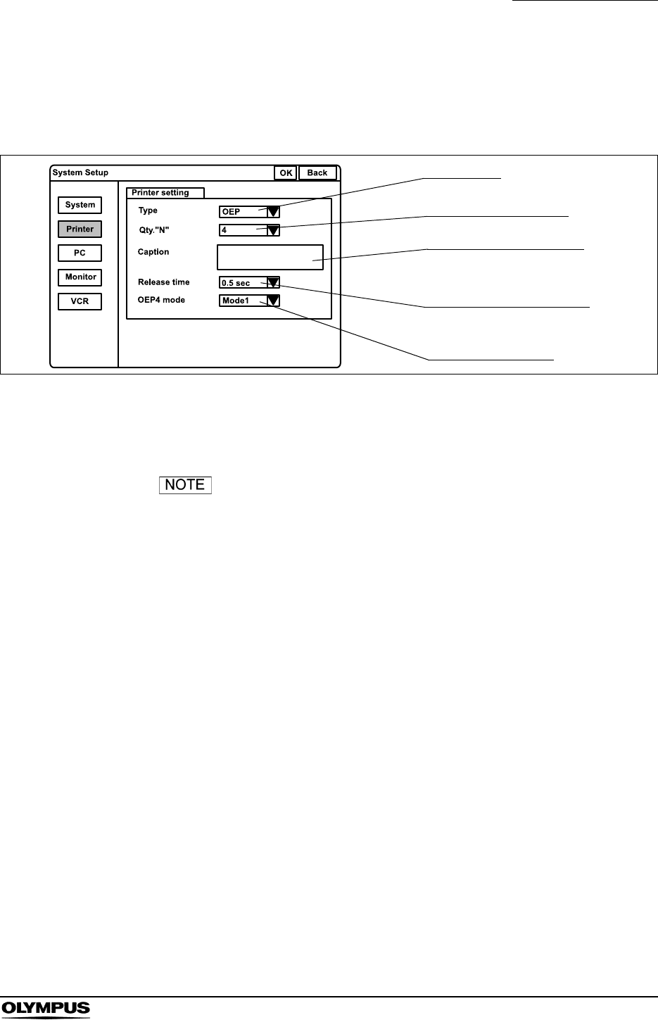
Chapter 9 Function setup
201
EVIS EXERA II VIDEO SYSTEM CENTER CV-180
Printer
This operation sets the type of video printer, number of print sheets, caption to
be printed on the print sheets, and release time of the video printer.
Figure 9.6
Click “Printer” on the system setup menu. The setting items appear on the right
part of the window (see Figure 9.6).
When the video printer of HDTV non-correspondence is
used, the HDTV image can only be printed in SDTV quality.
Printer type
Number of print sheets
Caption on print sheet
Release time of printer
Image signal output
202
Chapter 9 Function setup
EVIS EXERA II VIDEO SYSTEM CENTER CV-180
Printer type
1. Click “ ” of the “Type”. The video printer types appear in the pull-down
menu.
2. Click the video printer type to be used. The selected printer type is
displayed.
Number of print sheets
1. Click “ ” of the “Qty.“N””. The numbers of the print sheet (4, 5, 6, 7, 8 and
9) appear in the pull-down menu (see Figure 9.6). These numbers are
assigned to “N” of the “PRINT QTY.” key on the keyboard.
2. Click the desired number. The selected number is displayed.
• The number 1, 2 or 3 are selected with the “PRINT QTY.” key
on the keyboard.
• When “Foot switch” of the printer type is selected, “Qty.“N””
cannot be set.
Setting value Applicable video printer model HDTV printing
OEP OEP (OLYMPUS) NO
OEP4 OEP-4 (OLYMPUS) YES
OEP3 OEP-3 (OLYMPUS) NO
2900 UP-2900MD (SONY) NO
2950 UP-2950MD (SONY) NO
5200 UP-5200MD (SONY) NO
5250 UP-5250MD (SONY) NO
5000 UP-5000MD (SONY) NO
1800 UP-1800 (SONY) NO
1850 UP-1850 (SONY) NO
UP21 UP-21MD (SONY) NO
YP22 function not available NO
Foot switch NO
Table 9.8
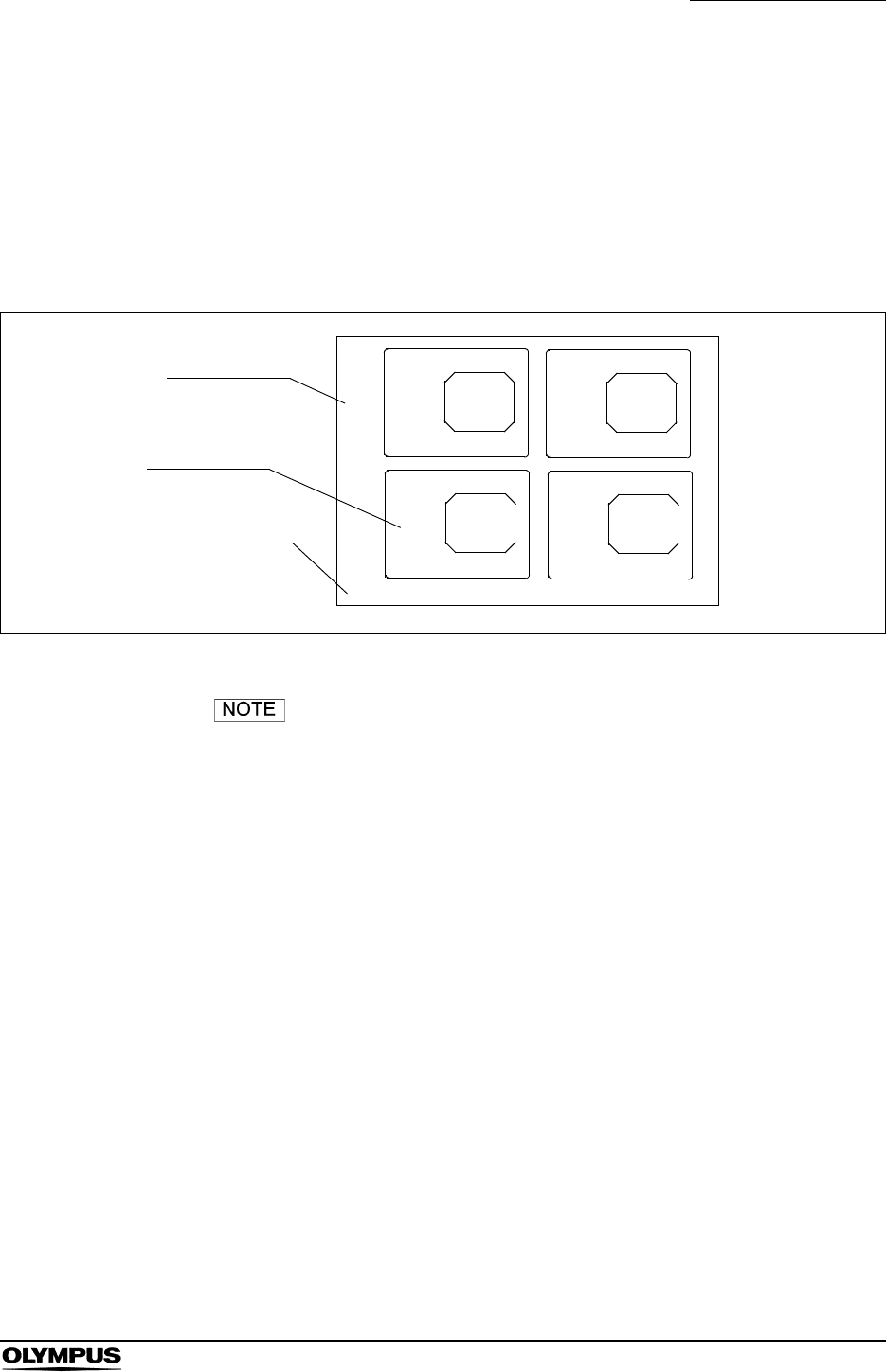
Chapter 9 Function setup
203
EVIS EXERA II VIDEO SYSTEM CENTER CV-180
Caption
1. Click the text box of “Caption”. The cursor moves into the dialog box.
2. Enter the caption in the dialog box.
• Max. 32 alphanumeric and symbol characters can be entered.
Figure 9.7 shows an example of a print sheet with caption.
Figure 9.7
• When OEP is used, set “TIMER” of the OEP printer OFF. For
details, refer to the instruction manual of the OEP printer.
• When “Foot switch” of the printer type is used, “Caption”
cannot be set.
• When the UP-1800/1850 is used, set the “LIVE MODE”
option of the printer OFF. For details, refer to the instruction
manual for the UP-1800/1850.
• When the UP-1800/1850 is used and “MODE” is set to “1”,
set “Separate” of the printer OFF. The remote operation from
the CV-180 becomes disabled. For details, refer to the
instruction manual for the UP-1800/1850.
• Set the baud rate of the RS-232C printer, referring to its
instruction manual.
Baud rate: 4800 bps
Follow-up observation
Print sheet
Print images
Caption
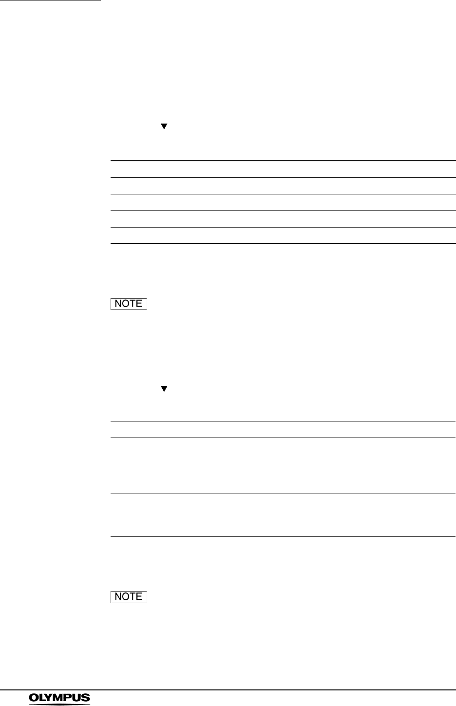
204
Chapter 9 Function setup
EVIS EXERA II VIDEO SYSTEM CENTER CV-180
Release time
The endoscopic live image pauses when the “RELEASE” button is pressed to
record the image onto the PC card. This operation sets the length of time to
pause.
1. Click “ ” of the “Release time (see Figure 9.6 on page 201). The release
times appear in the pull-down menu.
2. Click the desired release time. The selected time is displayed.
The actual release time is the longest one among all release
time settings of the instruments.
Output signal to OEP-4
1. Click “ ” of the “OEP4 mode” (see Figure 9.6 on page 201). The output
signal appear in the pull-down menu.
2. Click the desired mode. The selected mode is displayed.
• During displaying in PinP in mode1, the image output is
always SDTV format without reference to the endoscope.
• OEP-4 cannot print HDTV and SDTV images on the same
page. If HDTV images follow SDTV images, the Printing
Forcibly message will appear and the HDTV images will be
Setting value Release time
0.5 sec 0.5 second
1 sec 1 second
1.5 sec 1.5 seconds
2 sec 2 seconds
Table 9.9
Setting value Explanation
Mode 1 The video signal (HDTV or SDTV) compatible with the endoscope
type is output. In the mode1, HDTV images and SDTV images
cannot be printed on the same print sheet. In this case, HDTV
images and SDTV images are printed on the separate print sheets.
Mode 2 The SDTV signal is output regardless of the endoscope type.
However, the HDTV signal is output only when the number of
images on the print form is one.
Table 9.10

Chapter 9 Function setup
205
EVIS EXERA II VIDEO SYSTEM CENTER CV-180
printed first. After the HDTV images are printed, SDTV
images can be captured and printed as usual. The Printing
Forcibly message does not indicate a malfunction.
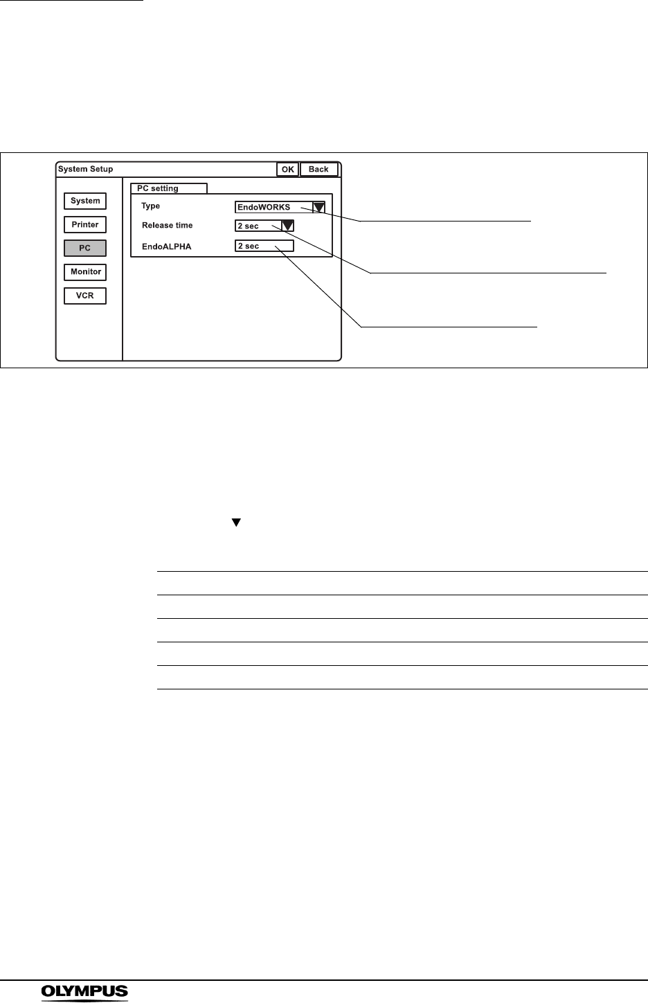
206
Chapter 9 Function setup
EVIS EXERA II VIDEO SYSTEM CENTER CV-180
Image filing system
This operation sets the type of image filing system that is connected to the “PC”
terminal on the rear panel and the release time of the image filing system.
Figure 9.8
Click “PC” on the system setup menu. The setting items appear on the right side
of the window (see Figure 9.8).
Type of image filing system
1. Click “ ” of the “Type”. The types of image filing system appear in the pull-
down menu.
2. Click the system to use. The selected system is displayed.
Setting value Explanation
EndoWORKS The images can be recorded on the EndoWORKS system.
EndoALPHA The images can be recorded on the EndoALPHA system.
EndoBASE The images can be recorded on the EndoBASE system.
OFF The images cannot be recorded on any image filing system.
Table 9.11
Type of digital filing system
Release time of the digital filing system
Release time of EndoALPHA
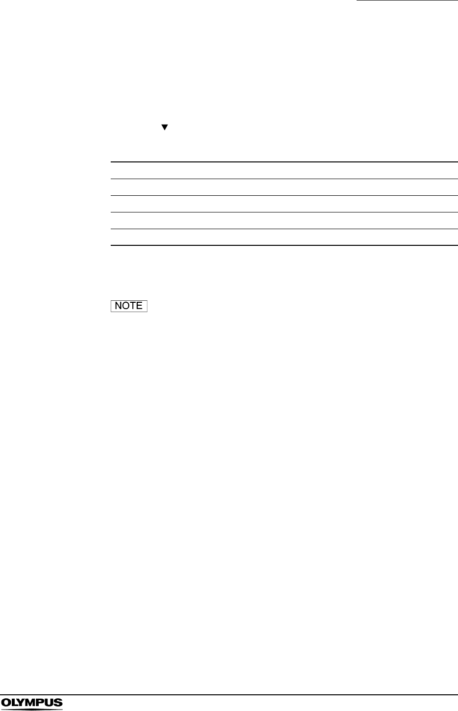
Chapter 9 Function setup
207
EVIS EXERA II VIDEO SYSTEM CENTER CV-180
Release time
The endoscopic live image pauses when the “RELEASE” button is pressed to
record the images into the image filing system. This operation sets the length of
time to pause.
1. Click “ ” of the “Release time (see Figure 9.8). The release times appear
in the pull-down menu.
2. Click the desired release time. The selected release time is displayed.
The actual release time is the longest one among all release
time settings of instruments.
EndoALPHA
The release time set by EndoALPHA is displayed. The setting value cannot be
changed on this screen.
Setting value Release time
0.5 sec 0.5 second
1 sec 1 second
1.5 sec 1.5 seconds
2 sec 2 seconds
Table 9.12
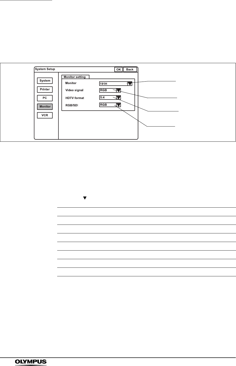
208
Chapter 9 Function setup
EVIS EXERA II VIDEO SYSTEM CENTER CV-180
Monitor
This operation sets the monitor type, video signal, aspect ratio of the HDTV
(horizontal and vertical ratio of the display area) and the terminal to be activated
by the monitor for video output.
Figure 9.9
Click “Monitor Setting” on the system setup menu. The setting items appear on
the right side of the window (see Figure 9.9).
Monitor type
1. Click “ ” of “Monitor”. The monitor types appear in the pull-down menu.
2. Click the desired monitor type. The selected monitor type is displayed.
Setting value Video signal Applicable monitor model
141/201 SDTV OEV141, OEV201
142/143/202/203 SDTV OEV142, OEV143, OEV202, OEV203
181H HDTV OEV181H
191H HDTV OEV191H
191 SDTV OEV191
Remote OFF (HD) HDTV
Remote OFF (SD) SDTV
Table 9.13
Monitor type
Video signal
Aspect ratio
Video signal
Chapter 9 Function setup
209
EVIS EXERA II VIDEO SYSTEM CENTER CV-180
Video signal format
Improper setting of the video signal may displays abnormal
images.
1. Click “ ” of the “Video signal”. The video signals appear in the pull-down
menu.
2. Click the desired video signal. The selected video signal is displayed.
Aspect ratio
1. Click “ ” of “HDTV format”. The aspect ratios appear in the pull-down
menu.
2. Click the desired aspect ratio. The selected aspect ratio is displayed.
• When the aspect ratio settings of the monitor and CV-180 are
not the same, the top and bottom or right and left of the
image may not be displayed properly on the monitor. It is
recommended to use the default settings of the aspect ratio
and to use the remote control cable described in Section 8.5,
“Monitor” on page 170
• A common aspect ratio of HDTV is 16:9, but this instrument
displays only the central part of the image in the ratio 5:4 or
4:3.
Setting value Explanation
RGB For RGB compatible monitors
YPbPr For analog component signal compatible monitors
Table 9.14
Setting value Explanation
5:4 For the monitors whose horizontal to vertical ratio is 5:4.
4:3 For the monitors whose horizontal to vertical ratio is 4:3.
Table 9.15

210
Chapter 9 Function setup
EVIS EXERA II VIDEO SYSTEM CENTER CV-180
The video signal output
This operation sets the type of video signal output to the monitor.
1. Click “ ” of “RGB/SDI”. The video signals appear in the pull-down menu.
2. Click the desired video signal. The selected video signal is displayed.
CV-180 does not have and SDI input and is therefore not
capable of transferring images from external devices to the
monitor via SDI.
Setting value Explanation
RGB Output RGB video signal to the monitor.
SDI Outputs SDI video signal to the monitor.
Table 9.16
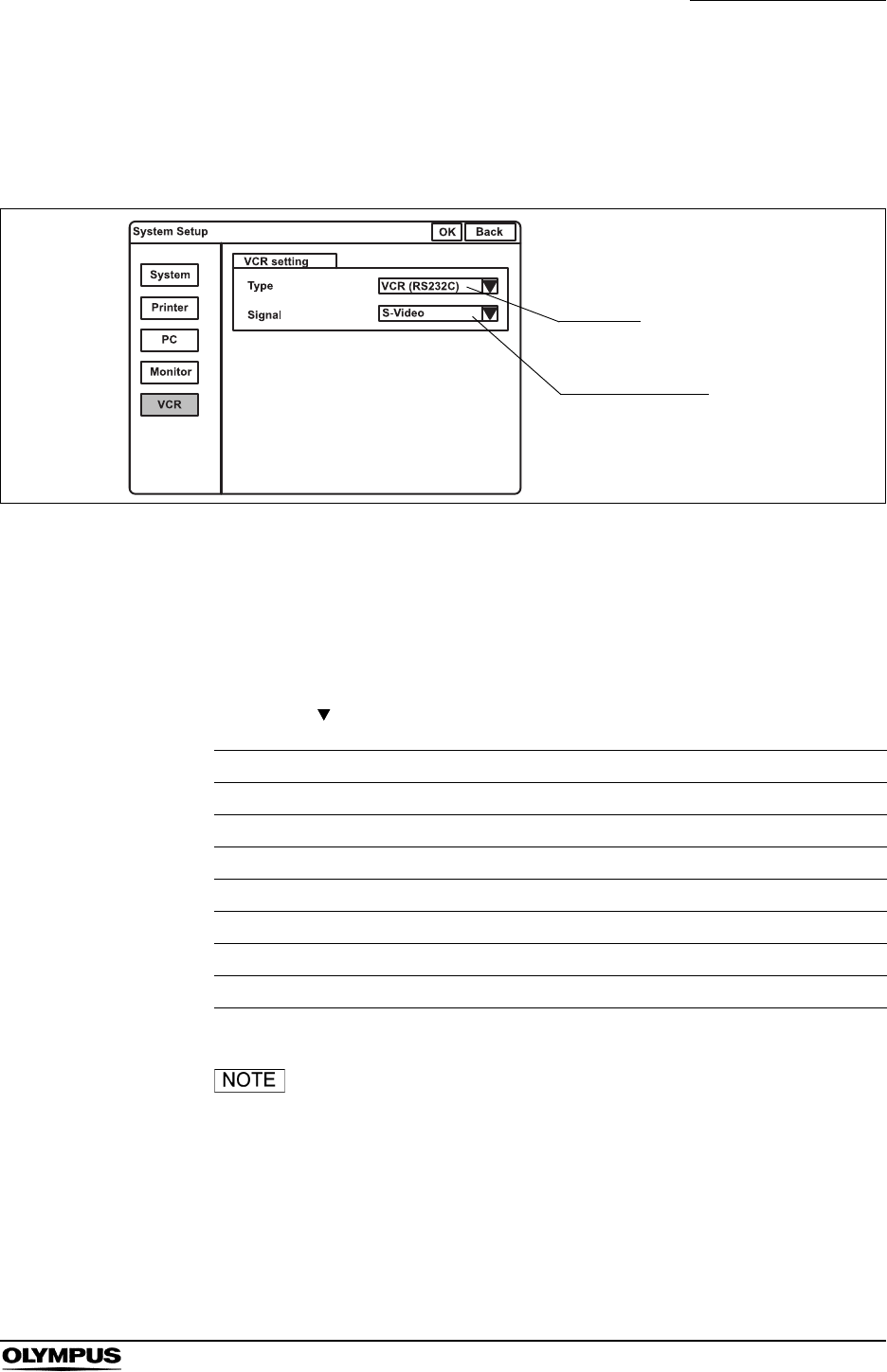
Chapter 9 Function setup
211
EVIS EXERA II VIDEO SYSTEM CENTER CV-180
Videocassette recorder
This operation sets the type of videocassette recorder (VCR) and the video
signal to the VCR.
Figure 9.10
Click “VCR” on the system setup menu. The setting items appear on the right
side of the window (see Figure 9.10).
VCR type
1. Click “ ” of “Type”. The VCR types appear in the pull-down menu.
When “DVD (IEEE1394)” of the VCR type is selected, only
the recording and pause operations can be operated.
2. Click the desired type. The selected VCR type is displayed.
Setting value VCR
VCR (RS232C) SVO-9500MD (SONY), DSR-20MD (SONY)
VCR (IEEE 1394) DSR-20MD (SONY)
Foot switch SVO-9500MD (SONY)
HD-VCR (RS232C) PDW-70MD (SONY), PDW-75MD (SONY)
DVD (RS232C) DVO-1000MD (SONY)
DVD (IEEE1394) DVO-1000MD (SONY)
Option Reserved for future system expansion.
Table 9.17
VCR type
Video signal type

212
Chapter 9 Function setup
EVIS EXERA II VIDEO SYSTEM CENTER CV-180
Video signal type
1. Click “ ” of “Signal”. The video signals appear in the pull-down menu.
2. Click the desired video signal. The selected video signal is displayed.
• When using the VTR remote cable MH-989 or MAJ-906, set
the RS-232C transfer rate of the videocassette recorder as
shown below following the instructions given in the
instruction manual for the VTR remote cable.
Baud rate: 9600 bps
• When using the VTR remote cable MH-922, set the
videocassette recorder’s foot switch control mode in “Low
level trigger mode” following the instructions given in the
instruction manual for the videocassette recorder (SVO-
9500MD).
• For further information, refer to the instruction manual for the
VTR remote cable.
Setting value Explanation
S-Video The type of input signal when playing back is S-video.
Composite The type of input signal when playing back is composite.
Table 9.18
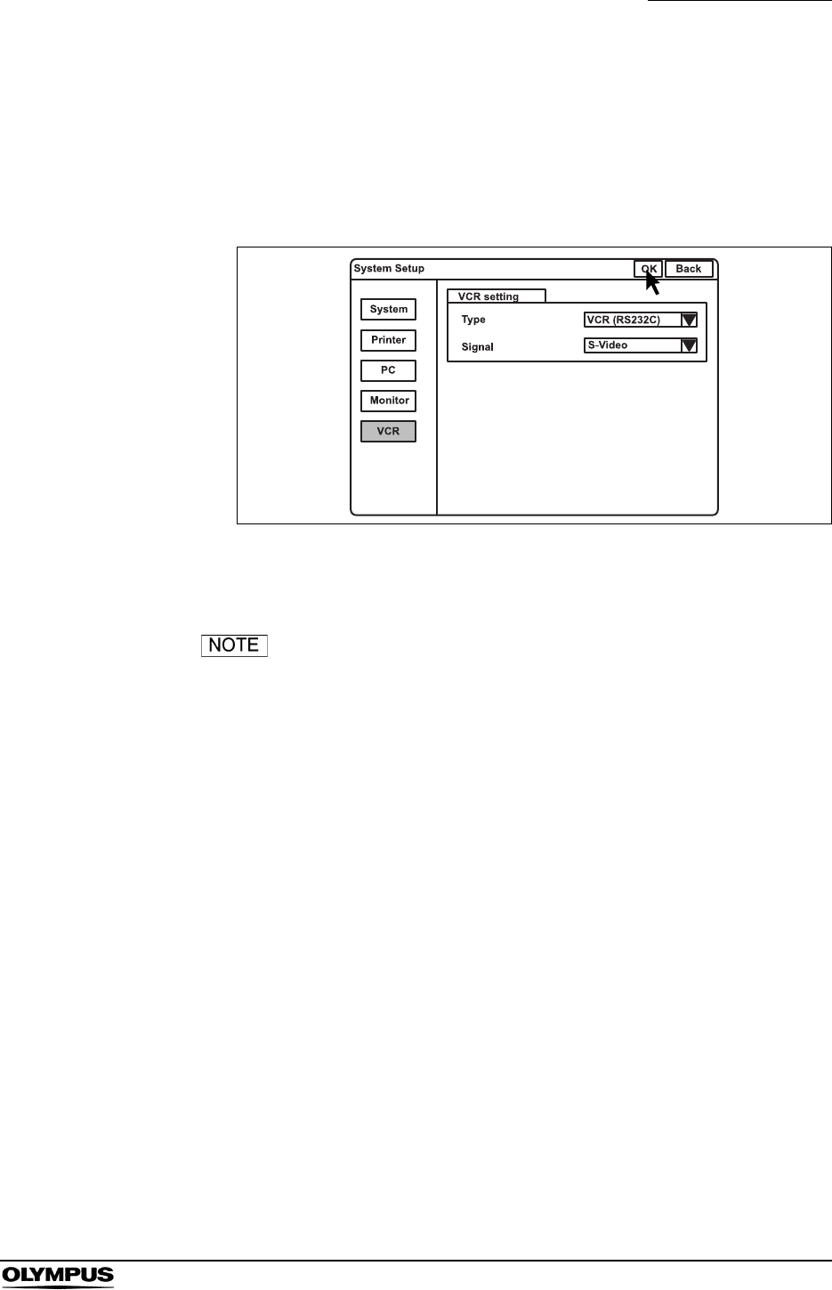
Chapter 9 Function setup
213
EVIS EXERA II VIDEO SYSTEM CENTER CV-180
Saving the system setup
This operation finalizes and saves the settings of the system setup.
1. Click “OK” of the system setup menu (see Figure 9.11). All settings are
saved in the video system center.
Figure 9.11
2. Click “Back” to go back to the endoscopic image.
To cancel the settings, click “Back” instead of “OK”. The input
values are canceled and the display returns to the
endoscopic image.
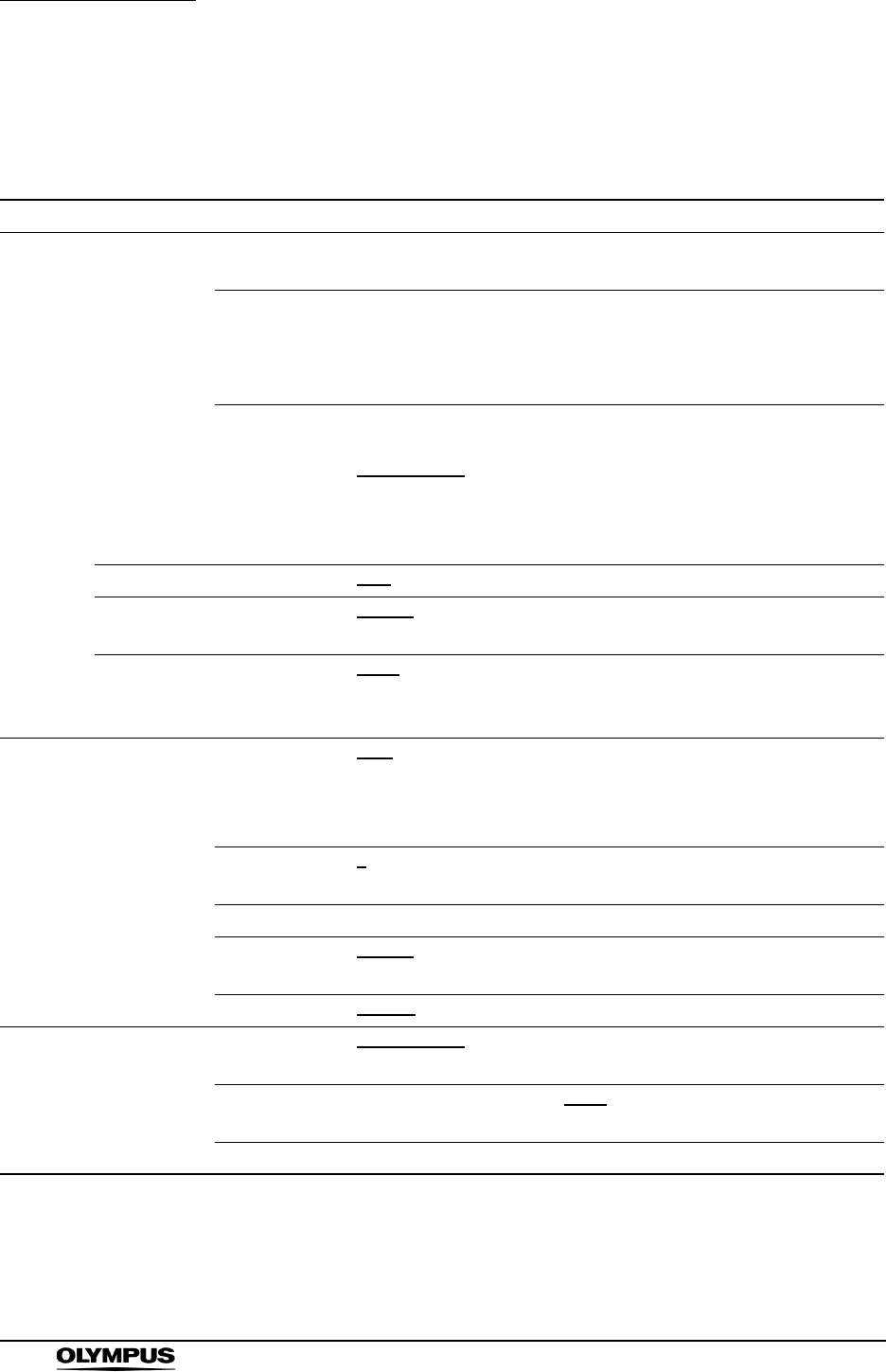
214
Chapter 9 Function setup
EVIS EXERA II VIDEO SYSTEM CENTER CV-180
Summary of settings
Table 9.19 shows the options of the system setup menu. The bold and
underscored options listed are the factory default settings.
Setup item Setting value Note
System Clock Date Display format is according to the
type of format selected below.
System date
Time Display format is [HH:MM:SS] System clock
HH: hour
MM: minute
SS: second
Format [YYYY/MM/DD], [YYYY.MM.DD],
[YYYY-MM-DD], [YYYY MM DD],
[MM/DD/YYYY], [MM-DD-YYYY],
[MM.DD.YYYY], [MM DD YYYY],
[DD/MM/YYYY], [DD-MM-YYYY],
[DD.MM.YYYY], [DD MM YYYY]
Display format for the system
date
YYYY: year
MM: month
DD: day
Light source Type [CLV], [CLV-S], [CLE] Type of light source or lamp bulb
PC card Release time [0.5 sec], [1 sec], [1.5 sec], [2 sec] Length of time to pause the
image for recording
Patient data Remove data [Clear], [Save] The display status of text data
such as patient data is saved or
not saved.
Printer Printer
setting
Type [OEP], [OEP3], [OEP4], [2900],
[2950], [5200], [5250], [5000],
[1800], [1850], [UP21], [YP22],
[Foot switch]
Printer types to connect to
“Printer Remote” terminal
Qty.“N” [4], [5], [6], [7], [8], [9] The number of sheet assigned
to “N”
Caption --
Release time [0.5 sec], [1 sec], [1.5 sec], [2 sec] Length of time to pause the live
image
OEP4 mode [Mode 1], [Mode 2] Video signal out put to OEP4
PC PC setting Type [EndoWORKS], [EndoBASE],
[EndoALPHA], [OFF]
Image filing systems
Release time [0.5 sec], [1 sec], [1.5 sec], [2 sec] Length of time to pause the live
image
EndoALPHA [0.5 sec], [1 sec], [1.5 sec], [2 sec]
Table 9.19
Chapter 9 Function setup
215
EVIS EXERA II VIDEO SYSTEM CENTER CV-180
Monitor Monitor
setting
Monitor [141/201], [142/143/202/203],
[181H], [191H], [191],
[Remote OFF (HD)],
[Remote OFF (SD)]
Monitor type to connected to
“Monitor OUT”
Video signal [RGB], [YPbPr] Video signal format to monitor
HDTV format [5:4], [4:3] Aspect ratio of HDTV
RGB/SDI [RGB], [SDI] Terminal to be activated by the
monitor for video output
VCR VCR setting Type [VCR (RS232C)],
[VCR (IEEE1394)], [Foot switch],
[HD-VCR (RS232C)],
[DVD (RS232C)],
[DVD (IEEE1394)], [Option]
VCR type
Signal [S-Video], [Composite] VCR signal type
Setup item Setting value Note
Table 9.19
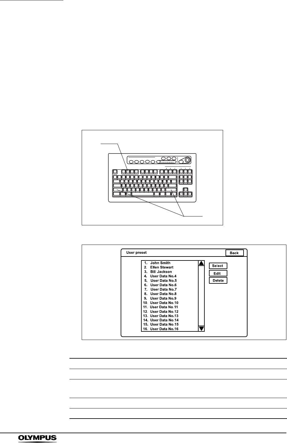
216
Chapter 9 Function setup
EVIS EXERA II VIDEO SYSTEM CENTER CV-180
9.3 User preset
The user preset menu sets the functions of this instrument for each individual
user, endoscope, etc., for up to 20 users. Each preset data has been set to the
factory defaults.
Basic operation of the user preset
1. Press the “Shift” and “F2” key together (see Figure 9.12). The “User preset”
menu appears on the monitor (see Figure 9.13).
Figure 9.12
Figure 9.13
Button Function
Back Cancels the inputs, and returns to the endoscopic image.
Select Loads the selected user preset data on the video system center, then
returns to the endoscopic image.
Edit Edits the user preset data in the video system center.
Delete Deletes the user name and its data from the user name list.
Table 9.20
F2
Shift
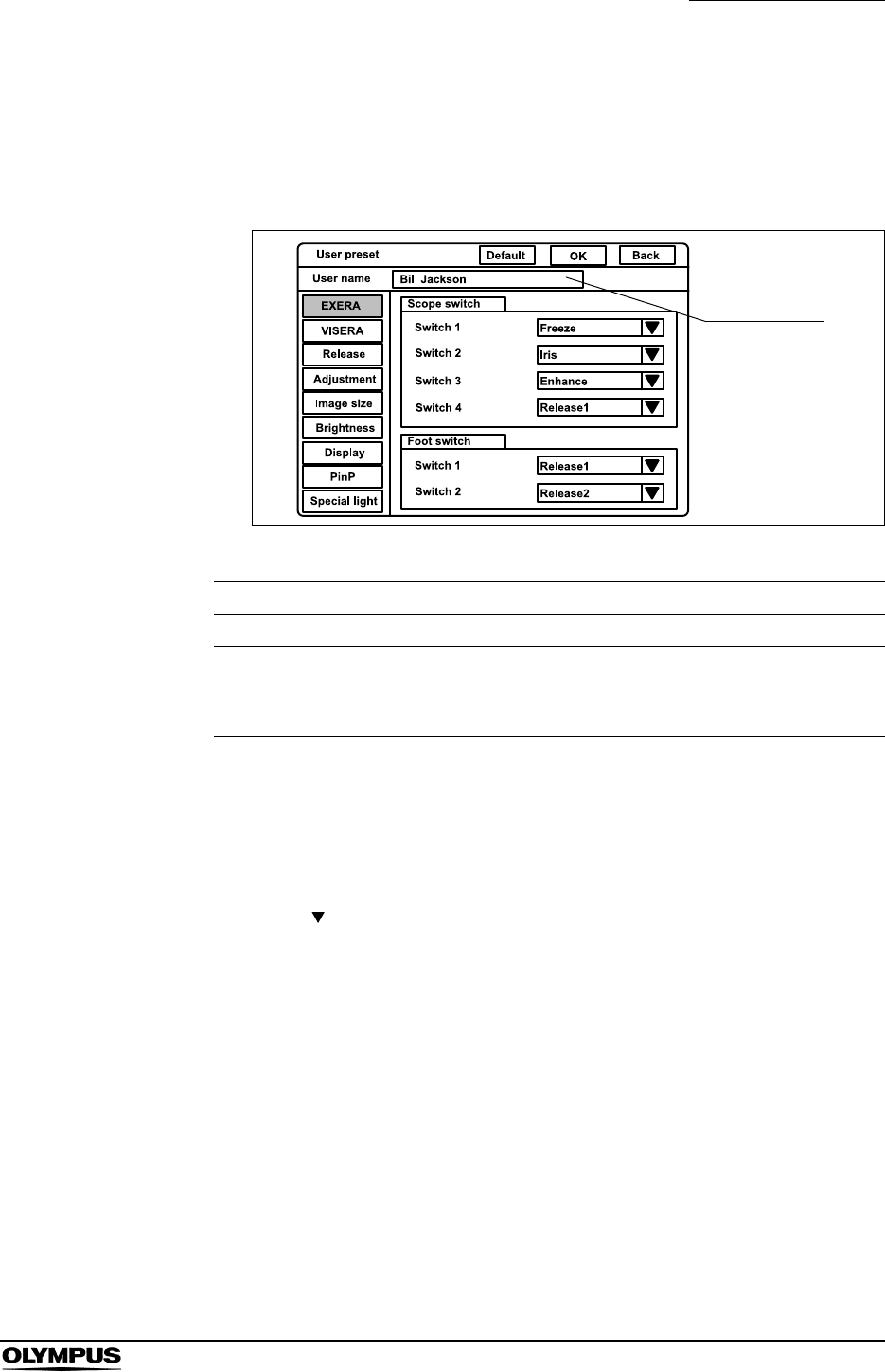
Chapter 9 Function setup
217
EVIS EXERA II VIDEO SYSTEM CENTER CV-180
2. Click “User Data No. #” in the user name list, in which no user is registered.
The background of the selected user name is blue.
3. Click “EDIT” on the right side of the screen (see Figure 9.13).The edit
screen of the user preset menu appears (see Figure 9.14).
Figure 9.14
4. Enter the user name in the text box of “User name”. Otherwise, entered data
cannot be saved.
• Max. 20 alphanumeric and symbol characters can be entered.
5. Click “ ” of the dialog box of the setting values to open the pull down
menu, and click the data on the pull down.
6. To cancel the input, click “Back” or “Esc”. The input values are canceled and
the display returns to the user list menu.
7. After completing data input, click “OK”. A confirmation message appears on
the monitor.
8. Click “N” to return to step 2.
Click “Y” or press “Enter” to save the input values and return to the
endoscopic image.
Button Function
Back Returns to the endoscopic image.
OK Determines and saves the input data (returns to the
endoscopic image).
Default Resets the user preset data to the factory default settings.
Table 9.21
User’s name
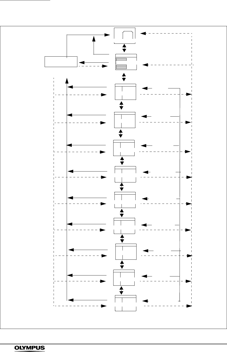
218
Chapter 9 Function setup
EVIS EXERA II VIDEO SYSTEM CENTER CV-180
Figure 9.15
User preset
User preset
User preset
User preset
User preset
User preset
User preset
User preset
User preset
Endoscopic image
Shift + F2
Shift + F2
Are you sure?
Y/N User list
EXERA
Release
Adjustment
Back, Esc
N, Esc
Delete
EXERA
Release
Adjustment
Image Size
Image size
Brightness
Brightness
Display
Edit
Display
PinP
PinP
Special light
Y
OK
Arrow key
N, Esc
page 194
page 219
page 223
page 226
page 229
page 232
page 241
page 246
page 250
Special light
Arrow key
Arrow key
Arrow key
Arrow key
Arrow key
Arrow key
Transition between the screens can be performed when the according box is highlighted.
User preset
VISERA VISERA
Arrow key
page 219
Select,
Back,
Esc
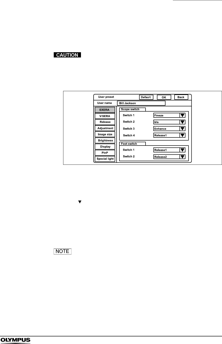
Chapter 9 Function setup
219
EVIS EXERA II VIDEO SYSTEM CENTER CV-180
Remote switch and foot switch (EXERA and VISERA)
This operation assigns the functions to the remote switches on the endoscope
and the foot switch.
Use the “EXERA” menu to assign the functions for the EVIS
series endoscopes and use the “VISERA” menu for the
VISERA series endoscopes and the camera heads.
Otherwise the functions are invalid.
Figure 9.16
1. Click “EXERA” or “VISERA” on the user preset menu. The setting items
appear on the right side of the window.
2. Click “ ” of “Scope switch” or “Foot switch” (see Figure 9.16). The
available functions appear in the pull-down menu.
3. Click the desired function. The selected function is displayed.
4. Follow steps 1. to 3. to assign the functions to all the scope and foot
switches in the same way.
• It is possible to assign the same function to all the remote
switches and foot switches.
• The positions and shapes of the scope switches are different
depending on the endoscope model as shown in Figure 9.17.
For details, refer to the instruction manual of the endoscope.
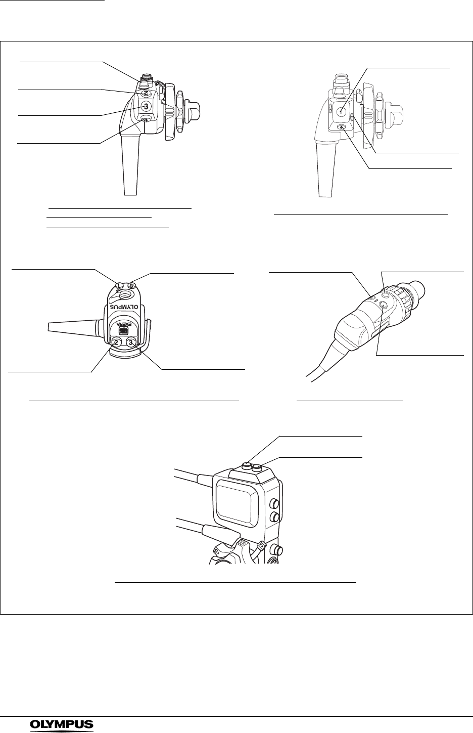
220
Chapter 9 Function setup
EVIS EXERA II VIDEO SYSTEM CENTER CV-180
Figure 9.17
Note) These illustrations are examples. For details, refer to the instruction manuals of the endoscopes used.
Remote switch 2
Remote switch 3
Remote switch 4
Remote switch 1
Remote switch 2
Remote switch 1
Remote switch 4
Remote switch 3
Remote switch 1
Remote switch 3
Remote switch 2
Remote switch 1
Remote switch 3
Remote switch 4
EVIS 160/180 series videoscopes,
Ultrasonic videoscopes
(GF-UC series, GF-UE series)
Ultrasonic videoscope (only GF-UM160)
BF-160/180 series, VISERA series videoscopes OTV-S7H camera heads
Remote switch 4
Remote switch 1
Ultrasonic videoscope (GF-UM series except GF-UM160)
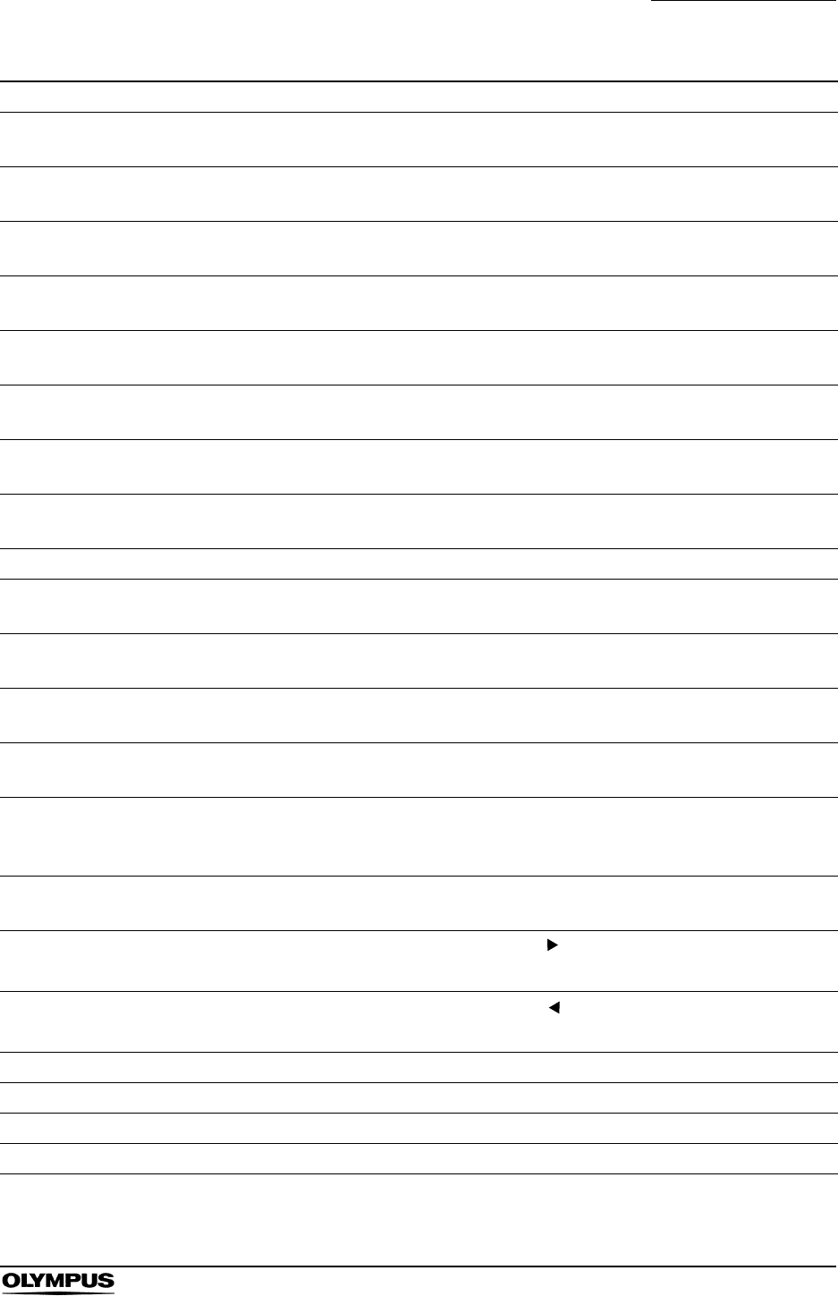
Chapter 9 Function setup
221
EVIS EXERA II VIDEO SYSTEM CENTER CV-180
Function Outline
Freeze Toggles back and forth between live image and frozen image of the endoscopic image. Same
function as “FREEZE” on the keyboard. See “Freeze (“FREEZE”)” on page 99.
Release 1 Records the images on the monitor into the recording devices. Same function as “RELEASE” on
the keyboard. See “Release (“RELEASE”)” on page 101.
Release 2 Records the images on the monitor into the recording devices. See “Release function” on
page 223.
Iris Switches to the iris mode (type of optical measurement). Same function as the “Iris mode switch”
on the front panel. See “Iris mode” on page 69.
Enhance Switches the image enhancement mode (OFF/1/2/3). Same function as the “Image enhancement
button” on the front panel. See “Image enhancement mode (ENH.)” on page 67.
Contrast Switches the contrast of the endoscopic image. Same function as the “Shift”+”F6” keys on the
keyboard. See “Contrast mode (“Shift” + “F6”)” on page 90.
AGC Toggles back and forth the AGC function between ON and OFF. Same function as the “F6” key on
the keyboard. See “Automatic gain control (AGC) (“F6”)” on page 89.
Image size Changes the image size. Same function as the “F8” key on the keyboard. See “Image size (“F8”)”
on page 94.
VCR Operates either VCR recording or pause. See “Videocassette recorder (VCR)” on page 129.
Capture Takes in the image into the video printer. Same function as the “CAPTURE” key on the keyboard.
See “Video printer” on page 131.
Stop watch Uses the clock on the monitor as a stopwatch. Same function as the “F5” key on the keyboard. See
“Stopwatch (“F5”)” on page 88.
Remove data Erases and displays the patient data on the monitor. Same function as the “F1” key on the
keyboard. See “Clearing characters from the screen (“F1”)” on page 82.
Zoom Enlarges the image. Same function as the “F7” key on the keyboard. See “Image zooming (“F7”)”
on page 91.
White balance Press and hold to execute the white balance adjustment. Same function as the “Shift” + “F9” keys
on the keyboard and “White balance button” on the front panel. See Section 4.5, “White balance
adjustment” on page 52.
Exposure
area
Changes the exposure area of the auto brightness control. See “Exposure area” on page 239.
Exposure (up) Increases the brightness of the image. Same function as “ ” of the brightness adjustment button
on the front panel. See “Brightness adjustment (Exposure)” on page 71.
Exposure
(down)
Decreases the brightness of the image. Same function as “ ” of the brightness adjustment button
on the front panel. See “Brightness adjustment (Exposure)” on page 71.
NBI Activates the NBI observation mode. See “NBI (narrow band imaging)” on page 151.
PDD mode This function is not available.
PDD gain This function is not available.
OP.2 Reserved for future system expansion. Currently not used.
Table 9.22
222
Chapter 9 Function setup
EVIS EXERA II VIDEO SYSTEM CENTER CV-180
OFP Turns the OLYMPUS flushing pump (OFP) ON and OFF. This function is available only on the
remote switches and foot switches.
Light Turns the light source ON and OFF. This function is active only when the CLV-180 is used. Press to
light up. Press and hold to light OFF.
Arrow Displays or erases the arrow pointer. Same function as the “Shift” and any arrow key on the
keyboard. See “Arrow pointer (“Shift” + arrow keys and domepoint)” on page 102.
PinP Toggles back and forth the display between the normal display and PinP. Same function as the
“PinP” button on the front panel. See “PinP (picture in picture) display” on page 64 or “PinP (picture
in picture) function” on page 246.
Shutter Turns the electronic shutter function ON and OFF. This function is available only on the remote
switches and foot switches.
US freeze Reserved for future system expansion. Currently not used.
Freeze mode Toggles back and forth between frame and field of the freeze mode. Same function as “F4” key on
the keyboard.
Option Operates functions of the image filing system such as EndoWORKS, etc.
None No function
Function Outline
Table 9.22
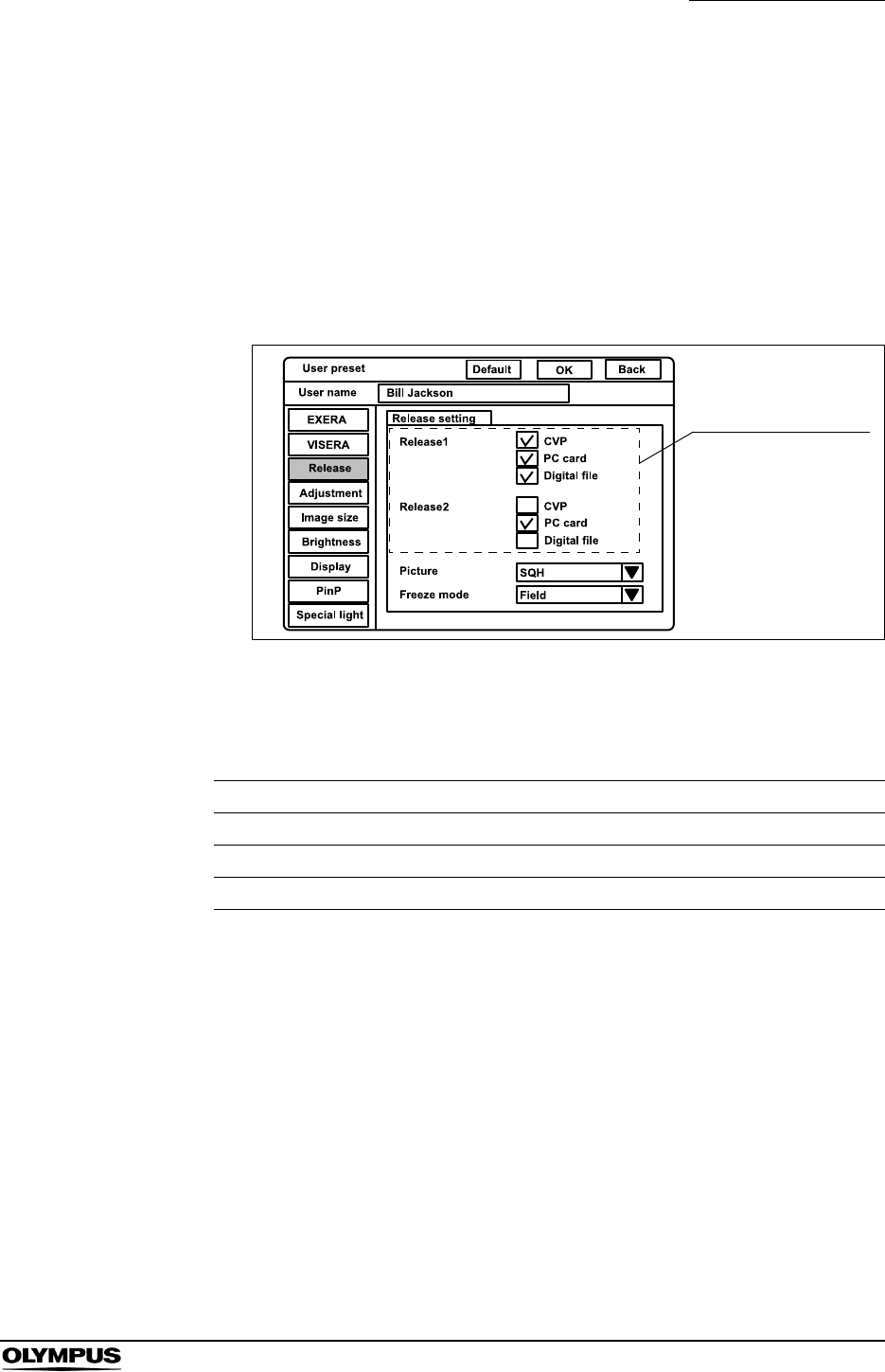
Chapter 9 Function setup
223
EVIS EXERA II VIDEO SYSTEM CENTER CV-180
Release function
Release is a function to take pictures or start recording the endoscopic image.
This operation assigns the recording devices to “Release 1” and “Release 2” that
is assigned to the remote switches and foot switches. The setting of “Release 1”
is assigned to the “RELEASE” key on the keyboard, too.
1. Click “Release” on the system setup menu. The setting items appear on the
right side of the window (see Figure 9.18).
Figure 9.18
2. Click the check box of the device to be used. The selected check box is
highlighted (see Figure 9.18). It is possible to select multiple devices.
3. Follow steps 1. and 2. to assign the instruments to “Release 2” in the same
way.
Setting value Device
CVP Color video printer
PC card PC card slot of this instrument
Digital file Image filing system
Table 9.23
Release assignment
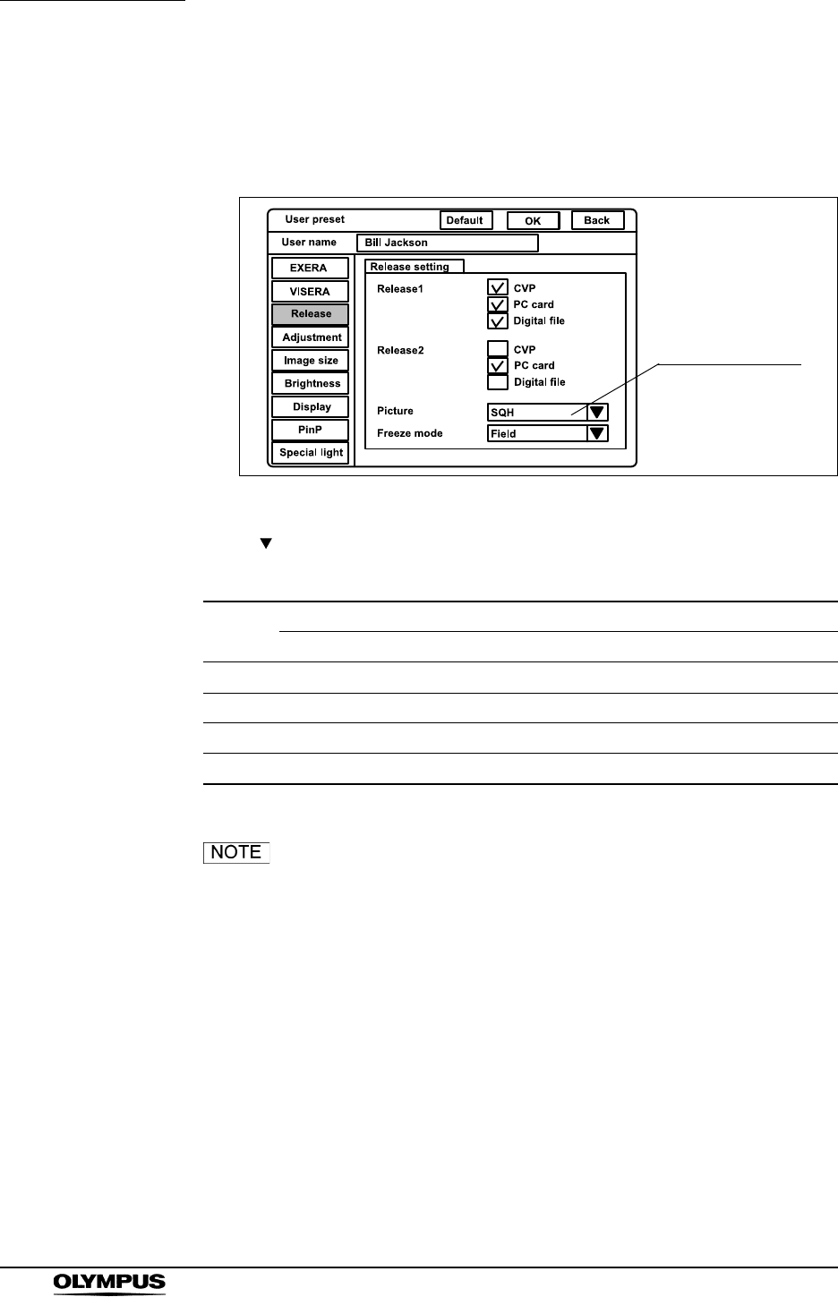
224
Chapter 9 Function setup
EVIS EXERA II VIDEO SYSTEM CENTER CV-180
Recording format for PC card
This operation sets the recording format of the frozen image to be recorded on
the PC card.
Figure 9.19
Click “ ” of “Picture” (see Figure 9.19). The recording formats appear in the
pull-down menu.
• The PinP function is OFF in Table 9.24.
• Even the approximate number of recordable images can vary
significantly depending on different factors.
Setting
value
Number of full-screen images that can be stored in 32 MB PC card
SDTV HDTV
SHQ Approx. 310 images Approx. 110 images
HQ Approx. 2000 images Approx. 760 images
SQ Approx. 2570 images Approx. 970 images
Tiff Approx. 30 images Approx. 6 images
Table 9.24
Recording format
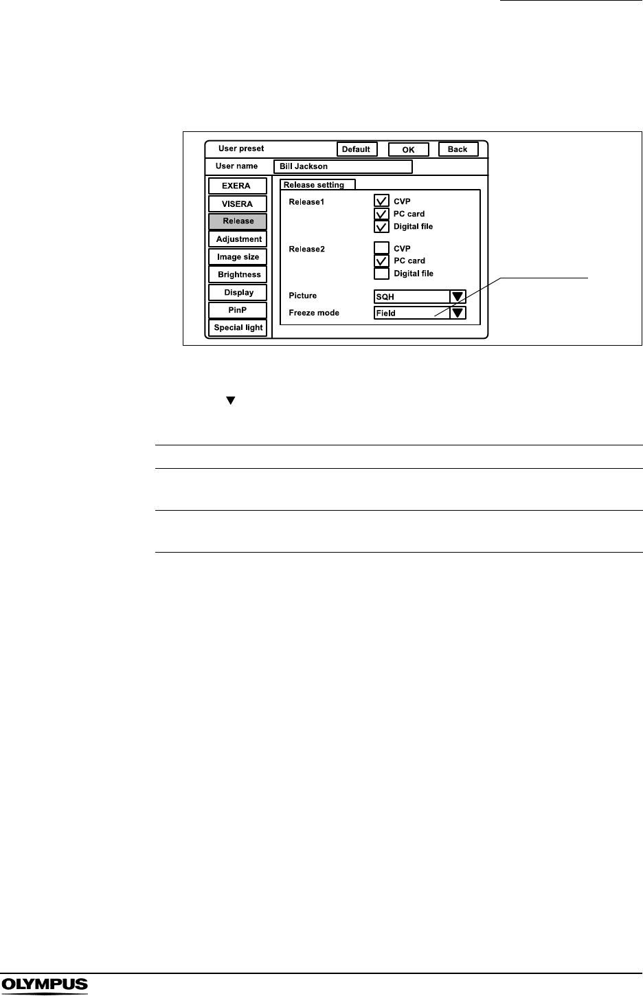
Chapter 9 Function setup
225
EVIS EXERA II VIDEO SYSTEM CENTER CV-180
Freeze function
This function sets the method of “FREEZE” to pause the endoscopic image.
Figure 9.20
1. Click “ ” of “Freeze mode” (see Figure 9.20). The freeze modes appear in
the pull-down menu.
2. Click the desired freeze mode. The selected mode is displayed.
Setting value Explanation
Field Less blur image can be obtained than frame freeze when the object
moves fast. (Use this mode for routine use.)
Frame Clearer image can be obtained than field freeze when the object does
not move.
Table 9.25
Freeze mode
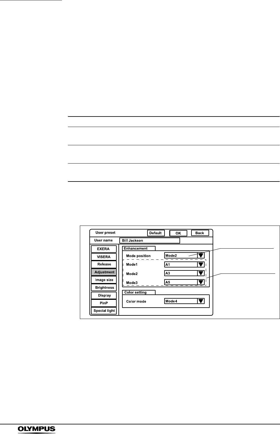
226
Chapter 9 Function setup
EVIS EXERA II VIDEO SYSTEM CENTER CV-180
Image enhancement (normal observation)
This function electrically increases the sharpness of the endoscopic image. The
three kinds of enhancement types can be set by combining desired
enhancement types and levels (see Table 9.26). These modes can be selected
during observation, and the selected mode is indicated by indicators 1, 2 or 3
above the “Image enhance button” on the front panel.This menu sets the three
enhancement modes and the initial mode when the user preset is called up.
Refer to “Image enhancement (NBI observation)” on page 250 for setting the
Image enhancement for NBI observation.
1. Click “Adjustment” on the system setup menu. The setting items appear on
the right side of the window (see Figure 9.21).
Figure 9.21
Enhancement method Function Level
A: Structural enhancement A Enhancement of contrast of the fine
patterns in the image.
A1, A2, A3, A4,
A5, A6, A7, A8
B: Structural enhancement B Enhancement of contrast of more
finer patterns than A in the image.
B1, B2, B3, B4,
B5, B6, B7, B8
E: Edge enhancement Enhancement of edges of the
endoscopic image.
E1, E2, E3, E4,
E5, E6, E7, E8
Table 9.26
Initial enhancement
mode
Setting each
enhancement mode
Chapter 9 Function setup
227
EVIS EXERA II VIDEO SYSTEM CENTER CV-180
Initial enhancement mode
1. Click “ ” of “Mode position” (see Figure 9.21). The numbers of
enhancement mode appear in the pull-down menu.
2. Click the enhancement mode number to use. The selected number is
displayed.
Enhancement modes
1. Click “ ” of “Mode 1” (see Figure 9.21).The enhancement types appear in
the pull-down menu.
• A1 to A8, B1 to B8 and E1 to E8, 24 kinds are displayed.
The larger the number, the higher the enhancement.
2. Click the desired enhancement type. The selected type is displayed.
3. Follow steps 1. and 2. to assign the enhancement type to “Mode 2” and
“Mode 3” in the same way.
• The image enhancement mode used before the video
system center OFF comes up when the instrument is turned
ON. The setting set in “Mode position” comes up when the
user preset is called up.
• B1 to B8 is valid for Scope 1, Scope 4, and Scope 5 shown in
Table 9.30 on page 229. When an endoscope other than
these endoscopes is used, the enhancement automatically
becomes A1 to A8.
• Mesh-like noise may be observed in the image, when the
image enhancement function is on during use of a fiberscope
or hybrid scope. Switch the image enhancement OFF, or use
the recommended camera head below:
OTV-S7H-1N
OTV-S7H-1D
Setting value Explanation
Mode 1 Corresponds to Mode 1 in “Enhancement modes” below.
Mode 2 Corresponds to Mode 2 in “Enhancement modes” below.
Mode 3 Corresponds to Mode 3 in “Enhancement modes” below.
OFF No enhancement
Table 9.27
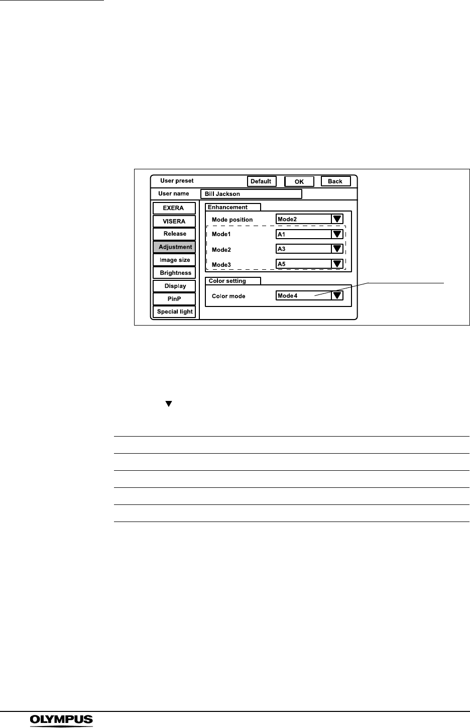
228
Chapter 9 Function setup
EVIS EXERA II VIDEO SYSTEM CENTER CV-180
Color mode
This menu sets the color tone of the monitor from four modes. The color mode
can be changed during observation. See “Color mode (“Shift” + “Alt” + “1”, “2”,
“3”, “4”)” on page 104 The setting of “Color Mode” is valid only to “Scopes B”,
“Scopes C” and “Scopes D” in Table 9.33 on page 233.
The color mode in this menu is not valid for NBI observation, because NBI
observation uses the exclusive color mode for each endoscope.
Figure 9.22
1. Click “Enhancement” on the system setup menu. The setting items appear
on the right side of the window (see Figure 9.22).
2. Click “ ” of “Color mode” (see Figure 9.22). The color modes appear in the
pull-down menu.
3. Click the desired color mode. The selected mode is displayed.
Setting value Explanation
Mode 1 Same as the color mode 1 of VISERA video system center OTV-S7V
Mode 2 Less reddish color than the Mode 1
Mode 3 More yellowish color than the Mode1
Mode 4 Standard color mode
Table 9.28
Initial color mode

Chapter 9 Function setup
229
EVIS EXERA II VIDEO SYSTEM CENTER CV-180
Image size
This menu sets the image size on the monitor when the user preset menu is
called up. The available image sizes (see Table 9.29) depend on the endoscope
models (see Table 9.30). Refer to Figure 9.24 on page 231 for a rough
comparison of the image sizes.
Type of
Endoscope
Image size
Small Medium Semi-Full Full-Height Full
Scope 1
Scope 2
Scope 3
Scope 4
Scope 5
: applicable, : not applicable
Table 9.29
Type of Endoscope Model
Scope 1 EVIS Q series
Scope 2 GIF-XP160, BF-P160, BF-3C160, BF-XT160, LF-V, HYF-V
Scope 3 • EVIS series other than Scope 1, Scope 2 and Scope 4
• ENF-V/V2, CYF-V/VA/V2/VA2, PEF-V
• Ultrasonic videoscopes
Scope 4 EVIS H series
Scope 5 Other than above
Table 9.30
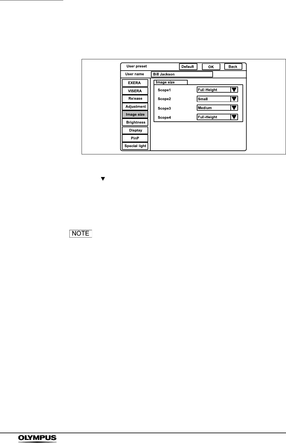
230
Chapter 9 Function setup
EVIS EXERA II VIDEO SYSTEM CENTER CV-180
1. Click “Image size” on the system setup menu. The setting items appear on
the right side of the window (see Figure 9.23).
Figure 9.23
2. Click “ ” of the scope type to use (see Figure 9.23).The image sizes
appear in the pull-down menu.
3. Click the desired image size. The selected image size is displayed.
4. Follow steps 1. to 3. to set the image size of all endoscopes to be used.
• The image size can be changed during observation. For
details, see “Image size (“F8”)” on page 94
• When this instrument is turned ON, the image size before
turning the video system center OFF comes up when turning
the instrument ON. When the user preset is called up, the
image size set in this menu comes up.
• “Scope 5” is not displayed on this menu because the
endoscopes of “Scope 5” are compatible only with “Full” size.
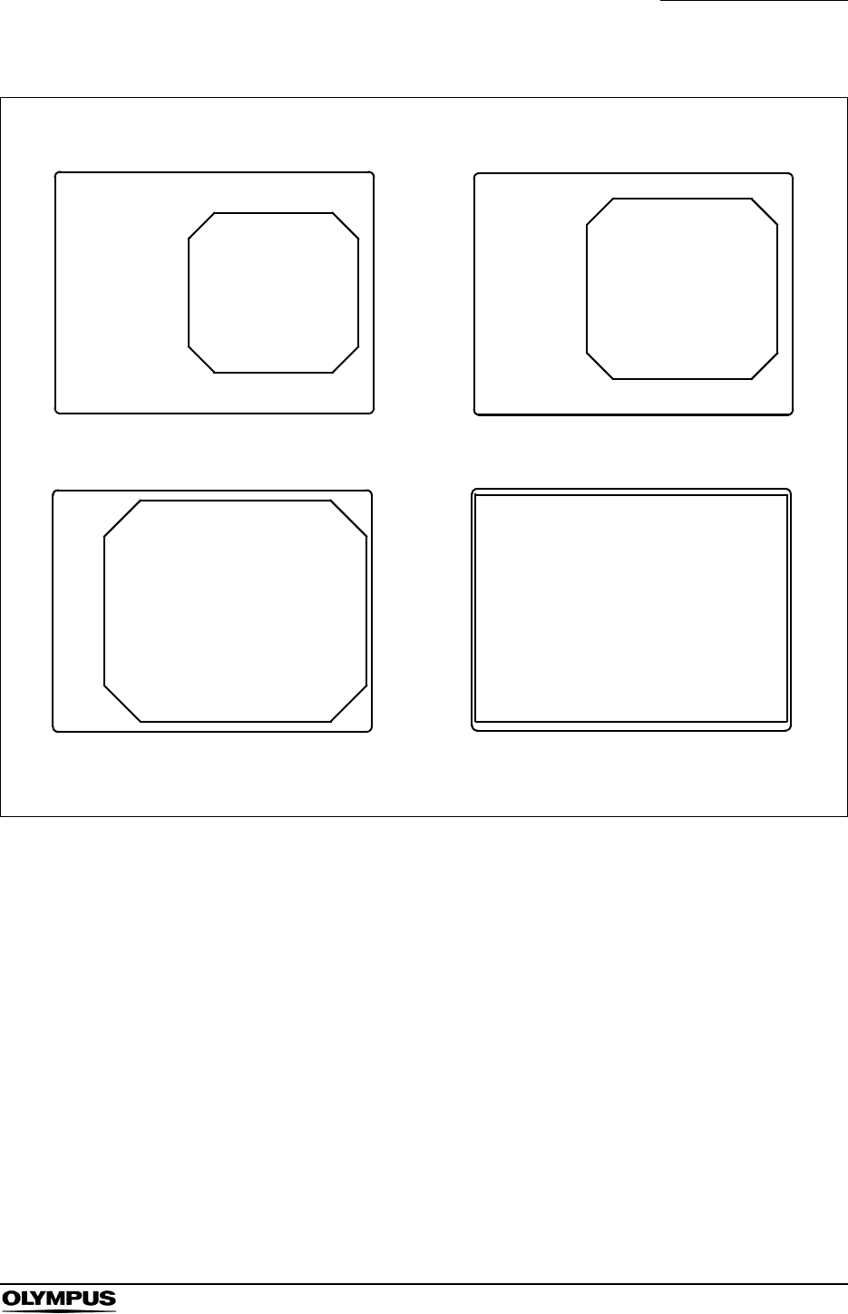
Chapter 9 Function setup
231
EVIS EXERA II VIDEO SYSTEM CENTER CV-180
Figure 9.24
ABC123
M 51
03/03/1954
12/12/2005
12:12:12
CVP: A4/4
D.F: 99
VCR
Ct: N Eh: A8
Z: x1.5
Pump
Media:
John Smith
Cardiac end of the stomach
ABC123
M 51
03/03/1954
12/12/2005
12:12:12
CVP: A4/4
D.F: 99
VCR
Ct: N Eh: A8
Z: x1.5
Pump
Media:
John Smith
Cardiac end of the stomach
Mike Johnson Mike Johnson
Medium
Small
Full-height Full
ABC123
Mike Johnson
M 51
03/03/1954
12/12/2005
12:12:12
CVP: A4/4
D.F: 99
VCR
Ct: N Eh: A8
Z: x1.5
Pump
Media:
John Smith
Cardiac end of the stomach
ABC123
Mike Johnson
M 51
03/03/1954
12/12/2005
12:12:12
CVP: A4/4
D.F: 99
VCR
Ct: N Eh: A8
Z: x1.5
Pump
Media:
John Smith
Cardiac end of the stomach

232
Chapter 9 Function setup
EVIS EXERA II VIDEO SYSTEM CENTER CV-180
Iris
This video system center automatically measures the brightness of objects, and
displays live images with proper brightness on the monitor. There are two
methods, “Auto” and “Peak”. The functions slightly differ depending on the
endoscope type (see Table 9.33) even with the same methods. The methods
can be changed during use (see “Iris mode” on page 69).
This menu sets the measuring method that measures the image brightness.
Setting value Explanation
Auto The methods to measure the brightness of the central and peripheral part of the image. One of
the following three measuring methods can be used. They differ in how the measuring results are
combined. Select “AUTO 1” or “AUTO 2” when white wash is observed on the image.
1) AVE.: The brightness is adjusted based on the average brightness of the two parts.
2) AUTO 1: The brightness is adjusted based on the brightest part of the central part and the
average brightness of the periphery part.
3) AUTO 2: The entire image is brighter than “AUTO 1”.
Peak The brightness is adjusted based on the brightest part of the image.
Table 9.31 Method of photometry for endoscopes of “Scope A” (see Table
9.33)
Setting value Explanation
Auto The image brightness is adjusted based on the average brightness of the exposure area set in
the image. The three exposure areas can be selected. Select “Full” or “Center” when white wash
is observed, or the image becomes dark.
1) Mask: The exposure area is set automatically to obtain optimum brightness for the
subject. (This exposure area become the same with “Full” for scope D.)
2) Center: Center-weighted measuring.
3) Full: The brightness of the whole image is measured.
Peak The same measuring method as the “Auto” but slightly darker.
Table 9.32 Method of photometry for endoscopes of “Scope B”, “Scope C”,
and “Scope D” (see Table 9.33)
Chapter 9 Function setup
233
EVIS EXERA II VIDEO SYSTEM CENTER CV-180
Endoscope type Endoscope model
Scope A GIF-N180/H180/Q180, CF-H180AL/I, CF-Q180AL/I, PCF-Q180AL/I,
BF-P180/Q180/1T180, EVIS EXERA160 series, EVIS140/130/100 series,
Ultrasonic videoscopes
Scope B ENF-V/V2, LF-V, HYF-V/V2, CYF-V/VA/VA2, PEF-V
Scope C OTV-SP1H-N-12E/12Q, OTV-SP1H-NA-12E/12Q, OTV-S7H-N/1N/1D/NA/1NA,
OTV-S7H-1D-F08E/L08E/1MD/D-L08E, OTV-S7H-NA-10E/1NA-10E, OTV-S7H-NA-12E/1NA-
12E, OTV-S7H-NA-10Q/NA-12Q, OTV-S7H-1NA-12Q, OTV-S7H-FA-E/1FA-E/FA-Q, OTV-S7H-
VA,
OTV-SP1H-NA-12E/12Q, OTV-SP1H-N-12E/12Q
OTV-S7ProH-HD-10E/10Q/12E/12Q
OTV-S7ProH-HD-L08E
Scope D LTF-V3/VP, A50001A, A50003A, A50021A, A50023A, WA50003L, WA50005L, WA50011A,
WA50013A, WA50013L, WA50015L, WA50121A, WA50201A, ENF-VQ
Table 9.33 Types of the endoscope
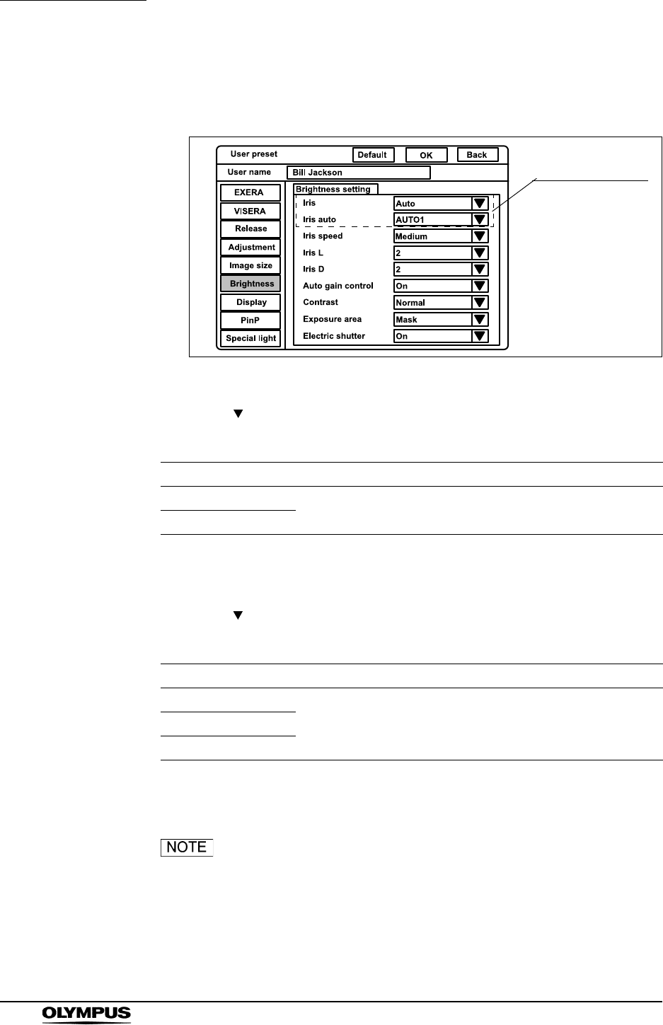
234
Chapter 9 Function setup
EVIS EXERA II VIDEO SYSTEM CENTER CV-180
1. Click “Brightness” on the system setup menu. The “Brightness setting”
appears on the right side of the window (see Figure 9.25).
Figure 9.25
2. Click “ ” of “Iris” (see Figure 9.25). The measuring methods appear in the
pull-down menu.
3. Click the desired method. The selected mode is displayed.
4. Click “ ” of “Iris auto” (see Figure 9.25). The measuring methods of “Auto”
appear in the pull-down menu.
5. Click the desired method. The selected method is displayed.
When this instrument is turned ON, the latest iris mode
before the video system center is turned OFF comes up
when the instrument is turned ON. When the user preset is
called up, the iris mode that is set in this menu comes up.
Setting value Explanation
Auto See Table 9.31 and Table 9.32 on page 232.
Peak
Table 9.34
Setting value Explanation
AUTO 1
See Table 9.31 on page 232.AUTO 2
AVE.
Table 9.35
Measuring method
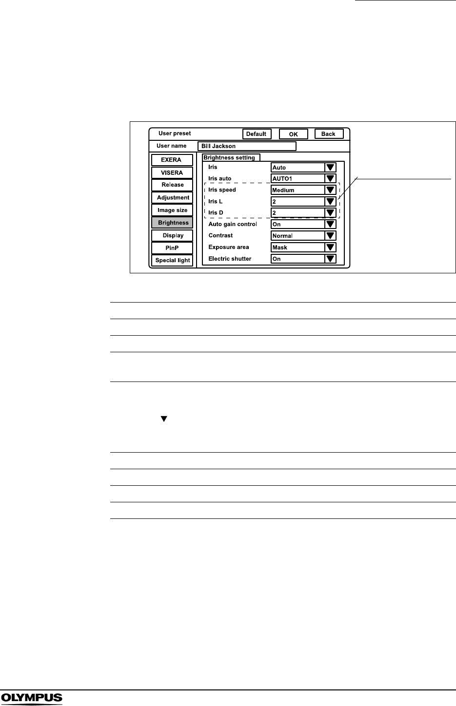
Chapter 9 Function setup
235
EVIS EXERA II VIDEO SYSTEM CENTER CV-180
Iris speed
This menu sets the speed to adjust the brightness of the image according to the
result of the measuring method. The setting in this menu is effective only for the
endoscopes of “Scope A” in see Table 9.33 on page 233.
Figure 9.26
1. Click “ ” of “Iris speed” (see Figure 9.26). The iris speeds appear in the
pull-down menu.
2. Click the desired iris speed. The selected iris speed is displayed.
Setting value Explanation
Iris speed Changes the brightness adjustment speed from three speeds.
Iris L Fine-tunes the darkening speed in the each settings of “Iris speed”.
Iris D Fine-tunes the brightening speed in the each settings of “Iris
speed”.
Table 9.36
Setting value Explanation
Low The brightness adjustment speed is low.
Medium The brightness adjustment speed is medium.
High The brightness adjustment speed is high.
Table 9.37
Speed for adjusting the
brightness

236
Chapter 9 Function setup
EVIS EXERA II VIDEO SYSTEM CENTER CV-180
3. Click “ ” of “Iris L (Iris D)” (see Figure 9.26). The fine-tuning speeds
appear in the pull-down menu.
4. Click the desired speed. The selected speed is displayed.
Setting value Explanation
1 The fine-tuning of the brightness adjustment is low.
2 The fine-tuning of the brightness adjustment is medium.
3 The fine-tuning of the brightness adjustment is high.
Table 9.38
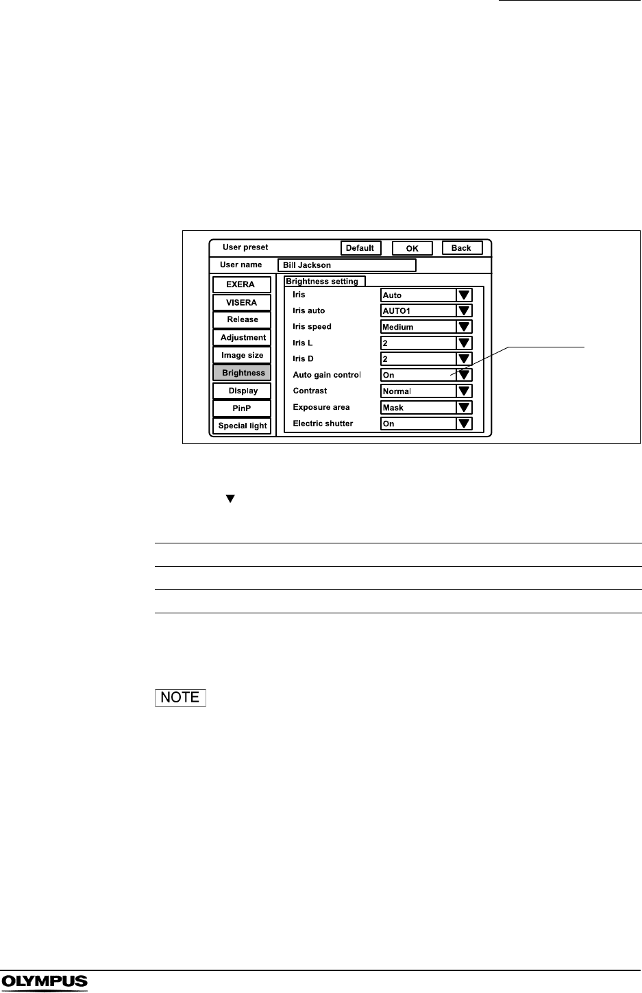
Chapter 9 Function setup
237
EVIS EXERA II VIDEO SYSTEM CENTER CV-180
Auto gain control (AGC)
The AGC (Automatic Gain Control) is used to increase the brightness of an
endoscopic image electrically, when the brightness of the image is dark because
the distance between the endoscope's distal end and the object is too long. This
menu sets if the AGC function is used. When the AGC function is set, the
function can be switched ON and OFF from the keyboard and remote switch
during observation (see “Automatic gain control (AGC) (“F6”)” on page 89).
Figure 9.27
1. Click “ ” of “Auto gain control” (see Figure 9.27). The setting values ON
and OFF appear in the pull-down menu.
2. Click “ON” or “OFF”. The selected status is displayed.
Image noises may appear in the image when the AGC
function is ON.
Setting value Explanation
On AGC function is ON.
Off AGC function is OFF.
Table 9.39
AGC setting
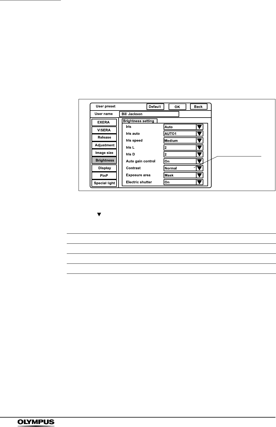
238
Chapter 9 Function setup
EVIS EXERA II VIDEO SYSTEM CENTER CV-180
Contrast
The contrast modes widen or narrow the difference between brightness and
darkness of the endoscopic image. This operation sets the contrast mode when
the video system center is turned ON. Three contrast modes are available and
can be changed by the keyboard during observation (refer to “Contrast mode
(“Shift” + “F6”)” on page 90). The setting is invalid during special light
observation (refer to “Special light observation” on page 250).
Figure 9.28
1. Click “ ” of “Contrast” (see Figure 9.28). The exposure areas appear in
the pull-down menu.
2. Click the desired contrast mode. The selected mode is displayed.
Setting value Explanation
Normal Standard setting
Low Darkens the bright part, and brightens the dark part.
High Brightens the bright part, and darkens the dark part.
Table 9.40
Contrast setting
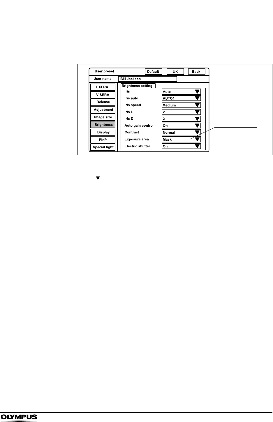
Chapter 9 Function setup
239
EVIS EXERA II VIDEO SYSTEM CENTER CV-180
Exposure area
This menu sets the exposure area for endoscopes of “Scope B”, “Scope C” and
“Scope D” in see Table 9.33 on page 233.
Figure 9.29
1. Click “ ” of “Exposure area” (see Figure 9.29). The exposure areas
appear in the pull-down menu.
2. Click the desired exposure area. The selected exposure area is displayed.
Setting value Explanation
Mask
See Table 9.32 on page 232.Center
Full
Table 9.41
Exposure area
setting
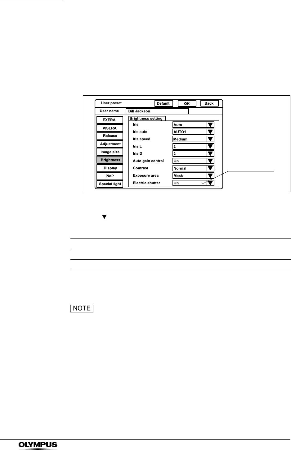
240
Chapter 9 Function setup
EVIS EXERA II VIDEO SYSTEM CENTER CV-180
Electronic shutter
This menu sets whether the electronic shutter function of the endoscope is ON
or OFF. The applicable endoscopes are EVIS H series endoscopes, camera
heads, LTF endoscopes. When using an endoscope compatible with the
electronic shutter function, the light exposure is automatically adjusted using the
electronic shutter function of the endoscope.
Figure 9.30
1. Click “ ” of “Electric shutter” (see Figure 9.30). The setting values ON and
OFF appear in the pull-down menu.
2. Click “ON” or “OFF”. The selected option is displayed.
When using an endoscope not compatible with the electronic
shutter function of the video system center, the electronic
shutter function does not work even if “ON” is selected.
Setting value Explanation
On Activates the electronic shutter function.
Off De-activates the electronic shutter function.
Table 9.42
Electric shutter
setting
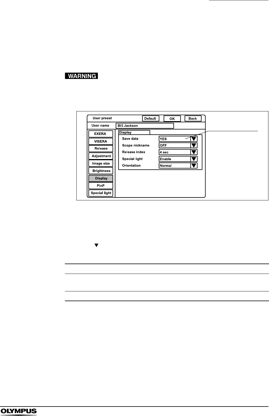
Chapter 9 Function setup
241
EVIS EXERA II VIDEO SYSTEM CENTER CV-180
Patient data display
This menu sets whether the patient data, which had been displayed right before
the video system center has been turned OFF, is displayed on the monitor or not
when the video system center is turned ON.
When “Save data” is set to YES, confirm that the name on
the monitor is identical to the name of the patient to be
observed before observation.
Figure 9.31
1. Click “Display” on the system setup menu. The setting items appear on the
right side of the window (see Figure 9.31).
2. Click “ ” of “Save data” (see Figure 9.31). The setting values of “Yes” and
“No” appear in the pull-down menu.
3. Click “Yes” or “No”. The selected option is displayed.
Setting value Explanation
Yes Displays the patient data that had been displayed right before the
video system center has been turned OFF.
No No display of last patient data.
Table 9.43
Saving patient data
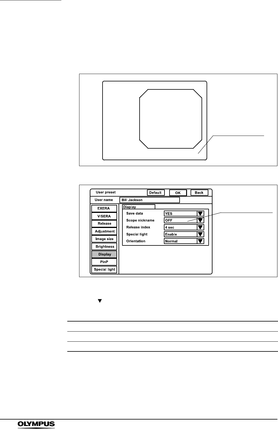
242
Chapter 9 Function setup
EVIS EXERA II VIDEO SYSTEM CENTER CV-180
Scope nickname
The scope nickname is displayed on the monitor, when the endoscope with the
scope nickname function is connected. This menu sets whether the nickname is
displayed on the monitor or not. For the V-scope, “V” is displayed on the monitor.
Figure 9.32
Figure 9.33
1. Click “ ” of “Scope nickname” (see Figure 9.33). The settings “Enable”
and “OFF” appear in the pull-down menu.
2. Click the desired setting. The selected setting is displayed.
Setting value Explanation
Enable Displays the scope nickname.
OFF No display of the scope nickname
Table 9.44
ID
Name:
Sex: Age:
D.O.B.
12/12/2005
12:12:12
Physician: V
Scope nickname
Scope nickname
setting
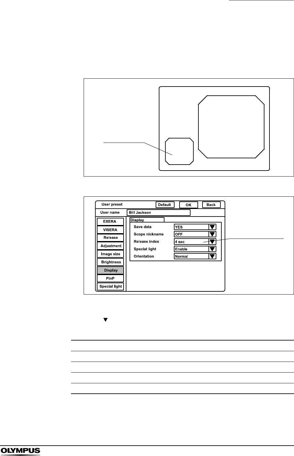
Chapter 9 Function setup
243
EVIS EXERA II VIDEO SYSTEM CENTER CV-180
Release index time
The image taken using the release function can be displayed in the lower-left of
the monitor. This image is called index image. This menu sets the time an index
image is displayed.
Figure 9.34
Figure 9.35
1. Click “ ” of “Release index” (see Figure 9.35). The display times appear in
the pull-down menu.
2. Click the desired display time. The selected time is displayed.
Setting value Explanation
Off No display of the index image
2 sec Displays the index image for 2 seconds.
4 sec Displays the index image for 4 seconds.
Always Displays the index image all the time.
Table 9.45
ID:
Name:
Sex: Age:
D.O.B
12/12/2005
12:12:12
Physician:
Comment:
Index image
Setting the Index
image
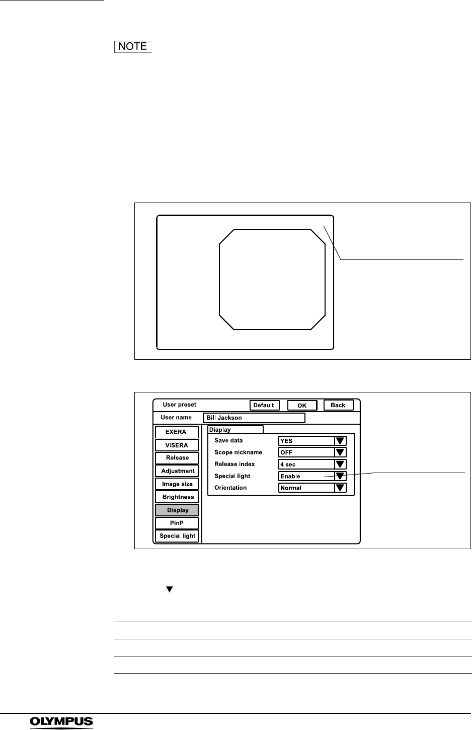
244
Chapter 9 Function setup
EVIS EXERA II VIDEO SYSTEM CENTER CV-180
• The index image may overlap the characters such as the
patient data.
• Index image does not appear in the PinP screen (see “PinP
(picture in picture) display” on page 64).
Indication of the special light observation
This menu sets whether the indication of the special light observation is shown
on the monitor or not (see Figure 9.36).
Figure 9.36
Figure 9.37
1. Click “ ” of “Special light” (see Figure 9.37). The settings “Enable” and
“Disable” appear in the pull-down menu.
Setting value Explanation
Enable Display
Disable No display
Table 9.46
ID
Name:
Sex: Age:
D.O.B.
12/12/2005
12:12:12
Physician:
NBI
NBI observation
Display of the special
light observation
Display of the special
light observation
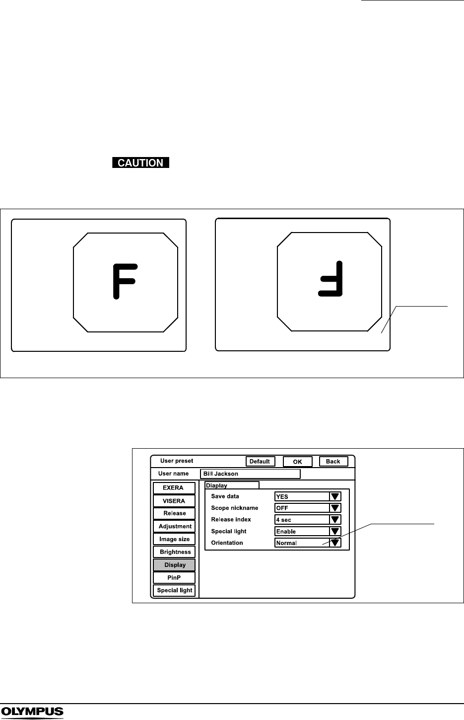
Chapter 9 Function setup
245
EVIS EXERA II VIDEO SYSTEM CENTER CV-180
2. Click “OFF” or “ON”. The selected option is displayed.
Monitor orientation function
The orientation function rotates the monitor image into a 180 reverse image.
The following operation sets whether or not the orientation function is activated
(see Figure 9.38).
Please note that during image rotation a time delay of the
image display might occur. Please deal appropriately.
Figure 9.38
The orientation setup affects all video outputs as well as the monitor output. The
recorded images are also reversed. The external PinP image does not reverse.
Figure 9.39
ID
Name:
Sex: Age:
D.O.B.
12/12/2005
12:12:12
Physician:
ID
Name:
Sex: Age:
D.O.B.
12/12/2005
12:12:12
Physician: R
“R” mark
Normal image Rotated image
Orientation
setting
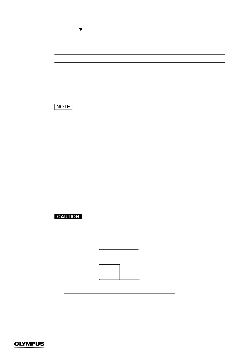
246
Chapter 9 Function setup
EVIS EXERA II VIDEO SYSTEM CENTER CV-180
1. Click “ ” of “Orientation” (see Figure 9.39). The settings of “Normal” and
“Rotation” appear in the pull-down menu.
2. Click “Normal” or “Rotation”. The selected option is displayed.
“R” mark is always displayed at the bottom right of the display
when the orientation function and PinP function are ON.
PinP (picture in picture) function
The PinP function displays an image of an external device (such as an ultrasonic
observation unit) connected to the video system center, as a sub or main image
on the monitor together with the endoscopic image (see Figure 9.40). The
images from one of the devices connected to the following connectors can be
displayed as the PinP image.
• PinP composite terminal on the front panel
(has priority to the PinP Y/C terminal on the rear panel)
• PinP Y/C terminal on the rear panel
Please note that during PinP is displayed a time delay of the
image display might occur. Please deal appropriately.
Figure 9.40
Setting value Explanation
Normal Displays the endoscopic image on the monitor normally.
Rotation Displays the endoscopic image rotated by 180 on the
monitor.
Table 9.47
Main image
Sub
image
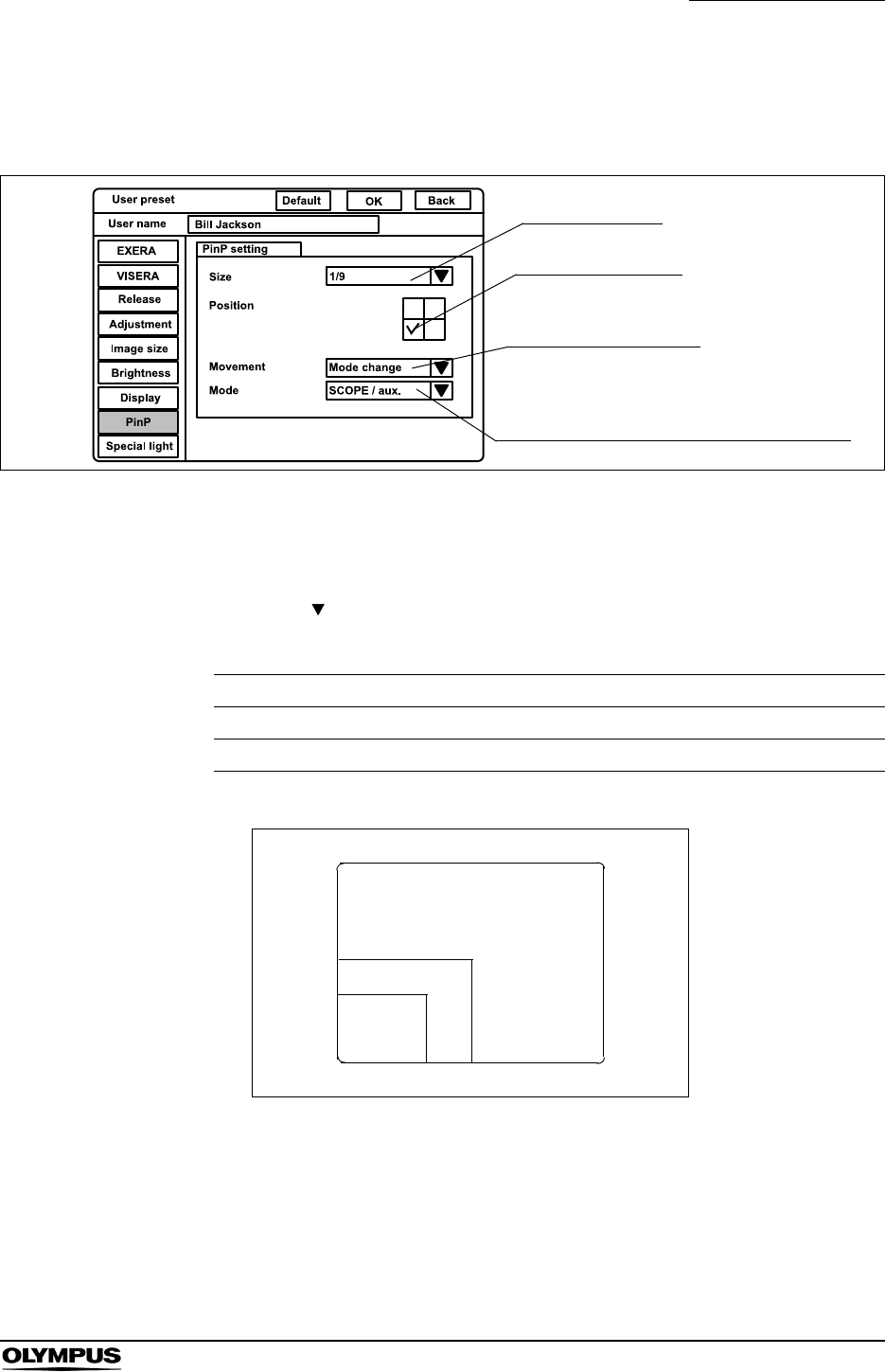
Chapter 9 Function setup
247
EVIS EXERA II VIDEO SYSTEM CENTER CV-180
Click “PinP” on the system setup menu. The setting items appear on the right
side of the window (see Figure 9.41).
Figure 9.41
Image size of PinP
1. Click “ ” of “Size” (see Figure 9.41). The PinP sub image sizes appear in
the pull-down menu (see Figure 9.42).
Figure 9.42
2. Click the desired sub image size. The selected size is displayed.
Setting value Explanation
1/4 The size of sub image is about one-fourth that of the main image.
1/9 The size of sub image is about one-ninth that of the main image.
Table 9.48
Sub image size
Sub image position
Way of screen change
Combination of the image in ON/OFF mode
1/9
sub image
Main image
1/4 sub image
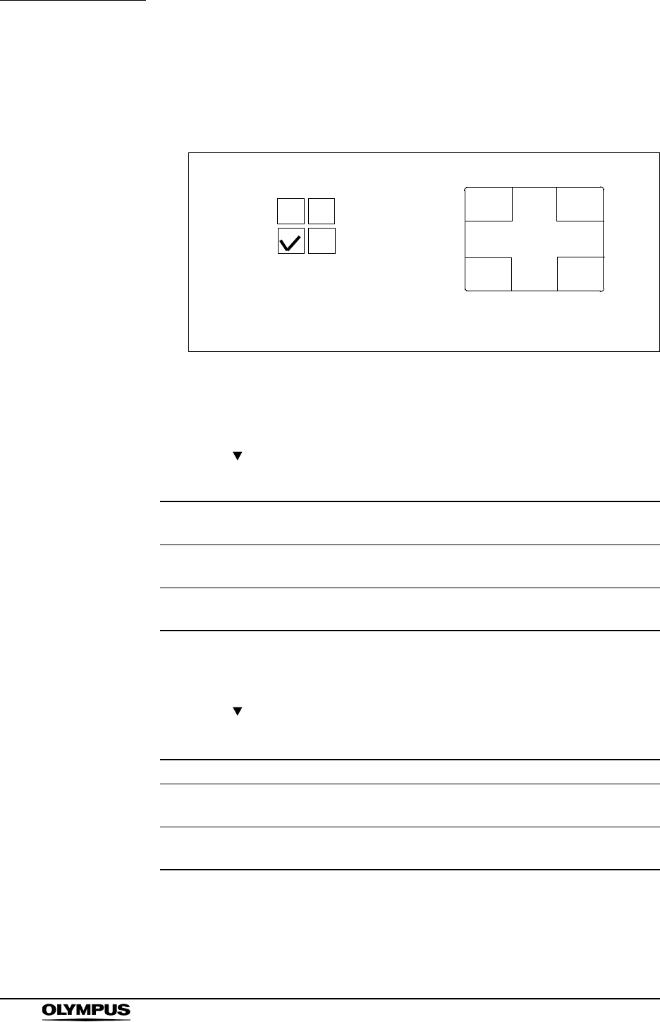
248
Chapter 9 Function setup
EVIS EXERA II VIDEO SYSTEM CENTER CV-180
Position of the PinP sub image
Click the check box for the position to display the PinP sub image. The selected
check box turns black.
Figure 9.43
Display mode of PinP
1. Click “ ” of “Movement” (see Figure 9.41).The PinP modes appear in the
pull-down menu.
2. Click the desired PinP mode. The selected PinP mode is displayed.
3. Click “ ” of “Mode” if “ON/OFF” of “Movement” is selected (see Figure
9.41). The display modes appear in the pull-down menu.
Setting value Explanation (see “PinP (picture in picture) display” on
page 64.)
On/Off The PinP button toggles back and forth between the non-PinP
mode and the PinP mode.
Mode change The PinP button switches between the PinP mode, endoscopic
image and external image.
Table 9.49
Setting value Explanation
SCOPE/aux. The endoscopic image is the main image and the external image
is the sub image.
AUX./scope The external image is the main image and the endoscopic image
is the sub image.
Table 9.50
The highlighted check box shows
the position of the sub image
AB
CD
D
AB
C
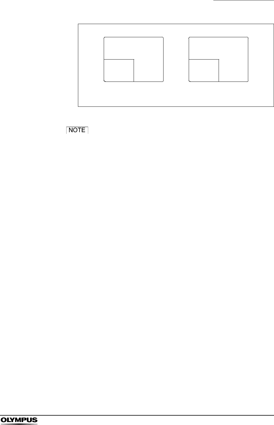
Chapter 9 Function setup
249
EVIS EXERA II VIDEO SYSTEM CENTER CV-180
Figure 9.44
If “Mode change” of “Movement” is selected, the mode
setting is invalid.
4. Click the desired display mode. This selected mode is displayed.
Endoscopic image
External
Image
SCOPE + aux.
External image
AUX. + scope
Endoscopic
image
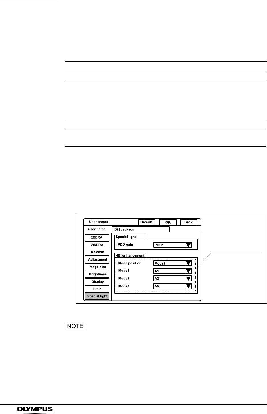
250
Chapter 9 Function setup
EVIS EXERA II VIDEO SYSTEM CENTER CV-180
Special light observation
The following special light observation function is available on the video system
center.
For these observations, use the endoscopes and the light source indicated
below.
Image enhancement (NBI observation)
This menu sets the image enhancement mode of NBI observation in the same
way as normal observation. Refer to the “Image enhancement (normal
observation)” on page 226 for the procedure.
Figure 9.45
“PDD gain” is reserved for future system expansion.
Currently not used.
Observation Outline
NBI (narrow band imaging) Uses filtered light of a specific wavelength.
Table 9.51
CV-180 Light source Compatible endoscope
NBI No change CLV-180 Refer to “NBI (narrow band
imaging)” on page 151.
Table 9.52
Image enhancement
for NBI
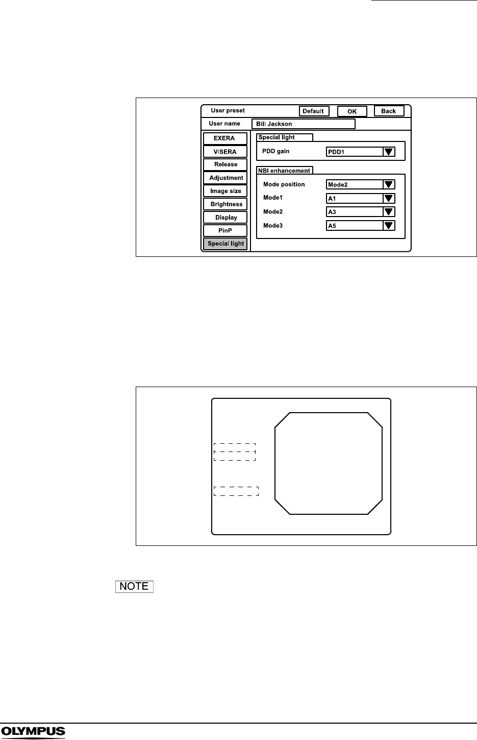
Chapter 9 Function setup
251
EVIS EXERA II VIDEO SYSTEM CENTER CV-180
Saving the user preset
This procedure finalizes and saves the user preset data.
Figure 9.46
1. Click “OK” in the user preset menu (see Figure 9.46). A confirmation
message appears on the monitor.
2. Click “N” to return to the user preset menu.
Click “Y” or press “Enter” to finalize and save the settings in the video
system center. The endoscopic image appears (see Figure 9.47).
Figure 9.47
• To cancel the data entered, click “Back” instead of “OK”. The
data entered are canceled and the display returns to the user
list menu.
• The functions are activated immediately after saving the user
preset data.
ABC123
Mike Johnson
M 51
03/03/1954
12/12/2005
12:12:12
CVP: A4/4
D.F: 99
VCR
Ct: N Eh: A8
Z: x1.5
Pump
Media:
Bill Jackson
Cardiac end of the stomach
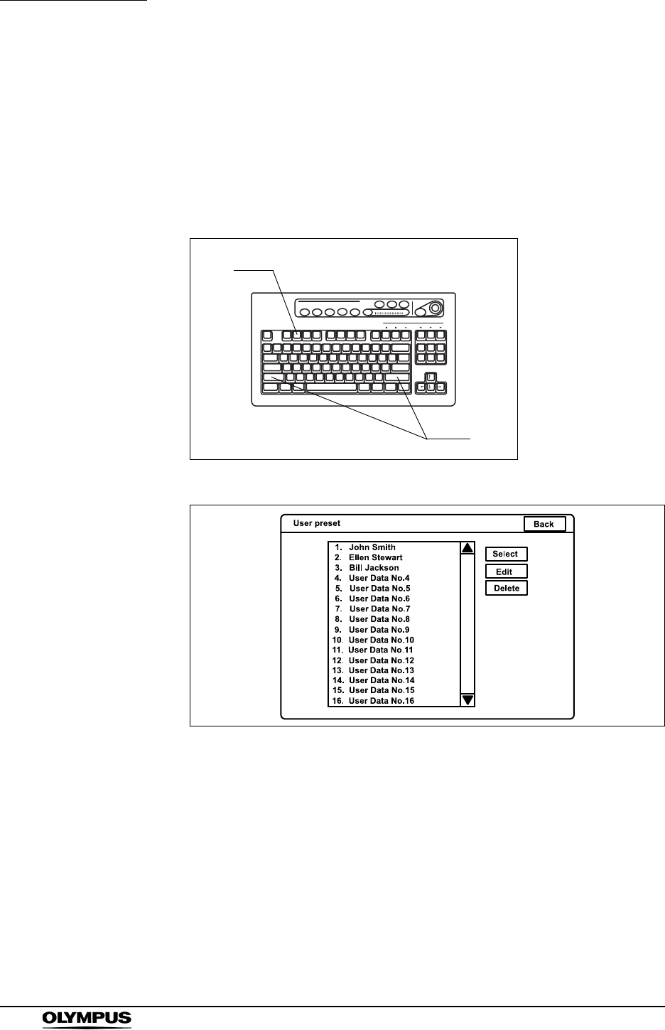
252
Chapter 9 Function setup
EVIS EXERA II VIDEO SYSTEM CENTER CV-180
Resetting the user preset data to the factory defaults
This operation resets the settings of the user preset to the factory default setting.
The user name is not reset in this operation. See Table 9.53 on page 255 for the
default settings.
1. Press the “Shift” and “F2” keys together (see Figure 9.48). The user name
list of the user preset menu appears on the monitor (see Figure 9.49).
Figure 9.48
Figure 9.49
2. Click the user name in the user name dialog box to be reset to the factory
default setting. The user name is highlighted.
3. Click “Edit”. The user preset menu appears.
4. Click “Default” on top of the menu. A confirmation message appears on the
monitor.
5. Click “No” to return to the user preset menu.
Click “Yes” to reset the settings of the selected user to the factory default
setting.
F2
Shift
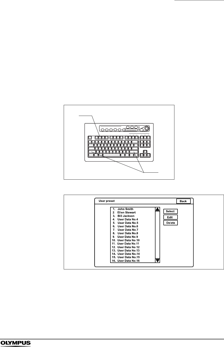
Chapter 9 Function setup
253
EVIS EXERA II VIDEO SYSTEM CENTER CV-180
6. Click “Back” to return to the user name list.
7. To reset the settings of other registered users, repeat steps 2. to 6.
8. Click “Back” to return to the endoscopic image.
Deleting user preset data
This operation deletes the user preset data with the user name.
1. Press the “Shift” and “F2” keys together (see Figure 9.50). The user name
list of the user preset menu appears on the monitor (see Figure 9.51).
Figure 9.50
Figure 9.51
2. Click the user name in the user name dialog box to be deleted. The user
name is highlighted.
3. Click “Delete” to delete the user preset data. A confirmation message
appears on the monitor.
F2
Shift

254
Chapter 9 Function setup
EVIS EXERA II VIDEO SYSTEM CENTER CV-180
4. Click “No” to return to step 2.
Click “Yes” to delete the user being selected. The user name changes to
“User Data No. #”.
5. To delete the settings of other registered users, repeat steps 2. to 4.
6. Click “Back” to return to the endoscopic image.
Chapter 9 Function setup
255
EVIS EXERA II VIDEO SYSTEM CENTER CV-180
Summary of settings
The following table shows the options of the user preset menu. The bold and
underscored options listed are the factory default settings.
Setup item Setting value Note
EXERA Scope switch Switch 1
Switch 2
Switch 3
Switch 4
[Freeze], [Release 1], [Release 2],
[Iris], [Enhance], [Contrast], [AGC],
[Image size], [VCR], [Capture],
[Stop watch], [Remove data], [Zoom],
[White balance], [Exposure area],
[Exposure (up)], [Exposure (down)],
[NBI], [OP.2], [OFP], [Light], [Arrow],
[PinP], [Shutter], [US freeze],
[Freeze mode], [Option], [None]
The same functions can be
assigned to more than one
switch.
The default settings are:
Switch 1: Freeze
Switch 2: Iris
Switch 3: Enhance
Switch 4: Release 1
Foot switch Switch 1
Switch 2
The same functions can be
assigned to more than one
switch.
The default settings are:
Switch 1: Release 1
Switch 2: Release 2
VISERA Scope switch Switch 1
Switch 2
Switch 3
Switch 4
[Freeze], [Release 1], [Release 2],
[Iris], [Enhance], [Contrast], [AGC],
[Image size], [VCR], [Capture],
[Stop watch], [Remove data], [Zoom],
[White balance], [Exposure area],
[Exposure (up)], [Exposure (down)],
[NBI], [OP.2], [OFP], [Light], [Arrow],
[PinP], [Shutter], [US freeze],
[Freeze mode], [Option], [None]
The same functions can be
assigned to more than one
switch.
The default settings are:
Switch 1: Release 1
Switch 2: Enhance
Switch 3: VCR
Switch 4: Freeze
Foot switch Switch 1
Switch 2
The same functions can be
assigned to more than one
switch.
The default settings are:
Switch 1: Release 1
Switch 2: Release 2
Release Release
setting
Release 1 [CVP], [PC card], [Digital file] Devices to be operated by
“Release1” and “Release 2”.
Release 2 [CVP], [PC card], [Digital file]
Picture [SHQ], [HQ], [SQ], [TIFF] Recording format for PC
card
Freeze
mode
[Field], [Frame] Freeze method
Table 9.53
256
Chapter 9 Function setup
EVIS EXERA II VIDEO SYSTEM CENTER CV-180
Adjustment Enhancement Mode
position
[Mode1], [Mode2], [Mode3], [OFF] Initial mode of enhancement
Mode 1
Mode 2
Mode 3
[A1] - [A8], [B1] - [B8], [E1] - [E8] Setting of each
enhancement mode
The default settings are:
Mode 1: A1
Mode 2: A3
Mode 3: A5
Color setting Color mode [Mode 1], [Mode 2], [Mode 3],
[Mode 4]
Color tone of the monitor
Image size Image size Scope 1 [Medium], [Full-height] The display area size of the
endoscopic image
Scope 2 [Small], [Medium]
Scope 3 [Medium], [Full-height]
Scope 4 [Medium], [Full-height]
Brightness Brightness
setting
Iris [Auto], [Peak] Initial method of metering
Iris auto [AUTO 1], [AUTO 2], [AVE] Metering method for “Auto”
Iris speed [High], [Medium], [Low] Speed of the automatic
brightness control
Iris L [1], [2], [3] Fine-tuning of “Iris Speed”
Iris D [1], [2], [3] Fine-tuning of “Iris Speed”
AGC [ON], [OFF] AGC function ON or OFF
Contrast [Normal], [Low], [High] Initial setting of contrast
Exposure
area
[Mask], [Center], [Full] Setting of the metering area
Electronic
shutter
[ON], [OFF] Electric shutter function ON
or OFF
Display Display Save data [YES], [NO]
Scope
nickname
[Enable], [OFF] The display function of the
scope nickname is ON or
OFF.
Release
index time
[OFF], [2 sec], [4 sec], [Always] Setting of display time of the
index image
Special light [Enable], [Disable] The display function of the
special light observation is
ON or OFF.
Orientation [Normal], [Rotation] Setting of the orientation of
the image
Setup item Setting value Note
Table 9.53
Chapter 9 Function setup
257
EVIS EXERA II VIDEO SYSTEM CENTER CV-180
PinP PinP setting Size [1/4], [1/9] Image size of the sub image
Position [Upper-left], [Upper-right],
[Lower-left], [Lower-right]
Position of the sub image
Movement [ON/OFF], [Mode change]
Mode [SCOPE/aux.], [AUX./scope] Setting of main and sub
image
Special
Light
PDD setting PDD gain [PDD1], [PDD2] Initial method of PDD
NBI
enhancement
Mode
position
[Mode 1], [Mode 2], [Mode 3], [OFF] Initial mode of enhancement
Mode 1
Mode 2
Mode 3
[A1] - [A8], [B1] - [B8], [E1] - [E8] Setting of each
enhancement mode
The default settings are:
Mode 1: A1
Mode 2: A3
Mode 3: A5
Setup item Setting value Note
Table 9.53

258
Chapter 9 Function setup
EVIS EXERA II VIDEO SYSTEM CENTER CV-180
Chapter 10 Troubleshooting
259
EVIS EXERA II VIDEO SYSTEM CENTER CV-180
Chapter 10 Troubleshooting
If the video system center is visibly damaged, does not function as expected, or
is found to have irregularities during the inspection described in Chapter 3,
“Inspection” and Chapter 8, “Installation and Connection”, or the use described
in Chapter 4, “Operation”, do not use the video system center and contact
Olympus. Some problems that appear to be malfunctions may be correctable by
referring to Section 10.1, “Troubleshooting guide”. If the problem cannot be
resolved by the described remedial action, stop using the video system center
and contact Olympus.
Never use the video system center if an abnormality is
suspected. Damage or irregularity in the instrument may
compromise patient or user safety and may result in more
severe equipment damage.
10.1 Troubleshooting guide
The following table shows the possible causes of and countermeasures against
troubles that may occur due to equipment setting errors or deterioration of
consumable.
Troubles or failures other than those listed in the following table need repair. As
repair performed by persons who are not qualified by Olympus could cause
patient or user injury and/or equipment damage, be sure to contact Olympus for
repair.
If an abnormality is suspected, turn the light source OFF
once and turn it ON again. If the abnormality cannot be
solved, turn the light source OFF and disconnect the power
cord to stop the flow of electricity completely.
260
Chapter 10 Troubleshooting
EVIS EXERA II VIDEO SYSTEM CENTER CV-180
Irregularity description Possible cause Solution
The endoscope cannot be
connected to the video
system center.
The endoscope is not
compatible with the video
system center.
Use one of the endoscopes
shown in the “System
chart” in the Appendix.
The power fails to come on. The power switch of the
video system center is set
to OFF.
Set the power switch to
ON.
The power cord is not
connected.
Connect to a wall mains
outlet as described in
Chapter 8, “Installation and
Connection”.
The power switch of the
mobile workstation is set to
OFF.
Set the power switch to
ON.
The fuses are blown. Replace the fuses as
described in Chapter 6,
“Fuse replacement”.
The endoscopic image
does not appear on the
monitor.
The switches on the rear
and/or front of the monitor
are not set correctly.
Set them correctly by
referring to the monitor's
instruction manual.
The monitor output setting
is incorrect.
Select the “SCOPE” output
on the front panel as
described in “Image source
buttons” on page 62.
The color bar image has
been displayed.
Press the “Esc” key on the
keyboard to restore the
endoscopic image.
The “System setup” menu
appears.
Press the “Esc” key on the
keyboard to restore the
endoscopic image.
The endoscope or camera
head is not connected
correctly.
Connect the endoscope or
camera head as described
in its instruction manual.
(See also Section 4.2,
“Connection of an
endoscope” on page 46.)
The monitor cable is not
connected.
Connect the monitor cable
as described in Section 8.5,
“Monitor” on page 170.
The videoscope cable
EXERA II is not connected.
Connect the videoscope
cable EXERA II as
described in Section 4.2,
“Connection of an
endoscope” on page 46.

Chapter 10 Troubleshooting
261
EVIS EXERA II VIDEO SYSTEM CENTER CV-180
The endoscopic image is
too dark.
The iris mode selection is
incorrect.
Set it properly as described
in “Iris mode” on page 69.
The exposure setting is
incorrect.
Set it properly as described
in “Brightness adjustment
(Exposure)” on page 71.
The monitor brightness
setting is improper.
Set a proper brightness as
described in the monitor's
instruction manual.
The monitor contrast
setting is improper.
Set a proper contrast as
described in the monitor's
instruction manual.
The following problems may be due to the light source.
Refer to the light source's instruction manual for further
details.
• The lamp is not installed
correctly.
Reinstall the lamp correctly.
• The lamp is broken. Replace it with a new lamp.
• The lamp is not ignited. Press the lamp ignition
button on the light source.
• The brightness level is
improper.
Set to a proper level.
• The filter is improper. Set to a proper filter setting.
The endoscopic image is
too bright.
The iris mode selection is
incorrect.
Set it properly as described
in “Iris mode” on page 69.
The exposure setting is
incorrect.
Set it properly as described
in “Brightness adjustment
(Exposure)” on page 71.
The light control cable is
not connected.
Connect the light control
cable as described in
Section 4.2, “Connection of
an endoscope” on page 46.
The light source brightness
level is improper.
Set a proper brightness as
described in the light
source's instruction
manual.
The monitor brightness
setting is improper.
Set a proper brightness as
described in the monitor's
instruction manual.
The monitor contrast
setting is improper.
Set a proper contrast as
described in the monitor's
instruction manual.
Irregularity description Possible cause Solution

262
Chapter 10 Troubleshooting
EVIS EXERA II VIDEO SYSTEM CENTER CV-180
The color tone of the
endoscopic image is
unusual.
The color tone is set
improperly.
Set it properly as described
in “Color tone adjustment
(“COLOR”)” on page 98.
The color mode is set
improperly.
Set it properly as described
in “Color mode” on
page 228.
The white balance is
incorrect.
Adjust it properly as
described in Section 4.5,
“White balance adjustment”
on page 52.
NBI mode is selected. Set the light source to
normal mode as described
in the instruction manual of
the light source or Section
5.8, “Special light
observation” on page 151.
The monitor cable is
connected incorrectly.
Connect the monitor cable
properly as described in
Section 8.5, “Monitor” on
page 170.
The following problems may be due to the monitor. Refer
to the monitor's instruction manual for further details.
• PHASE setting is
improper.
Set a proper PHASE
setting.
• CHROMA setting is
improper.
Set a proper CHROMA
setting.
• Color temperature setting
is improper.
Set a correct color
temperature.
The endoscopic image
remains frozen.
The freeze switch is still
set.
Press the freeze switch to
restore the real-time image.
The endoscopic image is
drifting.
The monitor cable is
connected incorrectly.
Connect the monitor cable
correctly as described in
Section 8.5, “Monitor” on
page 170.
The monitor is set
incorrectly.
Initialize the monitor.
The endoscopic image is
vibrating.
There is a strong magnetic
field near the monitor.
Move the source of the
magnetic field away from
the monitor.
Irregularity description Possible cause Solution

Chapter 10 Troubleshooting
263
EVIS EXERA II VIDEO SYSTEM CENTER CV-180
Characters do not appear
on the screen.
The color bar image is
displayed.
Press the “Shift”+”F7” keys
on the keyboard to display
the endoscopic image.
The screen is in “All clear
mode”.
Press the “F1” key on the
keyboard to display
characters.
PinP display is ON. Press the “SCOPE” button
to change to the
endoscopic image.
Patient data cannot be
entered.
The keyboard is not
connected securely.
Connect the keyboard
securely as described in
Section 8.10, “Foot switch”
on page 184.
The release destination
device cannot be selected.
The ancillary equipment
settings are incorrect.
Set the ancillary equipment
correctly as described in its
instruction manual.
Recording and playback of
the videotape recorder
cannot be performed.
The VTR remote cable is
not connected.
Connect the VTR remote
cable properly as described
in Section 8.7,
“Videocassette recorder
(VCR)” on page 179.
The monitor output setting
is incorrect.
Select the “VTR” output on
the front panel as described
in “Image source buttons”
on page 62.
The monitor remote control
setting is incorrect.
Set it properly as described
in “Monitor” on page 208.
The remote cable is not
connected to the monitor.
Connect the optional
monitor remote cable.
The monitor brightness
setting is improper.
Set a proper brightness as
described in the monitor's
instruction manual.
The VCR remote control
setup is incorrect.
Set it properly as described
in “Videocassette recorder”
on page 211.
Irregularity description Possible cause Solution

264
Chapter 10 Troubleshooting
EVIS EXERA II VIDEO SYSTEM CENTER CV-180
Image capturing and
display with the video
printer cannot be
performed.
The remote cable is not
connected.
Connect the remote cable
correctly as described in
“Printer” on page 201.
The monitor output setting
is incorrect.
Select the “Printer” output
on the front panel as
described in “Image source
buttons” on page 62.
The monitor remote control
setting is incorrect.
Set it correctly as described
in “Monitor” on page 208.
The remote cable is not
connected to the monitor.
Connect the optional
monitor remote cable.
The monitor brightness
setting is improper.
Set a proper brightness as
described in the monitor's
instruction manual.
The printer setup in the
system setup is incorrect.
Set it properly as described
in “Printer” on page 201.
The image cannot be
stored with the PC card.
The PC card adapter is not
inserted into the PC card
slot of the video system
center properly.
Insert the PC card adapter
properly.
The PC card is not inserted
into the PC card adapter
properly.
Insert the PC card properly.
Available space of the PC
card is low.
Delete unnecessary
images or use a new PC
card.
The image stored with the
PC card cannot be played
back.
The PC card adapter is not
inserted into the PC card
slot of the video system
center properly.
Insert the PC card adapter
properly.
The PC card is not inserted
into the PC card adapter
properly.
Insert the PC card properly.
The image was edited on a
personal computer or
another equipment.
The image cannot be
played back.
Irregularity description Possible cause Solution

Chapter 10 Troubleshooting
265
EVIS EXERA II VIDEO SYSTEM CENTER CV-180
The endoscope's remote
switches are inoperative.
The endoscope or
videoscope cable EXERA II
is not connected.
Connect the endoscope or
scope cable EXERA II as
described in Section 4.2,
“Connection of an
endoscope” on page 46.
The user preset for the
endoscope's remote
switches are incorrect.
Set them properly as
described in “Remote
switch and foot switch
(EXERA and VISERA)” on
page 219.
The data is not displayed
on the scope information
menu.
The endoscope is not
connected or a videoscope
other than that with the
scope information function
is connected.
Connect an videoscope
with scope information
function as described in its
instruction manual. (Also
see Section 5.7, “Scope
information” on page 148.
The endoscope information
data cannot be entered.
The endoscope is not
connected or a videoscope
other than that with the
scope information function
is connected.
Connect an videoscope
with scope information
function as described in its
instruction manual. (Also
see Section 5.7, “Scope
information” on page 148.
The sub image is not
displayed.
The external equipment is
not connected properly.
Connect the external
equipment is properly.
The internal clock shows
the wrong time and/or data.
The internal clock is not set
correctly.
Set it properly as described
in “Date and time” on
page 197.
NBI observation is not
enabled.
The light source (CLV-180)
is not connected.
Connect the CLV-180.
An endoscope incompatible
with NBI is connected.
Connect an NBI compatible
endoscope.
The light source is OFF. Turn the light source ON.
The NBI mode is not
selected.
Select the NBI mode and
confirm that the color of the
“NBI” indicator is changed
from green to white.
Irregularity description Possible cause Solution

266
Chapter 10 Troubleshooting
EVIS EXERA II VIDEO SYSTEM CENTER CV-180
Error messages
Message Possible cause Solution
No connect PC! The image filing system
and/or the cable is not
connected.
Connect the image filing
system and the cable.
No connect DV/VCR! The DV/VCR and/or the
cable is not connected.
Connect the DV/VCR and
the cable.
No connect CVP! The video printer and/or the
cable is not connected.
Connect the video printer
and the cable.
No input for PinP Only external image or no
image is inputted.
Input both endoscopic and
external image.
Not available with this
scope.
The zoom operation is
performed using an
incompatible endoscope.
Use an endoscope
compatible with the zoom
function.
White balance incomplete!
Perform again!
The white balance
adjustment is not
completed normally.
Adjust the white balance
again.
NBI white balance
incomplete! Perform again!
The white balance
adjustment for the NBI
observation is not
completed normally.
Adjust the white balance for
the NBI observation again.
Check PC card! No PC card is inserted. Insert a PC card to the PC
card slot.
Media full The storage capacity of the
PC card is insufficient. It
was attempted to create
over 900 folders on the PC
card or attempted to save
over 9999 files in a folder.
Delete unnecessary folders
or files. Or use a new PC
card.
No data! There is no data in the PC
card.
Use a card that the data is
stored on.
Failed in format Failed to format PC card Use a new PC card.
File down! The communication with
the image filing system is
not available.
Check the setting of the
image filing system.
Light source mismatch! The light source is not
compatible with the special
light observation.
Use the light source
compatible with the special
light observation.
Scope mismatch! The endoscope is not
compatible with the special
light observation.
Use the endoscope
compatible with the special
light observation.
Data NG! Invalid data is inputted. Input the valid data.

Chapter 10 Troubleshooting
267
EVIS EXERA II VIDEO SYSTEM CENTER CV-180
When the following messages appear, contact Olympus.
VCR error 1 The VCR remote cable is
not connected correctly.
Connect the VCR remote
cable correctly.
VCR error 2 Incompatible format of
media
Use a compatible media
referring to the instruction
manual for the media.
Message Possible cause
PC Card Error 01 Failure to call on the image from the PC card
PC Card Error 02 The number of releases for a folder exceeds 9999
PC Card Error 03 Failure to delete the image
PC Card Error 04 Failure to call on the image management file
PC Card Error 05 Failure to write to the image management file
PC Card Error 06 Failure to call on the folder
PC Card Error 07 The number of releases for the PC card exceeds 900
PC Card Error 08 Failure to delete the folder
PC Card Error 09 Failure to call on the annotation file
PC Card Error 10 Failure to create the annotation file
PC Card Error 11 Failure to delete the annotation file
PC Card Error 12 Failure to call on the image attached to the annotation file
PC Card Error 13 Failure to write the image to the annotation file
PC Card Error 14 Failure to call on the patient data
PC Card Error 15 Failure to create the patient data
PC Card Error 16 Failure to call on the setting file
PC Card Error 17 Failure to create the setting file
PC Card Error 18 Failure to create the log file
PC Card Error 19 Failure to compress the file
PC Card Error 20 Failure to decompress the file
PC Card Error 21 Released with full media
Message Possible cause Solution

268
Chapter 10 Troubleshooting
EVIS EXERA II VIDEO SYSTEM CENTER CV-180
10.2 Returning the video system center for repair
• Olympus is not liable for any injury or damage which occurs
as a result of repairs attempted by non-Olympus personnel.
• Before returning the video system center for repair, delete
the patient data from the instrument (refer to “Clearing all
patient data previously entered” on page 143) and remove
the PC card from the instrument.
When returning the video system center for repair, contact Olympus. With the
video system center, include a description of the malfunction or damage and the
name and telephone number of the individual at your location who is most
familiar with the problem. Include a repair purchase order.
If an accessory of the instrument (keyboard, power cord,
scope cable EXERA II, HDTV/SDTV monitor cable, foot
holder, spare fuse, white cap, scope cable holder, cable color
sheet) needs to be replaced, contact Olympus to purchase a
replacement.

Appendix
269
EVIS EXERA II VIDEO SYSTEM CENTER CV-180
Appendix
System chart
The recommended combinations of equipment that can be used with this video
system center are listed below. New products released after the introduction of
the video system center may also be compatible for use in combination with it.
For further details, contact Olympus.
If combinations of equipment other than those shown below
are used, the full responsibility should be assumed by the
medical treatment facility. Such combinations do not only
allow the equipment to manifest their full functionality but
may also imperil the safety of the patient and medical
personnel. In addition, the endurance of the video system
center and ancillary equipment is not guaranteed. Troubles
caused in this case are not covered by free-of-charge repair.
Be sure to use the equipment in one of the recommended
combinations.
For combination with the camera head for microsurgery OTV-S7H-1MD, refer to
the instruction manual for the OTV-S7H-1MD.
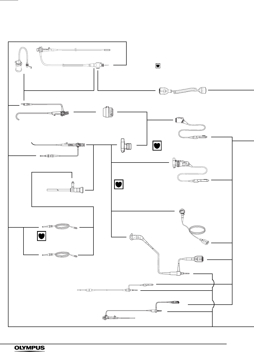
270
Appendix
EVIS EXERA II VIDEO SYSTEM CENTER CV-180
System chart for non-ultrasonic endoscopes
The OLYMPUS EVIS EXERA II video system can be
configured for cardiac applications only when TYPE CF
applied part (camera heads and light guide cables)
bearing the symbol are used.
Videoscope
(EVIS 100/130/140/160 /1801series)
Water container
(see page 274)
Fiberscope Video adapter
(A10-T1/T2)
Camera head
(OTV-S7H-1D-F08E/L08E
/D-L08E2,
OTV-S7ProH-HD-L08E12)
Rigidscope
Light guide cable
Videoscope cable EXERA II
(MAJ-1430)
Video adapter
Fiberscope
Light guide cable
Rigid videoscope
(A500A series, WA50A/L1 series)
Flexible videoscope
(ENF-V/V21/VQ1, CYF-V/VA/V21/VA21,
HYF-V, LTF-V3/VP/VH1, LF-V)
Camera head
(OTV-S7H-N/1N/1D/NA/1NA,
OTV-S7H-D-6M)
Camera head
(OTV-S7H-NA-10E/12E/10Q/12Q,
OTV-S7H-1NA-10E/12E/12Q,
OTV-S7H-FA-E/Q, OTV-S7H-1FA-E,
OTV-SP1H-NA-12E/12Q, OTV-SP1H-N-12E/12Q,
OTV-S7ProH-HD-10E/10Q/12E/12Q)
Camera head
(OTV-S7H-VA)
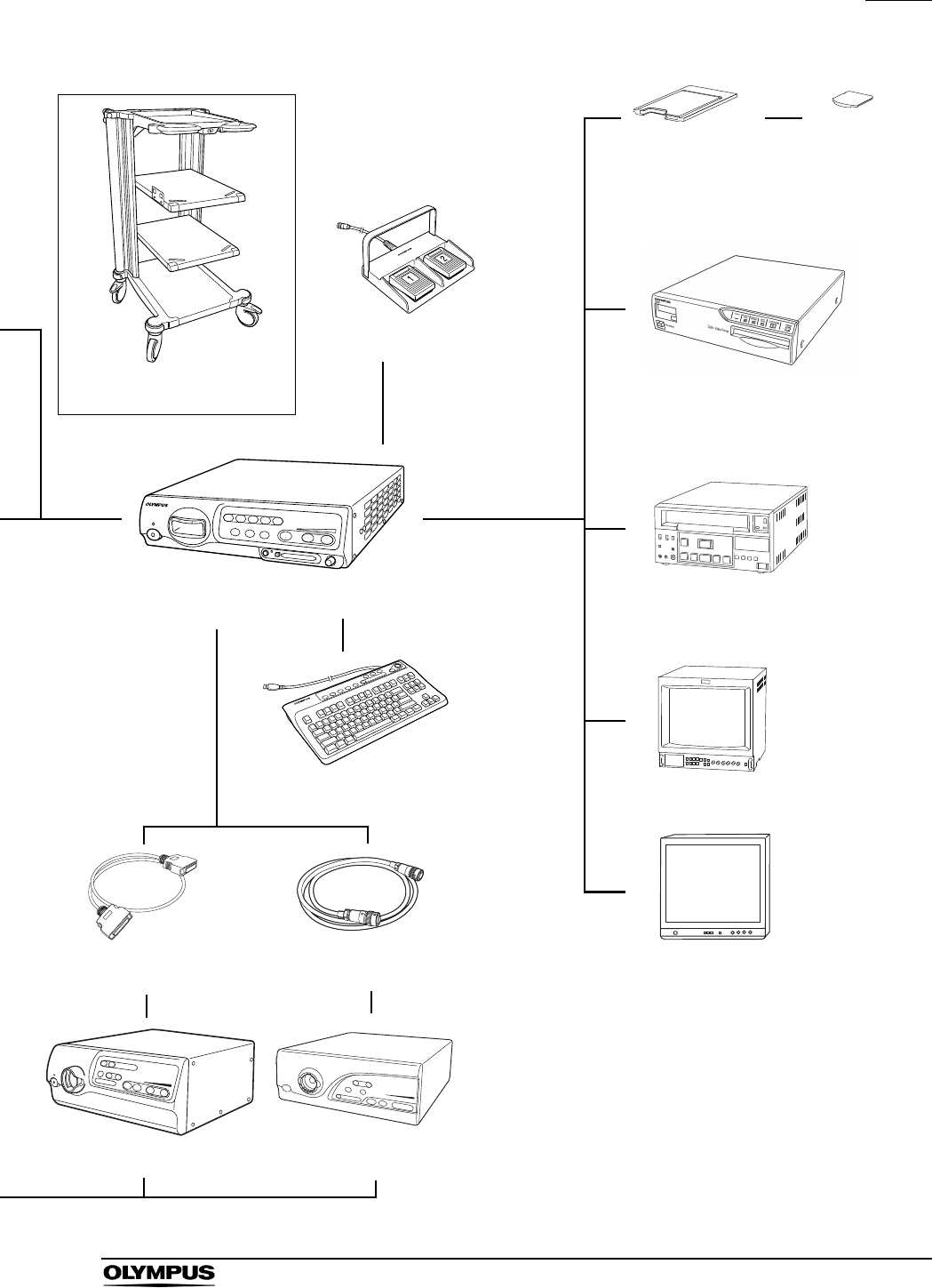
Appendix
271
EVIS EXERA II VIDEO SYSTEM CENTER CV-180
Mobile workstation
(WM-NP1, WM-WP1)
Foot switch
(MAJ-1391)
Color video printer
(OEP, OEP-3, OEP-4, UP-1800, UP-1850,
UP-2900MD, UP-2950MD, UP-5200MD,
UP-5250MD, UP-21MD)
VCR
(SVO-9500MD, DSR-20MD, DVO-1000MD,
PDW-70MD, PDW-75MD)
EVIS EXERA II
Video system center (CV-180)
Keyboard
(MAJ-1428)
Light source cable
(MAJ-1411)
Light control cable
(see page 274)
Color video monitor
(OEV203, OEV143)
Xenon light source
(CLV-160/U40/S40)
Xenon light source
(CLV-180)
High definition LCD monitor
(OEV191H)
High definition monitor
(OEV181H)
LCD monitor
(OEV191)
PC card adapter
(MAPC-10)
xD picture card
(M-XD32P, M-XD64P, M-
XD128P, M-XD256P, M-
XD512P, M-XD1GM, M-
XD1GMA, M-XD2GMA)
1 The followings are compatible with NBI:
GIF-H180, CF-H180AL/I, GIF-Q180, CF-Q180AL/I,
PCF-Q180AL/I, GIF-N180, BF-Q180, BF-P180,
BF-1T180, ENF-V2/VQ, CYF-V2, CYF-VA2,
OTV-S7ProH-HD-L08E, LTF-VH, WA5001A/L series
2 This product is not available in some areas.
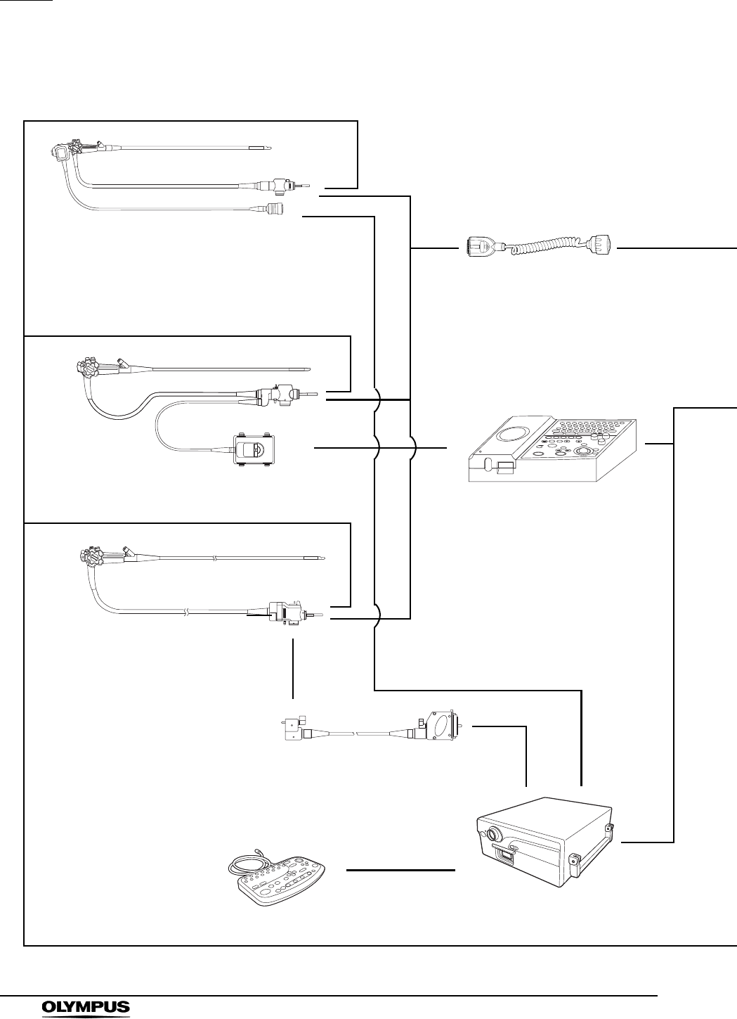
272
Appendix
EVIS EXERA II VIDEO SYSTEM CENTER CV-180
System chart for ultrasonic endoscopes
Endoscopic ultrasound center
(EU-M60, EU-M30, EU-MA)
Ultrasonic endoscope
Videoscope cable EXERA II
(MAJ-1430)
Keyboard
Ultrasonic cable A (MAJ-953)
Ultrasonic cable B (MAJ-954)
Ultrasonic gastrovideoscope
(GF-UM160)
Compact endoscopic
ultrasound center
(EU-C60)
Ultrasonic endoscope
(EU-M60 only)
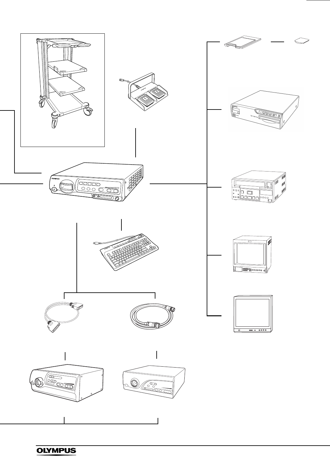
Appendix
273
EVIS EXERA II VIDEO SYSTEM CENTER CV-180
Foot switch
(MAJ-1391)
EVIS EXERA II
Video system center (CV-180)
Keyboard
(MAJ-1428)
Light source cable
(MAJ-1411)
Light control cable
(see page 274)
Xenon light source
(CLV-160, CLV-U40)
Xenon light source
(CLV-180)
Color video printer
(OEP, OEP-3, OEP-4, UP-1800, UP-1850,
UP-2900MD, UP-2950MD, UP-5200MD,
UP-5250MD, UP-21MD)
VCR
(SVO-9500MD, DSR-20MD,
DVO-1000MD)
Color video monitor
(OEV203, OEV143)
High definition LCD monitor
(OEV191H)
High definition monitor
(OEV181H)
LCD monitor
(OEV191)
PC card adapter
(MAPC-10)
xD picture card
(M-XD32P, M-XD64P, M-
XD128P, M-XD256P, M-
XD512P, M-XD1GM, M-
XD1GMA, M-XD2GMA)
Mobile workstation
(WM-NP1, WM-WP1)
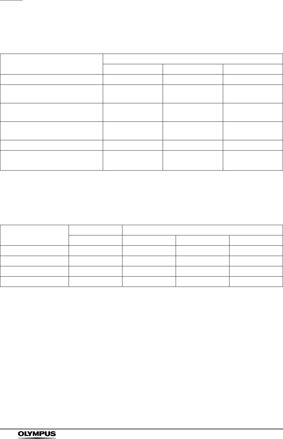
274
Appendix
EVIS EXERA II VIDEO SYSTEM CENTER CV-180
Water container
BF endoscopes do not use the water container.
Connection cable (light source)
Endoscope
Water container
MD-431 MH-884 MAJ-901
EVIS EXERA II 180 series –
EVIS EXERA 160 series,
Ultrasonic videoscope 160 series –
EVIS 140 series,
Ultrasonic videoscope 140 series –
EVIS 100 and 130 series,
Ultrasonic videoscope 130 series ––
OES 40 series –
OES 10, 20, 30, E and E3 series1,
Ultrasonic fiberscope ––
compatible, – incompatible
1 The OES E and E3 series are not available in some areas.
Light source
Light source cable Light control cables
MAJ-1411 MH-966 MAJ-1567 MH-993
CLV-180 –––
CLV-160 – ––
CLV-U40 –
CLV-S40 – – –
compatible, incompatible
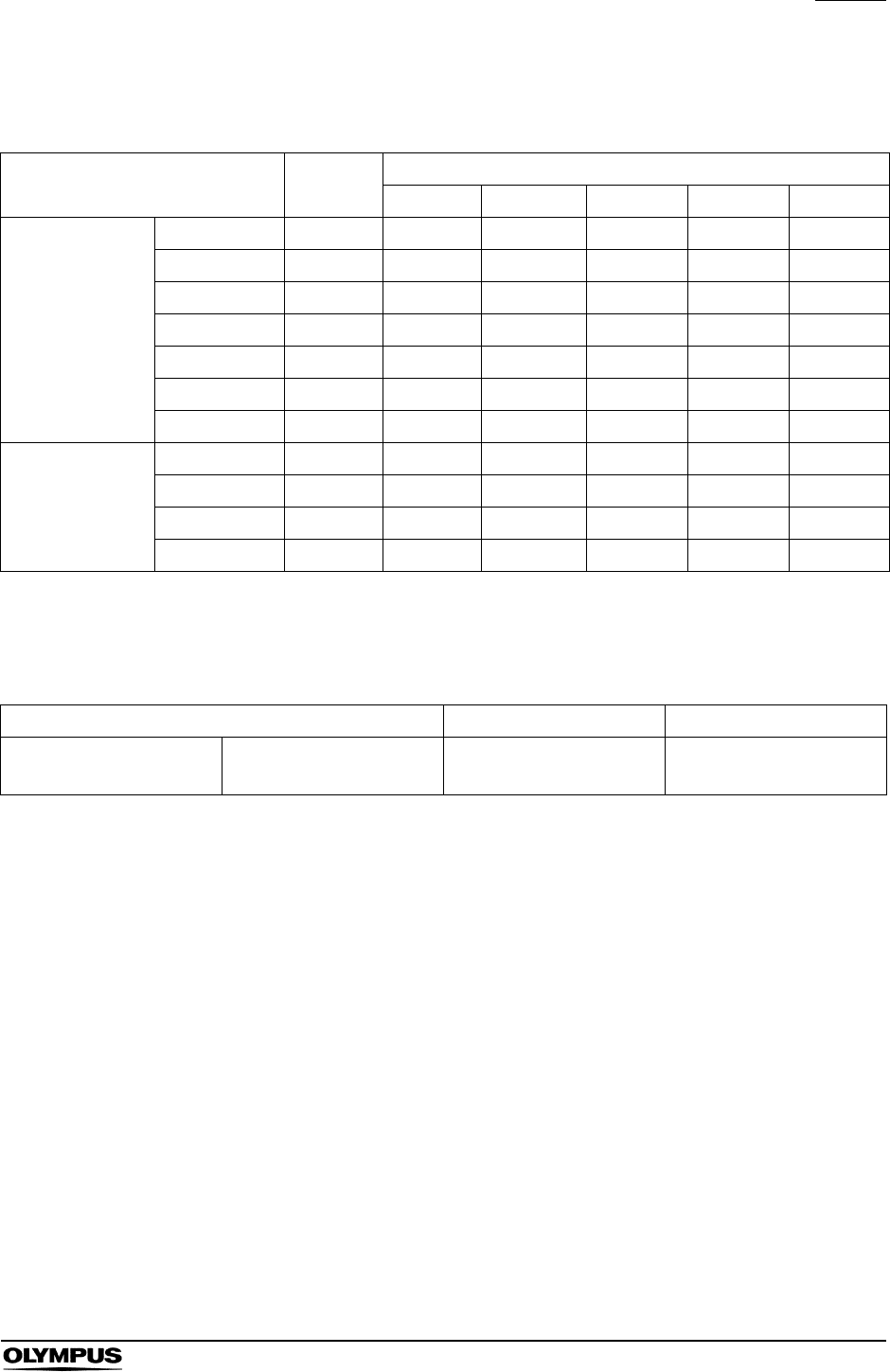
Appendix
275
EVIS EXERA II VIDEO SYSTEM CENTER CV-180
Connection cable (OEV143, OEV203, OEV191, OEV181H,
OEV191H)
Connection cables (color video printer)
Length
Monitor
OEV143 OEV203 OEV191 OEV181H OEV191H
Monitor cable
MAJ-921 1.5 m
MAJ-970 4 m
MAJ-1462 7 m
MAJ-1584 15 m
MAJ-1586 2 m
MAJ-846 7 m ––
MAJ-971 15 m ––
Monitor remote
cable
MAJ-227 4 m –––
MAJ-1161 7 m – –
MAJ-1230 4 m – –
MAJ-1465 15 m – –
compatible, – incompatible
Video cable (print) Video cable (display) Remote cable
MH-984 (3 m) MD-445 (7 m)
with MAJ-1165 MB-677 (3 m) MH-995 (2.8 m)

276
Appendix
EVIS EXERA II VIDEO SYSTEM CENTER CV-180
Transportation, storage, and operation environment
Specifications
Transportation
and storage
environment
Ambient temperature 25 to +70C (13 to +158F)
Relative humidity 10 - 90%
Atmospheric pressure 700 - 1060 hPa
Operation
environment
Ambient temperature 10 - 40C (50 - 104F)
Relative humidity 30 - 85% (without condensation)
Atmospheric pressure 700 - 1060 hPa
(0.7 - 1.1 kgf/cm2)
(10.2 - 15.4 psia)
Item Specifications
Power supply Voltage 100 - 240 V AC
Voltage fluctuation Within 10%
Frequency 50/60 Hz
Frequency
fluctuation Within 1Hz
Consumption
electric power 150 VA
Fuse rating 5 A, 250 V
Fuse size ø5 20 mm
Size Dimensions 382 (W) 91 (H) 490 (D) mm
(maximum)
Weight 10 kg
Classification
(medical electrical
equipment)
Type of protection
against electric
shock
Class I
Degree of
protection against
electric shock of
applied part
Depend on applied part
See also applied part (camera head
or videoscope).
Degree of
protection against
explosion
This instrument should be kept
away from flammable gases.

Appendix
277
EVIS EXERA II VIDEO SYSTEM CENTER CV-180
Observation HDTV signal output Either RGB or YPbPr output can be
selected.
SDTV signal output VBS composite (NTSC), Y/C and
RGB; simultaneous outputs
possible.
White balance
adjustment
White balance adjustment is
possible using the white balance
button on the front panel.
Standard color chart
output
A color bar chart can be displayed
by pressing the “Shift” + “F7” keys
on the keyboard.
Color tone
adjustment
The following color tone
adjustments are possible using the
“COLOR” key and arrow keys on the
keyboard.
• Red adjustment: 8 steps
• Blue adjustment: 8 steps
• Chroma adjustment: 8 steps
Automatic gain
control (AGC)
The image can be electrically
amplified when the light is
inadequate due to the distal end of
the endoscope being too far from
the object.
Contrast The image contrast can be set to
one of the following three modes (N,
H, L) using the “Shift”+ “F6” key on
the keyboard.
• N (normal): Normal image
• H (high): The dark areas are
darker and the bright areas are
brighter than in the normal image.
• L (low): The dark areas are
brighter and bright areas are
darker than in the normal image.
Item Specifications

278
Appendix
EVIS EXERA II VIDEO SYSTEM CENTER CV-180
Observation
(continued)
Iris The auto iris modes can be selected
using the “Iris mode” button on the
front panel.
• Peak: For use when observing by
focusing on a small bright area.
• Auto: For use when observing by
focusing on the image center.
Image enhancement
setting
Fine patterns or edges in the
endoscopic images can be
enhanced electrically to increase
the image sharpness.
Either the structural enhancement
or edge enhancement can be
selected according to the user
setup.
Switching the
enhancement
modes
The enhancement level can be
selected from 4 levels (OFF, 1, 2
and 3) using the image
enhancement button on the front
panel.
Image size selection The size of the endoscopic image
can be changed using the “Shift” +
“F8” key on the keyboard.
Reset to defaults The following settings can be reset
to their defaults using the rest button
on the front panel.
• User preset
• Image source
• Color tone
• Freeze
• Release index
•Zoom
• Special light observation
• Arrow pointer
•Stop watch
• Characters on screen
• Exposure
•PinP
Freeze An endoscopic image is frozen
using an endoscope or the
“FREEZE” key on the keyboard.
Item Specifications

Appendix
279
EVIS EXERA II VIDEO SYSTEM CENTER CV-180
Documentation Remote control The following ancillary equipment
can be controlled from the front
panel, keyboard or endoscope's
remote switches (specified models
only).
•VCR
• Video printer
• Image filing system
• Endoscopic ultrasound center,
etc.
Patient data The following data can be displayed
on the monitor using the keyboard.
• Patient ID No.
• Patient name
• Sex and age
• Date of birth
• Date of recording (time,
stopwatch)
• Image frame No.
• Videotape recorder mode
• Display image setting
• Physician name
• Comments
Advance
registration of
patient data
The following data of up to 40
patients can be entered prior to
surgery using the keyboard.
• Patient ID No.
• Patient name
• Sex and age
• Date of birth
• Physician
Item Specifications

280
Appendix
EVIS EXERA II VIDEO SYSTEM CENTER CV-180
PC card Media xD-Picture Card (2/1 GB,
512/256/128/64/32 MB), specified
by Olympus.
MAPC-10 can be used as PC card
adapter.
Recording format TIFF: no compression
SHQ: approx. 1/5
HQ: approx. 1/7
SQ: approx. 1/10
Number of
Recording images
in 32 MB, SDTV / HDTV
TIFF: approx. 30 / 6 images
SHQ: approx. 310 / 110 images
HQ: approx. 2000 / 760 images
SQ: approx. 2570 / 970 images
Image storage and
retrieval
Monitor output Using the monitor out switches on
the front panel, it is possible to
select an image from the endoscope
or ancillary equipment for display on
the monitor.
Memory backup Memorization of
selected setting
The following settings are held in
memory even after the video system
center is turned OFF.
• White balance
• Iris mode
• Enhancement
• Image size
• Color tone
Lithium battery Life: 5 years
Item Specifications

Appendix
281
EVIS EXERA II VIDEO SYSTEM CENTER CV-180
EMC Applied standards;
IEC 60601-1-2: 2001
This instrument complies with the
standards listed in the left column.
CISPR 11 of emission:
Group 1, Class B
This instrument complies with the EMC
standard for medical electrical
equipment; edition 2 (IEC 60601-1-2:
2001). However, when connecting to an
instrument that complies with the EMC
standard for medical electrical
equipment; edition 1 (IEC 60601-1-2:
1993), the whole system complies with
edition 1.
Year of
manufacture The last digit of the year of manufacture
is the second digit of the serial number.
7612345
282
Appendix
EVIS EXERA II VIDEO SYSTEM CENTER CV-180
EMC information
This model is intended for use in the electromagnetic environments specified
below. The user and the medical staff should ensure that it is used only in these
environments.
Magnetic emission compliance information and
recommended electromagnetic environments
Emission standard Compliance Guidance
RF emissions
CISPR 11
Group 1 This instrument uses RF (Radio Frequency) energy only for its
internal function. Therefore, its RF emissions are very low and are not
likely to cause any interference in nearby electronic equipment.
RF emissions
CISPR 11
Class B This instrument's RF emissions are very low and are not likely to
cause any interference in nearby electronic equipment.
Main terminal
conducted emissions
CISPR 11
Harmonic emissions
IEC 61000-3-2
Class A This instrument's harmonic emissions are low and are not likely to
cause any problem in the typical commercial power supply connected
to this instrument.
Voltage
fluctuations/flicker
emissions
IEC 61000-3-3
Complies This instrument stabilizes own radio variability and has no affect such
as flicker of a lighting apparatus.
Appendix
283
EVIS EXERA II VIDEO SYSTEM CENTER CV-180
Electromagnetic immunity compliance information and
recommended electromagnetic environments
UT is the a.c. mains power supply prior to application of the
test level.
Immunity test IEC 60601-1-2
test level Compliance level Guidance
Electrostatic
discharge (ESD)
IEC 61000-4-2
Contact:
±2, ±4, ±6kV
Air:
±2, ±4, ±8kV
Same as left Floors should by be made of wood, concrete,
or ceramic tile that hardly produces static. If
floors are covered with synthetic material that
tends to produce static, the relative humidity
should be at least 30%.
Electrical fast
transient/burst
IEC 61000-4-4
±2kV
for power supply lines
±1kV
for input/output lines
Same as left Mains power quality should be that of a typical
commercial (original condition feeding the
facilities) or hospital environment.
Surge
IEC 61000-4-5
Differential mode:
± 0.5, ± 1 kV
Common mode:
±0.5, ±1, ±2kV
Same as left Mains power quality should be that of a typical
commercial or hospital environment.
Voltage dips,
short interruptions
and voltage
variations on
power supply
input lines
IEC 61000-4-11
< 5% UT
(>95% dip in UT)
for 0.5 cycle
Same as left Mains power quality should be that of a typical
commercial or hospital environment. If the
user of this instrument required continued
operation during power mains interruptions, it
is recommended that this instrument be
powered from an uninterruptible power supply
or a battery.
40% UT
(60% dip in UT)
for 5 cycles
70% UT
(30% dip in UT)
for 25 cycles
< 5% UT
(>95% dip in UT)
for 5 seconds
Power frequency
(50/60 Hz)
magnetic field
IEC 61000-4-8
3 A/m Same as left It is recommended to use this instrument by
maintaining enough distance from any
equipment that operates with high current.
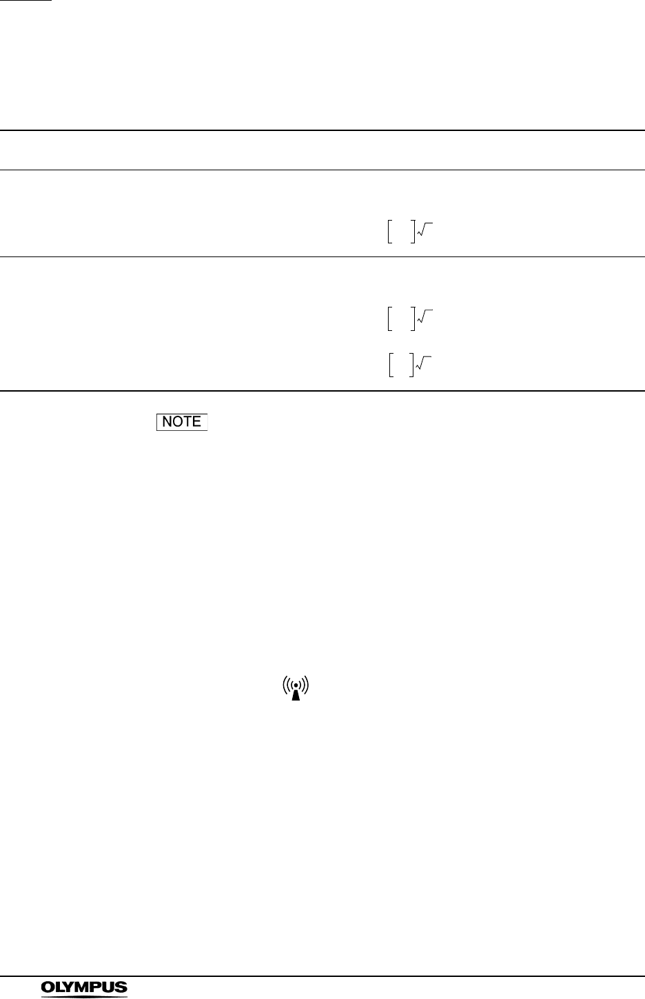
284
Appendix
EVIS EXERA II VIDEO SYSTEM CENTER CV-180
Cautions and recommended electromagnetic environment
regarding portable and mobile RF communications
equipment such as a cellular phones
• Where “P” is the maximum output power rating of the
transmitter in watts (W) according to the transmitter
manufacturer and “d” is the recommended separation
distance in meters (m).
• This instrument complies with the requirements of
IEC 60601-1-2: 2001. However, under electromagnetic
environment that exceeds its noise level, electromagnetic
interference may occur on this instrument.
• Electromagnetic interference may occur on this instrument
near a high-frequency electrosurgical equipment and/or other
equipment marked with the following symbol:
Immunity test IEC 60601-1-2
test level
Compliance
level Guidance
Conducted RF
IEC 61000-4-6
3V
rms
(150 kHz - 80 MHz)
3V (V
1) Formula for recommended separation distance
(V1=3 according to the compliance level)
Radiated RF
IEC 61000-4-3
3V/m
(80 MHz - 2.5 GHz)
3V/m (E
1) Formula for recommended separation distance
(E1=3 according to the compliance level)
80 MHz to 800 MHz
800 MHz to 2.5 GHz
d3.5
V1
--------P=
d3.5
E1
--------P=
d7
E1
------ P=
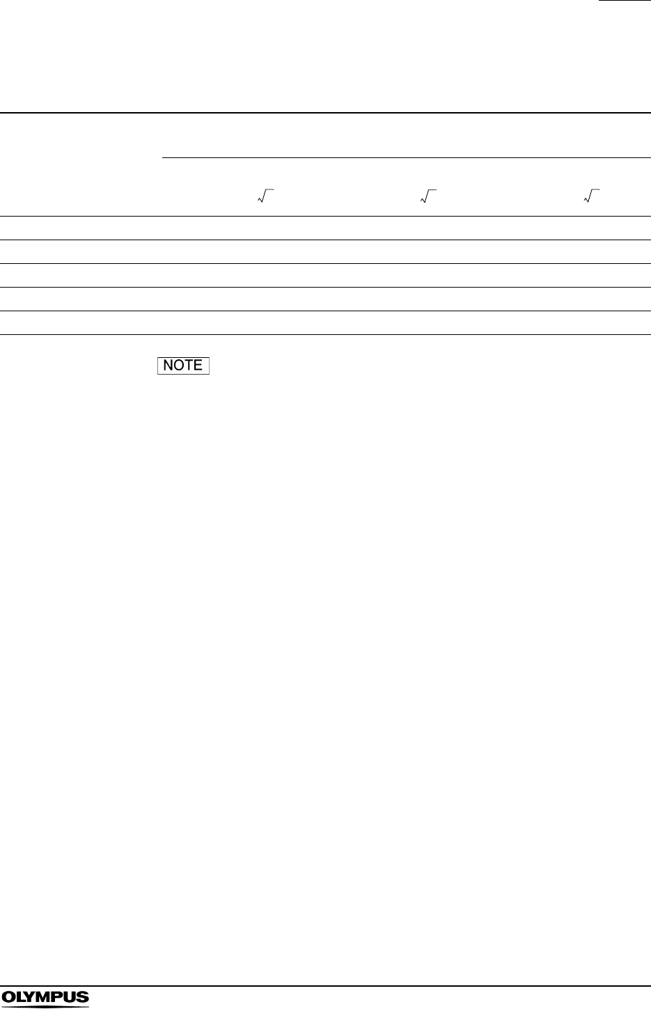
Appendix
285
EVIS EXERA II VIDEO SYSTEM CENTER CV-180
Recommended separation distance between portable and
mobile RF communications equipment and this instrument
The guidance may not apply in some situations.
Electromagnetic propagation is affected by absorption and
reflection from structures, objects and people.
Portable and mobile RF communications equipment such as
cellular phones should be used no closer to any part of this
instrument, including cables than the recommended
separation distance calculated from the equation applicable
to the frequency of the transmitter.
Rated maximum output
power of transmitter
P (W)
Separation distance according to frequency of transmitter (m)
(calculated as V1=3 and E1=3)
150 kHz - 80 MHz 80 MHz - 800 MHz 800 MHz - 2.5 GHz
0.01 0.12 0.12 0.23
0.1 0.38 0.38 0.73
11.2 1.2 2.3
10 3.8 3.8 7.3
100 12 12 23
d1.2P=
d1.2P=
d2.3P=

©2006 OLYMPUS MEDICAL SYSTEMS CORP. All rights reserved.
No part of this publication may be reproduced or distributed without the
express written permission of OLYMPUS MEDICAL SYSTEMS CORP.
OLYMPUS is a registered trademark of OLYMPUS CORPORATION.
Microsoft, Windows, Windows NT, PowerPoint, and MS-DOS are
either registered trademarks or trademarks of Microsoft Corporation in
the United States and/or other countries.
Other trademarks, product names, logos, or trade names used in this
document are generally registered trademarks or trademarks of each
company.

Printed in Japan 20100906 *0000
GT1245 16
Manufactured by
2951 Ishikawa-cho, Hachioji-shi, Tokyo 192-8507, Japan
Fax: (042)646-2429 Telephone: (042)642-2111
(Premises/Goods delivery) Wendenstrasse 14-18, 20097 Hamburg, Germany
(Letters) Postfach 10 49 08, 20034 Hamburg, Germany
3500 Corporate Parkway, P.O. Box 610, Center Valley, PA
18034-0610, U.S.A.
Fax: (484)896-7128 Telephone: (484)896-5000
KeyMed House, Stock Road, Southend-on-Sea, Essex SS2 5QH, United Kingdom
Fax: (01702)465677 Telephone: (01702)616333
491B, River Valley Road #12-01/04, Valley Point Office Tower, Singapore 248373
Fax: 6834-2438 Telephone: 6834-0010
A8F, Ping An International Financial Center, No. 1-3, Xinyuan South Road,
Chaoyang District, Beijing, 100027 P.R.C.
Fax: (86)10-5976-1299 Telephone: (86)10-5819-9000
117071, Moscow, Malaya Kaluzhskaya 19, bld. 1, fl.2, Russia
Fax: (095)958-2277 Telephone: (095)958-2245
31 Gilby Road, Mount Waverley, VIC., 3149, Australia
Fax: (03)9543-1350 Telephone: (03)9265-5400
5301 Blue Lagoon Drive, Suite 290 Miami, FL 33126-2097, U.S.A.
Fax: (305)261-4421 Telephone: (305)266-2332
Distributed by
Olympus-Tower, 114-9 Samseong-Dong, Gangnam-Gu, Seoul 135-090 Korea
Fax: (02)6255-3494 Telephone: (02)6255-3210
Fax: (040)23773-4656 Telephone: (040)23773-0

