**3539 00 BRAIN FACTS.LYT 3539 Brainfacts
User Manual: 3539
Open the PDF directly: View PDF ![]() .
.
Page Count: 64

Brain Facts
A PRIMER ON THE BRAIN AND NERVOUS SYSTEM
SOCIETY FOR NEUROSCIENCE

SOCIETY FOR NEUROSCIENCE
The Society for Neuroscience is the world’s largest organization of scientists
and physicians dedicated to understanding the brain, spinal cord, and periph-
eral nervous system.
Neuroscientists investigate the molecular and cellular levels of the nervous
system; the neuronal systems responsible for sensory and motor function; and
the basis of higher order processes, such as cognition and emotion. This
research provides the basis for understanding the medical fields that are con-
cerned with treating nervous system disorders. These medical specialties
include neurology, neurosurgery, psychiatry, and ophthalmology.
Founded in 1969, the Society has grown from 500 charter members to
more than 36,000 members. While a predominantly North American organi-
zation, SfN also has many members who live in Europe, Asia, Latin America,
and Australia/Oceania. The Society has more than 100 regional chapters. With
activities ranging from lectures to networking events and information sharing,
SfN chapters enable individual members to engage their colleagues at the local
level.
The mission of the Society is to:
∫Advance the understanding of the brain and the nervous system by bring-
ing together scientists of diverse backgrounds, by facilitating the integration
of research directed at all levels of biological organization, and by encourag-
ing translational research and the application of new scientific knowledge to
develop improved disease treatments and cures.
∫Provide professional development activities, information, and educational
resources for neuroscientists at all stages of their careers, including under-
graduates, graduates, and postdoctoral fellows, and increase participation of
scientists from a diversity of cultural and ethnic backgrounds.
∫Promote public information and general education about the nature of
scientific discovery and the results and implications of the latest neuroscience
research. Support active and continuing discussions on ethical issues relating
to the conduct and outcomes of neuroscience research.
∫Inform legislators and other policymakers about new scientific knowledge
and recent developments in neuroscience research and their implications for
public policy, societal benefit, and continued scientific progress.
The exchange of scientific information occurs at an annual fall meeting
where more than 16,000 reports of new scientific findings are presented and
more than 30,000 people attend. This meeting, the largest of its kind in the
world, is the arena for the presentation of new results in neuroscience.
The Society’s weekly journal, The Journal of Neuroscience,contains articles
spanning the entire range of neuroscience research and has subscribers world-
wide. The Society’s ongoing education and professional development e∑orts
reach teachers and help promote the education of Society members. Print and
electronic publications inform members about Society activities.
A major goal of the Society is to inform the public about the progress and
benefits of neuroscience research. The Society accomplishes this goal by pro-
viding information about neuroscience to schoolteachers and encouraging its
members to speak to young people about the human brain and nervous system.

Contents
INTRODUCTION . . . . . . . . . . . . . . . . . . . . . . . . . . . . . . . . . . . . . . . . . . . . . . . . . . . . . . . . 4
THE NEURON . . . . . . . . . . . . . . . . . . . . . . . . . . . . . . . . . . . . . . . . . . . . . . . . . . . . . . . . 6
Neurotransmitters ∫Second Messengers
BRAIN DEVELOPMENT . . . . . . . . . . . . . . . . . . . . . . . . . . . . . . . . . . . . . . . . . . . . . . . . 10
Birth of Neurons and Brain Wiring ∫Paring Back ∫Critical Periods
SENSATION AND PERCEPTION . . . . . . . . . . . . . . . . . . . . . . . . . . . . . . . . . . . . . . . . 14
Vision ∫Hearing ∫Taste and Smell ∫Touch and Pain
LEARNING, MEMORY, AND LANGUAGE . . . . . . . . . . . . . . . . . . . . . . . . . . . . . . . . 20
MOVEMENT . . . . . . . . . . . . . . . . . . . . . . . . . . . . . . . . . . . . . . . . . . . . . . . . . . . . . . . . . . 22
SLEEP . . . . . . . . . . . . . . . . . . . . . . . . . . . . . . . . . . . . . . . . . . . . . . . . . . . . . . . . . . . . . . . 25
The Stu∑ of Sleep ∫Sleep Disorders ∫How is Sleep Regulated?
STRESS . . . . . . . . . . . . . . . . . . . . . . . . . . . . . . . . . . . . . . . . . . . . . . . . . . . . . . . . . . . . . . 28
The Immediate Response ∫Chronic Stress
AGING . . . . . . . . . . . . . . . . . . . . . . . . . . . . . . . . . . . . . . . . . . . . . . . . . . . . . . . . . . . . . . 31
Aging Neurons ∫Intellectual Capacity
ADVANCES . . . . . . . . . . . . . . . . . . . . . . . . . . . . . . . . . . . . . . . . . . . . . . . . . . . . . . . . . . 33
Bipolar Disorder ∫Epilepsy ∫Major Depression
Pain ∫Parkinson’s Disease
CHALLENGES . . . . . . . . . . . . . . . . . . . . . . . . . . . . . . . . . . . . . . . . . . . . . . . . . . . . . . . . 36
Addiction ∫Attention Deficit Hyperactivity Disorder
Alzheimer’s Disease ∫Amyotrophic Lateral Sclerosis ∫Anxiety Disorders
Autism ∫Brain Tumors ∫Down Syndrome ∫Huntington’s Disease
Learning Disorders ∫Multiple Sclerosis ∫Neurological AIDS
Neurological Trauma ∫Schizophrenia ∫Stroke ∫Tourette Syndrome
NEW DIAGNOSTIC METHODS . . . . . . . . . . . . . . . . . . . . . . . . . . . . . . . . . . . . . . . . . 49
Imaging Techniques ∫Gene Diagnosis
POTENTIAL THERAPIES . . . . . . . . . . . . . . . . . . . . . . . . . . . . . . . . . . . . . . . . . . . . . . . 52
New Drugs ∫Trophic Factors ∫Engineered Antibodies
Small Molecules and RNAs ∫Cell and Gene Therapy
NEUROETHICS . . . . . . . . . . . . . . . . . . . . . . . . . . . . . . . . . . . . . . . . . . . . . . . . . . . . . . . 55
GLOSSARY . . . . . . . . . . . . . . . . . . . . . . . . . . . . . . . . . . . . . . . . . . . . . . . . . . . . . . . . . . 57
INDEX . . . . . . . . . . . . . . . . . . . . . . . . . . . . . . . . . . . . . . . . . . . . . . . . . . . . . . . . . . . . . . . 61
NEUROSCIENCE RESOURCES . . . . . . . . . . . . . . . . . . . . . . . . . . . . . . . . . . . . . . . . . . 63

4
t sets humans apart from all other species by allowing us to
achieve the wonders of walking on the moon and compos-
ing masterpieces of literature, art, and music. The human
brain—a spongy, three-pound mass of fatty tissue—has
been compared to a telephone switchboard and a super-
computer.
But the brain is much more complicated than either of these
devices, a fact scientists confirm almost daily, with each new dis-
covery.The extent of the brain’s capabilities is unknown, but it is
the most complex living structure known in the universe.
This single organ controls all body activities, ranging from
heart rate and sexual function to emotion, learning, and mem-
ory. The brain is even thought to influence the immune system’s
response to disease and to determine, in part, how well people
respond to medical treatments. Ultimately, it shapes our
thoughts, hopes, dreams, and imaginations. In short, the brain is
what makes us human.
Neuroscientists have the daunting task of deciphering the
mystery of this most complex of all machines: how as many as a
trillion nerve cells are produced, grow, and organize themselves
into e∑ective, functionally active systems that ordinarily remain
in working order throughout a person’s lifetime.
The motivation of researchers is twofold: to understand
human behavior better—from how we learn to why people have
trouble getting along together—and to discover ways to prevent
or cure many devastating brain disorders.
The more than 1,000 disorders of the brain and nervous sys-
tem result in more hospitalizations than any other disease group,
including heart disease and cancer. Neurological illnesses a∑ect
more than 50 million Americans annually, at costs exceeding $400
billion. In addition, mental disorders, excluding drug and alco-
hol problems, strike 44 million adults a year at a cost of some $148
billion.
However, during the congressionally designated Decade of
the Brain, which ended in 2000, neuroscience made significant
discoveries in these areas:
∫Genetics. Disease genes were identified that are key to several
neurodegenerative disorders—including Alzheimer’s disease,
Huntington’s disease, Parkinson’s disease, and amyotrophic lat-
eral sclerosis. This has provided new insights into underlying dis-
ease mechanisms and is beginning to suggest new treatments.
With the mapping of the human genome, neuroscientists will
be able to make more rapid progress in identifying genes that
either contribute to human neurological disease or that directly
cause disease. Mapping animal genomes will aid the search for
genes that regulate and control many complex behaviors.
∫Brain Plasticity. Scientists began to uncover the molecular
basis of neural plasticity, revealing how learning and memory
occur and how declines might be reversed. These discoveries are
leading to new approaches to the treatment of chronic pain.
∫New Drugs. Researchers gained new insights into the mech-
anisms of molecular neuropharmacology, which provides a new
understanding of the mechanisms of addiction. These advances
also have led to new treatments for depression and obsessive
compulsive disorder.
∫Imaging.Revolutionary imaging techniques, including mag-
netic resonance imaging and positron emission tomography, now
reveal brain systems underlying attention, memory, and emotions
and indicate dynamic changes that occur in schizophrenia.
∫Cell Death. The discovery of how and why neurons die, as
well as the discovery of stem cells, which divide and form new
neurons, has many clinical applications. This has dramatically
improved the outlook for reversing the e∑ects of injury in both
the brain and the spinal cord. The first e∑ective treatments for
stroke and spinal cord injury based on these advances have been
brought to clinical practice.
∫Brain Development. New principles and newly discovered
molecules responsible for guiding nervous system development
now give scientists a better understanding of certain disorders of
childhood. Together with the discovery of stem cells, these
advances are pointing to novel strategies for helping the brain or
spinal cord regain functions lost as a result of injury or develop-
mental dysfunction.
Federal neuroscience research funding of more than $5 bil-
lion annually and private support should vastly expand our
knowledge of the brain in the years ahead.
This book only provides a glimpse of what is known about
the nervous system, the disorders of the brain, and some of the
exciting avenues of research that promise new therapies for many
neurological diseases.
Introduction
I
5
THE TOLL OF SELECTED BRAIN AND NERVOUS SYSTEM DISORDERS*
Condition Total Cases Costs Per Year
Hearing Loss 28 million $ 56 billion
All Depressive Disorders 20.5 million 44 billion
Alzheimer’s Disease 4.5 million 100 billion
Huntington’s Disease 30,000 2 billion
Stroke 4.7 million 51 billion
Schizophrenia 2 million 32.5 billion
Parkinson’s Disease 1 million 5.6 billion
Traumatic Head Injury 5 million 56.3 billion
Multiple Sclerosis 2.5 million 9.5 billion
Spinal Cord Injury 250,000 10 billion
* Estimates provided by the National Institutes of Health and voluntary organizations.
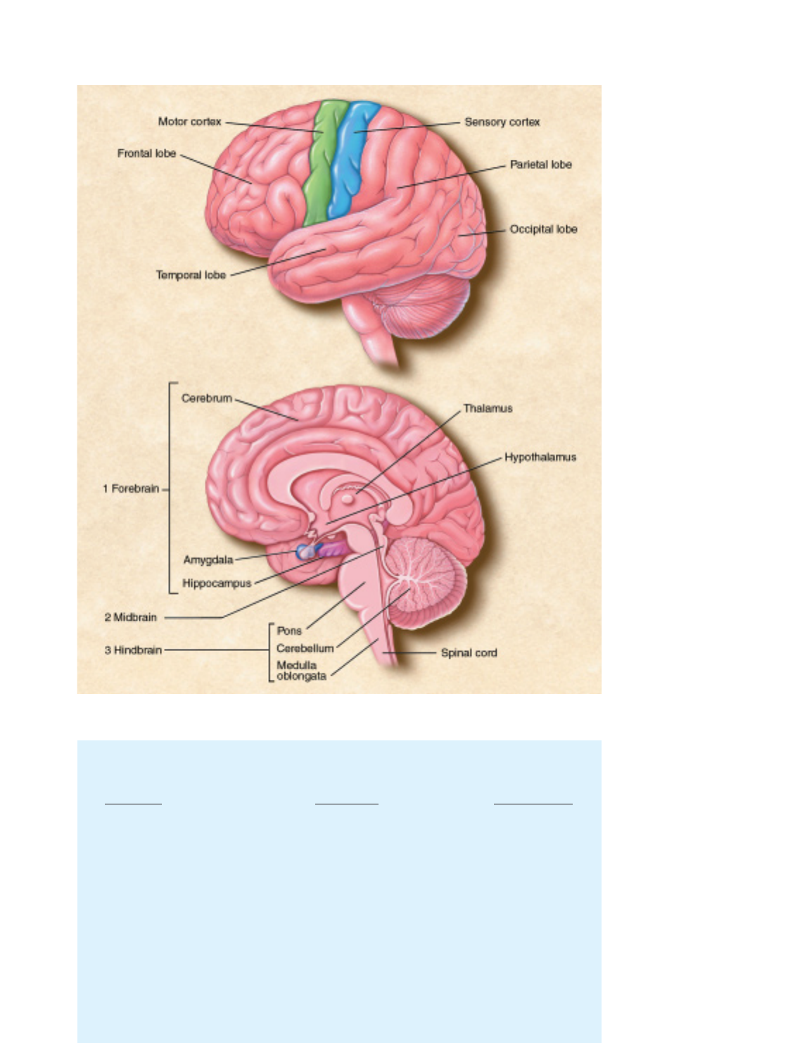
THE BRAIN. Cerebral cortex
(above). This part of the brain is
divided into four sections: the
occipital lobe, the temporal
lobe, the parietal lobe, and the
frontal lobe. Functions, such as
vision, hearing, and speech, are
distributed in selected regions.
Some regions are associated
with more than one function.
Major internal structures (below).
The (1) forebrain is credited with
the highest intellectual func-
tions—thinking, planning, and
problem-solving. The hippocam-
pus is involved in memory.
The thalamus serves as a relay
station for almost all of the
information coming into the
brain. Neurons in the hypothal-
amus serve as relay stations for
internal regulatory systems by
monitoring information coming
in from the autonomic nervous
system and commanding the
body through those nerves and
the pituitary gland. On the
upper surface of the (2) midbrain
are two pairs of small hills, col-
liculi, collections of cells that
relay specific sensory informa-
tion from sense organs to the
brain. The (3) hindbrain consists
of the pons and medulla
oblongata, which help control
respiration and heart rhythms,
and the cerebellum, which helps
control movement as well as
cognitive processes that require
precise timing.

6
Aspecialized cell designed to transmit informa-
tion to other nerve cells, muscle, or gland cells,
the neuron is the basic working unit of the
brain. The brain is what it is because of the
structural and functional properties of inter-
connected neurons. It contains between one
billion and one trillion neurons, depending on the species.
The neuron consists of a cell body containing the nucleus,
cytoplasm, and an electrically excitable output fiber, the axon.
Most axons also give rise to many smaller branches before ending
at nerve terminals.Synapses,from the Greek word meaning “to
clasp together,” are the contact points where one neuron commu-
nicates with another. Other structures, dendrites,Greek for “tree
branches,” extend from the neuron cell body and receive messages
from other neurons. The dendrites and cell body are covered with
synapses formed by the ends of axons of other neurons.
Neurons signal by transmitting electrical impulses along their
axons, which can range in length from a tiny fraction of an inch
to three or more feet. Many axons are covered with a layered insu-
lating myelin sheath, made of specialized cells called oligoden-
drocytes in the brain and Schwann cells in the peripheral nervous
system, which speeds the transmission of electrical signals along
the axon.
Nerve impulses involve the opening and closing of ion chan-
nels,water-filled molecular tunnels that pass through the cell
membrane and allow ions—electrically charged atoms—or
small molecules to enter or leave the cell. The flow of these ions
creates an electrical current that produces tiny voltage changes
across the membrane.
The ability of a neuron to fire—that is, to become su≈-
ciently activated by incoming synapses to discharge and com-
municate to its own synaptic target neurons—depends on a
small di∑erence in electrical charge between the inside and out-
side of the cell. When a nerve impulse begins, a dramatic rever-
sal occurs at one point on the cell’s membrane. The change, called
an action potential,then passes along the membrane of the axon
at speeds up to several hundred miles per hour. In this way, a neu-
ron may be able to fire impulses scores of times every second.
Upon reaching the end of an axon, these voltage changes trig-
ger the release of neurotransmitters,the brain’s chemical messen-
gers. Neurotransmitters are released at nerve ending terminals,
di∑use across the intrasynaptic space, and bind to receptors on
the surface of the target neuron.
These receptors act as on and o∑ switches for the next cell.
Each receptor has a distinctly shaped part that selectively recog-
nizes a particular chemical messenger. A neurotransmitter fits
into this region in much the same way as a key fits into a lock.
And when the transmitter is in place, this alters the neuron’s outer
membrane potential (or excitability) and triggers a change, such
as the contraction of a muscle or increased activity of an enzyme
in the cell.
Knowledge of neurotransmitters in the brain and the action
of drugs on these chemicals—gained largely through the study
of animals—is one of the largest fields in neuroscience. Armed
with this information, scientists hope to understand the circuits
responsible for disorders such as Alzheimer’s disease and Parkin-
son’s disease. Sorting out the various chemical circuits is vital to
understanding how the brain stores memories, why sex is such a
powerful motivation, and what the biological basis of mental ill-
ness is.
Neurotransmitters
Acetylcholine The first neurotransmitter, identified about 75
years ago, was acetylcholine (ACh). This chemical is released by
neurons connected to voluntary muscles (causing them to con-
tract) and by neurons that control the heartbeat. ACh also serves
as a transmitter in many regions of the brain.
ACh is formed at the axon terminals. When an action poten-
tial arrives at the terminal, the electrically charged calcium ion
rushes in, and ACh is released into the synapse and attaches to
ACh receptors. In voluntary muscles, this opens sodium channels
and causes the muscle to contract. ACh is then broken down and
resynthesized in the nerve terminal. Antibodies that block the
receptor for ACh cause myasthenia gravis,a disease characterized
by fatigue and muscle weakness.
Much less is known about ACh in the brain. Recent discov-
eries suggest, however, that it may be critical for normal atten-
tion, memory, and sleep. Since ACh-releasing neurons die in
Alzheimer’s patients, finding ways to restore this neurotransmit-
ter is one goal of current research.
A
The neuron
7
Amino acids Amino acids, widely distributed throughout the
body and the brain, serve as the building blocks of proteins. Cer-
tain amino acids can also serve as neurotransmitters in the brain.
The neurotransmitters glutamate and aspartate act as excita-
tory signals. Glycine and gamma-aminobutyric acid (GABA)
inhibit the firing of neurons. The activity of GABA is increased
by benzodiazepine (Valium) and by anticonvulsant drugs. In
Huntington’s disease, a hereditary disorder that begins during
midlife, the GABA-producing neurons in the brain centers coor-
dinating movement degenerate, thereby causing uncontrollable
movements.
Glutamate or aspartate activates N-methyl-d-aspartate
(NMDA) receptors, one of three major classes of glutamate
receptors, which have been implicated in activities ranging from
learning and memory to development and specification of nerve
contacts in a developing animal. The stimulation of NMDA
receptors may promote beneficial changes in the brain, whereas
overstimulation can cause nerve cell damage or cell death in
trauma and stroke.
Key questions remain about this receptor’s precise structure,
regulation, location, and function. For example, developing drugs
to block or stimulate activity at NMDA receptors holds promise
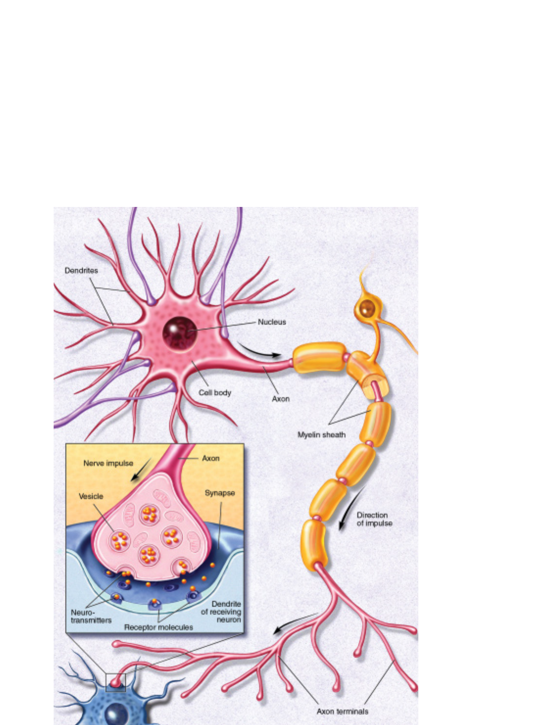
NEURON. A neuron fires by
transmitting electrical signals
along its axon. When signals
reach the end of the axon, they
trigger the release of neuro-
transmitters that are stored in
pouches called vesicles. Neuro-
transmitters bind to receptor
molecules that are present on
the surfaces of adjacent neurons.
The point of virtual contact is
known as the synapse.

8
for improving brain function and treating neurological disorders.
But this work is still in the early stage.
Catecholamines Dopamine and norepinephrine are widely
present in the brain and peripheral nervous system. Dopamine,
which is present in three circuits in the brain, controls movement,
causes psychiatric symptoms such as psychosis, and regulates
hormonal responses.
The dopamine circuit that regulates movement has been
directly linked to disease. The brains of people with Parkinson’s
disease—with symptoms of muscle tremors, rigidity, and di≈-
culty in moving—have practically no dopamine. Thus, medical
scientists found that the administration of levodopa,a substance
from which dopamine is synthesized, is an e∑ective treatment for
Parkinson’s, allowing patients to walk and perform skilled move-
ments successfully.
Another dopamine circuit is thought to be important for
cognition and emotion; abnormalities in this system have been
implicated in schizophrenia. Because drugs that block dopamine
receptors in the brain are helpful in diminishing psychotic symp-
toms, learning more about dopamine is important to under-
standing mental illness.
In a third circuit, dopamine regulates the endocrine system.
It directs the hypothalamus to manufacture hormones and hold
them in the pituitary gland for release into the bloodstream or to
trigger the release of hormones held within cells in the pituitary.
Nerve fibers containing norepinephrine are present through-
out the brain. Deficiencies in this transmitter occur in patients
with Alzheimer’s disease, Parkinson’s disease, and Korsako∑’s syn-
drome,a cognitive disorder associated with chronic alcoholism.
Thus, researchers believe norepinephrine may play a role in both
learning and memory. Norepinephrine is also secreted by the
sympathetic nervous system in the periphery to regulate heart
rate and blood pressure. Acute stress increases the release of nor-
epinephrine.
Serotonin This neurotransmitter is present in many tissues,
particularly blood platelets, the lining of the digestive tract, and
the brain. Serotonin was first thought to be involved in high
blood pressure because it is present in blood and induces a very
powerful contraction of smooth muscles. In the brain, serotonin
has been implicated in sleep, mood, depression, and anxiety.
Because serotonin controls the di∑erent switches a∑ecting vari-
ous emotional states, scientists believe these switches can be
manipulated by analogs, chemicals with molecular structures
similar to that of serotonin. Drugs that alter serotonin’s action,
such as fluoxetine (Prozac), have relieved symptoms of depres-
sion and obsessive-compulsive disorder.
Peptides These are chains of amino acids linked together.
Brain peptides called endorphins act like opium to kill pain or
cause sleepiness. (Peptides di∑er from proteins, which are much
larger and more complex combinations of amino acids.)
In 1973, scientists discovered receptors for opiates on neurons
in several regions of the brain, suggesting that the brain must
make substances very similar to opium. Shortly thereafter, scien-
tists made their first discovery of an opiate produced by the brain
that resembles morphine, an opium derivative used medically to
kill pain. They named it enkephalin,literally meaning “in the
head.” Soon after, the endorphins—another type of opioid pep-
tide, whose name comes from endogenous morphine—were dis-
covered.
The precise role of the opioid peptides in the body is unclear.
A plausible guess is that they are released by brain neurons in
times of stress to minimize pain and enhance adaptive behavior.
The presence of opioid peptides may explain, for example, why
injuries received during the stress of combat are often not noticed
until hours later.
Opioids and their receptors are closely associated with path-
ways in the brain that are activated by painful or tissue-damag-
ing stimuli. These signals are transmitted to the central nervous
system—the brain and spinal cord—by special sensory nerves,
small myelinated fibers, and tiny unmyelinated C fibers.
Scientists have discovered that some C fibers contain a pep-
tide called substance P that causes the sensation of burning pain.
The active component of chili peppers, capsaicin, causes the
release of substance P.
Trophic factors Researchers have discovered several small
proteins in the brain that are necessary for the development,
function, and survival of specific groups of neurons. These small
proteins are made in brain cells, released locally in the brain, and
bind to receptors expressed by specific neurons. Researchers have
also identified genes that code for receptors and are involved in
the signaling mechanisms of trophic factors. These findings are
expected to result in a greater understanding of how trophic fac-
tors work in the brain. This information should also prove use-
ful for the design of new therapies for brain disorders of devel-
opment and for degenerative diseases, including Alzheimer’s
disease and Parkinson’s disease.
Hormones After the nervous system, the endocrine system is
the second great communication system of the body. The pan-
creas, kidneys, heart, adrenal glands, gonads, thyroid, thymus,
and pituitary gland are sources of hormones. The endocrine sys-
tem works in large part through the pituitary gland, which
secretes hormones into the blood. Because endorphins are
released from the pituitary gland into the bloodstream, they
might also function as endocrine hormones. Hormones activate
specific receptors in target organs that release other hormones
into the blood, which then act on other tissues, the pituitary itself,
and the brain. This system is very important for the activation
and control of basic behavioral activities such as sex, emotion,
responses to stress, and the regulation of body functions such as
growth, energy use, and metabolism. Actions of hormones show
the brain to be very malleable and capable of responding to envi-
ronmental signals.
The brain contains receptors for both the thyroid hormone
and the six classes of steroid hormones—estrogens, androgens,

9
progestins, glucocorticoids,mineralocorticoids,and vitamin D.The
receptors are found in selected populations of neurons in the
brain and relevant organs in the body. Thyroid and steroid hor-
mones bind to receptor proteins that in turn bind to the DNA
genetic material and regulate the action of genes. This can result
in long-lasting changes in cellular structure and function.
In response to stress and changes in our biological clocks,such
as day and night cycles and jet lag, hormones enter the blood and
travel to the brain and other organs. In the brain, hormones alter
the production of gene products that participate in synaptic neu-
rotransmission as well as the structure of brain cells. As a result,
the circuitry of the brain and its capacity for neurotransmission
are changed over a course of hours to days. In this way, the brain
adjusts its performance and control of behavior in response to a
changing environment. Hormones are important agents of pro-
tection and adaptation, but stress and stress hormones can also
alter brain function, including learning. Severe and prolonged
stress can cause permanent brain damage.
Reproduction is a good example of a regular, cyclic process
driven by circulating hormones: The hypothalamus produces
gonadotropin-releasing hormone (GnRH), a peptide that acts on
cells in the pituitary. In both males and females, this causes two
hormones—the follicle-stimulating hormone (FSH) and the
luteinizing hormone (LH)—to be released into the bloodstream.
In males, these hormones are carried to receptors on cells in the
testes, where they release the male hormone testosterone into the
bloodstream. In females, FSH and LH act on the ovaries and
cause the release of the female hormones estrogen and proges-
terone. In turn, the increased levels of testosterone in males and
estrogen in females act back on the hypothalamus and pituitary
to decrease the release of FSH and LH. The increased levels also
induce changes in cell structure and chemistry that lead to an
increased capacity to engage in sexual behavior.
Scientists have found statistically and biologically significant
di∑erences between the brains of men and women that are sim-
ilar to sex di∑erences found in experimental animals. These
include di∑erences in the size and shape of brain structures in the
hypothalamus and the arrangement of neurons in the cortex and
hippocampus. Some functions can be attributed to these sex
di∑erences, but much more must be learned in terms of percep-
tion, memory, and cognitive ability. Although di∑erences exist,
the brains of men and women are more similar than they are
di∑erent.
Recently, several teams of researchers have found anatomical
di∑erences between the brains of heterosexual and homosexual
men. Research suggests that hormones and genes act early in life
to shape the brain in terms of sex-related di∑erences in structure
and function, but scientists are still putting together all the pieces
of this puzzle.
Sex di∑erences go well beyond sexual behavior and repro-
duction and a∑ect many brain regions and functions, ranging
from mechanisms for perceiving pain and dealing with stress to
strategies for solving cognitive problems.
Gases Very recently, scientists identified a new class of neu-
rotransmitters that are gases. These molecules—nitric oxide and
carbon monoxide—do not obey the “laws” governing neuro-
transmitter behavior. Being gases, they cannot be stored in any
structure, certainly not in synaptic storage structures. Instead,
they are made by enzymes as they are needed. They are released
from neurons by di∑usion. And rather than acting at receptor
sites, they simply di∑use into adjacent neurons and act upon
chemical targets, which may be enzymes.
Though only recently characterized, nitric oxide has already
been shown to play important roles. For example, nitric oxide
neurotransmission governs erection in neurons of the penis. In
nerves of the intestine, it governs the relaxation that contributes
to the normal movements of digestion. In the brain, nitric oxide
is the major regulator of the intracellular messenger molecule—
cyclic GMP.In conditions of excess glutamate release, as occurs
in stroke, neuronal damage following the stroke may be attribut-
able in part to nitric oxide. Exact functions for carbon monoxide
have not yet been shown.
Second messengers
Substances that trigger biochemical communication within cells,
after the action of neurotransmitters at their receptors, are called
second messengers; these intracellular e∑ects may be responsible
for long-term changes in the nervous system. They convey the
chemical message of a neurotransmitter (the first messenger)
from the cell membrane to the cell’s internal biochemical
machinery. Second messenger e∑ects may endure for a few mil-
liseconds to as long as many minutes.
An example of the initial step in the activation of a second
messenger system involves adenosine triphosphate (ATP), the
chemical source of energy in cells. ATP is present throughout the
cell. For example, when norepinephrine binds to its receptors on
the surface of the neuron, the activated receptor binds G proteins
on the inside of the membrane. The activated G protein causes
the enzyme adenylyl cyclase to convert ATP to cyclic adenosine
monophosphate (cAMP). The second messenger, cAMP, exerts a
variety of influences on the cell, ranging from changes in the
function of ion channels in the membrane to changes in the
expression of genes in the nucleus, rather than acting as a mes-
senger between one neuron and another. cAMP is called a second
messenger because it acts after the first messenger, the transmit-
ter chemical, has crossed the synaptic space and attached itself to
a receptor.
Second messengers also are thought to play a role in the man-
ufacture and release of neurotransmitters, intracellular move-
ments, carbohydrate metabolism in the cerebrum—the largest
part of the brain, consisting of two hemispheres—and the
processes of growth and development. Direct e∑ects of these sub-
stances on the genetic material of cells may lead to long-term
alterations of behavior.

10
Three to four weeks after conception, one of the
two cell layers of the gelatinlike human embryo,
now about one-tenth of an inch long, starts to
thicken and build up along the middle. As this
flat neural plate grows, parallel ridges, similar to
the creases in a paper airplane, rise across its sur-
face. Within a few days, the ridges fold in toward each other and
fuse to form the hollow neural tube. The top of the tube thickens
into three bulges that form the hindbrain, midbrain, and fore-
brain. The first signs of the eyes and then the hemispheres of the
brain appear later.
How does all this happen? Although many of the mecha-
nisms of human brain development remain secrets, neuroscien-
tists are beginning to uncover some of these complex steps
through studies of the roundworm, fruit fly, frog, zebrafish,
mouse, rat, chicken, cat, and monkey.
Many initial steps in brain development are similar across
species, although later steps are di∑erent. By studying these sim-
ilarities and di∑erences, scientists can learn how the human brain
develops and how brain abnormalities, such as mental retarda-
tion and other brain disorders, can be prevented or treated.
Neurons are initially produced along the central canal in the
neural tube. These neurons then migrate from their birthplace to
a final destination in the brain. They collect together to form each
of the various brain structures and acquire specific ways of trans-
mitting nerve messages. Their axons grow long distances to find
and connect with appropriate partners, forming elaborate and
specific circuits. Finally, sculpting action eliminates redundant or
improper connections, honing the specific purposes of the cir-
cuits that remain. The result is a precisely elaborated adult net-
work of 100 billion neurons capable of body movement, percep-
tion, emotion, and thought.
Knowing how the brain is put together is essential for under-
standing its ability to reorganize in response to external influences
or injury. Such studies also shed light on brain functions such as
learning and memory. Brain diseases such as schizophrenia and
mental retardation are thought to result from a failure to con-
struct proper connections during development. Neuroscientists
are beginning to discover some general principles to understand
the processes of development, many of which overlap in time.
Birth of neurons and brain wiring
The embryo has three layers that undergo many interactions in
order to grow into organ, bone, muscle, skin, or neural tissue.
Brain development
BRAIN DEVELOPMENT. The human brain and nervous system begin to develop at about three weeks’ gestation with the closing of the
neural tube (left). By four weeks, major regions of the human brain can be recognized in primitive form, including the forebrain, midbrain,
hindbrain, and optic vesicle (from which the eye develops). Irregular ridges, or convolutions, are clearly seen by six months.
T
11
Skin and neural tissue arise from one layer, the ectoderm,in
response to signals provided by the next layer, the mesoderm.
A number of molecules interact to determine whether the
ectoderm becomes neural tissue or develops in another way to
become skin. Studies of spinal cord development in frogs show
that one major mechanism depends on specific molecules that
inhibit the activity of various proteins. If nothing interrupts the
activity of such proteins, the tissue becomes skin. If other mole-
cules, which are secreted from the mesoderm, block protein sig-
naling, then the tissue becomes neural.
Once the ectodermal tissue has acquired its neural fate, more
signaling interactions determine the type of neural cell to which
it gives rise. The mature nervous system contains a vast array of
cell types, which can be divided into two main categories: the
neurons, responsible primarily for signaling, and supporting cells
called glial cells.
Researchers are finding that the destiny of neural tissue
depends on a number of factors, including position, that define
the environmental signals to which the cells are exposed. For
example, a key factor in spinal cord development is a secreted
protein called sonic hedgehog that is similar to a signaling protein
found in flies. The protein, initially secreted from mesodermal
tissue lying beneath the developing spinal cord, marks young
neural cells that are directly adjacent to become a specialized class
of glial cells. Cells farther away are exposed to lower concentra-
tions of sonic hedgehog, and they become the motor neurons
that control muscles. An even lower concentration promotes the
formation of interneurons that relay messages to other neurons,
not muscles.
A combination of signals also determines the type of chem-
ical messages, or neurotransmitters, that a neuron will use to
communicate with other cells. For some, such as motor neurons,
the type of neurotransmitter is fixed, but for others it is a matter
of choice. Scientists found that when certain neurons are main-
tained in a dish with no other cell type, they produce the neuro-
transmitter norepinephrine. In contrast, if the same neurons are
maintained with other cells, such as cardiac or heart tissue cells,
they produce the neurotransmitter acetylcholine. Since all neu-
rons have genes containing the information for the production
of these molecules, it is the turning on of a particular set of genes
that begins the production of specific neurotransmitters. Many
researchers believe that the signal to engage the gene and, there-
fore, the final determination of the chemical messengers that a
neuron produces, is influenced by factors coming from the tar-
gets themselves.
As neurons are produced, they move from the neural tube’s
ventricular zone,or inner surface, to near the border of the mar-
ginal zone,or outer surface. After neurons stop dividing, they
form an intermediate zone where they gradually accumulate as
the brain develops.
The migration of neurons occurs in most structures of the
brain but is particularly prominent in the formation of a large
cerebral cortex in primates, including humans. In this structure,
neurons slither from the place of origin near the ventricular sur-
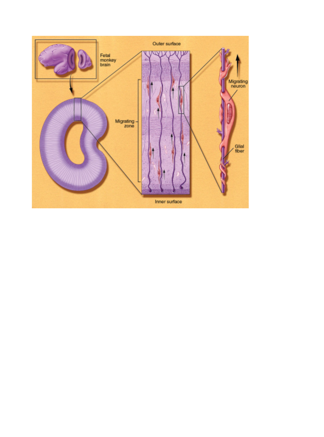
NEURON MIGRATION.
A cross-sectional view of the
occipital lobe (which processes
vision) of a three-month-old
monkey fetus brain (center)
shows immature neurons migrat-
ing along glial fibers. These
neurons make transient connec-
tions with other neurons before
reaching their destination. A sin-
gle migrating neuron, shown
about 2,500 times its actual size
(right), uses a glial fiber as a
guiding scaffold. To move, it
needs adhesion molecules, which
recognize the pathway, and
contractile proteins to propel it
along.

12
face, along nonneuronal fibers that form a trail to their proper
destination. Proper neuron migration requires multiple mecha-
nisms, including the recognition of the proper path and the abil-
ity to move long distances. One such mechanism for long-dis-
tance migration is the movement of neurons along elongated
fibers that form transient sca∑olding in the fetal brain. Many
external forces, such as alcohol, cocaine, or radiation, prevent
proper neuronal migration and result in misplacement of cells,
which may lead to mental retardation or epilepsy. Furthermore,
mutations in genes that regulate migration have recently been
shown to cause some rare genetic forms of retardation and
epilepsy in humans.
Once the neurons reach their final location, they must make
the proper connections for a particular function, such as vision
or hearing, to occur. They do this through their axons. These
wirelike appendages can stretch out a thousand times longer than
the cell body from which they arise. The journey of most axons
ends when they meet thicker appendages, called dendrites, on
other neurons. These target neurons can be located at a consid-
erable distance, sometimes at opposite sides of the brain. In the
case of a motor neuron, the axon may travel from the spinal cord
all the way down to a foot muscle.
Axon growth is directed by growth cones. These enlarge-
ments of the axon’s tip actively explore the environment as they
seek out their precise destinations. Researchers have discovered
many special molecules that help guide growth cones. Some mol-
ecules lie on the cells that growth cones contact, whereas others
are released from sources found near the growth cone. The
growth cones, in turn, bear molecules that serve as receptors for
the environmental cues. The binding of particular signals with
receptors tells the growth cone whether to move forward, stop,
recoil, or change direction. These molecules include proteins
with names such as cadherin, netrin, semaphorin, ephrin, neu-
ropilin, and plexin. In most cases, these are families of related
molecules; for example, there are at least 15 semaphorins and at
least 10 ephrins.
Perhaps the most remarkable finding is that most of these
proteins are common to worms, insects, and mammals, includ-
ing humans. Each family is smaller in flies or worms than in mice
or people, but their functions are quite similar. It has therefore
been possible to use the simpler animals to gain knowledge that
can be directly applied to humans. For example, the first netrin
was discovered in a worm and shown to guide neurons around
the worm’s “nerve ring.” Later, vertebrate netrins were found to
guide axons around the mammalian spinal cord. Worm receptors
for netrins were then found and proved invaluable in finding the
corresponding, and again related, human receptors.
Once axons reach their targets, they form synapses, which
permit electric signals in the axon to jump to the next cell, where
they can either provoke or prevent the generation of a new sig-
nal. The regulation of this transmission at synapses, and the inte-
gration of inputs from the thousands of synapses each neuron
receives, are responsible for the astounding information-pro-
cessing capacity of the brain. For processing to occur properly,
the connections must be highly specific. Some specificity arises
from the mechanisms that guide each axon to its proper target
area. Additional molecules mediate “target recognition,” whereby
the axon chooses the proper neuron, and often the proper part
of the target, once it arrives at its destination. Few of these mol-
ecules have been identified. There has been more success, how-
ever, in identifying the ways in which the synapse forms once
contact has been made. The tiny portion of the axon that con-
tacts the dendrite becomes specialized for the release of neuro-
transmitters, and the tiny portion of the dendrite that receives
the contact becomes specialized to receive and respond to the sig-
nal. Special molecules pass between the sending and receiving
cells to ensure that the contact is formed properly and that the
sending and receiving specializations are precisely opposed to
each other so that transmission can be fast and e≈cient.
Paring back
After growth, the network is pared back to create a more sturdy
system. Only about half the neurons generated during develop-
ment survive to function in the adult. Entire populations of neu-
rons are removed through internal suicide programs initiated in
the cells. The programs are activated if a neuron loses its battle
with other neurons to receive life-sustaining nutrients called
trophic factors. These factors are produced in limited quantities
by target tissues. Each type of trophic factor supports the survival
of a distinct group of neurons. For example, nerve growth factor
is important for sensory neuron survival. It has recently become
clear that the internal suicide program is maintained into adult-
hood and constantly held in check. On the basis of this idea,
researchers have found that injuries and some neurodegenerative
diseases kill neurons not directly by the damage they inflict but
rather by activating the cells’ own death programs. This discov-
ery—and its implication that death need not inevitably follow
insult—have led to new avenues for therapy.
Brain cells also form too many connections at first. For exam-
ple, in primates, the projections from the two eyes to the brain
initially overlap and then sort out to separate territories devoted
only to one eye or the other. Furthermore, in the young primate
cerebral cortex, the connections between neurons are greater in
number and twice as dense as those in an adult primate. Com-
munication between neurons with chemical and electrical signals
is necessary to weed out the connections. The connections that
are active and generating electrical currents survive, whereas
those with little or no activity are lost. Thus, the circuits of the
adult brain are formed, at least in part, by sculpting away incor-
rect connections to leave only the correct ones.
Critical periods
The brain’s refining and building of the network in mammals,
including humans, continues after birth. An organism’s interac-
13
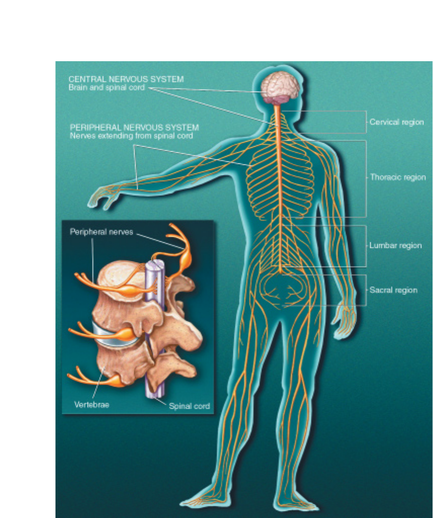
SPINAL CORD AND
NERVES. The mature central
nervous system (CNS) consists of
the brain and spinal cord. The
brain sends nerve signals to spe-
cific parts of the body through
peripheral nerves, known as the
peripheral nervous system
(PNS). Peripheral nerves in the
cervical region serve the neck
and arms; those in the thoracic
region serve the trunk; those in
the lumbar region serve the legs;
and those in the sacral region
serve the bowels and bladder.
The PNS consists of the somatic
nervous system that connects
voluntary skeletal muscles with
cells specialized to respond
to sensations, such as touch and
pain. The autonomic nervous
system is made of neurons con-
necting the CNS with internal
organs. It is divided into the sym-
pathetic nervous system, which
mobilizes energy and resources
during times of stress and
arousal, and the parasympa-
thetic nervous system, which
conserves energy and resources
during relaxed states.
tions with its surroundings fine-tune connections.
Changes occur during critical periods. These are windows of
time during development when the nervous system must obtain
certain critical experiences, such as sensory, movement, or emo-
tional input, to develop properly.
After a critical period, connections diminish in number and
are less subject to change, but the ones that remain are stronger,
more reliable, and more precise. Injury or sensory or social depri-
vation occurring at a certain stage of postnatal life may a∑ect one
aspect of development, whereas the same injury at a di∑erent
period may a∑ect another aspect.
In one example, a monkey is raised from birth to 6 months of
age with one eyelid closed. The animal permanently loses useful
vision in that eye because of diminished use. This gives cellular
meaning to the saying “use it or lose it.” Loss of vision is caused by
the actual loss of functional connections between that eye and
neurons in the visual cortex. This finding has led to earlier and bet-
ter treatment for the eye disorders of congenital cataracts and
“crossed eyes” in children.
Research also shows that enriched environments can bolster
brain development during postnatal life. For example, studies
show that animals brought up in toy-filled surroundings have
more branches on their neurons and more connections than iso-
lated animals. In one recent study, scientists found that enriched
environments resulted in more neurons in a brain area involved in
memory.
Scientists hope that new insights into brain development will
lead to treatments for those with learning disabilities, brain dam-
age, and neurodegenerative disorders, as well as helping us under-
stand aging.

14
Vision. This wonderful sense allows us to
image the world around us, from the genius of
Michelangelo’s Sistine Chapel ceiling to mist-
filled vistas of a mountain range. Vision is one
of the most delicate and complicated senses.
It is also the most studied. About one-fourth
of the brain is involved in visual processing, more than for any
other sense. More is known about vision than any other verte-
brate sensory system, with most of the information derived from
studies of monkeys and cats.
Vision begins with the cornea,which does about three-quar-
ters of the focusing, and then the lens,which varies the focus.
Both help produce a clear image of the visual world on the
retina—the sheet of photoreceptors that process vision, and neu-
rons lining the back of the eye.
As in a camera, the image on the retina is reversed: Objects
to the right of center project images to the left part of the retina
and vice versa, and objects above the center project to the lower
part and vice versa. The shape of the lens is altered by the mus-
cles of the iris so that near or far objects can be brought into focus
on the retina.
Visual receptors, about 125 million in each eye, are neurons
specialized to turn light into electrical signals. They occur in two
forms. Rods are most sensitive to dim light and do not convey color.
Cones work in bright light and are responsible for acute detail,
black-and-white vision, and color vision. The human eye contains
three types of cones that are sensitive to red, green, and blue, but
working together they convey information about all visible colors.
Primates, including humans, have well-developed vision
using two eyes. Visual signals pass from each eye along the mil-
lion or so fibers of the optic nerve to the optic chiasm, where
some nerve fibers cross over, so both sides of the brain receive sig-
nals from both eyes. Consequently, the left halves of both retinas
project to the left visual cortex and the right halves project to the
right visual cortex.
The e∑ect is that the left half of the scene you are watching
registers in your right hemisphere. Conversely, the right half of
the scene registers in your left hemisphere. A similar arrangement
applies to movement and touch: Each half of the cerebrum is
responsible for the opposite half of the body.
Scientists know much about the way cells encode visual infor-
mation in the retina, the lateral geniculate nucleus—an interme-
diate point between the retina and visual cortex—and the visual
cortex.These studies give us the best knowledge so far about how
the brain analyzes and processes information.
The retina contains three stages of neurons. The first, the layer
of rods and cones, sends its signals to the middle layer, which
relays signals to the third layer. Nerve fibers from the third layer
assemble to form the optic nerve. Each cell in the middle or third
layer typically receives input from many cells in the previous layer,
but the number of inputs varies widely across the retina. Near the
center of gaze, where visual acuity is highest, each cell in the third
layer receives inputs—via the middle layer—from one or a few
cones, thus allowing us to resolve very fine details. Near the mar-
gins of the retina, each cell in the third layer receives signals from
a cluster of rods and cones, explaining why we cannot see fine
details o∑ to either side. Whether large or small, this region of
visual space is called the receptive field of the third-layer cell.
About 55 years ago, scientists discovered that the receptive
field of such a cell is activated when light hits a tiny region in its
receptive field center and is inhibited when light hits the part of
the receptive field surrounding the center. If light covers the entire
receptive field, the cell reacts only weakly and perhaps not at all.
Thus, the visual process begins with a comparison of the
amount of light striking any small region of the retina and the
amount of light around it. Located in the occipital lobe, the pri-
mary visual cortex—two millimeters thick (a bit larger than a
half-dollar) and densely packed with cells in many layers—
receives messages from the lateral geniculate. In the middle layer,
which also receives input from the lateral geniculate, scientists
found patterns of responsiveness similar to those observed in the
retina and lateral geniculate cells. Cells above and below this
layer responded di∑erently. They preferred stimuli in the shape
of bars or edges. Further studies showed that di∑erent cells pre-
ferred edges at particular angles, edges that moved, or edges
moving in a particular direction.
Although the process is not yet completely understood,
recent findings suggest that visual signals are fed into at least
three separate processing systems. One system appears to process
information about shape; a second, color; and a third, movement,
V
Sensation and perception
15
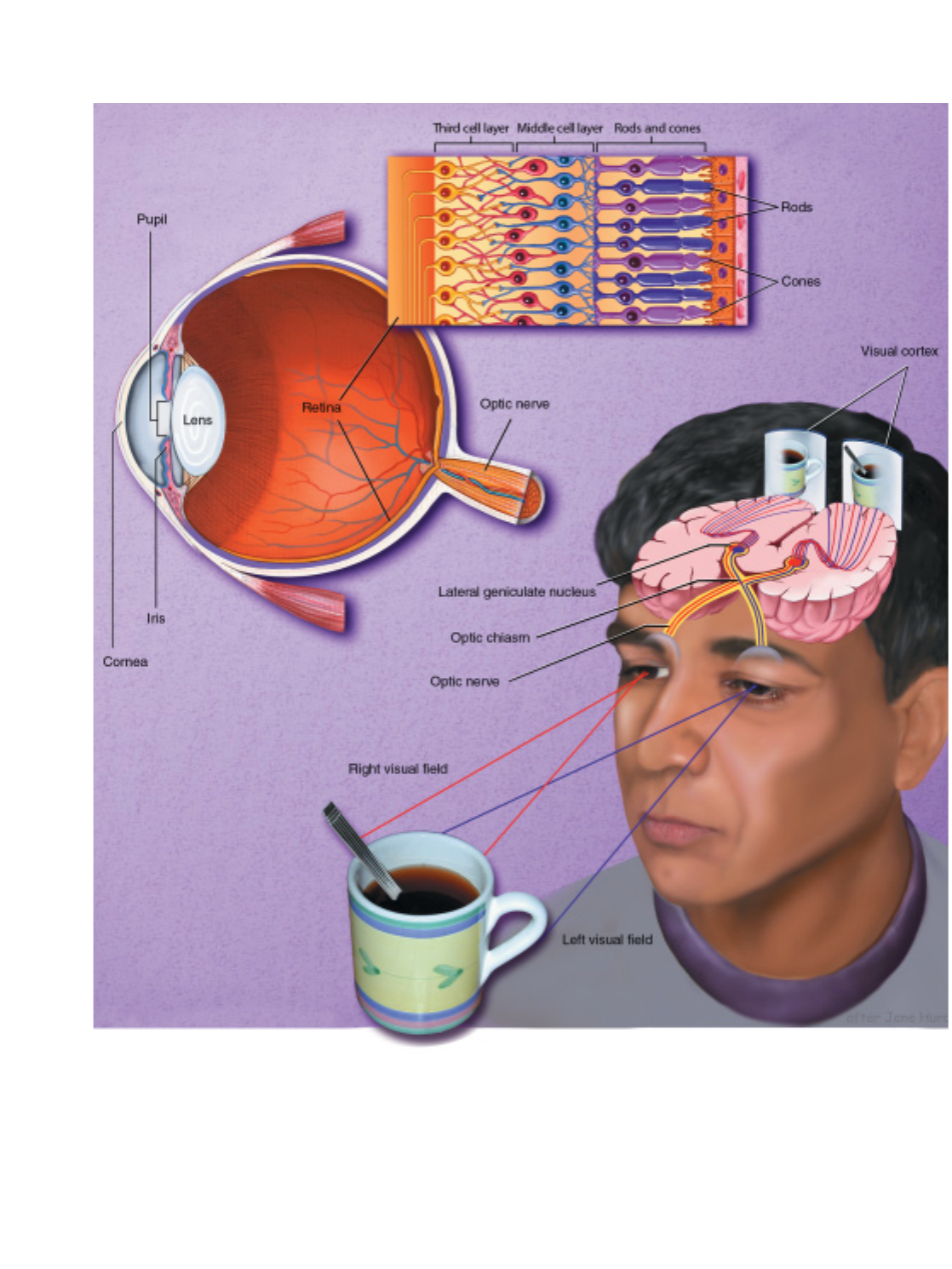
VISION. The cornea and lens help produce a clear image of the visual world on the retina, the sheet of photoreceptors and neurons lining the
back of the eye. As in a camera, the image on the retina is reversed: Objects to the right of the center project images to the left part of the
retina and vice versa. The eye’s 125 million visual receptors — composed of rods and cones — turn light into electrical signals. Rods are most
sensitive to dim light and do not convey the sense of color; cones work in bright light and are responsible for acute detail, black and white
vision, and color vision. The human eye contains three types of cones that are sensitive to red, green, and blue, but, in combination, convey
information about all visible colors. Rods and cones connect with a middle cell layer and third cell layer (see inset, above). Light passes through
these two layers before reaching the rods and cones. The two layers then receive signals from rods and cones before transmitting the signals
onto the optic nerve, optic chiasm, lateral geniculate nucleus, and, finally, the visual cortex.

16
location, and spatial organization. These findings of separate pro-
cessing systems come from monkey anatomical and physiologi-
cal data. They are verified by human psychological studies show-
ing that the perception of movement, depth, perspective, the
relative size of objects, the relative movement of objects, shading,
and gradations in texture all depend primarily on contrasts in
light intensity rather than in color.
Why movement and depth perception should be carried out
by only one processing system may be explained by a school of
thought called Gestalt psychology. Perception requires various ele-
ments to be organized so that related ones are grouped together.
This stems from the brain’s ability to group the parts of an image
together and also to separate images from one another and from
their individual backgrounds.
How do all these systems combine to produce the vivid
images of solid objects that we perceive? This involves extracting
biologically relevant information at each stage and associating
firing patterns with past experience.
Vision studies also have led to better treatment for visual dis-
orders. Information from research in cats and monkeys has
improved the therapy for strabismus,or squint,a term for “cross-
eye” or wall-eye. Children with strabismus initially have good
vision in each eye. But because they cannot fuse the images in the
two eyes, they tend to favor one eye and often lose useful vision
in the other. Vision can be restored in such cases, but only dur-
ing infancy or early childhood. Beyond the age of 6 or so, the
blindness becomes permanent. But until a few decades ago, oph-
thalmologists waited until children reached the age of 4 before
operating to align the eyes, or prescribing exercises or an eye
patch. Now strabismus is corrected very early in life—before age
4, when normal vision can still be restored.
Hearing
Often considered the most important sense for humans, hearing
allows us to communicate with each other by receiving sounds
and interpreting speech. It also gives us information vital to sur-
vival. For example, the sound of an oncoming train tells us to stay
clear of the railroad track.
Like the visual system, our hearing system distinguishes sev-
eral qualities in the signal it detects. However, our hearing sys-
tem does not blend di∑erent sounds, as the visual system does
when two di∑erent wavelengths of light are mixed to produce
color. We can follow the separate melodic lines of several instru-
ments as we listen to an orchestra or rock band.
From the chirping of crickets to the roar of a rocket engine,
most of the sounds processed by the ear are heard by a mecha-
nism known as air conduction.In this process, sound waves are
first funneled through the externally visible part of the ear, the
pinna (or external ear) and the external auditory canal,to the
tympanic membrane (eardrum), which vibrates at di∑erent
speeds. The malleus (hammer), which is attached to the tympanic
membrane, transmits the vibrations to the incus (anvil). This
structure passes them onto the stapes (stirrup), which delivers
them, through the oval window, to the inner ear.
The fluid-filled spiral passages of each cochlea contain 16,000
hair cells, whose microscopic, hairlike projections respond to the
vibrations produced by sound. The hair cells, in turn, excite the
28,000 fibers of the auditory nerve, which terminate in the
medulla of the brain. Auditory information flows via the thala-
mus to the temporal gyrus,the part of the cerebral cortex involved
in receiving and perceiving sound.
The brain’s analysis of auditory information follows a pattern
similar to that of the visual system. Adjacent neurons respond to
tones of similar frequency. Some neurons respond to only a small
range of frequencies, others react to a wide range; some react only
to the beginning of a sound, others only respond to the end.
Speech sounds, however, may be processed di∑erently than
others. Our auditory system processes all the signals that it
receives in the same way until they reach the primary auditory
cortex in the temporal lobe of the brain. When speech sound is
perceived, the neural signal is funneled to the left hemisphere for
processing in language centers.
Taste and smell
Although di∑erent, the two sensory experiences of taste and smell
are intimately entwined. They are separate senses with their own
receptor organs. However, these two senses act together to allow
us to distinguish thousands of di∑erent flavors. Alone, taste is a
relatively focused sense concerned with distinguishing among
sweet, salty, sour, bitter, and umami (Japanese for savory). The
interaction between taste and smell explains why loss of the sense
of smell apparently causes a serious reduction in the overall taste
experience,which we call flavor.
Tastes are detected within taste buds, special structures of
which every human has some 5,000 to 10,000. Taste buds are
embedded within papillae,or protuberances, located mainly on
the tongue, with others found in the back of the mouth and on
the palate. Taste substances stimulate specialized sensory cells.
Each taste bud consists of 50 to 100 of these cells, which respond
to salts, acidity, sweet substances, bitter compounds, and mono-
sodium glutamate and related amino acids.
Taste signals in the sensory cells are transferred by synapses to
the ends of nerve fibers, which send impulses along cranial nerves
to taste regions in the brain. From here, the impulses are relayed
to other brainstem centers responsible for the basic responses of
acceptance or rejection of the tastes, and to the thalamus and on
to the cerebral cortex for conscious perception of taste.
Specialized smell receptor cells are located in a small patch of
mucus membrane lining the roof of the nose. Axons of these sen-
sory cells pass through perforations in the overlying bone and
enter two elongated olfactory bulbs lying on top of the bone. The
portion of the sensory cell that is exposed to odors possesses hair-
like cilia. These cilia contain the receptor sites that are stimulated
by odorants carried by airborne molecules. These dissolve in the
17
mucus lining in order to stimulate receptor proteins in the cilia to start the smell response. An odor-
ant acts on many receptors to di∑erent degrees. Similarly, a receptor interacts with many di∑erent
odorants to varying degrees.
The pattern of activity set up in the receptor cells is projected to the olfactory bulb, where it forms
a spatial image of the odor. Impulses created by this stimulation pass to other smell regions, giving
rise to conscious perceptions of odor in the frontal lobe and emotional responses in the limbic sys-
tem of the brain.
Touch and pain
Touch is the sense by which we determine the characteristics of objects: size, shape, and texture. We
do this through touch receptors in the skin. In hairy skin areas, some receptors consist of webs of
sensory nerve cell endings wrapped around the base of hairs. The nerve endings are remarkably sen-
sitive, being triggered by slight movement of the hairs. Other receptors are more common in non-
hairy areas, such as the lips and fingertips, and consist of nerve cell endings that may be free or sur-
rounded by bulblike structures.
Signals from touch receptors pass via sensory nerves to the spinal cord, where they synapse (make
contact) and then travel to the thalamus and sensory cortex. The transmission of this information is
highly topographic, meaning that the body is represented in an orderly fashion at di∑erent levels of
the nervous system. Larger areas of the cortex are devoted to sensations from the hands and lips;
much smaller cortical regions represent less sensitive parts of the body.
Di∑erent parts of the body vary in their sensitivity to touch discrimination and painful stimuli
according to the number and distribution of receptors. The cornea is several hundred times more
sensitive to painful stimuli than are the soles of the feet. The fingertips are good at touch discrimi-
nation, but the chest and back are less sensitive.
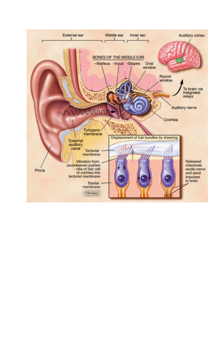
HEARING. From the chirping
of crickets to the roar of a
rocket engine, almost all of the
thousands of single tones
processed by the human ear are
heard by a mechanism known
as air conduction. In this
process, sound waves are first
funneled through the external
ear — the pinna and the exter-
nal auditory canal — to the
middle ear — the tympanic
membrane (eardrum)— that
vibrates at different speeds. The
malleus (hammer), which is
attached to the tympanic mem-
brane, transmits the vibrations
to the incus (anvil). The vibra-
tions are then passed onto the
stapes (stirrup) and oval win-
dow that, in turn, pass them
onto the inner ear. In the inner
ear, the fluid-filled spiral pas-
sage of the cochlea contains
cells with microscopic, hairlike
projections that respond to the
vibrations produced by sound.
The hair cells, in turn, excite the
28,000 fibers of the auditory
nerve that end in the medulla in
the brain. Auditory information
flows via the thalamus to the
temporal gyrus, the part of the
cerebral cortex involved in
receiving and perceiving sound.
18
Not surprisingly, acuity is greatest in the most densely nerve-packed areas of the body. This fea-
ture, in fact, is used to test clinically for the integrity of these somatosensory pathways. For example,
neurologists can run tests by using a two-point threshold.This method involves touching the skin with
calipers at two points. The two-point threshold is the distance between the two points that is neces-
sary for the individual to distinguish two distinct stimuli from one.
Until recently, pain was thought to be a simple message by which neurons sent electrical impulses
from the site of injury directly to the brain. We now know that the process is far more complicated.
Nerve impulses from sites of injury that persist for hours, days, or longer lead to changes in the ner-
vous system that result in an amplification and increased duration of the pain. These changes involve
dozens of chemical messengers and receptors. Persistent pain is in many respects a disease of the ner-
vous system, not merely a symptom of some other disease process.
The sensory fibers that respond to stimuli that injure tissue and can cause pain are called noci-
ceptors,special receptors that respond to tissue-damaging stimuli. In addition to directly activating
the nociceptor and evoking a pain sensation, tissue injury causes the release of numerous chemicals
at the site of damage and inflammation. One such family of chemicals includes the prostaglandins,
which enhance the sensitivity of receptors to tissue damage and ultimately can induce more intense
pain sensations. Prostaglandins also contribute to the clinical condition in which innocuous stimuli
can produce pain (such as in sunburned skin) because the threshold of the nociceptor is significantly
reduced. This phenomenon is called allodynia.
Pain messages are transmitted to the spinal cord via small myelinated fibers and C fibers—very
small unmyelinated fibers. Myelin is a covering sheath around nerve fibers that helps them send their
messages more rapidly. The small myelinated pain-sensitive nerve fibers probably evoke the sharp,
fast pain that is produced by, for example, a pin prick. C fiber-induced pain, by contrast, is generally
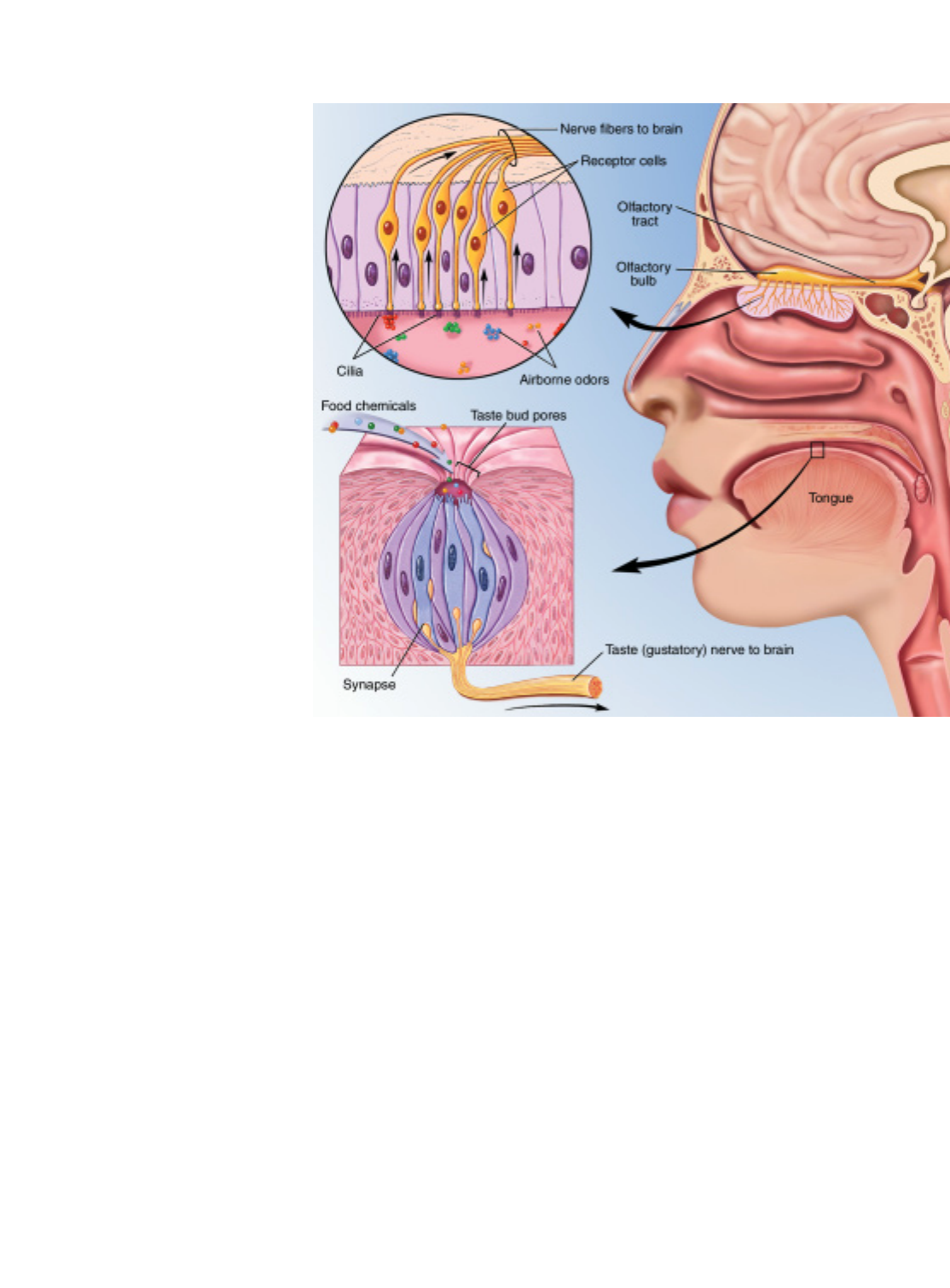
TASTE AND SMELL . Special-
ized receptors for smell are
located in a patch of mucus
membrane lining the roof of the
nose. Each cell has several fine
hairlike cilia containing receptor
proteins, which are stimulated
by odor molecules in the air,
and a long fiber (axon), which
passes through perforations in
the overlying bone to enter the
olfactory bulb. Stimulated cells
give rise to impulses in the
fibers, which set up patterns in
the olfactory bulb that are
relayed to the brain’s frontal
lobe to give rise to smell per-
ception, and to the limbic sys-
tem to elicit emotional responses.
Tastes are detected by special
structures, taste buds, of which
every human has some 5,000
to 10,000. Taste buds are
embedded within papillae (pro-
tuberances) mainly on the
tongue, with a few located in
the back of the mouth and on
the palate. Each taste bud con-
sists of about 100 receptors that
respond to the four types of
stimuli — sweet, salty, sour, and
bitter — from which all tastes
are formed. A substance is
tasted when chemicals in foods
dissolve in saliva, enter the
pores on the tongue, and come
in contact with taste buds. Here
they stimulate hairs projecting
from the receptor cells and
cause signals to be sent from
the cells, via synapses, to cra-
nial nerves and taste centers
in the brain.
19
slower in onset, dull, and more di∑use.
In the ascending system,the impulses are relayed from the
spinal cord to several brain structures, including the thalamus and
cerebral cortex, that are involved in the process by which “pain”
messages become conscious experience. The experience of pain is
not just a function of the magnitude of the injury, or even the
intensity of the impulse activity generated by the injury. The set-
ting in which the injury occurs contributes (e.g., the pain of child-
birth or that produced in a car accident). The emotional compo-
nent of the experience is a major contributor to the overall pain.
Pain messages can also be suppressed by a system of neurons
that originate within the gray matter in the brainstem. This
descending system sends messages to the dorsal horn of the spinal
cord,where it suppresses the transmission of pain signals to the
higher brain centers. Some of these descending systems use nat-
urally occurring chemicals similar to opioids. The three major
families of opioid peptides identified in the brain—enkephalins,
beta-endorphins, and dynorphins—originate from three pre-
cursor proteins encoded by three di∑erent genes. They act at mul-
tiple opioid receptors in the brain and spinal cord. Knowledge of
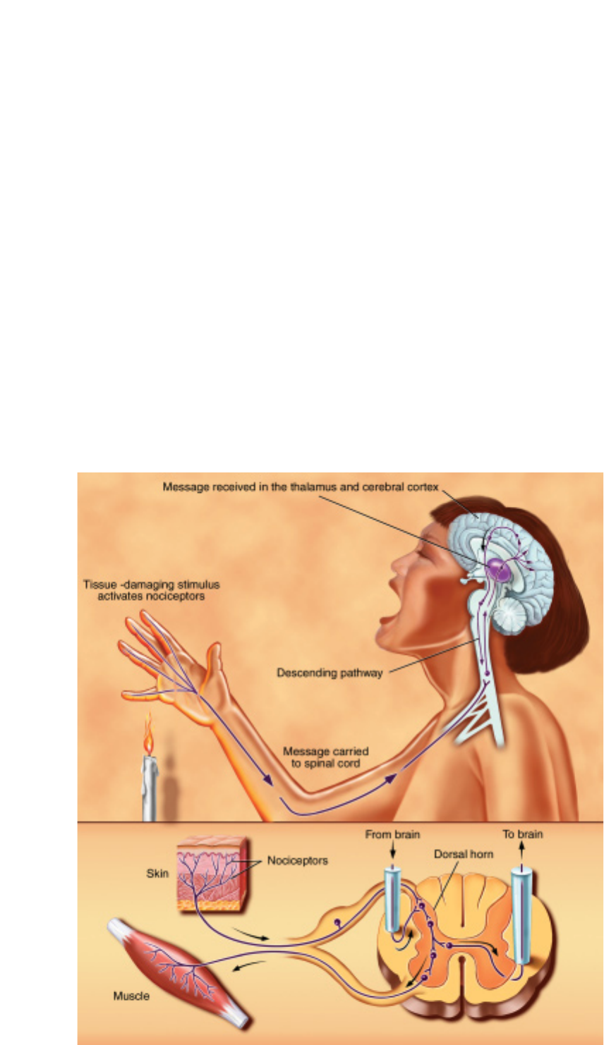
PAIN. Messages about tissue
damage are picked up by recep-
tors and transmitted to the
spinal cord via small myelinated
fibers and very small unmyeli-
nated fibers. From the spinal
cord, the impulses are carried to
the brainstem, thalamus, and
cerebral cortex and ultimately
perceived as pain. These mes-
sages can be suppressed by a
system of neurons that origi-
nates in the gray matter of the
midbrain. This descending path-
way sends messages to the
spinal cord where it suppresses
the transmission of tissue dam-
age signals to the higher brain
centers. Some of these descend-
ing pathways use naturally
occurring, opiate-like chemicals
called endorphins.
the way pain messages are transmitted has led to new treatments
for pain. For example, scientists began studying the spinal deliv-
ery of opioids when they discovered a dense distribution of opi-
oid receptors in the spinal cord horn. Such treatments were
begun in humans after the method was successfully used in ani-
mals; the technique is now common in treating pain after surgery.
Because the spinal opioid does not interact at all levels of the ner-
vous system, this technique bypasses many potentially negative
opioid side e∑ects.
Many new insights into the pain experience are coming from
studies in which modern imaging tools are used to monitor brain
activity when pain is experienced. One finding is that there is no
single area in the brain where pain is generated; rather, there are
both emotional and sensory components. Interestingly, when
people are hypnotized so that a painful stimulus is not experi-
enced as unpleasant, activity in only some areas of the brain is
suppressed. As such techniques for brain study improve, it should
be possible to better monitor the changes in the brain that occur
in people with persistent pain and to better evaluate the di∑erent
analgesic drugs being developed.

20
The conscious memory of a patient known as
H.M. is limited almost entirely to events that
occurred years before his surgery, in which part
of the medial temporal lobe of his brain was
removed to relieve epilepsy. H.M. can remember
recent events for only a few minutes. Talk with
him awhile and then leave the room. When you return, he has no
recollection of ever having seen you.
The medial temporal lobe, which includes the hippocampus
and adjacent brain areas, seems to play a role in converting mem-
ory from a short-term to a long-term, permanent form. The fact
that H.M. retains memories for events that are remote to his
surgery is evidence that the medial temporal region is not the site
of permanent storage but that it plays a role in the formation of
new memories. Other patients like H.M. have also been described.
Additional evidence comes from patients undergoing elec-
troconvulsive therapy (ECT) for depression. ECT is thought to
temporarily disrupt the function of the hippocampus and related
structures. These patients typically have difficulty with new learn-
ing and have amnesia for events that occurred during the several
years before treatment. Memory of earlier events is unimpaired.
As time passes after treatment, much of the lost part of memory
becomes available once again.
The hippocampus and the medial temporal region are con-
nected to widespread areas of the cerebral cortex, especially the
vast regions responsible for thinking and language. Whereas the
medial temporal region is important for forming and organizing
memory, cortical areas are important for the long-term storage
of knowledge about facts and events and for how this knowledge
is used in everyday situations.
Working memory,a type of transient, “online” memory that
enables us to retain what someone has said just long enough to
reply, depends in part on the prefrontal cortex. Researchers dis-
covered that certain neurons in this area are influenced by neu-
rons releasing dopamine and other neurons releasing glutamate.
Although much remains to be discovered about learning and
memory, scientists have already put together important pieces
of the puzzle. For example, the brain appears to process differ-
ent kinds of information in separate ways and then store it dif-
ferently.
Declarative knowledge requires processing in the medial tem-
poral region and parts of the thalamus and can be grouped into
working memory, episodic memory, and semantic memory.
Working memory allows us to keep and use information in our
minds and is mediated by a network of areas in the cerebral cor-
tex. Episodic memory lets us store and replay events in our minds
and depends on the hippocampus. Semantic memory includes
raw facts and data and is stored throughout the cerebral cortex.
The hippocampus may play a role in integrating new episodic
memories into the semantic memory storehouse.
In contrast, nondeclarative knowledge,the knowledge of how
to do something, is expressed in skilled behavior and learned
habits and requires processing by the basal ganglia.
The amygdala appears to play an important role in the emo-
tional aspects of memory. An important factor that influences
what is stored and how strongly it is stored is whether the action
is followed by reward, punishment, or highly emotional conse-
quences. These consequences help determine what behaviors an
organism will learn and remember.
Memory of motor learning tasks in which precise timing is
involved depends on the cerebellum.
How exactly does memory occur? After years of study, there
is much support for the idea that memory involves a persistent
change in the connection between neurons. In animal studies, sci-
entists found that this occurs in the short term through two bio-
chemical events that affect the strength of the relevant synapses.
The stability of long-term memory is conferred by turning on
genes that may lead to modifications within neurons that change
the strength and number of synapses. For example, researchers
can correlate specific chemical and structural changes in the rel-
evant cells with several simple forms of memory exhibited by the
sea slug Aplysia californica.
Another important model for the study of memory is the
phenomenon of long-term potentiation (LTP), a long-lasting
increase in the strength of a synaptic response following stim-
ulation. LTP occurs prominently in the hippocampus, as well as
in other brain areas. Studies of rats suggest that LTP occurs
through changes in synaptic strength at contacts involving
NMDA receptors. It is now possible to study LTP and learning
in genetically modified mice that have alterations in specific
Learning, memory, and
language
T
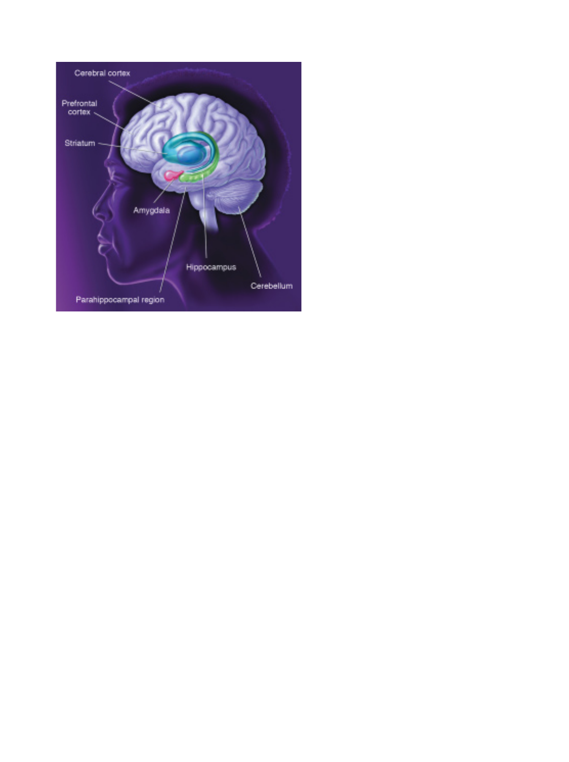
21
genes. Examples of these modified genes can be limited both to
particular brain areas and to specific times, such as during
learning.
Much of what we have learned about memory comes from
studies of amnesia due to damage to the hippocampus and cor-
tical areas—called the parahippocampal region—in the medial
part of the temporal lobe. Patients with damage in these areas can
remember recent events only while actively engaged in the mate-
rial—yet they often retain childhood memories quite well. This
pattern suggests that the temporal lobe is critical in integrating
early memories into a permanent storehouse that can be accessed
whenever needed.
Several types of memory are spared in amnesia. Reports indi-
cate that the sense of familiarity one has with a face or a scene is
spared, even though the specific context of the experience may be
lost. The emotional association one might develop with a given
item is also commonly spared in amnesia.
Scientists believe that no single brain center stores memory.
It most likely is stored in distributed collections of cortical pro-
cessing systems that are also involved in the perception, process-
ing, and analysis of the material being learned. In short, each part
of the brain most likely contributes differently to permanent
memory storage.
One of the most prominent intellectual activities dependent
on memory is language. Although the neural basis of language is
not fully understood, scientists have learned a great deal about
this function of the brain from studies of patients who have lost
speech and language abilities due to stroke, and from behavioral
and functional neuroimaging studies of normal people.
A prominent and influential model, based on studies of these
patients, proposes that the underlying structure of speech com-
prehension arises in part of the left hemisphere of the brain,
called Wernicke’s area. This temporal lobe region is connected
with another region, Broca’s area, in the frontal lobe, where a pro-
gram for vocal expression is created. This program is then trans-
mitted to a nearby area of the motor cortex that activates the
mouth, tongue, and larynx.
This same model proposes that, when we read a word, the
information is transmitted from the primary visual cortex to the
angular gyrus, where the message is somehow matched with the
words when they are spoken. The auditory form of the word is
then processed for comprehension in Wernicke’s area as if the
word had been heard. Writing in response to an oral instruction
requires information to be passed along the same pathways in
the opposite direction—from the auditory cortex to Wernicke’s
area to the angular gyrus. This model accounts for much of the
data from patients and is the most widely used for clinical diag-
nosis and prognosis. Some refinements to this model may be
necessary, however, because of both recent studies with patients
and functional neuroimaging studies in healthy people.
For example, using an imaging technique called positron
emission tomography (PET), scientists have demonstrated that
some reading tasks performed by normal people do not activate
Wernicke’s area or the angular gyrus. These results suggest that,
at least under some conditions, there is a direct reading route that
does not involve speech-sound recoding of the visual stimulus
before the processing of either meaning or speaking. Other stud-
ies with patients also have indicated that it is likely that familiar
words need not be recoded into sound before they can be under-
stood.
Although the understanding of how language is implemented
in the brain is far from complete, there are now several tech-
niques that may be used to gain important insights into this crit-
ical aspect of brain function.
LEARNING AND MEMORY. Different brain areas and systems
mediate distinct forms of memory. The hippocampus, parahip-
pocampal region, and areas of the cerebral cortex (including pre-
frontal cortex) compose a system that supports declarative, or cog-
nitive, memory. Different forms of nondeclarative, or behavioral,
memory are supported by the amygdala, striatum, and cerebellum.

22
From the stands, we marvel at the perfectly placed
serves of professional tennis players and the light-
ning-fast double plays executed by big league
infielders. But in fact, each of us in our daily activi-
ties performs a host of complex, skilled movements,
such as walking upright, speaking, and writing, that
are just as remarkable. This is made possible by a finely tuned and
highly complex central nervous system, which controls the
actions of hundreds of muscles. Through learning, the nervous
system can adapt to changing movement requirements to accom-
plish these everyday marvels, and to perform them more skill-
fully with practice.
To understand how the nervous system performs such tricks,
we have to start with the muscles, for these are the body parts
that produce movement under the control of the brain and
spinal cord.
Most muscles attach to points on the skeleton that cross one
or more joints, so they are called skeletal muscles.Activation ofa
given muscle can open or close the joints that it spans, depend-
ing upon whether it is a joint flexor (closer) or extensor (opener).
In addition, if flexors and extensors at the same joint are activated
together, they can “sti∑en” a joint, thus maintaining limb posi-
tion in the face of unpredictable external forces that would oth-
erwise displace it. Muscles that move a joint in an intended direc-
tion are called agonists,and those that oppose this direction of
movement are antagonists.Skilled movements at high speed are
started by agonists and stopped by antagonists, thus placing the
joint or limb at a desired position.
Some muscles act on soft tissue, such as the muscles that
move the eyes and tongue and those that control facial expres-
sion. These muscles are also under control of the central nervous
system, and their principles of operation are similar to those that
attach to bone.
Each skeletal muscle is made up of thousands of individual
muscle fibers, and each of these is controlled by one alpha motor
neuron in either the brain or the spinal cord. On the other hand,
each single alpha motor neuron controls many muscle fibers
(ranging from a few to a hundred or more), forming a functional
unit referred to as a motor unit.These motor units are the criti-
cal link between the brain and muscles. If they die, which can
happen in certain diseases that a∑ect motor neurons directly, a
person is no longer able to move, either voluntarily or through
reflexes.
Perhaps the simplest and most fundamental of movements
are reflexes. These are relatively fixed, automatic muscle responses
to particular stimuli, such as sudden withdrawal of the foot when
you step on a sharp object, or the slight extension of the leg when
a physician taps your knee with a small rubber hammer. All
reflexes involve the activation of small sensory receptors in the
skin, the joints, or even in muscles themselves. For example, the
knee movement referred to above is produced by a slight stretch
of the knee extensor muscles when the physician taps the muscle
tendon at the knee. This slight muscle stretch is “sensed”by recep-
tors in the muscle, called muscle spindles.Innervated by sensory
fibers, the spindles send information to the spinal cord and brain
about the length and speed of shortening or lengthening of a mus-
cle. This information is used in reflex control of the joint at which
the muscle acts, and also for control of voluntary movements.
Sudden muscle stretch sends a barrage of impulses into the
spinal cord along the muscle spindle sensory fibers. This, in turn,
activates motor neurons in the stretched muscle, causing a con-
traction called the stretch reflex.The same sensory stimulus
causes inactivation, or inhibition, in the motor neurons of the
antagonist muscles through connecting neurons, called inhibitory
neurons,within the spinal cord. Thus, even the simplest of
reflexes involves a coordination of activity across motor neurons
that control agonist and antagonist muscles.
Even more amazing is the fact that the brain can control not
only the actions of motor neurons and muscles, but also the
nature of the feedback that it receives from sensory receptors in
the muscles as movements occur. For example, the sensitivity of
the muscle spindle organs is controlled by the brain through a
separate set of gamma motor neurons that control the specialized
muscle fibers and allow the brain to fine-tune the system for
di∑erent movement tasks.
In addition to such exquisite sensing and control of muscle
length by muscle spindles, other specialized sense organs in mus-
cle tendons—the golgi tendon organs—detect the force applied
by a contracting muscle, allowing the brain to also sense and con-
trol the muscular force exerted during movement.
F
Movement
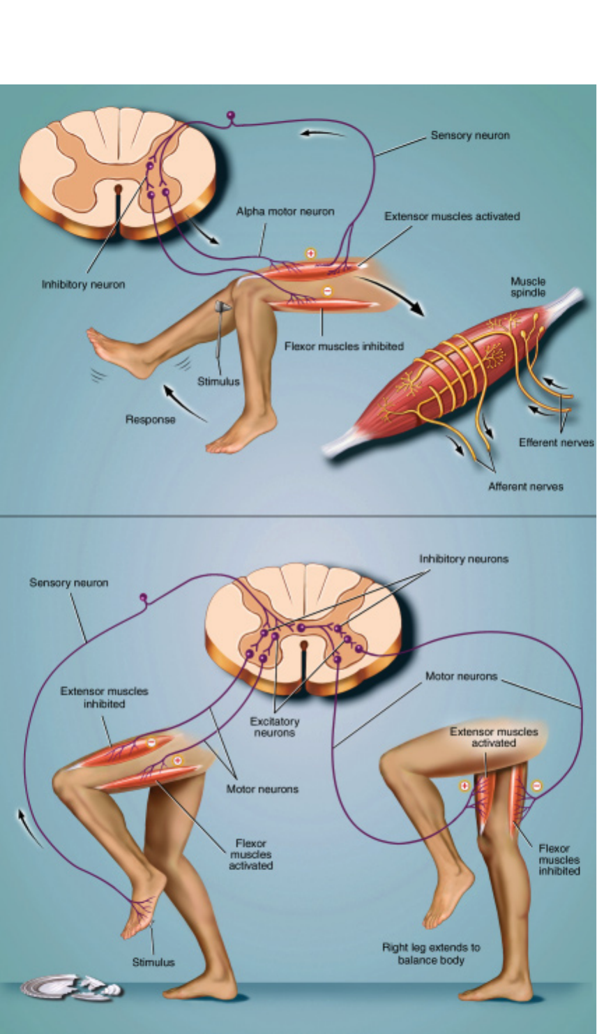
23
MOVEMENT. The stretch reflex
(above) occurs when a doctor
taps a muscle tendon to test your
reflexes. This sends a barrage
of impulses into the spinal cord
along muscle spindle sensory
fibers and activates motor neu-
rons to the stretched muscle to
cause contraction (stretch reflex).
The same sensory stimulus
causes inactivation, or inhibition,
of the motor neurons to the
antagonist muscles through con-
nection neurons, called inhibitory
neurons, within the spinal cord.
Afferent nerves carry messages
from sense organs to the spinal
cord; efferent nerves carry motor
commands from the spinal cord
to muscles. Flexion withdrawal
(below) can occur when your
bare foot encounters a sharp
object. Your leg is immediately
lifted (flexion) from the source of
potential injury, but the opposite
leg responds with increased
extension in order to maintain
your balance. The latter event is
called the crossed extension
reflex. These responses occur
very rapidly and without your
attention because they are built
into systems of neurons located
within the spinal cord itself.

24
We now know that these complex systems are coordinated
and organized to respond di∑erently for tasks that require pre-
cise control of position, such as holding a full teacup, and for
those requiring rapid, strong movement, such as throwing a ball.
You can experience such changes in motor strategy when you
compare walking down an illuminated staircase with the same
task done in the dark.
Another useful reflex is the flexion withdrawal that occurs if
your bare foot encounters a sharp object. Your leg is immediately
lifted from the source of potential injury (flexion) but the oppo-
site leg responds with increased extension in order to maintain
your balance. The latter event is called the crossed extension reflex.
These responses occur very
rapidly and without your atten-
tion because they are built into
systems of neurons that are
located within the spinal cord
itself. It seems likely that the
same systems of spinal neurons
also participate in controlling
the alternating action of the
legs during normal walking. In
fact, the basic patterns of mus-
cle activation that produce
coordinated walking can be
generated in four-footed ani-
mals within the spinal cord
itself. These spinal mechanisms, which evolved in primitive ver-
tebrates, are likely still present in the human spinal cord.
The most complex movements that we perform, including
voluntary ones that require conscious planning, involve control
of these basic spinal mechanisms by the brain. Scientists are only
beginning to understand the complex interactions that take place
among di∑erent brain regions during voluntary movements,
mostly through careful experiments on animals.
One important brain area in the control of voluntary move-
ment is the motor cortex,which exerts powerful control over the
spinal cord, in part through direct control of its alpha motor
neurons. Some neurons in the motor cortex appear to specify the
coordinated action of many muscles, to produce organized
movement of a limb to a particular place in space. Others appear
to control only two or three functionally related muscles, such
as those of the hand or arm, that are important for finely tuned,
skilled movement.
In addition to the motor cortex, movement control also
involves the interaction of many other brain regions, including
the basal ganglia, thalamus, cerebellum,and a large number of
neuron groups located within the midbrain and brainstem—
regions that connect cerebral hemispheres with the spinal cord.
The brain regions devoted to such control are large, containing
millions of intricately interconnected neurons.
Scientists know that the basal ganglia and thalamus have
widespread connections with sensory and motor areas of the
cerebral cortex. Loss of regula-
tion of the basal ganglia by
dopamine depletion can cause
serious movement disorders,
such as Parkinson’s disease. Loss
of dopamine neurons in the
substantia nigra of the mid-
brain, which connects with the
basal ganglia, is a major factor
in Parkinson’s disease.
Another brain region that is
crucial for skilled movement
and for the learning of new
movements is the cerebellum. A
disturbance of cerebellar func-
tion, for example, leads to poor coordination of muscle control,
to disorders of balance, and even to di≈culties in speech, one of
the most intricate forms of movement control.
The cerebellum receives direct and powerful sensory infor-
mation from the muscle receptors and the sense organs of the
inner ear, which signal head position and movement, and sig-
nals from the cerebral cortex. The cerebellum apparently acts to
integrate all this information to ensure smooth coordination of
muscle action, enabling us to perform skilled movements more
or less automatically. There is evidence that as we learn to walk,
speak, or play a musical instrument, the necessary, detailed con-
trol information is stored within the cerebellum, where it can be
called upon by commands from the cerebral cortex.
We now know that complex
systems are coordinated and organized
to respond differently for tasks that
require precise control of position,
such as holding a teacup, and for those
requiring rapid, strong movement,
such as throwing a ball.

25
Sleep remains one of the great mysteries of mod-
ern neuroscience. We spend nearly one-third of
our lives asleep, but the function of sleep still is
not known. Fortunately, over the past few years
researchers have made great headway in under-
standing some of the brain circuitry that con-
trols wake-sleep states.
Scientists now recognize that sleep consists of several di∑-
erent stages; that the choreography of a night’s sleep involves the
interplay of these stages, a process that depends upon a complex
switching mechanism; and that the sleep stages are accompanied
by daily rhythms in bodily hormones, body temperature, and
other functions.
Sleep disorders are among the nation’s most common health
problems, a∑ecting up to 70 million people, most of whom are
undiagnosed and untreated. These disorders are one of the least
recognized sources of disease, disability, and even death, costing
an estimated $100 billion annually in lost productivity, medical
bills, and industrial accidents. Research holds promise for devis-
ing new treatments to allow millions of people to get a good
night’s sleep.
The stuff of sleep
Although sleep appears to be a passive and restful time, it actu-
ally involves a highly active and well-scripted interplay of brain
circuits to produce its various stages.
The stages of sleep were discovered in the 1950s in experi-
ments using electroencephalography (EEG) that examined human
brain waves during sleep. Researchers also measured movements
of the eyes and the limbs during sleep. They found that over the
course of the first hour or so of sleep each night, the brain pro-
gresses through a series of stages during which the brain waves
progressively slow down. This period of slow wave sleep is accom-
panied by relaxation of the muscles and the eyes. Heart rate, blood
pressure, and body temperature all fall. If awakened at this time,
most people recall only fragmented thoughts, not an active dream.
Over the next half hour or so, brain activity alters drastically
from the deep slow wave sleep to generate neocortical EEG waves
that are indistinguishable from those observed during waking.
Paradoxically, the fast, waking-like EEG activity is accompanied
by atonia,or paralysis of the body’s muscles (only the muscles
that allow breathing remain active). This state is often called
rapid eye movement (REM) sleep. During REM sleep, there is
active dreaming. Heart rate, blood pressure, and body tempera-
ture become much more variable. Men often have erections dur-
ing this stage of sleep. The first REM period usually lasts 10 to 15
minutes.
During the night, these cycles of slow wave and REM sleep
alternate, with the slow wave sleep becoming less deep and the
REM periods more prolonged until waking occurs.
Over the course of a lifetime, the pattern of sleep cycles
changes. Infants sleep up to 18 hours per day, and they spend
much more time in deep slow wave sleep. As children mature,
they spend less time asleep and less time in deep slow wave sleep.
Older adults may sleep only six to seven hours per night, often
complain of early waking that they cannot avoid, and spend very
little time in slow wave sleep.
Sleep disorders
The most common sleep disorder, and the one most people are
familiar with, is insomnia.Some people have di≈culty falling
asleep initially, but other people fall asleep and then awaken part-
way through the night and cannot fall asleep again. Although
there are a variety of short-acting sedatives and sedating antide-
pressant drugs available to help, none of these produces a truly
natural and restful sleep state, because they tend to suppress the
deeper stages of slow wave sleep.
Excessive daytime sleepiness may have many causes. The
most common are disorders that disrupt sleep and result in inad-
equate amounts of sleep, particularly of the deeper stages. These
are usually diagnosed in the sleep laboratory, where the EEG, eye
movements, and muscle tone are monitored electrically as the
individual sleeps. In addition, the heart, breathing, and oxygen
content of the blood can be monitored.
Obstructive sleep apnea causes the airway muscles in the
throat to collapse as sleep deepens. This prevents breathing,
which causes arousal from sleep, and prevents the su∑erer from
entering the deeper stages of slow wave sleep. This condition can
also cause high blood pressure and may increase the risk of heart
attack. The increased daytime sleepiness leads to an increased risk
Sleep
S

26
of daytime accidents, especially automobile accidents. Treatment
may include a variety of attempts to reduce airway collapse dur-
ing sleep. Whereas simple things like losing weight, avoiding alco-
hol and sedating drugs prior to sleep, and avoiding sleeping on
one’s back can sometimes help, most people with sleep apnea
require devices that induce continuous positive airway pressure
to keep the airway open. This can be provided by fitting a small
mask over the nose that provides an air stream under pressure
during sleep. In some cases, surgery is needed to correct the air-
way anatomy.
Periodic limb movements of sleep are intermittent jerks of the
legs or arms that occur as the individual enters slow wave sleep
and can cause arousal from sleep. Other people have episodes in
which their muscles fail to be paralyzed during REM sleep, and
they act out their dreams. This REM behavior disorder can also be
very disruptive to a normal night’s sleep. Both disorders are more
common in people with Parkinson’s disease and both can be
treated with drugs that treat Parkinson’s or with a drug called
clonazepam.
Narcolepsy is a relatively uncommon condition—only one
case per 2,500 people—in which the switching mechanism for
REM sleep does not work properly. Narcoleptics have sleep
attacks during the day, in which they suddenly fall asleep. This is
socially disruptive, as well as dangerous, for example, if they are
driving. They tend to enter REM sleep very quickly as well and
may even enter a dreaming state while still partially awake, a con-
dition known as hypnagogic hallucination.They also have attacks
during which they lose muscle tone, similar to what occurs dur-
ing REM sleep, but while they are awake. These attacks of paral-
ysis, known as cataplexy,can be triggered by emotional experi-
ences, even by hearing a funny joke.
Recently, studies into the mechanism of narcolepsy have given
major insights into the processes that control these mysterious
transitions between waking, slow wave, and REM sleep states.
How is sleep regulated?
During wakefulness, the brain is kept in an alert state by the inter-
actions of two major systems of nerve cells. Nerve cells in the
upper part of the pons and in the midbrain, which produce
acetylcholine, send inputs to activate the thalamus. When the
thalamus is activated, it in turn activates the cerebral cortex and
produces a waking EEG pattern. Another important wakefulness
center is in the basal forebrain, whose neurons project directly to
the cerebral cortex. In addition to acetylcholine, other neuro-
transmitters promote wakefulness, including norepinephrine,
serotonin, histamine, and glutamate.
During REM sleep, the cholinergic nerve cells and the thala-
mus and cortex are in a condition similar to wakefulness, but the
brain is not very responsive to external stimuli. The di∑erence is
in the activity of three sets of monoamine nerve cells: the brain-
stem nerve cells in the locus coeruleus that use the neurotrans-
mitter norepinephrine; the dorsal and median raphe groups that
contain serotonin; and, in the hypothalamus, the tuberomammil-
lary cell group that uses histamine. These monoamine neurons
fire most rapidly during wakefulness, but they slow down during
slow wave sleep and stop during REM sleep. These monoamine
neurons act to suppress the occurrence of REM sleep.
The brainstem cell groups that control arousal from sleep are,
in turn, influenced by two groups of nerve cells in the hypothal-
amus, part of the brain that controls basic body cycles. One group
SLEEP PATTERNS. During a night of sleep, the brain waves of a young adult recorded by the electroencephalogram (EEG) gradually slow
down and become larger as the individual passes into deeper stages of slow wave sleep. After about an hour, the brain re-emerges through
the same series of stages, and there is usually a brief period of REM sleep (on dark areas of graph), during which the EEG is similar to wake-
fulness. The body is completely relaxed; the person is deeply unresponsive and usually is dreaming. The cycle repeats over the course of the
night, with more REM sleep, and less time spent in the deeper stages of slow wave sleep as the night progresses.
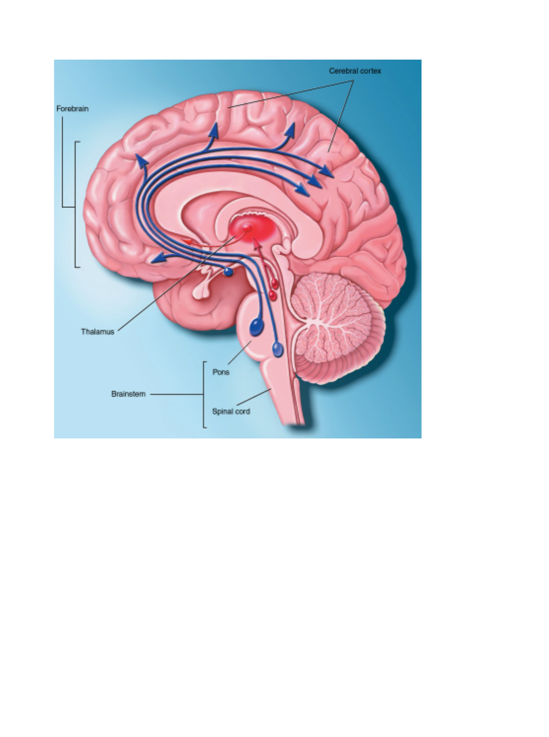
27
THE WAKING AND
SLEEPING BRAIN. Wakeful-
ness is maintained by activity in
two systems of neurons. Neu-
rons that make the neurotrans-
mitter acetylcholine are located
in two main arousal centers,
one in the brainstem and one in
the forebrain (red pathways).
The brainstem arousal center
supplies the acetylcholine for the
thalamus and brainstem, and
the forebrain arousal center
supplies that for the cerebral
cortex. Activation of these cen-
ters alone can create rapid eye
movement sleep. Activation of
other neurons that make
monoamine neurotransmitters
such as norepinephrine, sero-
tonin, and histamine (blue path-
ways) is needed for waking.
of nerve cells, in the ventrolateral preoptic nucleus, contains the
inhibitory neurotransmitters galanin and GABA. When the ven-
trolateral preoptic neurons fire, they are thought to turn o∑ the
arousal systems, causing sleep. Damage to the ventrolateral pre-
optic nucleus produces irreversible insomnia.
A second group of nerve cells in the lateral hypothalamus
influences and suppresses REM sleep. They contain the neuro-
transmitter orexin, which provides an excitatory signal to the
arousal system, particularly to the monoamine neurons. In exper-
iments in which the gene for the neurotransmitter orexin was
experimentally removed in mice, the animals became narcolep-
tic. Similarly, in two dog species with naturally occurring nar-
colepsy, an abnormality was discovered in the gene for the type 2
orexin receptor. Recent studies show that in humans with nar-
colepsy, the orexin levels in the brain and spinal fluid are abnor-
mally low. Thus, orexin appears to play a critical role in activat-
ing the monoamine system and in preventing abnormal
transitions, particularly into REM sleep.
Two main signals control our need for sleep and its circuitry.
First, there is homeostasis, or the body’s need to seek a natural
equilibrium of rest and sleep followed by wakefulness. Several
mechanisms for the signal of accumulating sleep have been sug-
gested. There is evidence that a chemical called adenosine, which
is linked to brain energy depletion, accumulates in the brain dur-
ing prolonged wakefulness and that it may drive sleep home-
ostasis. Interestingly, the drug ca∑eine, which is widely used to
prevent sleepiness, acts as an adenosine blocker.
If an individual does not get enough sleep, the sleep debt pro-
gressively accumulates and leads to a degradation of mental func-
tion. When the opportunity to sleep again comes, the individual
will sleep much more, to “repay” the debt. The slow wave sleep
debt is usually “paid o∑” first.
The other major influence on sleep cycles is the body’s circa-
dian clock, the suprachiasmatic nucleus. This small group of
nerve cells in the hypothalamus contains clock genes, which go
through a biochemical cycle of about 24 hours, setting the pace
for daily cycles of activity, sleep, hormones, and other bodily
functions. The suprachiasmatic nucleus also receives input
directly from the retina, and the clock can be reset by light, so that
it remains linked to the outside world’s day-night cycle. The
suprachiasmatic nucleus provides signals to the brain areas reg-
ulating sleep and arousal.

The ability to react in response to threatening
events has been with us since the time of our
ancient ancestors. In response to impending dan-
ger, muscles are primed, attention is focused, and
nerves are readied for action—“fight or flight.”
But in today’s complex and fast-paced world, this
response to stress is not enough, and the continued stimulation
of the systems that respond to threat or danger may contribute
to heart disease, obesity, arthritis, and depression, as well as accel-
erating the aging process.
Nearly two-thirds of ailments seen in doctors’ o≈ces are
commonly thought to be stress induced; indeed, stress can both
cause diseases and exacerbate existing ones. Surveys indicate that
60 percent of Americans feel they are under a great deal of stress
at least once a week. Costs due to stress from absenteeism, med-
ical expenses, and lost productivity are estimated at $300 billion
annually.
Stress is di≈cult to define because its e∑ects vary with each
individual. Specialists now define stress as any external stimulus
that threatens homeostasis—the normal equilibrium of body
function. Stress also can be induced by the belief that homeosta-
sis might soon be disrupted. Among the most powerful stressors
are psychological and psychosocial stressors that exist between
members of the same species. Lack or loss of control is a partic-
ularly important feature of severe psychological stress that can
have physiological consequences. Most harmful are the chronic
aspects of stress.
During the last six decades, researchers using animals found
that stress both helps and harms the body. When confronted with
a crucial physical challenge, properly controlled stress responses
can provide the extra strength and energy needed to cope. More-
over, the acute physiological response to stress protects the body
and brain and helps to reestablish or maintain homeostasis. But
stress that continues for prolonged periods can repeatedly elevate
the physiological stress responses or fail to shut them o∑ when
not needed. When this occurs, these same physiological mecha-
nisms can badly upset the body’s biochemical balance and accel-
erate disease.
Scientists also believe that the individual variation in
responding to stress is somewhat dependent on a person’s per-
ception of external events. This perception ultimately shapes his
or her internal physiological response. Thus, by controlling your
perception of events, you can do much to avoid the harmful con-
sequences of the sorts of mild to moderate stressors that typically
aΩict Westernized humans.
The immediate response
A stressful situation activates three major communication sys-
tems in the brain that regulate bodily functions. Scientists have
come to understand these complex systems through experiments
primarily with rats, mice, and nonhuman primates, such as mon-
keys. Scientists then verified the action of these systems in
humans.
The first of these systems is the voluntary nervous system,
which sends messages to muscles so that we may respond to sen-
sory information. For example, the sight of a growling bear on a
trail in Yellowstone National Park prompts you to run as quickly
as possible.
The second communication system is the autonomic nervous
system.It combines the sympathetic or emergency branch, which
gets us going in emergencies, and the parasympathetic or calm-
ing branch, which keeps the body’s maintenance systems, such as
digestion, in order and calms the body’s responses to the emer-
gency branch.
Each of these systems has a specific task. The emergency
branch causes arteries supplying blood to the muscles to relax in
order to deliver more blood, allowing greater capacity to act. At
the same time, the emergency system reduces blood flow to the
skin, kidneys, and digestive tract and increases blood flow to the
muscles. In contrast, the calming branch helps to regulate bodily
functions and soothe the body once the stressor has passed, pre-
venting the body from remaining too long in a state of mobiliza-
tion. Left mobilized and unchecked, these body functions could
lead to disease. Some actions of the calming branch appear to
reduce the harmful e∑ects of the emergency branch’s response to
stress.
The brain’s third major communication process is the neu-
roendocrine system,which also maintains the body’s internal
functioning. Various “stress hormones” travel through the blood
and stimulate the release of other hormones, which a∑ect bodily
Stress
T
28
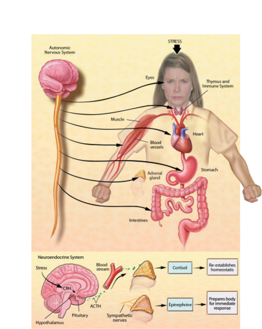
29
THE STRESS REACTION.
When stress occurs, the sympa-
thetic nervous system is trig-
gered. Norepinephrine is
released by nerves, and epi-
nephrine is secreted by the
adrenal glands. By activating
receptors in blood vessels and
other structures, these sub-
stances ready the heart and
working muscles for action.
Acetylcholine is released in the
parasympathetic nervous sys-
tem, producing calming effects.
The digestive tract is stimulated
to digest a meal, the heart rate
slows, and the pupils of the
eyes become smaller. The neu-
roendocrine system also main-
tains the body’s normal
internal functioning. Corti-
cotrophin-releasing hor-
mone (CRH), a peptide
formed by chains of amino
acids, is released from the
hypothalamus, a collection of
cells at the base of the brain
that acts as a control center for
the neuroendocrine system.
CRH travels to the pituitary
gland, where it triggers the
release of adrenocorticotropic
hormone (ACTH). ACTH travels
in the blood to the adrenal
glands, where it stimulates the
release of cortisol.
processes such as metabolic rate and sexual functions.
Major stress hormones are epinephrine (also known as adre-
naline) and cortisol.When the body is exposed to stressors, epi-
nephrine, which combines elements of hormones and the ner-
vous system, is quickly released into the bloodstream to put the
body into a general state of arousal and enable it to cope with a
challenge.
The adrenal glands secrete glucocorticoids, which are hor-
mones a∑ecting glucose metabolism. In primates, the main glu-
cocorticoid is cortisol (hydrocortisone), whereas in rodents, it is
corticosterone. Some of the actions of glucocorticoids help to
mediate the stress response, while some of the other, slower
actions counteract the primary response to stress and help
reestablish homeostasis. Over the short run, adrenaline mobilizes
energy and delivers it to muscles for the body’s response. With
prolonged exposure, cortisol enhances feeding and helps the
body recover from energy mobilization. Cortisol also raises blood
pressure and increases the risk of adult-onset diabetes, immuno-
suppression, reproductive impairments, and depression, among
other di≈culties.
Acute stress enhances memory of threatening situations and
events, increases activity of the immune system, and helps pro-

tect the body from pathogens. The two major stress hormones,
cortisol and adrenaline, facilitate the movement of immune cells
from the bloodstream and storage organs such as the spleen into
tissue where they are needed to defend against infection.
Glucocorticoids also a∑ect food intake during the sleep-wake
cycle. Cortisol levels peak in the body in the early morning hours
just before waking. This hormone acts as a wake-up signal and
helps to turn on appetite and physical activity. This e∑ect of glu-
cocorticoids may help to explain disorders such as jet lag, which
results when the light-dark cycle is altered by travel over long dis-
tances, causing the body’s biological clock to reset itself more
slowly. Until that clock is reset, cortisol secretion and hunger, as
well as sleepiness and wakefulness, occur at inappropriate times
of day in the new location.
Glucocorticoids do more than help the body respond to
stress. In fact, they are an integral part of daily life and the adap-
tation to environmental change. The adrenal glands help protect
us from stress and are essential for survival.
Chronic stress
When glucocorticoids or adrenaline are secreted in response to
the prolonged psychological stress commonly encountered by
humans, the results are not ideal. Normally, bodily systems gear
up under stress and release hormones to improve memory,
increase immune function, enhance muscular activity, and
restore homeostasis. If you are not fighting or fleeing, but stand-
ing frustrated in a supermarket checkout line or sitting in a
tra≈c jam, you are not engaging in muscular exercise. Yet these
systems continue to be stimulated, and when they are stimulated
chronically, there are di∑erent consequences: Memory is
impaired, immune function is suppressed, and energy is stored
as fat.
Overexposure to cortisol also can lead to weakened muscles
and the suppression of major bodily systems. Elevated epineph-
rine production increases blood pressure. Together, elevated cor-
tisol and epinephrine can contribute to chronic hypertension
(high blood pressure), abdominal obesity, and atherosclerosis
(hardening of the arteries). Adrenaline also increases the activity
of body chemicals that contribute to inflammation, and these
chemicals add to the burden of chronic stress, potentially leading
to atherosclerosis, arthritis, and possibly also aging of the brain.
Scientists have identified a variety of stress-related disorders,
including colitis, high blood pressure, clogged arteries, impotency
and loss of sex drive in males, irregular menstrual cycles in
females, adult-onset diabetes, and possibly cancer. Aging rats
show impairment of neuronal function in the hippocampus—
an area of the brain important for learning, memory, and emo-
tion—as a result of cortisol secretion throughout their lifetimes.
Overexposure to glucocorticoids also increases the number of
neurons damaged by stroke. Moreover, prolonged exposure
before or immediately after birth can cause a decrease in the nor-
mal number of brain neurons and smaller brain size.
The immune system,which receives messages from the ner-
vous system, also is sensitive to many of the circulating hormones
of the body, including stress hormones. Moderate to high levels
of glucocorticoids act to suppress immune function, although
acute elevations of stress hormones actually facilitate immune
function.
Although acute stress-induced immunoenhancement can be
protective against disease pathogens, the glucocorticoid-induced
immunosuppression can also be beneficial. It reduces inflamma-
tion and counteracts allergic reactions and autoimmune responses,
which occur when the body’s defenses turn against body tissue.
Synthetic glucocorticoids like hydrocortisone and prednisone are
used often to decrease inflammation and autoimmunity. But glu-
cocorticoids may be harmful in the case of increased tumor
growth associated with stress in experiments on animals—an
area of intense research yet to yield any final conclusions.
One important determinant of the immune system’s resis-
tance or susceptibility to disease may be a person’s sense of con-
trol as opposed to a feeling of helplessness. This phenomenon
may help explain large individual variations in response to dis-
ease. Scientists are trying to identify how the perception of con-
trol or helplessness influences physiological processes that regu-
late immune function.
The cardiovascular system receives many messages from the
autonomic nervous system, and stressful experiences have an
immediate and direct e∑ect on heart rate and blood pressure. In
the short run, these changes help in response to stressors. But
when stressors are chronic and psychological, the e∑ect can be
harmful and result in accelerated atherosclerosis and increased
risk for heart attack. Research supports the idea that people hold-
ing jobs that carry high demands and low control, such as tele-
phone operators, waiters, and cashiers, have higher rates of heart
disease than people who can dictate the pace and style of their
working lives.
Behavioral type a∑ects a person’s susceptibility to heart
attack. People at greatest risk are hostile, irritated by trivial things,
and exhibit signs of struggle against time and other challenges.
Researchers found that two groups of men—one with high
hostility scores and the other with low hostility scores—exhib-
ited similar increases in blood pressure and muscle blood flow
when performing a lab test. This finding confirmed that hostility
scores do not predict the biological response to simple mental
tasks.
Then the researchers added harassment to the test by leading
the subjects to believe that their performances were being
unfairly criticized. Men with high hostility scores showed much
larger increases in muscle blood flow and blood pressure and
showed slower recovery than those with low hostility scores. Sci-
entists found that harassed men with high hostility scores had
larger increases in levels of stress hormones. Thus, if you have
personality traits of hostility, learning to reduce or avoid anger
could be important to avoid cardiovascular damage.
30

31
Peuroscientists believe that the brain can remain
relatively healthy and fully functioning as it ages
and that diseases cause the most severe decline in
memory, intelligence, verbal fluency, and other
tasks. Researchers are investigating the normal
changes that occur over time and their e∑ect on
reasoning and other intellectual activities.
It appears that the e∑ects of age on brain function vary
widely. Almost everyone gets a bit forgetful in old age, particu-
larly in forming memories of recent events. For example, once
you reach your 70s, you may start to forget names or phone num-
bers or respond more slowly to conflicting information. This is
not disease. However, other individuals develop senile dementia,
the progressive and severe impairment in mental function that
interferes with daily living. The senile dementias include
Alzheimer’s and cerebrovascular diseases and a∑ect about 1 per-
cent of people younger than age 65, with the incidence increas-
ing to nearly 50 percent in those older than 85. In a small, third
group, mental functioning seems una∑ected by age. Many peo-
ple do well throughout life and continue to do well even when
old. The oldest human, Jeanne Calment, kept her wits through-
out her 122-year lifespan.
It’s important to understand that scientific studies measure
trends and reflect what happens to the norm—they don’t tell
what happens to everybody. Some people in their 70s and 80s
function as well as those in their 30s and 40s. The wisdom and
experience of older people often make up for deficits in perfor-
mance.
The belief that pronounced and progressive mental decline
is inevitable was and still is popular for several reasons. For one,
until the 20th century, few people lived past 65. In 1900, when
average life expectancy was about 47 years, 3 million people, or 4
percent of the population, were older than age 65 and were typ-
ically ill. In 2003, when life expectancy was more than 77 years,
nearly 36 million people, or more than 12 percent of the popula-
tion, were older than age 65. A generation ago, frailty was seen
among people in their 60s; today it is more typical among those
in their 80s. Moreover, few people challenged the notion that
aging meant inevitable brain decline because scientists knew lit-
tle about the brain or the aging process. Today’s understanding
of how the normal brain ages comes from studies of the nervous
system that began decades ago and are just now bearing results.
Modern technologies now make it possible to explore the struc-
ture and function of the brain in more depth than ever before
and to ask questions about what actually happens in its aging
cells.
Thus, neuroscientists are increasingly able to distinguish
between the processes of normal aging and disease. Although
some changes do occur in normal aging, they are not as severe as
scientists once thought.
All human behavior is determined by how well the brain’s
communication systems work. Often a failure in the cascade of
one of these systems results in a disturbance of normal function.
Such a failure may be caused by an abnormal biochemical process
or a loss of neurons.
The cause of brain aging still remains a mystery. Dozens of
theories abound. One says that specific “aging genes”are switched
on at a certain time of life. Another points to genetic mutations
or deletions. Other theories implicate hormonal influences, an
immune system gone awry, and the accumulation of damage
caused by free radicals,cell byproducts that destroy fats and pro-
teins vital to normal cell function.
Aging neurons
The brain reaches its maximum weight near age 20 and slowly
loses about 10 percent of its weight over a lifetime. Subtle changes
in the chemistry and structure of the brain begin at midlife for
most people. During a lifetime, the brain is at risk for losing some
of its neurons, but widespread neuron loss is not a normal
process of aging. Brain tissue can respond to damage or loss of
neurons in Alzheimer’s disease or after stroke by expanding den-
drites and fine-tuning connections between neurons. A damaged
brain neuron can readjust to damage only if its cell body remains
intact. If it does, regrowth can occur in dendrites and axons.
When neurons are destroyed, nearby surviving neurons can com-
pensate, in part, by growing new dendrites and connections.
Intellectual capacity
From the first large studies to monitor the same group of healthy
humans for many years, scientists have uncovered unexpected
Aging
N
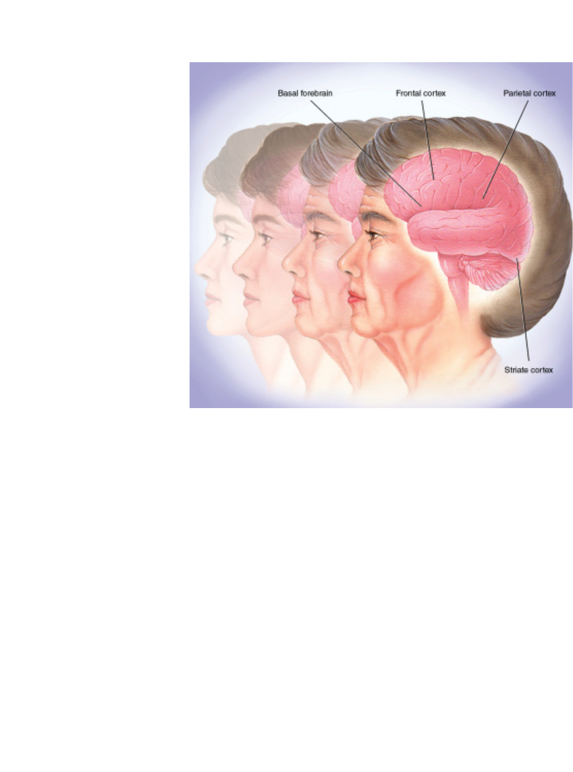
results. They report declines in some mental functions and
improvements in others. In several studies, the speed of carrying
out certain tasks became slower, but vocabulary improved. Sev-
eral studies found less severe declines in the type of intelligence
relying on learned or stored information compared with the type
that uses the ability to deal with new information.
This research is supported by animal studies in which scien-
tists found that changes in mental function are subtle. For exam-
ple, in rodents and primates in which only minor brain abnor-
malities can be detected, certain spatial tasks, such as navigating
to find food, tend to become more di≈cult with age.
The aging brain is only as resilient as its circuitry. Scientists
debate whether this circuitry is changed only by neuron atrophy
or whether some neuron loss over time also is inevitable. In any
event, when the circuitry begins to break down, remaining neu-
rons can adapt by expanding their roles.
Learning conditions may dictate what happens to brain cells.
Studies of rats shed light on some of the changes that occur in
brain cells when the animals live in challenging and stimulating
environments. In tests of middle-aged rats exposed to such envi-
ronments, researchers found that dendrites in the cerebral cor-
tex developed more and longer branches than did rats housed in
isolated conditions. Another study showed that brain cells in rats
given acrobatic training had more synapses per cell than rats
given only physical exercise or rats that were inactive. The scien-
tists concluded that motor learning generates new synapses.
Physical exercise, however, improved blood circulation in the
brain. Aerobic exercise can also improve human cognitive per-
formance.
Other scientists report that rats reared in a stimulating envi-
ronment made significantly fewer errors in a maze test than did
similar rats kept in an isolated environment. Moreover, the stim-
ulated rats showed an increase in brain weight and cortical thick-
ness compared with animals in the control group.
In response to enriched environments, older rats tend to
form new dendrites and synapses, just as younger animals do. But
the response is more sluggish and not as large. Compared with
younger rats, older rats have less growth of the new blood vessels
that nourish neurons.
Although much has been learned about the aging brain,
many questions remain. For instance, does the production of
proteins decline with age in all brain neurons? In a given neuron,
does atrophy cause a higher likelihood of death? How does aging
a∑ect gene expression in the brain—the organ with the greatest
number of active genes? Are there gender di∑erences in brain
aging that may be due to hormonal changes at menopause?
Neuroscientists speculate that certain genes may be linked to
events leading to death in the nervous system. By understanding
the biology of the proteins produced by genes, scientists hope to
be able to influence the survival and function of neurons.
THE AGING BRAIN. Studies
of people who have died contra-
dict the popular belief that
adults lose an enormous number
of neurons every day. In fact,
many areas of the brain, pri-
marily in the cortex, maintain
most of their neurons. Examples
include the parietal cortex,
which plays a role in sensory
processes and language, and
the striate cortex, which
processes visual information.
The connectivity between neu-
rons changes with aging, so that
the brain is constantly capable
of being modified or improved.
32

33
ipolar disorder. Patients with bipolar disor-
der, previously known as manic-depressive ill-
ness, usually experience episodes of deep depres-
sion and manic highs, with a return to relatively
normal functioning in between. They also have
an increased risk of suicide. Bipolar disorder
a∑ects 1.2 percent of Americans age 18 or older annually, or 2.2
million individuals. Approximately equal numbers of men and
women su∑er from this disorder.
Bipolar disorder tends to be chronic, and episodes can be-
come more frequent without treatment. Because bipolar disor-
der runs in families, e∑orts are underway to identify the respon-
sible gene or genes.
Bipolar patients can benefit from a broad array of treatments.
One of these is lithium. During the 1940s, researchers showed that
lithium injections into guinea pigs made them placid, which
implied mood-stabilizing e∑ects. When given to manic patients,
lithium calmed them and enabled them to return to work and
live relatively normal lives. Regarded as both safe and e∑ective,
lithium is often used to prevent recurrent episodes.
Other useful medications include certain anticonvulsants, such
as valproate or carbamazepine, which can have mood-stabilizing
e∑ects, like lithium, and may be especially useful for di≈cult-to-
treat bipolar episodes. Newer anticonvulsant medications are being
studied to determine how well they work in stabilizing mood cycles.
Epilepsy
Epilepsy, a chronic neurological disorder characterized by sudden,
disorderly discharge of brain cells, is marked by recurrent, unpro-
voked seizures that temporarily alter one or more brain functions.
The disorder a∑ects approximately 1 percent of the population.
Many di∑erent types of epilepsy have been recognized.
Epilepsy can start at any age and can be idiopathic (having an
uncertain cause) or symptomatic. Most idiopathic epilepsies are
likely due to inheriting a mutant gene, more than a dozen of
which have been identified during the last decade. Symptomatic
epilepsies result from a wide variety of brain diseases or
injuries, including birth trauma, brain infection such as abscess
or meningitis, brain tumors, and stroke.
Seizures are of two types, generalized and partial. Generalized
seizures, which typically result in loss of consciousness, can cause
several behavioral changes, including convulsions or sudden
changes in muscle tone, and arise when there is simultaneous exces-
sive electrical activity over a wide area of the brain. Partial seizures
may occur with maintained consciousness or with altered aware-
ness and behavioral changes. Partial seizures can produce localized
visual, auditory, and skin sensory disturbances; repetitive uncon-
trolled movements; or confused, automatic behaviors. Such seizures
arise from excessive electrical activity in a limited area of the brain.
There are more than a dozen antiseizure medications, approx-
imately half of which have been introduced in the last several
years, available to prevent seizures. The principal targets of anti-
seizure drugs are voltage-gated ion channels permeable to sodium
or calcium and synapses using the transmitter GABA, a naturally
occurring substance in the brain that acts to inhibit electrical dis-
charge. Identification of the mutant genes underlying human
epilepsy may provide new targets for the next generation of anti-
seizure drugs. In many instances, epilepsy can be controlled with
a single antiseizure drug that lessens the frequency of seizures, but
sometimes a combination of drugs is necessary. Complete control
of seizures can be achieved in more than 50 percent of patients,
and another 25 percent can be improved significantly. It is hoped
that the newly available antiseizure drugs will provide complete
control in additional patients.
Surgery, considered for patients who do not respond to anti-
seizure drugs, should be performed only at specialized medical
centers qualified to evaluate patients and perform epilepsy
surgery. Epilepsy surgery requires precise location and removal
of the area of the brain where the seizures originate. After surgery,
about 90 percent of properly selected patients experience strik-
ing improvement or complete remission of seizures.
A new form of epilepsy treatment, electrical stimulation ther-
apy, was introduced during the mid-1990s as another option for
hard-to-control seizures. The implantable pacemaker-like device
delivers small bursts of electrical energy to the brain via the vagus
nerve on the side of the neck.
Major depression
This condition, with its harrowing feelings of sadness, hopeless-
ness, pessimism, loss of interest in life, and reduced emotional
Advances
B
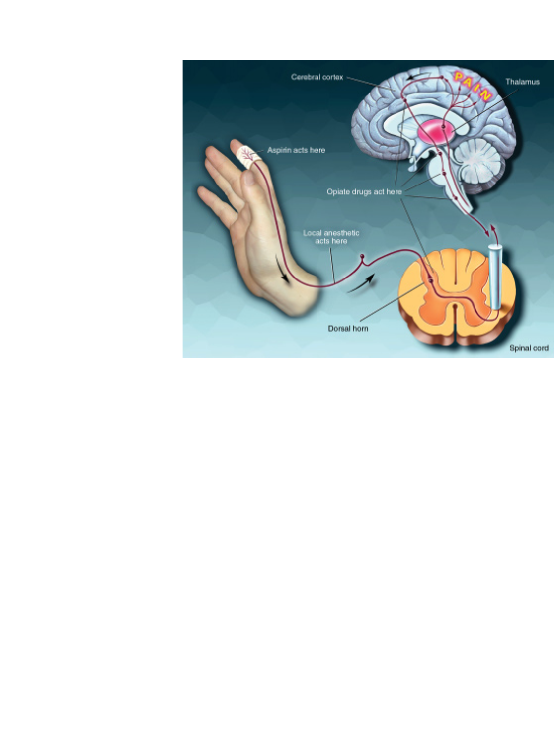
34
well-being, is one of the most common and debilitating mental
disorders and one of the leading causes of morbidity worldwide.
Depression is as disabling as heart disease or arthritis. Depressed
individuals are 18 times more likely to attempt suicide than peo-
ple with no mental illness.
Annually, major depression a∑ects 5 percent of the popula-
tion, or 9.8 million Americans, aged 18 years and older. Fortu-
nately, 80 percent of patients respond to drugs, psychotherapy, or
a combination of the two. Some severely depressed patients can
be helped with electroconvulsive therapy.
Depression arises from many causes: biological (including
genetic), psychological, environmental, or a combination of
these. Stroke, hormonal disorders, antihypertensives, and birth
control pills also can play a part.
Physical symptoms—disturbances of sleep, sex drive, energy
level, appetite, and digestion—are common. Some of these
symptoms may reflect the fact that the disorder a∑ects the deli-
cate hormonal feedback system linking the hypothalamus, the
pituitary gland, and the adrenal glands. For example, many
depressed patients secrete excess cortisol, a stress hormone, and
do not respond appropriately to a hormone that should counter
cortisol secretion. When tested in sleep laboratories, depressed
patients’ electroencephalograms (EEGs) often exhibit abnormal-
ities in their sleep patterns.
The modern era of drug treatment for depression began in
the late 1950s. Most antidepressants a∑ect norepinephrine or
serotonin in the brain, apparently by correcting the abnormal sig-
nals that control mood, thoughts, and other sensations. The tri-
cyclic antidepressants, such as imipramine, primarily block the
reabsorption and inactivation of serotonin and norepinephrine
to varying degrees. Another class of antidepressant medications
is the monoamine oxidase inhibitors (MAOIs). These agents
inhibit monoamine oxidase, an enzyme that breaks down sero-
tonin and norepinephrine, allowing these chemicals to remain
active. MAOIs available for use include isocarboxazid, phenel-
zine, and tranylcypromine.
The popular medication fluoxetine (Prozac) is the first of a
class of drugs called selective serotonin reuptake inhibitors, or
SSRIs. SSRIs block the reabsorption and inactivation of serotonin
and keep it active in certain brain circuits. Hence, they are func-
tionally similar to the tricyclic antidepressants, but act selectively
on the serotonin system. There are also several newer antidepres-
sants available, such as bupropion, that are also very e∑ective but
seem to have a di∑erent and as yet unknown mechanism of action.
Pain
If there is a universal experience, pain is it. Each year, more than
97 million Americans su∑er chronic, debilitating headaches or a
bout with a bad back or the pain of arthritis—all at a total cost
of some $100 billion. But it need not be that way. New discover-
ies about how chemicals in the body transmit and regulate pain
messages have paved the way for new treatments for both chronic
and acute pain.
Until the mid-19th century, pain relief during surgery relied
on natural substances, such as opium, alcohol, and cannabis. All
were inadequate and short-lived. Not until 1846 did doctors dis-
HOW PAIN KILLERS
WORK. At the site of injury,
the body produces prosta-
glandins that increase pain sen-
sitivity. Aspirin, which acts
primarily in the periphery, pre-
vents the production of prosta-
glandins. Acetaminophen is
believed to block pain impulses
in the brain itself. Local anes-
thetics intercept pain signals
traveling up the nerve. Opiate
drugs, which act primarily in the
central nervous system, block
the transfer of pain signals from
the spinal cord to the brain.

cover the anesthetic properties of ether, first in animals and then
in humans. Soon, the usefulness of chloroform and nitrous oxide
became known and heralded a new era in surgery. The dozens of
drugs used today during surgery abolish pain, relax muscles, and
induce unconsciousness. Other agents reverse these e∑ects.
Local anesthesia is used in a limited area of a person’s body to
prevent pain during examinations, diagnostic procedures, treat-
ments, and surgical operations. The most famous of these agents,
which temporarily interrupt the action of pain-carrying nerve
fibers, is Novocain, which dentists used as a local anesthetic for
many years; lidocaine is more popular today. Very recently, lido-
caine has been used in a slow-release patch to provide long-last-
ing pain in localized, specific parts of the body.
Analgesia refers to loss of pain sensation without loss of sen-
sitivity to touch. The two main types of analgesics are nonopioids
(aspirin and related nonsteroidal anti-inflammatory drugs
[NSAIDs] such as ibuprofen, naproxen, and acetaminophen) and
opioids (morphine, codeine). Nonopioid analgesics are useful for
treating mild or moderate pain, such as headache or toothache.
Because NSAIDs are anti-inflammatory, they are e∑ective for
treating such inflammatory conditions as arthritis. Moderate
pain also can be treated by combining a mild opioid, such as
codeine, with aspirin. Opioids are the most potent painkillers and
are used for severe pain, such as that occurring after major chest
or abdominal surgery.
Studies of the body’s own pain-control system not only
demonstrated the existence of naturally occurring opioids (the
endorphins) but also identified the receptors (targets) through
which opioids exert their e∑ects. These findings led to the use of
injections of morphine and endorphins, and other opioids, into
the cerebrospinal fluid (in which the spinal cord is bathed) with-
out causing paralysis, numbness, or other severe side e∑ects. This
technique came about through experiments with animals that
first showed that injecting opioids into the spinal cord could pro-
duce profound pain control. This technique is now commonly
used in humans to treat pain after surgery and is a mainstay for
pain relief after caesarean section.
Although NSAIDs and opioids are quite e∑ective for pain
produced by tissue injury, they are much less e∑ective when the
pain results from injury to the nervous system. These so-called
neuropathic pains include the pain of diabetic neuropathy, post-
therapeutic neuralgia, phantom limb pain, and post-stroke pain.
For these pains, anticonvulsants are more e∑ective, and some
patients can be helped with low doses of antidepressants.
New targets, however, are on the horizon. Molecular biology
has identified many molecules (ion channels and receptors) that
are predominantly, if not exclusively, expressed by the nociceptor,
which is the first-order nerve fiber in the pain pathway. Because
adverse side e∑ects of drugs arise from the widespread location
of the molecules targeted by analgesics (e.g., constipation results
from morphine’s action on opioid receptors in the gut), new anal-
gesics that target only the nociceptor may have a better side-e∑ect
profile. Among the many new targets are glutamate receptors,
vanilloid receptors (which are targeted by capsaicin, the active
ingredient in hot peppers), and a variety of acid-sensing ion chan-
nels. Blocking the activity of many of these molecules has proven
e∑ective in animal studies, suggesting that the development of
drugs that target these molecules in humans may have great value
for the treatment of persistent pain.
Parkinson’s disease
This neurologic disorder aΩicts 1 million individuals in the
United States, most of whom are older than 50. Parkinson’s dis-
ease is characterized by symptoms of slowness of movement,
muscular rigidity, tremor, and postural instability.
The discovery in the late 1950s that the level of dopamine was
decreased in the brains of Parkinson’s patients was followed in
the 1960s by the successful treatment of this disorder by admin-
istration of the drug levodopa, which is converted to dopamine
in the brain. The successful treatment of Parkinson’s by replace-
ment therapy is one of the greatest success stories in neurology.
Levodopa is now combined with another drug, carbidopa, that
reduces the peripheral breakdown of levodopa, thus allowing
greater levels to reach the brain and reducing side e∑ects. Also
playing an important role are newer drugs, such as inhibitors of
dopamine breakdown and dopamine agonists, that act directly
on dopamine receptors.
Genetic studies have demonstrated several heritable gene
abnormalities in certain families, but almost all cases of Parkin-
son’s occur sporadically. It is believed that hereditary factors may
render some individuals more vulnerable to environmental fac-
tors such as pesticides. The discovery in the late 1970s that a
chemical substance, MPTP, can cause parkinsonism in drug
addicts stimulated intensive research on the causes of the disor-
der. MPTP was accidentally synthesized by illicit drug designers
seeking to produce a heroin-like compound. MPTP was found to
be converted in the brain to a substance that destroys dopamine
neurons. Parkinson’s is now being intensively studied in primate
MPTP models.
In the past several decades, scientists have shown in primate
models of Parkinson’s that specific regions in the basal ganglia,
collections of cell bodies deep in the brain, are abnormally over-
active. Most importantly, they found that surgical destruction of
these overactive nuclei—the pallidum and subthalamic
nucleus—can greatly reduce symptoms of Parkinson’s disease.
The past decade has witnessed a resurgence in this surgical pro-
cedure, pallidotomy, and more recently chronic deep-brain stim-
ulation. These techniques are highly successful for treating
patients who have experienced significant worsening of symp-
toms and are troubled by the development of drug-related invol-
untary movements. The past decade has also seen further
attempts to treat such patients with surgical implantation of cells,
such as fetal cells, capable of producing dopamine. Replacement
therapy with stem cells also is being explored.
35

Addiction. Drug abuse is one of the nation’s
most serious health problems. Indeed, 6 per-
cent of Americans, roughly 15 million people,
abuse drugs on a regular basis. Recent esti-
mates show that the abuse of drugs, including
alcohol and nicotine from tobacco, costs the
nation more than $276 billion each year.
If continued long enough, drug abuse—often defined as
harmful drug use—can eventually alter the very structure and
chemical makeup of the brain, producing a true brain disorder.
This disorder is called drug addiction or drug dependence.Drug
addiction is defined as having lost control over drug taking, even
in the face of adverse physical, personal, or social consequences.
People abuse drugs for a simple reason: Drugs produce feel-
ings of pleasure or remove feelings of stress and emotional pain.
Neuroscientists have found that almost all abused drugs produce
pleasure by activating a specific network of neurons called the
brain reward system.The circuit is normally involved in an
important type of learning that helps us to stay alive. It is acti-
vated when we fulfill survival functions, such as eating when we
are hungry or drinking when we are thirsty. In turn, our brain
rewards us with pleasurable feelings that teach us to repeat the
task. Because drugs inappropriately turn on this reward circuit,
people want to repeat drug use.
Neuroscientists also have learned specifically how drugs a∑ect
neurons to exert their influence. Neurons release special chemi-
cals, called neurotransmitters,to communicate with each other.
Abused drugs alter the ways neurotransmitters carry their mes-
sages from neuron to neuron. Some drugs mimic neurotrans-
mitters, whereas others block them. Still others alter the way that
the neurotransmitters are released or inactivated. The brain
reward system is inappropriately activated because drugs alter the
chemical messages sent among neurons in this circuit.
Finally, neuroscientists also have learned that addiction
requires more than the activation of the brain reward system. The
process of becoming addicted appears to be influenced by many
factors. Motivation for drug use is an important one. For exam-
ple, people who take drugs to get high may get addicted, but peo-
ple who use them properly as medicine rarely do. Genetic sus-
ceptibility or environmental factors, such as stress, may also alter
the way that people respond to drugs. In addition, the develop-
ment of tolerance—the progressive need that accompanies
chronic use for a higher drug dose to achieve the same e∑ect—
varies in di∑erent people, as does drug dependence—the adap-
tive physiological state that results in withdrawal symptoms when
drug use stops. Tolerance and dependence are standard responses
of the brain and body to the presence of drugs. However, addic-
tion requires that these occur while a motivational form of depen-
dence—the feeling that a person can’t live without a drug,
accompanied by negative a∑ective states—is also developing.
Together, these insights on abuse and addiction are leading to
new therapies.
Nicotine In 2003, more than 70 million people smoked, at
least occasionally, making nicotine one of the most widely abused
substances. Tobacco kills more than 430,000 U.S. citizens each
year—more than alcohol, cocaine, heroin, homicide, suicide, car
accidents, fire, and AIDS combined. Tobacco use is the leading
preventable cause of death in the United States. Smoking is
responsible for approximately 7 percent of total U.S. health care
costs, an estimated $80 billion each year. The direct and indirect
costs of smoking are estimated at more than $138 billion per year.
Nicotine, the addicting substance in tobacco, acts through the
well-known cholinergic nicotinic receptor. This drug can act as
both a stimulant and a sedative. Immediately after exposure to
nicotine, there is a “kick” caused in part by the drug’s stimulation
of the adrenal glands and resulting discharge of epinephrine. The
rush of adrenaline stimulates the body and causes a sudden
release of glucose as well as an increase in blood pressure, respi-
ration, and heart rate. Nicotine also suppresses insulin output
from the pancreas, which means that smokers are always slightly
hyperglycemic. In addition, nicotine releases dopamine in the
brain regions that control pleasure and motivation. This mecha-
nism is thought to underlie the pleasurable sensations experi-
enced by many smokers.
Much better understanding of addiction, coupled with the
identification of nicotine as an addictive drug, has been instru-
mental in the development of treatments. Nicotine gum, the
transdermal patch, nasal spray, and inhalers all appear to be
equally e∑ective in treating more than 1 million people addicted
to nicotine. These techniques are used to relieve withdrawal
A
Challenges
36
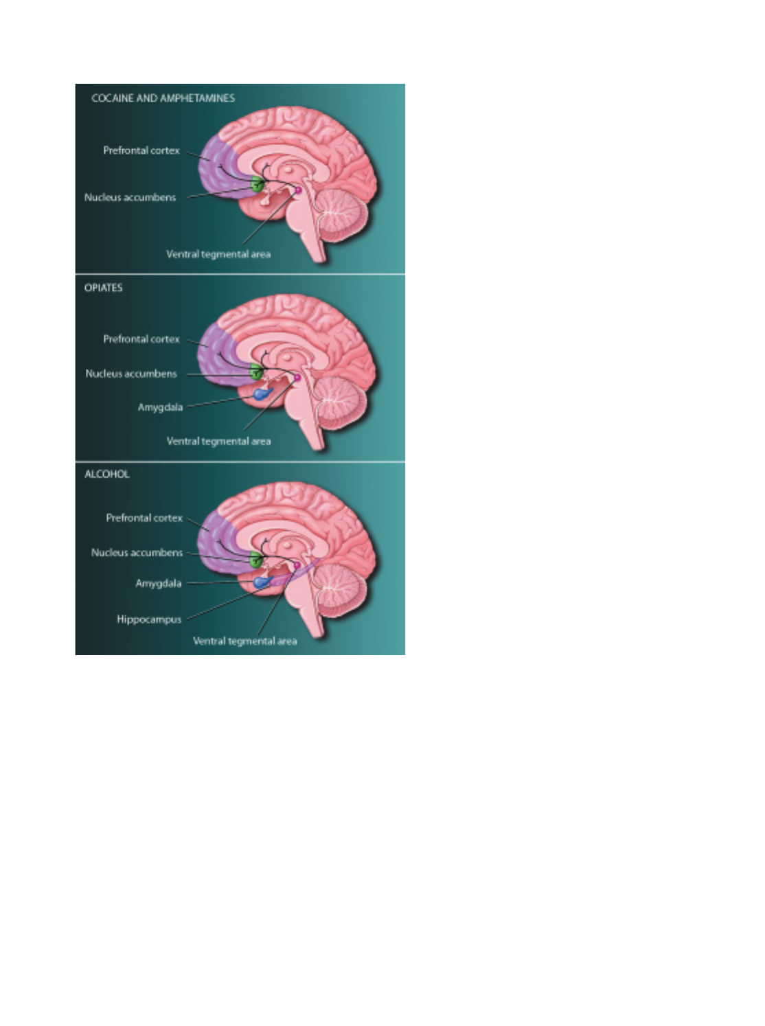
37
symptoms and produce less severe physiological alterations than
tobacco-based systems. They generally provide users with lower
overall nicotine levels than they receive with tobacco, as well as
totally eliminating exposure to smoke and its deadly contents.
The first non-nicotine prescription drug, bupropion, an antide-
pressant marketed as Zyban, has been approved for use as a phar-
macological treatment for nicotine addiction. Behavioral treat-
ments are important for helping an individual learn coping skills
for both short- and long-term prevention of relapse.
Psychostimulants In 2003, there were an estimated 2.3 mil-
lion chronic cocaine users and 5.9 million occasional cocaine
users in the United States. A popular chemically altered form of
cocaine, crack, is smoked. It enters the brain in seconds, pro-
ducing a rush of euphoria and feelings of power and self-
confidence. The key biochemical factor that underlies the rein-
forcing e∑ects of psychostimulants is the brain chemical
dopamine. We feel pleasure when dopamine-containing neurons
release dopamine into specific brain areas that include a special
portion of the nucleus accumbens.Cocaine and amphetamines
produce intense feelings of euphoria by increasing the amount
of dopamine that is available to send messages within the brain
reward system.
Cocaine users often go on binges, consuming a large amount
of the drug in just a few days. A crash occurs after this period of
intense drug-taking and includes symptoms of emotional and
physical exhaustion and depression. These symptoms may result
from an actual crash in dopamine function and the activity of
another brain chemical, serotonin, as well as an increased
response of the brain systems that react to stress. Vaccines to
produce antibodies to cocaine in the bloodstream are in clinical
trials.
Opiates Humans have used opiate drugs, such as morphine,
for thousands of years. Monkeys and rats readily self-administer
heroin or morphine and, like humans, will become tolerant and
physically dependent with unlimited access. Withdrawal symp-
toms range from mild flu-like discomfort to major physical ail-
ments, including severe muscle pain, stomach cramps, diarrhea,
and unpleasant mood.
Opiates, like psychostimulants, increase the amount of
dopamine released in the brain reward system and mimic the
e∑ects of endogenous opioids such as opioid peptides. Heroin
injected into a vein reaches the brain in 15 to 20 seconds and
binds to opiate receptors found in many brain regions, including
the reward system. Activation of the receptors in the reward cir-
cuits causes a brief rush of intense euphoria, followed by a cou-
ple of hours of a relaxed, contented state.
Opiates create e∑ects like those elicited by the naturally
occurring opioid peptides. They relieve pain, depress breathing,
cause nausea and vomiting, and stop diarrhea—important med-
ical uses. In large doses, heroin can make breathing shallow or
stop altogether—the cause of death in thousands of people who
have died of heroin overdose.
BRAIN DRUG REWARD SYSTEMS. Scientists are not certain
about all the structures involved in the human brain reward system.
However, studies of rat and monkey brains, and brain imaging stud-
ies in humans, have provided many clues. These illustrations show
what areas are most likely part of the reward systems in the human
brain. A central group of structures is common to the actions of all
drugs. These structures include a collection of dopamine-containing
neurons found in the ventral tegmental area. These neurons are
connected to the nucleus accumbens and other areas, such as the
prefrontal cortex. Cocaine exerts its effects mainly through this sys-
tem. Opiates act in this system and many other brain regions, includ-
ing the amygdala, that normally use opioid peptides. Opioids are
naturally occurring brain chemicals that induce the same actions as
drugs, such as heroin and morphine. Alcohol activates the core
reward system and additional structures throughout the brain because
it acts where GABA and glutamate are used as neurotransmitters.
GABA and glutamate are widely distributed in the brain, including
the cortex, hippocampus, amygdala, and nucleus accumbens.
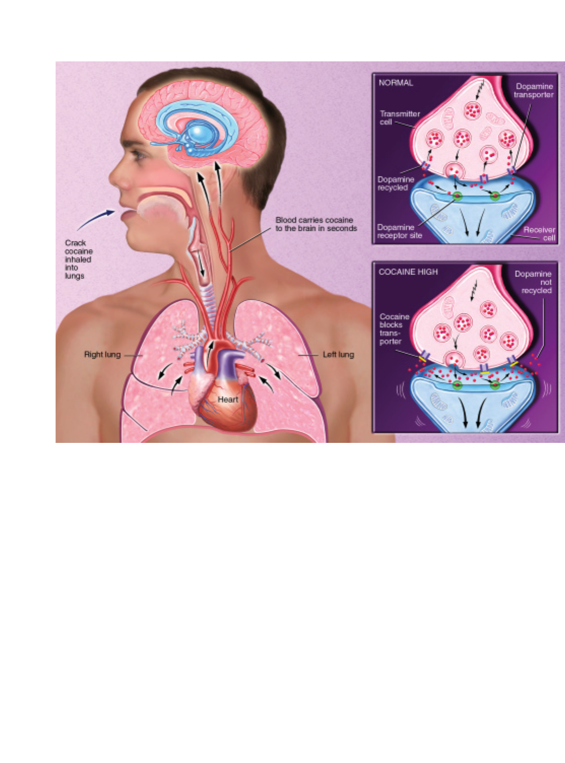
A standard treatment for opiate addiction involves metha-
done,a long-acting oral opiate that helps keep craving, with-
drawal, and relapse under control. Methadone helps opiate
addicts rehabilitate themselves by preventing withdrawal symp-
toms that are powerful motivators of drug use. A synthetic opi-
ate, known as LAAM, can exert its e∑ects on heroin for up to 72
hours with minimal side e∑ects when taken orally. In 1993, the
Food and Drug Administration approved the use of LAAM for
treating patients addicted to heroin. Its long duration of action
permits dosing just three times per week, eliminating the need
for daily dosing. LAAM will be increasingly available in clinics
that already dispense methadone. Naloxone and naltrexone are
medications that also block the e∑ects of morphine, heroin, and
other opiates. As antagonists, they are especially useful as anti-
dotes. Another medication to treat heroin addiction, buprenor-
phine, causes weaker opiate e∑ects and is less likely to cause over-
dose problems. Buprenorphine is expected to become an
important treatment.
Alcohol Although legal, alcohol is highly addictive. Alcohol
abuse and alcohol addiction—sometimes referred to as alco-
holism or alcohol dependence—are the nation’s major drug
problem, with some people being more susceptible than others.
Nearly 14 million people abuse alcohol or are alcoholic. Fetal alco-
hol syndrome,a∑ecting about 0.5 to 3 of every 1,000 babies born
in the United States, is the leading preventable cause of mental
retardation. Chronic liver diseases, including cirrhosis—the main
HOW CRACK COCAINE AFFECTS THE BRAIN. Crack cocaine takes the same route as nicotine by entering the bloodstream through the
lungs. Within seconds, it is carried by the blood to the brain. The basis for increased pleasure occurs at the gap where the impulses that repre-
sent neural messages are passed from one neuron to another. This gap is called a synapse. Dopamine-containing neurons normally relay their
signals by releasing dopamine into many synapses. Dopamine crosses the synapse and fits into receptors on the surface of the receiving cell.
This triggers an electrical signal that is relayed through the receiver. Then, to end the signal, dopamine molecules break away from the recep-
tors and are pumped back into the nerve terminals that released them. Cocaine molecules block the pump or “transporter,” causing more
dopamine to accumulate in the synapse. Pleasure circuits are stimulated again and again, producing euphoria.
38

39
chronic health problem associated with alcohol addiction—are
responsible for more than 25,000 deaths each year. The annual
cost of alcohol abuse and addiction is estimated at $185 billion.
Genetic and environmental factors contribute to alcoholism,
but no single factor or combination of factors enables doctors to
predict who will become an alcoholic.
Ethanol,the active ingredient in alcoholic beverages, reduces
anxiety, tension, and inhibitions. In low doses it may act as a
stimulant, whereas at higher doses, it acts as a depressant. In both
cases, it significantly alters mood and behavior. It can also cause
heat loss and dehydration.
The drug, which is easily absorbed into the bloodstream and
the brain, a∑ects several neurotransmitter systems. For example,
alcohol’s interaction with the
GABA receptor can calm anxi-
ety, impair muscle control, and
delay reaction time. At higher
doses, alcohol also decreases the
function of NMDA receptors
that recognize the neurotrans-
mitter glutamate. This interac-
tion can cloud thinking and
eventually lead to coma.
Researchers are developing treatments that interfere with
molecules, such as the opioid peptides, that trigger alcohol’s pos-
itive reinforcing e∑ects. One such drug, naltrexone, recently has
been approved for treating alcoholism.
Marijuana This drug can distort perception and alter the
sense of time, space, and self. In certain situations, marijuana can
produce intense anxiety.
In radioactive tracing studies, scientists found that tetrahy-
drocannabinol (THC), the active ingredient in marijuana, binds
to specific receptors, many of which coordinate movement. This
may explain why people who drive after they smoke marijuana
are impaired. The hippocampus, a structure involved with mem-
ory storage and learning, also contains many THC receptors. This
may explain why heavy users or those intoxicated on marijuana
have poor short-term memory and problems processing complex
information. Scientists recently discovered that these receptors
normally bind to a natural internal chemical called anandamide,
and they are now working to see how this chemical a∑ects brain
function.
Club Drugs Ecstasy, herbal ecstasy, rohypnol (“roofies”),
GHB, and ketamine are among the drugs used by some teens and
young adults as part of rave and trance events, which are gener-
ally night-long dances, often held in warehouses. The drugs are
rumored to increase stamina and to produce intoxicating highs
that are said to deepen the rave or trance experience. Recent hard
science, however, is uncovering the serious damage that can occur
in several parts of the brain from use of some of these drugs.
Many users tend to experiment with a variety of club drugs
in combination. This practice creates a larger problem, because
combinations of any of these drugs, particularly with alcohol,
can lead to unexpected adverse reactions and even death after
high doses. Physical exhaustion also can enhance some toxicities
and problems.
MDMA, called “Adam,” “ecstasy,” or “XTC” on the street, is a
synthetic, psychoactive drug with hallucinogenic and ampheta-
mine-like properties. Users encounter problems similar to those
found with the use of amphetamines and cocaine. Recent research
also may link ecstasy use to long-term damage to those parts of the
brain critical to thought, memory, and pleasure.
Rohypnol, GHB (gamma hydroxy-butyrate), and ketamine
are predominantly central nervous system depressants. Because
they are often colorless, tasteless, and odorless, they can be eas-
ily added to beverages and
ingested unknowingly. These
drugs have emerged as the so-
called date-rape drugs.When
mixed with alcohol, rohypnol
can incapacitate victims and
prevent them from resisting
sexual assault. Also, rohypnol
may be lethal when mixed with
alcohol and other depressants.
Since about 1990, GHB has been abused in the United States for
euphoric, sedative, and anabolic (body building) e∑ects. It, too,
has been associated with sexual assault. Ketamine is another cen-
tral nervous system depressant abused as a date-rape drug. Ket-
amine, or “Special K,” is a fast-acting general anesthetic. It has
sedative, hypnotic, analgesic, and hallucinogenic properties. It is
marketed in the United States and a number of foreign countries
for use as a general anesthetic in both human and veterinary
medical practice.
Attention deficit hyperactivity disorder
Attention deficit hyperactivity disorder (ADHD) was first
described more than 100 years ago. Today it is the focus of hun-
dreds of studies. Characterized by excessively inattentive, hyper-
active, or impulsive behaviors, ADHD a∑ects an estimated 2 mil-
lion children in the United States, or 3 percent to 5 percent of
children. Studies show that 30 percent to 70 percent of these chil-
dren will continue to experience ADHD symptoms as adults.
By definition, symptoms of ADHD appear before age 7, last
for six months or longer, and impair normal functioning in at
least two areas of a child’s life—at school, among friends, or at
home, for example. Adults must show impairment at home and
at work. Normal children sometimes show similar behavior, and
other conditions, disorders, or environmental triggers—such as
novelty—may also be present with ADHD children; therefore,
diagnosis requires a comprehensive evaluation, using parent and
teacher rating scales, a clinical interview, and testing. Currently,
ADHD is diagnosed solely on the basis of behavioral symptoms.
Some studies show a correlation between ADHD and changes
Attention deficit hyperactivity
disorder (ADHD) was first described
more than 100 years ago. Today it is
the focus of hundreds of studies.

40
in brain structure, suggesting the possibility of using neuroimag-
ing techniques in the future to help identify targets for treatment
or to help distinguish ADHD symptoms from those stemming
from a learning disability. ADHD also is thought to have a strong
genetic influence.
In addition to behavioral therapy, ADHD is commonly
treated with medication—largely stimulants. Ritalin, one brand
name under which the stimulant methylphenidate is sold, is one
of the most widely prescribed drugs for treating ADHD. Its use
is controversial. Debate about treatment with Ritalin centers on
the benefits of a more focused child, on the one hand, and doubts
about the long-term risks of exposing children to psychotropic
drugs, on the other.
Alzheimer’s disease
One of the most frightening and
devastating of all neurological
disorders is the dementia that
occurs in the elderly. The most
common cause of this illness is
Alzheimer’s disease (AD). Rare
before age 60 but increasingly
prevalent in each decade there-
after, AD a∑ects an estimated 4
to 5 million Americans. By the
year 2040, it is predicted to
a∑ect approximately 14 million individuals in the United States.
The earliest symptoms are forgetfulness and memory loss;
disorientation to time or place; and di≈culty with concentration,
calculation, language, and judgment. Some patients have severe
behavioral disturbances and may even become psychotic. The ill-
ness is progressive. In the final stages, the a∑ected individual is
incapable of self-care. Unfortunately, no e∑ective treatments
exist, and patients usually die from pneumonia or some other
complication. AD, which kills 100,000 people a year, is one of the
leading causes of death in the United States.
In the earliest stages, the clinical diagnosis of possible or
probable AD can be made with greater than 80 percent accuracy.
As the course of the disease progresses, the accuracy of diagno-
sis at Alzheimer’s research centers exceeds 90 percent. The diag-
nosis depends on medical history, physical and neurological
examinations, psychological testing, laboratory tests, and brain
imaging studies. At present, final confirmation of the diagnosis
requires examination of brain tissue, usually obtained at autopsy.
The causes and mechanisms of the brain abnormalities are
not yet fully understood, but great progress has been made
through genetics, biochemistry, cell biology, and experimental
treatments. Microscopic examination of AD brain tissue shows
abnormal accumulations of a small fibrillar peptide, termed a
beta amyloid,in the spaces around synapses (neuritic plaques) and
by abnormal accumulations of a modified form of the protein tau
in the cell bodies of neurons (neurofibrillary tangles). The plaques
and tangles are mostly in brain regions important for memory
and intellectual functions.
In cases of AD, reductions occur in levels of markers for sev-
eral neurotransmitters, including acetylcholine, somatostatin,
monoamine, and glutamate, that allow cells to communicate with
one another. Damage to these neural systems, which are critical
for attention, memory, learning, and higher cognitive abilities, is
believed to cause the clinical symptoms.
Rare individuals with AD have a dominantly inherited form
of the disease. These patients often have early-onset illness.
Recently, scientists have identified mutations in AD-linked genes
on three chromosomes. The gene encoding the amyloid precur-
sor protein is on chromosome 21. In other families with early-
onset AD, mutations have been
identified in the presenilin 1 and
2 genes, which are on chromo-
somes 14 and 1, respectively.
Apolipoprotein E (apoE), a chro-
mosome 19 gene, which influ-
ences susceptibility in late life,
exists in three forms, with apoE4
clearly associated with enhanced
risk.
Treatments are available
mostly only for some symptoms
of AD, such as agitation, anxiety,
unpredictable behavior, sleep disturbances, and depression. Three
drugs treat cognitive symptoms in patients with mild to moder-
ate Alzheimer’s. These agents improve memory deficits tem-
porarily. Several other approaches, such as antioxidants, are being
tested.
An exciting area of research is the use of approaches in which
genes are introduced in mice. These transgenic mice carrying
mutant genes linked to inherited AD develop behavioral abnor-
malities and some of the cellular changes that occur in humans.
It is anticipated that these mice models will prove very useful for
studying the mechanisms of AD and testing novel therapies.
Moreover, researchers have begun to knock out genes play-
ing critical roles in the production of amyloid. These enzymes,
termed beta and gamma secretase, which cleave the amyloid pep-
tide from the precursor, are clearly targets for development of
drugs to block amyloid.
Amyotrophic lateral sclerosis
This progressive disorder strikes more than 5,000 Americans
annually, with an average survival time of just three to five years
from symptom onset. It is the most common disorder within a
group of diseases a∑ecting motor neurons and costs Americans
some $300 million annually.
Commonly known as Lou Gehrig’s disease, amyotrophic lat-
eral sclerosis (ALS) a∑ects neurons that control voluntary muscle
movements such as walking. For reasons that are not completely
One of the most frightening and
devastating of all neurological disorders
is the dementia that occurs in the
elderly. The most common cause of this
illness is Alzheimer's disease.

41
understood, motor neurons in the brain and spinal cord begin to
disintegrate. Because signals from the brain are not carried by
these damaged nerves to the body, the muscles begin to weaken
and deteriorate from the lack of stimulation and resulting disuse.
The first signs of progressive paralysis are usually seen in the
hands and feet. They include weakness in the legs, di≈culty walk-
ing, and clumsiness of the hands when washing and dressing.
Eventually, almost all muscles under voluntary control, including
those of the respiratory system, are a∑ected. Despite the paralysis,
however, the mind and the senses remain intact. Death is usually
caused by respiratory failure or pneumonia.
No specific test identifies ALS, but muscle biopsies, blood
studies, electrical tests of muscle activity, CT and MRI scans, and
X-rays of the spinal cord help
identify the disease and rule out
other disorders. Still, diagnosis
is often di≈cult because its
causes remain unknown. Poten-
tial causes or contributors to the
disease include glutamate toxic-
ity, oxidative stress, environ-
mental factors, and an autoim-
mune response in which the
body’s defenses turn against
body tissue.
In more than 90 percent of
cases, ALS is sporadic, arising in individuals with no known fam-
ily history of the disorder. In the other 5–10 percent of cases, ALS
is familial—transmitted to family members because of a gene
defect.
Scientists have now identified several genes that are respon-
sible for some forms of ALS. The most common and well stud-
ied of these are mutations in the gene that codes for superoxide
dismutase,located on chromosome 21, that were linked to the
presence of this disorder. Scientists believe that whatever they
learn from studying this gene and others will have relevance for
understanding the more common sporadic form of motor neu-
ron disease.
Once diagnosed, physical therapy and rehabilitation meth-
ods help strengthen unused muscles. Various drugs can ease
specific problems, such as twitching and muscle weakness, but
there is no cure. An anti-glutamate drug moderately slows the
disease. Additional drugs are now under study. Protecting or
regenerating motor neurons using nerve growth factors, other
more potent drugs, and stem cells may someday provide signifi-
cant hope for patients.
Anxiety disorders
The most widespread mental illnesses, anxiety disorders annu-
ally a∑ect an estimated 12.6 percent of the adult population, or
24.8 million Americans. They include phobias such as fear of
heights, agoraphobia, and social anxiety disorder; generalized
anxiety disorder; post-traumatic stress disorder; panic disorder;
and obsessive-compulsive disorder (OCD). Some can keep peo-
ple completely housebound or, as in the case of panic disorder,
contribute to suicide. Many of these disorders occur with depres-
sion, and individuals so aΩicted are at high risk of suicide.
In OCD, people become trapped, often for many years, in
repetitive thoughts and behaviors, which they recognize as
groundless but cannot stop, such as repeatedly washing hands, or
checking that doors are locked or stoves turned o∑. The illness is
estimated to a∑ect 3.8 million Americans annually. Social learn-
ing and genetics likely play a role in developing the disorder.
Positron emission tomography (PET) scans reveal abnormalities
in both cortical and deep areas of the brain, suggesting central
nervous system changes in OCD
patients.
Scientists recently discov-
ered that certain breeds of large
dogs that develop acral lick syn-
drome,severely sore paws from
compulsive licking, respond to
the serotonergic antidepressant
clomipramine, which was the
first e∑ective treatment devel-
oped for OCD in people.
Serotonergic antidepressants
—especially the tricyclics, such
as clomipramine, and the selective serotonin reuptake inhibitors
(SSRIs), such as sertraline (Zoloft) and paroxetine (Paxil)—are
e∑ective in treating OCD. A specialized type of behavioral inter-
vention, exposure and response prevention,is also e∑ective in many
patients.
Panic disorder, which a∑ects 2.4 million Americans annually,
usually starts “out of the blue.” Patients experience an overwhelm-
ing sense of impending doom, accompanied by sweating, weak-
ness, dizziness, and shortness of breath. With repeated attacks,
patients may develop anxiety in anticipation of another attack and
avoid public settings where attacks might occur. If these patients
are untreated, their lives may constrict until they develop agora-
phobia,becoming virtually housebound.
Phobia is an intense, irrational fear of a particular object or
situation. Individuals can develop phobias of almost anything,
such as dogs, dating, or driving over bridges. Exposure to the
feared object or situation can trigger an extreme fear reaction that
may include a pounding heart, shortness of breath, and sweating.
Experiencing or witnessing a crime or being a victim of sex-
ual abuse can lead to a form of stress that can last a lifetime.
Termed post-traumatic stress disorder, the condition aΩicts 5.2
million Americans aged 18 to 54 each year.
The recent discovery of brain receptors for the benzodi-
azepine antianxiety drugs has sparked research to identify the
brain’s own antianxiety chemical messengers. This finding may
lead to ways to regulate this brain system and correct its possible
The most widespread
mental illnesses, anxiety disorders
annually affect an estimated
12.6 percent of the adult population,
or 24.8 million Americans.

42
defects in panic anxiety disorders. PET scans reveal that during
such attacks, the tip of the brain’s temporal lobe is unusually
active compared with controls. When normal people expect to
receive a shock to the finger, the same general area is activated.
The SSRIs, the serotonin-norepinephrine reuptake inhibitors
(SSRNs), cognitive behavior therapy, or a combination of these
are now the first-choice treatments of most anxiety disorders. Tri-
cyclic antidepressants, monoamine oxidase inhibitors (MAOIs),
and high-potency benzodiazepines are also e∑ective for many of
these disorders.
Autism
Almost one in every 166 babies born in the United States, or
about 1.5 million Americans, will develop some form of autism.
The numbers are staggering, considering that in the 1970s, the
estimates were just a few in 10,000 births. The rapid rise in inci-
dence is a key mystery to be solved.
Characterized by communication di≈culties, delayed devel-
opment of language, impaired social skills, communication di≈-
culties, and narrow, obsessive interests or repetitive behaviors,
autism is extremely isolating. There is no cure, but children with
autism respond well to a highly structured environment and spe-
cialized education and language intervention programs. The ear-
lier the interventions begin, the better the outcome.
Currently, autism is diagnosed on the basis of behavioral
symptoms. New research shows that brain imaging data is 95
percent accurate in identifying how the brains of individuals
with autism di∑er from those of typically developing young
children.
Research has also revealed that genetic factors contribute to
the development of autism. Success in identifying the so-called
“vulnerability” genes for autism may allow scientists to develop
an improved diagnostic technique that combines the detection
of behavioral indicators with biological abnormalities to better
identify infants and toddlers at risk for autism.
A clear understanding of the biological abnormalities that
alter brain development in autism could guide the formulation
of new therapies that target the disorder on a molecular level.
These research e∑orts will mean that health-care practitioners
will be better armed with the necessary tools for early diagnosis
and more e∑ective interventions.
Brain tumors
Although brain tumors are not always malignant—a condition
that spreads and becomes potentially lethal—these growths are
always serious because they can cause pressure in the brain and
compression of nearby structures, interfering with normal brain
activity.
Primary brain tumors arise within the brain, whereas sec-
ondary brain tumors spread from other parts of the body
through the bloodstream. For tumors starting in the brain, about
60 percent of which are malignant, the cause is unknown. Tumors
that begin as cancer elsewhere and then spread to the brain are
always malignant.
The incidence of primary brain tumors is about 12 per
100,000 population. About 36,000 new cases occur in the United
States annually. Because of di≈culties in diagnosing and classi-
fying brain tumors, exact statistics on secondary tumors are
unknown.
Symptoms vary according to location and size. The com-
pression of brain tissue or nerve tracts, as well as expansion of
the tumor, can cause symptoms such as seizures, headaches, mus-
cle weakness, loss of vision or other sensory problems, and speech
di≈culties. An expanding tumor can increase pressure within the
skull, causing headache, vomiting, visual disturbances, and
impaired mental functioning. Brain tumors are diagnosed with
MRI (magnetic resonance imaging) and CT (computed tomog-
raphy) scanning.
Surgery is a common treatment if the tumor is accessible and
vital structures will not be disturbed. Radiation is used to stop a
tumor’s growth or cause it to shrink. Chemotherapy destroys
tumor cells that may remain after surgery and radiation. Steroid
drugs relieve swelling and other symptoms.
Available treatments are primarily palliative and at best pro-
long life by a few weeks. A number of promising experimental
therapies, however, are currently being explored. These include
antiangiogenic therapy,in which the tumor’s blood supply is
restricted; immunotherapy,which uses the body’s own immune
system against the tumor; gene therapy,in which bioengineered
genes are delivered to the cancer cells to kill them; and several
approaches for a targeted delivery of antibodies, toxins, or
growth-inhibiting molecules that attach specifically to the can-
cer cells and kill them.
Down syndrome
Down syndrome, the most frequently occurring chromosomal
abnormality, appears in one of every 800 to 1,000 babies. It occurs
when an extra copy of chromosome 21—or part of its long
arm—is present in the egg or, less commonly, in the sperm, at
the time of conception. It is not known why this error occurs, and
it is not linked to any environmental or behavioral factors, either
before or during pregnancy, but the risk is markedly increased
with the age of the mother. At age 35, the risk is about one in 365
births; at age 40, it is one in 110. It is important to note, however,
that the average age of women who give birth to children with
Down syndrome is 28, because younger women give birth more
often. Prenatal screening tests, such as the Triple Screen and
Alpha-fetaprotein Plus, can accurately detect Down syndrome in
about 60 percent of fetuses.
Down syndrome is associated with approximately 50 physical
and developmental characteristics. An individual with Down syn-
drome is likely to possess, to various degrees, some of these char-
acteristics: mild to moderate mental retardation; low muscle tone;
an upward slant to the eyes; a flat facial profile; an enlarged tongue;

43
and an increased risk of congenital heart defects, respiratory
problems, and digestive tract obstruction. All people with Down
syndrome show the neuropathological changes of Alzheimer’s
disease by age 40, and most show cognitive decline by age 60.
Babies with Down syndrome develop much like typical chil-
dren, but at a somewhat slower rate. They will learn to sit, walk,
talk, and toilet train, just like their peers. Early intervention pro-
grams can begin shortly after birth and can help foster an infant’s
development.
Thanks to medical advances and a greater understanding of
the potential of those with this condition, people with Down syn-
drome have been able to have longer and fuller lives. Individuals
with Down syndrome are being
educated in their neighborhood
schools, participating in com-
munity activities, and finding
rewarding employment and
relationships.
Although there is no cure
for or means of preventing
Down syndrome, scientists are
moving closer to understanding
the role that the genes on chro-
mosome 21 play in a person’s
development. Once this mystery
is understood, they hope to decode the biochemical processes
that occur in Down syndrome and learn to treat or cure this dis-
order.
Huntington’s disease
A∑ecting some 30,000 Americans and placing another 200,000 at
risk, Huntington’s disease (HD) is now considered one of the
most common hereditary brain disorders. The disease, which
killed folk singer Woody Guthrie in 1967, progresses slowly over
a 10- to 20-year period and eventually robs the a∑ected individ-
ual of the ability to walk, talk, think, and reason. HD usually
appears between the ages of 30 and 50. It a∑ects both the basal
ganglia, which control coordination, and the brain cortex, which
serves as the center for thought, perception, and memory.
The most recognizable symptoms include involuntary jerk-
ing movements of the limbs, torso, and facial muscles. These are
often accompanied by mood swings, depression, irritability,
slurred speech, and clumsiness. As the disease progresses, com-
mon symptoms include di≈culty swallowing, unsteady gait, loss
of balance, impaired reasoning, and memory problems. Eventu-
ally, the individual becomes totally dependent on others for care,
with death often due to pneumonia, heart failure, or another
complication.
Diagnosis consists of a detailed clinical examination and
family history. Brain scans may be helpful. The identification in
1993 of the gene that causes HD has simplified genetic testing,
which can be used to help confirm a diagnosis. However, HD
researchers and genetic counselors have established specific pro-
tocols for predictive testing to ensure that the psychological and
social consequences of a positive or negative result are under-
stood. Predictive testing is available only for adults, though chil-
dren under 18 may be tested to confirm a diagnosis of juvenile-
onset HD. Prenatal testing may be performed. The ethical issues
of testing must be considered, and the individual must be ade-
quately informed, because there is no e∑ective treatment or cure.
The HD mutation is an expanded triplet repeat in the HD
gene—a kind of molecular stutter in the DNA. This abnormal
gene codes for an abnormal protein called huntingtin.The hunt-
ingtin protein, whose normal function is still unknown, is widely
distributed in the brain and
appears to be associated with
proteins involved in transcrip-
tion, protein turnover, and
energy production. But the
cause of HD probably involves
the gain of a new and toxic func-
tion. Cell and transgenic animal
models can replicate many fea-
tures of the disease and are now
being used to test new theories
and therapies. Clinical and
observational trials are being
conducted. Any of these may yield an e∑ective treatment that
would slow the progression of or delay onset of the disease while
researchers continue working toward a cure.
Learning disorders
An estimated 10 percent of the population, as many as 25 million
Americans, have some form of learning disability involving
di≈culties in the acquisition and use of listening, speaking, read-
ing, writing, reasoning, or mathematical abilities. These chal-
lenges often occur in people with normal or high intelligence.
Dyslexia,or specific reading disability, is the most common
and most carefully studied of the learning disabilities. It a∑ects
80 percent of all of those identified as learning-disabled.
Dyslexia is characterized by an unexpected di≈culty in reading
in children and adults who otherwise possess the intelligence,
motivation, and schooling considered necessary for accurate and
fluent reading. Studies indicate that dyslexia is a persistent,
chronic condition. It does not represent a transient “develop-
mental lag.”
There is now a strong consensus that the central di≈culty in
most forms of dyslexia reflects a deficit within the language sys-
tem—and more specifically, in a component of the language sys-
tem called phonology. This is illustrated in di≈culty transform-
ing the letters on the page to the sounds of language. A current
debate exists as to whether this di≈culty reflects a general sound-
processing deficit or a problem specific to sounds of language,
phonemes, and conscious awareness of these sounds.
Affecting some 30,000 Americans and
placing another 200,000 at risk,
Huntington’s disease is now considered
one of the most common hereditary
brain disorders.

44
As children approach adolescence, one manifestation of
dyslexia may be a very slow reading rate. Children may learn to
read words accurately, but they will not be fluent or automatic,
reflecting the lingering e∑ects of a phonologic deficit. Because
they can read words accurately—albeit very slowly—dyslexic
adolescents and young adults may mistakenly be assumed to have
“outgrown” their dyslexia. The ability to read aloud accurately,
rapidly, and with good expression, as well as facility with spelling,
may be most useful clinically in distinguishing students who are
average from those who are poor readers.
A range of investigations indicates that there are di∑erences
in brain regions between dyslexic and nonimpaired readers,
especially the temporo-parieto-
occipital and frontal opercular
regions. Recent data using func-
tional brain imaging indicate
that dyslexic readers demon-
strate a functional disruption in
an extensive system in the pos-
terior portion of the brain. The
disruption occurs within the
neural systems linking visual
representations of the letters to
the phonologic structures they
represent. The specific cause of
the disruption of neural sys-
tems in dyslexia is thought to
result from developmental missteps relating to neuronal migra-
tion to the cerebral cortex. It is clear that dyslexia runs in fami-
lies, and active research aims to identify what appear to be sev-
eral dyslexia-susceptibility genes.
Interventions to help children with dyslexia focus on teach-
ing the child that words can be segmented into smaller units of
sound and that these sounds are linked with specific letter pat-
terns. In addition, children with dyslexia require practice in read-
ing stories, both to allow them to apply their newly acquired
decoding skills to reading words in context and to experience
reading for meaning.
Multiple sclerosis
The most common central nervous system disease of young adults
after epilepsy, multiple sclerosis (MS) is a lifelong ailment of
unknown origin that a∑ects more than 400,000 Americans. MS is
diagnosed mainly in individuals between the ages of 20 and 50,
with two of three cases occurring in women. MS results in earn-
ing losses of about $2 billion annually for families with MS.
Although a cause has yet to be found, MS is thought to be an
autoimmune disease in which the body’s natural defenses act
against the myelin and nerve fibers in the central nervous system
as though they were foreign tissue. Some nerve fibers are actually
cut in association with the loss of myelin. In MS, when brain tis-
sue is destroyed, it is replaced by scars of hardened sclerotic patches
of tissue. Such lesions are called plaques and appear in multiple
places within the central nervous system. These e∑ects are com-
parable to the loss of insulating material around an electrical wire,
which interferes with the transmission of signals.
Siblings of people with MS are 10 to 15 times more likely
than others to be diagnosed with the disorder, whereas the risk
for disease concordance for identical twins is about 30 percent.
In addition, the disease is as much as five times more prevalent
in temperate zones, such as the northern United States and
northern Europe, than it is in the tropics. Thus, both genetic
and environmental factors are probably involved in the cause.
An infection acquired during the first 15 years of life may be
responsible for triggering the
disease in a genetically suscep-
tible individual.
The most common symp-
toms are blurred vision, awk-
ward gait, numbness, and fatigue.
These can occur singly or in
combination, vary in intensity,
and last from several weeks to
months. In some patients, symp-
toms include slurred speech,
weakness, loss of coordination,
uncontrollable tremors, loss of
bladder control, memory prob-
lems, depression, and paralysis.
Muscle spasticity can a∑ect balance and coordination, causing
pain and involuntary jerking movement—and, if untreated, can
create contractures,or the “freezing” of a joint that prevents
movement.
MS cannot be cured at present, but several medications con-
trol relapsing forms of MS. A wide range of medications and ther-
apies are available to control symptoms such as spasticity, pain,
fatigue, and mood swings, as well as bladder, bowel, or sexual dys-
functions. Steroids, which have been used to treat MS for more
than three decades, may e∑ectively shorten attacks and speed
recovery from MS-related acute attacks. Promising new agents to
control MS or to alleviate its symptoms are in clinical trials.
Neurological AIDS
In 2003, about 4.8 million people became infected with human
immunodeficiency virus (HIV), the largest number since the onset
of the AIDS epidemic; 38 million are now living with HIV. The
epidemic is still the most intense in sub-Saharan Africa but is
gaining speed in Asia and Eastern Europe. The impact of AIDS
in the United States has been muted because of life-prolonging
drugs, but in developing countries only 400,000 of 6 million peo-
ple are receiving such treatment. Women now represent nearly
half of all worldwide cases.
Although the principal target of HIV is the immune system,
the nervous system also may be profoundly a∑ected. Some 20
An estimated 10 percent of the
population, as many as 25 million
Americans, have some form of learning
disability involving difficulties
in the acquisition and use of listening,
speaking, reading, writing,
reasoning, or mathematical abilities.

45
percent to 40 percent of patients with full-blown AIDS also
develop clinically significant dementia that includes movement
impairment, with a smaller percentage still su∑ering from an
overt dementia. Those a∑ected have mental problems ranging
from mild di≈culty with concentration or coordination to pro-
gressive, fatal dementia.
Despite advances in treating other aspects of the disease,
AIDS dementia remains incompletely understood. Most current
hypotheses center on an indirect e∑ect of HIV infection related
to secreted viral products or cell-coded signal molecules called
cytokines.Nonetheless, HIV infection appears to be the prime
mover in this disorder because antiviral treatment may prevent
or reverse this condition in some patients.
Experts believe that serious neurologic symptoms are
uncommon early in HIV/AIDS infection. Later, however, patients
develop di≈culty with concentration and memory and experi-
ence general slowing of their mental processes. At the same time,
patients may develop leg weakness and a loss of balance. Imag-
ing techniques, such as computed tomography (CT) and mag-
netic resonance imaging (MRI), show that the brains in these
patients have undergone some shrinkage. The examination of
brain cells under a microscope suggests that abnormalities are
present principally in subcortical areas. Neurons in the cortex
also may be altered or lost, however.
Recent studies indicate that highly active combination anti-
retroviral treatment—cocktails of three or more drugs active
against HIV—is e∑ective in reducing the incidence of AIDS
dementia. Such treatment also can e∑ectively reverse the cogni-
tive abnormalities attributed to brain HIV infection.
Peripheral neuropathy is also a major neurological problem
seen commonly in HIV patients. It is believed that the virus trig-
gers a distal sensory neuropathy through neurotoxic mechanisms.
This has often been unmasked or exacerbated by certain of the
antiretroviral drugs that have mitochondrial toxicity and tend to
make the neuropathies more frequent and serious. In current
cohorts of advanced patients, more than half have neuropathy,
making it a major area for preventive and symptomatic thera-
peutic trials.
Despite remarkable progress in developing therapy, some
patients develop these problems and fail to respond to treatment,
thus requiring additional approaches to prevention and treatment
of these symptoms. In addition, because of immunodeficiency in
HIV patients, encountering otherwise rare opportunistic infec-
tions and malignancies is still relatively common.
Neurological trauma
No magic bullet has yet been found, but doctors have discovered
several methods to stave o∑ severe neurological damage caused
by head and spinal cord injuries and to improve neurological
function. These treatments include better imaging techniques,
methods to understand and improve the brain’s ability to regen-
erate and repair itself, and improved rehabilitation techniques.
Some 750,000 people su∑er traumatic head injuries requir-
ing hospitalization each year, and roughly 100,000 die—many
before reaching the hospital. Economic costs approach $25 bil-
lion annually.
Greater access to and use of computed tomography (CT) and
magnetic resonance imaging (MRI) o∑er physicians the oppor-
tunity to diagnose the extent of trauma and to avoid secondary
injury related to edema, or swelling, and a reduction in blood
flow to the brain (ischemia). In general, patients who arrive in
the emergency room and are diagnosed with a severe head injury
have a pressure-monitoring device inserted into their brain, usu-
ally within the lateral ventricle. As swelling progresses, the CT or
MRI images of the brain show the surface of the brain being
pressed against the inside of the skull. This pressure inside the
skull increases and can become life-threatening. Patients so
injured are not allowed to lie flat on their backs in bed. Rather,
they are positioned in a modified sitting position, which raises
the head to reduce pressure e∑ects within the skull.
Treatments for increases in intracranial pressure include the
removal of cerebrospinal fluid, moderate hyperventilation to
decrease blood volume, and the administration of drugs to
reduce cellular metabolism or to remove water from the injured
tissue. Treatments for the injury-induced reduction of cerebral
blood flow include the administration of drugs that increase
mean arterial blood pressure. In combination with the reduction
in intracranial pressure, this results in an increase in blood flow,
allowing more blood to reach vital areas.
In addition to helping the physician avoid cerebral edema
and reductions in cerebral blood flow following traumatic brain
injury, imaging can reveal mass lesions produced by the initial
injury. These mass lesions can consist of bleeding on the surface
or within the brain as well as the formation of contusions. Once
blood leaves its respective vessels and comes into direct contact
with brain tissue, it can add focal pressure, thereby reducing cere-
bral blood flow, or it can by itself be toxic to brain cells. As a con-
sequence, once detected, it is usually surgically removed. Contu-
sions can also be troubling, because they can increase pressure as
well as contribute to the development of post-traumatic epilepsy.
Depending on the location and type, they are also candidates for
surgical removal.
An estimated 250,000 individuals are living with spinal cord
injury in the United States. Some 11,000 new injuries are reported
annually and are caused mostly by motor vehicle accidents, vio-
lence, and falls. Economic costs approach $10 billion a year.
Researchers have found that people who su∑er spinal cord
injuries become less severely impaired if they receive high intra-
venous doses of a commonly used steroid drug, methylpred-
nisolone, within eight hours of the injury. Building on this knowl-
edge, researchers hope to decipher the precise order of chemical
reactions that lead to damage and to develop new therapies to
block these reactions.
Scientists have known that, after a spinal cord injury, animals

can regain the ability to bear their weight and walk at various
speeds on a treadmill belt. More recently, scientists have recog-
nized that the level of this recovery depends to a large degree on
whether these tasks are practiced—that is, trained for—after
injury. It appears that humans with spinal cord injury also
respond to training interventions.
Recently, scientists have discovered that new nerve cells can
be born in the adult brain, but these new cells do not seem capa-
ble of helping the injured brain regenerate. Studies are underway
to determine how to “jumpstart” the pathway that stimulates
neurogenesis,the birth of new nerve cells. Researchers are also try-
ing to decipher how certain environmental cues can be used or
overcome to attract these new
cells—or transplanted stem or
progenitor cells—to areas of
brain injury to facilitate regen-
eration and repair.
These and other recent dis-
coveries are pointing the way
toward new therapies to pro-
mote nerve regeneration after
brain and spinal cord injury.
Although these new therapies
have not yet reached the clinic,
several approaches are on the path to clinical trials.
Schizophrenia
Marked by disturbances in thinking, emotional reactions, and
social behavior, schizophrenia usually results in chronic illness
and personality change. Delusions, hallucinations, and thought
disorder are common.
A∑ecting about 1 percent of the population, or 2 million
Americans each year, schizophrenia is disabling and costly. On a
given day, these patients occupy up to 100,000 hospital beds.
Annual costs total about $32.5 billion.
Schizophrenia is thought to reflect changes in the brain, pos-
sibly caused by disease or injury at the time of birth, and a genetic
disposition that may be exacerbated by environmental stress.
Recently, several genes have been identified that appear to
increase the risk of developing schizophrenia. Brain systems using
the chemicals dopamine, glutamate, and GABA appear to be par-
ticularly involved in the pathogenesis of the disorder. Brain scans
and postmortem studies show abnormalities in some people with
schizophrenia, such as enlarged ventricles (fluid-filled spaces)
and reduced size of certain brain regions. Functional neuro-
imaging scans such as positron emission tomography (PET) and
functional magnetic resonance imaging (fMRI) taken during intel-
lectual tasks show abnormal functioning in specific brain areas
of people with this illness.
The disorder usually begins between the ages of 15 and 25.
Some patients fully recover following treatment, but most con-
tinue to have moderate or severe symptoms, particularly in
response to stress. About 15 percent of patients return to normal
life after a single episode, 60 percent will have intermittent
episodes throughout their lives, and another 25 percent will not
recover their ability to live as independent adults. Deficits in cog-
nition, particularly involving attention and memory, are fre-
quent, lifelong manifestations in most patients, even in those
who show good recovery from acute symptoms.
After a long search for an e∑ective antipsychotic medication,
scientists synthesized the drug chlorpromazine during the late
1940s. By the 1950s, it was found to be useful in treating psychotic
states and later became a mainstay of drug treatment.
Since that time, many agents similar to chlorpromazine have
been developed. When given as
long-acting injections, these
drugs reduce some symptoms
and aid patients’ readiness for
adjustment back into their com-
munities. Chronic use of the
drugs, however, may cause ab-
normal muscle movements and
tremors in some patients. Safer
treatments are being sought.
Thus far, most drugs are
successful in treating hallucina-
tions and thought disorder. Clozapine acts somewhat di∑erently
from other antipsychotic drugs. It treats the approximately 30
percent of patients who are not helped by conventional med-
ications. The drug can, however, induce a potentially fatal blood
disorder, agranulocytosis,in about 1 percent of patients. To pre-
vent this disorder, patients must take regular weekly to biweekly
blood tests, a precaution that makes using the drug very costly.
Several new antipsychotics—risperidone, olanzapine, quetiap-
ine, ziprasadone,and aripiprazole—o∑er some of the benefits
of clozapine without risk of angranulocytosis, but their long-
term side e∑ect profiles are not fully known.
Stroke
Until recently, if you or a loved one had a stroke, your doctor
would tell your family there was no treatment. In all likelihood,
the patient would live out the remaining months or years with
severe neurological impairment.
This dismal scenario is now brightening. For one, use of the
clot-dissolving bioengineered drug, tissue plasminogen activator
(tPA), is now a standard treatment in many hospitals. This
approach rapidly opens blocked vessels to restore circulation
before oxygen loss causes permanent damage. Given within three
hours of a stroke, it often can help in limiting the ensuing brain
damage. Also, attitudes about the nation’s third leading cause of
death are changing rapidly. Much of this has come from new and
better understandings of the mechanisms that lead to the death
of neurons following stroke and devising ways to protect these
neurons.
46
Schizophrenia is thought to reflect
changes in the brain, possibly caused by
disease or injury at the time of birth,
and a genetic disposition that may be
exacerbated by environmental stress.
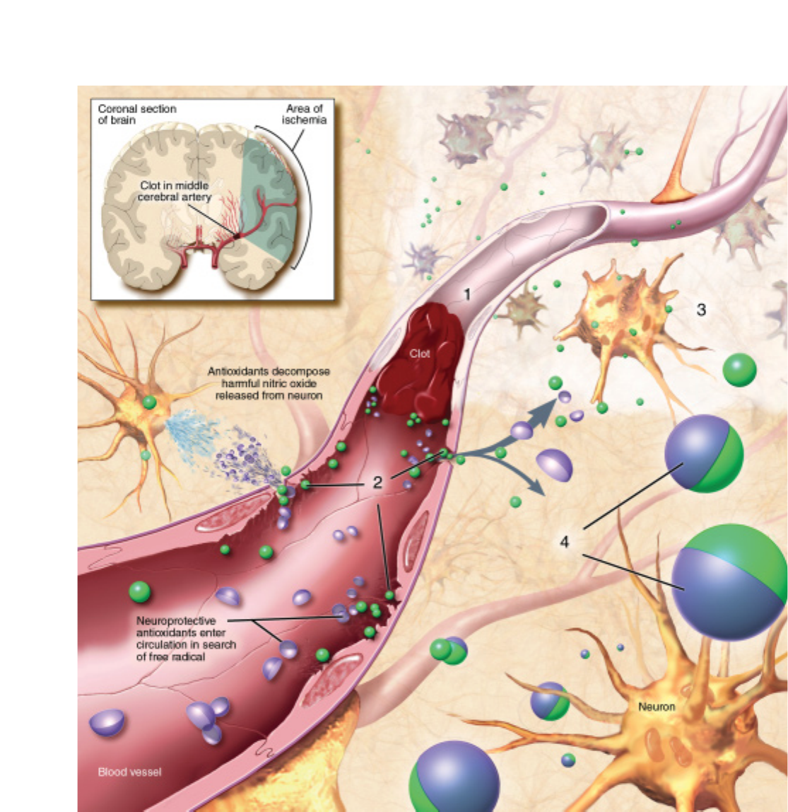
47
STROKE. A stroke occurs when a blood vessel bringing oxygen and nutrients to the brain bursts or is clogged by a blood clot (1). This lack of
blood leads to a cascade of neurochemical abnormalities that can cause cell death within minutes. Free radicals are released, causing dam-
age to endothelial cells (2) and the mitochondria (3) of neurons. Normally the body readily disarms free radicals (4), but in stroke, endothelial
cell damage allows many more than can be controlled to move into brain tissue. Depending on its location, a stroke can have different symp-
toms such as paralysis on one side of the body or a loss of speech.

48
Stroke a∑ects roughly 700,000 Americans a year—150,000 of
whom die; total annual costs are estimated at $51.2 billion.
A stroke occurs when a blood vessel bringing oxygen and
nutrients to the brain bursts or is clogged by a blood clot or some
other particle. This deprives the brain of blood, causing the death
of neurons within minutes. Depending on its location, a stroke
can cause many permanent disorders, such as paralysis on one
side of the body and loss of speech.
Stroke often occurs in individuals over 65 years of age, yet a
third are younger. Stroke tends to occur more in males and blacks
and in those with diabetes, high blood pressure, heart disease,
obesity, high cholesterol, and a family history of stroke.
In addition to tPA, increased use of preventive measures is
battling the disorder. Controlling risk factors such as obesity,
blood pressure, diabetes, and high cholesterol can help prevent
stroke. Other specific treatments involving surgery can clear clogs
in the arteries of the neck region and help prevent a cuto∑ of
blood supply.
Treatments that target the heart’s blood flow can prevent
stroke. Surgery can help repair damaged heart valves. Anticoag-
ulant drugs can reduce the chance of clots forming, traveling to
the brain and causing a stroke.
Other experimental therapies under investigation may lead
to even bigger payo∑s for patients in the future. Some strategies
target mechanisms inside the neuron. In this way, the vicious
cycle of local damage followed by a widening fringe of biochem-
ical-induced neuronal death can be slowed. A number of classes
of drugs have been shown to be e∑ective in animal studies.
Another promising possibility is the use of neural stem cells.
Some animal studies have shown that an injection of stem cells
aids recovery even if administered several days after the injury.
Administration of growth factors may further enhance the
benefits of stem cell transplantation.
Tourette syndrome
One of the most common and least understood neurobiological
disorders, Tourette syndrome (TS) is an inherited disorder that
a∑ects an estimated one in 500 Americans, roughly 200,000 peo-
ple. Males are a∑ected three to four times as often as females.
Symptoms usually appear between the ages of four and eight,
but in rare cases may emerge in the late teenage years. The symp-
toms include motor and vocal tics—repetitive, involuntary
movements or utterances that are rapid and sudden and persist
for more than one year. The types of tics may change frequently
and increase or decrease in severity over time. Generally, this dis-
order lasts a lifetime, but one-third of patients may experience a
remission or decrease in symptoms as they get older. Most peo-
ple with TS do not require medication; their symptoms are mild
and do not a∑ect functioning.
A high percentage of people with TS also have associated
conditions such as problems with learning, di≈culties with atten-
tion span, and obsessive behaviors. Sometimes these manifesta-
tions are more troublesome to individuals than the tics them-
selves, so physicians must consider them when choosing a
treatment regimen.
The disorder seems to result from a hypersensitivity of dopa-
mine receptors. Another neurotransmitter, serotonin, also has
been implicated. The most e∑ective drugs for control of move-
ments, such as haloperidol, act by blocking the overactive system.
Other symptoms, such as obsessive-compulsive traits and atten-
tion deficit disorder, often require treatment with other classes of
drugs that act on serotonin.
The neuroleptic drugs haloperidol and pimozide have been
the mainstays of treatment for the movements and vocalizations,
but not for the associated conditions that so many people with
TS experience. The neuroleptics are not perfect medications and
are prescribed only when the movements are fairly severe and
interfere with functioning. They can cause disturbing side
e∑ects—abnormal involuntary movements, sti∑ness of the face
and limbs, or sedation and weight gain—in some patients.
Recently, newer medications, such as low doses of selective sero-
tonin reuptake inhibitors like risperidone, have been found
e∑ective in some patients. Other classes of medications are pre-
scribed to reduce the symptoms of the comorbid conditions.

49
any of the recent advances in understand-
ing the brain are due to the development of
techniques that allow scientists to directly
monitor neurons throughout the body.
Electrophysiological recordings trace
brain electrical activity in response to a
specific external stimulus. In this method, electrodes placed in
specific parts of the brain—depending on which sensory system
is being tested—make recordings that are then processed by a
computer. The computer makes an anaylsis based on the time
lapse between stimulus and response. It then extracts this infor-
mation from background activity.
Following the discovery that material is transported within
neurons, methods have been developed to visualize activity and
precisely track fiber connections within the nervous system. This
can be done by injecting a radioactive amino acid into the brain
of an experimental animal; the animal is killed a few hours later,
and then the presence of radioactive cells is visualized on film. In
another technique, the enzyme horseradish peroxidase is injected
and taken up by nerve fibers that can be later identified under a
microscope.
These and other methods have resulted in many advances in
knowledge about the workings of the nervous system and are still
useful today. New methods, safely applicable to humans, promise
to give even more precise information.
Imaging techniques
Positron emission tomography (PET) PET is one of the most
important techniques for measuring blood flow or energy con-
sumption in the brain. This method of measuring brain function
is based on the detection of radioactivity emitted when positrons,
positively charged particles, undergo radioactive decay in the
brain. Small amounts of radiation are introduced into the blood,
which is then taken up into di∑erent brain areas in proportion to
how hard the neurons are working. Computers build three-
dimensional images of the brain based on the amount of radia-
tion emitted in these di∑erent areas.
PET studies have helped scientists understand more about
how drugs a∑ect the brain and what happens during learning,
when using language, and in certain brain disorders—such as
stroke, depression, and Parkinson’s disease. Within the next few
years, PET could enable scientists to identify the biochemical
nature of neurological and mental disorders and determine how
well therapy is working in patients. PET has revealed marked
changes in the depressed brain. Knowing the location of these
changes helps researchers understand the causes of depression
and monitor the e∑ectiveness of specific treatments.
Another technique, single photon emission computed tomog-
raphy (SPECT), is similar to PET, but its pictures are not as
detailed. SPECT is much less expensive than PET because the
tracers it uses have a longer half-life and do not require an accel-
erator nearby to produce them.
Magnetic resonance imaging (MRI) Providing a high-qual-
ity, three-dimensional image of organs and structures inside the
body without X-rays or other radiation, MRIs are unsurpassed in
anatomical detail and may reveal minute changes that occur over
time.
MRIs tell scientists when structural abnormalities first appear
in the course of a disease, how they a∑ect subsequent develop-
ment, and precisely how their progression correlates with men-
tal and emotional aspects of a disorder.
During the 15-minute MRI procedure, a patient lies inside a
massive, hollow, cylindrical magnet and is exposed to a power-
ful, steady magnetic field. Di∑erent atoms in the brain resonate
to di∑erent frequencies of magnetic fields. In MRI, a background
magnetic field lines up all the atoms in the brain. A second mag-
netic field, oriented di∑erently from the background field, is
turned on and o∑ many times a second; at certain pulse rates,
particular atoms resonate and line up with this second field.
When the second field is turned o∑, the atoms that were lined up
with it swing back to align with the background field. As they
swing back, they create a signal that can be picked up and con-
verted into an image. Tissue that contains a lot of water and fat
produces a bright image; tissue that contains little or no water,
such as bone, appears black.
MRI images can be constructed in any plane, and the tech-
nique is particularly valuable in studying the brain and spinal
cord.It reveals the precise extent of tumors rapidly and vividly.
And MRI provides early evidence of potential damage from
stroke, allowing physicians to administer proper treatments early.
New diagnostic methods
M

50
Magnetic resonance spectroscopy (MRS), a technique related to
MRI, uses the same machinery but measures the concentration of
specific chemicals—such as neurotransmitters—in di∑erent
parts of the brain instead of blood flow. MRS also holds great
promise: by measuring the molecular and metabolic changes that
occur in the brain, this technique has already provided new infor-
mation on brain development and aging, Alzheimer’s disease,
schizophrenia, autism, and stroke. Because it is noninvasive, MRS
is ideal for studying the natural course of a disease or its response
to treatment.
Functional magnetic resonance imaging (fMRI) Among the
most popular neuroimaging techniques today is fMRI. This tech-
nique compares brain activity under resting and activated condi-
tions. It combines the high-spatial-resolution, noninvasive imag-
ing of brain anatomy o∑ered by standard MRI with a strategy for
detecting increases in blood oxygen levels when brain activity
brings fresh blood to a particular area of the brain. This technique
allows for more detailed maps of brain areas underlying human
mental activities in health and disease. To date, fMRI has been
applied to the study of various functions of the brain, ranging
from primary sensory responses to cognitive activities.
Magnetoencephalography (MEG) MEG is a recently devel-
oped technique that reveals the source of weak magnetic fields
emitted by neurons. An array of cylinder-shaped sensors moni-
tors the magnetic field pattern near the patient’s head to deter-
mine the position and strength of activity in various regions of
the brain. In contrast with other imaging techniques, MEG can
characterize rapidly changing patterns of neural activity—down
to millisecond resolution—and can provide a quantitative mea-
sure of the strength of this activity in individual subjects. More-
over, by presenting stimuli at various rates, scientists can deter-
mine how long neural activation is sustained in the diverse brain
areas that respond.
One of the most exciting developments in imaging is the com-
bined use of information from fMRI and MEG. The former pro-
vides detailed information about the areas of brain activity in a
particular task, whereas MEG tells researchers and physicians
when certain areas become active. Together, this information leads
to a much more precise understanding of how the brain works in
health and disease.
Optical imaging techniques Optical imaging relies on shin-
ing weak lasers through the skull to visualize brain activity. These
techniques are inexpensive and relatively portable. They are also
silent and safe: Because only extremely weak lasers are used, they
can be used even to study young infants. In a technique called near
infrared spectroscopy (NIRS), technicians shine lasers through the
skull at near-infrared frequencies, which renders the skull trans-
parent. Blood with oxygen in it absorbs di∑erent frequencies of
light than blood in which the oxygen has been consumed. By
observing how much light is reflected back from the brain at each
frequency, researchers can track blood flow. Di∑use optical tomog-
raphy is then used to create maps of brain activity. A related tech-
nique, the event-related optical signal,records how light is scat-
tered in response to cellular changes that arise when neurons fire
and potentially can assess very fast—well under a second—
changes in neural activity.
Gene diagnosis
The inherited blueprint for all human characteristics, genes con-
sist of short sections of deoxyribonucleic acid (DNA), the long, spi-
raling, double-helix structure found on the 23 pairs of chromo-
somes in the nucleus of every human cell.
New gene diagnosis techniques now make it possible to find
the chromosomal location of genes responsible for neurologic and
psychiatric diseases and to identify structural changes in these
genes that are responsible for causing disease.
This information is useful for identifying individuals who
carry faulty genes and thereby improving diagnosis, for under-
standing the precise cause of diseases in order to improve meth-
ods of prevention and treatment, and for evaluating the malig-
nancy of and susceptibility to certain tumors.
So far, scientists have identified defective genes for more than
50 neurological disorders and the chromosomal location of the
defect in up to 100. Prenatal or carrier tests exist for many of the
most prevalent of these illnesses.
Scientists have tracked down the gene on chromosome 4 that
goes awry in Huntington’s patients. The defect is an expansion of
a CAG repeat. CAG is the genetic code for the amino acid gluta-
mine, and the expanded repeat results in a long string of gluta-
mines within the protein. This expansion appears to alter the pro-
tein’s function. Scientists have found that the size of the expanded
repeat in an individual is predictive of Huntington’s disease. Other
neurodegenerative disorders have been attributed to expanded
CAG repeats in other genes. The mechanisms by which these
expansions caused adult-onset neurodegeneration is the focus of
intense research.
Sometimes patients with single-gene disorders are found to
have a chromosomal abnormality—a deletion or break in the
DNA sequence of the gene—that can lead scientists to a more
accurate position of the disease gene. This is the case with some
abnormalities found on the X chromosome in patients with
Duchenne muscular dystrophy and on chromosome 13 in patients
with inherited retinoblastoma,a rare, highly malignant childhood
eye tumor that can lead to blindness and death.
Gene mapping has led to the localization on chromosome 21
of the gene coding for the beta amyloid precursor protein that is
abnormally cut to form the smaller peptide, beta amyloid. It is this
peptide that accumulates in the senile plaques that clog the brains
of patients with Alzheimer’s disease. This discovery shed light on
why individuals with Down syndrome invariably accumulate
amyloid deposits: they make too much amyloid due to having
three rather than two copies of this gene (trisomy 21). Mutations
in this gene have been shown to underlie Alzheimer’s in a distinct
subset of these patients.
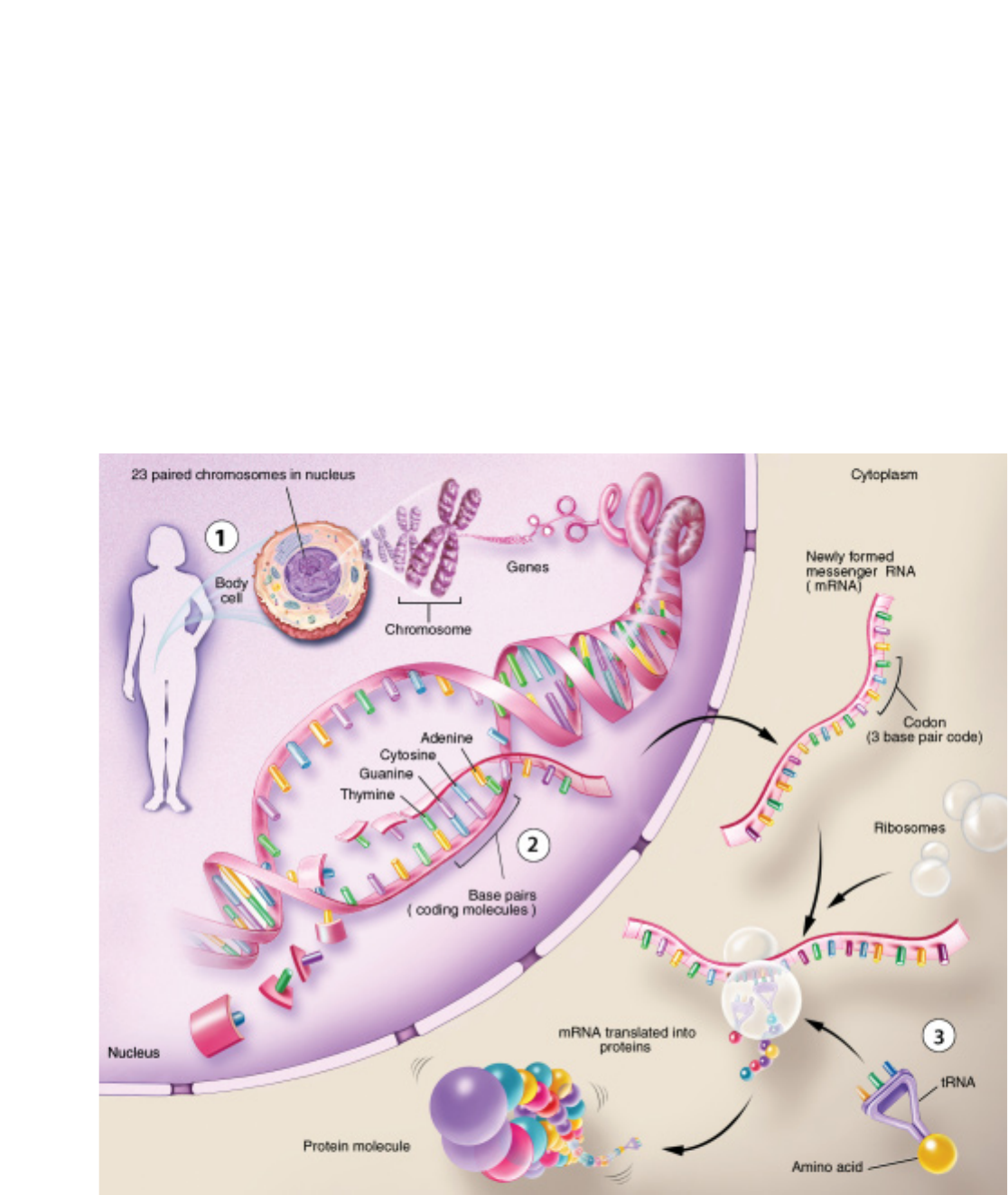
51
Several other genetic factors have been identified in Alz-
heimer’s disease, including genes for two proteins, presenilin 1
and presenilin 2, located on chromosomes 14 and 1, respectively.
A risk factor for late-onset Alzheimer’s is the gene for the apo-
lipoprotein E protein located on chromosome 19.
Gene mapping has enabled doctors to diagnose fragile X men-
tal retardation,the most common cause of inherited mental
retardation. Scientists have now identified this gene, which is
important for neuronal communication. Several groups of sci-
entists are investigating whether there are genetic components to
schizophrenia, bipolar disorder, and alcoholism, but their find-
ings are not yet conclusive.
Overall, the characterizations of the structure and function
of individual genes causing diseases of the brain and nervous sys-
tem are in the early stages. Factors that determine variations in
the genetic expression of a single-gene abnormality—such as
what contributes to the early or late start or severity of a disor-
der—are still largely unknown.
Scientists also are studying the genes in mitochondria, struc-
tures found outside the cell nucleus that have their own DNA and
are responsible for the production of energy used by the cell.
Recently, di∑erent mutations in mitochondrial genes were found
to cause several rare neurological disorders. Some scientists spec-
ulate that an inheritable variation in mitochondrial DNA may
play a role in diseases such as Alzheimer’s, Parkinson’s, and some
childhood diseases of the nervous system.
CHROMOSOMES, GENES, AND PROTEINS. Every trait and chemical process in the body is controlled by a gene or group of genes on
23 paired chromosomes in the nucleus of every cell (1). Each gene is a discrete segment along the two tightly coiled strands of DNA that make
up these chromosomes. DNA strands bear four different types of coding molecules — adenine (A), cytosine (C), guanine (G), and thymine (T)—
the sequence of which contains the instructions for making all the proteins necessary for life (2). During protein production, a gene uses a mol-
ecule called mRNA to send a message with instructions for the amino acids needed to manufacture a protein (3).

52
Iew drugs. Most medicines used today were
developed using trial-and-error techniques,
which often do not reveal why a drug produces a
particular e∑ect. But the expanding knowledge
gained from the new methods of molecular biol-
ogy—the ability to determine the structure of
receptors or other proteins—makes it possible to design safer
and more e∑ective drugs.
In a test tube, the potency of an agent can be determined by
how well it attaches to a receptor or other protein target. A sci-
entist then can vary the drug’s structure to enhance its action on
the desired target. Thus, subsequent generations of drugs can be
designed to interact more selectively with the target or, in many
cases, specific subtypes of the target, producing better therapeu-
tic e∑ects and fewer side e∑ects.
While this “rational drug design” holds promise for develop-
ing drugs for conditions ranging from stroke and migraine
headaches to depression and anxiety, it will take considerable
e∑ort to clarify the role of the di∑erent potential drug targets in
these disorders.
Other promising candidates for drug therapies include
trophic factors, antibodies engineered to specifically modify the
interactions and toxicity of misfolded proteins, small molecules
that take advantage of specific biochemical pathways, interfering
RNAs (RNAi) that reduce toxic levels of individual proteins, and
stem cells that could replace dead or dying neurons.
Trophic factors
One result of basic neuroscience research is the discovery of
numerous growth factors or trophic factors in the brain, which
control the development and survival of specific groups of neu-
rons. Once the specific actions of these molecules and their recep-
tors are identified and their genes cloned, procedures can be
developed to modify trophic factor-regulated functions in ways
that might be useful in the treatment of neurological disorders.
Once a trophic factor for a particular cell is found, copies of
the factor might be genetically targeted to the area of the brain
where this type of cell has died. The treatment may not cure a
disease, but could improve symptoms and delay progression.
Already, researchers have demonstrated the possible value of
at least one of these factors, nerve growth factor (NGF). Infused
into the brains of rats, NGF prevented cell death and stimulated
the regeneration and sprouting of damaged neurons that are
known to die in Alzheimer’s disease. When aged animals with
learning and memory impairments were treated with NGF, sci-
entists found that these animals were able to remember a maze
task as well as healthy aged rats. NGF, which slows the destruc-
tion of neurons that use acetylcholine, also holds promise for
slowing the memory deficits associated with normal aging.
Recently, several new factors have been identified. They are
potentially useful for therapy, but scientists must first understand
how they may influence neurons. Alzheimer’s disease, Parkinson’s
disease, and amyotrophic lateral sclerosis (ALS) may be treated
in the future with trophic factors or their genes.
In an interesting twist on growth factor therapy, researchers
demonstrated that a neutralization of inhibitory molecules can
help repair damaged nerve fiber tracts in the spinal cord. Using
antibodies to Nogo-A, a protein that inhibits nerve regeneration,
Swiss researchers succeeded in getting some nerves of damaged
spinal cords to regrow in rats. Treated rats showed large improve-
ments in their ability to walk after spinal cord damage.
In these experiments, scientists cut one of the major groups
of nerve fiber tracts in the spinal cord that connect it to the brain.
When an antibody directed against the factor Nogo-A was
administered to the spinal cords or brains of adult rats, enhanced
sprouting of nerve fibers occurred where the spinal cord had been
cut. Within two to three weeks, some fibers grew to the lower level
of the spinal cord and, in some animals, along its whole length.
In untreated spinal cord-injured rats, the maximum distance of
nerve regrowth rarely exceeded one-tenth of an inch. This
research could eventually have clinical implications for spinal
cord- or brain-damaged people.
Engineered antibodies
The immune system has evolved to very specifically identify and
modify factors both inside and outside of cells. It is sometimes
possible to trick the body into attacking proteins that cause neu-
rological diseases by “vaccinating” patients against these proteins.
This approach has shown some promise in Alzheimer’s disease,
although it also carries risks. Another new approach combines
N
Potential therapies
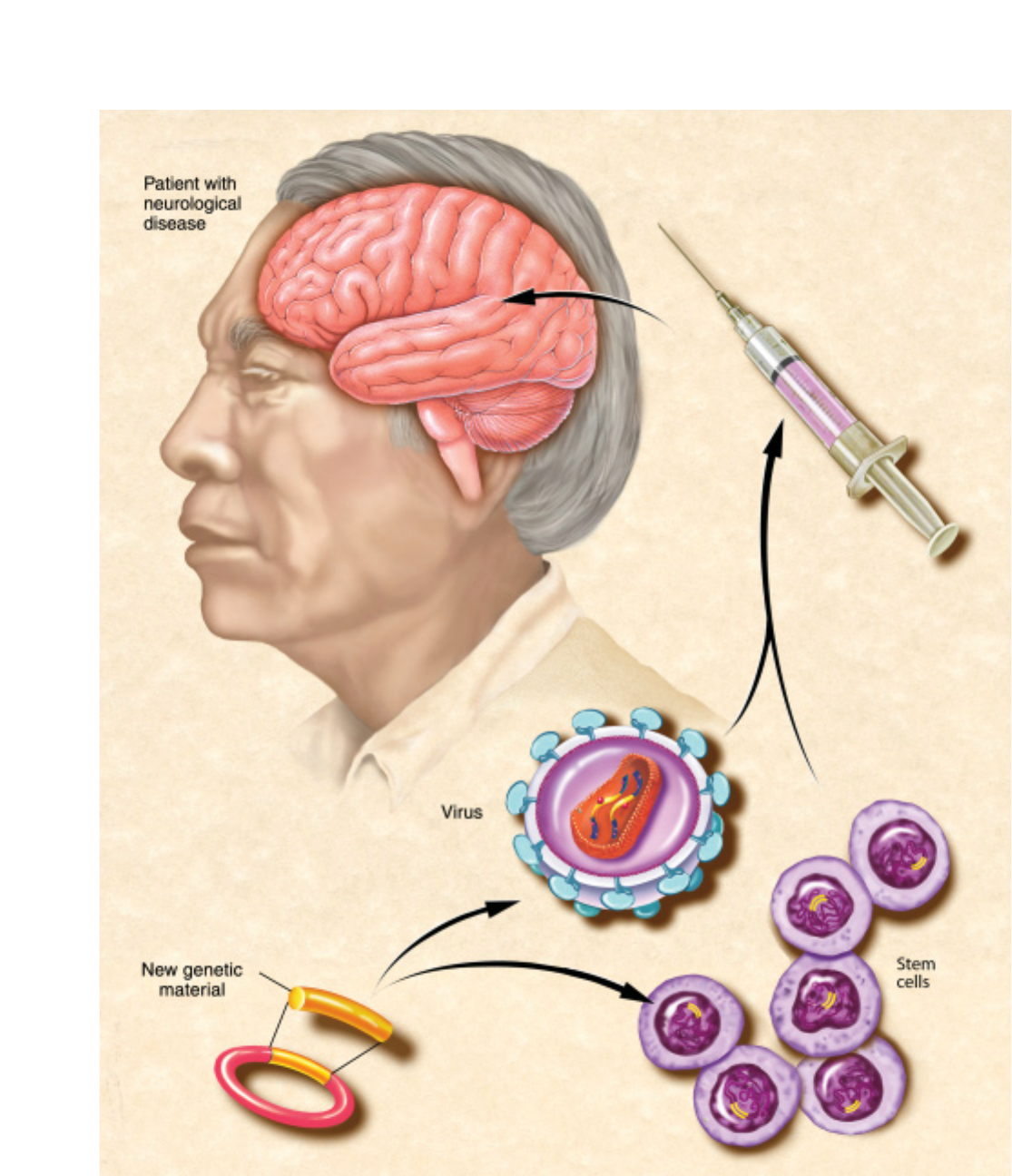
53
CELL AND GENE THERAPY. In potential therapy techniques, scientists plan to insert genetic material for a beneficial neurotransmitter or
trophic factor into stem cells or a virus. The cells or virus are then put into a syringe and injected into the patient where they will produce the
beneficial molecule and, it is hoped, improve symptoms.

54
genetic engineering with immunology to engineer antibodies or
fragments of antibodies that can bind to and alter the disease
characteristics of specific proteins. These therapies can be deliv-
ered either as proteins or as genes.
Promising preliminary results have been obtained for Hunt-
ington’s, Parkinson’s, Alzheimer’s, and prion diseases. For exam-
ple, fruit flies (Drosophila) that get Huntington’s disease (HD)
because they have been modified to carry the mutant human gene
are generally too weak and uncoordinated to break out of their
pupal case the way normal insects do. However, when they also
express the gene for an anti-HD antibody, all of them can emerge
as young adults. Furthermore, these treated flies live longer than
the untreated ones that do man-
age to emerge, and the treated
ones show less pathology in
their brains.
Small molecules and
RNAs
Clarifying the processes that
underlie brain damage will open
up the potential to use small
molecule drugs to alter these
processes. Some success has occurred in developing animal mod-
els using approaches based on known mechanisms of drugs.
Examples include drugs such as antibiotics and anti-tumor drugs,
which appear to reduce the neuronal damage in ALS, HD, and
Parkinson’s disease. Thousands of small molecule drug candidates
can be tested using high throughput screening to alter a cellular
property that represents an important part of a disease process.
Because many neurodegenerative diseases involve proteins that
misfold and clump abnormally, lasers are used to measure
whether proteins are clumped inside cells that have been roboti-
cally distributed into tiny wells, along with the small molecules to
be tested. A machine then scans the wells and reports whether par-
ticular drugs have changed the protein clumping, so that these
drugs can be tested further. New leads for Alzheimer’s and prion
drugs have recently been described using these methods.
Several neurodegenerative diseases are caused by the accu-
mulation of abnormal proteins. If the cells made much less of such
proteins to begin with, then the disease would progress much
more slowly. A new class of potential drugs is based on removing
the RNAs that code for the proteins that are causing damage.
Mouse models of HD and ALS appear to have responded posi-
tively to such treatments, which are delivered via gene therapies.
Cell and gene therapy
Researchers throughout the world are pursuing a variety of new
ways to repair or replace neurons and other cells in the brain.
These experimental approaches are still being worked out in ani-
mals and cannot be considered therapies for humans at this time.
Scientists have identified em-
bryonic neuronal stem cells—
unspecialized cells that give rise
to cells with specific functions—
in the brain and spinal cord of
embryonic and adult mice. Stem
cells can continuously produce
all three major cell types of the
brain: neurons; astrocytes,the
cells that nourish and protect
neurons; and oligodendrocytes,
the cells that surround axons and allow them to conduct their sig-
nals e≈ciently. The production abilities of stem cells may some-
day be useful for replacing brain cells lost to disease. A more lim-
ited type of stem cell also has been discovered in the adult nervous
system in various kinds of tissue, raising the possibility that these
adult stem cells might be pharmacologically directed to replace
damaged neurons.
In other work, researchers are studying a variety of viruses
that may ultimately be used to act as “Trojan horses,” carrying
therapeutic genes to the brain to correct nervous system diseases.
The viruses include herpes simplex type 1 virus (HSV),adenovirus,
lentivirus,adeno-associated virus,and others naturally attracted
to neurons. It has been found that all can be modified to carry
new genes to cells in tissue culture and in the rodent central ner-
vous system. HSV and adenovirus vectors have also been evalu-
ated in early-stage human trials for treating brain tumors.
Thousands of small molecule drug
candidates can be tested using high
throughput screening to alter a cellular
property that represents an important
part of a disease process.

55
Breaking a confidence. Going along to get along. Telling a “white lie” to protect a friend.
Everyone faces ethical dilemmas—in school, at home, and nearly everywhere in
everyday life. And this is no di∑erent for neuroscientists. With the tremendous
advances in the field, many scientists and nonscientists alike have sensed a critical
turning point. Researchers are now on the threshold of understanding some funda-
mental principles of brain function, and are even beginning to develop treatments for
some of the most devastating neurological diseases and conditions. At the same time, much of this
knowledge and implications for treatments and diagnostics raise ethical questions.
For example, some recent brain imaging studies have sought to define areas responsible for phe-
nomena such as deception. The post-9/11 era has created much interest in lie detection for security
purposes in screening immigrants. How should privacy be balanced with national security? Is the
technology accurate enough to provide useful data upon which to base decisions? Pursuing these
lines of scientific inquiry in a responsible way requires neuroscientists to examine how what they do
a∑ects the world beyond the laboratory or clinic.
This self-examination makes up a field known as neuroethics.Scientists and ethicists are begin-
ning to reflect on the implications of neuroscience in areas such as moral reasoning, decision-mak-
ing, and behavior. Much discussion will focus on neuroethics in the coming years.
Neuroethics is the part of bioethics that considers the intended and unintended consequences of
neuroscience in medical practice, research, and society at large. Neuroethics also deals with issues
that touch no other area of science—our sense of self, our personalities, and our behavior. And brain
science can change these aspects in significant ways. Neuroethics has been the subject of several con-
ferences that have attracted a wide range of thinkers, basic and clinical neuroscientists, economists,
philosophers, journalists, sociologists, lawyers, and others. Some major topics include the subjects
listed below.
Need for ethical framework It is very likely that the potential application of new knowledge to
human behavior will generate a great deal of ethical and public policy concern. Neuroethics spans
fields as diverse as forensic psychiatry, athletics, education, college admissions, corporate hiring, polic-
ing, admission to seminaries, and the law. Information about why people, and categories of people,
behave as they do will lead those in these fields to eagerly use new knowledge about the human brain.
As the brain sciences advance, researchers are working toward developing a paradigm of morals that
might help guide the use of the new knowledge.
Morality Learning right from wrong enhances skills in cooperative behavior and helps people
know when to modify their social interactions. Discoveries in neuroscience indicate that learning
right from wrong may depend on the development of brain tissue during the adolescent years. Given
this possibility, researchers are considering the implications of how problems in brain development
may a∑ect moral behaviors such as self-control. For example, could some individuals have a biolog-
ical handicap that impairs their ability to obey the law? Researchers also are investigating the ethical
issues related to using drugs that influence behaviors like self-control.
Social behavior Neurobiological factors may play a role in disturbances in social behavior.
Patients who have malfunctions caused by disease in selected brain regions exhibit antisocial behav-
B
Neuroethics

56
ioral change. Because modern neuroscience can investigate the
mechanisms behind disturbed behaviors, society must ponder
the manner in which it manages individuals who violate its rules
by taking into account those with medical conditions. This situ-
ation raises questions regarding the punishment and treatment
of such individuals.
Social policy Since neuroscience has the potential to trans-
form our understanding of human nature, we may be able to
make predictions about an individual’s future, including the risk
for ill health and cognitive impairment, potential success in school
or employment, and violent behavior or addiction to drugs. Of
particular concern is the potential role for neuroscience in delin-
eating the boundary between
what society views as normal
and what is deemed patho-
logical. For example, many boys
are being prescribed medicines
for hyperactivity, such as Ri-
talin, for conditions that many
worry may not be clear-cut
brain diseases. Troubling ques-
tions related to this phenome-
non include: Who should have
access to such agents? If learn-
ing can be sped up and atten-
tion enhanced through phar-
maceuticals, should such drugs
be available to all, or should re-
sources be devoted to trans-
forming only the environment
of the classroom?
Genetics Sequencing the
human genome—identifying all the approximately 20,000 to
25,000 genes in humans—went hand-in-hand with an invest-
ment in studying the ethical, legal, and social implications of
human genetics. Neuroethics may play an important role in
issues arising from genetics, such as the social consequences of
using DNA to predict the future. Should patients with a family
history of a neurological or psychiatric disorder be genetically
tested even though no treatment is available?
Brain injury Since traumatic brain injury may cause a per-
son to experience significant cognitive, personality, emotional,
and behavioral changes, su∑erers may become legally incompe-
tent. Traumatic brain injury may excuse or mitigate a person’s
responsibility for acts that otherwise would be classified as
crimes. But the medical profession sometimes can have trouble
determining a person’s preinjury behavior compared with postin-
jury behavior. This poses serious problems for clinical evaluators
as well as for the legal system.
Neurological disorders With the likelihood of developing
e∑ective treatments for disorders such as Alzheimer’s disease
(AD), in which brain cells die at a progressive rate, it will be
important to identify individuals who are at risk in order to pre-
vent brain damage at the earliest stage. Yet there is little prece-
dence for such early intervention. Moreover, AD is a genetically
complex disorder with multiple genes and degrees of risk associ-
ated with each. Thus, the medical profession faces challenges in
ensuring that candidates for treatment understand their risk for
severity of AD and evaluating the potential risks and benefits of
treatments that may be harmful.
Informed consent in research Special care must be taken in
the informed-consent process and throughout the research pro-
tocol when individuals have
thinking or emotional impair-
ments that might a∑ect their
decision-making capacity. Con-
sent is an ongoing process that
should involve education of the
potential research participant
and, when appropriate, family
members. Researchers are dis-
cussing potential needs to exer-
cise greater scrutiny, ensure
safeguards, and enhance partic-
ipants’ grasp of a study, includ-
ing risks and benefits.
Environmental influences
Many are concerned about the
possible implications of research
indicating that most of the brain
gets built after birth and that it
uses experiences from the out-
side world to form its circuits. With this as background, scientists
are discussing whether society has a moral obligation to ensure
that millions of children no longer grow up in violent and im-
poverished environments that can stunt their brains.
At this stage, the field of neuroethics raises more questions
than answers. It poses challenges to scientists and to the public
to work through the social implications of new discoveries. The
issues are too broad-based to expect that scientists alone will sup-
ply the answers. But neuroscientists are well-positioned to help
shape and contribute to the debate and discussion.
One of the hallmarks of the field has always been the drive
toward integrating information from disparate fields and special-
izations to increase knowledge. Sorting through the complex issues
captured under the umbrella of neuroethics provides an impor-
tant opportunity for informed and rich discussions among scien-
tists and with the public. Continuing study of neuroethics will help
all segments of society deal with the challenges posed by emerging
technologies that investigate the brain and how it works.
Researchers are now on the threshold
of understanding some fundamental
principles of brain function, and are
even beginning to develop treatments for
some of the most devastating neuro-
logical diseases and conditions. At the
same time, much of this knowledge and
implications for treatments and
diagnostics raise ethical questions.

57
ACETYLCHOLINE A neurotransmitter active both in the brain,
where it regulates memory, and in the peripheral nervous system,
where it controls the actions of skeletal and smooth muscle.
ACTION POTENTIAL An electrical charge travels along the axon
to the neuron’s terminal, where it triggers the release of a neuro-
transmitter. This occurs when a neuron is activated and tem-
porarily reverses the electrical state of its interior membrane from
negative to positive.
ADRENAL CORTEX An endocrine organ that secretes corticos-
teroids for metabolic functions; for example, in response to stress.
ADRENAL MEDULLA An endocrine organ that secretes epi-
nephrine and norepinephrine in concert with the activation of the
sympathetic nervous system; for example, in response to stress.
AGONIST A neurotransmitter, drug, or other molecule that
stimulates receptors to produce a desired reaction.
ALZHEIMER’S DISEASE The major cause of dementia most
prevalent in the elderly, it inflicts enormous human financial cost
on society. The disease is characterized by death of neurons in the
hippocampus, cerebral cortex, and other brain regions.
AMINO ACID TRANSMITTERS The most prevalent neuro-
transmitters in the brain, these include glutamate and aspartate,
which have excitatory actions on nerve cells, and glycine and
gamma-amino butyric acid (GABA), which also have inhibitory
actions on nerve cells.
AMYGDALA A structure in the forebrain that is an important
component of the limbic system and plays a central role in emo-
tional learning, particularly within the context of fear.
ANDROGENS Sex steroid hormones, including testosterone,
found in higher levels in males than females. They are responsi-
ble for male sexual maturation.
ANTAGONIST A drug or other molecule that blocks receptors.
Antagonists inhibit the e∑ects of agonists.
APHASIA Disturbance in language comprehension or produc-
tion, often as a result of a stroke.
APOPTOSIS Programmed cell death induced by specialized bio-
chemical pathways, often serving a specific purpose in the devel-
opment of the animal.
Glossary
AUDITORY NERVE A bundle of nerve fibers extending from the
cochlea of the ear to the brain that contains two branches: the
cochlear nerve, which transmits sound information, and the
vestibular nerve, which relays information related to balance.
AUTONOMIC NERVOUS SYSTEM A part of the peripheral ner-
vous system responsible for regulating the activity of internal
organs. It includes the sympathetic and parasympathetic nervous
systems.
AXON The fiberlike extension of a neuron by which it sends
information to target cells.
BASAL GANGLIA Clusters of neurons, which include the cau-
date nucleus, putamen, globus pallidus, and substantia nigra,
located deep in the brain that play an important role in the initi-
ation of movements. Cell death in the substantia nigra contributes
to Parkinson’s disease.
BRAINSTEM The major route by which the forebrain sends infor-
mation to and receives information from the spinal cord and
peripheral nerves. The brainstem controls, among other things,
respiration and the regulation of heart rhythms.
BROCA’S AREA The brain region located in the frontal lobe of
the left hemisphere that is important for the production of speech.
CATECHOLAMINES The neurotransmitters dopamine, epineph-
rine, and norepinephrine, which are active in both the brain and
the peripheral sympathetic nervous system. These three molecules
have certain structural similarities and are part of a larger class of
neurotransmitters known as monoamines.
CEREBELLUM A large structure located at the roof of the hind-
brain that helps control the coordination of movement by mak-
ing connections to the pons, medulla, spinal cord, and thalamus.
It also may be involved in aspects of motor learning.
CEREBRAL CORTEX The outermost layer of the cerebral hemi-
spheres of the brain. It is largely responsible for all forms of con-
scious experience, including perception, emotion, thought, and
planning.
CEREBRAL HEMISPHERES The two specialized halves of the
brain. For example, in right-handed people, the left hemisphere is
specialized for speech, writing, language, and calculation; the right

58
hemisphere is specialized for spatial abilities, visual face recogni-
tion, and some aspects of music perception and production.
CEREBROSPINAL FLUID A liquid found within the ventricles of
the brain and the central canal of the spinal cord.
CHOLECYSTOKININ A hormone released from the lining of the
stomach during the early stages of digestion that acts as a pow-
erful suppressant of normal eating. It also is found in the brain.
CIRCADIAN RHYTHM A cycle of behavior or physiological
change lasting approximately 24 hours.
CLASSICAL CONDITIONING Learning in which a stimulus that
naturally produces a specific response (unconditioned stimulus)
is repeatedly paired with a neutral stimulus (conditioned stimu-
lus). As a result, the conditioned stimulus can come to evoke a
response similar to that of the unconditioned stimulus.
COCHLEA A snail-shaped, fluid-filled organ of the inner ear
responsible for transducing motion into neurotransmission to
produce an auditory sensation.
COGNITION The process or processes by which an organism
gains knowledge or becomes aware of events or objects in its
environment and uses that knowledge for comprehension and
problem-solving.
CONE A primary receptor cell for vision located in the retina. It
is sensitive to color and is used primarily for daytime vision.
CORPUS CALLOSUM The large bundle of nerve fibers linking
the left and right cerebral hemispheres.
CORTISOL A hormone manufactured by the adrenal cortex. In
humans, cortisol is secreted in the greatest quantities before
dawn, readying the body for the activities of the coming day.
DEPRESSION A mental disorder characterized by depressed
mood and abnormalities in sleep, appetite, and energy level.
DENDRITE A tree-like extension of the neuron cell body. The
dendrite is the primary site for receiving and integrating infor-
mation from other neurons.
DOPAMINE A catecholamine neurotransmitter known to have
varied functions depending on where it acts. Dopamine-con-
taining neurons in the substantia nigra of the brainstem project
to the caudate nucleus and are destroyed in Parkinson’s victims.
Dopamine is thought to regulate key emotional responses such
as reward and plays a role in schizophrenia and drug abuse.
DORSAL HORN An area of the spinal cord where many nerve
fibers from peripheral pain receptors meet other ascending and
descending nerve fibers.
DRUG ADDICTION Loss of control over drug intake or com-
pulsive seeking and taking of drugs, despite adverse conse-
quences.
ENDOCRINE ORGAN An organ that secretes a hormone
directly into the bloodstream to regulate cellular activity of cer-
tain other organs.
ENDORPHINS Neurotransmitters produced in the brain that
generate cellular and behavioral e∑ects like those of morphine.
EPILEPSY A disorder characterized by repeated seizures, which
are caused by abnormal excitation of large groups of neurons in
various brain regions. Epilepsy can be treated with many types of
anticonvulsant medications.
EPINEPHRINE A hormone, released by the adrenal medulla and
specialized sites in the brain, that acts with norepinephrine to
a∑ect the sympathetic division of the autonomic nervous system.
Sometimes called adrenaline.
ESTROGENS A group of sex hormones found more abundantly
in females than males. They are responsible for female sexual
maturation and other functions.
EVOKED POTENTIALS A measure of the brain’s electrical activ-
ity in response to sensory stimuli. This is obtained by placing
electrodes on the surface of the scalp (or more rarely, inside the
head), repeatedly administering a stimulus, and then using a
computer to average the results.
EXCITATION A change in the electrical state of a neuron that is
associated with an enhanced probability of action potentials.
FOLLICLE-STIMULATING HORMONE A hormone released by
the pituitary gland that stimulates the production of sperm in the
male and growth of the follicle (which produces the egg) in the
female.
FOREBRAIN The largest part of the brain, which includes the
cerebral cortex and basal ganglia. The forebrain is credited with
the highest intellectual functions.
FRONTAL LOBE One of the four divisions (the other lobes are
the parietal, temporal, and occipital) of each hemisphere of the
cerebral cortex. The frontal lobe has a role in controlling move-
ment and in the planning and coordinating of behavior.
GAMMA-AMINO BUTYRIC ACID (GABA) An amino acid
transmitter in the brain whose primary function is to inhibit the
firing of nerve cells.
GLIA Specialized cells that nourish and support neurons.
GLUTAMATE An amino acid neurotransmitter that acts to excite
neurons. Glutamate stimulates N-methyl-d-aspartate (NMDA)
and alpha-amino-3-hydroxy-5-methylisoxazole-4-propionic acid
(AMPA). AMPA receptors have been implicated in activities
ranging from learning and memory to development and
specification of nerve contacts in developing animals. Stimula-
tion of NMDA receptors may promote beneficial changes,
whereas overstimulation may be a cause of nerve cell damage or
death in neurological trauma and stroke.
GONAD Primary sex gland: testis in the male and ovary in the
female.
GROWTH CONE A distinctive structure at the growing end of
most axons. It is the site where new material is added to the axon.

59
HIPPOCAMPUS A seahorse-shaped structure located within the
brain and considered an important part of the limbic system. One
of the most studied areas of the brain, it functions in learning,
memory, and emotion.
HORMONES Chemical messengers secreted by endocrine glands
to regulate the activity of target cells. They play a role in sexual
development, calcium and bone metabolism, growth, and many
other activities.
HUNTINGTON’S DISEASE A movement disorder caused by the
death of neurons in the basal ganglia and other brain regions. It
is characterized by abnormal movements called chorea—sudden,
jerky movements without purpose.
HYPOTHALAMUS A complex brain structure composed of many
nuclei with various functions, including regulating the activities
of internal organs, monitoring information from the autonomic
nervous system, controlling the pituitary gland, and regulating
sleep and appetite.
INHIBITION A synaptic message that prevents a recipient neu-
ron from firing.
IONS Electrically charged atoms or molecules.
LIMBIC SYSTEM A group of brain structures—including the
amygdala, hippocampus, septum, basal ganglia, and others—that
help regulate the expression of emotion and emotional memory.
LONG-TERM MEMORY The final phase of memory, in which
information storage may last from hours to a lifetime.
MANIA A mental disorder characterized by excessive excitement,
exalted feelings, elevated mood, psychomotor overactivity, and
overproduction of ideas. It may be associated with psychosis, for
example, delusions of grandeur.
MELATONIN Produced from serotonin, melatonin is released by
the pineal gland into the bloodstream. Melatonin a∑ects physio-
logical changes related to time and lighting cycles.
MEMORY CONSOLIDATION The physical and psychological
changes that take place as the brain organizes and restructures
information to make it a permanent part of memory.
METABOLISM The sum of all physical and chemical changes that
take place within an organism and all energy transformations that
occur within living cells.
MIDBRAIN The most anterior segment of the brainstem. With
the pons and medulla, the midbrain is involved in many functions,
including regulation of heart rate, respiration, pain perception,
and movement.
MITOCHONDRIA Small cylindrical organelles inside cells that
provide energy for the cell by converting sugar and oxygen into
special energy molecules, called ATP.
MONOAMINE OXIDASE (MAO) The brain and liver enzyme
that normally breaks down the catecholamines norepinephrine,
dopamine, and epinephrine, as well as other monoamines such as
serotonin.
MOTOR NEURON A neuron that carries information from the
central nervous system to muscle.
MYASTHENIA GRAVIS A disease in which acetylcholine recep-
tors on muscle cells are destroyed so that muscles can no longer
respond to the acetylcholine signal to contract. Symptoms include
muscular weakness and progressively more common bouts of
fatigue. The disease’s cause is unknown but is more common in
females than in males; it usually strikes between the ages of 20 and
50.
MYELIN Compact fatty material that surrounds and insulates the
axons of some neurons.
NECROSIS Cell death due to external factors, such as lack of oxy-
gen or physical damage, that disrupt the normal biochemical
processes in the cell.
NERVE GROWTH FACTOR A substance whose role is to guide
neuronal growth during embryonic development, especially in the
peripheral nervous system. Nerve growth factor also probably
helps sustain neurons in the adult.
NEURON A nerve cell specialized for the transmission of infor-
mation and characterized by long, fibrous projections called axons
and shorter, branchlike projections called dendrites.
NEUROPLASTICITY A general term used to describe the adaptive
changes in the structure or function of nerve cells or groups of
nerve cells in response to injuries to the nervous system or alter-
ations in patterns of their use and disuse.
NEUROTRANSMITTER A chemical released by neurons at a
synapse for the purpose of relaying information to other neurons
via receptors.
NOCICEPTORS In animals, nerve endings that signal the sensa-
tion of pain. In humans, they are called pain receptors.
NOREPINEPHRINE A catecholamine neurotransmitter, pro-
duced both in the brain and in the peripheral nervous system.
Norepinephrine is involved in arousal and in regulation of sleep,
mood, and blood pressure.
OCCIPITAL LOBE One of the four subdivisions of the cerebral
cortex.The occipital lobe plays a role in processing visual infor-
mation.
ORGANELLES Small structures within a cell that maintain the
cell and do the cell’s work.
PARASYMPATHETIC NERVOUS SYSTEM A branch of the auto-
nomic nervous system concerned with the conservation of the
body’s energy and resources during relaxed states.
PARIETAL LOBE One of the four subdivisions of the cerebral cor-
tex. The parietal lobe plays a role in sensory processes, attention,
and language.

60
PARKINSON’S DISEASE A movement disorder caused by death
of dopamine neurons in the substantia nigra, located in the mid-
brain. Symptoms include tremor, shuΩing gait, and general
paucity of movement.
PEPTIDES Chains of amino acids that can function as neuro-
transmitters or hormones.
PERIPHERAL NERVOUS SYSTEM A division of the nervous sys-
tem consisting of all nerves that are not part of the brain or spinal
cord.
PHOSPHORYLATION A process that modifies the properties of
neurons by acting on an ion channel, neurotransmitter receptor,
or other regulatory protein. During phosphorylation, a phos-
phate molecule is placed on a protein and results in the activa-
tion or inactivation of the protein. Phosphorylation is believed
to be a necessary step in allowing some neurotransmitters to act
and is often the result of second-messenger activity.
PINEAL GLAND An endocrine organ found in the brain. In
some animals, the pineal gland serves as a light-influenced bio-
logical clock.
PITUITARY GLAND An endocrine organ closely linked with the
hypothalamus. In humans, the pituitary gland is composed of
two lobes and secretes several di∑erent hormones that regulate
the activity of other endocrine organs throughout the body.
PONS A part of the hindbrain that, with other brain structures,
controls respiration and regulates heart rhythms. The pons is a
major route by which the forebrain sends information to and
receives information from the spinal cord and peripheral nervous
system.
PSYCHOSIS A severe symptom of mental disorders character-
ized by an inability to perceive reality. Psychosis can occur in
many conditions, including schizophrenia, mania, depression,
and drug-induced states.
RECEPTOR CELL A specialized sensory cell, designed to pick up
and transmit sensory information.
RECEPTOR MOLECULE A specific protein on the surface of or
inside a cell with a characteristic chemical and physical structure.
Many neurotransmitters and hormones exert their e∑ects by
binding to receptors on cells.
REUPTAKE A process by which released neurotransmitters are
absorbed for later reuse.
ROD A sensory neuron located in the periphery of the retina.
The rod is sensitive to light of low intensity and is specialized for
nighttime vision.
SCHIZOPHRENIA A chronic mental disorder characterized by
psychosis (e.g., hallucinations and delusions), flattened emotions,
and impaired cognitive function.
SECOND MESSENGERS Substances that trigger communica-
tions among di∑erent parts of a neuron. These chemicals play a
role in the manufacture and release of neurotransmitters, intra-
cellular movements, carbohydrate metabolism, and processes of
growth and development. The messengers’ direct e∑ects on the
genetic material of cells may lead to long-term alterations of
behavior, such as memory and drug addiction.
SEROTONIN A monoamine neurotransmitter believed to play
many roles, including, but not limited to, temperature regulation,
sensory perception, and the onset of sleep. Neurons using sero-
tonin as a transmitter are found in the brain and gut. Several anti-
depressant drugs are targeted to brain serotonin systems.
SHORT-TERM MEMORY A phase of memory in which a limited
amount of information may be held for several seconds or min-
utes.
STIMULUS An environmental event capable of being detected
by sensory receptors.
STROKE The third-largest cause of death in the United States,
stroke is an impeded blood supply to the brain. Stroke can be
caused by a rupture of a blood vessel wall, an obstruction of
blood flow caused by a clot or other material, or pressure on a
blood vessel (as by a tumor). Deprived of oxygen, which is car-
ried by blood, nerve cells in the a∑ected area cannot function and
die. Thus, the part of the body controlled by those cells cannot
function either. Stroke can result in loss of consciousness and
death.
SYMPATHETIC NERVOUS SYSTEM A branch of the autonomic
nervous system responsible for mobilizing the body’s energy and
resources during times of stress and arousal.
SYNAPSE A physical gap between two neurons that functions
as the site of information transfer from one neuron to another.
TEMPORAL LOBE One of the four major subdivisions of each
hemisphere of the cerebral cortex. The temporal lobe functions
in auditory perception, speech, and complex visual perceptions.
THALAMUS A structure consisting of two egg-shaped masses of
nerve tissue, each about the size of a walnut, deep within the
brain. The key relay station for sensory information flowing into
the brain, the thalamus filters out information of particular
importance from the mass of signals entering the brain.
VENTRICLES Comparatively large spaces filled with cere-
brospinal fluid. Of the four ventricles, three are located in the
forebrain and one in the brainstem. The lateral ventricles, the two
largest, are symmetrically placed above the brainstem, one in each
hemisphere.
WERNICKE’S AREA A brain region responsible for the com-
prehension of language and the production of meaningful
speech.

61
Acetylcholine 6
Action potential 6
Addiction 3639
Aging 3132
and intellectual capacity 3132
AIDS 4445
Alcohol 37,3839
Alcoholism 3839
Alpha motor neurons 2223,24
Alzheimer’s disease 5, 40
Amino acid transmitters 78
Amphetamines 37
Amyloid protein 40
Amyotrophic lateral sclerosis (ALS)
4041
Analgesia 35
Androgen 8
Anxiety disorders 4142
Attention deficit hyperactivity disorder
(ADHD) 3940
Autoimmune response 30
Autonomic nervous system 13,28, 29
Axon 67
Basal ganglia 24
Biological clock 9
Bipolar disorder 33
Brain
aging 3132
anatomical organization 5
development 1013
diseases 4, 5
tumors 42
Catecholamines 8
Central nervous system 8, 13
Cerebellum 5,21,24
Cerebral cortex 5,11, 19,21,27
Club drugs 39
Cocaine 37,38
Cortisol 2930
Costs of brain diseases 45
Crossed extension reflex 2324
Declarative knowledge 20
Dementia 31, 40, 45
Dendrite 67
Depression
major 3334
Dopamine 8, 37,38
Down syndrome 4243
Drug reward system 37
Endocrine system 89, 28, 29,30
Endorphins 8, 19
Epilepsy 33
Epinephrine 2930
Estrogen 8
Fetal alcohol syndrome 38
Firing of neurons 67
Flexion withdrawal 2324
Fluoxetine 8
Forebrain 5,10,27
Fragile X mental retardation 51
Functional magnetic resonance imaging
(fMRI) 46, 50
Gammaaminobutyric acid (GABA) 7, 37
Gamma motor neurons 22
Gene 5051
diagnosis 50
therapy 53,54
Glucocorticoids 9, 2930
Glutamate 7, 37
Hearing 1617
Heroin 3738
Hippocampus 5,2021,37
Huntington’s disease 5, 43
Hypothalamus 5,8,29
Immune system 30
Information processing 12
and hearing 1617
and learning and memory 2021
and movement 22, 2324
and pain 1719
and taste and smell 1618
and vision 14, 1516
Inhibitory neurons 2223
Ion channels 6
Language 2021
Learning 2021
Learning disorders 4344
Levodopa 8
Limbic system 17
Longterm potentiation 2021
Lou Gehrig’s disease 4041
Magnetic resonance imaging (MRI) 49
Magnetic resonance spectroscopy (MRS)
50
Magnetoencephalography (MEG) 50
Marijuana 39
Memory 2021
Methylprednisolone 45
Midbrain 5,10,19,24
Mitochondria 47,51
Monoamine oxidase inhibitors (MAOIs)
34
Morphine 8, 35, 3738
Motor cortex 5, 24
Motor neuron 11, 2223,24
Motor unit 22
Movement 2223,24
MPTP 35
Multiple sclerosis 5, 44
Myasthenia gravis 6
Myelin 67
Index
Numbers in bold refer to illustrations.

62
Narcolepsy 26
Nerve growth factor (NGF) 52
Nerve impulse 67
Neuroethics 5556
Neurofibrillary tangles 40
Neurological trauma 4546
Neuron 67
birth 1011
migration 1011
pathfinding 1112
survival 1213
Neurotransmitters 69
Nicotine 3637
NMDA receptors 7
Nondeclarative knowledge 2021
Norepinephrine 8
Obsessivecompulsive disorder (OCD)
41
Occipital lobe 5,11,14
Olfactory bulbs 1618
Opiates 3738
Optical imaging 50
Pain 1719,3435
Panic disorder 4142
Parietal lobe 5
Parkinson’s disease 5, 35
Peptides 8
Peripheral nervous system 13
Phobias 4142
Pituitary gland 89, 29
Pons 5,27
Positron emission tomography (PET)
21, 49
Primary visual cortex 14, 15
Prostaglandins 18, 34
Psychostimulants 37
Receptive field 14
Receptors 67
Reflex 2223
Regeneration 46, 52
Reproduction 9
Schizophrenia 5, 46
Second messengers 9
Serotonin 8
Single photon emission computed
tomography (SPECT) 49
Sleep 25, 26,27
REM sleep 2526,27
stages 26
disorders 2526
Smell 1617, 18
Spinal cord 5, 13,19,23,4546
Strabismus 16
Stress 2830
in arousal 2829
chronic 30
and endocrine system 2830
and schizophrenia 46
Stroke 5, 4647,48
Substance P 8
Synapse 67
Taste 1617, 18
Tempor a l lobe 5,2021
Testosterone 9
Thalamus 5
Touch 1719
Tourette syndrome 48
Tricyclic antidepressants 34
Trophic factors 8, 52
Vision 14, 1516
Working memory 20

63
National Institutes of
Health
National Eye Institute
31 Center Drive, MSC 2510
Bethesda, MD 20892–2510
(301) 496–2234
http://www.nei.nih.gov
National Institute on Aging
31 Center Drive, MSC 2292
Bethesda, MD 20892–2292
(301) 496–9265
http://www.nia.nih.gov
National Institute on Alcohol
Abuse and Alcoholism
5635 Fishers Lane, MSC 9304
Bethesda, MD 20892–9304
(301) 443–3885
http://www.niaaa.nih.gov
National Institute of
Biomedical Imaging and Bio-
engineering
6707 Democracy Blvd., Ste.
202, MSC 5469
Bethesda, MD 20892–5469
(301) 451–6768
http://www.nibib.nih.gov
National Institute of Child
Health and Human Develop-
ment
Public Information and
Communications Branch
31 Center Drive, MSC 2425
Bethesda, MD 20892–2425
(301) 496–3454
http://www.nichd.nih.gov
National Institute on Deafness
and Other Communication
Disorders
O≈ce of Health Communica-
tion and Public Liaison
31 Center Drive, MSC 2320
Bethesda, MD 20892–2320
(301) 402–0900
http://www.nidcd.nih.gov
National Institute of Dental
and Craniofacial Research
Public Information and
Liaison Branch
45 Center Drive, MSC 6400
Bethesda, MD 20892–6400
(301) 496–3571
http://www.nidcr.nih.gov
National Institute on Drug
Abuse
6001 Executive Blvd., Rm. 5213
Bethesda, MD 20892–9561
(301) 443–6480
http://www.nida.nih.gov
National Institute of
Environmental Health
Sciences
P.O. Box 12233
Research Triangle Park, NC
27709–2233
(919) 541–3201
http://www.niehs.nih.gov
National Institute of General
Medical Sciences
45 Center Drive, MSC 6200
Bethesda, MD 20892–6200
(301) 594–2172
http://www.nigms.nih.gov
National Institute of Mental
Health
O≈ce of Communications
6001 Executive Blvd.,
MSC 9663
Bethesda, MD 20892–9663
(301) 443–3673
http://www.nimh.nih.gov
National Institute of
Neurological Disorders and
Stroke
P.O. Box 5801
Bethesda, MD 20824–5801
(301) 496–9746
http://www.ninds.nih.gov
National Institute of Nursing
Research
31 Center Drive
Bldg. 31, Rm. 5B10
Bethesda, MD 20892–2178
(301) 496–8230
http://ninr.nih.gov/ninr
National Library of Medicine
8600 Rockville Pike
Bethesda, MD 20894
(301) 496–8834
http://www.nlm.nih.gov
National Center for Research
Resources
6701 Democracy Boulevard,
MSC 4874
Bethesda, MD 20892–4874
(301) 435–0888
http://www.ncrr.nih.gov
National Center for Comple-
mentary and Alternative
Medicine
31 Center Drive, MSC 2182
Bethesda, MD 20892–2182
(301) 435–6826
http://nccam.nih.gov
Other Resources
The Society for Neuroscience
1121 14th Street, NW,
Suite 1010
Washington, DC 20005
http://www.sfn.org
Dana Alliance for Brain
Initiatives
745 Fifth Ave., Ste. 900
New York, NY 10151
dabiinfo@dana.org
http://www.dana.org
Neuroscience Resources

Copyright ©2005, 2006 Society for Neuroscience
1121 14th Street, NW, Suite 1010
Washington, DC 20005 USA
www.sfn.org
All rights reserved. No portion of this publication may be reproduced,
stored in a retrieval system, or transmitted in any from or by any means,
electronic, mechanical, photocopying, recording, or otherwise without
permission of the Society for Neuroscience.
To acquire additional copies of this book, please visit our Web site
www.sfn.org, go to Publications,and click on Brain Facts.
The Society gratefully acknowledges the contributions of the many neuro-
scientists who volunteered their expertise and guidance in the development
of this book.
society for neuroscience
Editor: Joseph Carey, Senior Director, Communications and Public A∑airs
Managing Editor: Dawn McCoy
Designer: Marc Alain Meadows, Meadows Design O≈ce Inc.,
Washington, DC www.mdomedia.com
Illustrator: Lydia V. Kibiuk, Baltimore, Maryland
Printed and bound in Canada
Fifth Edition, revised 2006
06 07 08 2 3 4
