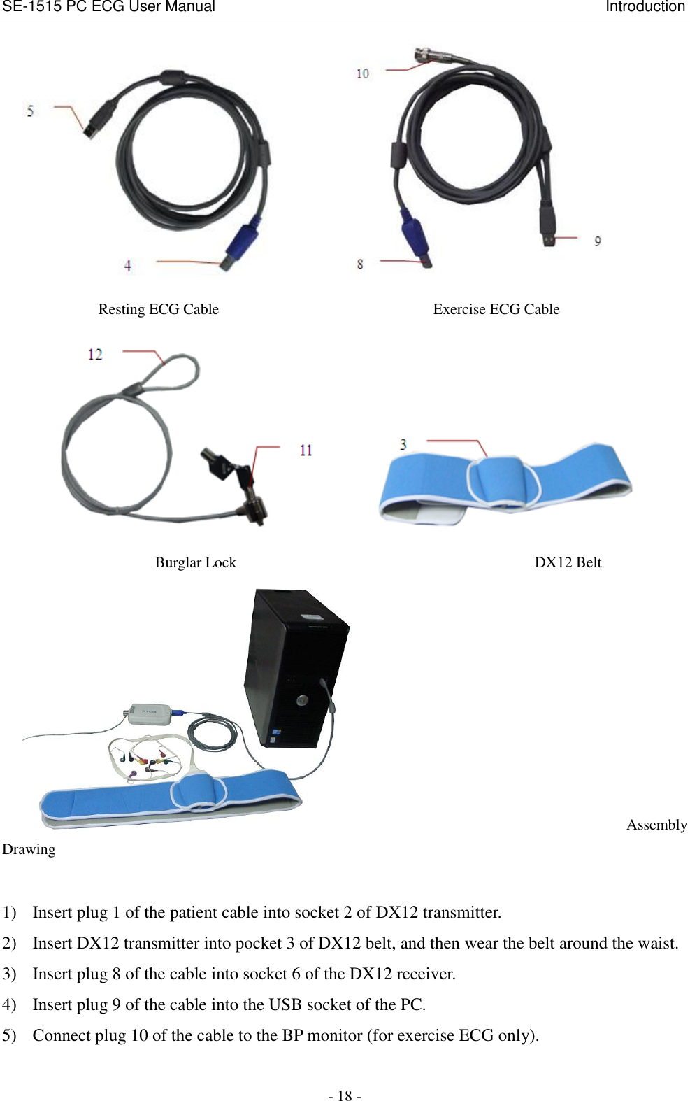
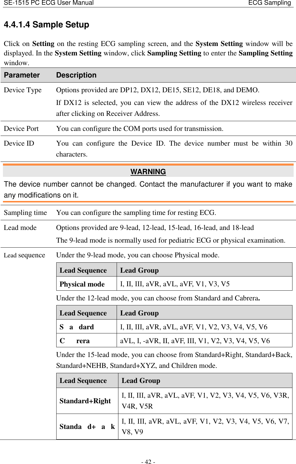
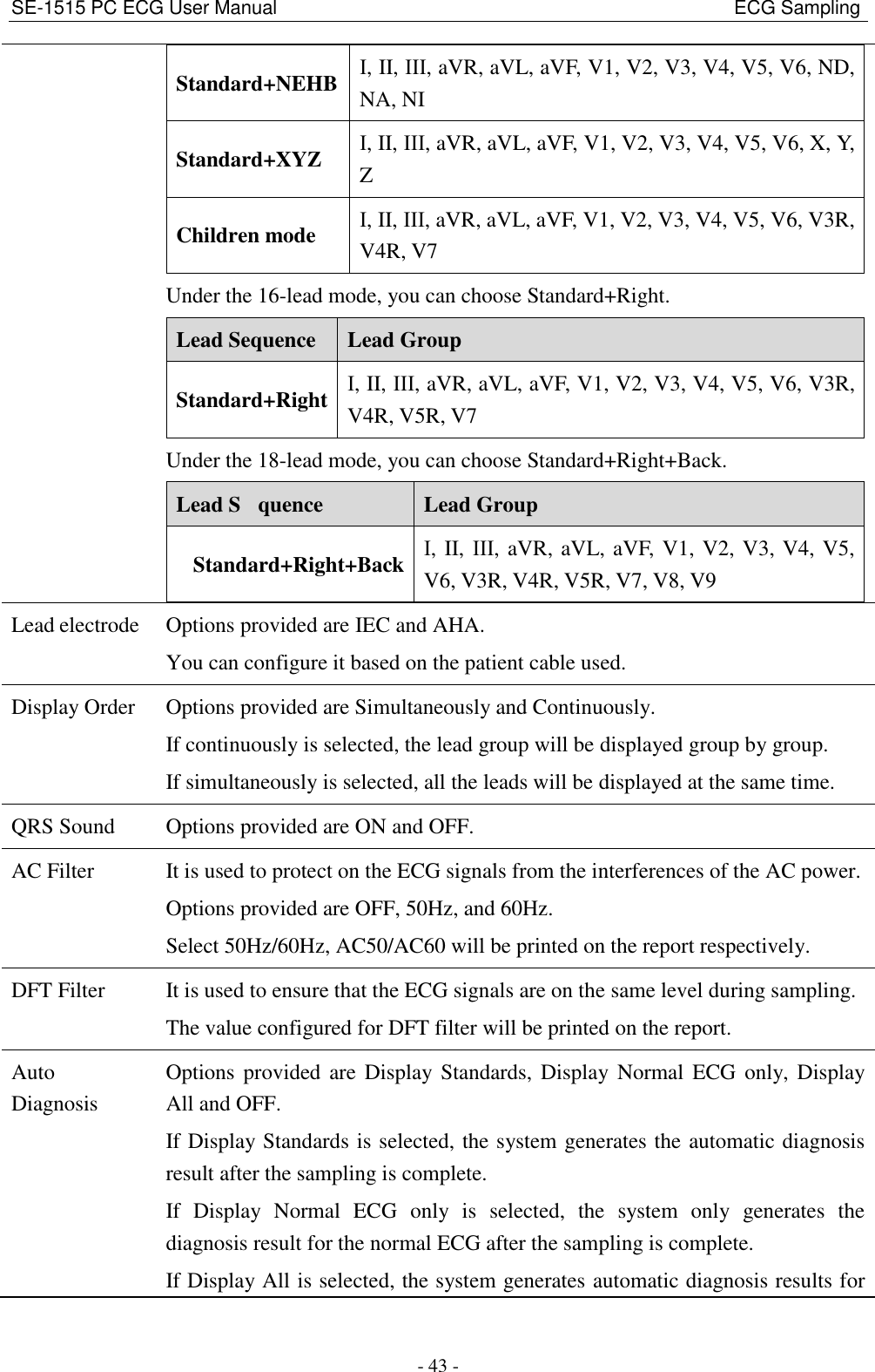
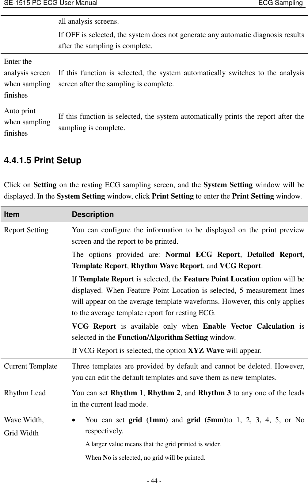
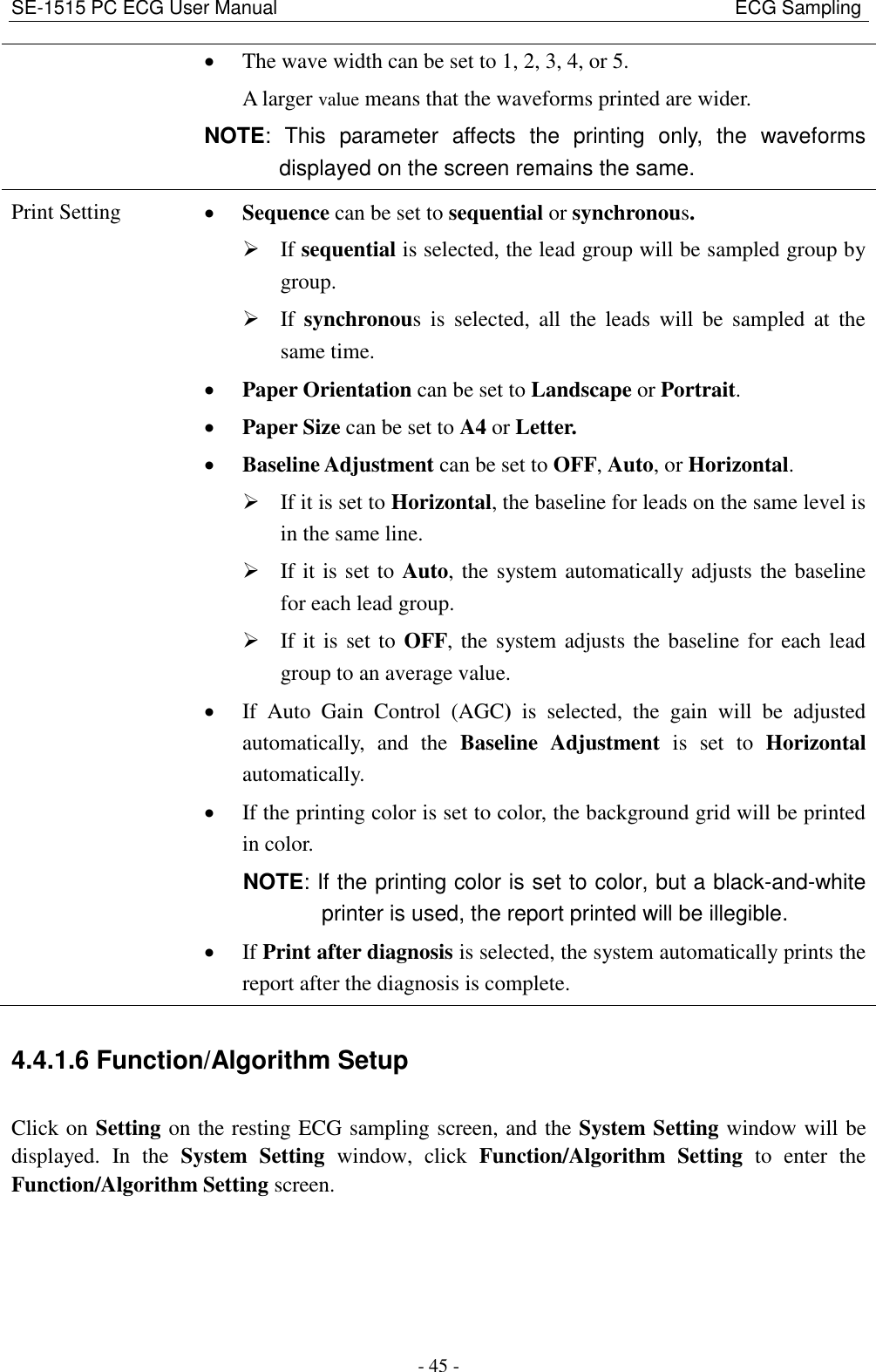
EDAN INSTRUMENTS DX12REEDAN ECG sampling box with Bluetooth User Manual SE 1515 PC ECG

EDAN INSTRUMENTS, INC. ECG sampling box with Bluetooth SE 1515 PC ECG

User manual

 I About this Manual P/N: 01.54.455911 MPN: 01.54.455911018 Release Date: Oct. 2016 © Copyright EDAN INSTRUMENTS, INC. 2013-2016. All rights reserved. Statement This manual will help you understand the operation and maintenance of the product better. It is reminded  that  the  product  shall  be  used  strictly  complying  with  this  manual.  User's  operation failing  to  comply  with  this  manual  may  result  in  malfunction  or  accident  for  which  EDAN INSTRUMENTS, INC. (hereinafter called EDAN) cannot be held liable. EDAN  owns  the  copyrights  of  this  manual.  Without  prior  written  consent  of  EDAN,  any materials contained in this manual shall not be photocopied, reproduced or translated into other languages. Materials protected by  the copyright law, including but not limited to  confidential information such as technical information and patent information are contained in this manual, the user shall not disclose such information to any irrelevant third party. The user shall understand that nothing in this manual grants  him,  expressly or implicitly,  any right or license to use any of the intellectual properties of EDAN. EDAN holds the rights to modify, update, and ultimately explain this manual. Responsibility of the Manufacturer EDAN only considers itself responsible for any effect on safety, reliability and performance of the equipment if: Assembly  operations,  extensions,  re-adjustments,  modifications  or  repairs  are  carried  out  by persons authorized by EDAN, and   The electrical installation of the relevant room complies with national standards, and   The instrument is used in accordance with the instructions for use. Terms Used in this Manual This guide is designed to give key concepts on safety precautions.
 II WARNING A  WARNING  label  advises  against  certain  actions  or  situations  that  could  result  in  personal injury or death. CAUTION A  CAUTION label advises against actions or situations that could damage equipment, produce inaccurate data, or invalidate a procedure. NOTE A NOTE provides useful information regarding a function or a procedure.
 III Contents Chapter 1 Safety Guidance ........................................................................................................... 1 1.1 Indications for Use/Intended Use ........................................................................................ 1 1.2 Warnings and Cautions ........................................................................................................ 1 1.2.1 General Warnings ...................................................................................................... 1 1.2.2 General Cautions ....................................................................................................... 4 1.2.3 Operation Warnings for Wireless System ................................................................. 5 1.2.4 Preparation and Operation Warnings (for Exercise ECG) ........................................ 6 1.2.5 Contraindications (for Exercise ECG) ...................................................................... 7 1.3 List of Symbols ................................................................................................................... 8 Chapter 2 Introduction ................................................................................................................ 10 2.1 Wired SE-1515 System ..................................................................................................... 11 2.1.1 Wired System Connection Diagram ........................................................................ 11 2.1.2 Assembling the Wired System ................................................................................ 12 2.1.3 ECG Sampling Box ................................................................................................. 13 2.2 Wireless SE-1515 System ................................................................................................. 14 2.2.1 Wireless System Connection Diagram .................................................................... 14 2.2.2 Assembling Wireless System .................................................................................. 17 2.2.3 ECG Sampling Box ................................................................................................. 19 2.3 Installing the Software ...................................................................................................... 21 2.3.1 Requirements of the PC .......................................................................................... 21 2.3.2 About Installation Window ..................................................................................... 22 2.4 Features ............................................................................................................................. 22 Chapter 3 Preparations Before Operation ................................................................................. 24 3.1 Preparing the Patient ......................................................................................................... 24 3.1.1 Instructing the Patient ............................................................................................. 24 3.1.2 Cleaning the Skin .................................................................................................... 24 3.2 Connecting the Electrodes of Wired System ..................................................................... 25 3.3 Connecting the Electrodes of Wireless System ................................................................. 26 3.4 Attaching Electrodes ......................................................................................................... 27 3.4.1 Attaching Electrodes for Resting ECG ................................................................... 27 3.4.2 Electrode Placement for Exercise ECG .................................................................. 32 3.4.3 Attaching the Reusable Electrodes .......................................................................... 33 3.4.4 Attaching Disposable Electrodes............................................................................. 34 3.5 Inspection Before Test ....................................................................................................... 35 Chapter 4 ECG Sampling ............................................................................................................ 37 4.1 Initial Configuration .......................................................................................................... 37 4.2 Entering Patient Information ............................................................................................. 37 4.2.1 Entering Patient Information Manually .................................................................. 38
 IV 4.2.2 Entering Patient Information by Using a Bar Code Reader .................................... 39 4.2.3 Retrieve Patient Information ................................................................................... 39 4.3 Selecting the ECG Sampling Type .................................................................................... 39 4.4 ECG Sampling .................................................................................................................. 40 4.4.1 Resting ECG Sampling ........................................................................................... 40 4.4.2 STAT ECG ............................................................................................................... 47 4.4.3 Exercise ECG Sampling .......................................................................................... 47 4.4.4 VCG Sampling ........................................................................................................ 54 4.4.5 HRV ECG Sampling ............................................................................................... 54 Chapter 5 ECG Analysis .............................................................................................................. 55 5.1 Resting ECG ..................................................................................................................... 55 5.1.1 Wave Analysis ......................................................................................................... 55 5.1.2 Average Template .................................................................................................... 57 5.1.3 About the Detail Information Window .................................................................... 57 5.1.4 About the Rhythm Wave Window ........................................................................... 58 5.1.5 History Record ........................................................................................................ 58 5.1.6 About the Parameters .............................................................................................. 58 5.2 Exercise ECG .................................................................................................................... 59 5.2.1 About the Summary Screen ..................................................................................... 59 5.2.2 About the All View Screen ...................................................................................... 61 5.2.3 About the ECG Strip Screen ................................................................................... 62 5.2.4 ST Analysis ............................................................................................................. 62 5.2.5 ST Trend .................................................................................................................. 63 5.3 VCG .................................................................................................................................. 63 5.3.1 Displaying Vector ECG with All Plane and All Loop ............................................. 64 5.3.2 Displaying Vector ECG with Frontal Plane and QRS Loop ................................... 66 5.3.3 Displaying 3D Vector ECG ..................................................................................... 67 5.4 HRV ................................................................................................................................... 68 5.5 Report Previewing ............................................................................................................. 69 5.6 Report Printing .................................................................................................................. 69 5.7 Saving ECG Reports ......................................................................................................... 69 Chapter 6 Archives ....................................................................................................................... 71 6.1 All List ............................................................................................................................... 71 6.1.1 Record Display ........................................................................................................ 71 6.1.2 Modifying Patient Information ............................................................................... 71 6.1.3 Viewing Examination Records ................................................................................ 72 6.1.4 Deleting Examination Records ............................................................................... 72 6.1.5 Merging Examination Records ............................................................................... 72 6.1.6 Searching Patient Records....................................................................................... 72 6.1.7 Import ...................................................................................................................... 73
 V 6.1.8 Export ...................................................................................................................... 73 6.2 Order List .......................................................................................................................... 74 6.2.1 New Order ............................................................................................................... 74 6.2.2 Inbox ....................................................................................................................... 74 6.2.3 Searching Information ............................................................................................. 74 Chapter 7 Statistics ...................................................................................................................... 75 Chapter 8 System Setup ............................................................................................................... 76 8.1 Basic Setup ........................................................................................................................ 76 8.2 Display Setup .................................................................................................................... 77 8.3 Transmission Setup ........................................................................................................... 77 8.4 Output File Setup .............................................................................................................. 78 8.5 GDT Setup ........................................................................................................................ 79 8.6 DICOM Setup ................................................................................................................... 79 8.7 HL7 Setup ......................................................................................................................... 80 8.8 Barcode Setup ................................................................................................................... 80 8.9 Other Setup ....................................................................................................................... 81 8.9.1 Product Authorization ............................................................................................. 82 8.9.2 Advanced Setting .................................................................................................... 82 Chapter 9 Hint Information ........................................................................................................ 84 Chapter 10 Cleaning, Care and Maintenance ........................................................................... 85 10.1 General Points ................................................................................................................. 85 10.2 Cleaning .......................................................................................................................... 85 10.2.1 Cleaning the Sampling Box .................................................................................. 86 10.2.2 Cleaning the Patient Cable .................................................................................... 86 10.2.3 Cleaning the Reusable Electrodes (for Resting ECG) .......................................... 86 10.3 Disinfection ..................................................................................................................... 87 10.3.1 Disinfecting the Sampling Box ............................................................................. 87 10.3.2 Disinfecting the Patient Cable ............................................................................... 87 10.3.3 Disinfecting the Reusable Electrodes (for Resting ECG) ..................................... 87 10.4 Maintenance of ECG Sampling Box ............................................................................... 88 Chapter 11 Accessories ................................................................................................................ 89 Chapter 12 Warranty & Service ................................................................................................. 93 12.1 Warranty .......................................................................................................................... 93 12.2 Contact Information ........................................................................................................ 93 Appendix 1 Technical Specifications .......................................................................................... 94 A1.1 Safety Specifications ...................................................................................................... 94 A1.2 Environment Specifications ........................................................................................... 94 A1.3 Physical Specifications ................................................................................................... 95 A1.4 Power Supply Specifications .......................................................................................... 95 A1.5 Performance Specifications ............................................................................................ 96
 VI Appendix 2 EMC Information .................................................................................................... 99 Appendix 3 Abbreviations ......................................................................................................... 103
SE-1515 PC ECG User Manual                                                                                            Safety Guidance - 1 - Chapter 1 Safety Guidance This chapter provides important safety information related to the use of SE-1515. 1.1 Indications for Use/Intended Use SE-1515 PC ECG is intended to acquire, process and store ECG signals from adult and pediatric patients undergoing stress exercise test or resting test. The SE-1515 PC ECG is intended to be used only in hospitals and healthcare facilities by doctors and trained healthcare professionals. The cardiogram recorded by the SE-1515 PC ECG can help users to analyze and diagnose heart diseases.  However,  the  ECG  with  measurements  and  interpretive  statements  is  offered  to clinicians on an advisory basis only. WARNING 1.  This system is not designed for intracardiac use or direct cardiac application. 2.  This system is not intended for home use. 3.  This system is not intended for treatment or monitoring. 4.  This system is intended for use on adult and pediatric patients only. 5.  The results given by the system should be examined  based on the overall clinical condition of the patient, and they cannot substitute for regular checking. 1.2 Warnings and Cautions To  use  the  system  safely  and  effectively,  firstly  be  familiar  with  the  operation  method  of Windows and read the user manual in detail to be familiar with the proper operation method for the purpose of avoiding the possibility of system failure. The following warnings and cautions must be paid more attention to during the operation of the system. 1.2.1 General Warnings WARNING 1.  The system is intended to be used by qualified physicians or personnel professionally trained.  They  should  be  familiar  with  the  contents  of  this  user  manual  before operation.
SE-1515 PC ECG User Manual                                                                                            Safety Guidance - 2 - WARNING 2.  Only  qualified  service  engineers  can  install  this  equipment,  and  only  service engineers  authorized  by  the  manufacturer  can  open  the  shell.  Otherwise,  safety hazards may happen. 3. EXPLOSION  HAZARD  -  Do  not  use  the  system  in  the  presence  of  flammable anesthetic mixtures with oxygen or other flammable agents. 4. SHOCK HAZARD - The power receptacle must be a hospital grade grounded outlet. Never try to adapt the three-prong plug to fit a two-slot outlet. 5.  Only the patient cable and other accessories supplied by the manufacturer can be used. Or else, the performance and electric shock protection cannot be guaranteed. The system has been safety tested with the recommended accessories, peripherals, and  leads,  and  no  hazard  is  found  when  the  system  is  operated  with  cardiac pacemakers or other stimulators. 6.  The use of patient cable and other accessories not supplied by the manufacturer may result in increased emissions or decreased immunity of the equipment. 7.  Make sure that all electrodes are connected to the patient correctly before operation. 8.  Ensure that the conductive parts of electrodes and associated connectors, including neutral electrodes, do not come in contact with earth or any other conducting objects. 9.  Disposable electrodes must be used during defibrillation. 10. Electrodes of dissimilar metals should not be  used; otherwise it may cause a  high polarization voltage. 11. The disposable electrodes can only be used for one time. 12. Do not touch the patient, bed, table or the equipment while using the ECG together with a defibrillator. 13. Do not touch accessible parts of electrical equipment and the patient simultaneously. 14. The use of equipment that applies high frequency voltages to the patient (including electrosurgical  equipment  and  some  respiration  transducers)  is  not  supported  and may  produce  undesired  results.  Disconnect  the  patient  data  cable  from  the  ECG workstation, or detach the leads from the patient prior to performing any procedure that uses high frequency surgical equipment. 15. Fix attention on the examination to avoid missing important ECG waves. 16. SHOCK HAZARD - Don't connect non-medical electrical equipment, which has been supplied  as  a part  of  the  system,  directly  to  the  wall  outlet  when  the  non-medical equipment  is  intended  to  be  supplied  by  a  multiple  portable  socket-outlet  with  an isolation transformer.
SE-1515 PC ECG User Manual                                                                                            Safety Guidance - 3 - WARNING 17. SHOCK HAZARD - Don't connect electrical equipment, which has not been supplied as a part of the system, to the multiple portable socket-outlet supplying the system. 18. Do  not  connect  any  equipment  or  accessories  that  are  not  approved  by  the manufacturer or that are not IEC/EN 60601-1 approved to the system. The operation or use of non-approved equipment or accessories with the system is not tested or supported, and system operation and safety are not guaranteed. 19. Any non-medical equipment (such as the external printer) is not allowed to be used within the patient vicinity (1.5m/6ft.). 20. Do  not  exceed  the  maximum  permitted  load  when  using  the  multiple  portable socket-outlet(s) to supply the system. 21. Multiple portable socket-outlets shall not be placed on the floor. 22. Do  not  use  the  additional  multiple  portable  socket-outlet  or  extension  cord  in  the medical electrical system, unless it's specified as part of the system by manufacturer. And the multiple portable socket-outlets provided with the system shall only be used for supplying power to equipment which is intended to form part of the system. 23. Accessory equipment connected to the analog and digital interfaces must be certified according to the respective IEC/EN standards (e.g. IEC/EN 60950 for data processing equipment  and  IEC/EN  60601-1  for  medical  equipment).  Furthermore  all configurations shall comply with the valid version of the standard IEC/EN  60601-1. Therefore anybody, who connects additional equipment to the signal input or output connector to configure a medical system, must make sure that it complies with the requirements of the valid version of the system standard IEC/EN 60601-1. If in doubt, consult our technical service department or your local distributor. 24. Connecting  any  accessory  (such  as  external  printer)  or  other  device  (such  as  the computer) to this ECG workstation makes a medical system. In that case, additional safety measures should be taken during installation of the system, and the system shall provide: a)  Within the patient environment, a level of safety comparable to that provided by medical electrical equipment complying with IEC/EN 60601-1, and   b)  Outside the patient environment, the level of safety appropriate for non-medical electrical equipment complying with other IEC or ISO safety standards. 25. All the accessories connected to system must be installed outside the patient vicinity, if they do not meet the requirement of IEC/EN 60601-1. 26. Make sure that there is no intense electromagnetic interference source around when using the wireless system of SE-1515. Furthermore, keep an unobstructed distance of at most 5 meters between DX12 transmitter and the PC.
SE-1515 PC ECG User Manual                                                                                            Safety Guidance - 4 - WARNING 27. You should purchase computer, printer, treadmill, ergometer, BP monitor and bar code reader  from  the  manufacturer.  Otherwise,  the  manufacturer  will  not  be  held responsible  for  the  maintenance  of  the  PC  hardware,  operating  system  and  other accessories. 28. If multiple instruments are connected to a patient, the sum of the leakage currents may exceed the limits given in the IEC/EN 60601-1 and may pose a safety hazard. Consult your service personnel. 29. Connecting  to  other  devices  may  decrease  the  antistatic  gradation  of  the  system during operation. 30. The system shall not be serviced or maintained while in use with a patient. 31. The appliance coupler or mains plug is used as isolation means from supply mains. Position the ECG workstation in a location where the operator can easily access the disconnection device. 32. The medical electrical equipment needs to be installed and put into service according to Appendix 2 EMC information. 33. Portable  and  mobile  RF  communications  equipment  can  affect  medical  electrical equipment, refer to the recommended separation distances provided in Appendix 2 EMC Information. 34. The equipment should not be used adjacent to or stacked with other equipment, refer to the recommended separation distances provided in Appendix 2 EMC Information. 35. Assembly of the ECG workstation and modifications during actual service life shall be evaluated based on the requirements of IEC60601-1. 36. The device is MR unsafe. It is not intended for use in an MRI environment. 37. Magnetic and electrical fields are capable of interfering with the proper performance of  the device. For this  reason make sure that  all external devices operated in the vicinity of the device comply with the relevant EMC requirements. X-ray equipment or MRI devices are a possible source of interference as they may emit higher levels of electromagnetic radiation. 1.2.2 General Cautions   CAUTION 1.  Avoid  liquid  splash  and  excessive  temperature.  The  temperature  must  be  kept between 5ºC and 40ºC during operation, and it should be kept between -20ºC and 55ºC  during transportation and storage. 2.  Do  not  use  the  equipment  in  a  dusty  environment  with  bad  ventilation  or  in  the presence of corrosive.
SE-1515 PC ECG User Manual                                                                                            Safety Guidance - 5 - CAUTION 3.  Make sure that there is no intense electromagnetic interference source around the equipment, such as radio transmitters or mobile phones etc. Attention: large medical electrical equipment such as electrosurgical equipment, radiological equipment and magnetic  resonance  imaging  equipment  etc.  is  likely  to  bring  electromagnetic interference. 4.  The device and accessories are to be disposed of according to local regulations after their useful lives. Alternatively, they can be returned to the dealer or the manufacturer for recycling or proper disposal. Batteries are hazardous waste. Do NOT dispose of them together with house-hold garbage. At the end of their lives hand the batteries over to the applicable collection points for the recycling of waste batteries. For more detailed  information  about  recycling of  this  product  or  battery,  please  contact  your local Civic Office, or the shop where you purchased the product. 5.  Federal (U.S.) law restricts this device to sale by or on the order of a physician. 1.2.3 Operation Warnings for Wireless System WARNING 1.  Make sure that there is no intense electromagnetic interference source around the wireless system. 2.  Do not open the battery cover of the transmitter during operation. 3.  Improper  operations  may  heat  the  battery,  make  it  on  fire,  explode,  destroyed  or reduce its capacity. Please read the manual and the cautions carefully before using the battery. 4.  Batteries of the same model and specification as manufacture configuration should be used. 5. DANGER  OF  EXPLOSION --  Do  not  reverse  the  anode  and  the  cathode  when installing the battery. 6. Do not use the battery near fire or in an environment where the temperature is over 60ºC ; do not heat or splash the battery or throw it into fire or water. 7.  Do not destroy the battery; do not pierce battery with a sharp object such as a needle; do not hit with a hammer, step on or throw or drop to cause strong shock; do not disassemble or modify the battery. 8.  When leakage or foul smell is found, stop using the battery immediately. If your skin or cloth comes into contact with the leakage liquid, cleanse it with clean water at once. If the leakage liquid splashes into your eyes, do not wipe them. Irrigate them with clean water first and go to see a doctor immediately.
SE-1515 PC ECG User Manual                                                                                            Safety Guidance - 6 - WARNING 9.  Properly dispose of or recycle the depleted battery according to local regulations. 10. Remove the battery from the transmitter if the system won't be used for a long time. 1.2.4 Preparation and Operation Warnings (for Exercise ECG) WARNING 1.  Test  the  safety stop  (mushroom  type)  and  safety stop (cord  type)  of  the  treadmill before using the system. 2.  During the exercise test, ensure that such tests are supervised by properly trained technician who meets competency requirements for exercise test supervision, fully trained in cardiopulmonary resuscitation, and is supported by a physician skilled in exercise  testing  or  emergency  medicine  who  is  in  close  proximity  for  pretest assessment or compliance that may raise. 3.  Make  sure  that  there  is  necessary  valid  first-aid  equipment  such as  defibrillators, blood-pressure meters etc, and necessary valid medication in the exercise test room. 4.  Turn off the system power and disconnect the power cord from the wall outlet after using the system. 5.  Make sure that the power is turned off and the power cord is disconnected from the AC socket before defibrillation. 6.  Keep  the  four  feet  of  the  machine  on  the  ground  and  make  sure  that  it's  stably working. 7.  The treadmill must be powered by the specific power outlet. 8.  Examine the treadmill/ergometer carefully before using it. 9.  The patient undergoing the exercise test should wear suitable clothes and shoes. 10. Keep hands, hair, jewelry, and loose clothing away from moving parts. 11. Don't let the patient stand on the running belt when starting the treadmill. The patient should stand on the foot rails and hold the handrails during start-up. Wait until the running belt is moving before placing feet on the belt. 12. To avoid the static electricity, the patient should not wear loose clothing or clothing (such as nylon) that easily produces static electricity. 13. Stop  exercising  immediately  when  the  patient  feels  uncomfortable  or  something abnormal in the operation. 14. Press down the safety stop (mushroom type) or pull out the safety stop (cord type) to stop the treadmill immediately when an emergency happens.
SE-1515 PC ECG User Manual                                                                                            Safety Guidance - 7 - 1.2.5 Contraindications (for Exercise ECG) Absolute Contraindications: 1.  Acute MI (within 2 days) 2.  High-risk unstable angina 3.  Hemodynamic compromise caused by uncontrolled cardiac arrhythmia 4.  Symptomatic severe aortic stenosis 5.  Heart failure with clinic episode uncontrolled 6.  Acute pulmonary embolus or pulmonary infarction 7.  Acute myocarditis or pericarditis 8.  Acute aortic dissection 9.  The patient opposes the test. Relative Contraindications: 1.  Left main coronary stenosis 2.  Moderate stenotic valvular heart disease 3.  Serum Electrolyte abnormalities 4. Severe  hypertension  (systolic  blood  pressure  >200  mmHg  or  diastolic  blood pressure >110 mmHg) 5.  Tachyarrhythmias or bradyarrhythmias 6.  Hypertrophic cardiomyopathy 7.  Patients can not cooperate because of mental impairment or physical disability 8.  High-degree AV block
SE-1515 PC ECG User Manual                                                                                            Safety Guidance - 8 - 1.3 List of Symbols No. Symbol Description 1  DEFIBRILLATION-PROOF TYPE CF APPLIED PART 2  Caution 3  Operating instructions 4  General symbol for recovery/recyclable 5  Part Number 6  SERIAL NUMBER 7  Date of manufacture 8  MANUFACTURER 9  CLASS II equipment 10  AUTHORISED  REPRESENTATIVE  IN  THE  EUROPEAN COMMUNITY 11  Transmission Status Indicator of Bluetooth 12  Power Supply Indicator of DX12 Receiver
SE-1515 PC ECG User Manual                                                                                            Safety Guidance - 9 - 13  Start printing button (on DE15&DE18) 14  CE marking 15  Disposal method 16  Caution: Federal (U.S.) law restricts this device to sale by or on the order of a physician. 17   Federal Communications Commission:   FCC ID:SMQDX12TREDAN (for DX12 Transmitter) FCC ID:SMQDX12REEDAN (for DX12 Receiver) 18  Refer to User Manual (Background: Blue; Symbol: White) 19  Warning (Background: Yellow; Symbol&Outline: Black) 20  Non- ionizing electromagnetic radiation 21  MR  Unsafe–Keep  away  from  magnetic  resonance imaging (MRI) equipment NOTE: The user manual is printed in black and white.
SE-1515 PC ECG User Manual                                                                                                   Introduction - 10 - Chapter 2 Introduction The SE-1515 system consists of the following equipment:  18-lead ECG sampling system (18-lead ECG sampling box) Or, 16-lead ECG sampling system (16-lead ECG sampling box) Or, 12-lead wired ECG sampling system (12-lead ECG sampling box) Or, 12-lead wireless ECG sampling system (wireless DX12 transmitter and receiver)  PC ECG software  Patient cable  Disposable electrodes  USB cable According to the configurations of different types of workstations, the following purchased accessories can also be included: tablet, computer, screen, printer, treadmill/ergometer, and exercise BP monitor. NOTE: The pictures and windows in this manual are for reference only. WARNING 1.  Use a special grounded socket to get accurate voltage and current. 2.  When using a laptop with a two-prong plug, please connect a grounded printer to avoid power interference. 3.  Only stress BP monitors can be used.
SE-1515 PC ECG User Manual                                                                                                   Introduction - 11 - 2.1 Wired SE-1515 System 2.1.1 Wired System Connection Diagram 1. Resting ECG of Wired SE-1515 System  2. Exercise ECG of Wired SE-1515 System
SE-1515 PC ECG User Manual                                                                                                   Introduction - 12 - 2.1.2 Assembling the Wired System The DE15 ECG sampling box is used as an example here:                Patient Cable                                                                      DE15 ECG sampling box                         Resting ECG Cable                                                                  Exercise ECG Cable  Assembly Drawing  1 7 8 6 5 4 2 3
SE-1515 PC ECG User Manual                                                                                                   Introduction - 13 - 1) Insert plug 1 of the patient cable into socket 2 of DE15 ECG sampling box. 2) Insert plug 7 of the cable into socket 3 of DE15 ECG sampling box. 3) Insert plug 8 of the cable into the USB socket of the PC. 4) Connect plug 6 of the cable to the BP monitor (for exercise ECG only). 5) Connect a treadmill or an ergometer to the PC (for exercise ECG only). 6) Connect a printer to the PC. 7) Insert the Sentinel into the USB socket of the PC if the Sentinel is purchased. 8) Make  sure  that  the  above  parts  are  properly  connected,  and  then  connect  the  PC, treadmill/ergometer and printer to the power supply. 2.1.3 ECG Sampling Box WARNING 1.  When the computer connected to the USB cable is powered on, do not connect the USB  cable  to  the  ECG  sampling  box;  when  the  system  is  powered  on,  do  not disconnect the USB cable from the ECG sampling box. 2.  It  is not necessary or recommended to regularly disconnect  the USB  cable from the ECG sampling box. Disconnect the USB cable from the PC if necessary. 3.  When connecting the sampling box to the PC, do not connect them via an USB HUB. 4.  When a sampling box is connected the PC, no other device should be connected to the PC for battery enchargement through an USB cable. 2.1.3.1 DE15/DE18 ECG sampling box  Name Explanation Indicator When  the  ECG  sampling  box  is  powered  by  the  PC,  the indicator will be lit. USB Interface Connecting to the USB socket of the PC with a USB cable  USB Interface Indicator
SE-1515 PC ECG User Manual                                                                                                   Introduction - 14 - 2.1.3.2 DP12 ECG Sampling Box  Front Panel  Name Explanation Indicator When  the  ECG  sampling  box  is  powered  by  the  PC,  the indicator will be lit. USB Interface Connecting to the USB socket of the PC with a USB cable 2.2 Wireless SE-1515 System WARNING This device complies with Part 15 of the FCC Rules. Operation is subject to the following two conditions:     1)  this device may not cause harmful interference, and   2)  this device must accept any interference received, including interference that may cause undesired operation. NOTE: 1.  This equipment has been tested and found to comply with the limits for a Class B digital device, pursuant to part 15 of the  FCC Rules. These limits are designed to provide reasonable protection against harmful interference in a residential installation. This equipment generates, uses and can radiate radio frequency energy and, if not installed  and  used  in  accordance  with  the  instructions,  may  cause  harmful interference  to  radio  communications.  However,  there  is  no  guarantee  that interference will not occur in a particular installation. If this equipment does cause harmful  interference  to  radio  or  television  reception,  which  can  be  determined  by turning  the  equipment  off  and  on,  the  user  is  encouraged  to  try  to  correct  the interference by one or more of the following measures: -  Reorient or relocate the receiving antenna. -  Increase the separation between the equipment and receiver. -  Connect the equipment into an outlet on a circuit different from that to which the Indicator USB Interface
SE-1515 PC ECG User Manual                                                                                                   Introduction - 15 - receiver is connected. -  Consult the dealer or an experienced radio/TV technician for help. 2. Any  changes  or  modifications  to  this  unit  not  expressly  approved  by  the  party responsible for compliance could void the user's authority to operate the equipment. 2.2.1 Wireless System Connection Diagram The DX12 device which consists of transmitter and receiver has passed FCC certification. This device  complies  with  Part  15  of  the  FCC  Rules.  Operation  is  subject  to  the  following  two conditions: (1) this device may not cause harmful interference, and (2) this device must accept any interference received, including interference that may cause undesired operation.
SE-1515 PC ECG User Manual                                                                                                   Introduction - 16 - 1 Resting ECG of Wireless System    2 Exercises ECG of Wireless System  NOTE: The manufacturer is not responsible for any radio or TV interference caused by unauthorized modifications to this  equipment. such modifications could  void  the user’s authority to operate this equipment.  WARNING 1.  Accessory equipment connected to the analog and digital interfaces must be certified according to the respective IEC/EN standards (e.g. IEC/EN 60950 for data processing equipment and IEC/EN 60601-1 for medical equipment). Furthermore all configurations shall comply with the valid version of the standard IEC/EN 60601-1. Therefore anybody, who connects additional equipment to the signal input or output connector to configure a medical system, must make sure that it complies with the requirements of the valid version  of  the  system  standard  IEC/EN  60601-1.  If  in  doubt,  consult  our  technical
SE-1515 PC ECG User Manual                                                                                                   Introduction - 17 - service department or your local distributor. 2.  The system should be installed by a qualified service engineer. Do not power on the system until all cables are properly connected and verified. 3.  If multiple instruments are connected to a patient, the sum of the leakage currents may exceed the limits given in the IEC/EN 60601-1 and may pose a safety hazard. Consult your service personnel. 4.  DX12 transmitter of the wireless system uses the Bluetooth technology, which could make the patient with the pacemaker uncomfortable. Keep DX12 transmitter far away from the pacemaker when using the wireless system of SE-1515. 2.2.2 Assembling Wireless System  Patient Cable              DX12 Transmitter                                         DX12 Receiver
SE-1515 PC ECG User Manual                                                                                                   Introduction - 18 -             Resting ECG Cable                                                        Exercise ECG Cable                   Burglar Lock                                                                              DX12 Belt   Assembly Drawing 1) Insert plug 1 of the patient cable into socket 2 of DX12 transmitter. 2) Insert DX12 transmitter into pocket 3 of DX12 belt, and then wear the belt around the waist. 3) Insert plug 8 of the cable into socket 6 of the DX12 receiver. 4) Insert plug 9 of the cable into the USB socket of the PC. 5) Connect plug 10 of the cable to the BP monitor (for exercise ECG only).
SE-1515 PC ECG User Manual                                                                                                   Introduction - 19 - 6) Connect a treadmill or an ergometer to the PC (for exercise ECG only). 7) Connect a printer to the PC. 8) Insert the Sentinel into the USB socket of the PC if the sentinel is purchased. 9) Unlock burglar lock 11 and insert into the burglar lock keyhole of DX12 receiver, and fix the wireless system via the other end of the burglar lock. 10) Make  sure  that  the  above  parts  are  properly  connected,  and  then  connect  the  PC, treadmill/ergometer and printer to the power supply. 2.2.3 ECG Sampling Box 2.2.3.1 Receiver   2.2.3.2 Transmitter  Button and Symbol    Figure 2-1 DX12 Transmitter Main Screen USB interface Keyhole
SE-1515 PC ECG User Manual                                                                                                   Introduction - 20 - Button Explanation  Switch on DX12 receiver and install batteries on DX12 transmitter. Press the button to start up DX12 transmitter, the main screen will be displayed.. When the main screen is displayed, press the button to return to previous menu.  When the  main screen or the  setting screen is displayed,  press the  button  to enter the next menu. Press  the  button  first,  and  press    within  1.2s  to  lock  or  unlock  the keyboard.  When the main screen is displayed, press the button to switch leads. When  the  menu  screen is  displayed, press  the  button  to  display  one  item  in black.  Bluetooth Connection Icon   If the Bluetooth connection icon    is not displayed on the main screen, you have to match the device manually.  Keyboard Lock Icon No  operation  within  8s,  the  keyboard  will  be  locked  automatically  and  the main screen is displayed.  Battery Capacity Icon If  the  battery  in  the  DX12  transmitter  is  low,  hint  will  appear  in  the  ECG station software.  Menu Settings  Make some settings on the DX12 transmitter according to actual use. Menu Description
SE-1515 PC ECG User Manual                                                                                                   Introduction - 21 - Back Light Select On to turn on the backlight of LCD screen. Select Off to turn off the backlight of LCD screen. Auto Sleep Select  On  to  display  Sleeping  on  the  screen  and  make  DX12 transmitter  be  in  low  power  consumption  mode  after  lead  off  for  5 minutes. Select Off to turn off auto sleep function. Language You can set the system language. Lead Electrode You can select IEC or AHA. Match Device Inquiring…will  be  displayed  (for  10  seconds)  to  search  DX12 receiver.  The  address  of  DX12  receiver  will  be  displayed  (for  8 seconds) if a matching DX12 receiver is found. No device found. Try again  later  will  be  displayed  (for  1  second)  if  no  matching  DX12 receiver is found. Device Information You  can  see  the  related  information,  such  as  software  version,  ID, address  of  DX12  receiver,  manufacture  and  release  time  about  the device.   NOTE: The device information is for reference only. 2.3 Installing the Software 2.3.1 Requirements of the PC CPU Pentium P4, Celeron D 310 or above System Memory (RAM) 1G or above Main Board Recommend the main board of Intel chipset Hard Disk 128G or above Printer ink jet printer of more than 300dpi or laser printer Recommend HP2035, HP2010, CANON iP1980 Display 17"  TFT  (Resolution:  1280*1024,  1366*768),  19"  TFT (resolution: 1440×900), 21" TFT(1920*1080) Operating System Windows  XP  SP3  (32  bit),  Windows  7  SP1  (32/64  bit)  or Windows  8  (32/64  bit), Windows 8.1  (32/64  bit), Windows 10 (32/64 bit)
SE-1515 PC ECG User Manual                                                                                                   Introduction - 22 - Complied Standard IEC/EN 60950 NOTE: Ensure that there is a graphic driver installed in the PC. Otherwise, the displayed ECG waves may be abnormal. 2.3.2 About Installation Window Insert  the  installation  CD  into  CD-ROM,  double-click  on  Setup.exe  and  then  follow  the directions to finish the installation. For details on installing SE-1515 software, please refer to SE-1515 PC ECG Installation Guide. 2.4 Features  3-/6-/9-/12-/15-/16-/18-channel ECG waves are displayed and printed  ECG waves can be frozen and reviewed  Measurement point adjustment  and re-analysis,  manual measurement with  an electronic ruler of high precision  High performance filters guarantee stable ECG waveforms  Perfect data management and processing functions  Multiple report formats, including PDF, Word, JPG, and BMP  Supporting multi-language  Supporting auto measurement and diagnosis  Editing the diagnosis templates The following features are only for the exercise test function    High-performance ECG filter, which ensures wave stability  During  sampling,  it  supports  analyzing  real-time  HR,  ST  segment  and  ST  trend,  and displays and prints the simultaneous 12-lead ECG in real time.  During sampling, you can perform ST segment analysis on the data of 12 leads, and adjust the ST segment place involved in lead analysis at any time.  Analyzing arrhythmia automatically  Providing summaries, ST analysis, wave reviews and trends  Providing specific statistic data of each lead in each stage  Providing average waves of each lead in each stage for you to observe the changes of ST segments among different stages  Automatically generating delicate reports and providing report preview  Providing classical exercise protocols; new exercise protocols can be added to the system
SE-1515 PC ECG User Manual                                                                                                   Introduction - 23 -  Storing massive patient data in the computer, which enables you to review and analyze the exercise ECG in any time  Automatically controlling and adjusting the speed and the elevation of the treadmill  Supporting many kinds of treadmills and ergometers
SE-1515 PC ECG User Manual                                                                    Preparations Before Operation - 24 - Chapter 3 Preparations Before Operation 3.1 Preparing the Patient NOTE: Correct operation for the best-quality ECG is very important. 3.1.1 Instructing the Patient 1. Before attaching the electrodes, greet the patient and explain the procedure. Explaining the procedure decreases the patient's anxiety. 2. Privacy is  important  for relaxation. When  possible, prepare the patient in a quiet  room or area where others can't  see the patient.  Use hangings beside the bed  during ECG  exam if other people are in the room.   3. Reassure the patient that the procedure is painless. 4. Make sure that the patient is comfortable. Once the electrodes and patient cable are connected, inform the patients that: 1) No talking 2) Breathe smoothly   3) Try to be calm 4) Not to chew or keep his teeth firmly The more relaxed the patient is, the less will the ECG wave is disturbed. 3.1.2 Cleaning the Skin The skin is a poor conductor of electricity and frequently creates artifacts that distort the ECG signals. There is natural resistance on the skin surface due to dry, dead epidermal cells, oils and dirt To clean the skin 1. Shave hair from electrode sites, if necessary. Excessive hair prevents a good connection. 2. Wash the area thoroughly with soap and water. 3. Dry the skin to increase capillary blood flow and to remove the dead, dry skin cells and oils. 4. Use the disposable frosting film in the standard accessory list to get good ECG waveform.   NOTE: Rub the skin with a gauze pad to increase capillary blood flow if you don't operate the steps above.
SE-1515 PC ECG User Manual                                                                    Preparations Before Operation - 25 - WARNING The  performance and electric  shock  protection  can  be  guaranteed  only if  the  original patient cable and electrodes of the manufacturer are used. 3.2 Connecting the Electrodes of Wired System  DE15 patient cable (Example)  12-Lead Patient Cable (Example) The  patient  cable  includes  patient  cable  plugs  and  lead  wires  which  can  be  connected  to electrodes according to the colors and identifiers. The lead wires have 10 chest leads and 4 limb leads.  Insert the patient cable plugs to the socket of ECG sampling box.  Align all lead wires of the patient cable to avoid twisting, and connect the lead wires to the corresponding electrodes according to the colors and identifiers.   Connecting to Electrodes  Lead Wires Connecting to ECG Sampling Box
SE-1515 PC ECG User Manual                                                                    Preparations Before Operation - 26 - 3.3 Connecting the Electrodes of Wireless System  Patient Cable (Snap Style)  Patient Cable (Banana Style) The  patient  cable  includes  patient  cable  plugs  and  lead  wires  which  can  be  connected  to electrodes according to the colors and identifiers. The lead wires have 10 chest leads and 4 limb leads.  Insert the patient cable plugs to the socket of DX12 transmitter.  Align all lead wires of the patient cable to avoid twisting, and connect the lead wires to the corresponding electrodes according to the colors and identifiers.   Connecting to Electrodes  Lead Wires Connecting to ECG Sampling Box  Main Cable  Main Cable  Lead Wires  Connecting to the ECG Sampling Box  Connecting to Electrodes
SE-1515 PC ECG User Manual                                                                    Preparations Before Operation - 27 - 3.4 Attaching Electrodes 3.4.1 Attaching Electrodes for Resting ECG WARNING Make sure that the conductive parts of electrodes and associated connectors, including neutral electrodes, do not come in contact with earth or any other conducting objects. The quality of ECG waveform will be affected by the contact resistance between the patient and the electrode. In order to get a high-quality ECG, the skin-electrode resistance must be minimized while connecting electrodes. The identifiers and color codes of electrodes used comply with IEC/EN requirements. In order to avoid incorrect connections, the electrode identifiers and color codes are specified in Table 3-1. Table 3-1 Electrodes and their identifiers and color codes IEC AHA Electrodes Color Code Electrodes Color Code R Red RA White L Yellow LA Black N/RF Black RL Green F Green LL Red C1 White/Red V1 Brown/Red C2 White/Yellow V2 Brown/Yellow C3 White/Green V3 Brown/Green C4 White/Brown V4 Brown/Blue C5 White/Black V5 Brown/Orange C6 White/Violet V6 Brown/Violet C3R White/Pink V3R Brown/Yellow C4R White/Gray V4R Brown/Red C5R White/Green V5R Brown/Green C7 White/Orange V7 Brown/Black C8 White/Blue V8 Brown/Blue
SE-1515 PC ECG User Manual                                                                    Preparations Before Operation - 28 - C9 White/Yellow V9 Brown/Yellow H Light blue/Violet/ H Orange/Violet E Light blue/ Yellow E Orange/Yellow I Light blue/ Red I Orange/ Red M Light blue/ Black M Orange/Black  Standard 12-Lead Placement  Only for the Reusable Electrodes              Only for the Disposable Electrodes IEC AHA Electrode Placement C1 V1 Fourth intercostal space at the right border of the sternum C2 V2 Fourth intercostal space at the left border of the sternum C3 V3 Fifth rib between C2 and C4 C4 V4 Fifth intercostal space on the left midclavicular line C5 V5 Left anterior axillary line at the horizontal level of C4 C6 V6 Left midaxillary line at the horizontal level of C4 L LA Left arm/Left deltoid
SE-1515 PC ECG User Manual                                                                    Preparations Before Operation - 29 - R RA Right arm/Right deltoid F LL Left leg/Upper leg as close to torso as possible N RL Right leg/Upper leg as close to torso as possible  Standard+XYZ    IEC AHA Electrode Placement C1 V1 Fourth intercostal space at the right border of the sternum C2 V2 Fourth intercostal space at the left border of the sternum C3 V3 Fifth rib between C2 and C4 C4 V4 Fifth intercostal space on the left midclavicular line C5 V5 Left anterior axillary line at the horizontal level of C4 C6 V6 Left midaxillary line at the horizontal level of C4 L LA Left arm/Left deltoid R RA Right arm/Right deltoid F LL Left leg/Upper leg as close to torso as possible N RL Right leg/Upper leg as close to torso as possible
SE-1515 PC ECG User Manual                                                                    Preparations Before Operation - 30 - H H Back of neck, avoid the carotid artery and jugular vein. E E Mid-sternum on the same horizontal level as C4 and C6. I I Right mid-axillary line on the same horizontal level as C4 and C6. M M Center of spine on the same horizontal level as C4 and C6  Frank Lead Placement (for VCG)        IEC AHA Electrode Placement C1 (Corresponding to I) V1 (Corresponding to I) Right mid-axillary line on the same horizontal level as C3 and C4 C2 (Corresponding to E) V2 (Corresponding to E) Sternum at the level of C3 and C4 C3 (Corresponding to C) V3 (Corresponding to C) Mid-clavicular  line  in  the  fifth  intercostals space C4 (Corresponding to A) V4 (Corresponding to A) Left mid-axilary line on the same horizontal level as C3 C5 (Corresponding to M) V5 (Corresponding to M) Center of spine on the same horizontal level as C3 and C4 C6 (Corresponding to H) V6 (Corresponding to H) Neck, avoid carotid artery and jugular vein L LA Left arm/Left deltoid   R RA Right arm/Right deltoid F LL Left  leg/Upper  leg  as  close  to  torso  as possible
SE-1515 PC ECG User Manual                                                                    Preparations Before Operation - 31 - N RL Right  leg/Upper  leg  as  close  to  torso  as possible  NEHB Placement  IEC AHA Electrode Placement Nst/C3R A1/V3R Attachment point of the second rib to the right sternal edge Nax/C4R A2/V4R Fifth intercostal space on the left posterior axillary line Nap/C4 V4 Left mid-clavicular line in the fifth intercostal space  V3R+V4R +V5R (Right)  IEC AHA Electrode Placement C3R V3R Right anterior chest opposite of C3 C4R V4R Right anterior chest opposite of C4 C5R V5R Right anterior chest opposite of C5
SE-1515 PC ECG User Manual                                                                    Preparations Before Operation - 32 -  V7+V8+V9 (Back)  DE18: IEC AHA Electrode Placement C7 V7 Left posterior axillary line on the same horizontal level as C4 and C6   C8 V8 Left midscapular line on the same horizontal level as C4 and C7 C9 V9 Left paraspinal border on the same horizontal level as C4 and C8 DE15: IEC AHA Electrode Placement C7 V7 Left posterior axillary line on the same horizontal level as C4 and C6   C8/V5R V8/V5R Left midscapular line on the same horizontal level as C4 and C7 C9/V4R V9/V4R Left paraspinal border on the same horizontal level as C4 and C8 3.4.2 Electrode Placement for Exercise ECG
SE-1515 PC ECG User Manual                                                                    Preparations Before Operation - 33 - The Precordial Electrodes' Positions on Body Surface: IEC AHA Electrode Placement C1 V1 Fourth intercostal space at the right border of the sternum C2 V2 Fourth intercostal space at the left border of the sternum C3 V3 Fifth rib between C2 and C4 C4 V4 Fifth intercostal space on the left midclavicular line C5 V5 Left anterior axillary line at the horizontal level of C4 C6 V6 Left midaxillary line at the horizontal level of C4 The Extremity Electrodes' Positions on Body Surface: IEC AHA Electrode Placement R / L RA / LA Below the right/left clavicle N / F RL / LL Below the right/left rib 3.4.3 Attaching the Reusable Electrodes 3.4.3.1 Attaching the Limb Electrodes  Limb Electrode        Limb Electrode Connection: 1) Ensure that the electrodes are clean; 2) Clean the electrode area which is a short distance above the ankle or the wrist with 75% alcohol; 3) Daub the electrode area on the limb with gel evenly; 4) Place  a  small  amount  of  gel  on  the  metal  part  of  the limb electrode clamp; 5) Connect the electrode to  the limb,  and make sure  that the metal part is placed on the electrode area above the ankle or the wrist; 6) Attach all limb electrodes in the same way. Reed Connecting  to  a  Lead  Wire Clamp
SE-1515 PC ECG User Manual                                                                    Preparations Before Operation - 34 - 3.4.3.2 Attaching the Chest Electrodes  Chest Electrode Chest Electrode Connection: 1) Ensure that the electrodes are clean; 2) Clean the electrode area on the chest surface with 75% alcohol; 3) Daub the round area of 25mm in diameter on each electrode site with gel evenly; 4) Place a small amount of gel on the brim of the chest electrode's metal cup; 5) Place the electrode on the chest electrode site and squeeze the suction bulb. Unclench it and the electrode is adsorbed on the chest; 6) Attach all chest electrodes in the same way. NOTE: Long-time measurement with a strong negative pressure on the suction bulb may cause reddening of the skin. When using the electrode on  kids or patients with delicate skin, squeeze the suction bulb lightly. 3.4.4 Attaching Disposable Electrodes    Disposable Electrode (clip style):                       Alligator Clip:              Disposable Electrode Connection (Clip Style) 1) Align all lead wires of the patient cable to avoid twisting, and connect the alligator clips to the patient cable. Suction Bulb Connecting to a Lead Wire Metal  Cup
SE-1515 PC ECG User Manual                                                                    Preparations Before Operation - 35 - 2) Clean the electrode areas on the body surface with 75% alcohol. 3) Attach the disposable electrodes to the electrode positions on the body surface. 4) Clip the disposable electrodes with the alligator clips. Snap/Banana Socket Adapters                    Disposable Electrode (Snap Style)          Disposable Electrode Connection (Snap Style) 1) Align  all  lead  wires  of  the  patient  cable  to  avoid  twisting,  and  cnnect  Snap/Banana Socket Adapters to connector of patient cable. 2) Clean the electrode areas on the body surface with 75% alcohol. 3) Attach the disposable electrodes to the electrode positions on the body surface. 4) Connect Snap/Banana Socket Adapters to the disposable electrodes.    WARNING 1.  The disposable electrodes can only be used for one time. 2.  The clip electrodes should be used together with the alligator clips. 3.5 Inspection Before Test In order to avoid safety hazards and get good ECG records, the following inspection procedure is recommended before operation. 1) Environment:  Make sure that there is no electromagnetic interference source around the equipment, especially  large  medical  electrical  equipment  such  as  electrosurgical  equipment, radiological equipment, magnetic resonance imaging equipment etc. Switch off these devices when necessary.  Keep the  examination  room  warm  (above  18℃)  to  avoid  muscle  action  voltages in ECG signals caused by cold.
SE-1515 PC ECG User Manual                                                                    Preparations Before Operation - 36 - 2) Power Supply: Check whether the power cord is connected well. The grounded outlet should be used. 3) Ground Connection Check whether the ground cable is firmly connected. 4) Patient Cable: Check whether the patient cable is connected to the ECG sampling box firmly, and keep it far away from the power cord. 5) Electrodes: ♦ Check whether all electrodes are connected to lead wires of the patient cable correctly. ♦ Ensure that the electrodes do not contact. 6) Patient: ♦ The patient should not come into contact with conducting objects such as earth, metal parts etc. ♦ Ensure the patient is warm and relaxed, and breathes calmly.
SE-1515 PC ECG User Manual                                                                                              ECG Sampling - 37 - Chapter 4 ECG Sampling Plug in the right sentinel, and double-click    on the screen to start the software. The main screen will be displayed as follows:   Figure 4-1 Main screen 4.1 Initial Configuration When the software is started the first time after installation, an Initial Setting window will be displayed, and you can configure parameters such as the hospital name, lead mode, and sequence. NOTE:  If  you  need  to  reinstall  the  software,  reinstall  it  under  the  original  directory. Otherwise, you will have to reconfigure it. 4.2 Entering Patient Information You can enter the patient information with the following two ways: 1. New Patient Click New Patient on the main screen (Figure 4-1), and the New Patient window will be displayed as follows:
SE-1515 PC ECG User Manual                                                                                              ECG Sampling - 38 -      Figure 4-2 New Patient window You can also enter the New Patient window by clicking New Patient on the analysis screen. 2. New Order On  the  Archives screen,  click  Order List,  and you  enter  patient  information  in  the  New Order area. NOTE: The Order List is not displayed in the Archives screen by default. You have to select Display Order List in the Basic Setting window of System Setting. 4.2.1 Entering Patient Information Manually NOTE: Customize_1/2 can be customized in the Basic Information window; pacemaker and other patient information options can be configured in the Display Setting window. Before the configuration is complete, these options won't be displayed in the New Patient window. For details, see section 8.1.1 and 8.2. Enter basic information: You can fill out the basic information of a patient. If Pacemaker is selected on the Display Setting window, a checkbox for the pacemaker will be provided on the New Patient window. If Pacemaker is selected, SE-1515 is very sensitive and can  detect  very  small  pacemaker  pulses.  Therefore,  SE-1515  should  be  placed  far  from  the devices emitting high frequency radiation to avoid interferences on the pacemaker pulse detection and normal ECG acquisition. NOTE:   1.  In the New Patient window, patient ID is a must. You can use the number generated by the system or input a  number manually. Patient  ID  can be a  random  character string excluding '/', '\', ':', '*', '?', '<', '>' and '|'. 2.  Select  Pacemaker  only  when  the  system  is  mostly  used  for  patients  with pacemakers.
SE-1515 PC ECG User Manual                                                                                              ECG Sampling - 39 - 4.2.2 Entering Patient Information by Using a Bar Code Reader The procedure is as follows: 1. Configure the bar code For  more  detailed  information  about  configuring  the  bar  code,  please  contact  the manufacturer or the local distributor. NOTE:  If the  two-dimensional  bar  code  reader  is  used,  you  should  install  Symbol COM Port Emulation Driver manually. For details, please refer to SE-1515 PC ECG Installation Guide. 2. Connect the USB bar code reader to the computer. 3. Start the SE-1515 PC ECG software. 4. Use the barcode reader to scan the code: One-dimensional: Place the cursor on the Patient ID in the New Patient window or order screen and then scan the barcode. Two-dimensional: On the Archives screen, scan the barcode and the New Patient window will be displayed with the patient information filled out automatically; or place the cursor on the patient ID in the New Patient window before scanning.   NOTE: 1.  Only the USB bar code reader recommended by the manufacturer can be used. 2.  The USB bar code reader can only read the basic patient information. 4.2.3 Retrieve Patient Information On the Archives screen, you can use the following two methods to retrieve the information of a patient recorded before: Select an order record and click Exam.; or select an examination record, and click Retrieve. 4.3 Selecting the ECG Sampling Type When  creating  a  new  patient  record,  you  can  configure  the  ECG  sampling  type  in  the  New Patient window.   When  placing  a  new  order,  you  can  select  the  ECG  sampling  type  in  the  New  Order  area.   Options provided are Resting ECG, Exercise ECG, VCG/SAECG, and HRV.
SE-1515 PC ECG User Manual                                                                                              ECG Sampling - 40 - 4.4 ECG Sampling 4.4.1 Resting ECG Sampling Click on OK after filling out the patient information and selecting the ECG sampling type, the ECG sampling screen will be displayed. NOTE: When placing a new order, if you want to start ECG sampling right after placing the  order,  select  Start  examination  after  order  in  the  Display  Setting  of System Setting.   Figure 4-3 Resting ECG Sampling 4.4.1.1 Buttons NOTE:   1.  You  can  directly  press  F1,  F4  and  F5  on  the  keyboard  to  control  the  ECG sampling screen. 2.  The Stop, Keep, and Comment buttons can be used only after the Start button is pressed. Button Description Start F1 Once clicked, the system starts sampling and automatically saves the sampled ECG data to the specified directory. Stop F4 Once clicked, the system stops ECG sampling.
SE-1515 PC ECG User Manual                                                                                              ECG Sampling - 41 - Edit Once clicked, the New Patient window will be displayed and you can edit the patient information. Freeze Once clicked, the frozen window will be displayed. On the displayed window, you can review the ECG sampled before, and print the displayed ECG by clicking on the Print button.   Keep If  this  button  has  been  clicked,  the  system  continues  the  sampling  when  the sampling time extends the preset time.   During  the  extended  sampling,  you  can  click  Stop  at  any  time  to  stop  the sampling process, or cancel Keep and finish sampling within the preset time. Event F5 If this button is clicked, you can mark the sampled waveforms at any time. After the sampling  process  is  complete,  you can find  the  corresponding waveforms with the event marks on the rhythm wave screen. The marked waveforms are stressed with yellow lines. Settings If  this  button  is  clicked,  you  can  configure  the  parameters  provided  on  the displayed window. 4.4.1.2 Signal Strength Indication At the bottom right  corner of  the  ECG  sampling  screen,  a  signal  strength  indication  model  is displayed.   You can check the placement of each lead according to the signal strength indication model, and identify the signal strength of each electrode based on the background color. Electrode indication color  Green: The waveforms are good and are free from interferences.  Yellow: The waveforms are interfered.  Red: The lead is off. Indication  AAA: Noise type.  BX: Lead For example, AAA: B1, B2, B3 means that the EMG interferences on lead I, II, and V1 are serious. 4.4.1.3 Display Mode Setup You can configure the display mode, gain, paper speed, filter, lead sequence, and lead mode. Rhythm 1 and Rhythm 3 can be selected for display mode. The lead order is related to the lead mode. Different lead modes have different lead sequences. NOTE: To pass the distortion test, the ECG workstation has to be configured with the highest bandwidth in filter settings. Otherwise, ECG signal may be distorted.
SE-1515 PC ECG User Manual                                                                                              ECG Sampling - 42 - 4.4.1.4 Sample Setup Click on Setting on the resting ECG sampling screen, and the System Setting window will be displayed. In the System Setting window, click Sampling Setting to enter the Sampling Setting window. Parameter Description Device Type Options provided are DP12, DX12, DE15, SE12, DE18, and DEMO. If DX12 is selected, you can view the address of the DX12 wireless receiver after clicking on Receiver Address. Device Port You can configure the COM ports used for transmission. Device ID You  can  configure  the  Device  ID.  The  device  number  must  be  within  30 characters. WARNING The device number cannot be changed. Contact the manufacturer if you want to make any modifications on it. Sampling time You can configure the sampling time for resting ECG. Lead mode Options provided are 9-lead, 12-lead, 15-lead, 16-lead, and 18-lead The 9-lead mode is normally used for pediatric ECG or physical examination. Lead sequence Under the 9-lead mode, you can choose Physical mode. Lead Sequence Lead Group   Physical mode І, II, ІІІ, aVR, aVL, aVF, V1, V3, V5 Under the 12-lead mode, you can choose from Standard and Cabrera. Lead Sequence Lead Group   Sadard І, II, ІІІ, aVR, aVL, aVF, V1, V2, V3, V4, V5, V6 Crera aVL, І, -aVR, II, aVF, ІІІ, V1, V2, V3, V4, V5, V6 Under the 15-lead mode, you can choose from Standard+Right, Standard+Back, Standard+NEHB, Standard+XYZ, and Children mode. Lead Sequence Lead Group   Standard+Right І, II, ІІІ, aVR, aVL, aVF, V1, V2, V3, V4, V5, V6, V3R, V4R, V5R Standad+ak І, II, ІІІ, aVR, aVL, aVF, V1, V2, V3, V4, V5, V6, V7, V8, V9
SE-1515 PC ECG User Manual                                                                                              ECG Sampling - 43 - Standard+NEHB І, II, ІІІ, aVR, aVL, aVF, V1, V2, V3, V4, V5, V6, ND, NA, NI Standard+XYZ І, II, ІІІ, aVR, aVL, aVF, V1, V2, V3, V4, V5, V6, X, Y, Z Children mode І, II, ІІІ, aVR, aVL, aVF, V1, V2, V3, V4, V5, V6, V3R, V4R, V7 Under the 16-lead mode, you can choose Standard+Right.   Lead Sequence Lead Group   Standard+Right І, II, ІІІ, aVR, aVL, aVF, V1, V2, V3, V4, V5, V6, V3R, V4R, V5R, V7 Under the 18-lead mode, you can choose Standard+Right+Back. Lead Squence Lead Group   Standard+Right+Back І, II, ІІІ, aVR, aVL, aVF, V1, V2, V3, V4, V5, V6, V3R, V4R, V5R, V7, V8, V9  Lead electrode Options provided are IEC and AHA.   You can configure it based on the patient cable used. Display Order Options provided are Simultaneously and Continuously. If continuously is selected, the lead group will be displayed group by group. If simultaneously is selected, all the leads will be displayed at the same time. QRS Sound Options provided are ON and OFF. AC Filter It is used to protect on the ECG signals from the interferences of the AC power.   Options provided are OFF, 50Hz, and 60Hz. Select 50Hz/60Hz, AC50/AC60 will be printed on the report respectively. DFT Filter It is used to ensure that the ECG signals are on the same level during sampling. The value configured for DFT filter will be printed on the report. Auto Diagnosis Options  provided are  Display Standards, Display Normal  ECG  only, Display All and OFF. If Display Standards is selected, the system generates the automatic diagnosis result after the sampling is complete. If  Display  Normal  ECG  only  is  selected,  the  system  only  generates  the diagnosis result for the normal ECG after the sampling is complete. If Display All is selected, the system generates automatic diagnosis results for
SE-1515 PC ECG User Manual                                                                                              ECG Sampling - 44 - all analysis screens. If OFF is selected, the system does not generate any automatic diagnosis results after the sampling is complete. Enter the analysis screen when sampling finishes If this  function  is  selected, the  system  automatically switches to  the analysis screen after the sampling is complete. Auto print when sampling finishes If this function is selected, the system automatically prints the report after the sampling is complete. 4.4.1.5 Print Setup Click on Setting on the resting ECG sampling screen, and the System Setting window will be displayed. In the System Setting window, click Print Setting to enter the Print Setting window. Item Description Report Setting You  can  configure  the  information  to  be  displayed  on  the  print  preview screen and the report to be printed. The  options  provided  are:  Normal  ECG  Report,  Detailed  Report, Template Report, Rhythm Wave Report, and VCG Report. If Template Report is selected, the Feature Point Location option will be displayed. When Feature Point Location is selected, 5 measurement lines will appear on the average template waveforms. However, this only applies to the average template report for resting ECG. VCG  Report  is  available  only  when  Enable  Vector  Calculation  is selected in the Function/Algorithm Setting window. If VCG Report is selected, the option XYZ Wave will appear. Current Template Three templates are provided by default and cannot be deleted. However, you can edit the default templates and save them as new templates. Rhythm Lead You can set Rhythm 1, Rhythm 2, and Rhythm 3 to any one of the leads in the current lead mode. Wave Width, Grid Width  You  can  set  grid  (1mm)  and  grid  (5mm)to  1,  2,  3,  4,  5,  or  No respectively. A larger value means that the grid printed is wider. When No is selected, no grid will be printed.
SE-1515 PC ECG User Manual                                                                                              ECG Sampling - 45 -  The wave width can be set to 1, 2, 3, 4, or 5. A larger value means that the waveforms printed are wider. NOTE:  This  parameter  affects  the  printing  only,  the  waveforms displayed on the screen remains the same. Print Setting  Sequence can be set to sequential or synchronous.  If sequential is selected, the lead group will be sampled group by group.  If synchronous  is  selected,  all  the  leads  will  be  sampled  at  the same time.    Paper Orientation can be set to Landscape or Portrait.  Paper Size can be set to A4 or Letter.  Baseline Adjustment can be set to OFF, Auto, or Horizontal.  If it is set to Horizontal, the baseline for leads on the same level is in the same line.  If it is set to Auto, the system automatically adjusts the baseline for each lead group.  If it is set to  OFF, the system adjusts the baseline for each lead group to an average value.  If  Auto  Gain  Control  (AGC)  is  selected,  the  gain  will  be  adjusted automatically,  and  the  Baseline  Adjustment  is  set  to  Horizontal automatically.  If the printing color is set to color, the background grid will be printed in color. NOTE: If the printing color is set to color, but a black-and-white printer is used, the report printed will be illegible.  If Print after diagnosis is selected, the system automatically prints the report after the diagnosis is complete. 4.4.1.6 Function/Algorithm Setup Click on Setting on the resting ECG sampling screen, and the System Setting window will be displayed.  In  the  System  Setting  window,  click  Function/Algorithm  Setting  to  enter  the Function/Algorithm Setting screen.
SE-1515 PC ECG User Manual                                                                                              ECG Sampling - 46 - Parameter Description Enable Vector Calculation Once enabled,  you can  view the vector  calculation results  on the  analysis screen for resting ECG. NOTE:  This  function  is  unavailable  in  9-Lead  mode  and Standard+XYZ mode.. Parameter Setting  Axis Calculation Method can be set to Area Method or Amplitude Method.  Tachycardia  Criterion  (greater  than):  Manual  input,  default:  100 bpm. When  the  patient's  heart  rate  exceeds  the  Tachycardia  Criterion,  a Tachycardia  hint  will  appear  in  the  diagnosis  result,  and  the  HR information will be stressed with red.  Bradycardia Criterion (less than) : Manual input, default: 60 bpm ECG key Options provided are Start and Forbidden. When  Start  is  selected,  the  ECG  key  on  the  DE15/DE18  sampling  box functions as the Start button on the ECG sampling Screen. When Forbidden is selected, there will be no response when pressing the ECG key. 4.4.1.7 Other Setup Click on Setting on the resting ECG sampling screen, and the System Setting window will be displayed. In the System Setting window, click Others to enter the Others screen. Item Description Display  When Anti-aliasing is selected, the waveforms displayed or printed will be smoother.  When  1mV  marker  is  enabled,  the  1mV  calibration  mark  will  be displayed at the start of a line of waveforms in the sampling screen or analysis screen.  Grid View can be set to:  5mm: Only the 5mm  grid is dispplayed on the  waveform screens. The 1mm grid will not be displayed.  1mm:  Both  5mm  grid  and  1mm  grid  will  be  displayed  on  the waveform screens.  1s/1mV: On the waveform screens, 1s is regarded as a grid (5mm) horizontally, and 1mV is regarded as a grid (5mm) vertically.
SE-1515 PC ECG User Manual                                                                                              ECG Sampling - 47 -  No: No grid will be displayed on the waveform screens.  Arrhythmia hint, can be set to On or OFF. If it is set to On, when arrhthmia data is sampled during the sampling process, the  system  will  highlight  the  related  waveforms  and  provides the arrhthmia type. Printer type You can select a type of printers in the operating system. Comment when marking an event When this function is selected, you can add a mark on an event. 4.4.2 STAT ECG Click on STAT ECG on the main screen and you can enter the sampling screen for resting ECG directly. The system automatically generates a patient ID. NOTE: The difference between the resting ECG sampling and STAT ECG sampling is that, during resting ECG sampling, you have to configure information for a new patient or use the information of an existing patient. All operations for the STAT ECG are the same as those for the resting ECG data sampling. 4.4.3 Exercise ECG Sampling NOTE: The exercise test duration of SE-1515 can be as long as 2 hours. Normally, the test duration is within 40 minutes.  Figure 4-4 Exercise ECG sampling window You can configure the display mode, print mode, paper speed, gain, and filter in   .
SE-1515 PC ECG User Manual                                                                                              ECG Sampling - 48 - 4.4.3.1 Buttons NOTE:   1.  You can use F1–F9 on the keyboard directly to control the ECG sampling screen. 2.  No  matter  how  many  changes  on  the  speed  or  slope  you  make  in  a  stage,  the software only saves the latest one. Button Description Pretest F1 On the presampling screen, you can click Pretest to enter the pretest stage. On the sampling screen, you can click Pretest to move into the next pretest stage. Exercise F2 In the Pretest stage you can click Exercise to move into the exercise stage. In the Exercise stage, you can click Exercise to move into the next exercise stage. NOTE: This function cannot be used when the sampling has been in the last Exercise stage. Recovery F3 In the  Exercise stage,  you  can  click  Recovery to  move  into  the  Recovery stage. In  the  Recovery  stage,  you  can  click  Recovery  to  move  into  the  next Recovery stage. NOTE: This function cannot be used when the test in the last Recovery stage. Stop F4 Once Stop is clicked, the system stops sampling and refreshes the waveforms, and displays a window. In the displayed window, you can select or manually input the reason for test termination. The reason input manually will be saved as an option for the next time. Print/Setting F5  In the presampling stage, Setting is displayed. Click  Setting  and  the  Setting  window  will  be  displayed.  You  can configure the related parameters in this window. NOTE: The Sample Setting, Print Setting, and Others windows for the exercise ECG are the same as those for the resting ECG. For details, see section 4.4.1.4, section 4.4.1.5, and section 4.4.1.7.  During the test, Print is displayed.   Click Print and the system will print the 12-lead waveform report in the last 10s.  If the paper used can only  be printed  with  the an  ECG of  Xs
SE-1515 PC ECG User Manual                                                                                              ECG Sampling - 49 - (X<10), the system will print the waveforms in the last Xs. Freeze F6 During the presampling stage and test, Freeze is displayed. Click Freeze and the frozen window will be displayed.   Edit/Event   F7  During presampling, you can click Edit to open the New Patient window and edit patient information.  During the test, Event is displayed. You can click it to mark an event. BP F8 Click  BP  in  the  auto  measurement  mode,  the  system  will  activate  the  BP monitor and start measuring. Click BP in the manual measurement mode, you can manually input BP in the displayed window. Start/Stop Tmill F9 Only available in the Exercise and Recovery stage. During the exercise test, you can click Stop Tmill to cool down the treadmill and stop the exercise test temporarily, and click Start Tmill to continue the exercise test. Keep Click Keep during the exercise test, and the system will maintain the current speed and slope before you click Keep again.   NOTE:  In  the Keep status, the  test  cannot move  into  the next  stage automatically. Next Click Next during the exercise test and the system will move into a new stage. NOTE: When the Pretest has lasted for less than 15s, you cannot click Next to move from the Pretest to Exercise. Speed  Up/Speed Down You can click Speed Up/Speed Down in the Exercise stage to raise/reduce the speed of the treadmill. You can click Speed Up/Speed Down in the Exercise stage to raise/reduce the output of the ergometer. Slope  Up/Slope Down You can click Slope Up/Slope Down in the Exercise stage to raise/reduce the slope of the treadmill. Exercise Amount Up/Down You  can  click  Exercise  Amount Up/Down  in  the  Exercise  stage  to add/reduce 5W exercise amount at one time.
SE-1515 PC ECG User Manual                                                                                              ECG Sampling - 50 - 4.4.3.2 Parameter Information Display  Heart rate display  NOTE: 1.  If the background color of the current HR displayed is yellow, it indicates that the current HR excesses the target value and should be paid attention to. 2.  If the current HR excesses the target value, you should click Recovery to move into the Recovery stage and observe the waveforms of in that stage.  BP display When a BP monitor is connected to the system, the blood pressure display area is as follows:  The  blood  pressure  data  automatically  refreshes  regularly  according  to  the  configured  blood pressure sampling mode. You can click BP to manually refresh to the blood pressure. The normal BP range can be configured in the Setting screen of the exercise ECG. NOTE: If the background color of the blood pressure display area is yellow, it indicates that the current systolic or diastolic is abnormal.  Information display area If a treadmill is used, the information display area displays the total time, protocol, stage time, stage, speed, slope, PVC/min, Max ST and Min ST, etc. If an ergometer is used, the information display area displays the total time, protocol, stage time, stage, exercise amount, PVC/min, Max ST and Min ST, etc. 4.4.3.3 Average Template On the sampling screen for exercise ECG, you can click on Amplify or Template to observe the average template of one or multiple waveforms. The ST value and ST slope, displayed on the mean wave of each lead, refreshes every 10s. The 3 measurement lines on each mean wave shifts with that of the waveform simultaneously. You can change the POSTj and the place of the measurement line to adjust the current ST and ST slope for each lead. However, you cannot change the POSTj and the place of the measurement line of the baseline ST. Current HR Target HR Percentage of the current HR against the target HR Systolic Diastolic
SE-1515 PC ECG User Manual                                                                                              ECG Sampling - 51 - When you click Re-identify after you have manually adjusted the measurement line, the system automatically calculates the place of the measurement line of the Q point and J point and their corresponding ST value and ST slope. The system also refreshes the place of the measurement line of the Q point and J point every 10s.   1. single amplified mean wave You can manage the lead to be displayed by system scan or manual configuration. 2. multiple original mean waves In the manual mode, double-click on any mean wave area and the system will automatically switch to its enlarged display window. 3. ST baseline Right-click on the area of a single amplified mean wave or multiple original mean waves and the system will display the Accumulate Baseline/Accumulate All.  Select the Accumulate Baseline and the baseline mean wave will be folded up to the average template of each lead. This operation is only available during the Exercise stage.  Select the Accumulate All and the mean wave of all leads will be folded up on the single amplified mean wave window. 4.4.3.4 ST Trend On the ST Trend screen, the ST run charts of 6 leads are displayed. You can right-click to switch between lead groups. 4.4.3.5 Sample Setup Click on Settings on the exercise ECG sampling screen, and the System Setting window will be displayed.  In  the  System Setting  window, click  Sample Setting  to  enter  the  Sample Setting window. In the Sampling Setting window, you can    Configure the model, port number, device ID of the sampling box.  Configure the parameters of the filter.  Configure the lead mode, lead sequence, lead electrode, display sequence and QRS voice. In addition, the lead sequence can be edited.  Select the Enter the analysis screen when sampling finishes.  Select the Auto print when sampling finishes.
SE-1515 PC ECG User Manual                                                                                              ECG Sampling - 52 - 4.4.3.6 Device Setup Click on Settings on the exercise ECG sampling screen, and the System Setting window will be displayed. In the System Setting window, click Device to enter the Device window. Parameter Description Device Type Options provided are Treadmill or Ergometer. Device Model You can configure the device model based on the device used. If RUNNER RUN-2011/T treadmill is to be used, set the Device Model to Trackmaster Treadmill. Protocol You can configure the protocol based on the device used or customize a protocol. You can click Edit to edit the protocol. For details, see section 4.5.3.7. You can click Restore to factory defaults and the system will to reset the protocols to the default value. BP monitor You can set BP monitor to the port that can be used on the computer. BP Sampling Mode  Options provided are:  Once per stage The  system  starts  measuring the  blood  pressure  immediately  after entering a new stage each time. However, in the Pretest stage, the system only starts the blood pressure measurement in the first 10s of the first stage.  Once every three/five/seven minutes After  the  first  time  the  blood  pressure  is  measured  in  the  pretest stage,  that  is,  the  first  10s  of  the  first  stage, the  system  starts  the measurement every 3/5/7 minutes.  Protocol-Based Control The  blood  pressure  measurement  starts  as  configured  in  the Protocol Edit screen.  Manual Input You have to input the blood pressure data manually. Triggering mode It can be set to Square wave or QRS. Unit The unit of Speed can be set to mph or km/h. The unit of Slope can be set to % or degree. ECG Key It can be set to Pretest, Print, or Forbidden.
SE-1515 PC ECG User Manual                                                                                              ECG Sampling - 53 - 4.4.3.7 Protocol Editing In  the  Device  window,  select  a  protocol  and  click  Edit  to  enter  the  editing  window  for non-default protocols. In this window, you can change any parameter of each stage or delete the non-default protocols. 4.4.3.8 Parameter Setup Item Description Target HR You can configure the Max predicted HR and the calculation method of the target HR. Normal BP range You can configure the systolic and diastolic to the value in the normal range. If the blood pressure of the patient excesses the configured value, an indication  message  will  be  displayed  on  the  main  screen  and  the background color of the blood pressure parameter will be yellow. POST J It can be set to Manual or Auto. NOTE: J point is the endpoint of the QRS waveform group and the startpoint of the ST segment. It is also the reference point used by the system to determine the place of the ST  segment.  Please  select  the  J  point  based  on  the actual ECG waveforms of the patient. ST Standards When  ST  Overrun  hint  is  selected,  the  system  will  display  an indication message ST Overrun when the ST value is detected to be beyond the threshold. You  can  set  the  threshold  for  ST  Elevation  or  ST  Depression  only after STOverrun hint is selected. ST Elevation can be set to 0.05 ~0.3mV, and ST Depression can be set to -0.05 ~-0.3mV. Manual Report Options provided are Print, Print and Save, Save. When Print/Print and Save/Save is  selected, you can click Print to Print/Print and Save/Save the corresponding ECG report during the exercise test. Auto Report Options provided are OFF, Print, Print and Save, Save. If OFF is selected, the 12-lead report will not be printed automatically on  the  scheduled  auto-print  time  (configured  on  the  Edit  Protocol window) during the exercise test.
SE-1515 PC ECG User Manual                                                                                              ECG Sampling - 54 - If Print/Print  and  Save/Save  is  selected,  the  12-lead  report  of  the Exercise stage will be Print/Print and Save/Save. Event Report Options provided are OFF, Print, Print and Save, Save. If OFF is selected, the system will not print or save the event marker report during the exercise test, but it will save the event marker report to the summary and analysis screen. If  Print/Print  and  Save/Save  is  selected,  the  system  automatically print/print and save/save the 12-lead report with comment. Arrhythmia Report Options provided are OFF, Print, Print and Save, Save. If  OFF  is  selected,  the  system  will  not  print  or  save  the  arrhythmia report during the exercise test. If  Print/Print  and  Save/Save  is  selected,  the  system  automatically Print/Print and Save/Save the arrhythmia report. ECG-Report-ST/ECG-Report-Analysis/Duke Score/FAI% If ECG Report-ST, ECG Report-Analysis, Duke Score or FAI% is selected,  corresponding  information  will  be  contained  in  the  printed report. 4.4.4 VCG Sampling Buttons Similar to those of resting ECG. For details, refer to section 4.4.1 "Buttons". Sampling Setting Auto Diagnosis Options: Display Standard and Off. For details about other parameters, refer to section 4.4.1.4 "Sample Setup". Print Setting Report Setting Options: VCG Report, XYZ Wave TVCG  Report  and  SAECG  Report  are  reserved functions, and are not supported currently. Rhythm Lead Select the lead to be analyzed in the VCG mode. For details about other parameters, refer to section 4.4.1.5 "Print Setup". Other Setting For details about other parameters, refer to section 4.4.1.7 "Other Setup". 4.4.5 HRV ECG Sampling In the HRV ECG sampling mode, the default sampling time is 5 minutes. For  details  about  operations  and  parameter  settings,  refer  to  section  4.4.1  "Resting  ECG Sampling".
SE-1515 PC ECG User Manual                                                                                                  ECG Analysis - 55 - Chapter 5 ECG Analysis Three methods can be used to enter the ECG analysis screen: 1. When the ECG sampling time has met the planed value, the system automatically stop ECG sampling and enters the ECG analysis screen. 2. Click Stop on the sampling screen, the system will automatically displays the analysis screen. 3. In the patient record area on the All List screen, double-click on a record to enter the analysis screen.   When Resting ECG is selected, the ECG analysis screen contains the resting ECG, rhythm ECG, and pharma study ECG. 5.1 Resting ECG 5.1.1 Wave Analysis Click  Waveform  to  enter  the  Waveform  screen  for  resting  ECG.  On  this  screen,  you  can configure the paper speed, gain, display format, and display order.  Figure 5-1 Resting ECG–Waveform screen 1. Click  Re-sample  and  you  can  resample  ECG  data  on  the  ECG  sampling  screen.  After resampling, you can click Comparison to analyze the comparison results of the two sampled ECG records.
SE-1515 PC ECG User Manual                                                                                                  ECG Analysis - 56 - 2. Click Pharma to open the pharma study screen start pharma study.  Test Time Setting In  the  parameter  setup  window  for  pharma  study,  you  can  set  the  report  output  time mode. The default value is 0-1-3-5-10-15. It means the system will automatically output a report in the beginning, 1st, 3rd, 5th, 10th, and 15th minute. This function is available only when the report format for pharma study is set to All-Lead ECG Report. You can also customize new time modes. A new time mode can contain a maximum of 10 time nodes and the time node value must be no larger than 30. When the cusmization is started, you should set the time nodes first and then close the customization window.  For details about other settings, refer to corresponding parameter descriptions for resting ECG.   3. If you find the hand electrodes or chest electrodes have been placed incorrectly after ECG sampling,  you  can  click  Inversion  to  adjust  the  electrode  settings  and  therefore  avoid resampling. 4. Click Re-analyze and the system automatically reanalyze the ECG data in the last 10s. 5. The  information  displayed  on  the  right  panel  includes  measurement  information,  feature description and diagnosis result.    Measurement  information:  Parameter  values  can  be  input  manually.  If  the  value  is beyond range, it will turn red automatically.  Shortcut  Keyboard:  Used  for  quick  modification  on  feature  description  or  diagnosis result.  Smart input: In the Feature Description or Diagnosis Result area, you can input only one letter and possible phrases will be provided.    Glossary: Click to open the Glossary window and  you can edit feature description or diagnosis result.  History:  Click  on  History  and  you  can  view  all  the  history  diagnosis  records  of  the current patient. 6. Right-click on the waveform area and a shortcut menu will be displayed. Options provided are R-R (bpm), R-R (ms) and Refilter.   Select R-R (bpm)/ R-R (ms) and related data will be displayed. Select Refilter and you can modify the parameter values in the Refilter window. 7. Double-click on the waveform area, you can view the magnified waveform around the click point on the magnified waveform screen.  Click on the lead symbol in the 1mV calibration mark and you can switch leads.  The inverted triangles correspond to R waves. Click on an inverted triangle, 5 mark lines will  appear around  the  corresponding R  wave  and  related R,  QRS,  PR, and  QT/QTC information will be displayed. Right click and you can disable the mark lines.
SE-1515 PC ECG User Manual                                                                                                  ECG Analysis - 57 -  On the magnified waveform screen, drag the mouse and an electronic measurement ruler (hereinafter  called  ruler)  and  corresponding measurement  data  will  be  displayed.  You can move the ruler by pressing Up/Down/Left/Right arrow keys. NOTE:  The  mark  line  and  ruler  cannot  be  used  at  the  same  time,  please  right-click  to  disable  either  of  them  first.  To  enable  it,  right-click  again. 8. VCG: If Enable Vector Calculation is selected in the Function/Algorithm Settings window, you can click VCG for vector calculation on the resting ECG analysis screen. For details, refer to section 5.3 "VCG".   5.1.2 Average Template Click Template to enter the Average Template window for resting ECG. In this window, you can analyze the data of waveforms on the Average Template.  When you press ALL, magnified average templates of all leads will be overlapped with the same central axis.  You can set the speed and the gain of average templates.  You can drag marker lines of P1, P2, Q, S and T on average templates. P1 is the onset of P wave, P2 is the offset of P wave, Q marks the onset of the QRS wave, S marks the offset of the QRS wave, and T is the offset of the T wave. You can move these lines by dragging on the mouse and the corresponding parameter values will change. You can also use the arrows key on the keyboard to move these marks.  Reset After you manually modify the position of a marker line, you can click Reset to restore it to the initial position. The ST and ST slope will be automatically updated at the same time.  Re-diagnose After you manually modify the position of a marker line, the diagnosis result which is based on  the  marker  line  position  and  corresponding  measurement  information  will  not  update automatically, you have to click Re-diagnose to update to update the diagnosis result. 5.1.3 About the Detail Information Window On the Average Template window for resting ECG, click on Detail Information at the bottom left corner to enter the Detail Information screen. You can click Export Excel to export an Excel file.
SE-1515 PC ECG User Manual                                                                                                  ECG Analysis - 58 - 5.1.4 About the Rhythm Wave Window Click on Rhythm to enter the Rhythm screen. On this screen, you can view the rhythm waves. Item Description Event Review Click to view the strips about arrhythmia and strips saved when marking an event. 5.1.5 History Record On the analysis screen for resting ECG, click History and the History Record window will be displayed. You can view all the history records of the current user.   Information displayed on the History Record window includes Exam. ID, Exam. Time, Exam. Type, Exam. Status, and Diagnosis Result. Click  on  a  record  and  you  can  view the  related  information  on  the analysis screen displayed. 5.1.6 About the Parameters On the analysis screen for resting ECG, if you made any modifications to the parameters, click Save to save the modifications. Common parameters used are listed in the following table: Designation Description HR Heart Rate P P-wave duration PR P-R interval QRS QRS complex duration   QT/QTc Q-T interval/Corrected QT interval P/QRS/T The electric axis of P/QRS/T wave. RV5/SV1 The amplitude of R wave of V5 lead/the amplitude of S wave of V1 lead RV5+SV1 The amplitude of R wave of V5 lead plus the amplitude of S wave of V1 lead RV6/SV2 The amplitude of R wave of V6 lead/the amplitude of S wave of V2 lead
SE-1515 PC ECG User Manual                                                                                                  ECG Analysis - 59 - 5.2 Exercise ECG 5.2.1 About the Summary Screen 5.2.1.1 Stage Information The stage information includes: 1. Stage information list:    If a treadmill is used,  you can view the information  such as stage, stage time, speed, slope, Workload, BP, PVC/min, Max ST and Min ST, etc. in every stage of the exercise test in the list.  If an ergometer is used, you can view the information such as stage, stage time, exercise time,  Workload,  BP,  HR,  DP,  PVC,  Max  ST  and  Min  ST,  etc.  in  every  stage  of  the exercise test in the list. NOTE: Double-click on HR/BP/METs/Max ST/Min ST/PVC/min and you can change its value. 2. Stage time It indicates the sampling time in a certain stage. 3. HR The last HR value before entering the next stage is regarded as the HR in that stage. 4. BP 5. Max ST/Min ST An ST value is calculated and saved every 10s in each stage during the test. The largest value is the Max ST/Min ST in that stage. 6. DP The DP value changes when the HR or BP value is modified manually. 7. PVC It refers to the ventricular premature contraction occurred per minute in a certain stage. NOTE: Only integers from 0 to 99 are allowed. 5.2.1.2 Summary Information On the Summary Information area, you can view the protocol of the exercise test, view and change the parameter values and diagnosis information. 1. Protocol information: including protocol name, total protocol time, and total exercise time. 2. Part of the parameters that can be edited are described as follows:
SE-1515 PC ECG User Manual                                                                                                  ECG Analysis - 60 -  DUKE score It  is  automatically  calculated  by  the  system  and  is  used  to  evaluate  the  follow-up conditions after the exercise test. DUKE Value Risk Level >5 Low -10~5 Medium <-10 High NOTE:  The  DUKE  value  cannot  be  changed  manually,  but  it  automatically changes after you manually change the Max ST Change or Pectoralgia Type.  HR Only integers from 0 to 350 are allowed.  BP When in the unit of mmHg, only integers from 0 to 350 are allowed. When in the unit of Kpa, the value must be a decimal fraction from 0-46.9, and only one decimal digit is allowed.  Max Workload The value  must  be  a decimal fraction  from 0  to  100.0,  and  only one  decimal  digit is allowed.  Max ST/Min ST Max ST: The value must be a decimal fraction from 0 to 0.80, and two decimal digits are allowed. Min ST: The value must be a decimal fraction from -0.80 to 0.80, and two decimal digits are allowed.  Max ST Change The  value  must  be  a  decimal  fraction  from  0  to  0.5,  and  only  one  decimal  digit  is allowed. 3. Diagnosis Every time the sampling for exercise ECG is complete, you can fill out the diagnosis result manually. The diagnosis result must be within 500 characters. When filling out the diagnosis result, you can use a common diagnosis template in the Glossary, or you can customize the glossary. 4. Diagnosis History Click on History and you can view all the history diagnosis records of the current patient.
SE-1515 PC ECG User Manual                                                                                                  ECG Analysis - 61 - 5.2.1.3 Trend On the summary analysis area, you can view the following:  HR Trend  BP Trend  DP Trend  Workload Trend 5.2.2 About the All View Screen On the All View screen, you can view the ECG wave of one lead throughout the whole test and easily  locate  the  abnormal  waveforms.  The  All  View  screen  consists  of  the  Thumbnail  ECG Display and Original ECG Display areas. 5.2.2.1 Thumbnail ECG Panorama On the Thumbnail ECG Panorama window, you can view the changes between the heart rate and waveforms during the test. 1. Select wave segment  Click  on  the  waveform  area  and  a  rectangle  will  appear,  covering  the  10s-waveform centered on where you clicked.  You can press the left or right direction key to shift the rectangle. 2. Select/print thumbnail ECG segment:  Click Seg Select and you can manually adjust the startpoint and endpoint for segment printing.  Snapshot: After selecting an  ECG segment, you can  click Snapshot  to save the ECG segment, which can be viewed on the ECG Strip screen.    Click Print to print a report of the selected segment. If  you  select  Single-Lead ECG  Report,  the  single-lead  ECG  will  be  printed;  if  you select All-Lead ECG Report, the All-Lead ECG Report in the PSI ECG region will be printed. 5.2.2.2 ECG Panorama of Three Rhythm Leads On the All View screen, you can view the primary rhythm lead ECG of a thumbnail waveform. On the rhythm lead ECG area, you can move the rollbar to view all the waveforms.
SE-1515 PC ECG User Manual                                                                                                  ECG Analysis - 62 - 5.2.2.3 12-Lead ECG Panorama Click  Full Screen on  the  All  View  screen  and  the  all  view window for  12-lead ECG  will  be displayed; more information about the leads will be displayed. On the 12-lead ECG panorama window, you can click Return to return to the PSI ECG window. 5.2.3 About the ECG Strip Screen 5.2.3.1 Strip On the ECG Strip screen, you can review:  ECG strips printed manually or automatically  Snapshots  ECG strips with event marks  ECG strips with arrhythmia Multi-selection: You can select multiple strips by clicking the strips one after another. Edit Label: Click Edit Label and you can add a comment to the selected strips (multiple strips can be selected simultaneously). 5.2.3.2 12-Lead ECG Double-click  on  a  strip  on  the  ECG  Strip  window,  you  can  open  the  corresponding  original 12-lead ECG to obtain more detailed information about the waveforms and parameters. 5.2.4 ST Analysis 5.2.4.1 Average Template On  the  Average Template  window,  you can  view  the  ST trend  for  every  30s  or  every  stage, which can be configured on the right. When the mouse pointer is settled in a waveform area, the pointer turns into a magnifier image. Click on the waveform and a magnified image will be displayed. The mark lines on the magnified image can be modified by dragging the mouse or pressing the arrow keys. 5.2.4.2 Max ST On the Max ST window, you can view the highest ST value and its occurrence time of each lead. When the mouse pointer is settled in a waveform area, the pointer turns into a magnifier image. Click on the waveform and a magnified image will be displayed
SE-1515 PC ECG User Manual                                                                                                  ECG Analysis - 63 - 5.2.5 ST Trend On the ST Trend window, you can view:  ST change trend  ST trend  STj trend  ST slope trend  ST/HR trend Click on any point of the curve and you can view the ST value corresponding to the point. Double-click on any point of the curve and you can open the all-lead waveform screen. 5.3 VCG Vector ECG displays 3D image of ECG activity. You can choose the plane and the vector loop on the VCG analysis screen. Plane choices include Frontal, Horizontal, Sagittal and ALL. Loop choices include P loop, QRS loop, T loop and ALL. Click Re-analyze and the system automatically reanalyze the ECG data in the last 10s.   Item Description Mode  Auto: The system automatically chooses beats for analysis.  Manual: When enabled, manually click on the triangle symbol displayed on the top to choose beats for analysis.
SE-1515 PC ECG User Manual                                                                                                  ECG Analysis - 64 - 5.3.1 Displaying Vector ECG with All Plane and All Loop Set the plane to ALL and the loop to ALL.  Figure 5-2 Vector ECG - Plane of ALL and Loop of ALL Figure 5-2 displays Vector ECG with the plane of ALL and the loop of ALL. A- Vector ECG of Frontal B- Vector ECG of Horizontal C- Vector ECG of Sagittal D- Average templates of X, Y and Z leads. Double-click on this figure to display the magnified average template. You can drag these lines marked P1, P2, Q, S, T1 and T2 on the wave. With the change of the line position, the corresponding parameter values change. E- Click on the Parameter button to display the following Vector ECG parameter list.            D                                C                     G             A                          B                                   E      F
SE-1515 PC ECG User Manual                                                                                                  ECG Analysis - 65 -   Designation Definition Max Vector The  position  of  the  maximal  amplitude  of  QRS/P/T  loop (ms) Amplitude The amplitude of the Max vector of QRS/P/T loop (mV) Angle The angle of the Max vector of QRS/P/T loop (degree) Direction Rotation direction of QRS/P/T loop CW Clockwise CCW Counter-clockwise 8 ‘8’ font ring 0.01 (amplitude) The amplitude at 0.01s from QRS loop 0.01 (angle) The angle at 0.01s from QRS loop 0.02 (amplitude) The amplitude at 0.02s from QRS loop 0.02 (angle) The angle at 0.02s from QRS loop 0.03 (amplitude) The amplitude at 0.03s from QRS loop
SE-1515 PC ECG User Manual                                                                                                  ECG Analysis - 66 - 0.03 (angle) The angle at 0.03s from QRS loop 0.04 (amplitude) The amplitude at 0.04s from QRS loop 0.04 (angle) The angle at 0.04s from QRS loop Start Vector Start point of QRS loop End Vector End point of QRS loop ST Vector The position of ST vector in vector loop Length/Width The ratio of length to width in T loop T-R angle The degree between the Max vector of T loop and the Max vector of QRS loop (degree) F- Click on 3D to display the 3D VCG graph. G- Diagnosis Field 1. Enter your own opinions in the Diagnosis textbox, and then click on the OK button. 2. Or, double-click on the necessary results required to be added in the Glossary textbox, and the selected results  will be  displayed in  the  Diagnosis  textbox, and then click on the OK button. 5.3.2 Displaying Vector ECG with Frontal Plane and QRS Loop  Figure 5-3 Vector ECG - Frontal & QRS loop
SE-1515 PC ECG User Manual                                                                                                  ECG Analysis - 67 - The percent values in the square represent the area percentages of QRS loop in every quadrant. 20 mm/mV indicates the magnified multiple (gain). The red curve is QRS loop. You can click on the Zoom in button or the Zoom out button to change the gain of the displayed graphics. You can click on the Play button to watch the forming process of the QRS loop. 5.3.3 Displaying 3D Vector ECG Click on 3D to display the 3D VCG graph. 3D (Three Dimensional Vector Loops) This function allows you to observe the Vector ECG in three dimensions.   Select Rotate, and then you can rotate the whole picture to view all directions of the vector loops by clicking the mouse.   Select Axis to display the axes.   Select Plane to display the planes. Click on Zoom In/ Zoom Out to magnify/minify the picture. Click on Reset to restore the magnified/minified picture to original size.    Click on Preview to preview the 3D graph. Click on Print to print the 3D graph. Click on Save to save the graph on the current screen. Click on Return to return to the ECG analysis screen.
SE-1515 PC ECG User Manual                                                                                                  ECG Analysis - 68 - 5.4 HRV The HRV ECG analysis screen includes two tabs: Auto Diagnosis result and Waveform. NOTE: 1.  The HRV sampling time can be set in the Sample Setting window. 2.  The HRV analysis lead can be selected in the Sample Setting window. Designation Definition Sampling Time Set sampling time Total Beat Number Total beat number during the measuring course Heart Rate Heart rate Average RR interval Average RR interval Max RR interval Maximum RR interval Min RR interval Minimum RR interval Max/Min Ratio of Maximum RR interval to Minimum RR interval SDNN Standard Deviation of Normal to Normal Intervals RMSSD Root Mean Square Successive Difference NN50   (the total beat number) The number of duration difference that is more than 50ms between the adjacent NN durations. PNN50 (unit: per centum) NN50 divide the total NN number   SDSD Standard  deviation  of  successive  differences  between  adjacent normal cycles TINN The triangular interpolation of NN interval histogram Triangle Index Total NN number divide the NN intervals of the highest percentage LF Low Frequency HF High Frequency LF/HF Ratio of low frequency to high frequency LF (norm) Standard LF power HF (norm) Standard HF power Total Power Total NN difference
SE-1515 PC ECG User Manual                                                                                                  ECG Analysis - 69 - Doctor Diagnosis Field 1. Enter your own opinions in the Diagnosis textbox, and then click on the OK button. 2. Or, double-click on the necessary results required to be added in the Glossary textbox, the selected results will be displayed in the Diagnosis textbox, and then click on the OK button. HRV waveform is displayed in the Waveform window. You can drag the mouse in the window to choose the wave field to be previewed or printed. Then click on the Preview or Print button to preview/print the selected wave field. 5.5 Report Previewing Click Preview on a screen and you can open the corresponding preview screen. The following operations can also be performed: 1. Click Pre Page/Next Page to move into the previous/next preview screen. 2. Click to zoom in/out the preview area. 3. Click Print to print a report using the default printer. 4. Click Exit to close the print preview screen and return to its upper-level screen.   5.6 Report Printing Click Print on the ECG analysis screen or print preview screen, and the default printer will print the report. For  exercise  ECG,  click  on  Print  on  the  Panorama  and  ECG  Strip  screen  and  only  the information displayed on the screen will be printed. On other screens, the report configured in the settings window will be printed. For resting ECG, click on Print on the Event Review screen, only the ECG strip report will be printed. On other screens, the report configured in the settings window will be printed. NOTE: The printer type is configured in the Printers and Faxes by the operating system. You can set Print Setting in System Settings to Color. However, if a black and white printer is used, only black and white reports can be printed. 5.7 Saving ECG Reports You can click on the Report Save button to save ECG reports. The report format includes PDF, WORD, JPG and BMP. Click on the Browse button to choose the save path and click on OK to save the sampled data to the designated directory.
SE-1515 PC ECG User Manual                                                                                                  ECG Analysis - 70 - If you select Send, the sampled data will be sent by Window Live Mail (Windows 7/Vista) when it is saved to the designated directory.   NOTE: In Windows 7/Vista, only if Window Live Mail is installed, can the report be sent by Email. WARNING The printouts need to be done with no scaling or sizing done to fit the page.
SE-1515 PC ECG User Manual                                                                                                            Archives - 71 - Chapter 6 Archives 6.1 All List Click Archives and then click All List, the following window will be displayed:  Figure 6-1 Archives window 6.1.1 Record Display The All List screen can be displayed in two ways: focus on examination and focus on patient, which can be configured by selecting or deselecting Focus on patient. 6.1.2 Modifying Patient Information Select a patient record in the patient list and click Modify, the New Patient window will be displayed. In the displayed window, you can modify the patient information. In this case, modifications are made to all the patient's examination records at the same time. Select an examination record in the examination record list and click Modify, the New Patient window will be displayed. In the displayed window, you can modify the patient information. In this case, the modifications are made only to the current examination record and other examination records remain the same.
SE-1515 PC ECG User Manual                                                                                                            Archives - 72 - 6.1.3 Viewing Examination Records Double-click on an examination record in the examination record list to open the ECG analysis screen. You can determine whether the examination record is confirmed by the doctor by checking the examination status in the examination record list. Unconfirmed:   The physician has not confirmed the diagnosis result, that is, the physician did not click on OK on the analysis screen to confirm the diagnosis result. Confirmed:   The physician has confirmed the diagnosis result, that is, the physician has clicked on OK on the analysis screen to confirm the diagnosis result. 6.1.4 Deleting Examination Records NOTE:  Data  cannot  be  recovered  after  being  deleted.  Be  careful  when  deleting examination records. Select one or multiple patient records in the patient list and click Delete to delete the selected patient records. Select  one  or  multiple  examination  records  in  the  examination  record  list  and  click  Delete  to delete the selected examination records. 6.1.5 Merging Examination Records Select one or multiple examination records in the examination record list and click Merge, the New Patient window will be displayed. In the displayed window, fill out the patient ID and click OK, the selected examination records will be allocated to the patient. 6.1.6 Searching Patient Records 6.1.6.1Searching Examination Records Configure the search criteria and click Search, all the examination records which meet the search criteria will be displayed on the Archives screen.
SE-1515 PC ECG User Manual                                                                                                            Archives - 73 - 6.1.6.2 Advanced Search Click More and the Exam. Record Advanced Search window will be displayed as follows:  6.1.7 Import Click Select File, select the right directory and the patient data to be imported, and click Import to import the patient data to the Archives screen. NOTE: Only ECG data in DAT format can be imported. 6.1.8 Export On the Archives screen, click Export and select the file format (SCP, FDA-XML, DICOM, and DAT) and directory, and then click OK to export the patient data to the specified directory.
SE-1515 PC ECG User Manual                                                                                                            Archives - 74 - 6.2 Order List Choose System Setting > Basic Setting, select Display Order List, and restart the system, the Order List will be displayed in the Archives screen.   6.2.1 New Order Fill out the patient information based on the patient and click OK. If the order is successful, the patient record will be displayed in the Order List below. 6.2.2 Inbox In the inbox, the doctor can quickly locate all the orders that are related to him/her. 6.2.3 Searching Information After configuring the search criteria, click Search and all the related order information will be displayed on the right.
SE-1515 PC ECG User Manual                                                      Statistics - 75 - Chapter 7 Statistics On the toolbar of the main screen, click Statistics to enter the Statistics window. The  Statistics  window  contains  the  following  categories:  Examination  Department  Workload Count,  Request  Department  Workload  Count,  Ref-Physician  Count,  Staff  Workload  Count, Operating Device Count, Cost Count, and Measurement Analysis Count. You can calculate the workload  of  the  department,  staff  and  operating  devices,  and  calculate  the  cost  of  each department or doctors, and calculate the patient data. NOTE: The exam records disabled and deleted will not be calculated to workload. 1. Statistic criteria If you want to select a department, tick it in the checkbox before it. Appoint exam date: You can specify a time range by clicking Assign and choose a time. Or, you can choose the starting date and the ending date in the time selection region. The default time is Last month. On the Measurement Analysis screen, if you want to cancel the configured conditions, select the condition to be canceled, and press Delete. 2. Statistic results After configuring the statistic criteria, click Statistics and the results will be displayed in the list. Right-click on the title of the Result List, and you can set the item to be displayed or hidden by the pop-up menu. The item with a tick on the left will be displayed. Cancel the tick and the item will not be displayed.   You can change the item sequence by dragging the item to the place you want.   Next time you log on to the system, you will find the item sequence is the same as you set last time. Click Export Excel and the statistic results will be exported as an Excel file.
SE-1515 PC ECG User Manual                                                                                             System Setup - 76 - Chapter 8 System Setup Click on the System Setting button on the main screen to open the System Setting window. There  are  eight  tabs  in  the  System  Setting  window:  Basic  Information,  Display  Setting, Transmission Setting, File Output Setting, GDT Setting, DicomWorklist Setting, Barcode Setting, and Other Setting. After you modify some information in the System Setting window, you can click on OK to save these modifications and exit or click on Cancel to cancel these modifications and exit. 8.1 Basic Setup Item Description Hospital Name and Logo Enter a hospital name in the Hospital Name textbox and it will be printed on the report. Click Set Hospital Logo and you can upload the hospital's logo. Customize_1/2 When you fill in the Customize_1/2 textbox, the corresponding items in the New Patient window will change into what is filled. Memory Select  Memory  in  Basic  Information  window,  the  content  of Customize_1 in the New Patient window will be saved If  Memory  is  not  selected,  the  content  of  Customize_1  in  the  New Patient window will be empty. ID Generating Method When  the  generating  method  is  set  to  Auto,  the  patient  ID  can  be automatically generated according to the examination date. When the generating method is set to Manual Input, you should enter the patient ID manually in the New Patient window. When the generating method is set to Accumulate, the patient ID can be increased by one automatically. You need to set the format and the starting number for ID. Display Order List When selected, the Order List will be displayed on the Archives screen.   Display All List When selected, the All List will be displayed on the Archives screen. Refresh Interval The refresh interval can be configured between 5 and 600s. Work Mode Configure the advanced options.
SE-1515 PC ECG User Manual                                                                                             System Setup - 77 - 8.2 Display Setup In the Display Setting window, you can select the items to be displayed on the New Patient and order window. You can also configure the order function. 8.3 Transmission Setup Item Description UART Transmission Enable UART transmission and select transmission protocol. When Ymodem is selected, choose UART (Ymodem) When EdanSerial is selected, choose UART. Synchronize  ECG Time When transmitting order records to an ECG machine, the system time of the ECG machine will also be synchronized automatically. Port   You can configure the COM ports used for serial transmission. Match Type Options provided are: Request No., Exam. ID., Patient ID, Outpatient ID, Inpatient ID, Physical Exam. ID, and None.    No When examination records are uploaded from the ECG machine, the records will not be matched with the order information.  Request No./Exam. ID./Patient ID/Outpatient ID/Inpatient No. When  downloading  order  information  to  the  ECG  machine,  the patient  ID  in  the  ECG  machine  must  be  the  same  as  the  Request No./Exam. ID./Patient ID/Outpatient /Inpatient No. of the patient. Exam. Device  ECG machine When  checking  the  order  information,  the  system  automatically receives data and waits for the data to be transmitted from the ECG machine.  PC sampling box When  checking  the  order  information,  the  system  opens  the corresponding sampling screen based on the checked item. Hint Mode Options provided are Sound, Blink, and Diaglog Box Hint Sound The  Hint  Sound  can  be  configured  only  when  Hint  Mode  is  set  to Sound.   Hint  Sound   The Hint Sound Interval can be configured only when Hint Mode is set
SE-1515 PC ECG User Manual                                                                                             System Setup - 78 - Interval to Sound.   Options provided are Hint once, Hint every 30s, Hint every 60s, Hint every 120s and Continuous Hint. When Hint once is selected, the system reminds the user only when data is received. Auto Diagnosis When  this  function  is  selected,  the  diagnosis  results  automatically displays on the analysis window after the data has been received. Reserve  Examined Order When  this  function  is  selected,  the  examined  orders  will  remain  in  the order list for specified time. Enter analysis after uploading When this function is selected, the system automatically switches to the analysis screen after the data has been uploaded. Deliver after diagnosis When  this  function  is  selected,  the  status  of  a  record  diagnosed  on  the analysis screen will be changed into Delivered automatically. 8.4 Output File Setup Item Description File name Setting The file name consists of the ID, name, exam time, age, and gender. The default file name is: Patient ID-Name. NOTE: The file name you configured cannot be empty and at least one item has to be selected or set. Auto export after sampling finishes When  selected,  the  system  will  automatically  output  files  in  the corresponding format when sampling finishes. Output when making diagnosis, When  selected,  the  system  will  automatically  output  files  in  the corresponding format when making diagnoses. Output path Click on Browse to specify the output path.
SE-1515 PC ECG User Manual                                                                                             System Setup - 79 - 8.5 GDT Setup NOTE:  This is  an  optional  advanced  function.  To  activate  it,  please  contact  the  local distributor. Item Description Enable GDT Select Enable GDT and then select .GDT or .001 according to the actual work stream. GDT Path Click on Browse to specify a path to exchange files with the EDP. Input File Name The command file name sent to the software from the EDP. Output File Name The data file name sent to the EDP from the software. ECG ID The name allocated to the software by the EDP. EDP ID 8315 or 8316 in the GDT protocol. Output GDT file when sampling stops When  selected,  the  system  automatically  outputs  a  GDT  file  after  the sampling is complete. Output GDT file when making diagnosis When  selected,  the  system  automatically  outputs  a  GDT  file  after  the diagnosis is complete. 8.6 DICOM Setup NOTE:  This is  an  optional  advanced function.  To  activate it,  please  contact  the  local distributor. Item Description Enable DICOM Worklist When selected, the DICOM Worklist function will be activated. Refresh Interval Set the refresh interval for automatic worklist download. Upper Download Limit Set  the  upper  limit  of  patient  records  during  an  automatic download. Auto Worklist When selected, the system automatically downloads worklists from the server in batch. ECHO Click to test whether DICOM connection is successful.
SE-1515 PC ECG User Manual                                                                                             System Setup - 80 - Server IP/Server Port/Server AE/Client AE Set  Server  IP/Server  Port/Server  AE/Client  AE  to  the  server IP/server port/server AE/client AE  used for  the DICOM Worklist system. DICOM Store Setting Set the time when DICOM files are stored to the server.     8.7 HL7 Setup NOTE:  This is  an  optional  advanced  function.  To  activate  it,  please  contact  the  local distributor. The ECG workstation allows obtaining patient information and uploading files through the HL7 workflow. 8.8 Barcode Setup Item Description Barcode Setting Configure the Start Address, End Address, Male Code/Female Code, and Device Port for each sub-item based on the actual situation. NOTE:  Only  the  USB  bar  code  reader  recommended  by  the manufacturer can be used. 1. If the bar code reader is connected before starting the software, the software automatically detects the reader. In this case, you can use it without port configuration or initialization. 2. If the bar code reader is connected after starting the software, you have to initialize the bar code reader in System Setting. 3. If  the  initialization  fails,  you  have  to  manually  configure  the  port  by  performing  the following operations: 1) Connect the USB bar code reader to the computer. 2) On  the  computer,  click  Start  and  right-click  on  My  Computer,  and  then  choose Manage.
SE-1515 PC ECG User Manual                                                                                             System Setup - 81 -  3) In  the  Computer  Management  window,  click  Device  Manager  and  select Human Interface Devices to view the ports.          4) After setting the device port to the viewed port on the Barcode Setting window, click Yes to restart the SE-1515 software. 8.9 Other Setup Item Description System Maintenance  Production Activation  System Password Setting After the password has been changed successfully, you have to enter the new password to enter the System Settings window.  Advanced Setting: Refer to section 8.9.2 for details. Regional Options The  system  language,  date  format,  time  format,  and  the  unit  of  blood
SE-1515 PC ECG User Manual                                                                                             System Setup - 82 - pressure, height and weight can be configured. Theme To set the system color scheme. Automatically  set to  normal  after printing If enabled, the system automatically changes the status of a exmination record from Emergent to Normal after printing its report. 8.9.1 Product Authorization For details, refer to SE-1515 PC ECG Installation Guide. 8.9.2 Advanced Setting 8.9.2.1 Examination Department and Device Management On  the  Exam.  Dept.  and  Device  Management  window,  fill  the  device  ID  to  the  Device  ID column and configure the  Parent Department and Device Name. Click  Save (S) to  save the configuration. You can modify the department name, add or delete data entries. 8.9.2.2 Examination Type and Item Management Click on Exam. Type and Item Management, you can modify the value and unit of the cost. 8.9.2.3 User Management Click on User Management and you can search the list by user ID or user name. Click Search and you can search for user information based on the conditions set. Under User List, you can click Add (A) to add new users and set their rights; click Modify (M) to modify basic user information. To  switch  between  multiple  user  accounts,  choose  Setting>Basic  Setting,  and  select  Enable System Login in local mode settings. 8.9.2.4 Data Maintenance Click on Data Maintenance, you can perform data backup and recovery settings.
SE-1515 PC ECG User Manual                                                                                             System Setup - 83 - The name of the backup folder does not allow special characters or letters in languages other than English. 8.9.2.5 Request Department Management Click  on  Request  Dept.  Management,  you  can  modify  the  department  name,  add  or  delete department data.
SE-1515 PC ECG User Manual                                                                            Hint Information - 84 - Chapter 9 Hint Information Hint information and the corresponding causes provided by the system are listed as follows: Table 9-1 Hint Information and Causes Hint Information Causes Lead off: X Electrodes fall off the patient or the patient cable falls off the ECG sampling box. No sentinel detected! The sentinel is not inserted. No sampling box detected, enter DEMO display? The  software  is  started  without  connecting  the sampling box. Communication  failure:  Please  check whether the USB cable is connected. You can  reconnect  the  USB  cable  and  try  it again. The  USB  cable  is  disconnected  or  the communication  between  the  ECG  sampling  box and the serial port is interrupted. 1. Reconnect the ECG sampling box to the PC. 2. Click on the Device tab in the System Setting window  of  the  SE-1515  system,  and  check whether the sampling device is set correctly. 3. The  USB  cable  falls  off  the  PC  during  the sampling process. Battery  of  sampling  device  is  weak, please change the battery after the test. Battery of DX12 transmitter is low. Battery  is  weak,  the  sampling  device  is closing. Battery of DX12 transmitter is low. Sampling Device is in sleep mode, please press "Power" to activate it. DX12 transmitter is in sleep mode. Overload The direct current offset voltage on an electrode is too high. Fail to create database! The system fails to create database. The current  HR  has  exceeded the  target HR! Current  heart  rate  value  exceeds  the  target  heart rate value. The  diastolic  BP  has  exceeded  the normal range! Diastolic  blood  pressure  exceeds  the  normal  BP range. The systolic BP has exceeded the normal range! Systolic  blood  pressure  exceeds  the  normal  BP range.
SE-1515 PC ECG User Manual                                                                Cleaning, Care and Maintenance - 85 - Chapter 10 Cleaning, Care and Maintenance Use only the EDAN-approved substances and methods listed in this chapter to clean or disinfect your  equipment.  Warranty  does  not  cover  damage  caused  by  using  unapproved  substances  or methods. Edan Instruments has validated the cleaning and disinfection instructions provided in this User Manual. It is the responsibility of the healthcare professional to ensure that the instructions are followed so as to ensure adequate cleaning and disinfection. 10.1 General Points Keep your sampling box and accessories free of dust and dirt. To prevent the device from damage, please follow the instructions:  Use only the recommended cleaning agents and disinfectants listed in this manual. Others may  cause  damage  (not  covered  by  warranty),  reduce  product  lifetime  or  cause  safety hazards.  Always dilute according to the manufacturer's instructions.  Unless otherwise specified, do not immerse any part of the equipment or any accessories in liquid.  Do not pour liquid onto the equipment.  Do not allow liquid to enter the case.  Never use abrasive material (such as steel wool or silver polish).  Inspect the sampling box and reusable accessories after they are cleaned and disinfected. CAUTION If you spill liquid on the equipment or accessories, or they are accidentally immersed in liquid, contact your service personnel or EDAN service engineer. 10.2 Cleaning If the equipment or accessory has been in contact with the patient, then cleaning and disinfection is required after each use. The validated cleaning agents for cleaning the sampling box and patient cable are:  Mild near neutral detergent  Ethanol (75%)  Isopropanol (70%) The validated cleaning agent for cleaning the reusable electrodes is:  Mild near neutral detergent
SE-1515 PC ECG User Manual                                                                Cleaning, Care and Maintenance - 86 - Cleaning agents should be applied or removed using a clean, soft, non-abrasive cloth or paper towel. 10.2.1 Cleaning the Sampling Box WARNING When the DX12 transmitter is used, turn off the power, disconnect the patient cable, and take out the battery before cleaning. 1. Wipe the exterior surface of the equipment using a soft cloth dampened with the cleaning solution until no visible contaminants remain. 2. Wipe off the  cleaning solution  with  a  fresh cloth  or  towel  dampened with  tap  water after cleaning until no visible cleaning agent remains. 3. Dry the equipment in a ventilated and cool place. 10.2.2 Cleaning the Patient Cable 1. Wipe the patient cable with a soft cloth dampened with the cleaning solution until no visible contaminants remain. 2. Wipe off the  cleaning solution  with  a  fresh cloth  or  towel  dampened with  tap  water after cleaning until no visible cleaning agent remains. 3. Wipe off with a dry cloth to remove residual moisture. 4. Leave the patient cable to air dry. CAUTION Any remainder of cleaning solution should be removed from the sampling box and the patient cable after cleaning. 10.2.3 Cleaning the Reusable Electrodes (for Resting ECG) 1. Wipe off with a soft cloth to remove residual gel. 2. Wipe the suction bulbs of chest electrodes and the clamps of limb electrodes with a soft cloth dampened with the cleaning solution until no visible contaminants remain. 3. Wipe off the  cleaning solution  with  a  fresh cloth  or  towel  dampened with  tap  water after cleaning until no visible cleaning agent remains. 4. Wipe off with a dry cloth to remove residual moisture. 5. Leave the suction bulbs and clamps to air dry.
SE-1515 PC ECG User Manual                                                                Cleaning, Care and Maintenance - 87 - 10.3 Disinfection To avoid permanent damage to the equipment, it is recommended that disinfection is performed only when it is considered as necessary according to your hospital' regulations. Clean  the  equipment  and  reusable  accessories  before  they  are  disinfected.  The  validated disinfectants for disinfecting the sampling box and patient cable are:  Ethanol (75%)  Isopropanol (70%) The validated disinfectant for disinfecting the reusable electrodes is:  Isopropanol (70%) If Ethanol or Isopropanol is used for both cleaning and disinfecting, then a new cloth is required to be used for the disinfection step. CAUTION 1. Do  not  use  high-temperature,  high-pressure  vapour  or  ionizing  radiation  as disinfection methods. 2.  Do not use chloric disinfectant such as chloride, sodium hypochlorite etc. 3.  Clean and disinfect reusable electrodes after each use. 10.3.1 Disinfecting the Sampling Box WARNING When the DX12 transmitter is used, turn off the power, disconnect the patient cable, and take out the battery before disinfection. 1. Wipe the exterior surface of the equipment using a soft cloth dampened with the disinfectant solution. 2. Wipe off the disinfectant solution with a dry cloth after disinfection if necessary. 3. Dry the equipment for at least 30 minutes in a ventilated and cool place. 10.3.2 Disinfecting the Patient Cable 1. Wipe the patient cable with a soft cloth dampened with the disinfectant solution. 2. Wipe off the disinfectant solution with a dry cloth after disinfection. 3. Leave the patient cable to air dry for at least 30 minutes. 10.3.3 Disinfecting the Reusable Electrodes (for Resting ECG) 1. Wipe the suction bulbs of chest electrodes and the clamps of limb electrodes with a soft cloth dampened with the disinfectant solution.
SE-1515 PC ECG User Manual                                                                Cleaning, Care and Maintenance - 88 - 2. Wipe off the disinfectant solution with a dry cloth after disinfection. 3. Leave the suction bulbs and clamps to air dry for at least 30 minutes. 10.4 Maintenance of ECG Sampling Box CAUTION Besides the maintenance requirements recommended in this manual, comply with local regulations on maintenance and measurement. The following safety checks should be performed at least every 12 months by a qualified person who has adequate training, knowledge, and practical experience to perform these tests. a) Inspect the equipment and accessories for mechanical and functional damage. b) Inspect the safety related labels for legibility. c) Verify that the device functions properly as described in the instructions for use. d) Test the protection earth resistance according to IEC/EN 60601-1: Limit: 0.1 ohm. e) Test the earth leakage current according to IEC/EN 60601-1:  Limit:  NC 500 μA,  SFC 1000 μA f) Test the enclosure leakage current according to  IEC/EN 60601-1:  Limit:  NC  100  μA, SFC 500 μA. g) Test the patient leakage current according to IEC/EN 60601-1: Limit: NC a.c. 10 μA, d.c. 10 μA; SFC a.c. 50 μA, d.c. 50 μA. h) Test the patient auxiliary current according to IEC/EN 60601-1: Limit: NC a.c. 10 μA, d.c. 10 μA; SFC a.c. 50 μA, d.c. 50 μA. i) Test the patient leakage current under single fault condition with mains voltage on the applied part according to IEC/EN 60601-1: Limit: 50 μA (CF). j) Test  the  essential  performance  according  to  IEC/EN  60601-2-25,  or  methods recommended by the hospital or local distributor. The data should be recorded in an equipment log. If the equipment is not functioning properly or fails any of the above tests, the equipment has to be repaired. WARNING Failure  on  the  part  of  the  responsible  individual  hospital  or  institution  employing  this equipment  to  implement  a  satisfactory  maintenance  schedule  may  cause  undue equipment failures and possible health hazards.
SE-1515 PC ECG User Manual                                                                                                      Accessories - 89 - Chapter 11 Accessories Accessory Part Number DE15 ECG Sampling Box (IEC) 02.01.210720 DE15 ECG Sampling Box (AHA) 02.01.210727 DE18 ECG Sampling Box (IEC) 02.01.211212 DE18 ECG Sampling Box (AHA) 02.01.211213 18-Lead Patient Cable (AHA) 01.57.471393 18-Lead Patient Cable (IEC) 01.57.471394 16-Lead Patient Cable (IEC) 01.57.471306 16-Lead Patient Cable (AHA) 01.57.471305 Adult Chest Electrodes 01.57.040163 Adult Limb Electrodes 01.57.040162 Resting ECG USB Cable 01.13.036134 Portable Bag 01.56.465623 DP12 ECG Sampling Box 02.01.210039 PC ECG&Stress ECG (DX12) Transmitter 02.06.260163   PC ECG&Stress ECG (DX12) Receiver 02.06.260164   DX12 Patient Cable (IEC) 01.57.471278 DX12 Patient Cable (AHA) 01.57.471279 DX12 Patient Cable (IEC) 01.57.471030 DX12 Patient Cable (AHA) 01.57.471055 12-Lead Patient Cable (IEC) 01.57.471500 12-Lead Patient Cable (IEC) 01.57.471613
SE-1515 PC ECG User Manual                                                                                                      Accessories - 90 - 12-Lead Patient Cable (AHA) 01.57.471499 12-Lead Patient Cable (AHA) 01.57.471614 12-Lead Patient Cable (IEC) 01.57.109850 12-Lead Patient Cable (AHA) 01.57.109851 Pediatric Chest Electrodes 01.57.040168 Pediatric Limb Electrodes 01.57.040169 Disposable Adult Snap Socket Electrode 01.57.471056 Disposable Pediatric Snap Socket Electrode 01.57.471057 Disposable Clip-on Electrode Adapter 01.57.471031 Disposable Snap Socket Electrode 01.57.471046 Snap Electrode Adaptation Cable 01.13.107449 Multi-Function Electrode Adapter 01.57.040172 Clip-on Electrode Adapter 01.57.040173 Patient Cable for Exercise ECG 01.13.036135 DP12 Belt 01.57.106750 DX12 Belt 01.57.471054 DE15 Belt 01.56.465354 Smart ECG Viewer Dongle 02.01.047227 Exercise ECG Dongle 01.18.047229 LS4208 Bar Code Reader (One-Dimension) 01.23.068023 1900GSR-2 Bar Code Reader (Two-Dimension) 21.18.052311 NOTE: 1.  Smart ECG Viewer Dongle and Exercise ECG Dongle are used for supporting the authorization of the data management and Exercise ECG function. 2.  The part name may vary depending on context, but the part number is constant.
SE-1515 PC ECG User Manual                                                                                                      Accessories - 91 - Recommended Optional Accessories Treadmill:  Model: TM-400 Manufacturer: EDAN INSTRUMENTS, INC. China   CE marking Model: Valiant Manufacturer: Lode B.V. The Netherlands CE marking Model: h/p/cosmos (all medical models) with coscom interface Manufacturer: Full Vision Inc. USA CE marking Model: mercury med 4.0, mercury 4.0 Manufacturer: h/p/cosmos sports & medical gmbh Germany CE marking Model: RUNNER RUN-2011/T Manufacturer: Runner srl CE marking Model: TMX428 Manufacturer: Full Vision, Inc. CE marking Ergometer:  Model: sana bike 120F, sana bike 150F, sana 250F Manufacturer: ergosana gmbh Germany CE marking Model: ergoselect 100P/100K, ergoselect 200P/200K Manufacturer: ergoline gmbh Germany CE marking Model: Corival Manufacturer: Lode B.V. The Nettherlands CE marking STRESS BP:  Model: Tango M2 Manufacturer: SunTech Medical Inc. USA CE marking
SE-1515 PC ECG User Manual                                                                                                      Accessories - 92 - WARNING 1. The electrical outlet and the isolating transformer shall only be used for supplying power to the part of the system. 2. It  will harm  the  wall outlet  to  connect the non-medical electrical equipment  of  the SE-1515  system  directly  to  the  wall  outlet,  because  the  non-medical  electrical equipment of the system is intended to be powered by using the electrical outlet and the isolating transformer. 3. An additional multiple portable socket-outlet or extension cord shall not be connected to the system. 4. The electrical outlet and the isolating transformer shall not be placed on the floor.
SE-1515 PC ECG User Manual                                                                                        Warranty & Service - 93 - Chapter 12 Warranty & Service 12.1 Warranty EDAN warrants that EDAN's products meet the labeled specifications of the products and will be free from defects in materials and workmanship that occur within warranty period. The warranty is void in cases of: 1. damage caused by mishandling during shipping. 2. subsequent damage caused by improper use or maintenance. 3. damage caused by alteration or repair by anyone not authorized by EDAN. 4. damage caused by accidents. 5. replacement or removal of serial number label and manufacture label. If a product covered by this warranty is determined to be defective because of defective materials, components, or workmanship, and the warranty claim is made within the warranty period, EDAN will, at its discretion, repair or replace the defective part(s) free of charge. EDAN will not provide a substitute product for use when the defective product is being repaired. 12.2 Contact Information If you have any question about maintenance, technical specifications or malfunctions of devices, contact your local distributor. Alternatively, you can send an email to EDAN service department at: support@edan.com.cn.
SE-1515 PC ECG User Manual                                                                                Technical Specifications - 94 - Appendix 1 Technical Specifications A1.1 Safety Specifications Comply with: IEC 60601-1:2005/A1:2012 EN 60601-1:2006/A1:2013 IEC 60601-1-2:2007 EN 60601-1-2:2007/AC:2010 IEC 60601-2-25:2011 Anti-electric-shock type: Class ІI Anti-electric-shock degree: Type CF with defibrillation-proof Degree of protection against harmful ingress of water: Ordinary equipment (Sealed equipment without liquid proof) Disinfection/sterilization method: Refer  to  the  user  manual  for  details  (Please  see  Chapter  10, "Cleaning, Care and Maintenance") Degree of safety of application in the presence of flammable gas: Equipment not suitable for use in the presence of flammable gas Working mode: Continuous operation EMC: CISPR 11, Group 1, Class A Patient Leakage Current: NC <10μA (AC) / <10μA (DC) SFC <50μA (AC) / <50μA (DC) Patient Auxiliary Current: NC <10μA (AC) / <10μA (DC) SFC <50μA (AC) / <50μA (DC) A1.2 Environment Specifications  Transport & Storage Working Temperature: DP12/DE15/DE18 ECG sampling box: -40ºC (-8ºF) ~ +55ºC (+131ºF) +5ºC (+41ºF) ~ +40ºC (+104ºF)
SE-1515 PC ECG User Manual                                                                                Technical Specifications - 95 - DX12 ECG sampling box: -20ºC (-4ºF)~+55ºC (+131ºF) Relative Humidity: 25%RH~93%RH Non-Condensing 25%RH~80%RH Non-Condensing Atmospheric Pressure: 70 kPa ~106 kPa 86 kPa ~ 106 kPa A1.3 Physical Specifications Dimensions DE18/DE15 ECG sampling box: 139mm× 96mm × 25mm, ±  5mm DP12 ECG sampling box: 148mm (L) ×100 mm (W) ×  40mm (H) , ±  2mm DX12 transmitter: 63mm(L)×107mm(W) ×23mm(H), ±  2mm DX12 receiver: 155mm(L)×100mm(W)×30mm(H), ±  2mm Weight DE15/DE18 ECG sampling box: Approx. 215g   DP12 ECG sampling box: Approx. 210g DX12 transmitter: Approx. 113g (not including battery) DX12 receiver: Approx. 173g   A1.4 Power Supply Specifications Power Supply: PC Operating Voltage: 110V-240V~ Operating Frequency: 50 Hz/60Hz DE18/DE15 ECG Sampling Box DC 5V Input Power: 1 VA(MAX), 0.5 VA(MIN) DP12  ECG Sampling Box 5V, 1VA (MAX), 0.5VA (MIN) DX12 transmitter Input Power: 2x1.5V Excell Alkaline AA IEC LR6; Operation life of battery ≥12 hours DX12 receiver DC 5V Input Power: 350mW
SE-1515 PC ECG User Manual                                                                                Technical Specifications - 96 - A1.5 Performance Specifications HR Recognition HR Range 30 bpm ~300 bpm Accuracy ±1 bpm ECG Sampling Box Performance Leads DE18 18 leads DE15 16 leads DP12/DX12 12 leads Acquisition Mode DE18 simultaneously 18 leads DE15 simultaneously 16 leads DP12/DX12 simultaneously 12 leads Sample Frequency DE18/DE15 1kHz (Analysis) 16kHz (Sampling) DP12 1kHz (Analysis) 1kHz (Sampling) DX12 0.5kHz (Analysis) 10kHz (Sampling) A/D DE18/DE15/DP12 24 bits DX12 18bits Resolution DE18/DE15 0.1575uV/LSB DP12/ DX12 2.52uV/LSB Input Voltage Range <±5mVp-p Time Constant ≥3.2s
SE-1515 PC ECG User Manual                                                                                Technical Specifications - 97 - CMRR DE18/DE15 ≥123dB (AC OFF)   DP12 ≥115dB (AC OFF)   DX12 ≥100dB (AC OFF)   Frequency Response DE18/DE15 0.01Hz~300Hz (-3dB) DP12/DX12 0.05Hz~150Hz (-3dB) Gain 2.5, 5, 10, 20, 10/5, AGC (mm/mV) Input Impedance DE18/DE15 ≥100MΩ DP12 ≥50MΩ DX12 ≥20MΩ Input Circuit Current ≤10 nA Calibration Voltage 1mV ± 2% DC Offset Voltage DE18/DE15/DP12 ±600mV DX12 ±500mV Minimum Amplitude:   20 μVp-p Noise DE18/DE15/DP12 ≤12.5μVp-p DX12 ≤15μVp-p Recovery  Time  After Defibrillation <5 s Multichannel crosstalk ≤0.5mm Patient Leakage Current: NC <10μA (AC) / <10μA (DC) SFC <50μA (AC) / <50μA (DC) Patient Auxiliary Current: NC <10μA (AC) / <10μA (DC) SFC <50μA (AC) / <50μA (DC)
SE-1515 PC ECG User Manual                                                                                Technical Specifications - 98 - Insulation 4000Vrms/min Filter AC DE18/DE15/DX12   50Hz/60Hz/Off EMG DE18/DE15/ DX12   25Hz/35Hz/45Hz/Off DFT DX12 0.05Hz/0.32Hz/0.67Hz DE18/DE15 0.01Hz/0.05Hz/0.32Hz/0.67Hz LOWPASS Filter DX12   150Hz/100Hz/75Hz DE18/DE15   300Hz/270Hz/150Hz/100Hz /150Hz/100Hz/75Hz Pacemaker Detection DE18/DE15 ±750uV ~ ±700mV, 50us ~ 2.0ms DP12 ±2mV ~ ±700mV, 0.1ms ~ 2.0ms DX12 ±2mV ~ ±500mV, 0.1ms ~ 2.0ms DX12 Bluetooth Transmitting Frequency 2402-2480Hz Frequency Band 2402-2480Hz Modulation Type FHSS Transmitting Power 4.65dBM Effective Radiated Power 2.65dBM NOTE: Operation of the equipment below the minimum amplitude may cause inaccurate results.
SE-1515 PC ECG User Manual                                                                                          EMC Information - 99 - Appendix 2 EMC Information Electromagnetic Emissions Guidance and manufacture's declaration - electromagnetic emission SE-1515 PC ECG is intended for use in the electromagnetic environment specified below. The customer or the user of SE-1515 PC ECG should assure that it is used in such an environment. Emission test Compliance Electromagnetic environment - guidance RF emissions CISPR 11 Group 1 SE-1515 PC ECG uses RF energy only for its internal function. Therefore, its RF emissions are very low and are  not  likely  to  cause  any  interference  in  nearby electronic equipment. RF emission CISPR 11 Class A SE-1515  PC  ECG  is  suitable  for  use  in  all establishments, other than domestic and those directly connected  to  the  public  low-voltage  power  supply network  that  supplies  buildings  used  for  domestic purposes. Harmonic emissions IEC 61000-3-2 Not applicable Voltage  fluctuations/ flicker emissions IEC 61000-3-3 Not applicable Electromagnetic Immunity Guidance and manufacture's declaration - electromagnetic immunity SE-1515 PC ECG is intended for use in the electromagnetic environment specified below. The customer or the user of SE-1515 PC ECG should assure that it is used in such an environment. Immunity test IEC 60601 test level Compliance level Electromagnetic environment - guidance Electrostatic discharge (ESD) IEC 61000-4-2 6 kV contact 8 kV air 6 kV contact 8 kV air Floors  should  be  wood, concrete  or  ceramic  tile.  If floor  are  covered  with synthetic  material,  the relative humidity should be at least 30%.
SE-1515 PC ECG User Manual                                                                                          EMC Information - 100 - Electrical  fast transient/burst IEC 61000-4-4 2  kV  for  power supply lines Not applicable Not applicable Surge IEC 61000-4-5 1 kV line to line 2 kV line to ground  Not applicable Not applicable Power frequency (50Hz/60Hz) magnetic field IEC 61000-4-8 3A/m 3A/m Power  frequency  magnetic fields  should  be  at  levels characteristic  of  a  typical location  in  a  typical commercial  or  hospital environment. Voltage  dips,  short interruptions  and voltage variations on power  supply  input lines IEC 61000-4-11 <5% UT   (>95% dip in UT) for 0.5 cycle  40% UT (60% dip in UT) for 5 cycles  70% UT (30% dip in UT) for 25 cycles  <5% UT   (>95% dip in UT) for 5 sec Not applicable Not applicable NOTE: UT is the a.c. mains voltage prior to application of the test level. Electromagnetic Immunity Guidance and manufacture's declaration - electromagnetic immunity SE-1515 PC ECG is intended for use in the electromagnetic environment specified below. The customer or the user of SE-1515 PC ECG should assure that it is used in such an environment.
SE-1515 PC ECG User Manual                                                                                          EMC Information - 101 - Immunity test IEC  60601 test level Compliance level Electromagnetic environment - guidance Conducted RF IEC61000-4-6 3 Vrms 150  kHz  to 80 MHz 3 Vrms Portable  and  mobile  RF  communications equipment should be used no closer to any part of SE-1515  PC  ECG,  including  cables,  than  the recommended separation distance calculated from the  equation  applicable  to  the  frequency  of  the transmitter. Recommended separation distance Pd 2.1 Radiated RF IEC61000-4-3 3 V/m 80  MHz  to 2.5 GHz 3 V/m Pd 2.1  80 MHz to 800 MHz    Pd 3.2  800 MHz to 2.5 GHz Where P is the maximum output power rating of the  transmitter  in  watts  (W)  according  to  the transmitter  manufacturer  and  d  is  the recommended separation distance in meters (m). Field  strengths  from  fixed  RF  transmitters,  as determined  by  an  electromagnetic  site  survey  a, should be less than the compliance level in each frequency range b.   Interference  may  occur  in  the  vicinity  of equipment marked with the following symbol:  NOTE 1  At 80 MHz and 800 MHz, the higher frequency range applies. NOTE 2  These  guidelines  may  not  apply  in  all  situations.  Electromagnetic  propagation  is affected by absorption and reflection from structures, objects and people. a  Field strengths from fixed transmitters, such as base stations for radio (cellular/cordless) telephones and land mobile radios, amateur radio, AM and FM radio broadcast and TV broadcast cannot be predicted theoretically with accuracy. To assess the electromagnetic
SE-1515 PC ECG User Manual                                                                                          EMC Information - 102 - environment  due  to  fixed  RF  transmitters,  an  electromagnetic  site  survey  should  be considered. If the measured field strength in the location in which  SE-1515 PC ECG is used  exceeds  the  applicable  RF  compliance  level  above,  SE-1515  PC  ECG  should  be observed  to  verify  normal  operation.  If  abnormal  performance  is  observed,  additional measures may be necessary, such as reorienting or relocating SE-1515 PC ECG. b    Over the frequency range 150 kHz to 80 MHz, field strengths should be less than 3 V/m. Recommended separation distances between portable and mobile RF communications equipment and the EQUIPMENT or SYSTEM Recommended separation distances between   portable and mobile RF communications equipment and SE-1515 PC ECG SE-1515 PC ECG is intended for use in an electromagnetic environment in which radiated RF disturbances are controlled. The customer or the user of SE-1515 PC ECG can help prevent electromagnetic interference by maintaining a minimum distance between portable and mobile RF communications equipment (transmitters) and SE-1515 PC ECG as recommended below, according to the maximum output power of the communications equipment. Rated maximum output  power of transmitter (W) Separation distance according to frequency of transmitter (m) 150 kHz to 80 MHz Pd 2.1 80 MHz to 800 MHz Pd 2.1 800 MHz to 2.5 GHz Pd 3.2 0.01 0.12 0.12 0.23 0.1 0.38 0.38 0.73 1 1.2 1.2 2.3 10 3.8 3.8 7.3 100 12 12 23 For  transmitters  rated  at  a  maximum  output  power  not  listed  above,  the  recommended separation  distance  d  in  metres  (m)  can  be  estimated  using  the  equation  applicable  to  the frequency of the transmitter, where P is the maximum output power rating of the transmitter in watts (W) according to the transmitter manufacturer. NOTE 1  At 80 MHz  and 800 MHz, the separation distance for  the higher frequency range applies. NOTE 2  These  guidelines  may  not  apply  in  all  situations.  Electromagnetic  propagation  is affected by absorption and reflection from structures, objects and people.
SE-1515 PC ECG User Manual                                                                                              Abbreviations - 103 - Appendix 3 Abbreviations Abbreviation Full Description LCD Liquid Crystal Display BP Blood Pressure ECG Electrocardiogram/Electrocardiograph HR Heart Rate AC Alternating Current USB Universal Serial Bus AGC Auto Gain Control NC Normal Condition SFC Single Fault Condition

Navigation menu