Given Imaging CAPDH-2 Endoscopy Capsule User Manual DOC xxxx 01 PillCam RAPID 8 5 EN
Given Imaging Limited Endoscopy Capsule DOC xxxx 01 PillCam RAPID 8 5 EN
Contents
- 1. Compliance Statement
- 2. User Manual
User Manual

PillCam® Capsule Endoscopy
User Manual
RAPID® v8.3
DOC-2601-02
February 2015

COPYRIGHT
Copyright ©2001–2015 Given Imaging Ltd.
TRADEMARKS
Copyright ©2001-2015 Given Imaging Ltd. GIVEN, GIVEN & Design, PILLCAM, PILLCAM & Logo, RAPID, RAPID ACCESS, BRAVO, BRAVO PH
SYSTEM, DIGITRAPPER, MANOSCAN, MANOSHIELD, MANOVIEW, GASTROTRAC, GEROFLEX, VERSAFLEX, ACCUTRAC, ACCUVIEW,
POLYGRAF ID, SMARTPILL, MOTILIGI, SMARTBAR, and THE MEASURE OF GI HEALTH are trademarks and/or registered trademarks of Given
Imaging Ltd., its subsidiaries and/or affiliates in the United States and/or other countries. All other company or product names are the trademarks or registered
trademarks of their respective holders. All rights not expressly granted are reserved.
This device complies with Part 15 of the FCC rules. Operation is subject to the following two conditions: (1) this device may not cause harmful interference, and
(2) this device must accept any interference received, including interference that may cause undesired operation.
Rx Only
֠
֠֠
֠Note
Changes or modifications not expressly approved by Given Imaging Limited
could void authority to operate the PillCam Capsule Endoscopy System.
Given Imaging
3950 Shackleford Road, Suite 500 Duluth GA
30096 USA supportUS@givenimaging.com
Given Imaging GmbH
Borsteler Chaussee 47 D-22453
Hamburg, Germany supportEU@givenimaging.com

Table of Contents i
Table of Contents
Chapter 1
Using This Guide .......................................................................................... 1
Conventions ............................................................................................................................. 1
Chapter 2
Indications, Contraindications, Warnings, Cautions ................................ 3
Indications for Use................................................................................................................... 3
PillCam SB.......................................................................................................................... 3
PillCam ESO ....................................................................................................................... 3
PillCam UGI ........................................................................................................................ 3
PillCam COLON.................................................................................................................. 4
Contraindications .................................................................................................................... 4
PillCam SB.......................................................................................................................... 4
PillCam ESO/PillCam UGI .................................................................................................. 4
PillCam COLON.................................................................................................................. 4
Adverse Events ........................................................................................................................ 5
Warnings................................................................................................................................... 5
Cautions.................................................................................................................................... 8
Benefits and Risks—PillCam Capsule Endoscopy............................................................... 8
Benefits ............................................................................................................................... 8
Risks ................................................................................................................................... 9
Essential Performance .......................................................................................................... 10
PillCam Video Capsules ................................................................................................... 10
PillCam Recorder DR2 and PillCam Recorder DR3 ......................................................... 10
Accuracy of the Device—SB................................................................................................. 10
Accuracy of the Device—ESO .............................................................................................. 12
Accuracy of the Device—UGI ............................................................................................... 13
Accuracy of the Device—COLON 2...................................................................................... 17
Clinical Validation Study and Interpretation of Results ..................................................... 17
Evaluation of Capsule Endoscopy with PillCam COLON 2 in Visualization of the Colon (MA-204) 17
Chapter 3
Welcome to PillCam Capsule Endoscopy ................................................ 29
What is PillCam Capsule Endoscopy? ................................................................................ 29
The PillCam Capsule Endoscopy Process .......................................................................... 29
PillCam Capsule Endoscopy System Components............................................................ 29
PillCam Capsules.............................................................................................................. 29
Handling the PillCam Capsule .......................................................................................... 31
PillCam Recorders ............................................................................................................ 31
PillCam Sensors ............................................................................................................... 32
RAPID for PillCam Software ............................................................................................. 32
Home Screen .......................................................................................................................... 32

PillCam Capsule Endoscopy
ii Table of Contents
Chapter 4
Preparing for PillCam Capsule Endoscopy ............................................. 37
Preparing the Patient ............................................................................................................ 37
Preparing the Required Equipment ..................................................................................... 38
Connecting the PillCam Recorder to RAPID for Check-in................................................ 38
Creating Patient Instructions for the Procedure ................................................................ 40
Pre-ingestion Instruction Handouts .................................................................................. 40
Post-ingestion Instructions for Procedures Involving Colon Visualization ........................ 41
General Patient Guidelines During the Procedure ............................................................. 42
Performing Patient Check-in ................................................................................................ 43
Updating Patient Details ................................................................................................... 50
Fitting Equipment on the Patient ......................................................................................... 51
Applying the PillCam Sensor Belt ..................................................................................... 51
Applying the PillCam Sensor Array .................................................................................. 52
Necessary Equipment and Accessories.................................................................................. 52
ESO/UGI Sensor Locations .................................................................................................... 54
SB Sensor Locations............................................................................................................... 55
COLON 2 Sensor Locations.................................................................................................... 56
Attaching the Sensors to the PillCam Recorder ............................................................... 57
Recorder Pouch ...................................................................................................................... 57
PillCam Recorder Belt ............................................................................................................. 58
PillCam Recorder DR2 and DR3............................................................................................. 58
Positioning PillCam Recorder DR3 ......................................................................................... 59
Positioning PillCam Recorder DR2 ......................................................................................... 60
Chapter 5
Know Your PillCam Recorder .................................................................... 63
Preparing the PillCam Recorder .......................................................................................... 63
Functions ............................................................................................................................... 63
Initialization....................................................................................................................... 63
Pairing for DR3 ................................................................................................................. 63
Real-Time Viewing ........................................................................................................... 63
Regimen Reminder........................................................................................................... 64
Download.......................................................................................................................... 64
PillCam Recorder DR3 .......................................................................................................... 64
General............................................................................................................................. 64
Turning On and Off........................................................................................................... 65
Regimen Reminder........................................................................................................... 66
Charging ........................................................................................................................... 66
Controls ............................................................................................................................ 67
Main Display............................................................................................................................ 67
LED Display ............................................................................................................................ 68
Acknowledge (ACK) Button, Designated for Patient Use........................................................ 68
Navigation Buttons .................................................................................................................. 69
Button Pressing Indication ...................................................................................................... 69
Battery and Capsule Icons ...................................................................................................... 70
PillCam Recorder DR3 LEDs .................................................................................................. 70
Screen Icons ........................................................................................................................... 71
Check-in Screen Icons ............................................................................................................ 72

Table of Contents iii
Error Messages....................................................................................................................... 73
PillCam Recorder DR2........................................................................................................... 74
General ............................................................................................................................. 74
Turning On and Off ........................................................................................................... 74
Charging ........................................................................................................................... 75
PillCam Recorder DR2 LEDs............................................................................................ 75
Connecting a PillCam Recorder to a Personal
Computer (PC)........................................................................................................................ 77
Connecting a PillCam Recorder to the External
Real-Time Viewer ................................................................................................................... 77
Chapter 6
Performing PillCam Capsule Endoscopy ................................................. 79
Setting Delay First Instruction in PillCam Recorder........................................................... 79
PillCam Recorder—Capsule Pairing (DR3 only) ................................................................. 80
Capsule Designation During Patient Check-in.................................................................. 80
Capsule Designation Before Capsule Ingestion ............................................................... 81
Capsule Ingestion .................................................................................................................. 82
PillCam Recorder DR3...................................................................................................... 83
PillCam Recorder DR2...................................................................................................... 83
After Capsule Ingestion......................................................................................................... 84
PillCam ESO/UGI.............................................................................................................. 84
PillCam SB........................................................................................................................ 84
PillCam Recorder DR3............................................................................................................ 84
PillCam Recorder DR2............................................................................................................ 84
PillCam COLON (DR3 only).............................................................................................. 84
Real-Time Viewing with PillCam Recorder DR3 Only—Internal Mode.............................. 85
Real-Time Viewing with PillCam Recorder DR2 Only—
External Mode ........................................................................................................................ 87
Removing Equipment from the Patient................................................................................ 87
Chapter 7
Creating RAPID Videos .............................................................................. 89
PillCam Recorder Download................................................................................................. 89
Creating a Video from the PillCam Recorder.................................................................... 90
Creating a Video from USB Storage Device or DVD ........................................................ 91
Batch Video Creation ........................................................................................................ 92
From PillCam Recorders......................................................................................................... 92
From Raw Data Files/USB Storage Devices........................................................................... 92
Pause/End Video Creation................................................................................................ 92
Managing RAPID Video Data ................................................................................................ 92
Copying Data from a PillCam Recorder............................................................................ 93
Managing Data Files ......................................................................................................... 95
Batch Data Copy............................................................................................................... 96
From PillCam Recorders......................................................................................................... 96
From Video Data Files/USB Storage Devices......................................................................... 96
Backing up Data................................................................................................................ 96

PillCam Capsule Endoscopy
iv Table of Contents
Chapter 8
Reviewing and Interpreting RAPID Videos .............................................. 97
Loading a Study with the Study Manager ........................................................................... 97
Archives............................................................................................................................ 99
Adding an Archive ................................................................................................................... 99
Archive Options..................................................................................................................... 100
Studies............................................................................................................................ 100
Study Columns...................................................................................................................... 101
Study Options........................................................................................................................ 101
Understanding the Status Bar ........................................................................................ 101
Action Buttons ................................................................................................................ 102
Search Function ............................................................................................................. 103
Managing Columns......................................................................................................... 104
Using the Study Manager ................................................................................................... 107
Open ............................................................................................................................... 107
Export ............................................................................................................................. 107
Burning a Study to a CD/DVD............................................................................................... 108
Saving a Study to Another Archive ....................................................................................... 110
Saving a Study as a Zip File ................................................................................................. 111
Delete ............................................................................................................................. 111
Offline Studies ................................................................................................................ 112
Overview of the RAPID Interface........................................................................................ 114
Dialog Box Launchers .................................................................................................... 114
Quick Access Toolbar..................................................................................................... 115
File View ......................................................................................................................... 116
View Screen ................................................................................................................... 117
View Ribbon ................................................................................................................... 118
Preview/Review Groups........................................................................................................ 118
Preview Group buttons ......................................................................................................... 119
Review Group Buttons ......................................................................................................... 120
Viewing Layout Group........................................................................................................... 120
Image Adjustment Group ...................................................................................................... 121
Zoom Group .......................................................................................................................... 124
Show Group .......................................................................................................................... 124
Using RAPID to View a Video ............................................................................................. 125
Reading a Capsule Endoscopy Video ............................................................................ 125
QuickView ............................................................................................................................. 125
Complementary QuickView................................................................................................... 126
Viewing a Video.............................................................................................................. 126
Dynamic Player Control.................................................................................................. 127
Dual Head View.............................................................................................................. 128
SBI View ........................................................................................................................ 129
Time Bar/Color Bar ......................................................................................................... 129
Time Indication...................................................................................................................... 130
Creating and Annotating Thumbnails ............................................................................. 130
Thumbnail Status .................................................................................................................. 131
Thumbnail Comments ........................................................................................................... 132
Thumbnail Editor ................................................................................................................... 134
Marking Tools........................................................................................................................ 134
Mark Circle ............................................................................................................................ 134
Mark Arrow ............................................................................................................................ 135
Undo Mark ............................................................................................................................ 135

Table of Contents v
Circumference Scale ............................................................................................................ 135
Polyp Size Estimation .......................................................................................................... 135
Using Localization and Landmarks ................................................................................. 137
Landmarks ............................................................................................................................ 137
Suggested Flexure Landmarks for COLON Videos .............................................................. 138
Suggested Landmarks for PillCam SB 3 Videos................................................................... 138
Localization ........................................................................................................................... 138
PillCam Progress Indicator.................................................................................................... 139
Colon Location Diagram........................................................................................................ 140
Passage Times ..................................................................................................................... 141
Comparing Thumbnails................................................................................................... 141
RAPID Video Files................................................................................................................ 143
Working with Findings ........................................................................................................ 143
Saving Your Findings...................................................................................................... 144
Opening a Findings File .................................................................................................. 144
Creating a PillCam Capsule Endoscopy Report ............................................................... 146
Overview ......................................................................................................................... 146
Report Ribbon................................................................................................................. 147
Configure Buttons Group ...................................................................................................... 147
Report Buttons Group ........................................................................................................... 149
Markings Buttons Group ....................................................................................................... 152
Clinical Tools Buttons Group................................................................................................. 152
RAPID Atlas .......................................................................................................................... 153
Comparing Video Images to Atlas Images...................................................................... 154
Atlas Image Export.......................................................................................................... 155
Lewis Score .......................................................................................................................... 155
Generating a Report ............................................................................................................ 158
Image Data Buttons Group ............................................................................................. 159
Appendix A1
Installing RAPID Software ....................................................................... 163
Setup Requirements ............................................................................................................ 163
RAPID Installation................................................................................................................ 163
Main Endoscopy System Components.............................................................................. 164
Connecting the Components.............................................................................................. 164
Connecting the PillCam Recorder Cradle.......................................................................... 165
Starting RAPID for the First Time....................................................................................... 165
Appendix A2
Configuring RAPID Software ................................................................... 169
Single or Multi-user Setting ................................................................................................ 169
RAPID Workstation Configuration................................................................................... 169
RAPID Settings..................................................................................................................... 170
General Tab .................................................................................................................... 171
User Information Section....................................................................................................... 171
Regional Settings Section ..................................................................................................... 172
Permissions Section.............................................................................................................. 173

PillCam Capsule Endoscopy
vi Table of Contents
Video Tab ....................................................................................................................... 173
Video Creation Section ......................................................................................................... 174
Video Data Management Section ......................................................................................... 174
Research Tools Section ........................................................................................................ 175
Report Tab...................................................................................................................... 176
Report Templates Section..................................................................................................... 177
Electronic Signature .............................................................................................................. 178
Use of the Electronic Signature............................................................................................. 179
Results Export and Patient Summary Sections .................................................................... 179
Regimen Format Section ...................................................................................................... 179
Check-in Tab .................................................................................................................. 180
Configure Check-in Fields Section ........................................................................................ 180
HIS Information Directory Section ......................................................................................... 180
Other Tab ....................................................................................................................... 181
Study Manager Section ......................................................................................................... 181
Regimen Settings Section..................................................................................................... 181
Customer Support Section .................................................................................................... 182
Regimen Manager................................................................................................................ 183
Using the Regimen Manager.......................................................................................... 183
Pre-Ingestion Patient Instructions................................................................................... 185
Post-Ingestion Patient Instructions ................................................................................. 187
Printing the Patient Instructions...................................................................................... 190
Print Layout of the Post-Capsule Ingestion Instructions ................................................. 190
Print in Pocket Format........................................................................................................... 191
Print in Page Format ............................................................................................................. 192
Additional Settings.............................................................................................................. 192
Backup/Restore Offline Studies...................................................................................... 193
Importing Reports ........................................................................................................... 195
Freeing Space on Your Computer .................................................................................. 195
Deleting Videos ..................................................................................................................... 195
Deleting Raw Data Files after Video Creation....................................................................... 196
Backup System Logs...................................................................................................... 196
CD/DVD Burning ............................................................................................................ 197
Roxio Drag-to-Disc 9 ............................................................................................................. 197
DirectCD 5............................................................................................................................. 198
Keyboard Shortcuts ............................................................................................................ 199
Appendix A3
PillCam Equipment Maintenance ............................................................ 201
PillCam Recorder Maintenance.......................................................................................... 201
Important Safety Instructions.......................................................................................... 201
PillCam Recorder DR3 ................................................................................................... 202
PillCam Recorder DR2 ................................................................................................... 203
Charging................................................................................................................................ 203
Manual Discharge ................................................................................................................. 204
PillCam Sensor Cleaning .................................................................................................... 205
Cleaning the PillCam Sensor Belt .................................................................................. 205
Cleaning the PillCam Sensor Array ................................................................................ 205
Cleaning the Recorder Pouch ........................................................................................ 205

Table of Contents vii
Appendix A4
Troubleshooting ....................................................................................... 207
RAPID Video ......................................................................................................................... 207
Saving and Opening Videos ............................................................................................... 207
Printer ................................................................................................................................... 208
CD/DVD ................................................................................................................................. 208
Sensor Array ........................................................................................................................ 208
Sensor Belt ........................................................................................................................... 209
Capsule ................................................................................................................................. 209
Cradle.................................................................................................................................... 209
PillCam Recorder DR3......................................................................................................... 209
PillCam Recorder DR2......................................................................................................... 210
Error Messages .................................................................................................................... 211
Low Signal ............................................................................................................................ 212
Appendix A5
Technical Description .............................................................................. 213
System Labeling .................................................................................................................. 213
Capsule Labeling ............................................................................................................ 214
Essential Performance ........................................................................................................ 214
PillCam Capsules............................................................................................................ 214
PillCam Recorder DR2 and PillCam Recorder DR3 ....................................................... 214
Warnings............................................................................................................................... 214
Cautions................................................................................................................................ 216
System Specifications......................................................................................................... 217
PillCam SB 2 Capsule..................................................................................................... 217
PillCam SB 3 Capsule..................................................................................................... 218
PillCam UGI Capsule ...................................................................................................... 219
PillCam COLON 2 Capsule............................................................................................. 220
Sensor Array PillCam Recorder DR2.............................................................................. 221
Sensor Array PillCam Recorder DR3.............................................................................. 221
PillCam Recorder DR2/DR2C ........................................................................................ 222
Cradle PillCam Recorder DR2 ........................................................................................ 222
PillCam Recorder DR3 ................................................................................................... 223
PillCam Recorder DR3 SDHC Memory Card.................................................................. 224
Cradle PillCam Recorder DR3 ........................................................................................ 224
DC Power Supply............................................................................................................ 224
RAPID for PillCam Software ........................................................................................... 225
Guidance and Manufacturer's Declarations ...................................................................... 225
PillCam Capsules............................................................................................................ 225
PillCam Recorder DR2/DR2C ........................................................................................ 229
PillCam Recorder DR3 ................................................................................................... 232
Index .......................................................................................................... 237

PillCam Capsule Endoscopy
viii Table of Contents

Conventions 1
Chapter 1
Using This Guide
Conventions
Screen elements, such as menus, button names, and screen names are in bold as follows: PillCam
Recorders.
System messages appear as follows: Your PillCam recorder needs an update.
A Note is a piece of information or a remark that receives emphasis and appears as follows:
A Caution warns you about possible damage to equipment, and appears as follows:
A Warning warns you about possible harm to people and appears as follows:
֠
֠֠
֠Note
When connecting more than one PillCam recorder DR2 to the computer,
use a USB-powered hub.
!
Caution
Make sure that there is no other PillCam capsule or other diagnostic capsule
in the patient’s gastrointestinal tract.
!
Warning
Never connect the PillCam recorder to the sensor array while the PillCam
recorder is in its cradle.

PillCam Capsule Endoscopy
2 Conventions

Indications for Use 3
Chapter 2
Indications, Contraindications, Warnings,
Cautions
Indications for Use
PillCam SB
The PillCam Capsule Endoscopy System with the PillCam SB capsule is intended for visualization of the
small bowel mucosa.
•The PillCam Capsule Endoscopy System with the PillCam SB capsule may be used in the
visualization and monitoring of lesions that may indicate Crohn's disease not detected by upper and
lower endoscopy.
•The PillCam Capsule Endoscopy System with the PillCam SB capsule may be used in the
visualization and monitoring of lesions that may be a source of obscure bleeding (either overt or
occult) not detected by upper and lower endoscopy.
•The PillCam Capsule Endoscopy System with the PillCam SB capsule may be used in the
visualization and monitoring of lesions that may be potential causes of iron deficiency anemia
(IDA) not detected by upper and lower endoscopy.
The Suspected Blood Indicator (SBI) feature is intended to mark frames of the video suspected of
containing blood or red areas.
The PillCam Capsule Endoscopy System with PillCam SB capsules may be used as a tool in the
detection of abnormalities of the small bowel and is intended for use in adults and children from two
years of age.
PillCam ESO
The PillCam Capsule Endoscopy System with PillCam ESO capsules is intended for the visualization of
esophageal mucosa in adults and children from 18 years of age.
PillCam UGI
The PillCam UGI capsule endoscopy system is intended for visualization of the upper gastrointestinal
tract (esophagus, stomach, duodenum). It may be used for visualization of blood in the upper
gastrointestinal tract (esophagus, stomach, duodenum) in patients who are hemodynamically stable and
at least 18 years of age.

PillCam Capsule Endoscopy
4 Contraindications
PillCam COLON
The PillCam COLON 2 capsule endoscopy system is intended to provide visualization of the colon. It may
be used for detection of colon polyps in patients after an incomplete optical colonoscopy with adequate
preparation, and a complete evaluation of the colon was not technically possible.
Contraindications
PillCam SB
The PillCam SB capsules are contraindicated for use under the following conditions:
•In patients with known or suspected gastrointestinal obstruction, strictures, or fistulas based on the
clinical picture or pre-procedure testing and profile.
•In patients with cardiac pacemakers or other implanted electromedical devices.
•In patients with dysphagia or other swallowing disorders.
PillCam ESO/PillCam UGI
The PillCam ESO/PillCam UGI capsule is contraindicated for use under the following conditions:
•In patients with known or suspected gastrointestinal obstruction, strictures, or fistulas based on the
clinical picture or preprocedure testing and profile.
•In patients with cardiac pacemakers or other implanted electromedical devices.
•In patients with dysphagia or other swallowing disorders.
PillCam COLON
The PillCam COLON capsule is contraindicated for use under the following conditions:
•In patients with known or suspected gastrointestinal obstruction, strictures, or fistulas based on the
clinical picture or pre-procedure testing and profile.
•In patients with cardiac pacemakers or other implanted electromedical devices.
֠
֠֠
֠Note
The procedure may involve intake of laxatives and prokinetic (“push”)
agents to aid in advancement of the capsule through the digestive tract.
Refer to the labeling of these agents for their contraindications.
֠
֠֠
֠Note
The SB PillCam Capsule may be deployed by using transendoscopic
delivery in patients who are either unable to ingest the PillCam capsule or
are known to have slow gastric emptying time. Placement into the
duodenum is recommended to prevent the patient from vomiting the
capsule.
֠
֠֠
֠Note
This device is not meant to replace upper endoscopy.

Indications, Contraindications, Warnings, Cautions
Adverse Events 5
•In patients with dysphagia or other swallowing disorders.
•In patients with allergies or known contraindication to the medications and preparation agents used
in the procedure as described in the relevant instructions for use.
Adverse Events
Potential adverse events associated with the use of this device may include delayed or no excretion of
the capsule, aspiration, obstruction, perforation, and mucosal injury or bleeding. In some instances,
intervention is required to remove the capsule.
Warnings
Procedure Related:
•The absence of blood when performing an evaluation with the PillCam UGI video capsule does not
exclude the presence of a significant bleeding site in the acute upper gastrointestinal (esophagus,
stomach and duodenum).
•The PillCam COLON 2 capsule may be used for individuals after an incomplete optical
colonoscopy with adequate preparation, and a complete evaluation of the colon, was not
technically possible.
•A normal or negative capsule endoscopy examination does not exclude the possibility of colon
polyps or colon cancer.
•A negative or normal result obtained by the PillCam video capsule does not exclude the presence
of pathology and if symptoms persist, further evaluation should be performed.
•The safety of the PillCam SB capsule has not been established in children below two years of age.
•The safety of the PillCam ESO/PillCam UGI capsule has not been established in patients below
age 18.
•The safety of the PillCam COLON capsule has not been established in patients below age 18.
•In a small number of cases, the PillCam COLON capsule used for Colon capsule endoscopy may
not image the entire large bowel (colon) due to variations in patient GI motility. In the MA-204
study 104 (11.8%) subjects were excluded due to issues with the capsule procedure including 77
(8.7%) subjects with inadequate cleansing or short transit time for the capsule.
•If an adequate cleansing level is not achieved and the total transit time of the capsule is less than 40
minutes related, a repeat evaluation should be considered with the PillCam COLON capsule, or
with alternative diagnostic modalities.
•The capsule should not be swallowed by children under the age of 8 years or patients where a
concern for aspiration of the capsule exists (e.g., due to cognitive or neurological deficits or a
history of aspiration). In these patients, it is recommended that a capsule endoscopic delivery
!
Warning
PillCam capsules are MR unsafe.

PillCam Capsule Endoscopy
6 Warnings
system be used to place the capsule directly in the duodenum. Placement of the capsule in the
duodenum will decrease the risk of aspiration of the device [by vomiting] and gastric retention.
•Examine both video streams when viewing the results of a COLON capsule endoscopy.
•If intestinal fistulas, strictures, or stenosis are suspected, or the patient has had prior abdominal or
pelvic surgery, the physician should consider performing an examination to ascertain patency for an
object the size of the PillCam video capsule.
•A thorough understanding of the technical principles, clinical applications and risks associated with
the PillCam system is necessary before using this product. Read the entire manual before using the
system for the first time.
•To prevent the patient from being exposed to unforeseen risks during passage of any PillCam video
capsule, make sure the patient thoroughly understands the procedure, and provide the patient with a
copy of the Patient Instructions.
•A patient with known or suspected delayed gastric emptying (whether disease related or drug induced)
could be at increased risk for incomplete PillCam capsule endoscopy of the small bowel or colon.
•When swallowing the capsule there is a possibility of choking on the capsule. If the patient exhibits
any symptoms and/or clinical signs of choking (labored breathing, wheezing, involuntary coughing,
etc.), the recommended first-aid procedure should be followed.
•If a child has accidentally swallowed any unused or spent PillCam video capsule, seek medical
attention.
•Instruct the patient not to sit on bare metal surfaces, such as chairs with metal seating area, during the
procedure.
•Instruct the patient to contact the physician immediately if, after ingesting any PillCam video capsule,
there is any abdominal pain, nausea, or vomiting.
•Only one PillCam video capsule should be ingested at a time and only after confirmation that no other
PillCam video capsules or ingestible diagnostic devices remain in the patient’s body.
•If, contrary to instructions, a patient ingests more than one PillCam video capsule, instruct the patient
to immediately contact the physician.
•In patients with unsuspected strictures of the GI tract, any PillCam video capsule can potentially cause
intestinal obstruction resulting in the need for hospitalization and surgery.
•The safety of this device in pregnant women has not been established.
•The safety of this device in patients with significant gastrointestinal diverticular disease is unknown.
•Final diagnosis based on the RAPID video should be made only by physicians who are trained in the
interpretation of capsule endoscopy images.
Product Related:
•If there is reasonable doubt concerning the integrity of the PillCam video capsule due to dropping,
biting, or any other eventuality, the capsule should be deactivated by returning it to its box and it
should not be used until consulting with an authorized Given Imaging representative.
•Store all PillCam video capsules in a safe place, out of the reach of children and infants.
•Do not use any PillCam video capsule after its expiration date.
•Instruct the patient to avoid biting the PillCam video capsule prior to swallowing.

Indications, Contraindications, Warnings, Cautions
Warnings 7
•Instruct the patient to wear the PillCam recorder throughout the procedure for as long as the
PillCam recorder LED continues to blink at the ingested capsule's blinking rate.
•Review the time bar of the RAPID video to determine if video gaps exist, which may result in the
need to repeat the capsule endoscopy procedure. This is important if the procedure results in a
normal or negative capsule endoscopy examination.
•Occasionally, some images may be lost (less than 3% for COLON 2 and UGI procedures and less
than 1% for SB procedures) which results in video gaps (shown as a gray section on the time bar
display of the RAPID video) due to radio interference (e.g., from amateur radio transmitters, RFID
(radio-frequency identification) systems, MRI). This may result in the need to repeat the capsule
endoscopy procedure. In such a case, advise the patient to stay within the premises of the clinic for
the duration of the second capsule endoscopy procedure to prevent this problem from recurring.
Electromagnetic Compatibility Related:
•After ingesting the PillCam video capsule and until it is excreted, the patient should not be near any
source of powerful electromagnetic fields such as one created near an MRI device.
•Keep the magnet of the PillCam video capsule’s packaging away from implants such as
pacemakers, defibrillators, nerve stimulators, and other devices that could be affected by proximity
to a DC magnetic field.
•PillCam Capsule Endoscopy System and its components need special precautions regarding
Electromagnetic Compatibility (EMC) to avoid loss of image transfer resulting in video gaps.
PillCam Capsule Endoscopy System needs to be installed and put into service according to the
Electromagnetic Compatibility (EMC) information provided in the accompanying documents.
•The use of the accessory with PillCam capsule other than those specified may result in increased
emissions or decreased immunity of the PillCam capsule.
Recorder Related:
•A PillCam video capsule should be ingested only in the presence of authorized medical personnel.
The patient should be instructed not to let relatives, neighbors or acquaintances use any PillCam
video capsule without medical attention.
•If excretion of the PillCam video capsule from the patient has not been positively verified, and the
patient develops unexplained post-procedure abdominal pain, vomiting, or other symptoms of
obstruction, he/she should contact the physician for evaluation and possible abdominal X-ray
examination.
•Never connect the PillCam recorder to the sensor array while the PillCam recorder is in its cradle.
•The PillCam video capsule and PillCam recorder should not be used adjacent to or stacked with
other equipment and that if adjacent or stacked use is necessary, the equipment or system should be
observed to verify normal operation in the configuration in which it will be used.
•Portable and mobile RF communications equipment can affect the PillCam video capsule and the
PillCam recorder.
•The PillCam video capsule may be interfered with by other equipment, even if that other
equipment complies with CISPR emission requirements.
•The Lithium-Ion battery pack in the PillCam recorder DR3 incorporates built-in safety devices.
•Do not use the PillCam recorder in a location where static electricity (greater than the
manufacturer’s guarantee) may be present. Otherwise, the safety devices can be damaged, possibly
leading to acid leakage, overheating, smoke emission, bursting and/or ignition.

PillCam Capsule Endoscopy
8 Cautions
Cautions
A caution indicates a condition that may damage the equipment.
•Make sure that only trained personnel, familiar with all of the PillCam Capsule Endoscopy System
operating procedures, use the system.
•Endoscopic video capsule placement requires skill and experience in endoscopic esophageal
intubations with an accessory device seated at the distal tip of the endoscope. Use of the device is not
recommended if the clinician lacks the required experience and proficiency.
•Use the system only with components purchased from Given Imaging Ltd. Use of other components
including power supply for the cradle, may damage the system and void the warranty.
•Occasionally, some images may be lost (less than 3% for COLON 2 and UGI procedures and less than
1% for SB procedures) which results in video gaps (shown as a gray section on the time bar display of
the RAPID video) due to radio interference (e.g., from amateur radio transmitters, RFID (radio-
frequency identification) systems, MRI). This may result in the need to repeat the capsule endoscopy
procedure. In such a case, advise the patient to stay within the premises of the clinic for the duration of
the second capsule endoscopy procedure to prevent this problem from recurring.
•In a small number of cases, the PillCam SB capsules used for Small Bowel Capsule Endoscopy may
not image the entire small bowel due to variations in patient GI motility. Similarly the PillCam
COLON capsule used for COLON Capsule Endoscopy may not image the entire large bowel (colon)
due to variations in patient GI motility.
•Final diagnosis based on the RAPID video should be made only by physicians who are trained in the
interpretation of capsule endoscopy images.
•The Lithium-Ion battery pack in the PillCam recorder DR3 incorporates built-in safety devices. Do not
use the PillCam recorder in a location where static electricity (greater than the manufacturer’s
guarantee) may be present. Otherwise, the safety devices can be damaged, possibly leading to acid
leakage, overheating, smoke emission, bursting and/or ignition.
Benefits and Risks—PillCam Capsule Endoscopy
Benefits
•PillCam capsule endoscopy is the most widely used patient-friendly tool for visualization of the GI
tract. To date, more than 2,000,000 patients worldwide have benefited from PillCam endoscopy.
•PillCam capsule endoscopy provides an alternative for those patients who have had an incomplete
optical colonoscopy with an adequate preparation, and a complete evaluation of the colon was not
technically possible.
•After the patient swallows the PillCam video capsule, images and data are transmitted wirelessly as the
capsule passes through the digestive system. The images are captured and stored in a PillCam recorder
worn by the patient; after the procedure is complete the images are reviewed by a physician.
•The procedure does not require sedation, intubation, bowel insufflation or radiation.
•Patients may continue with their normal daily activity during the procedure.

Indications, Contraindications, Warnings, Cautions
Benefits and Risks—PillCam Capsule Endoscopy 9
•PillCam capsule endoscopy offers a simple, safe and non-invasive alternative to traditional
imaging procedures.
•The PillCam patency capsule provides a simple and convenient means to verify functional patency
of the GI tract in patients with known or suspected strictures.
Risks
•A normal or negative capsule endoscopy examination does not exclude the possibility of colon
polyps or colon cancer.
•PillCam capsule endoscopy is not for everyone. PillCam video capsules are contraindicated in
patients with known or suspected gastrointestinal obstruction, strictures or fistulas, in patients with
cardiac pacemakers or other implantable electromedical devices and in patients with swallowing
disorders.
•Capsule retention has been reported in less than two percent of all capsule endoscopy and patency
procedures. Capsule retention is defined as having a capsule remain in the digestive tract for more
than two weeks.
•Causes of retention cited in the literature include: NSAID strictures, Crohn's disease, small bowel
tumors, intestinal adhesions, ulcerations, and radiation enteritis. Summaries in published literature
identify the overall risk of retention for capsule endoscopy to be 1.4%. The risk of retention for
obscure bleeding is estimated to be 1.2%, for suspected Crohn's disease to be 2.6%, for known
Crohn's the risk is higher at 5% and for neoplastic lesions the rate of retention is 2.1% as compared
to healthy volunteers [1]. To verify passage of the capsule from the GI tract, an abdominal X-ray
may be obtained at the discretion of the physician. The capsule can be removed using medical,
endoscopic or surgical intervention.
•There is an extremely rare risk of capsule aspiration while patients are attempting to swallow a
PillCam video capsule or Patency capsule.
•There is also a low risk of skin irritation from the sensor array sleeve adhesive or silicone exposure.
•The PillCam SB video capsule may be administered by using transendoscopic delivery in patients
who are either unable to ingest the capsule or are known to have slow gastric emptying time. If
using transendoscopic delivery potential complications include, but are not limited to: perforation,
hemorrhage, aspiration, fever, infection, hypertension, respiratory arrest, cardiac arrhythmia or
arrest, due to the transendoscopic procedure.
•PillCam patency capsules are contraindicated in patients with swallowing disorders. The PillCam
patency scanner is contraindicated in patients with cardiac pacemakers or other implanted
electromedical devices.
•All medical procedures carry some risks. Information on this site should not be used as a substitute
for talking with your doctor about diagnosis and treatment.
References:
[1] Liao et al., Indications and detection, completion, and retention rates of small-bowel capsule
endoscopy: a systematic review, Gastrointestinal Endoscopy, 2010; 71:280-286

PillCam Capsule Endoscopy
10 Essential Performance
Essential Performance
PillCam Video Capsules
ON-Mode
Data transmitting to PillCam recorder is considered to be essential performance of the PillCam capsules.
The PillCam capsules shall transmit data continuously monitored by on-line image display as received by
PillCam recorder.
OFF-Mode
No unintentional transmissions are allowed.
PillCam Recorder DR2 and PillCam Recorder DR3
Data receiving by PillCam recorder is considered to be essential performance of the PillCam recorder DR2
and PillCam recorder DR3.
Accuracy of the Device—SB
The PillCam Capsule Endoscopy System with the PillCam SB 1 capsule was studied in a series of 20
subjects with hemoccult positive stool, iron-deficiency anemia, and/or subacute hematochezia or melena.
All patients had undergone unrevealing colonoscopy, gastroscopy, enteroscopy, and small bowel X-rays
prior to enrolling in the study. When compared to repeated push enteroscopy, the PillCam video capsule
was able to detect a pathological abnormality in 12 (60%) of the patients whereas enteroscopy detected
abnormalities in 7 (35%) of these patients. The 5 patients in whom lesions were found by the PillCam video
capsule but not enteroscopy all had abnormalities in the distal jejunum or ileum, outside the reach of most
standard enteroscopy examinations. The average length of insertion during enteroscopy was 2.3 meters.
Specific findings detected by the imaging system included arterio-venous malformations (AVMs), mucosal
erosions and ulcerations, and a submucosal tumor. In one case (5%), though the PillCam video capsule
detected a small bowel AVM that was found by enteroscopy, one out of the two reviewing physicians did
not detect the AVM when reviewing the RAPID video.
Overall, the findings obtained from the PillCam Capsule Endoscopy System and standard enteroscopy
agreed in 14 cases (70%). The two methods revealed similar pathologies in 6 of these patients. Both exams
were normal in an additional 8 patients. [1]
A total of 14 separate small bowel findings were eventually noted in 13 patients by either of the two
imaging modalities or by laparoscopic surgery. The PillCam Capsule Endoscopy System was able to
identify 12 of the 14 lesions (86%) while the enteroscopy detected 7 of the 14 lesions (50%). Both repeated
enteroscopies, small bowel X-rays and the PillCam video capsule, failed to detect an ulcerated Meckel’s
diverticulum found at surgery.
PillCam video capsule localization is based on off-line processing of the strength of the radio frequency
signals emitted from the PillCam video capsule as received by each of the eight sensors. The information
helps estimate the relative two-dimensional location of the PillCam video capsule with respect to the
umbilicus (e.g., abdominal quadrant).

Indications, Contraindications, Warnings, Cautions
Accuracy of the Device—SB 11
The localization software was studied in a series of 17 healthy subjects. Multiple fluoroscopic images
(92 sets) were obtained at various times during the PillCam video capsule's passage through the small
bowel. The location was assessed in two dimensions relative to the umbilicus and then compared to the
position obtained from the localization software. When compared to the relative two-dimensional
location determined fluoroscopically, approximately 87% (80/92) of the PillCam video capsule
estimates were within 6cm (a “fist”). The mean error for PillCam video capsule localization was found
to be 3.8 cm. [2]
The Suspected Blood Indicator (SBI) feature is intended to mark frames suspected of containing fresh
blood. The feature may be activated only after labeling the first duodenal image and marks frames
contained only within the small bowel. The SBI feature should not serve as a substitute for a physician's
complete viewing of the video but rather to provide supplemental information afterwards. All events
marked by the SBI feature should be carefully reviewed by a physician. In a review of 27 patients with
at least one red or bleeding lesion found by a physician on capsule endoscopy, the SBI feature correctly
marked 439, or 88%, of the 498 individual lesions. In addition, a total of 561 false positive lesions were
marked by the feature, giving a positive predictive value (PPV) of 44%. [3]
References:
[1] Clinical report presented in K010312
[2] Clinical report presented in K020341
[3] Clinical report presented in K022980

PillCam Capsule Endoscopy
12 Accuracy of the Device—ESO
Accuracy of the Device—ESO
The PillCam Capsule Endoscopy System with the PillCam ESO 1 capsule was studied in a series of 107
subjects included in the study. Among patients included in this clinical study, 94 subjects had suspected
GERD at the time of the study, and a small group (13 subjects) were diagnosed with Barrett’s prior to the
study.
All patients underwent esophageal capsule endoscopy as well as esophagoscopy, to compare the results of
the esophageal capsule endoscopy with conventional video EGD (imperfect standard) in assessing
endoscopic findings of the esophagus.1
The study was too small to accurately assess the agreement with lesions other than the most common
findings—esophagitis and suspected Barrett’s esophagus. There were several other lesions which were
noted as depicted in the table below.
Rate of
Agreement
(95% CI)
Rate of False
Pos (95% CI)
Rate of False
Neg (95% CI)
Overall
Agreement
(95% CI)
Esophagitis 85%
(69%; 95%)
13%
(4%; 27%)
12%
(3%; 27%)
Suspected BE 81%
(62%; 94%)
15%
(6%; 30%)
15%
(4%; 34%)
Normal 70%
(53%; 83%)
OVERALL 78.3%
(69%; 86%)
1.Clinical report presented in K041149
# Cases Diagnosed
by EGD
# Cases correctly
Diagnosed by CE Agreement
Hiatal Hernia 37 6 16%
Stricture 5 1 20%
Polyps/Nodules 3 0 0%
Varices 1 1 100%
֠
֠֠
֠Note
The PillCam ESO 1 capsule was not able to reliably identify:
• Precise location of lesion(s)
• Length or extent of lesion(s)
• Severity or grade of lesion(s)

Indications, Contraindications, Warnings, Cautions
Accuracy of the Device—UGI 13
Accuracy of the Device—UGI
The PillCam Capsule Endoscopy System with the PillCam UGI capsule was evaluated in adults with
acute upper gastrointestinal hemorrhage who were hemodynamically stable presenting to the
emergency departments of two academic medical centers
There were 49 patients enrolled in the clinical study of which 46 patients underwent an UGI capsule
endoscopy (CE, PillCam ESO 2), and 47 patients underwent an upper endoscopy examination within 12
to 24 hours. One patient was unable to swallow the CE capsule. The results from the patients with
completed CE examinations demonstrated the presence of blood in the upper gastrointestinal tract in 15
patients. The results of the EGD examination for the 15 patients with blood in the upper gastrointestinal
tract included 8 (53%) patients with normal or non-clinically significant findings which included:
•Normal: n = 2
•Hiatal hernia: n = 1
•Esophagitis: n = 2
•Gastritis/Duodenitis: n = 3
There were 16 (35%) patients that had no evidence of blood in the upper gastrointestinal tract at the time
of the CE examination, but had clinically significant lesions identified at the time of upper endoscopy
examination which included:
•Mallory-Weiss tear: n = 1
•Esophageal varices: n = 1
•Gastric/Duodenal ulcers: n = 11
•Gastric masses: n = 3
The results are summarized in the below table [1]:
Diagnosis and detection of gross blood by capsule endoscopy compared to EGD
No Patient
Detection of
Gross blood
by Capsule
endoscopy
Detection of
Gross blood
by EGD
Diagnosis Based on CE
Findings
Diagnosis
Based on EGD
Findings
1 001SIT No No Gastric Ulcer in antrum Gastric Ulcer,
Gastritis
2 002GBA No No Duodenitis Duodenal
Ulcer, gastritis
3 003VSA Yes No Normal Normal
4 004KAY Yes Yes Esophageal Varices Esophageal
Varices
5 005ZOV No No Gastric Ulcer Normal
6 006ROY Yes No Coffee grounds Gastritis, duo-
denitis

PillCam Capsule Endoscopy
14 Accuracy of the Device—UGI
7 007MIV No No Normal Normal
8 008XKK Yes No Coffee grounds Normal
9 001-YN No No Gastritis, duodenitis Gastritis
10 002SLY No Yes Esophageal Varices Esophageal
Varices
11 003WLC No No Gastritis Normal
12 004YTL No No Gastritis Gastritis
13 005SCW Yes Yes Gastric Ulcer Gastric Ulcer
14 006WCL Yes Yes Gastric Ulcer Gastric and
duodenal AVMs
15 007SCC No No Normal Duodenum
Ulcer
16 008NMP Not applicable Yes Patient unable to swallow
capsule
Gastric and
Duodenal
ulcers
17 009CMT No No Duodenal Ulcer Duodenum
Ulcer
18 010PSN No No Normal Esophageal
nodule
19 011YYT Yes No Duodenal Ulcer Duodenal Ulcer
20 012FYL No No Erosive Esophagitis Erosive esoph-
agitis, Gastric
Ulcer, Duode-
nal ulcer
21 013-SH No No Normal Gastritis
22 014MFN Yes No Duodenitis Esophagitis
23 015LKN No No Gastritis Normal
24 016CWK No No Normal Gastritis, Duo-
denitis
25 017SLW No No Normal Gastritis
26 018-HW Yes No Gastric Ulcer Gastric Ulcer
27 019SCN No No Normal Normal
28 020CSC No No Gastric Ulcer Suspected gas-
tric cardia malig
Diagnosis and detection of gross blood by capsule endoscopy compared to EGD

Indications, Contraindications, Warnings, Cautions
Accuracy of the Device—UGI 15
29 021CKH Yes Yes Duodenum Ulcer Gastric and
Duodenal
Ulcers
30 022FKC Yes No Blood in duodenum Gastric Ulcer
31 023YTP No No Normal Gastritis
32 024KST Yes No Coffee grounds in stomach &
duodenum
Gastritis
33 025FHL Yes No Ulcer with overlying clot Hiatal Hernia
34 026KHW Not applicable Not applica-
ble
Patient withdrew Patient with-
drew
35 027SML No No Normal Gastric and
Duodenal
ulcers
36 028KHN No No Duodenal Ulcer Gastric Ulcer
37 029CTT No No Gastric Ulcer Gastritis
38 030MLC No No Duodenal Ulcer Gastroduode-
nal ulcer
39 031CKL No No Normal Gastritis,
Esopahgitis
40 032KCL No No Gastric Ulcer with visible
vessel
Duodenal Ulcer
41 033WLC No No Esophagitis, gastritis, duode-
nitis
Esophagitis,
gastritis, duo-
denitis
42 034YCL Not applicable Not applica-
ble
Patient withdrew Patient with-
drew
43 035LNC No No Normal Gastric mass
44 036SKH Yes No Esopahgitis and Duodenal
Ulcer
Esophagitis
45 037CYL No No Erosive esophagitis, gastritis Mallory-Weiss
Tear
46 038WSC No Yes Normal Gastric Ulcer
47 039Y-F No No Esophageal Varices, gastric
and duodenal erosions
Esophageal
Varices
48 040WCL No No Normal Duodenal Ulcer
Diagnosis and detection of gross blood by capsule endoscopy compared to EGD

PillCam Capsule Endoscopy
16 Accuracy of the Device—UGI
The absence of blood when performing an evaluation with the PillCam UGI video capsule does not exclude
the presence of a significant bleeding site in the acute upper gastrointestinal (esophagus, stomach and
duodenum).
Reference:
[1] Gralnek IM1, Ching JY, Maza I, et al. Capsule endoscopy in acute upper gastrointestinal hemorrhage: a
prospective cohort study. Endoscopy. 2013;45 (1):12-9.
49 041K-L Yes No Normal Gastritis
Diagnosis and detection of gross blood by capsule endoscopy compared to EGD

Indications, Contraindications, Warnings, Cautions
Accuracy of the Device—COLON 2 17
Accuracy of the Device—COLON 2
Clinical Validation Study and Interpretation of Results
Evaluation of Capsule Endoscopy with PillCam COLON 2 in
Visualization of the Colon (MA-204)
Study Design
A prospective, multi-center study (MA-204) was conducted to evaluate the clinical effectiveness of the
PillCam COLON 2 (colon capsule endoscopy or CCE) device. The primary objective of the study was
to compare CCE with optical colonoscopy (OC) for agreement on absence or presence of colon polyps
(≥6 mm or ≥10 mm). There were a total of 17 enrollment sites; 11 were located in the US and 6 were
located in Israel.
Study Design and Accountability
CCE was performed on subjects 6 weeks prior to their OC procedure in order for a central reader to
interpret the CCE results prior to OC. In the initial phase of the study, colonoscopists were blinded to
CCE results when evaluating their OC findings. The data analysis for this phase of the study is reported
here.
A total of 884subjects were enrolled using the following inclusion criteria:
•Subject is between the ages of 50 and 75 years
•Subject is classified as average risk per the American Gastroenterological Association (AGA)
Guidelines on colorectal cancer (CRC) Screening (individuals without a personal or family history
of CRC or adenomas, inflammatory bowel disease, or high-risk genetic syndromes).1
Among the 884 subjects enrolled, 184 subjects were excluded from the effectiveness analysis. A total of
104 subjects (11.8%) were excluded due to issues related to the performance of the CCE including 77
(8.7%) that were excluded on the basis of an inadequate colon preparation prior to CCE or a rapid
transit of the capsule through the colon. A total of 63 subjects withdrew from the study. Two subjects
were excluded because of OC procedure violations. One site was terminated due to major protocol
violations, accounting for 15 excluded subjects. The samples included in the study were average risk
asymptomatic, first time screening patients undergoing colonoscopy. The use of CCE has not been
evaluated in other populations.
A total of 700 subjects successfully completed an investigation with both CCE and OC and were
included in the effectiveness analysis. The data analysis of the effectiveness of CCE was undertaken on
a per subject basis. The comparison of CCE with OC was based on the presence or absence of at least
one finding of a polyp of size in diameter (≥6 mm or ≥10 mm) identified on OC.
PillCam COLON 2 bowel preparation:
PillCam COLON 2 bowel preparation included administration of 4 Senna tablets, a clear liquid diet and
administration of a purgative sulfate-free polyethylene glycol electrolyte lavage (SF-ELS) solution
divided into two doses: the first dose on the evening before the exam (2 liters) and the second dose on
the morning of the exam day (2 liters).
1.AGA Guidelines: Screening and Surveillance for the Early Detection of Colorectal Cancer and Adenomatous Pol-
yps, 2008: A Joint Guideline From the American Cancer Society, the US Multi-Society Task Force on Colorectal
and the American College of Radiology; Gastroenterology 2008;134:1570-1595.

PillCam Capsule Endoscopy
18 Accuracy of the Device—COLON 2
Following capsule ingestion and depending on capsule progression through the digestive tract, subjects
were required to take a boost of an additional volume of laxative (SuPrep) in order to enhance capsule
propulsion and maintain adequate cleansing of the colon. Per procedure progress, in case the capsule
remained in the stomach more than an hour after ingestion a prokinetic medication may have been
administered. Upon SB detection, a first boost of 6 ounces diluted to 16 ounces was administered. Unless
the PillCam capsule was excreted, a second boost of 3 ounces diluted to 8 ounces may have been
administered 3 hours after the first boost. If towards the end of the procedure (2 hours after the second
boost) the capsule was not excreted, the subject was asked to take a 10mg Bisacodyl suppository according
to package insert instructions. Subjects were allowed to eat 2 hours after suppository intake or after capsule
excretion.
Subjects were encouraged to drink clear liquids such as water, isotonic drinks, tea or other pulp-free juices
throughout their preparation and examination.
Descriptive Analysis Sizes of the largest colon polyp identified in a subject by OC and CCE, regardless of
segmental location, were compared (Table 1). In particular, on OC 219 subjects were identified as having a
colon polyp that measured 5 mm or less in diameter, 115 subjects were identified as having a colon polyp
equal to or greater than 6 mm in diameter, but less than 10 mm in diameter, and 77 subjects were identified
as having a colon polyp measuring at least 10 mm in diameter. The overall prevalence of subjects with any
polyp found on OC was 58.7% (411/700). The prevalence of subjects found to have at least one polyp at
least 6 mm in diameter was 27.4% (192/700), and the prevalence of subjects found to have at least one
polyp at least 10 mm in size was 11.0% (77/700).
Among 115 subjects with a polyp identified on OC that was greater than 6 mm but less than 10 mm in
diameter, CCE also identified 55 (47.8%) subjects with a polyp greater than 6 mm but less than 10 mm in
diameter anywhere in the colon, and 19 (16.5%) subjects with a 10 mm in diameter sized polyp, for a total
of 74 subjects (64.3%) with a CCE detected polyp of 6 mm or greater.
Among 77 subjects with a polyp identified on OC that was greater than 10 mm in diameter, CCE identified
a polyp anywhere in the colon less than 6 mm in diameter in 12 (15.6%) subjects, a polyp greater than 6 mm
but less than 10 mm in diameter in 18 (23.4%) subjects, and a polyp measuring at least 10 mm in diameter
in 37 (48.1%) subjects. Thus, CCE identified a total of 55 subjects (71.4%) with a polyp of 6 mm or greater.
CCE failed to identify any polyp of 6 mm or greater in 22 (28.6%) subjects and any polyp of 10 mm or
greater in 40 (51.9%) subjects.
Table 1: Comparison of the polyp with the largest diameter (mm) identified by OC and CCE in a subject,
regardless of segmental location
Max CCE
(mm)
Max OC (mm)
0 0<OC<6 6≤OC<10 10≤OC Total
0 168 84 20 10 282
0<CCE<6 90 71 21 12 194
6≤CCE<10 18 52 55 18 143
10≤CCE 13 12 19 37 81
Total 289 219 115 77 700

Indications, Contraindications, Warnings, Cautions
Accuracy of the Device—COLON 2 19
Analysis of Agreement
Location-based and size-based analyses of the agreement of CCE with OC were conducted. Two polyp
size thresholds, 6 mm and 10 mm, were considered for defining a subject as positive for a polyp. If one
or more polyps were identified on OC, the polyp with the largest estimated diameter was used for
comparison purposes and is referred to as the ‘reference’ polyp. The colon segment location of the
reference OC polyp was determined and recorded as the cecum, ascending colon, transverse colon,
descending-sigmoid colon or rectum. The largest polyp identified on CCE within the same or an
adjacent segment location of the “reference OC” polyp was used to determine agreement with OC based
on a size-matching algorithm. If there were two polyps of equal size that were “the largest” and located
in different segments of the colon, the location and size-matching algorithm was repeated on each of
these reference OC polyps to determine agreement of CCE with OC. In such instances, the reference
OC polyp chosen for the final determination was the one that was in favor of the device. A full
description of the location and size-matching algorithm is given in the Appendix.
For each subject, the CCE evaluation was classified into one of four categories of agreement with OC:
true positive (TP) agreement, false negative (FN) disagreement, false positive (FP) disagreement, or
true negative (TN) agreement. Letting TP, FN, FP, and TN denote the number of subjects that fall into
these categories (Table 2), the estimates of positive percent agreement (PPA) and negative percent
agreement (NPA) of CCE with OC are defined as follows:
PPA = 100 × TP / (TP+FN)
NPA = 100 × TN / (FP+TN)
Tables 3 and 4 display the observed 2 by 2 categorical data when, respectively, 6 mm and 10 mm are
used as the size thresholds for defining a subject as positive for a polyp. From these data tables, PPA and
NPA are estimated along with their 95% confidence intervals (Table 5).
Table 2: Format for reporting agreement results in a 2 x 2 categorical table
CCE
OC
negative positive
negative TN FN
positive FP TP
Table 3: Observed Categorical Data, 6 mm Threshold
CCE
OC
negative positive Total
negative 413 60 473
Positive 95 132 227
508 192 700

PillCam Capsule Endoscopy
20 Accuracy of the Device—COLON 2
Using the 6 mm or greater sized polyp threshold, the positive and negative percent agreements of CCE with
OC were, respectively, 68.8% (132/192, 95% CI 61.7-75.2%) and 81.3% (413/508, 95% CI 77.6-84.6%).
Using the 10 mm or greater sized polyp threshold, the positive and negative percent agreements were
64.9% (50/77, 95% CI 53.2-75.5%) and 92.9% (579/623, 95% CI 90.6-94.8%).
Following the completion of a colonoscopy when the polyp detected on CCE was not identified, the results
of the CCE evaluation were unblinded and the colonoscopy was repeated in a second attempt to identify the
polyp identified on CCE. The unblinded colonoscopy results for the >=6 mm threshold showed a 1% and
3% increase in the positive and negative percent agreement values, respectively. The unblinded
colonoscopy results for the >=10 mm threshold showed a 1% increase in the positive and negative percent
agreements.i ii
Notes:
i. Five of ninety-five patients who per protocol should have undergone a second OC procedure were determined not to require it by
the physician.
ii. The reported positive and negative percent agreement of the unblinded analysis are potentially biased because OC was repeated
only on those patients with an initial CCE false positive finding and not also on those patients with an initial CCE true negative
finding based on the blinded OC evaluation.
Table 4: Observed Categorical Data, 10 mm Threshold
CCE
OC
negative positive
negative 579 27 606
positive 44 50 94
623 77 700
Table 5: Percent Agreement of CCE with OC
Percent
Agreement
Polyp Size Threshold
≥ 6 mm ≥ 10 mm
Positive
Percent
Agreement
(95% CI)a
68.8% (132/
192)
95% CI
(61.7%,
75.2%)
64.9% (50/
77)
95% CI
(53.2%,
75.5%)
Negative
Percent
Agreement
(95% CI) a
81.3% (413/
508)
95% CI
(77.6%,
84.6%)
92.9% (579/
623)
95% CI
(90.6%,
94.8%)
a. 95% CI based on Clopper Pearson

Indications, Contraindications, Warnings, Cautions
Accuracy of the Device—COLON 2 21
Safety
Only one (0.1%) case (out of 884 cases) was reported within this study as a serious adverse event
(abdominal pain related to OC procedure). The event resolved the following day.
A total of 142 non-serious adverse events related to the study occurred in 101 (11%) out of 884 subjects.
•Three adverse events were reported as related to the capsule procedure and resolved within the
same day (i.e., severe gagging reflex and mild vomiting and abdominal cramping).
•Eleven adverse events were reported as related to the colonoscopy procedure as follows:
• 6 moderate adverse events, out of which 5 occurred in the same subject and resolved within the
same day (i.e., fever, headache, abdominal pain, bloating and nausea); a second subject suffered
from moderate abdominal pain that resolved within 3 days.
• 5 mild adverse events resolved within 8 days (i.e., abdominal pain / cramping (3 cases), fever
and bleeding-old blood).
The remaining 128 adverse events were reported as related to the colon preparation (prior to CE and
optical colonoscopy procedures).
Appendix
Definitions of categories of subject level agreement of CCE with OC used in location- and size-
based agreement analysis
Agreement Analysis, polyp size threshold 6 mm
When no OC polyp ≥ 6 mm was detected, a CCE finding of no polyp ≥ 6 mm would be a True Negative
finding. A CCE finding of a polyp ≥ 6 mm anywhere in the colon would be a False Positive finding.
When an OC polyp ≥ 6 mm was detected, a CCE polyp that was within the range of 50% smaller
diameter or any size larger in diameter, and located in the same or an adjacent colon segment would be
a True Positive finding. When an OC polyp ≥ 6 mm was detected, a CCE polyp that was below the 50%
smaller diameter in the same or an adjacent colon segment would be a False Negative finding. An
exception was made in the situation where an OC polyp ≥ 20 mm and a CCE polyp ≥ 10 mm were
identified in the same or an adjacent colon segment. This was also considered a True Positive finding.
Agreement Analysis, polyp size threshold 10 mm
The same algorithm would apply for an OC polyp ≥ 10 mm in terms of determining True Positive,
True Negative, False Positive and False Negative findings.
The use of the 50% margin of error rule for the comparison of the diameter of colon polyps as estimated
by both OC and CCE was incorporated into this study analysis in order to be consistent with the two
other published clinical trials comparing the PillCam COLON 2 device and optical colonoscopy.1 2
1.Eliakim R, Yassin K, Niv Y, et al. Prospective multicenter performance evaluation of the second- generation colon
capsule compared with colonoscopy. Endoscopy 2009 Dec;41(12):1026-1031.
2.Spada C, Hassan C, Munoz-Navas M, et al. Second-generation colon capsule endoscopy com-pared with colonos-
copy. Gastrointest Endosc 2011 Sep;74(3):581-589.

PillCam Capsule Endoscopy
22 Accuracy of the Device—COLON 2
Table 6: Examples of Subject Level Classificationsa
Subject Segment OC max
Polyp Size
CCE max
Polyp Size
CCE
Classification
for 6 mm
threshold
CCE
Classification
for 10 mm
threshold
1 cecum - -
1 ascending - -
1 transverse 5 -
1 desc-sigmoid - 6
1 rectum 20 -
Subject Level classification FN FN
2 cecum - -
2 ascending - -
2 transverse 5 -
2 desc-sigmoid - 5
2 rectum 8-
Subject Level classification TP TN
3 cecum - 4
3 ascending 9 -
3 transverse - -
3 desc-sigmoid - -
3 rectum 11 5
Subject Level classification FN FN
4 cecum - 6
4 ascending 9-
4 transverse - -
4 desc-sigmoid - -
4 rectum 954
Subject Level classification TP TN

Indications, Contraindications, Warnings, Cautions
Accuracy of the Device—COLON 2 23
5 cecum - 7
5 ascending - -
5 transverse - -
5 desc-sigmoid - -
5 rectum 5-
Subject Level classification FP TN
6 Cecum - 5
6 Ascending - 5
6 Transverse - 5
6 desc-sigmoid - 5
6 rectum 5-
Subject Level classification TN TN
7 cecum - -
7 ascending - -
7 transverse - -
7 desc-sigmoid - -
7 rectum - -
Subject Level classification TN TN
7 cecum - -
7 ascending 30 -
7 transverse - 11
7 desc-sigmoid 9 -
7 rectum - -
Subject Level classification TP TP
7 cecum 25 -
Table 6: Examples of Subject Level Classificationsa
Subject Segment OC max
Polyp Size
CCE max
Polyp Size
CCE
Classification
for 6 mm
threshold
CCE
Classification
for 10 mm
threshold

PillCam Capsule Endoscopy
24 Accuracy of the Device—COLON 2
7 ascending - 60
7 transverse - -
7 desc-sigmoid - 6
7 rectum - -
Subject Level classification TP TP
7 cecum - -
7 ascending 811
7 transverse - -
7 desc-sigmoid - -
7 rectum - -
Subject Level classification TP FP
7 cecum - -
7 ascending - 6
7 transverse 18 -
7 desc-sigmoid - -
7 rectum - -
Subject Level classification FN FN
a. OC index polyps appear in bold face
Table 6: Examples of Subject Level Classificationsa
Subject Segment OC max
Polyp Size
CCE max
Polyp Size
CCE
Classification
for 6 mm
threshold
CCE
Classification
for 10 mm
threshold

Indications, Contraindications, Warnings, Cautions
Accuracy of the Device—COLON 2 25
Published Studies
There have been two published studies utilizing PillCam COLON 2 which are summarized below. The
statistical analysis performed on the two published studies below is different from the FDA clinical
study presented above (Evaluation of Capsule Endoscopy with PillCam COLON 2 in Visualization of
the Colon (MA-204)).
Prospective multicenter performance evaluation of the second-generation colon capsule
compared with colonoscopy1
This trial was conducted in Israel by Eliakim et al to evaluate the ability of the PillCam COLON 2
capsule endoscopy system to detect polyps and other pathologies in the colon. A total of 104 patients
were enrolled using the following inclusion criteria:
1. Subject is between the ages of 18 and 57 years
2. Subject is able and agrees to sign the Informed Consent Form
• Subject was referred to colonoscopy for at least one of the following reasons:
• Colorectal cancer screening
• Clinical symptoms such as: rectal bleeding, hematochezia, melena, positive fecal occult blood
test (FOBT), recent change of bowel habits, or diarrhea/constipation of recent onset
• Positive findings in the colon on a GI radiographic study
• Personal history of colorectal cancer (CRC) or adenomatous polyps and at least 5 years since
last conventional colonoscopy
Patients were excluded from the study for the standard contraindications for small bowel capsule
endoscopy such as:
1. Dysphagia,
2. Life threatening conditions,
3. Use of a pacemaker,
4. Current pregnancy,
5. Use of nonsteroidal anti-inflammatory drugs (NSAIDs), or
6. Contraindications to bowel preparation and prokinetic agents used in the study.
98 patients were included in the accuracy analysis with 66% males and 34% females at an average age
of 49.8 years (18-57). For the detection of patients with polyps ≥6 mm, a true positive result was
considered when at least one polyp equal to or larger than 4 mm was identified by PillCam COLON 2
and at least one polyp equal to or larger than 6 mm was identified by colonoscopy. For the detection of
patients with polyps ≥ 10 mm, a true positive result was considered when at least one polyp equal to or
larger than 7 mm was identified by PillCam COLON 2 and at least one polyp equal to or larger than
10 mm was identified by colonoscopy.
1.Eliakim R, Yassin K, Niv Y, et al. Prospective multicenter performance evaluation of the second- generation colon
capsule compared with colonoscopy. Endoscopy 2009 Dec;41(12):1026-1031.

PillCam Capsule Endoscopy
26 Accuracy of the Device—COLON 2
The accuracy of the PillCam COLON 2 capsule endoscopy system for patients with polyps is listed in the
table below:
Second-generation colon capsule endoscopy compared with colonoscopy1
This trial was conducted in eight European sites by Spada et al to assess the ability of the PillCam
COLON 2 capsule endoscopy system to detect polyps and other pathologies in the colon. A total of 117
patients were enrolled using the following inclusion criteria:
1. Subject is between the ages of 18 and 80 years
2. Subject is referred to colonoscopy for at least one of the following reasons:
• Colorectal cancer screening,
• Clinical symptoms such as: rectal bleeding, hematochezia, melena, positive FOBT, recent change
of bowel habits for age ≤50,
• Positive findings in the colon (e.g. Polyp ≥ 10mm), or
• Personal history of significant polyps (≥ 6 mm) that were removed 3 or more years previously.
Patients were excluded from the study for the following reasons:
1. Dysphagia or any swallowing disorder,
2. Congestive heart failure,
3. Allergy or other known contraindication to the medications used in the study,
4. An increased risk for capsule retention (i.e. Crohn’s disease, previous abdominal surgeries, ongoing
nonsteroidal anti-inflammatory drug use), or
5. Cardiac pacemaker or other implanted electromedical device.
109 patients were included in the accuracy analysis with 59% males and 41% females at an average age of
58.0 years (26-79). For a given polyp to be considered a match between PillCam COLON 2 and
colonoscopy, it had to be assessed within 50% of the size of the largest estimate of the two studies and as
appearing within the same colon segment or in adjacent segments. Following the polyp-match algorithm,
for a patient to be considered to be a true-positive at least one polyp in a given size category (i.e., equal to or
larger than 6 mm or equal to or larger than 10 mm), had to be present in PillCam COLON 2 and
colonoscopy reports.
Accuracy ≥ 6 mm ≥ 10 mm
Sensitivity %
(95% CI)
89%
(70%–97%)
88%
(56%–98%)
Specificity %
(95% CI)
76%
(72%–78%)
89%
(86%–90%)
1.Spada C, Hassan C, Munoz-Navas M, et al. Second-generation colon capsule endoscopy compared with colonoscopy.
Gastrointest Endosc 2011 Sep;74(3):581-589.

Indications, Contraindications, Warnings, Cautions
Accuracy of the Device—COLON 2 27
The accuracy of the PillCam COLON 2 capsule endoscopy system for patients with polyps is listed in
the table below:
Accuracy ≥ 6 mm ≥10 mm
Sensitivity %
(95% CI)
84%
(74%–95%)
88%
(76%–99%)
Specificity %
(95% CI)
64%
(52%–76%)
95%
(90%–100%)

PillCam Capsule Endoscopy
28 Accuracy of the Device—COLON 2

What is PillCam Capsule Endoscopy? 29
Chapter 3
Welcome to PillCam Capsule Endoscopy
What is PillCam Capsule Endoscopy?
The PillCam capsule endoscopy procedure is a process that enables minimally invasive visualization of
the GI tract using an ingestible video capsule. The video capsule captures images that are later
presented to the physician for review and interpretation.
The PillCam Capsule Endoscopy Process
The PillCam capsule endoscopy (CE) process consists of the following steps:
•Preparing the patient and the system for the ingestion of the capsule
•Performing the capsule endoscopy procedure
•Video creation
•Video review and interpretation
PillCam Capsule Endoscopy System Components
The PillCam capsule endoscopy (CE) system components that support the PillCam capsule endoscopy
process consist of the following:
•PillCam capsules, which acquire pictures of the gastrointestinal tract and transmit them to the
PillCam recorder.
•PillCam recorders with PillCam sensors, which receive and store the images collected during the
procedure for subsequent video creation with the RAPID software.
•RAPID software, which processes and transforms the raw image data stored in the recorder into a
conveniently viewable RAPID video and allows review of the RAPID video.
PillCam Capsules
PillCam capsules are video cameras designed specifically for imaging the intestinal tract. Each capsule
is equipped with a tiny battery, a transmitter with antenna, and LEDs (light-emitting diodes) for each
video camera head. These components are enclosed in a biocompatible plastic casing. A capsule is
about the size of a large vitamin pill.
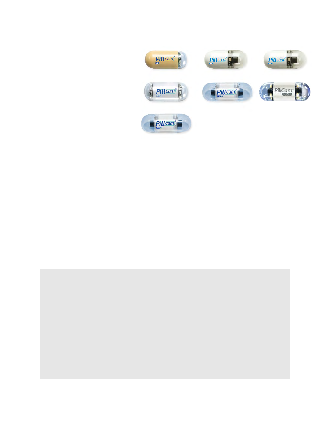
PillCam Capsule Endoscopy
30 PillCam Capsule Endoscopy System Components
There are three PillCam capsule types, supported by the current PillCam capsule endoscopy system. Each is
optimized for a different bowel segment, some with one video head and some with two video heads:
PillCam SB capsules contain one video camera. PillCam SB 1 is a fixed frame rate first generation capsule,
PillCam SB 2 is a fixed frame rate second generation capsule, while PillCam SB 3 is a third generation
capsule with enhanced imaging capabilities with adaptive frame rate (AFR).
PillCam ESO/UGI capsules contain two video cameras (one at each end) with prolonged operation time and
adaptive frame rate.
PillCam COLON capsules contain two video cameras (one at each end). The PillCam COLON 2 capsule
has enhanced imaging capabilities with adaptive frame rate (AFR).
After activation and ingestion, the capsule is propelled by peristalsis through the gastrointestinal tract.
During this process, the video cameras acquire images and the transmitter sends them, via the sensors, to
the PillCam recorder for storage.
For full technical specifications of PillCam capsules, see System Specifications on page 217. For
indications and contraindications, see Indications, Contraindications, Warnings, Cautions on page 3.
!
Caution
The PillCam capsule transmits at a specific frequency of 434.1MHz in a
bandwidth of ± 10MHz. Occasionally, interference from external devices
transmitting in the same bandwidth may occur that may interrupt or limit the
effective performance of the capsule transmission.
The following possible cases could occur:
• Car or house alarm: These operate with momentary transmissions.
Depending on the proximity of the car or house alarm transmitter to the
ingested capsule, there could be a momentary interference that should not
damage the capsule video.
• Police/Fire stations radio equipment: These may operate with more
extended transmission durations. Depending on the proximity of the car or
house alarm transmitter to the ingested capsule, this could cause longer
duration interference, possibly causing gaps in the capsule video.
PillCam SB capsules:
small bowel
PillCam COLON capsule:
colon
PillCam ESO/UGI capsules:
esophagus and upper GI
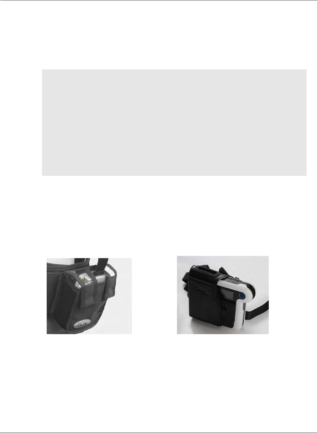
Welcome to PillCam Capsule Endoscopy
PillCam Capsule Endoscopy System Components 31
Handling the PillCam Capsule
PillCam capsules are packaged using a controlled process that ensures the capsule is activated only
when needed. Each PillCam capsule is packed in a separate box with an embedded magnet that
prevents it from activating when it is handled in the box prior to ingestion.
To avoid accidental activation (blinking) of the capsule while in its box, please observe the following:
PillCam Recorders
The PillCam recorder is a compact battery-operated unit worn by the patient during the procedure.
It receives and stores the image data transmitted by the PillCam capsule. There are two models currently
available: PillCam recorder DR3 and PillCam recorder DR2.
The PillCam recorder is supplied with a pouch to wear over the shoulder and with an adjustable strap to
secure to the waist.
!
Caution
• Keep the PillCam capsules in their box until use.
• Store the PillCam capsules only in the packaging supplied with the product.
• Do not use a PillCam capsule if the packaging is damaged.
• Keep the capsule package away from strong magnetic fields (such as MRI
devices).
• Stack PillCam capsule boxes with the clear lid facing up only; never stack
capsule boxes lid to lid.
• Keep metal objects away from the lid of the capsule box.
•For procedures involving the visualization of the colon: After ingesting
the PillCam COLON capsule, instruct the patient not to sit on bare metal
surfaces, such as chairs with a metal sitting area, during the procedure.
DR2 DR3

PillCam Capsule Endoscopy
32 PillCam Capsule Endoscopy System Components
PillCam Sensors
The PillCam sensors are physical receptors that receive transmission data from the PillCam capsule and
transfer it to the PillCam recorder. These sensors are placed on the patient as a sensor belt or a sensor
array:
•PillCam sensor belt: The sensor belt is worn around the patient’s waist over a thin shirt.
•PillCam sensor array: The sensors of the sensor array are attached to the patient's skin. The sensor
locations depend on the procedure type. Each sensor consists of a flexible printed circuit board (PCB)
and is attached with a disposable, medical adhesive sleeve.
The PillCam sensor array and belt are connected to the PillCam recorder by a flexible cable.
RAPID for PillCam Software
The RAPID for PillCam software (RAPID v8.3) supports the PillCam capsule endoscopy procedure by
providing access to the different steps in the PillCam capsule endoscopy procedure via the Home screen.
Home Screen
After starting the RAPID v8.3 software, the Home screen appears. The Home screen is the main screen
that provides access to all phases of the PillCam capsule endoscopy procedure.
The Home screen enables the following functions:
•Patient Check-in: Admitting the patient and initializing PillCam recorder.
•Recorder Download: Creating the RAPID video.
•View Study: Viewing videos and allowing interpretation as well as generating capsule endoscopy
report (see Reviewing and Interpreting RAPID Videos on page 97).
֠
֠֠
֠Note
The PillCam Capsule Endoscopy System does not contain any natural
rubber latex components.
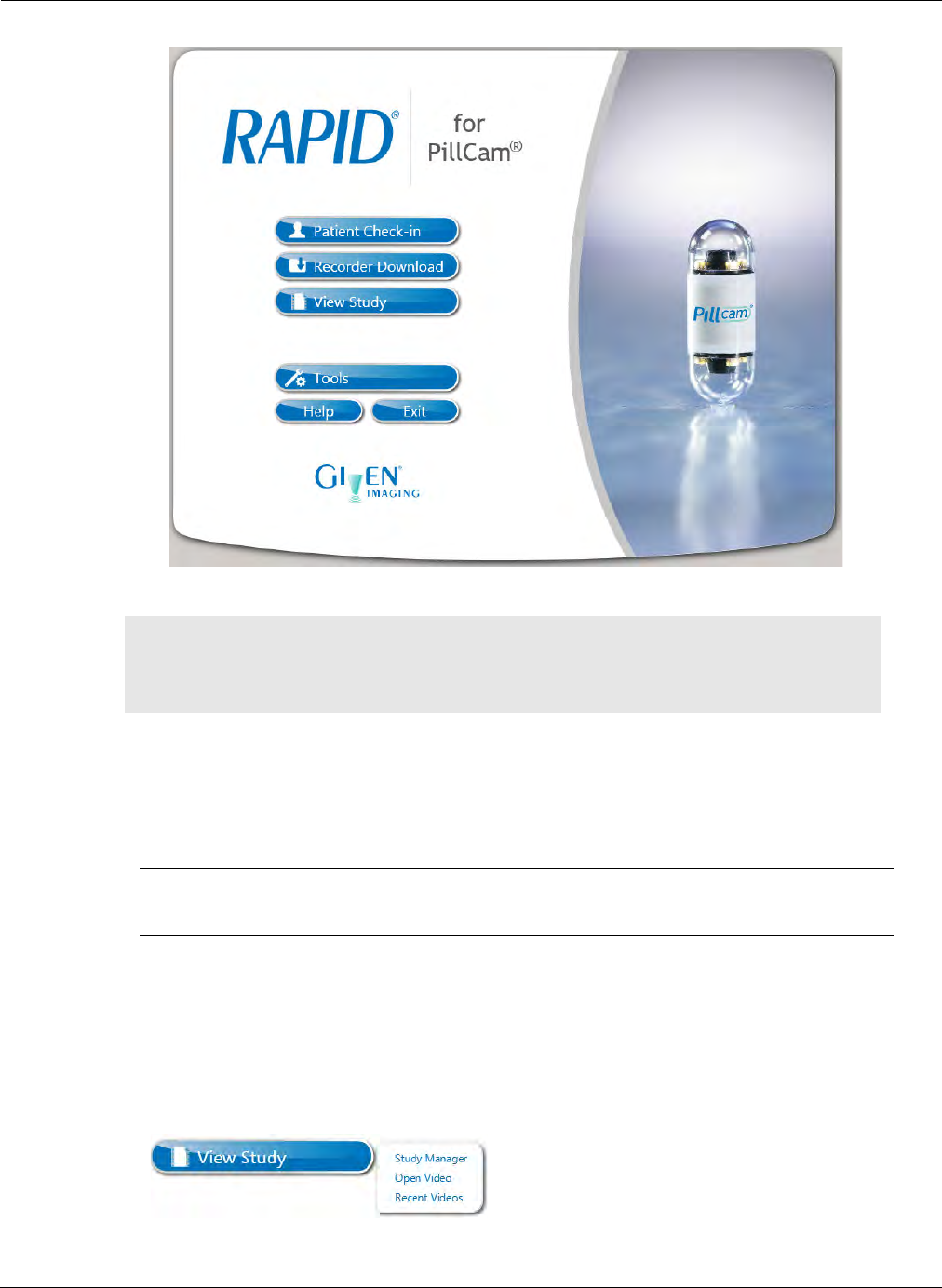
Welcome to PillCam Capsule Endoscopy
PillCam Capsule Endoscopy System Components 33
֠
֠֠
֠Note
Certain key functions described in this manual are accessed from the Home
screen. All future mention of Home screen, such as From the Home screen,
click the Tools button refers to the screen described above.
1
11
1Patient Check-in: Opens the patient check-in wizard to enter information about the
patient and the procedure and to initialize the PillCam recorder with this data. There are a
series of screens that walk you through the check-in process (see Performing Patient
Check-in on page 43).
2
22
2Recorder Download: Opens a screen to download data from a PillCam recorder (see
PillCam Recorder Download on page 89) and to create a RAPID video from the data.
3
33
3View Study: Clicking this button opens a menu that provides access to:
•Study Manager: Allows easy access to all studies. The Study Manager allows you to
open, search, sort, delete, and export studies (see Using the Study Manager on
page 107).
•Open Video: Open a saved RAPID video and findings, if available, by opening a
windows screen that enables you to locate the RAPID video.
•Recent Videos: Displays recently viewed RAPID videos that can be selected and
opened.
2
22
2
3
33
3
4
44
4
1
11
1
5
55
5 6
66
6
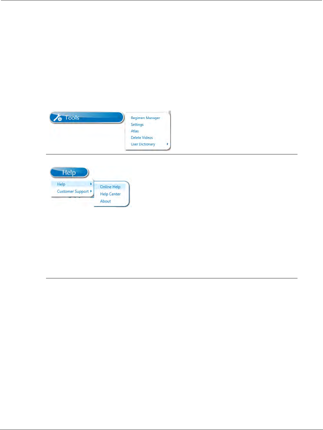
PillCam Capsule Endoscopy
34 PillCam Capsule Endoscopy System Components
RAPID software is supplied as:
•Software only: Installs on your PC.
•RAPID workstation: Dedicated PC with RAPID pre-installed.
4
44
4Tools: Clicking this button opens a menu that provides access to:
•Regimen Manager: Opens a software tool for creating or modifying capsule ingestion
regimen instructions for the patient (see Regimen Manager on page 183).
•Settings: Enables user configuration of RAPID settings (see RAPID Settings on
page 170).
•Atlas: Opens an atlas of typical disease images searchable by key descriptors and
used as a viewing aid for comparing study images with reference images (see RAPID
Atlas on page 153).
•Delete Videos: Delete a RAPID video folder and its contents (see Freeing Space on
Your Computer on page 195).
•User Dictionary: Import or export term dictionary used in writing comments and study
summary.
5
55
5Help: Clicking this button opens a menu that provides access to:
Help: Selecting Online Help in the submenu opens a searchable PDF version of this
user manual. Selecting Help Center will direct you to the RAPID Help Center. The Help
Center provides quick references for How to questions and additional tools including
video clips and step-by-step demonstrations to facilitate learning of Capsule Endoscopy
procedures and the RAPID software.
Customer Support: The menu provides a link to Given Imaging contact information for
customer support (see Customer Support Section on page 182), an option to collect
analysis files and to show the RAPID log.
6
66
6Exit: Closes the RAPID software.

Welcome to PillCam Capsule Endoscopy
PillCam Capsule Endoscopy System Components 35
•RAPID Reader: A limited version of the software; all RAPID features are available except for
creating videos.
Before using the RAPID software, you must configure and personalize it. Refer to Configuring RAPID
Software on page 169 for more details.
֠
֠֠
֠Note
• Your version of RAPID may include multiple types of RAPID (for example,
full software to install on your PC plus copies of RAPID Reader to install
on multiple PCs used only for video review and reporting).
• You cannot install RAPID Reader on a PC on which RAPID was already
installed.
Throughout this manual, RAPID implies any version unless specifically
noted.
֠
֠֠
֠Note
Videos created using capsule procedures prior to PillCam SB 2 can be
viewed in RAPID v8.3.
RAPID v8.3 does not support patient check-in for these earlier capsule
types.

PillCam Capsule Endoscopy
36 PillCam Capsule Endoscopy System Components

Preparing the Patient 37
Chapter 4
Preparing for PillCam Capsule Endoscopy
This chapter covers the following tasks, which must be performed before administering a PillCam
capsule:
•Preparing the patient (see Preparing the Patient on page 37)
•Preparing the equipment (see Preparing the Required Equipment on page 38)
•Creating patient instructions for the procedure (see Creating Patient Instructions for the Procedure
on page 40)
•Performing patient check-in (see Performing Patient Check-in on page 43)
•Fitting Equipment on patient (see Fitting Equipment on the Patient on page 51)
Preparing the Patient
Once it is decided that the patient should undergo capsule endoscopy:
1. Verify that no contraindications apply to the patient (see Indications, Contraindications, Warnings,
Cautions on page 3).
2. Inform the patient:
a. Inform the patient of the small possibility of bowel obstruction.
b. Inform the patient of the importance of a clean bowel for the success of his or her PillCam
examination.
c. Instruct the patient what to expect before, during, and after the procedure.
d. Instruct the patient on the proper use of the PillCam recorder:
• The patient must treat the PillCam recorder with care. Avoid any sudden movements. Avoid
bumping it.
• The patient should not remove or disconnect the PillCam recorder at any time during the
procedure.
• The patient should contact the medical staff if the PillCam recorder is blinking red or white.
• The patient must follow the dietary instructions from the medical staff or when alerted by the
PillCam recorder.
e. If the patient experiences any abdominal pain, nausea, or vomiting after ingesting the PillCam
capsule, the patient should immediately inform the medical staff.
f. After ingesting the PillCam capsule and until it is excreted, the patient should not go near any
source of a powerful electromagnetic field, such as one created near an MRI device and
should avoid direct exposure to bright sunlight.
g. If the patient experiences any post-procedure abdominal pain, vomiting, or other unexplained
symptoms and PillCam capsule excretion cannot be verified, the patient should contact the
physician for evaluation and possible abdominal X-ray procedure.

PillCam Capsule Endoscopy
38 Preparing the Required Equipment
h. Instruct the patient to contact the medical staff if one of the adhesive sleeves detaches from the
patient's body.
i. For procedures involving visualization of the colon: use the Regimen Manager to create and
select pre- and post-ingestion instructions (see Regimen Manager on page 183); print for the
patient and explain.
3. Obtain the patient’s informed consent.
4. Before the procedure, remind the patient about how to prepare for the procedure. Remind them about
appropriate clothing:
• If using the sensor belt, upper garment of thin, natural fiber cloth that is long enough to reach at
least to hip level and will not ride up above the belt.
• If using the sensor array, loose-fitting, two-piece opaque clothing.
Preparing the Required Equipment
Before the patient arrives for the procedure, verify that the following equipment and accessories are
available:
•RAPID (installed, configured, and open after accepting the license agreement)
•PillCam capsule (ESO/UGI, SB, COLON)
•PillCam recorder
•PillCam recorder pouch with shoulder strap
•Printed instructions for the patient
•Prepared PillCam sensor belt or PillCam sensor array with sensors already inserted in sleeves
•Sensor Location Guide (ESO/UGI, SB, SB Pediatric, or COLON) if you are using a sensor array
•Water and drinking cup
•Any medication prescribed for the patient during the procedure
Connecting the PillCam Recorder to RAPID for Check-in
The PillCam recorder must be connected to RAPID during patient check-in. The check-in process allows
entering patient and procedure data for the procedure and saves the correct patient and procedure
information to the PillCam recorder. The process is explained in detail in this section.
The PillCam recorder connects to the RAPID workstation or Personal Computer (PC) via the cradle.
1. Make sure RAPID is On.
2. Make sure the PillCam recorder is firmly inserted into the cradle.
!
Caution
Connection of the PillCam recorder to the RAPID software for patient check-
in, video download, and recorder upgrade must be done using the cradle.
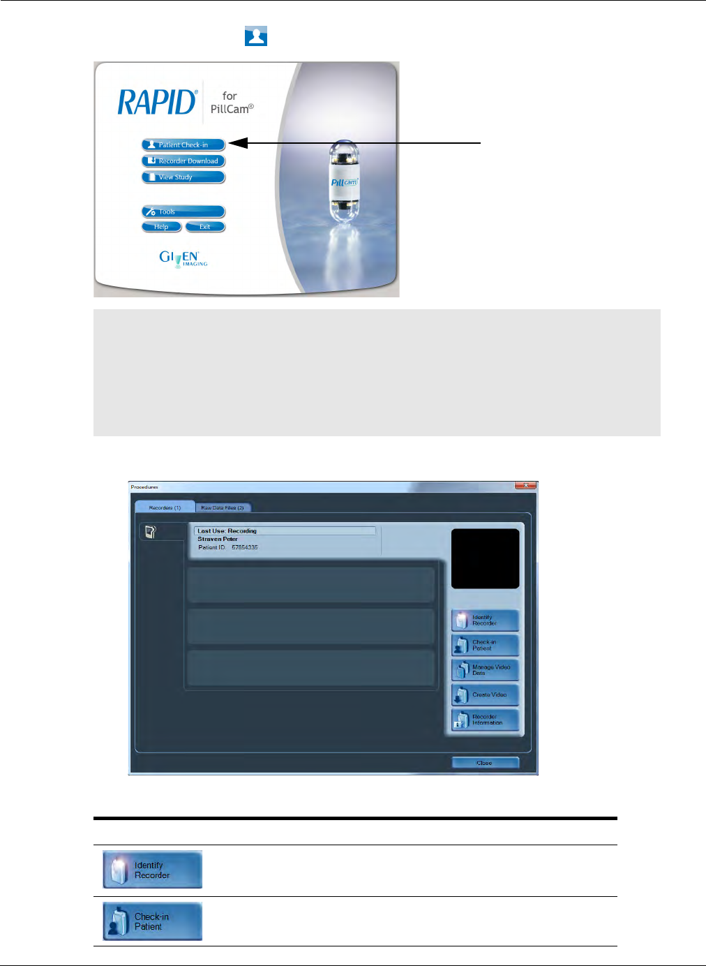
Preparing for PillCam Capsule Endoscopy
Preparing the Required Equipment 39
3. Click Patient Check-in on the RAPID Home screen.
4. In the Procedures screen, select the Recorders tab.
5. Click the Recorder bar to connect the function buttons to the PillCam recorder:
֠
֠֠
֠Note
• When the DR3 battery approaches 400 cycles, RAPID will display a
warning. Contact customer service for a replacement battery when this
battery warning appears, or when the battery reaches 3 years of service,
whichever occurs first.
• Do not remove the PillCam recorder from its cradle before the end of the
check-in process.
Button Action
Identifies the PillCam recorder for the selected recorder bar.
When you click this button, all the LEDs on the associated
PillCam recorder and cradle blink.
Prepares the PillCam recorder for the capsule endoscopy
procedure by entering patient data.

PillCam Capsule Endoscopy
40 Creating Patient Instructions for the Procedure
Creating Patient Instructions for the Procedure
Patient Instructions are handouts used to guide the patient in preparing for the PillCam capsule endoscopy
procedure.
Pre-ingestion Instruction Handouts
PillCam SB procedure goes with predefined pre-ingestion instruction text.
PillCam ESO/UGI procedure also goes with predefined pre-ingestion instruction text.
PillCam COLON procedures go with customizable pre-ingestion instructions that can be created by the
physician using the Regimen Manager tool. In general, pre-ingestion bowel preparation for PillCam capsule
endoscopy of the colon includes cessation of iron supplement intake several days before capsule ingestion,
then adherence to a Clear Liquid Diet starting from at least a full day before capsule ingestion, to be
followed by customizable laxative intake with water at least the evening before ingestion as well as the
morning of ingestion. All these instructions including the customized laxatives are embedded in the pre-
ingestion regimen instructions.
Pre-ingestion instructions for the respective PillCam procedures are printed and given to the patient before
the day of the procedure.
Copies the raw video data to the computer. This button only
appears after unchecking the checkbox Disable video data
management in the Video tab of the Settings screen (see
Video Data Management Section on page 174). (Not available
for RAPID Reader.)
Starts or stops video creation from the raw data on the PillCam
recorder.
Displays all technical details of the PillCam recorder.
֠
֠֠
֠Note
When performing check-in with RAPID v8.3, video download must be done
using RAPID v8.3.
Button Action

Preparing for PillCam Capsule Endoscopy
Creating Patient Instructions for the Procedure 41
Post-ingestion Instructions for Procedures Involving Colon
Visualization
In general, the post-ingestion instructions should result in the preservation of proper cleanliness of the
colon during the procedure and also facilitate the timely progress of the PillCam COLON capsule in
the colon to visualize the entire colon before capsule burn-out or excretion. This is achieved by
instructing the patient in real-time during the procedure to conditionally follow a set of instructions
involving timely ingestion of prokinetics and laxatives. The post ingestion instructions are grouped in
a regimen that is selected by the physician from a library of possible regimens and are loaded into the
PillCam recorder DR3 during patient check-in (see Performing Patient Check-in on page 43) and they
are also printed out in a handout to the patient. During the PillCam COLON procedure, the PillCam
recorder DR3 alerts the user to perform the instructions detailed in the corresponding post-ingestion
instructions handout.
The post-ingestion instructions, i.e. a regimen, consists in general of the following conditional
instructions:
•Continue fasting until capsule leaves the stomach.
•If capsule is delayed in stomach, ingest a customized prokinetic agent.
•Intake the first boost laxative dose (and water when necessary) directly after capsule leaves the
stomach.
•Intake the second boost laxative dose (and water when necessary) 2 - 4 hours after the capsule
leaves the stomach.
•Eat a light meal (depending on capsule progress) 1 - 3 hours after second laxative.
•Take a suppository 1 - 2 hours after the meal (if required).
The execution of these instructions in a regimen is triggered by appropriate alerts from the PillCam
recorder which senses the fulfillment of the conditions for the respective instructions and cues the
patient during the procedure to refer to the associated instruction and execute it.
The difference between the regimens is in the use of specific laxative and prokinetic materials as well as
some timing differences associated with these different materials.
It may happen that the execution of the instructions, some of which drive the capsule through the
gastrointestinal tract, needs to be delayed until the patient reaches suitable environment for the intake of
the associated laxatives. Such a case is when the ingestion is at the physician's office but the rest of the
procedure may take place at the patient's home. Under such circumstances, it is desirable to delay the
capsule-driving instructions until the patient reaches home. The setting for delay of prompting by alerts
may be achieved by appropriate programming of the PillCam recorder DR3, either through check-in
(see Performing Patient Check-in on page 43) or through direct programming before capsule pairing
(see Setting Delay First Instruction in PillCam Recorder on page 79).
The Regimen Manager tool enables the creation and editing of post-ingestion patient instructions for
colon procedures.
Refer to the Regimen Manager section (Regimen Manager on page 183) to familiarize yourself with
this tool.
For pre-capsule ingestion regimen refer to Pre-ingestion Instruction Handouts on page 40, and for post-
capsule ingestion regimen, refer to Post-ingestion Instructions for Procedures Involving Colon
Visualization on page 41.

PillCam Capsule Endoscopy
42 General Patient Guidelines During the Procedure
General Patient Guidelines During the Procedure
Make sure that the patient has the printed post-capsule ingestion instructions or patient instructions with
your contact information included, and instruct the patient as follows:
•For COLON procedures using a PillCam recorder DR3: Explain to the patient that the PillCam
recorder DR3 will alert him to perform instructions from the post-ingestion instructions handout by
beeping and vibrating and displaying on its screen the number of the instruction to be performed at the
alert time. When the PillCam recorder beeps and vibrates he or she should do the following:
a. Check the instruction number on the PillCam recorder LCD screen.
b. Press the Acknowledge button on the upper right corner of the PillCam recorder for at least 3
seconds.
c. Follow the corresponding post-capsule ingestion instruction on the supplied printed out.
d. Follow instructions only when prompted by the PillCam recorder and only the instruction that
matches the displayed number, even if the PillCam recorder skips a number.
e. The PillCam recorder DR3 displays the End of Procedure screen , beeps and vibrates
when the procedure is over and shuts down automatically after five minutes.
•Avoid any physical activity that involves sweating, bending, or stooping.
•Remain active. Do not sleep.
•Use the bathroom as often as needed (do not suppress the urge). The use of wet wipes and cream to
protect the skin is recommended.
•Avoid any source of powerful electromagnetic field (such as an MRI device).
•DR3 only: Do not remove the PillCam recorder until the capsule is excreted or the End of Procedure
instruction appears on the PillCam recorder.
•DR3 only: If the capsule LED at the top of the recorder is blinking red, move to a different location
until the capsule LED on the top of the PillCam recorder has resumed blinking blue.
•DR2 only: If the LED of the top of the PillCam recorder stops blinking blue before the timeouts
specified in the next section for the different capsules, contact the medical staff.
•DR3 only: The end of the procedure for a PillCam recorder DR3 is when the End Of Procedure
instruction is alerted and displayed on the recorder screen.
The end of a procedure may also be declared by the medical staff after proper consultation if the
blinking stopped before the above specified times.
•DR2 only: In the event that no transmissions are received from the capsule (the PillCam recorder stops
blinking blue) for more than 60 minutes, the PillCam recorder will shut down.
•At the end of the procedure, remove the PillCam recorder and sensors. If you need assistance, or were
instructed to do so, return to the clinic to have this done.
•Contact the medical staff in case of any unexpected event or doubt.
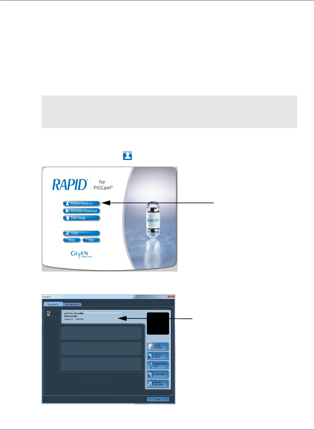
Preparing for PillCam Capsule Endoscopy
Performing Patient Check-in 43
Performing Patient Check-in
Patient check-in is the process of entering patient and procedure data into the PillCam recorder. This
information becomes part of the data in the RAPID video and capsule endoscopy report. During this
process, old data from previous procedures is cleared and the new patient and regimen information is
saved to the PillCam recorder.
For colon visualization procedures: patient check-in also programs the PillCam recorder to provide
post-ingestion patient alerts. It also allows you to print out the selected post-ingestion instructions for
the patient. Make sure that the RAPID workstation or PC is connected to a printer (for printing the post-
ingestion patient instructions).
1. Make sure the PillCam recorder is fully charged and connected to RAPID.
2. From the Home screen, click Patient Check-in.
3. In the Procedures screen, click the Recorder bar.
4. Click Check-in Patient.
֠
֠֠
֠Note
Make sure that an approved post-ingestion regimen is available in the
Regimen Manager before performing patient check-in (see Post-Ingestion
Patient Instructions on page 187).
Recorder bar
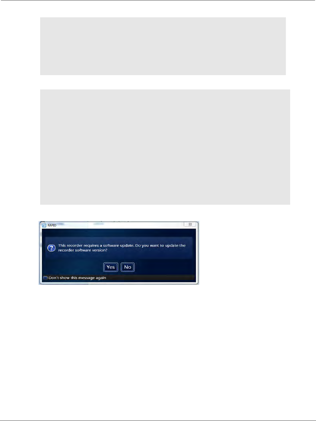
PillCam Capsule Endoscopy
44 Performing Patient Check-in
!
Caution
When placed in the cradle, RAPID 8.3 will perform updates to your existing
PillCam recorders. It is important to follow the instructions displayed on the
screen. Do not stop the update process until it is finished.
• For the PillCam recorder DR3, RAPID will perform a mandatory update.
• For the PillCam recorder DR2, follow the instructions detailed in the Note
below.
֠
֠֠
֠Note
PillCam recorder DR2 only: If the PillCam recorder needs a software
update, this message appears: This recorder requires a software update.
Do you want to update the recorder software version?
We highly recommend you perform this upgrade unless you are an ESO 2
user. Upgrading the DR2 to SB 3 support will deactivate ESO 2 support. If you
are working with several recorders, designate which will support ESO 2
procedures and which will support SB 3 procedures.
• Click Yes and follow the instructions on the screen. When the update is
done, remove the PillCam recorder from its cradle and reinsert it.
• To ignore the update, click No. You may continue the check-in and perform
the update later.
• Selecting the Don’t show this message again checkbox will disable
further updates to this specific recorder. This option is available only if you
are logged in as the administrator.
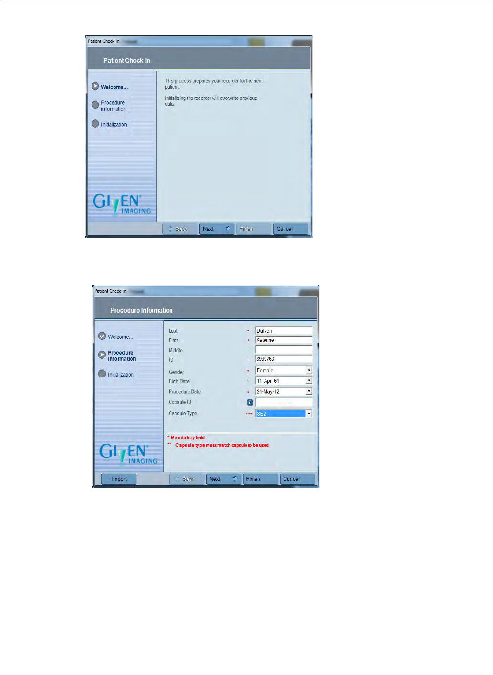
Preparing for PillCam Capsule Endoscopy
Performing Patient Check-in 45
The Patient Check-in wizard opens.
5. Click Next to continue.
The first Patient Check-in window appears.
6. Enter patient data by importing from HIS (Hospital Information System) or by typing in manually:
HIS Import: If you are working in a HIS-enabled networked environment, you can import the
patient check-in data to automatically complete the check-in fields.
a. Click Import.
The Import Patient Data screen appears.
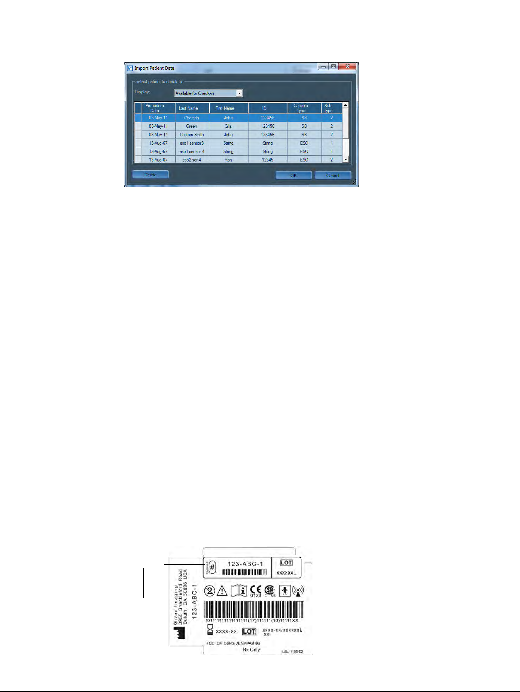
PillCam Capsule Endoscopy
46 Performing Patient Check-in
The available data for patients not yet checked in appears on the screen sorted by planned
Procedure Date. You can sort the studies by any of the column headings in either ascending or
descending order.
b. To select a patient, select the relevant line and click OK. This automatically adds check-in data
into the appropriate fields in RAPID.
Once imported, the patient data is removed from the Available for Check-in list and appears in the
Already imported list. In the list next to Display at the top of the screen, you can select which
patient list you wish to see: Available for Check-in or Already imported.
To delete a patient from the Already imported list, select the relevant line and click Delete.
Once you click OK, the first check-in screen appears again, with all available information already
entered. You may need to complete additional mandatory or optional procedure information (such
as capsule ID) not auto-populated from the HIS (see the RAPID v8.3 IT Guide). When all necessary
data entry is completed, you may proceed to the Procedure info confirmation screen by clicking
the Next or Finish buttons or by changing any of the information fields.
Manual Data Entry: You can enter patient information manually.
a. Enter the patient’s Last, First, and Middle names into the appropriate fields. Use alphanumeric,
underscore, hyphen, and space characters. Use the TAB key to move to the next field.
b. Enter the patient ID number in ID.
c. Select the Gender.
d. In Birth Date, set the patient’s date of birth.
e. In Procedure Date, enter the date on which the procedure is to be performed. By default, it is set
for current date.
f. In Capsule ID, if filling this field during check-in, enter the capsule ID code that is printed on
the bottom of the capsule box (by typing it in or by using a barcode). The capsule type field is
automatically populated according to the capsule type encoded in the capsule ID entered.
Capsule ID
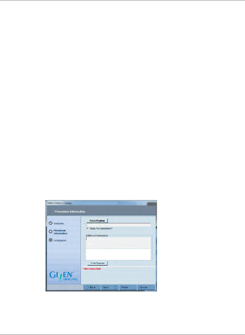
Preparing for PillCam Capsule Endoscopy
Performing Patient Check-in 47
g. In Capsule Type, if you have not filled in Capsule ID in the previous field, select the correct
type of procedure (SB 2, SB 3, ESO 3, COLON 2). If you have filled in the Capsule ID field,
this field will auto-populate with the corresponding capsule type.
h. For DR2 recorders, in the Sensor Type drop-down list, select the sensor type. Only those
sensor types appropriate for the selected capsule type field value will appear in the selection
menu. To ensure accurate localization information use only the sensor type that you defined
in the check-in process.
7. Click Next to continue.
8. For PillCam COLON 2 procedures only: Select a regimen and print it out to hand it out to the
patient before capsule ingestion. Use the Delay first instruction? checkbox (ON by default) to
determine if a predefined minimum 90 minute free-of-instructions period will or will not be forced
between the time of ingestion and the first instruction to the patient. If the delay first instruction is
set, the first instruction to the patient will be either alert 0 (if still relevant) or alert 1:
•Alert 0: An instruction #0 (based on time passed from capsule ingestion) to take prokinetics to
facilitate passage of the PillCam COLON 2 capsule to the small bowel. Threshold of time
passed from capsule ingestion for alert 0 can be modified in the regimen manager for a specific
regimen.
•Alert 1: An instruction #1 for taking laxative after detection by the PillCam recorder of PillCam
COLON 2 passage into the small bowel. It coincides with the activation of the AFR mode in the
PillCam COLON 2 capsule and the appearance of the AFR status icon in the right corner of the
status line at the top of the PillCam recorder DR3 display. The appearance of alert 1 can be
designated to appear either an additional 0 minutes or 15 minutes after original tentative alert 1
timing. Alert 0 will not occur if alert 1 was raised before it.
If you do not change this parameter during check-in, it remains in its default state (ON for “Delay
first instruction”, i.e. delayed) but you may set it also through the PillCam recorder DR3 before
pairing the PillCam COLON 2 capsule to the PillCam recorder DR3.
9. Click Next to continue.
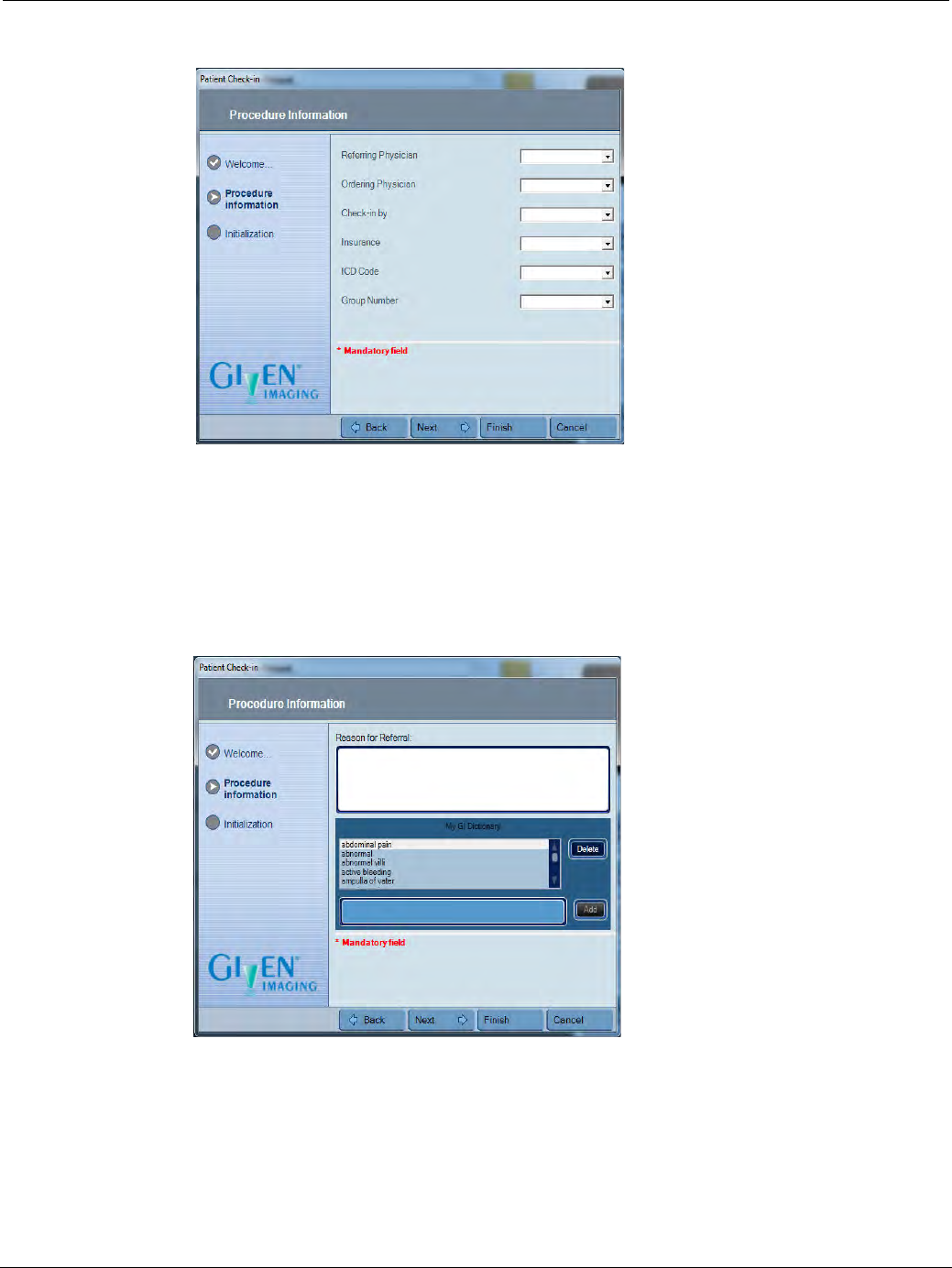
PillCam Capsule Endoscopy
48 Performing Patient Check-in
10. Complete physician and insurance details:
a. Enter Referring Physician and Ordering Physician.
b. In Check-in by, enter your name.
c. Enter Insurance, Group Number, and ICD Code, as required.
d. Click Next to continue.
11. Type in the Reason for Referral, if it is known, and click Next.
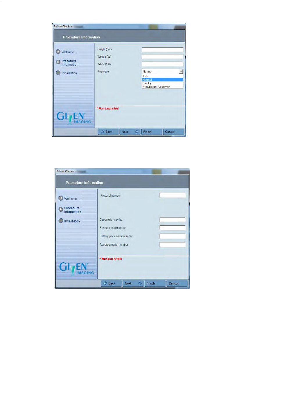
Preparing for PillCam Capsule Endoscopy
Performing Patient Check-in 49
12. Complete the patient physical description (Height, Weight, Waist, and Physique).
13. Click Next.
14. Complete the protocol and materials details:
a. Fill in Protocol number or Name if you are performing a clinical trial.
b. For Capsule lot number, enter the LOT # from the back of the capsule box.
c. For Sensor serial number, enter the SN from the sensor array cable.
d. For Battery pack serial number, enter the DR2 recorder battery pack serial number.
e. For Recorder serial number, enter the PillCam recorder serial number.
f. Click Next to continue.
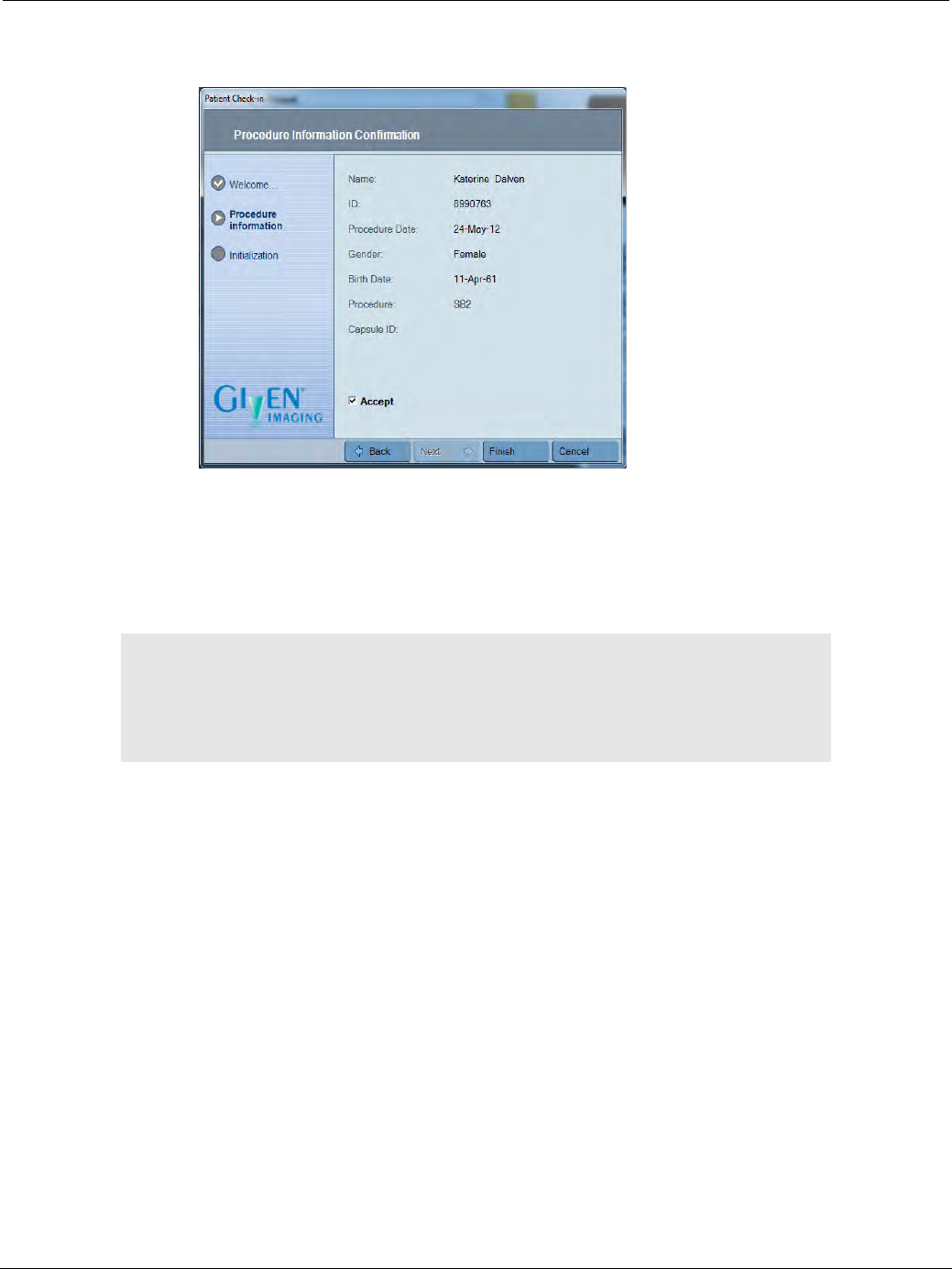
PillCam Capsule Endoscopy
50 Performing Patient Check-in
15. In the Procedure Information Confirmation screen, verify that the patient and procedure data is
correct:
• If the data is incorrect, click Back and return to a previous screen to correct the mistake.
• If the data is correct, select Accept, and then click Finish to continue. When the Patient Check-in
complete screen appears, click Ready to proceed.
Updating Patient Details
Patient details are collected during the Patient Check-in process. However, RAPID allows you to change
or update all the patient information after a video creation.
To update patient details:
1. Find the study in the Study Manager.
2. Right-click the study and select Update Patient Details.
The following screen appears. Make the desired changes and click Next until you have completed your
update.
֠
֠֠
֠Note
When the patient check-in is complete, the capsule LED on the PillCam
recorder lights up in orange.
After patient check-in, keep the PillCam recorder in its cradle until the
capsule ingestion procedure begins.
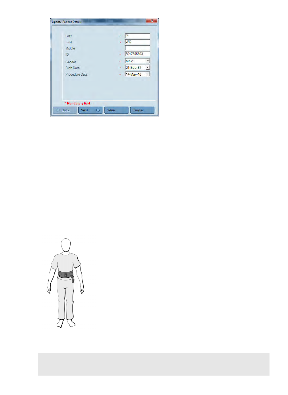
Preparing for PillCam Capsule Endoscopy
Fitting Equipment on the Patient 51
3. Click Save on the last screen.
Fitting Equipment on the Patient
The following section details the instruction for preparing and fitting the patient with the recording
equipment required for the capsule endoscopy procedure.
Applying the PillCam Sensor Belt
The sensor belt is used for SB or COLON procedures and consists of a flat, flexible belt-like sensor
arrangement worn around the patient's waist over a single, thin layer of natural fabric, such as a T-shirt.
Depending on the sensor belt, for sanitary purposes some models may require the fitting of a single use
disposable protective sleeve while other models feature a reusable washable external fabric sleeve.
The image above of the PillCam sensor belt is for reference only. Sensor belt models may differ from
image.
֠
֠֠
֠Note
Refer to the product insert supplied with your sensor belt for full instructions
on fitting, usage, cleaning, and technical description.
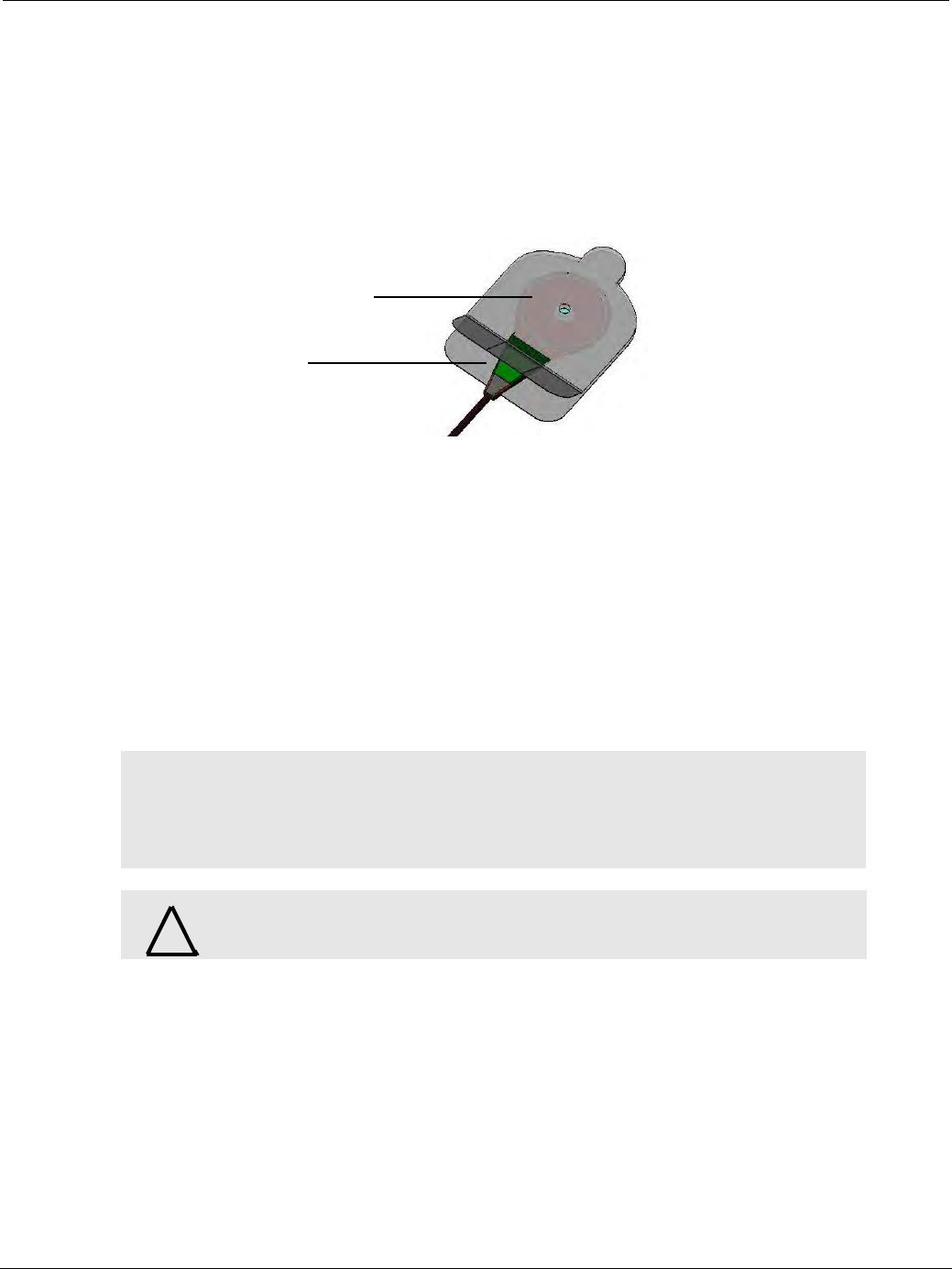
PillCam Capsule Endoscopy
52 Fitting Equipment on the Patient
Applying the PillCam Sensor Array
The sensor array allows the PillCam recorder to collect localization data during a procedure. The
prescribing physician may request this. To prepare the sensor array:
1. Insert each sensor into an adhesive sleeve. The sensor markings (dots or this side up) should face
away from the adhesive side of the sleeve.
2. To secure the sensor in the sleeve, remove the liner from the topside of the lower lip at the opening of
the adhesive sleeve and press both lips together.
3. Place the sensors on the patient according to the appropriate placement guides (see ESO/UGI Sensor
Locations on page 54, SB Sensor Locations on page 55, or COLON 2 Sensor Locations on page 56).
Necessary Equipment and Accessories
To attach the sensor array to the patient, you need the following equipment:
•Sensor array
•Sensor Location Guide (ESO/UGI, SB, COLON)
•Adhesive sleeves, to hold each one of the sensors securely in place
•Razor and disinfectant (not supplied), to shave the area of the sensors on the abdomen
To attach the sensor array:
1. DR3 only: With the patient standing and exposing the thorax and abdominal area, place the sensor
array loop on the left shoulder. If the loop is too long, gather and fasten the surplus in the fastener.
2. Ask the patient to lie down.
3. Use the appropriate Sensor Location Guide to identify the location of each sensor on the patient's body
and mark it with a dot.
4. The sensor array sleeves should be applied to hairless skin. If needed, wipe the patient's skin with
disinfectant and shave the areas where sensors are to be applied.
֠
֠֠
֠Note
Sensor arrays must be applied directly to smooth skin. Anything that comes
between the patient's skin and the sensors, including hair or air, and any
changes in the sensors' arrangement, may interfere with the quality of the
data.
!
Warning
Do not use the sensor array if it is torn or damaged.
Sensor inside adhesive
sleeve between two layers
Lower lip with liner on
the top side
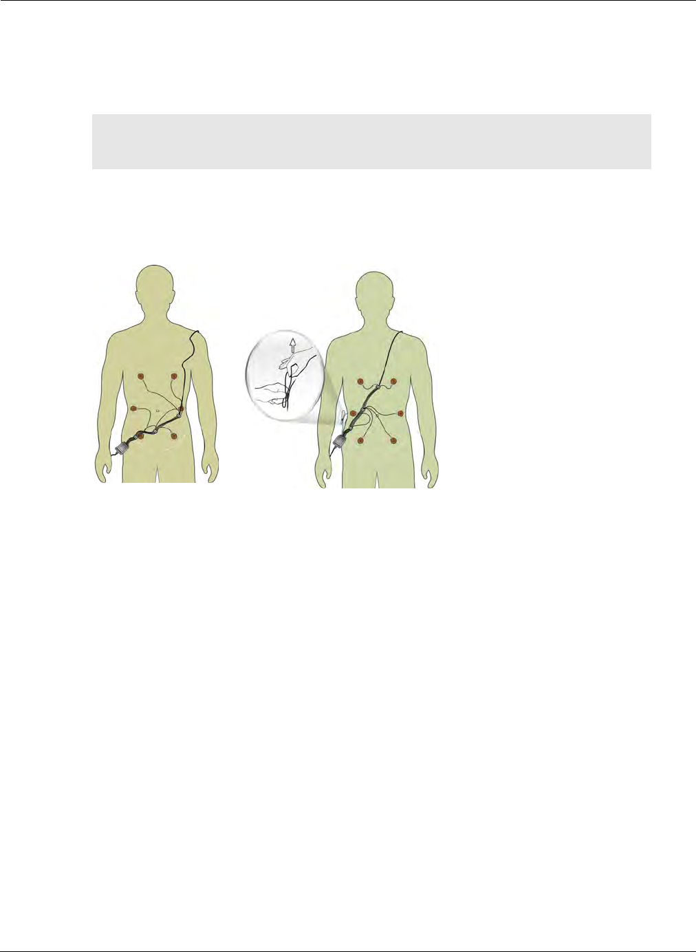
Preparing for PillCam Capsule Endoscopy
Fitting Equipment on the Patient 53
5. With the sensor array connector at the patient's side, lay the prepared sensor array on the patient's
abdomen and match the letters and colors on each sensor wire to the letters and colors on the
Sensor Location Guide.
6. To attach each sensor, remove the protective backing from its adhesive sleeve.
If you are using a sensor array with a downlink loop, adjust the downlink loop to remove excess
cable so that it fits closely to the patient's body. To prevent damage to the wires do not forcibly
bend the downlink loop in any way.
7. When the patient gets dressed, make sure that the sensor array connector remains outside of the
patient's clothing and make sure that the patient is not uncomfortable with the equipment.
֠
֠֠
֠Note
Since the sensors are placed on the body according to anatomical reference
points, the distances between sensors may vary from patient to patient.
X√
√√
√
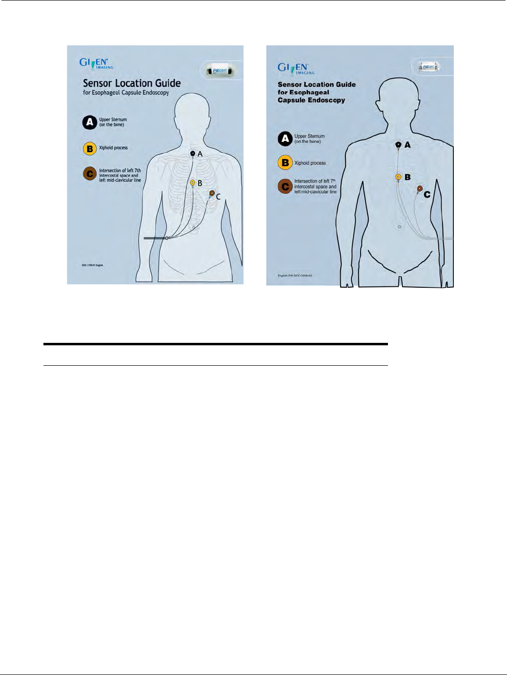
PillCam Capsule Endoscopy
54 Fitting Equipment on the Patient
ESO/UGI Sensor Locations
Sensor Label Sensor Color Sensor Location
A Black Upper Sternum (on the bone)
B Yellow Xiphoid process
C Brown Intersection of left 7th intercostal space
and left mid-clavicular line
PillCam recorder DR3 PillCam recorder DR2
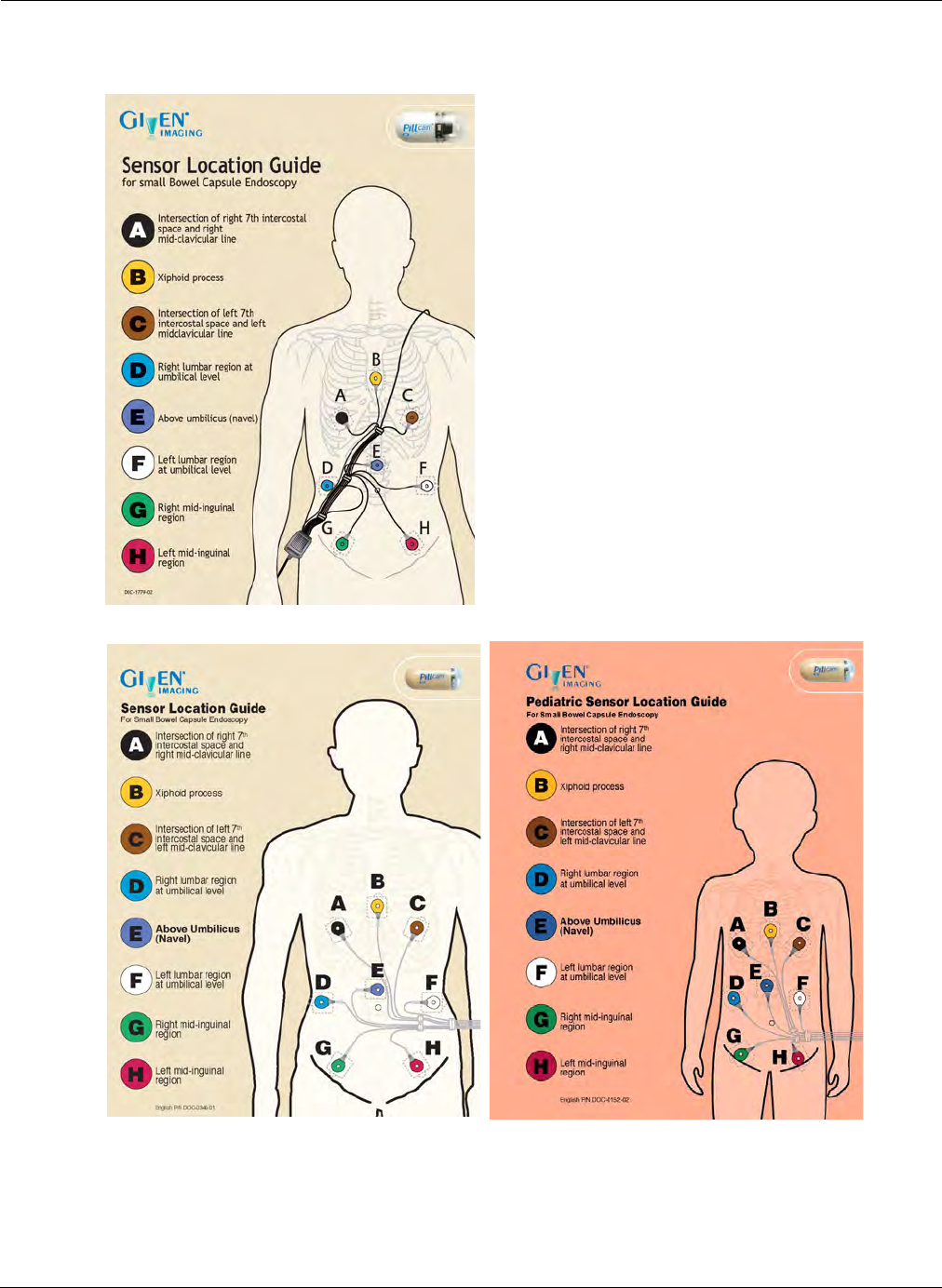
Preparing for PillCam Capsule Endoscopy
Fitting Equipment on the Patient 55
SB Sensor Locations
For DR3:
For DR2:
Standard Sensor Location Guide Pediatric Sensor Location Guide
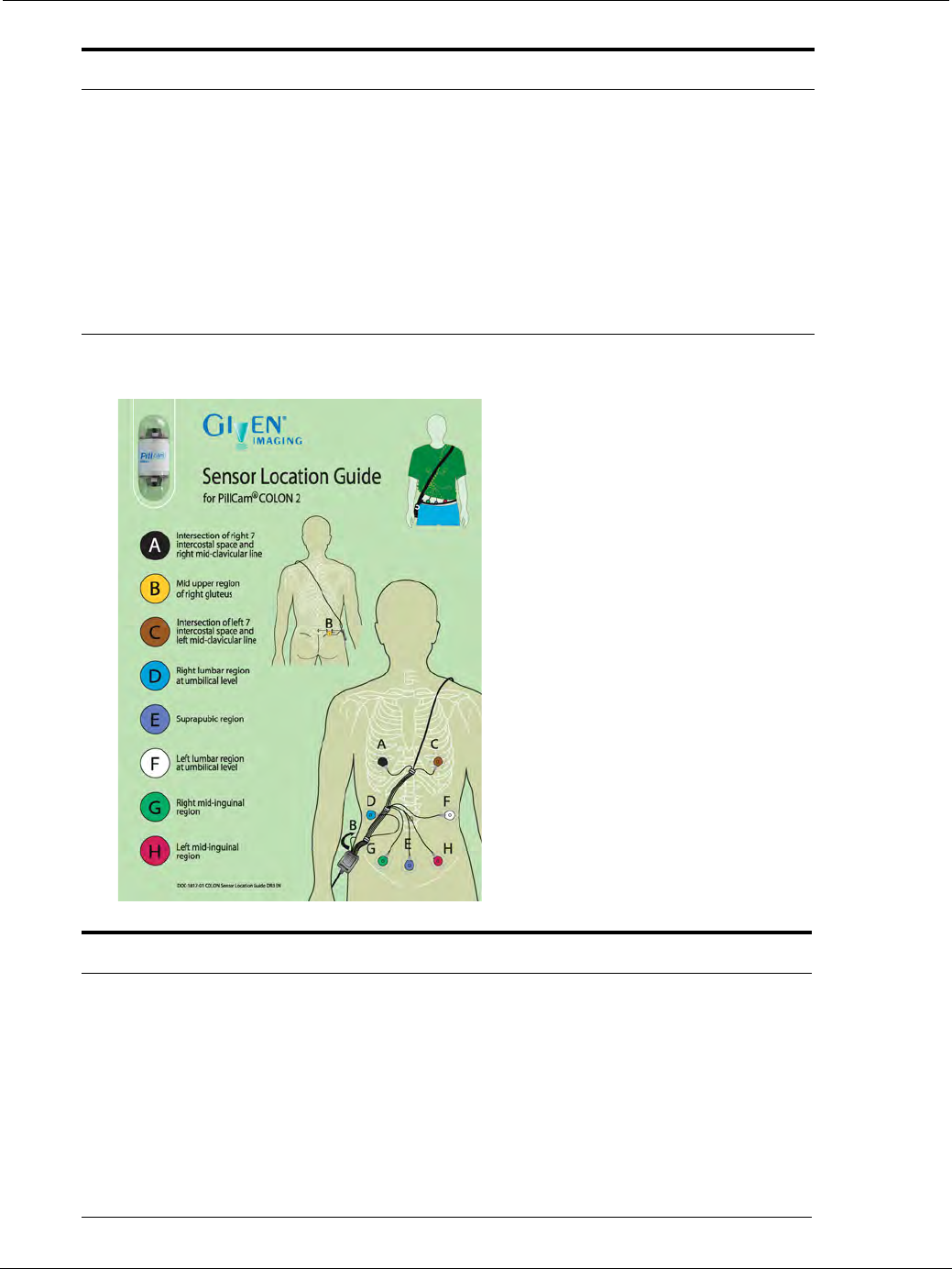
PillCam Capsule Endoscopy
56 Fitting Equipment on the Patient
COLON 2 Sensor Locations
Sensor Label Sensor Color Sensor Location
A Black Intersection of right 7th intercostal space and
right mid-clavicular line
B Yellow Xiphoid process
C Brown Intersection of left 7th intercostal space and left
mid-clavicular line
D Blue Right lumbar region at umbilical level
E Purple Above umbilicus (navel)
F White Left lumbar region at umbilical level
G Green Right mid-inguinal region
H Red Left mid-inguinal region
Sensor Label Sensor Color Sensor Location
A Black Intersection of right 7th intercostal space and
right mid-clavicular line
B Yellow Mid upper region of right gluteus
C Brown Intersection of left 7th intercostal space and left
mid-clavicular line
D Blue Right lumbar region at umbilical level
E Purple Suprapubic region
F White Left lumbar region at umbilical level
G Green Right mid-inguinal region
H Red Left mid-inguinal region
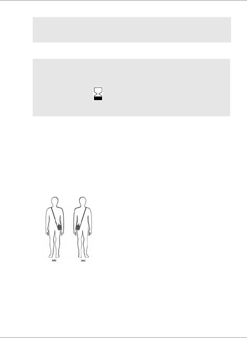
Preparing for PillCam Capsule Endoscopy
Fitting Equipment on the Patient 57
Before dispensing the PillCam capsule, the equipment must be prepared.
Attaching the Sensors to the PillCam Recorder
The PillCam recorder is worn by patients during the procedure in the recorder pouch with shoulder
strap or in the recorder belt with suspenders. Make sure that these accessories fit the patient
comfortably.
Recorder Pouch
To fit the recorder pouch:
1. With the patient standing, hang the recorder pouch from the patient’s shoulder as displayed in the
illustration.
2. Adjust the shoulder strap so that the recorder hangs at the patient's side at waist level with the
supplied strap securing the recorder to the waist.
֠
֠֠
֠Note
Adjust the downlink loop to remove excess cable so that it fits closely to the
patient's body. To prevent damage to the wires do not forcibly bend the
downlink loop in any way.
!
Caution
• Make sure that there is no other PillCam capsule or other diagnostic
capsule in the patient’s gastrointestinal tract.
• Verify that the capsule expiration date has not passed (see the date next
to the icon on the packaging).
• If you are performing the procedure for the first time, read the capsule
package insert.
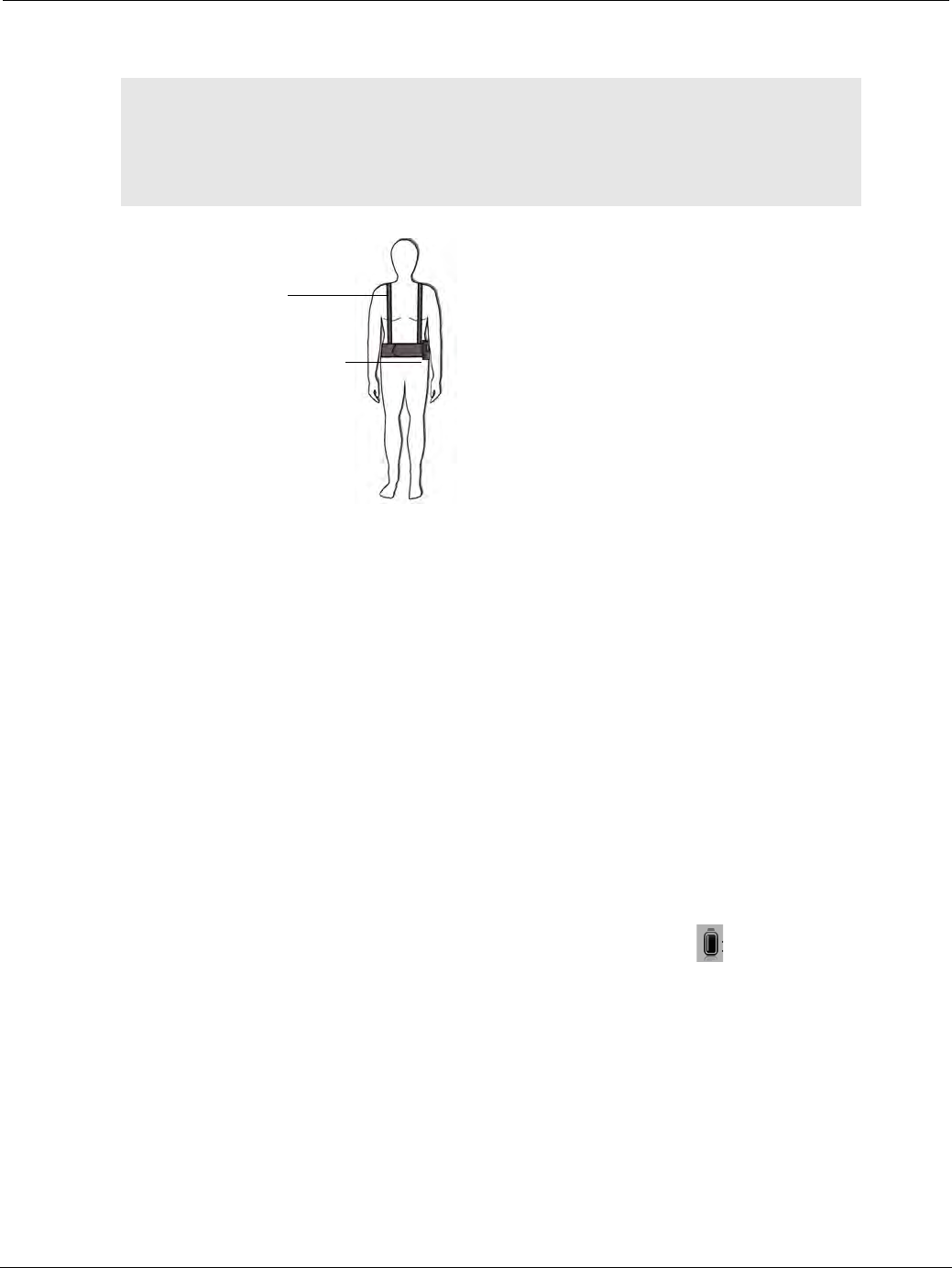
PillCam Capsule Endoscopy
58 Fitting Equipment on the Patient
PillCam Recorder Belt
To fit the recorder belt:
1. With the patient standing, place the belt around the patient’s waist.
2. Adjust the belt to fit the patient. Add the belt extension if needed.
3. Make sure that the PillCam recorder DR2 pouch is at the patient’s hip as shown here. The belt has a
Velcro strap for attaching the pouch, allowing the patient to adjust the pouch as needed.
4. Adjust suspender length and location to fit the patient.
PillCam Recorder DR2 and DR3
To assemble PillCam recorder accessories:
1. Remove the PillCam recorder from the cradle. If the PillCam recorder is properly initialized and ready
for the procedure, the capsule LED is constantly on in orange.
2. Verify that the battery is fully charged:
• For PillCam recorder DR2, all battery LEDs should light up.
• For PillCam recorder DR3, the battery icon on the screen should be .
֠
֠֠
֠Note
There are three belt configurations (PillCam recorder DR2 only):
• the standard recorder belt for patients over 85 lbs (40 kg),
• the small recorder belt for patients under 85 lbs (40 kg),
• the standard recorder belt with the belt extension for very large patients.
PillCam recorder DR2
in pouch on the left
Suspenders
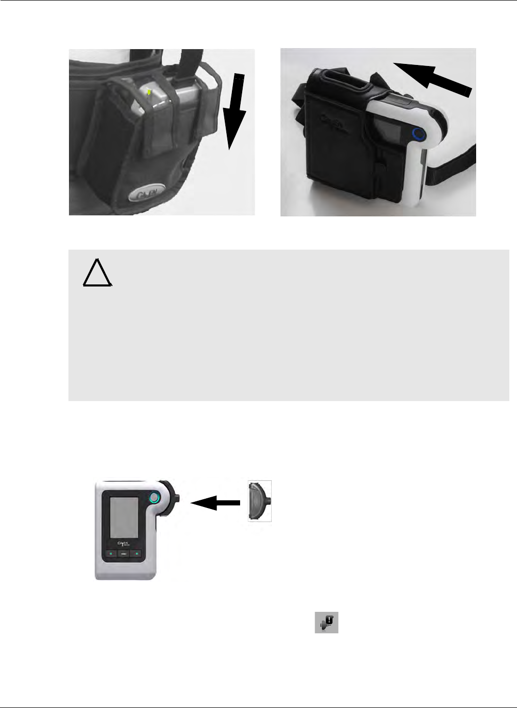
Preparing for PillCam Capsule Endoscopy
Fitting Equipment on the Patient 59
3. Insert the PillCam recorder into its pouch. Instruct the patient to keep wearing the PillCam recorder
during the examination.
The pouch or belt is ready and the patient can wear it.
Positioning PillCam Recorder DR3
1. Make sure the PillCam recorder is on (navigation button LEDs blink once every 5 seconds).
2. With the patient standing, hang the pouch from the patient’s left shoulder to the right hip.
3. Insert the sensor connector into the PillCam recorder’s slot until you feel and hear a click.
If the sensor is not properly connected to the recorder, appears on the PillCam recorder
screen (see PillCam Recorder DR3 on page 209 for troubleshooting details).
4. If using a PillCam sensor array, tuck the vibrating connector component between the recorder
pouch and the patient's abdomen. This will ensure that the tactile vibrating alerts will be felt by the
patient.
!
Warning
When a PillCam recorder is connected to a sensor array worn by a patient:
• Do not connect the PillCam recorder to a computer that is connected to an
electrical outlet.
• Do not put the PillCam recorder into a cradle or connect it to a charger.
• Attach the sensor connector to the PillCam recorder immediately prior to
capsule ingestion.
•PillCam recorder DR3 only: make sure that the connector component is
placed between the patient’s body and the PillCam recorder DR3 waist strap.
•PillCam recorder DR2 only: if the blue LED is blinking before you open the
PillCam capsule box, reinitialize the PillCam recorder.
DR2 DR3
A
B
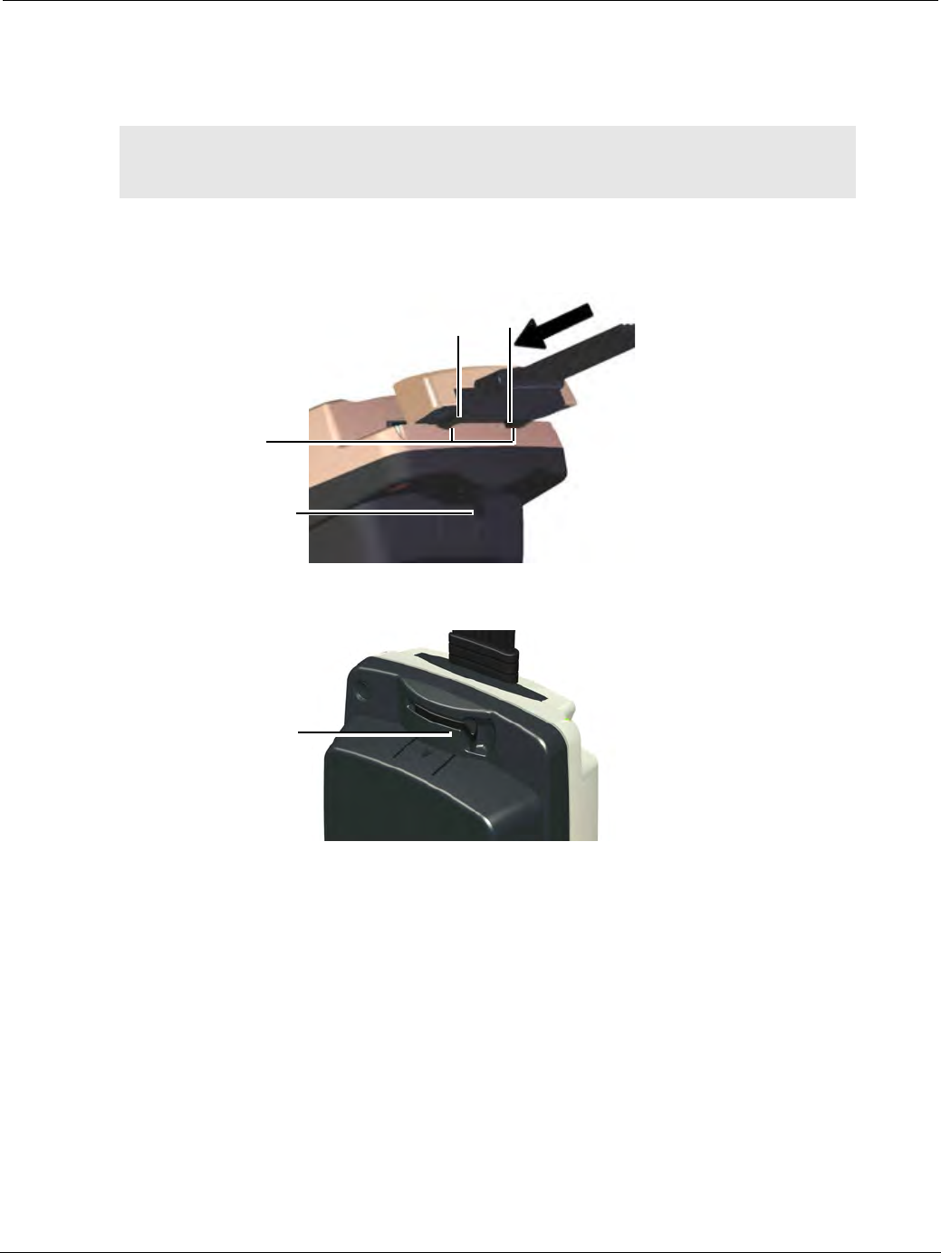
PillCam Capsule Endoscopy
60 Fitting Equipment on the Patient
Positioning PillCam Recorder DR2
1. If you are using the recorder pouch, hang it from the patient’s shoulder while the patient is standing.
If you are using the recorder belt, secure it around the patient’s waist, while the patient is standing.
2. Make sure the locking handle at the back of the PillCam recorder is open.
3. Slide the two protrusions on the connector into the matching grooves in the PillCam recorder.
4. Verify that the connector is inserted completely, and then lock it by closing the handle on the PillCam
recorder.
5. Make sure that the PillCam recorder is on (the battery LEDs light up once every 5 seconds).
֠
֠֠
֠Note
The sensor array connector and wire should hang over the top of the
recorder belt.
Locking handle
is open before
connecting
sensor array
Protrusions on
connector
Grooves in
PillCam
recorder
Closed handle

Preparing for PillCam Capsule Endoscopy
Fitting Equipment on the Patient 61
֠
֠֠
֠Note
• Remind the patient about wearing and handling the PillCam recorder
with care (see General Patient Guidelines During the Procedure on
page 42).
• Make sure the patient has the printed instructions (see Printing the
Patient Instructions on page 190).
• When securing the sensor belt, make sure the fabric of the patient's shirt
is not folded beneath the front portion of the sensor belt.
• Make sure nothing other than a single, thin layer of fabric is allowed to
come between the sensor belt and the abdomen.
• In order to avoid pulling the sensor belt out of position, do not attach or
anchor anything to the sensor belt.
• Be sure that the PillCam recorder is worn over the sensor belt and that
the PillCam recorder pouch is not attached to the PillCam sensor belt.
• Another layer of clothing may be worn over the sensor belt as long as
the sensor belt connecting wire can be attached to the PillCam recorder.

PillCam Capsule Endoscopy
62 Fitting Equipment on the Patient

Preparing the PillCam Recorder 63
Chapter 5
Know Your PillCam Recorder
Preparing the PillCam Recorder
The PillCam recorder is used to receive and store the captured images from the PillCam capsule during
the capsule endoscopy procedure for subsequent download to RAPID. The video created from the
downloaded data can then be reviewed by the physician.
There are two PillCam recorder models: DR3 and DR2. The DR3 model is the newest model and works
with all types of PillCam capsules and provides advanced functions of real-time viewing during the
procedure. The DR2 model is the previous model and it works with all types of PillCam capsules in a
fixed frame rate mode.
Both recorder models are provided with a cradle (for charging and connecting to a PC) and a pouch (to
allow the patient to wear the PillCam recorder during the procedure).
Functions
Initialization
Initialization is a mandatory operation before the procedure. With the PillCam recorder connected to
RAPID, the patient and procedure data are uploaded to the PillCam recorder so that the ensuing study
and procedure data are personalized. The initialization of the PillCam recorder is performed through the
RAPID check-in process (see Performing Patient Check-in on page 43), while the PillCam recorder is
connected to RAPID through its cradle. The PillCam recorder is a passive element in this process and
no control or operation on it is required.
Pairing for DR3
Pairing is performed before PillCam capsule ingestion, the capsule and PillCam recorder are made to
connect so the PillCam recorder is tuned to the transmissions only from the paired capsule.
Pairing for PillCam recorder DR3 may be performed either through the check-in process in RAPID by
entering the capsule ID during check-in, or directly using the recorder control buttons before ingestion
(see PillCam Recorder—Capsule Pairing (DR3 only) on page 80). Pairing is mandatory when working
with a PillCam recorder DR3 in order to enable recording.
Real-Time Viewing
Real-time viewing is an optional function whereby the images captured by the capsule and received by
the PillCam recorder are displayed to the user in real-time for review by the physician. The PillCam
recorder DR2 provides this functionality only in conjunction with a dedicated tablet PC connected to it
during the procedure. The PillCam recorder DR3 provides this functionality both through a dedicated
֠
֠֠
֠Note
For full technical specifications about the PillCam recorders and their
cradles, see System Specifications on page 217.

PillCam Capsule Endoscopy
64 PillCam Recorder DR3
tablet PC but also autonomously by its built-in display. Appropriate controls on the recorder activate and
control the operation of this function.
Regimen Reminder
Regimen Reminder is a reminder function whereby the PillCam recorder alerts the user to perform dietary
instructions during a PillCam COLON 2 procedure. For more details see Post-Ingestion Patient
Instructions on page 187.
Download
Download is the transfer of the stored raw procedure data from the PillCam recorder to RAPID and the
creation of a RAPID video for subsequent review.
Before you perform your first PillCam procedure, make sure that you are familiar with the controls and
functions of the PillCam recorder.
PillCam Recorder DR3
General
The PillCam recorder DR3 battery is limited to 400 recharge cycles. RAPID will display a notification at
approximately 385 cycles prompting you to replace the battery. Contact customer service for a replacement
battery when the battery notification appears, or when the battery reaches 3 years of service; whichever
occurs first.
RAPID will alert the user when the number of recorder uses is close to the allowed limit.
֠
֠֠
֠Note
SD Card: The SD card in the PillCam recorder DR3 may not be used
externally to perform a patient check-in.
Use of SD cards not supplied by Given Imaging in the PillCam recorder DR3
may cause the device to malfunction or lead to data corruption or loss.
!
Caution
The SD card should never be removed or reinserted when the PillCam
recorder DR3 is ON.
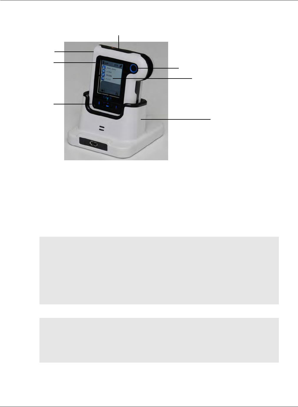
Know Your PillCam Recorder
PillCam Recorder DR3 65
Turning On and Off
The illustration below shows the PillCam recorder DR3 sitting in the cradle:
The PillCam recorder DR3 is automatically ON when it is in its cradle.
When removed from the cradle, it may be turned off and on again using the On/Off button.
The On/Off button is on the left side of the PillCam recorder DR3:
•To turn on, press and hold the On/Off button for 5 seconds until you see the startup screen. All
LEDs start to flash. (The full start-up sequence takes about one minute.)
•To turn off, press and hold the On/Off button for 5 seconds until the PillCam recorder DR3 beeps
and the screen turns off and the button LEDs turn black.
֠
֠֠
֠Note
Automatic Shutdown:
• After the PillCam recorder DR3 has been checked in, it goes into standby
mode ready to receive capsule signals when removed from its cradle. If
after 90 minutes no capsule pairing is performed, the PillCam recorder
DR3 automatically shuts down.
• After starting to receive signals from a paired capsule, if there is a gap of
30 minutes with no signal reception from the paired capsule, the PillCam
recorder DR3 will shut down.
֠
֠֠
֠Note
When the PillCam recorder DR3 is on, its screen backlight goes into an off
mode if the screen or recorder controls are idle for more than 3 minutes. If
the PillCam recorder DR3 is on, but the screen is off, press any key to turn
the screen on. Once the PillCam recorder DR3 screen is activated, proceed
with pressing the desired function button.
ON/OFF
button
Acknowledge button
Capsule and message LEDs
Line for
status icons
Message area
Navigation
buttons
Cradle
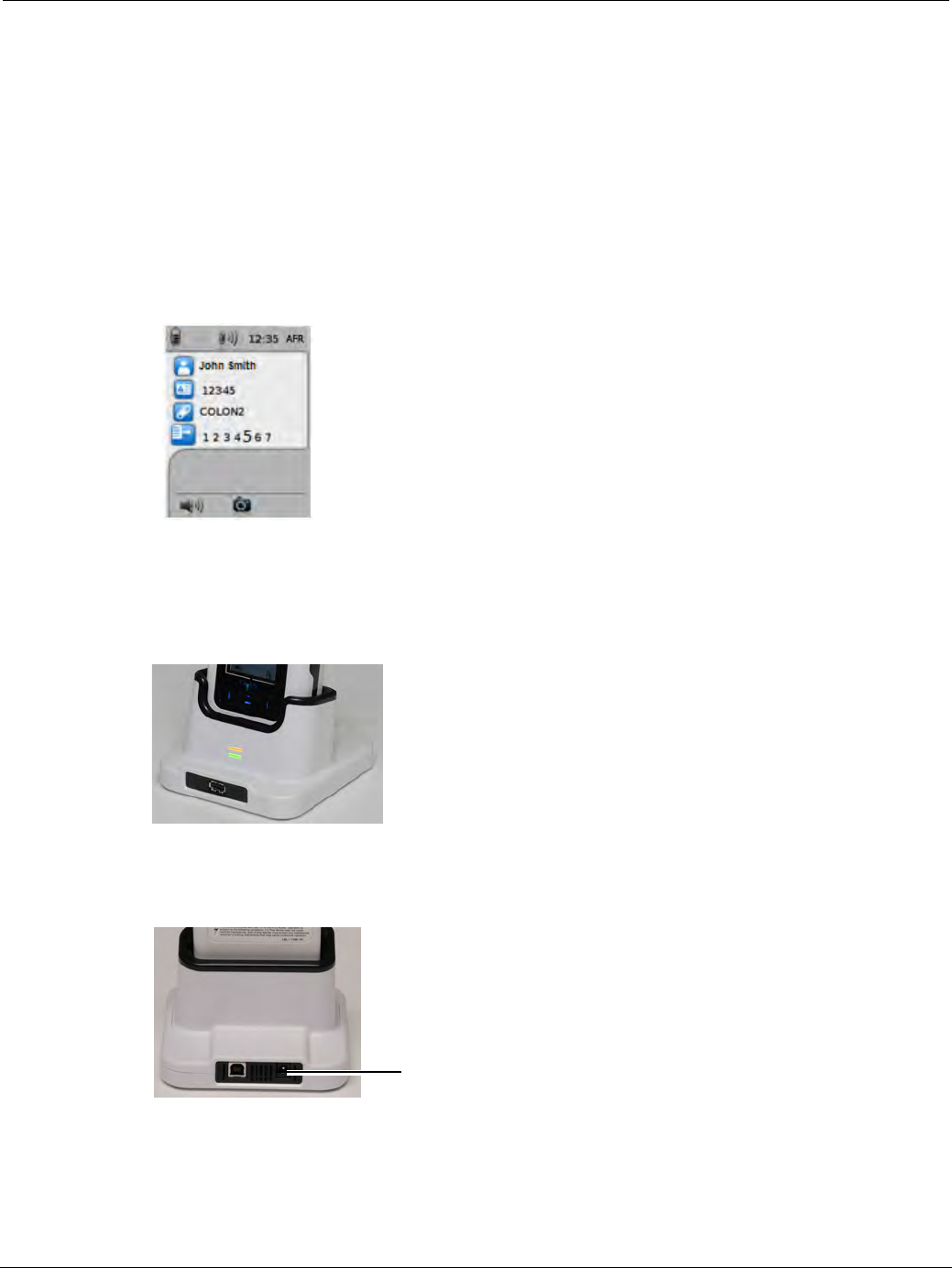
PillCam Capsule Endoscopy
66 PillCam Recorder DR3
Regimen Reminder
Regimen Reminder is a reminder function, whereby the PillCam recorder alerts the patient to perform
dietary instructions during a PillCam COLON 2 procedure. The alerts are timely instruction numbers that
appear on the recorder screen together with some audio and tactile alerts to draw the patient's attention to
the alerted instruction number. The patient is required to acknowledge the alert by pressing an
acknowledge button on the recorder and execute the associated instruction detailed on a patient instruction
sheet. The PillCam recorder DR3 is programmed during the initialization to remind the user according to
an uploaded regimen instruction set which is also printed out and handed to the patient. The timing of the
regimen instructions is synchronized to the moment of PillCam recorder DR3 pairing with the PillCam
capsule.
The last instruction alerted is displayed on the screen of the PillCam recorder DR3.
Charging
Charge the PillCam recorder DR3 by placing it in the cradle. The cradle charges the PillCam recorder DR3
and also connects it to the PC for performing patient check-in and creating videos. There are two LEDs at
the base of the cradle.
•The top LED is orange when the PillCam recorder DR3 is in the cradle.
•The bottom LED is orange while charging and green when the PillCam recorder is fully charged.
There is a power connector on the back panel of the cradle:
power connector
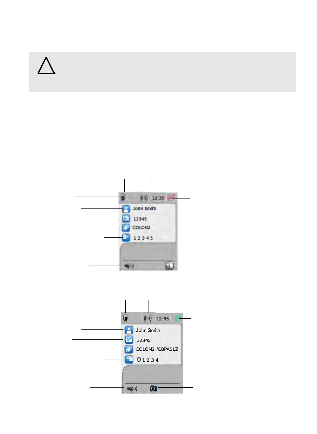
Know Your PillCam Recorder
PillCam Recorder DR3 67
The PillCam recorder is ready for operation when:
•the battery is at least 80% charged (eight or more bars displayed on battery icon),
•it has been removed from the cradle,
•it is connected to the sensor belt or sensor array.
Controls
Main Display
The main display of the PillCam recorder DR3 displays the relevant information during the different
phases of the capsule endoscopy procedure.
After Initialization (after check-in) and before pairing with the capsule, the recorder display shows the
relevant procedure information data, as follows:
After pairing with the capsule, the display shows the relevant procedure information data, as follows:
!
Warning
Never connect the PillCam recorder to the sensor array or the sensor belt while
the PillCam recorder is in the cradle.
Do not use the DR3 cradle for any USB devices except the PillCam recorder
DR3.
Battery charge level Capsule signal level
Status line
Patient name
Patient ID
Capsule type
Last Regimen instruction
Audio alert control Icon to toggle delay
first instruction
Recorder status: unpaired
Battery charge level Capsule signal level
Status line
Patient name
Patient ID
Capsule type
Last Regimen instruction
Audio alert control
Recorder status: paired
Real-Time viewing
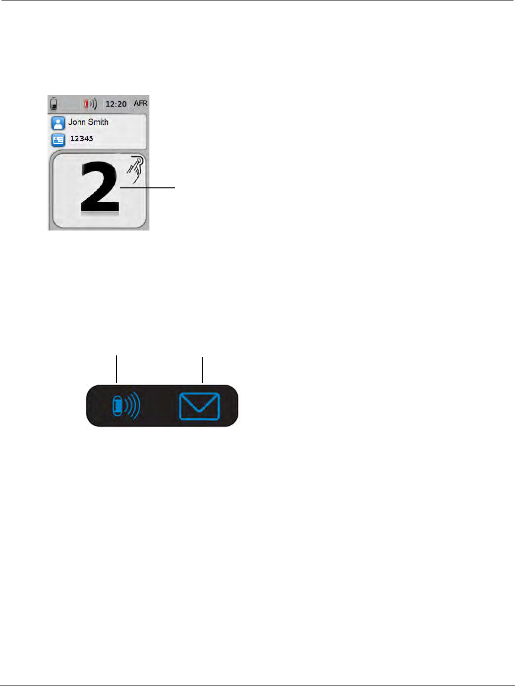
PillCam Capsule Endoscopy
68 PillCam Recorder DR3
To activate real-time viewing (only after pairing), use the navigation buttons located under the icons at the
bottom of the display: press the button below the camera icon, then the left button, and then the right button.
When a regimen instruction alert is activated, the instruction number appears all over the main screen and
remains until the Acknowledge button is pressed. The graphic indication in the top right corner of the
displayed alert indicates that an acknowledge response is required by pressing the acknowledge button.
Error messages appear similarly over the main screen (see Error Messages on page 73).
LED Display
The top of the PillCam recorder has a small screen on which the capsule and message LEDs are displayed.
These LEDs indicate the status of the PillCam recorder and the capsule endoscopy procedure. For
example, when the capsule LED on the PillCam recorder blinks in blue, this means that PillCam recorder
is receiving data from a capsule.
For a complete list of all capsule and message LEDs, see PillCam Recorder DR3 LEDs on page 70.
Acknowledge (ACK) Button, Designated for Patient Use
During a capsule endoscopy procedure, patients press the Acknowledge button in response to PillCam
recorder message alerts that appear on the recorder display. This ensures that the patient acknowledges the
instruction message. This can include regimen instruction messages during post-ingestion regimen (see
Post-Ingestion Patient Instructions on page 187).
•The acknowledge button will simultaneously blink with the display of any regimen reminder popup in
PillCam recorder DR3 LCD display and it will keep blinking until patient pressed the acknowledge
button for 3 sec or the popup display has reached its time out duration.
•At the end of 3 sec continuous pressing on the acknowledge button the blinking will go off.
Regimen reminder alert
Message LEDCapsule LED
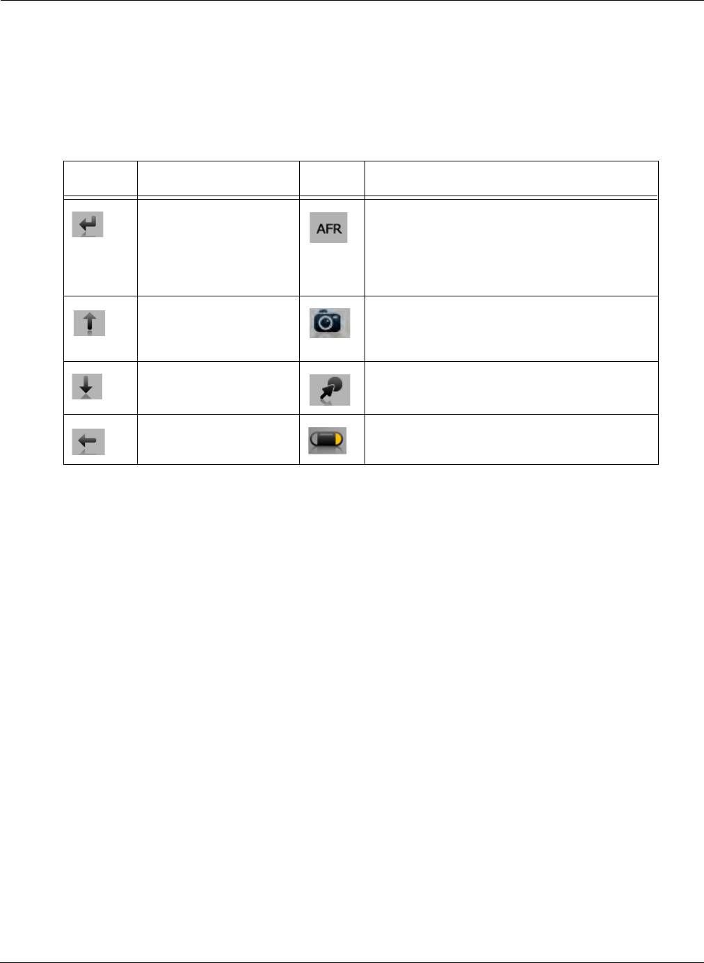
Know Your PillCam Recorder
PillCam Recorder DR3 69
Navigation Buttons
The navigation buttons are used to interact with the recorder by moving a cursor on a menu of icons on
the recorder LCD screen and to make a selection during the capsule pairing process (see PillCam
Recorder—Capsule Pairing (DR3 only) on page 80) and when selecting display modes. The icon
above the navigation button at the bottom of the display area indicates the functionality of the button.
The table below shows the navigation buttons and their actions.
Button Pressing Indication
The following audio (beep) and visual feedbacks alert the user when pressing buttons on the PillCam
recorder DR3:
•If the backlight is off: Pressing any button for the first time or turning on the PillCam
recorder DR3 turns the backlight on. This action is not accompanied by audio feedback.
•If the backlight is on:
• Momentarily pressing a button that affects a function, such as volume control, Real-Time
viewing combination, scrolling up/down or left/right, and selecting buttons, results in audio
(beep) feedback.
• Continuously pressing a button (i.e. press the Acknowledge button for 3 seconds), results in
audio (beep) feedback at the end of the required duration (i.e. at the end of 3 seconds).
• Momentarily pressing a button that is designed to activate a function after continued pressing
(i.e. Acknowledge), is not accompanied by audio or visual feedback.
•If the volumes is set to “off”, no audio indications should play.
Icon Action when pressed Icon Action when pressed
Confirm/select Manually activate AFR mode in PillCam
COLON 2 capsule and activate instruction #1
if gastric passage of the capsule is detected
and the AFR mode was not entered
automatically
Scroll up the cursor Activate Real-Time viewing (followed by
pressing the left then right buttons within 6
seconds)
Scroll down the cursor Mark displayed frame
Exit Real-Time viewing Switch video head (in Real-Time viewing
mode)
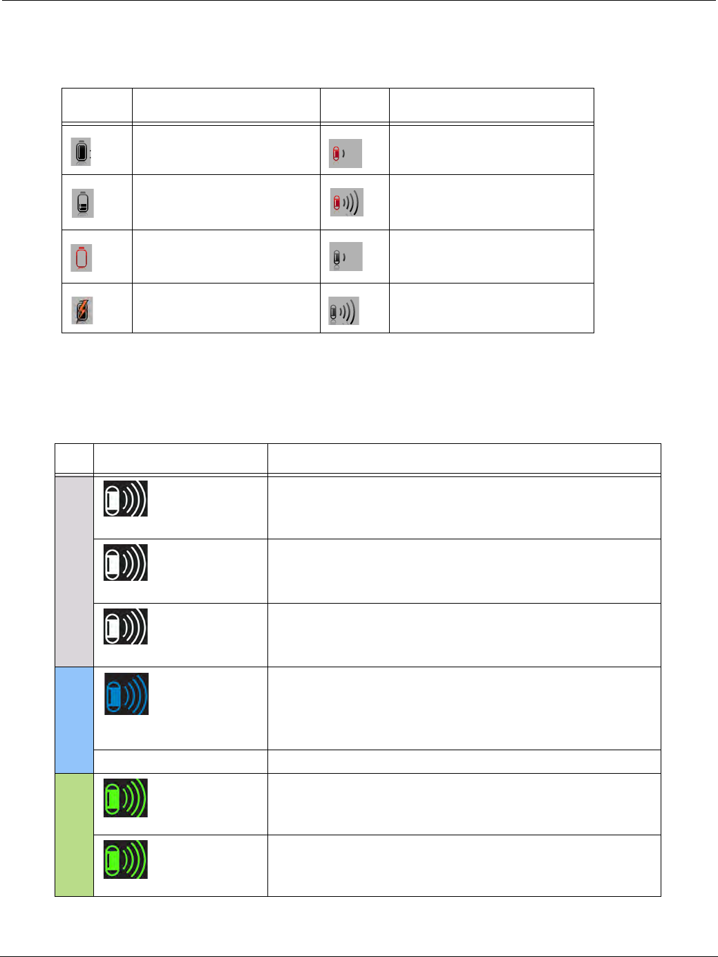
PillCam Capsule Endoscopy
70 PillCam Recorder DR3
Battery and Capsule Icons
The battery icon on the left side of the status line at the top of the display indicates the status of the battery
in 10% increments. These icons appear in the top status line of the PillCam recorder DR3 screen.
PillCam Recorder DR3 LEDs
The status indications of the LED indicators on the top of the PillCam recorder DR3 for the most common
PillCam recorder events are as follows:
Icon Battery Status Icon Capsule Reception Status
Battery fully charged Signal weak, recording with
noise
Battery charge level at 10%
intervals
Signal strong, recording with
noise
Battery empty, PillCam
recorder shuts down
Signal weak, but recording OK
Battery charging Signal strong, and recording
OK
LEDs PillCam Recorder DR3 Status
CHECK-IN
Blinking in white
PillCam recorder DR3 is being checked in.
Blinking rate = very fast.
Constant on in white
PillCam recorder DR3 is checked in and ready to receive
capsule signals.
Blinking in white
PillCam recorder DR3 is checked in and receiving signals
from an unpaired capsule.
Blinking rate = capsule frame rate.
RECORD
Blinking in blue at capsule
frame rate
PillCam recorder DR3 is receiving paired capsule signals and
recording.
Blinking rate = capsule frame rate.
LEDs off Recording done and raw data is available for downloading.
DOWNLOAD
Blinking in green
PillCam recorder DR3 has started downloading.
Constant on in green
PillCam recorder DR3 has completed downloading.
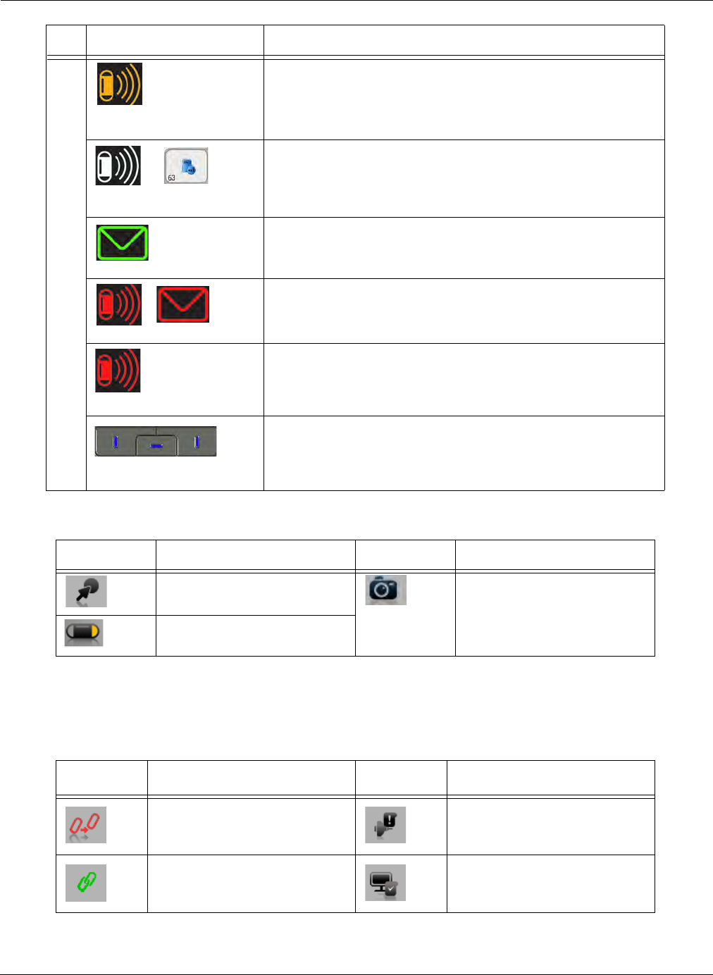
Know Your PillCam Recorder
PillCam Recorder DR3 71
Navigation Buttons Legend
Screen Icons
These icons appear as recorder status on the rightmost icon of the top status line of the PillCam
recorder DR3 screen:
General Indications
Blinks in yellow/orange every
5 seconds
PillCam recorder DR3 has stopped receiving capsule signals
for more than 5 seconds.
Constant on in white with error
message
PillCam recorder DR3 has stopped recording because the
memory card is full.
Blinking in green
There is an instruction on the PillCam recorder DR3 screen.
Constant on in red
PillCam recorder DR3 is malfunctioning.
Blinking in red
PillCam recorder DR3 detects capsule signal, but is not
recording it. This is a malfunction. Check the sensor
connection or have patient move to a different location.
Blinking in blue
The navigation buttons blink in blue every 5 seconds when
the PillCam recorder DR3 is on, either in or out of the cradle,
and the LCD screen is off. To turn the LCD screen back on,
press any button on the PillCam recorder DR3.
Icon Action when pressed Icon Action when pressed
Mark displayed frame. Activate Real-Time viewing
(followed by pressing the left
then right buttons within 6
seconds).
Switch video head (in Real-
Time viewing mode).
Icon Explanation Icon Explanation
No capsule is paired. No sensor is connected.
Pairing succeeded. PillCam recorder DR3 is
checked in.
LEDs PillCam Recorder DR3 Status
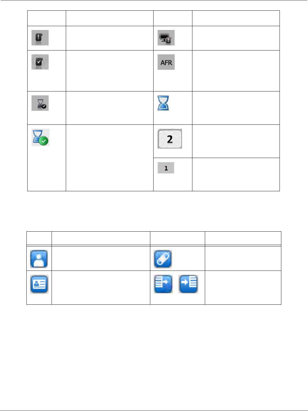
PillCam Capsule Endoscopy
72 PillCam Recorder DR3
Check-in Screen Icons
After performing patient check-in, these icons appear on the PillCam recorder screen data area:
Data has not downloaded. PillCam recorder DR3 is
waiting for check-in.
Data has downloaded. Indicates that AFR mode was
activated in the capsule
(PillCam SB 3 or COLON 2),
after the PillCam recorder DR3
detected gastric passage.
End of procedure. In Real-Time Viewing mode,
an End of Procedure
notification appears on the top
right corner of the screen.
End of procedure icon
appears on the main screen
and indicates that the
procedure has ended and
equipment may be removed. It
occurs when a predefined
reception gap from the paired
capsule is encountered.
Regimen reminder numbers
appear on the main screen to
alert the user to perform
dietary instructions.
In Real-Time Viewing mode,
the Regimen Reminder
number appears on the top
right corner of the screen.
Icon Explanation Icon Explanation
Patient name Procedure type and
capsule ID
Patient ID
On Off
Regimen Delay first
instruction status
indicator and navigation
button designator
Icon Explanation Icon Explanation
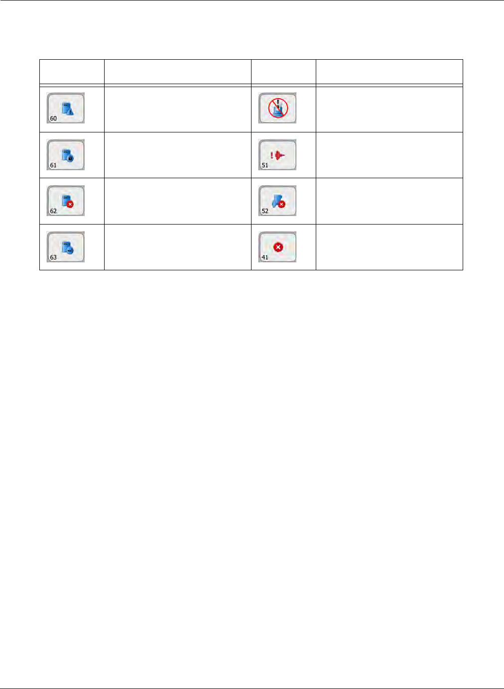
Know Your PillCam Recorder
PillCam Recorder DR3 73
Error Messages
During operation, the following messages may appear in the PillCam recorder main screen.
Popup Explanation Popup Explanation
No approved memory card is
detected. Verify you are using
an approved card.
Do not remove the PillCam
recorder DR3 from the cradle.
Memory card is write-
protected.
Sensor hardware failure.
Consult a technician.
Memory card error.
Remove and reinsert card.
Wrong sensor type.
Insufficient memory on card. Fatal error.
Consult a technician.
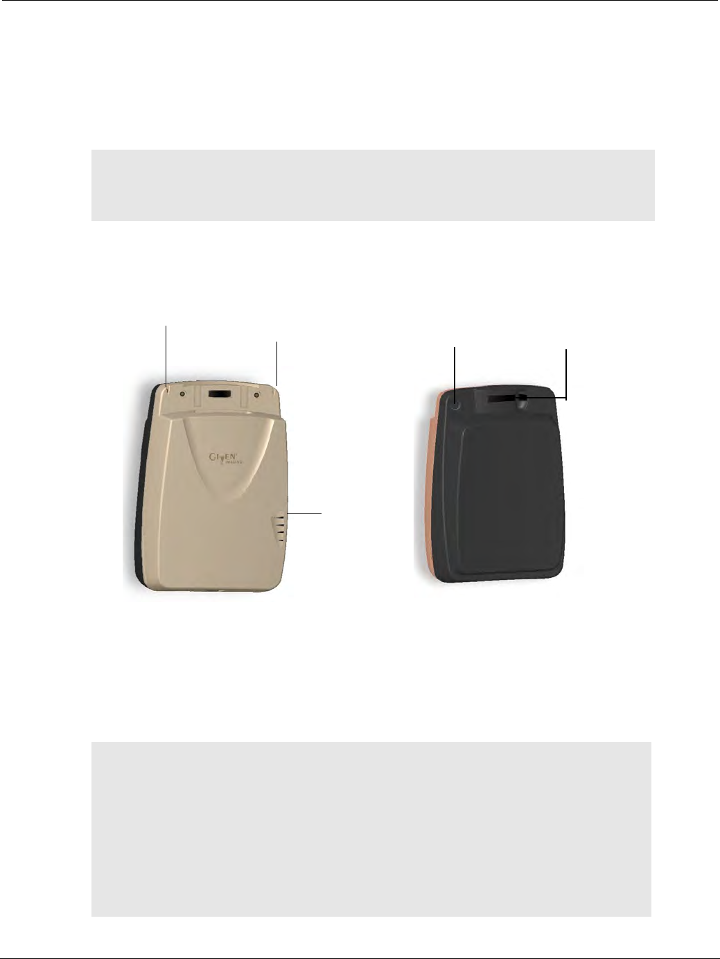
PillCam Capsule Endoscopy
74 PillCam Recorder DR2
PillCam Recorder DR2
General
The PillCam recorder DR2 battery is limited to 350 use cycles. Contact customer service for a
replacement battery when necessary.
Turning On and Off
This illustration shows the front and back view of the PillCam recorder DR2:
The On/Off button is on the top left side on the back of the PillCam recorder DR2:
•To turn on, press and hold the on/off button until you hear a long beep followed by a short one and the
LEDs start flashing.
•To turn off, press and hold the On/Off button until you hear a beep and the LEDs turn off.
֠
֠֠
֠Note
Once the number of uses exceeds 350, the following message appears (in
the log file only): DR2 battery must be replaced before further use. Contact
customer support to arrange replacement.
֠
֠֠
֠Note
Automatic shutdown: After the PillCam recorder DR2 has been checked
in, it goes into standby mode ready to receive capsule signals when
removed from its cradle and starts recording as soon as a signal is received
from a transmitting capsule. If after removal from the cradle no signal is
received for 90 minutes, the PillCam recorder DR2 automatically shuts
down.
After starting to receive signals from a capsule, if there is a gap of 60
minutes of no signal reception, the recorder shuts down.
Locking
handle in
open position
PillCam
recorder DR2
LED Capsule LED ON/OFF button
Battery
LEDs
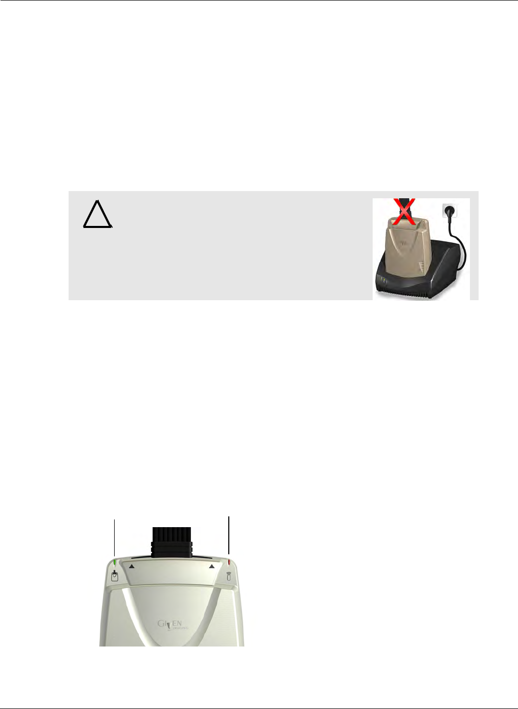
Know Your PillCam Recorder
PillCam Recorder DR2 75
Charging
The cradle provided with the PillCam recorder DR2:
•Charges the PillCam recorder when it is placed in the cradle
•Charges a spare battery externally when a stand-alone battery is placed in the cradle
•Performs battery maintenance by discharging the battery when needed (the cradle detects when the
battery needs refreshing and automatically discharges it before recharging)
To charge the PillCam recorder DR2:
1. Insert the PillCam recorder DR2 into the cradle.
2. Push it all the way down into the cradle and make sure you hear a series of beeps, indicating that
connection is complete.
.
There are four connections on the back panel of the cradle. Only two of them are used with standard
operation of the cradle: the power connector and the USB cable connection.
The cradle also connects the PillCam recorder to the Personal Computer (PC).
•The green LED on the cradle indicates that the PillCam recorder DR2 is fully charged and ready
for use.
•The red LED, when lit continuously, indicates a defective battery.
•The red LED, when blinking, indicates that there is a problem with the cradle.
PillCam Recorder DR2 LEDs
When the PillCam recorder DR2 is on, it starts recording as soon as it receives a signal from a PillCam
capsule. When the capsule LED blinks, the PillCam recorder DR2 is receiving data. When the signal
from the PillCam capsule is too weak, the LED does not blink.
!
Warning
Do not connect the PillCam recorder DR2 to the
sensor array while the PillCam recorder DR2 is in
its cradle.
Capsule LED
Recorder LED
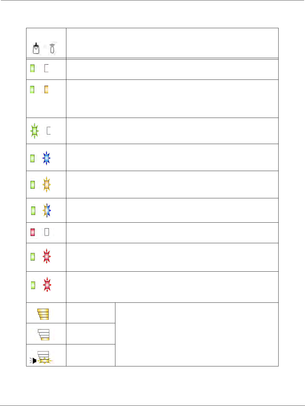
PillCam Capsule Endoscopy
76 PillCam Recorder DR2
These are the LED indicators and their statuses and colors for the most common PillCam recorder DR2
events and statuses:
LEDs PillCam Recorder DR2 Status
PillCam recorder is ON but not initialized. PillCam recorder does not capture
capsule signals.
PillCam recorder is initialized with patient data and ready to capture capsule
signals.
PillCam recorder shuts down if no capsule signals are received for more than
30, 60, or 90 minutes, depending on the PillCam recorder software version.
PillCam recorder is exchanging status or data with RAPID or RAPID RT.
LED blinking rate varies according to the communication flow.
PillCam recorder is capturing capsule signals.
Blinking rate = capsule frame rate.
PillCam recorder has stopped capturing capsule signals for more than 5
seconds.
PillCam recorder is detecting a capsule in sleep mode.
Blinking rate = every five seconds (in any color).
PillCam recorder is malfunctioning.
<20 seconds
PillCam recorder is synchronizing with a capsule. This is normal functioning.
>20 seconds
PillCam recorder detects capsule signal, but is not recording it. This is a
malfunction.
Check the sensor array connection or have patient move to a different location.
maximum Battery charge level:
When charging, the battery LEDs do not blink.
When PillCam recorder is out of the cradle, the battery
LEDS blink once every 5 seconds.
25%
below 10%

Know Your PillCam Recorder
Connecting a PillCam Recorder to a Personal Computer (PC) 77
Connecting a PillCam Recorder to a Personal
Computer (PC)
The PillCam recorder needs to be connected to a PC with RAPID in the following cases:
•Connect for initialization: For initialization before the procedure: to check-in a patient.
•Connect for downloading: For downloading data from the recorder after the procedure: to create a
video or copy the video data from the recorder to the computer.
•Connect for charging: For charging the PillCam recorder after the procedure: to prepare the
recorder for the next procedure.
•Connect for upgrading: When the software version of the PillCam recorder is lower than the latest
one internally specified in RAPID, the user is prompted to allow a recorder upgrade the first time
the recorder is connected to RAPID for check-in. It is highly recommended to allow the upgrade.
The latest software versions for both PillCam recorder DR2 and PillCam recorder DR3 are stored
in RAPID and when prompted and allowed, the appropriate upgrade will be performed.
The PillCam recorder is connected to the PC only through its cradle, each model through its cradle.
The PillCam recorder is passive during these cases and no controls need be operated on it.
Connecting a PillCam Recorder to the External
Real-Time Viewer
The PillCam recorder may be connected to an external real-time viewer, which is a dedicated tablet PC
with real-time viewing software and special setup, for viewing in real-time the images captured and
stored in the recorder. This functionality exists for both recorder types, PillCam recorder DR2 and
PillCam recorder DR3 (the latter has also the built-in capability to perform real-time viewing through
the recorder's display).
For a real-time viewing session, both recorder types connect to the external real-time viewer (tablet PC)
through a USB cable, while the tablet PC or anything connected to it must not be connected, directly or
indirectly to any wall outlet. The real-time viewing software in the tablet PC controls the session and the
PillCam recorder is passive in this configuration. There is no need to use any control button of the
recorders.
֠
֠֠
֠Note
It is recommended to use an external hub when connecting multiple PillCam
recorders.

PillCam Capsule Endoscopy
78 Connecting a PillCam Recorder to the External Real-Time Viewer
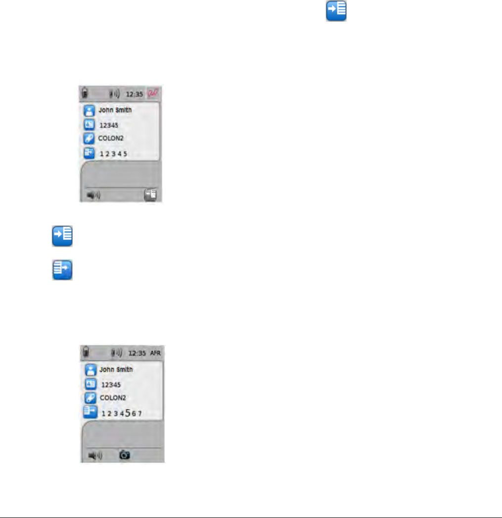
Setting Delay First Instruction in PillCam Recorder 79
Chapter 6
Performing PillCam Capsule Endoscopy
Setting Delay First Instruction in PillCam Recorder
Before ingestion of the PillCam COLON 2 capsule, you need to make sure that the settings match the
procedure circumstance: patient stays in clinic after ingestion or is instructed to go home immediately
after ingestion. If the patient stays in clinic, the Delay first instruction flag on the PillCam recorder
DR3 needs to be OFF, otherwise it needs to be ON. If this flag has not been set properly during check-
in, it needs to be set before ingestion directly on the PillCam recorder prior to pairing with the PillCam
COLON 2 capsule by using the right navigation button with the icon above it to toggle the flag.
The default setting for a PillCam recorder DR3 checked-in with RAPID v8.3, without changing the
Delay first instruction field during check-in, is ON—first instruction delayed. Repetitive use of the
right button will toggle the status of the PillCam recorder DR3 and its status will show in the fourth
icon (from above) in the left most side of the fourth procedure data line.
The fourth line status icon and the button designation icon in the lower right corner of the screen above
the right navigation button will always be opposite. After pairing, the Delay first instruction icon will
disappear and the status will not be changeable any more.
Delay first instruction—Off
Delay first instruction—On
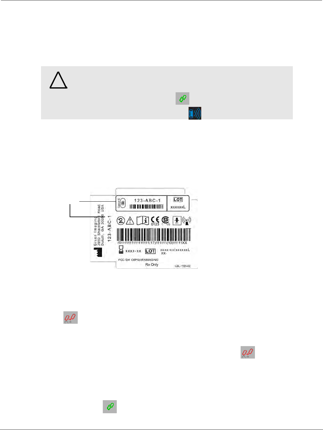
PillCam Capsule Endoscopy
80 PillCam Recorder—Capsule Pairing (DR3 only)
PillCam Recorder—Capsule Pairing (DR3 only)
PillCam recorder DR3 operates only with capsules that were paired to it. This allows the PillCam recorder
to recognize images from the correct capsule. Pairing is initiated when the PillCam recorder recognizes
transmission from a designated capsule. From that point on, the recorder is associated with the capsule.
The recorder and capsule are then paired.
Designation of the capsule to be used in the procedure may be performed during patient check-in or
immediately before capsule ingestion. The following section describes these two scenarios.
Capsule Designation During Patient Check-in
While entering the patient and procedure data during check-in, type the capsule ID in the Capsule ID field
or use a barcode scanner. The capsule ID is displayed on the capsule package.
During recorder initiation, the unique capsule ID will be transferred into the PillCam recorder together with
the rest of the initiation data. It will appear on the PillCam recorder LCD together with the rest of the
patient data.
As long as no transmission from the checked in capsule is received, pairing is not done. The Ready for
pairing icon remains in the top right corner of the screen.
If there are PillCam capsule transmissions received by the PillCam recorder, which are not from the
designated capsule, the capsule IDs of these other capsules (only of the checked-in capsule type) are
temporarily displayed on the PillCam recorder screen. Ready for pairing icon remains in the top right
corner of the screen.
When you open the designated capsule box, just before ingestion, the capsule starts blinking and
transmitting, each capsule type in its unique way. The PillCam recorder will automatically pair to the
designated capsule and start recording when it receives transmissions from it.
The Pairing success icon will appear in the top right corner of the screen.
!
Warning
The PillCam recorder starts recording only after it is paired with the
capsule. If pairing is not complete, no video can be produced. Make sure
you see the Pairing Success icon on the screen and the capsule LED
on top of the recorder blinking in BLUE before capsule ingestion.
Capsule ID
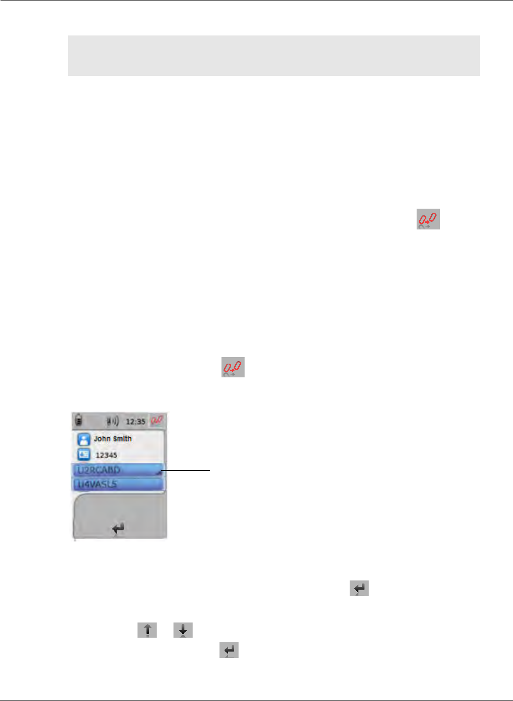
Performing PillCam Capsule Endoscopy
PillCam Recorder—Capsule Pairing (DR3 only) 81
Capsule Designation Before Capsule Ingestion
There are two situations when you need to perform capsule designation manually immediately prior to
ingestion:
•if designation was not performed during check-in, or
•if designation was performed, but the designated capsule is not available for the procedure.
As long as there are PillCam capsule transmissions received by the PillCam recorder but are not from a
designated capsule, the capsule IDs of these other capsules (only of the checked-in capsule type) are
temporarily displayed on the PillCam recorder screen. The Ready for pairing icon remains in the
top right corner of the screen.
To designate and pair a capsule to be used in the procedure before capsule ingestion:
1. Open the capsule box lid—the capsule starts blinking, each capsule in its unique blinking pattern
(see System Specifications on page 217). Right after opening the lid, the blinking rate for
PillCam SB 3 and PillCam COLON 2 capsules should still be 2 fps per head, the same as for
PillCam SB 2. For PillCam SB 2 (fixed 4 fps variant) the blinking rate should be 4 fps. For PillCam
UGI, the blinking rate is 35 fps per head. Hold the capsule close to the sensors worn by the patient
during this pairing process.
2. When the Ready for pairing icon is displayed in the top right corner of the screen and when
the PillCam recorder detects your PillCam capsule, its ID code is displayed on the screen (alone or
in addition to IDs from other capsules).
3. If only one capsule ID appears on the PillCam recorder screen, check the capsule ID shown in the
PillCam recorder screen against the capsule ID displayed on the PillCam capsule package. If the
codes match, press the middle navigation button, above which appears on the screen.
4. If more than one capsule ID appears on the PillCam recorder screen, use the navigation buttons
(located below or icons on the screen) to scroll to your capsule ID and press the middle
navigation button (located below icon on the screen) to designate and initiate pairing of your
capsule with the PillCam recorder.
֠
֠֠
֠Note
Capsule designation during patient check-in is the recommended method for
designating a capsule to be used in the procedure.
This mark indicates that this capsule ID will be
selected when pressing the middle navigation button.

PillCam Capsule Endoscopy
82 Capsule Ingestion
The Pairing success icon will appear in the top right corner of the screen.
Capsule Ingestion
Capsule ingestion is the process of having the patient swallow the PillCam capsule.
֠
֠֠
֠Note
• If a PillCam COLON 2 capsule blinks slower than 1 blink per second
during the pairing procedure, return it to its box so that it stops blinking
and start over.
• If, at any time before ingestion, the capsule does not blink for at least 20
seconds, do not use this capsule. Instead, use a different capsule and
repeat the pairing procedure.
֠
֠֠
֠Note
If after designating the capsule:
• the patient does not ingest it within ten minutes, or
• you suspect that the capsule is defective.
Return the capsule into its box. When you close the lid, if the capsule stops
blinking, it is deactivated.
If the capsule continues blinking, rotate it around its axis, without removing it
from its place holder in the box until it stops blinking when you close the lid.
To use this capsule later (if it is not defective), you must repeat the pairing
process.
!
Multiple Procedures
When performing more than one PillCam capsule endoscopy procedure in
the same vicinity, follow these guidelines to prevent signal interference with
other procedures:
• After patient check-in, keep the PillCam recorder in the cradle without
sensors attached until capsule ingestion procedure begins. Return it to
the cradle when the procedure is complete.
•DR2 only: If the blue PillCam recorder LED is blinking before you open
the box of the capsule you intend to use, repeat check-in with the PillCam
recorder (see Performing Patient Check-in on page 43).
• Perform only one capsule ingestion at a time with no other active PillCam
recorder or capsules present in the room.
• Attach sensors to the PillCam recorder immediately prior to the ingestion
after sensors are properly positioned on the patient.
• Do not permit patients wearing PillCam recorders to stay directly next to
other patients with ingested capsules.
• To minimize the potential for radio frequency interference from the
capsule after it is removed from the box, verify that the capsule LEDs are
blinking and have the patient ingest it immediately.
• Once the RAPID video is created, check to be sure the video is complete.

Performing PillCam Capsule Endoscopy
Capsule Ingestion 83
PillCam Recorder DR3
1. Make sure that the capsule and the capsule LED on the PillCam recorder are blinking in time
with each other.
2. Check the color of the capsule LED on the PillCam recorder:
• If (blue), go to step 3.
• If (white), pair the capsule with the PillCam recorder and verify the pairing success icon
appears in the top right corner of the PillCam recorder screen and the capsule LED blinks
blue .
3. Have the patient swallow the capsule with a sip of water. The ingestion procedure may take several
minutes.
PillCam Recorder DR2
1. Make sure that the capsule and the capsule LED on the PillCam recorder are blinking in time with
each other.
2. Position the patient on the bed, with a pillow (6 cm or 2.5 inch high) under the head to facilitate
drinking and ingestion.
3. Instruct the patient not to talk during the procedure.
4. To verify that the system is operating properly, hold the capsule in front of the patient's abdomen
very close to the sensors (practically touching through the clothes one of the sensors). Hold it for at
least 15 seconds and check that the capsule LED on the PillCam recorder blinks at the same rate as
the capsule.
5. Have the patient swallow the capsule with a sip of water.
֠
֠֠
֠Note
During capsule endoscopy, the capsule LED blinks blue at the recording
rate.
If the capsule LED blinks orange once every 5 seconds, the PillCam
recorder is not receiving capsule signals (see PillCam Recorder DR3 on
page 209 for troubleshooting details).

PillCam Capsule Endoscopy
84 After Capsule Ingestion
After Capsule Ingestion
PillCam ESO/UGI
The patient must stay at the medical facility until the end of the procedure (approximately 90 minutes after
ingestion of the capsule).
The procedure ends when the End of Procedure screen appears on the PillCam recorder. The PillCam
recorder also beeps and vibrates when the End of Procedure alert appears. In ESO/UGI capsule procedures,
the End of Procedure icon appears when 10 minutes have passed without paired capsule reception in the
PillCam recorder DR3.
At the end of the procedure, remove the PillCam recorder and sensors from the patient. The patient may
then be released and can return to a normal daily routine.
PillCam SB
Once the patient has ingested a PillCam SB capsule, the patient may leave the clinic. Make sure that the
patient knows which activities to avoid during the procedure and how to return the PillCam recorder and
sensors.
PillCam Recorder DR3
The procedure ends when the End of Procedure screen appears on the PillCam recorder. The PillCam
recorder also beeps and vibrates when the End of Procedure alert appears.
In SB capsule procedures, the End of Procedure icon appears when 25 minutes have passed without paired
capsule reception in the PillCam recorder DR3.
At the end of the procedure, remove the PillCam recorder and sensors from the patient. The patient may
then be released and can return to a normal daily routine.
PillCam Recorder DR2
The procedure lasts as long as the blue PillCam recorder LED is blinking, or until the capsule is excreted.
The PillCam recorder DR2 LED should blink for at least six hours, unless the PillCam SB capsule was
excreted.
At the end of the procedure, remove the PillCam recorder and sensors from the patient. The patient may
then be released and can return to a normal daily routine.
PillCam COLON (DR3 only)
After ingestion, explain the Patient Guidelines (see General Patient Guidelines During the Procedure on
page 42) to the patient.
Explain to the patient what to do when the PillCam recorder beeps or vibrates (see Post-Ingestion Patient
Instructions on page 187).
The procedure ends when the End of Procedure screen appears on the PillCam recorder. The PillCam
recorder also beeps and vibrates when the End of Procedure alert appears.
֠
֠֠
֠Note
When the capsule LED on the PillCam recorder blinks orange, this indicates
that the PillCam recorder is no longer receiving capsule signals.
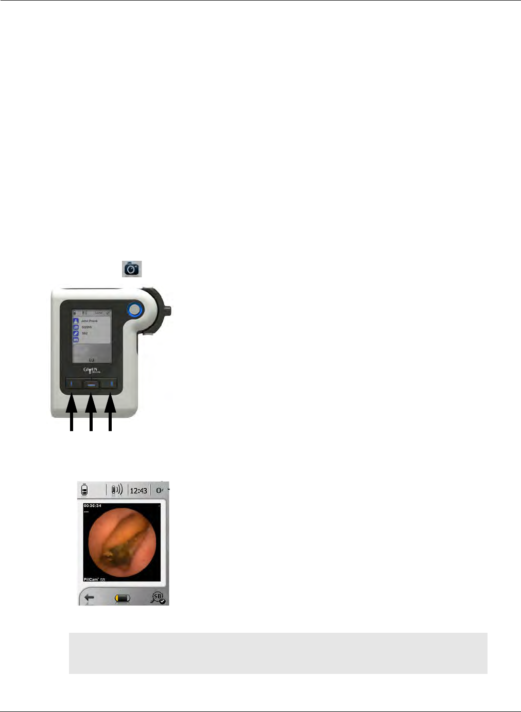
Performing PillCam Capsule Endoscopy
Real-Time Viewing with PillCam Recorder DR3 Only—Internal Mode 85
In COLON capsule procedures, the appearance of the End of Procedure icon depends on how much
time has passed without paired capsule reception in the PillCam recorder DR3:
•During the first 5 hours of the procedure, 25 minutes of no paired capsule reception.
•Between 5 hours and 10 hours of the procedure, decreasing incrementally from 25 minutes to 10
minutes of no paired capsule reception.
•After 10 hours into the procedure, 10 minutes of no paired capsule reception.
At the end of the procedure, the PillCam recorder and sensors may be removed from the patient. The
patient may then return to a normal daily routine.
Real-Time Viewing with PillCam Recorder DR3 Only—
Internal Mode
To activate real-time viewing with PillCam recorder DR3, press the middle navigation button below the
Real-Time icon , then immediately press the left then right buttons one after the other.
The following screen appears on the PillCam recorder.
֠
֠֠
֠Note
Using Real-time Viewing on the PillCam recorder with an external real-time
viewer (laptop) is not supported.
1 32
PillCam Capsule Endoscopy
86 Real-Time Viewing with PillCam Recorder DR3 Only—Internal Mode
In procedures with AFR mode capsules (PillCam SB 3 and PillCam COLON 2), the AFR icon appears
above the right navigation button while the capsule is not in AFR mode to allow the user to manually
activate the AFR mode of the capsule even before automatic activation. After activation of the AFR mode
in the capsule (either manually or automatically), the recorder status icon in the top status line changes from
paired to AFR . The icon displayed above the right navigation button is the mark icon that
allows the marking of the displayed image for automatic creation of a thumbnail and to facilitate further
scrutiny of the image off-line in the created video.
In procedures with non-AFR capsules (PillCam SB 2 and PillCam ESO/UGI) the icon that appears above
the right navigation button in real-time viewing is the mark icon .
For colon visualization procedures: instruction #1 on the PillCam recorder screen appears after the capsule
enters the small bowel, usually within two hours of ingestion. The AFR icon simultaneously appears in
the status line at the top of the PillCam recorder screen, designating the fact that the PillCam COLON 2
capsule passed into the AFR mode operation.
If the system does not show instruction #1 or the AFR icon two hours after ingestion, you can monitor the
capsule location in the GI tract using the real-time viewing. The system will activate by default the AFR
mode and instruction #1 at 4 hours post-ingestion at the latest.
If you need to verify (after instruction #1 on the PillCam recorder screen) that the capsule has left the
stomach, use the real-time viewer:
•If capsule is still in the stomach, repeat real-time viewing to check capsule location every 30 minutes
until you can confirm that it has left the stomach.
•If you visually confirm that the capsule has left the stomach while the AFR indication in the top right
corner of the status line still hasn't changed to , press the right navigation button under the AFR
icon at the right bottom of the screen for 5 seconds. This triggers instruction #1 and activates
recording mode AFR frame rate. The AFR icon appears at the top right corner in the status line of
the screen. The video created from this procedure will start at this point. The mark icon will
appear above the right navigation button instead of the icon.
•Once instruction #1 occurs, the PillCam recorder automatically continues to provide instructions
according to the post-ingestion regimen selected during check-in.
֠
֠֠
֠Note
For colon visualization procedures:
• If the PillCam recorder does not detect that the capsule has entered the small
bowel (either automatically or by the manual procedure described in step 2,
above), the following message appears when opening the video: Gastric-to-SB
passage not detected. In this case, the video is very short (the first 3 minutes
only).
• Some patients may require close supervision to ensure they comply with post-
ingestion instructions. Other patients may be able to function independently as
long as they have access to all needed doses of laxatives and/or prokinetics and
are capable of following instructions during the procedure.

Performing PillCam Capsule Endoscopy
Real-Time Viewing with PillCam Recorder DR2 Only— External Mode 87
When in Real-Time viewing the PillCam recorder screen turns off and returns to the main screen after:
•2 minutes of inactivity for colon visualization procedures/SB procedures,
•30 minutes of inactivity for ESO/UGI procedures.
Real-Time Viewing with PillCam Recorder DR2 Only—
External Mode
To activate real-time viewing with external RAPID Real-Time viewer, connect either PillCam recorder
model you are using to the external real-time viewer through the respective USB cable and activate the
RAPID Real-Time viewer. For instructions on the operation of the RAPID Real-Time viewer, see the
user manual for RAPID Real-Time viewer.
Removing Equipment from the Patient
If the patient has not already removed equipment, do so as follows:
1. Disconnect the sensors from the PillCam recorder.
2. Remove the PillCam recorder from the patient.
3. Remove the sensors from the patient.
4. Remove the PillCam recorder from the pouch and place it in the cradle.
5. After each procedure, make sure to clean the equipment (see PillCam Sensor Cleaning on
page 205) and charge the PillCam recorder (see Charging on page 203).
֠
֠֠
֠Note
AFR should be activated only after visually confirming entry into the small
bowel

PillCam Capsule Endoscopy
88 Removing Equipment from the Patient

PillCam Recorder Download 89
Chapter 7
Creating RAPID Videos
PillCam Recorder Download
After the capsule endoscopy examination, the video data in the PillCam recorder must be copied to a
computer and compiled into a video. There are two ways of doing this:
•Copy the data and compile the video as a single task. This method is the default; it is very
convenient, but takes longer to complete and free up the PillCam recorder.
•Copy the data only and perform compilation later. This method requires some additional steps, but
makes the PillCam recorder available faster (see Copying Data from a PillCam Recorder on
page 93).
When creating a video, the RAPID software:
•Creates a folder for the new video. This folder is inside the preset default folder (see RAPID Video
Files on page 143).
•Displays the images being copied from the PillCam recorder during video creation.
•Creates the RAPID video from this raw data and saves it in the new folder. The new video name and
its folder name are the same.
•Notifies you once video creation is complete so you may disconnect the PillCam recorder.
֠
֠֠
֠Note
• In the rare case that the video creation fails, RAPID prompts you to save the
raw data for customer support.
• You can reduce the file size while copying data (see Video Data
Management Section on page 174).
• When downloading from four recorders simultaneously ensure that there is
at least to 40 gigabytes of hard disk space.
• Make sure that the computer power settings are not set to sleep or to
hibernate modes.
!
Caution
Connection of the PillCam recorder to the RAPID software for patient check-in,
video download, and recorder upgrade must be done using the appropriate
cradle.
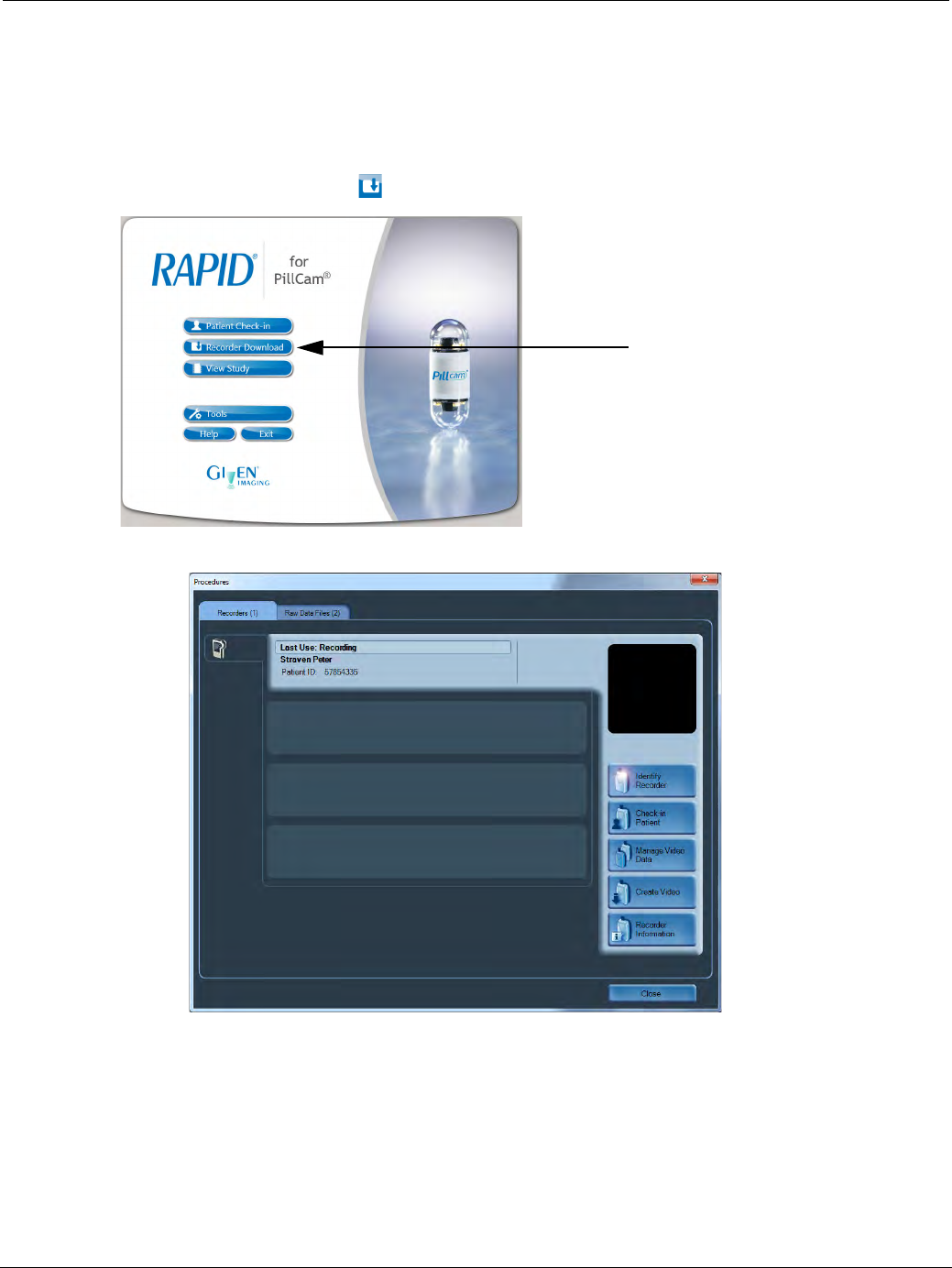
PillCam Capsule Endoscopy
90 PillCam Recorder Download
Creating a Video from the PillCam Recorder
To create a video from the PillCam recorder:
1. Place the PillCam recorder in the cradle. Make sure that the cradle is connected to the computer on
which RAPID is installed and open.
2. From the Home screen, click Recorder Download.
The Procedures screen appears with the Recorders tab on top.
3. Click the Recorder bar that corresponds to the PillCam recorder with the desired data:
• When the correct Recorder bar is activated and you click on the Identify Recorder button, the
corresponding PillCam recorder LEDs blink.
• The relevant patient information is displayed on the Recorder bar.
• PillCam recorder's last use, such as Recording, Creating video...
The Create Video and Check-in Patient buttons become available.
4. Click Create Video.
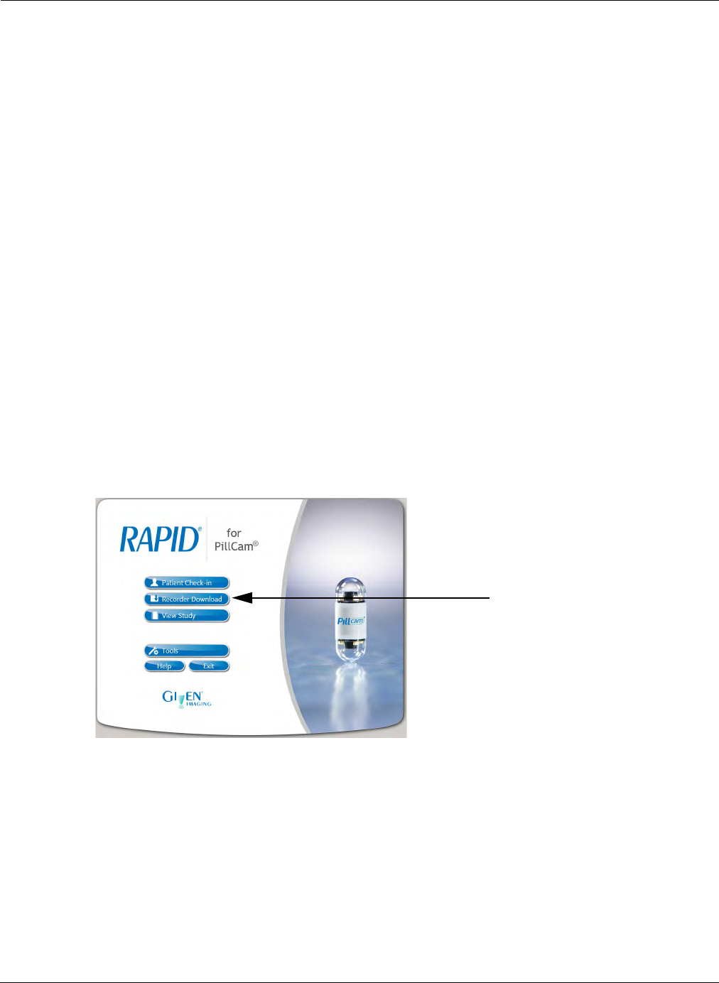
Creating RAPID Videos
PillCam Recorder Download 91
If there is enough space on the PC’s hard disk, the new video is created. If not, you are prompted to
free up space (see Freeing Space on Your Computer on page 195).
During video creation, in the PillCam recorders window, the Create Video button becomes the
End video creation button. The Recorder bar displays the following:
• The status and progress bar.
• The patient name and ID.
• The name of the video to be created.
• The battery status.
• The Do not remove recorder message flashes below the battery status until all data is
copied or compiled into a video.
The images that are being copied to the computer are displayed in the Compiling Images window
above the function buttons.
Creating a Video from USB Storage Device or DVD
RAPID supports video creation from media (a removable USB storage device or DVD). This can be
useful if you have copied raw data from the PillCam recorder.
To create a video from a media:
1. Plug the USB storage device into one of the USB 2.0 ports of the PC, or insert the DVD into the
DVD drive.
2. From the Home screen, click Recorder Download.
The Procedures screen appears.
3. Click the Raw Data Files tab.
Find the bar that corresponds to the USB storage device you want to select. The relevant patient
information is displayed on the button.
4. Click Create Video.
5. After a video has been created, click Safely Remove to safely remove the USB storage device.

PillCam Capsule Endoscopy
92 Managing RAPID Video Data
Batch Video Creation
RAPID can perform multiple video creations for any combination of up to four PillCam recorder cradles,
and for any number of raw data files. Once the process begins, the videos are created consecutively.
From PillCam Recorders
Place PillCam recorders into the connected cradles. In the Recorders screen, click Create Video for each
one, separately. The order in which you click Create Video determines the order in which the videos are
created. If you cancel one of the video creations after activating it, this PillCam recorder is skipped and the
next one starts automatically.
From Raw Data Files/USB Storage Devices
Connect USB Storage devices. In the Raw Data Files screen, click Create Video for each one of the
devices or any raw data files already on the computer. The order in which you click Create Video
determines the order in which the videos are created. If you cancel one of the video creations after
activating it, this raw data file is skipped and the next one starts automatically.
Pause/End Video Creation
During video creation, the End video creation button becomes available.
To cancel video creation:
1. Click End video creation.
2. Click Yes to end the compilation, or click No to continue compilation.
Managing RAPID Video Data
You can copy the video data from the PillCam recorder to the computer, a USB storage device, or a DVD
without compilation into a video. Video data management is disabled by default.
To enable video data management:
1. From the Home screen, select Tools > Settings.
2. Select the Video tab.
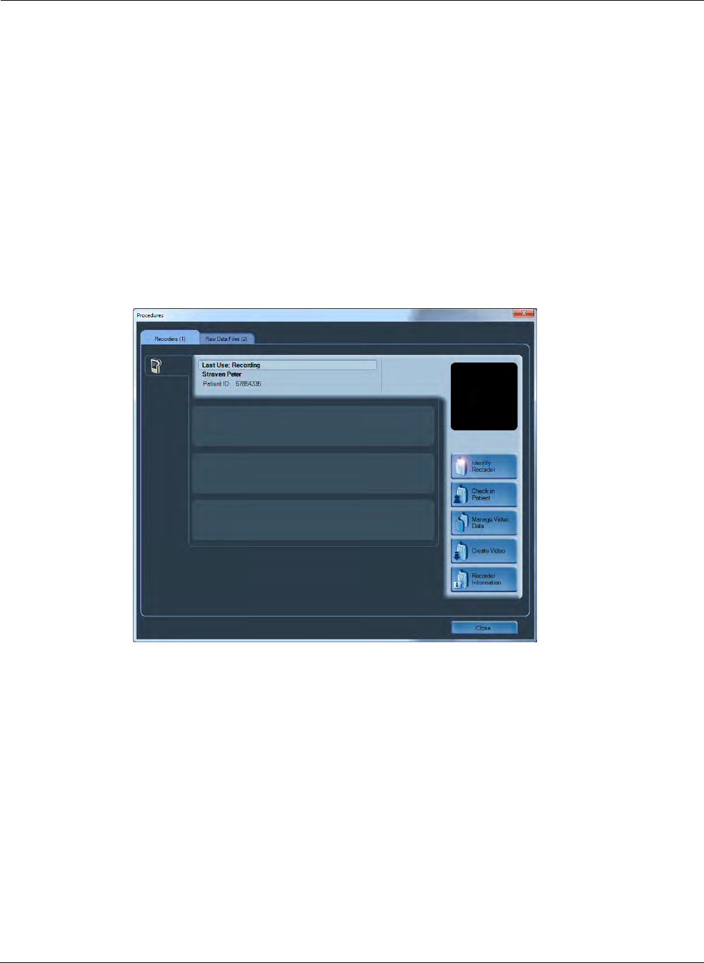
Creating RAPID Videos
Managing RAPID Video Data 93
3. Under Video Data Management, Disable video data management is selected by default. Select
either Copy raw data or Compile raw data.
You can copy the video data from the USB storage device or a DVD onto a workstation for video
creation (see From Raw Data Files/USB Storage Devices on page 92).
Copying Data from a PillCam Recorder
To copy video data from a PillCam recorder:
1. Place the PillCam recorder in the cradle. Make sure that the cradle is connected to the personal
Computer (PC).
2. From the Home screen, click Recorder Download.
The Procedures screen appears with the Recorders tab on top.
3. Click the Recorder bar that corresponds to the PillCam recorder with the desired data:
• When the correct Recorder bar is activated and you click on the Identify Recorder button, the
corresponding PillCam recorder’s LEDs blink.
• The relevant patient information is displayed on the Recorder bar.
• PillCam recorder's last use is displayed.
The Manage Video Data, Create Video, and Check-in Patient buttons become available.

PillCam Capsule Endoscopy
94 Managing RAPID Video Data
4. Click Manage Video Data.
5. Select the location for the copied data.
Click Change if you wish to copy the data to a different location (on the computer, network, or
external device) than the default (E:\datatransfer). To change the default directory, see Video Data
Management Section on page 174.
6. Click Start Copy.
During raw data copy the Recorder bar displays:
• The Compiling raw data or Copying data message, depending on the video settings.
• The status and progress bar.
• The patient name ID.
• The name of the video to be created.
• The battery status.
• The Do not remove recorder message flashes below the battery status until all data is copied.
Once the process is complete, a message appears on the screen notifying you that the data copy is complete.
The PillCam recorder may be disconnected at this point.
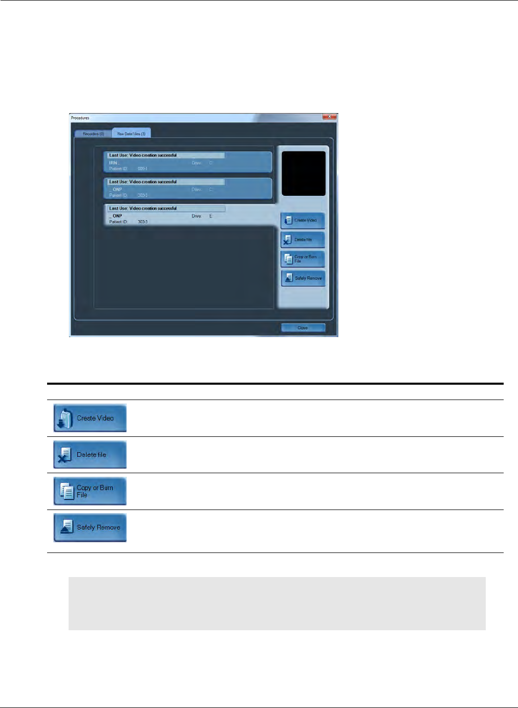
Creating RAPID Videos
Managing RAPID Video Data 95
Managing Data Files
The Raw Data Files screen allows you to manage and monitor all video data files on your PC or
connected USB storage device.
To open the Raw Data Files screen, select Recorder Download from the Home screen. The
Procedures screen appears. Select the Raw Data Files tab.
A list of raw data files appears; their status and location are displayed in the recorder bar.
On the right side, the following buttons appear:
Button Action
Creates a video from the video data file that was copied to the computer.
Deletes the video data file that was copied to the computer.
Copies the video data file to a CD/DVD, to a different folder, or a USB
storage device.
Ejects the USB device once the video data file is copied and deleted from a
USB storage device. This button appears only if raw data files are
connected to the computer via a USB device.
֠
֠֠
֠Note
Label all USB storage devices used for Video Data Copy or for transport of
RAPID videos, stating that they may contain clinical data and should not be
used for other purposes.

PillCam Capsule Endoscopy
96 Managing RAPID Video Data
Batch Data Copy
The RAPID software can perform multiple video data copy for any combination of up to four PillCam
recorder cradles and USB storage devices, and for any number of video data files.
From PillCam Recorders
Place PillCam recorders into the connected cradles. In the Recorders screen, click Manage Video Data
for each one, separately.
From Video Data Files/USB Storage Devices
Connect USB Storage devices. In the Raw Data Files screen, click Copy or Burn File for each one of the
devices or any video data files already on the computer.
Backing up Data
When you have confirmed that the video was created successfully, you may delete the raw data files from
your computer. We recommend this since these raw data files are very large.
Back up the RAPID folders (which contain created videos) by saving them on removable discs (CD/DVD
or USB storage device). After saving them on removable media, you may delete them from the PC's hard
disk if they are not to be reviewed on that PC.
֠
֠֠
֠Note
Make sure that the RAPID software is completely shut down before
removing the PillCam recorder from its cradle.
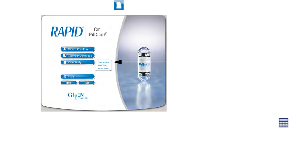
Loading a Study with the Study Manager 97
Chapter 8
Reviewing and Interpreting RAPID Videos
After completing the patient procedure and downloading the procedure data to the computer, the RAPID
software processes and transforms the raw image data from PillCam studies into conveniently viewable
RAPID videos. This section of the user manual describes the RAPID interface and how to open a study,
view the procedure video and then proceed to create a Report.
This chapter focuses on using RAPID to load a RAPID video for viewing, annotating images of interest,
adding comments and markings to the images and finally, creating a summary report.
This chapter guides you through the following main actions:
•Loading a Study with the Study Manager
•Reviewing and Interpreting RAPID Videos
•Creating a PillCam Capsule Endoscopy Report
Loading a Study with the Study Manager
The Study Manager allows you to conveniently access and manage patient studies. The studies are
organized in the Study Manager in a way that enables easy access and management of studies, sorting,
and searching by different criteria such as patient name and procedure dates.
The Study Manager connects to a directory of studies, called an Archive, at different locations, i.e. on the
local computer or on a network, on a removable drive connected to the computer, or on a CD/DVD
containing the studies. These studies are represented in a multi-line format where each line represents a
separate capsule procedure.
To access the Study Manager:
•From the Home screen, click View Study and select Study Manager in the drop-down list.
•From one of the RAPID screens, click File > Study Manager or click the Study Manager icon:
located on the Quick Access Toolbar.
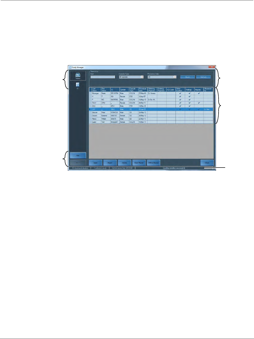
PillCam Capsule Endoscopy
98 Loading a Study with the Study Manager
The image below displays the Study Manager layout, divided into the following sections:
•Archives
•Studies
•Status Bar
•Action buttons
•Search
Archives Search
Studies
Action
buttons
Status bar
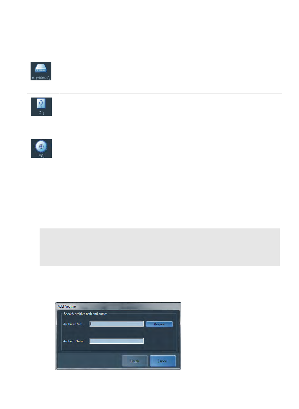
Reviewing and Interpreting RAPID Videos
Loading a Study with the Study Manager 99
Archives
The Archives section displays the current procedure study archives that are accessible in the RAPID
software. The Study Manager can connect to procedure study archives located on the local computer,
on an accessible network, or a removable storage media such as a flash drive or a CD/DVD. The
different archive icons are explained below:
Adding an Archive
The instructions below demonstrate how to add an archive containing procedure studies that can be
accessed from the RAPID software. Portable drives connected to the computer's USB and studies
contained on a CD/DVD are detected and added automatically. The archives on the Study Manager can
be renamed or deleted but this will not affect the source directory of the archive.
To add an archive:
1. Click the Add button at the bottom left of the Study Manager screen. The Add Archive screen
appears.
2. Enter the archive path and the archive name that will appear below the archive.
• To browse for the archive on the local computer or on the network, click the Browse button.
An archive on a fixed drive, such as the computer's hard disk or a drive on the
network. The default archive is the one selected in Settings > Video > Video
directory. The archive is added and defined by the user using the Add button. This
is explained below in Adding an Archive on page 99.
A portable drive containing procedure studies connected to the computer's USB.
Portable drives connected to the USB are automatically detected and cannot be
user defined.
Using the mouse, navigate to the USB connected device in My Computer, right
click and select Properties. Type the desired volume name and click OK.
Procedure studies stored on a CD/DVD in the computer's DVD bay. CD/DVD
archives are automatically detected and cannot be user defined.
֠
֠֠
֠Note
Adding an archive provides a visual link to the directory using the Study
Manager. It does not copy the data locally to your computer. Removing an
archive using the Delete button does not delete data from the directory, but
only the path to the directory.

PillCam Capsule Endoscopy
100 Loading a Study with the Study Manager
• The Browse For Folder screen appears. Navigate to the desired archive and click OK.
3. Click Finish.
When the Study Manager scans the archive, the archive icon will display Scanning… below the
archive.
The status bar will display Loading studies… until all studies are loaded and the status bar will
display Loading studies successful.
Archive Options
When right-clicking on a selected archive in the Study Manager the following options are available:
•Remove: Remove the archive path from the Study Manager. To remove the CD/DVD from the disk
drive or any portable drive from the USB connection click the Eject button.
•Rename: Rename the selected archive.
•Move Up: Move the selected archive up on the archive list display.
•Move Down: Move the selected archive down on the archive list display.
Studies
The study lines displayed are links to the studies located in the selected archive. Each line represents a link
to the study.
֠
֠֠
֠Note
During archive scanning, studies that already appear in the Study Manager
screen can be opened.
֠
֠֠
֠Note
After upgrading your software, click the Refresh button when connecting to
any archive for the first time. This may take time.
֠
֠֠
֠Note
DO NOT remove the USB storage device until after any actions performed
with it are completed.
֠
֠֠
֠Note
In some cases, especially when working with network archives, you may not
immediately see the updated Study file in the Study Manager, after making
changes to the file. In this event, click the Refresh button in the top right
corner of the Study Manager to make sure the most recent data is shown.
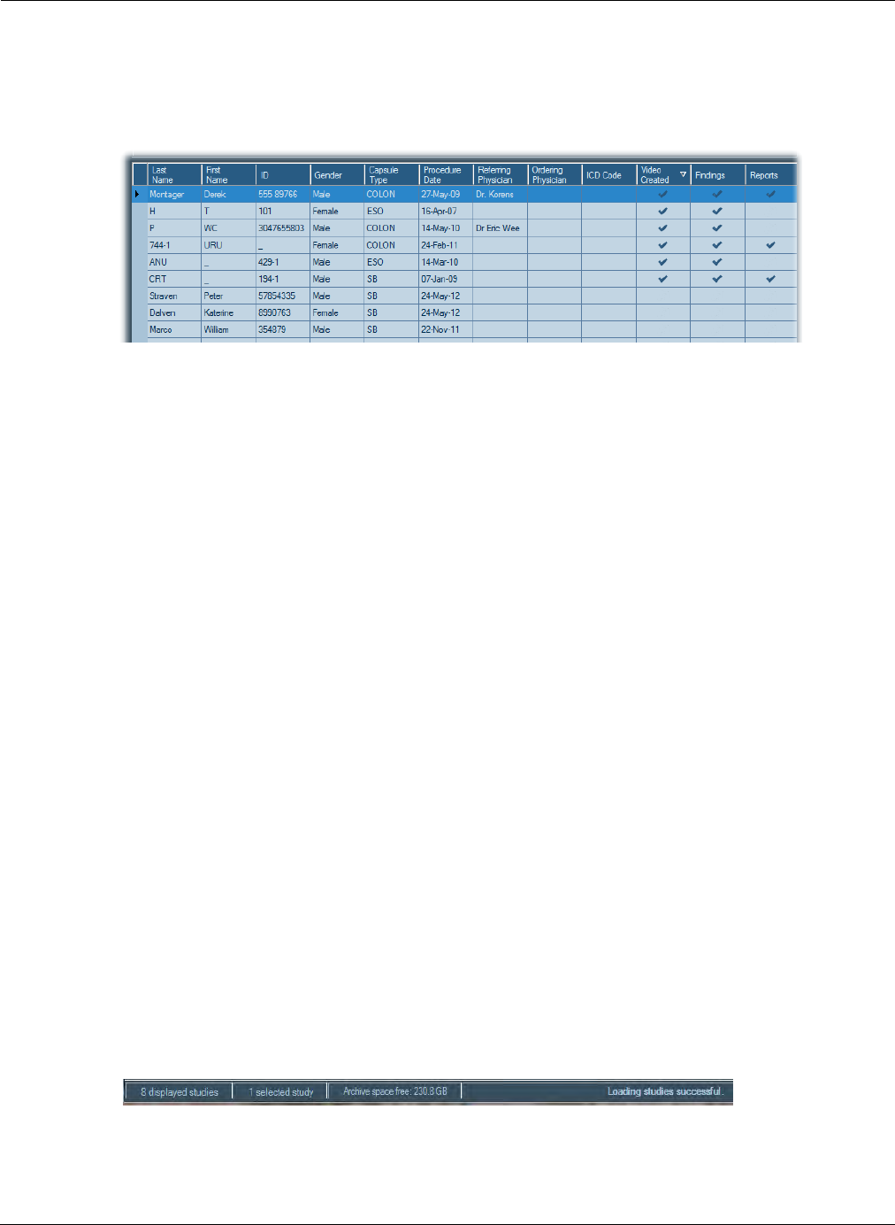
Reviewing and Interpreting RAPID Videos
Loading a Study with the Study Manager 101
Study Columns
By default, the studies are sorted by the last modified study, the Video Created column. The column
that is sorting is represented by an arrow. In the example below the studies are sorted by Video
Created, the date that the video was created.
The studies are identified by several parameters that define the study, such as patient name, capsule
procedure type, and referring physician. Additionally, a check mark in the following columns indicates
the availability of the following:
•Video Created: Study includes a video of the procedure.
•Findings: Study includes a findings file created for this procedure.
•Reports: Study includes a report of the procedure.
Study Options
When right-clicking on a selected study in the Study Manager, the following options are available:
•Manage Columns enables the adding, deleting and creating of Study Manager columns (see
Managing Columns on page 104).
•Update Patient Details changes or updates all the patient and procedure information even after a
video is created. Click Save when finished.
•Print Regimen opens a print preview layout of the Regimen for this procedure (see Print Layout of
the Post-Capsule Ingestion Instructions on page 190).
•Open Video Only opens the procedure video without the findings. If any findings are added during
review, when trying to save you will be prompted to overwrite any existing findings file (see Open
on page 107).
•Export Table Information saves the selected archive data to a static line and column format Excel
file displaying all the studies for that archive.
•Show all search results displays the screen of results of the search performed (see Search
Function on page 103).
Understanding the Status Bar
A status bar at the bottom of the screen shows the number of studies displayed, the number of studies
selected, the amount of free space on the selected archive, and loading status of the studies.
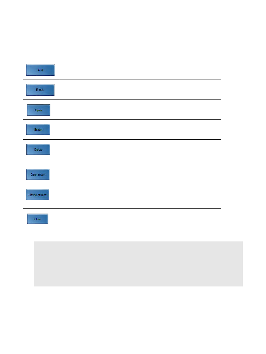
PillCam Capsule Endoscopy
102 Loading a Study with the Study Manager
Action Buttons
The action buttons located on the bottom of the Study Manager screen enable several commands like
opening a study in RAPID, deleting studies, and export. To perform an action on a study, select the study by
clicking the row containing the relevant study and click one of the following action buttons:
Button Action
Adds an archive to the Study Manager screen (see Adding an
Archive on page 99).
Ejects a CD/DVD from CD drive and ejects any flash drive connected
to USB. This will remove the archive from the Study Manager.
Opens the video and findings of the selected study in the RAPID
software (see Open on page 107).
Exports the selected study or specific files of the study to a CD/DVD
or another archive (see Export on page 107).
Deletes the selected study (see Delete on page 111). Deleting a
study in the Study Manager deletes all data including procedure
videos from the source directory.
Opens the report of the selected study (see Open on page 107).
Opens Offline study archive which displays the list of the studies
which were stored on a removable storage media but are now not
accessible (see Offline Studies on page 112).
Closes Study Manager screen. This can also be performed by
clicking the Escape button on the computer's keyboard.
֠
֠֠
֠Note
For Delete and Export commands you can select multiple studies to perform
the command. Click the row of the first study, press and hold SHIFT on your
keyboard and select the last study you wish to select. If not all the studies are
in rows next to each other, press and hold CTRL on your keyboard and select
the studies one by one. You can then either click Delete or Export. The
number of selected studies is displayed in the Status bar.
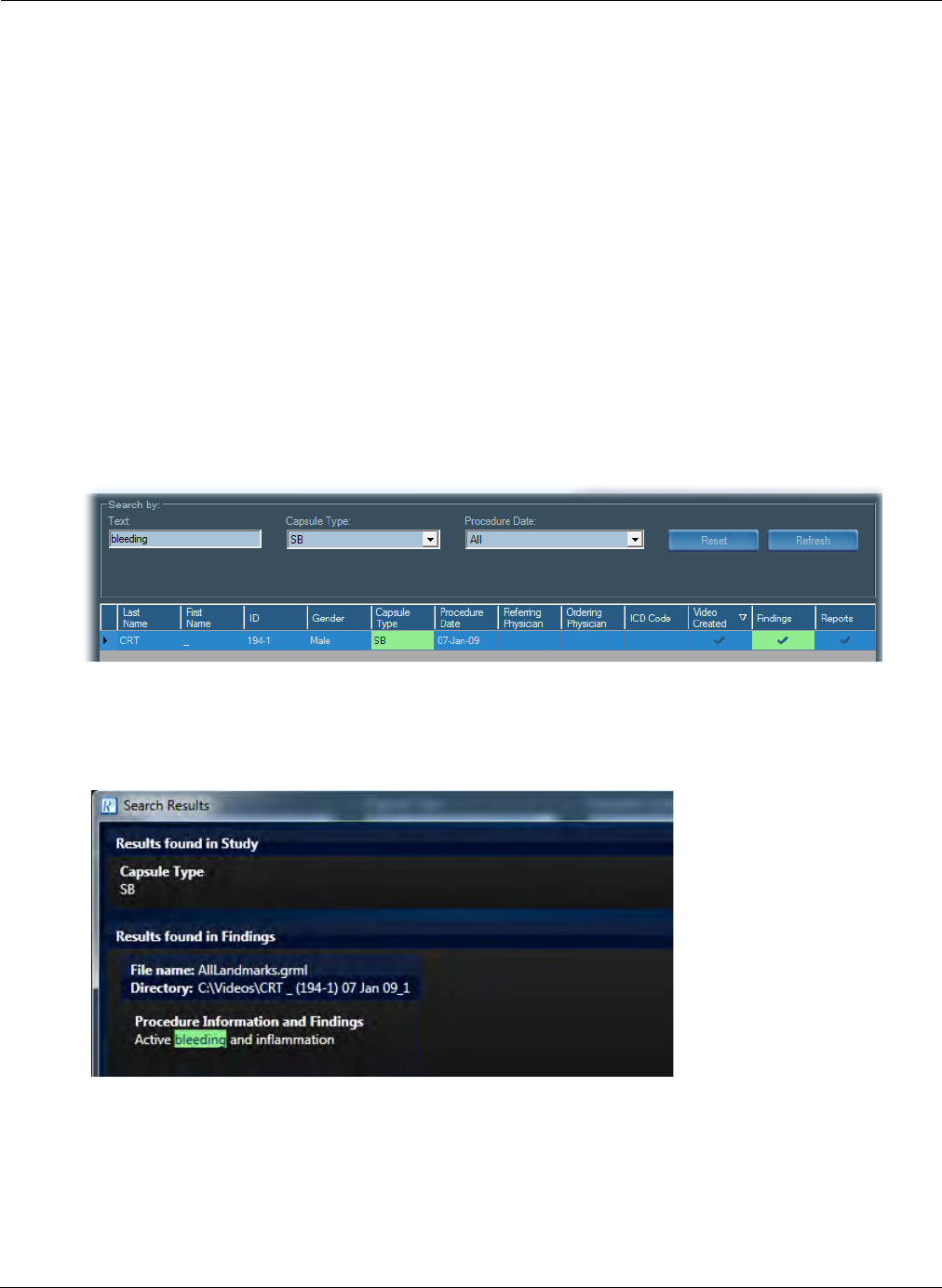
Reviewing and Interpreting RAPID Videos
Loading a Study with the Study Manager 103
Search Function
At the top of the Study Manager screen, the following search and filter functions enable to search for a
study in a selected archive:
•Text: Search by entering part of the name or the entire name of the patient, findings, procedure, or
other free text.
•Capsule Type: Filter the search by selecting from the drop-down list the type of capsule
procedure.
•Procedure Date: Filter the search by selecting from the drop-down list the procedures performed
in a specific time window (in the last days or weeks or even by a specific date).
After entering one or more search criteria the Study Manager will search and display only relevant
studies. The criteria that were searched will be highlighted in green.
In the example below, the following search criteria was entered:
•Text: bleeding
•Capsule Type: SB
•Procedure Date: All
All SB capsule procedures were displayed and highlighted. Additionally, one of the procedures displays
a highlighting on the Findings. Right-clicking the study and selecting Show all search results displays
additional information regarding the text searched bleeding and where it is located. In this example, the
bleeding text was located in the Findings, under the Reason for Referral.
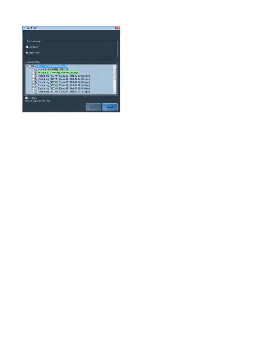
PillCam Capsule Endoscopy
104 Loading a Study with the Study Manager
Additionally, if opening, deleting, or exporting the study, the finding will remain highlighted to allow for its
selection as shown below:
To clear the search fields and again see all studies in the selected archive, click Reset.
To refresh the search view click Refresh to ensure that the latest data is displayed in the Study Manager.
Managing Columns
You can determine what information is displayed about each study. By default, Study Manager displays
the following columns containing information entered during the Check-in process:
These columns are mandatory and cannot be changed or removed. There are also columns that you can
optionally add by selecting from a list of additional check-in information fields. You can also add and name
a new column (User Column).
To add or remove columns:
1. Right-click anywhere in the area where the studies are displayed and select Manage Columns. The
Manage Columns screen appears. On the right is the list of currently Displayed Columns in the
• Last Name • Referring Physician
• First Name • Ordering Physician
• ID • ICD Code
• Gender • Video Created
• Capsule Type • Findings
• Procedure Date • Report
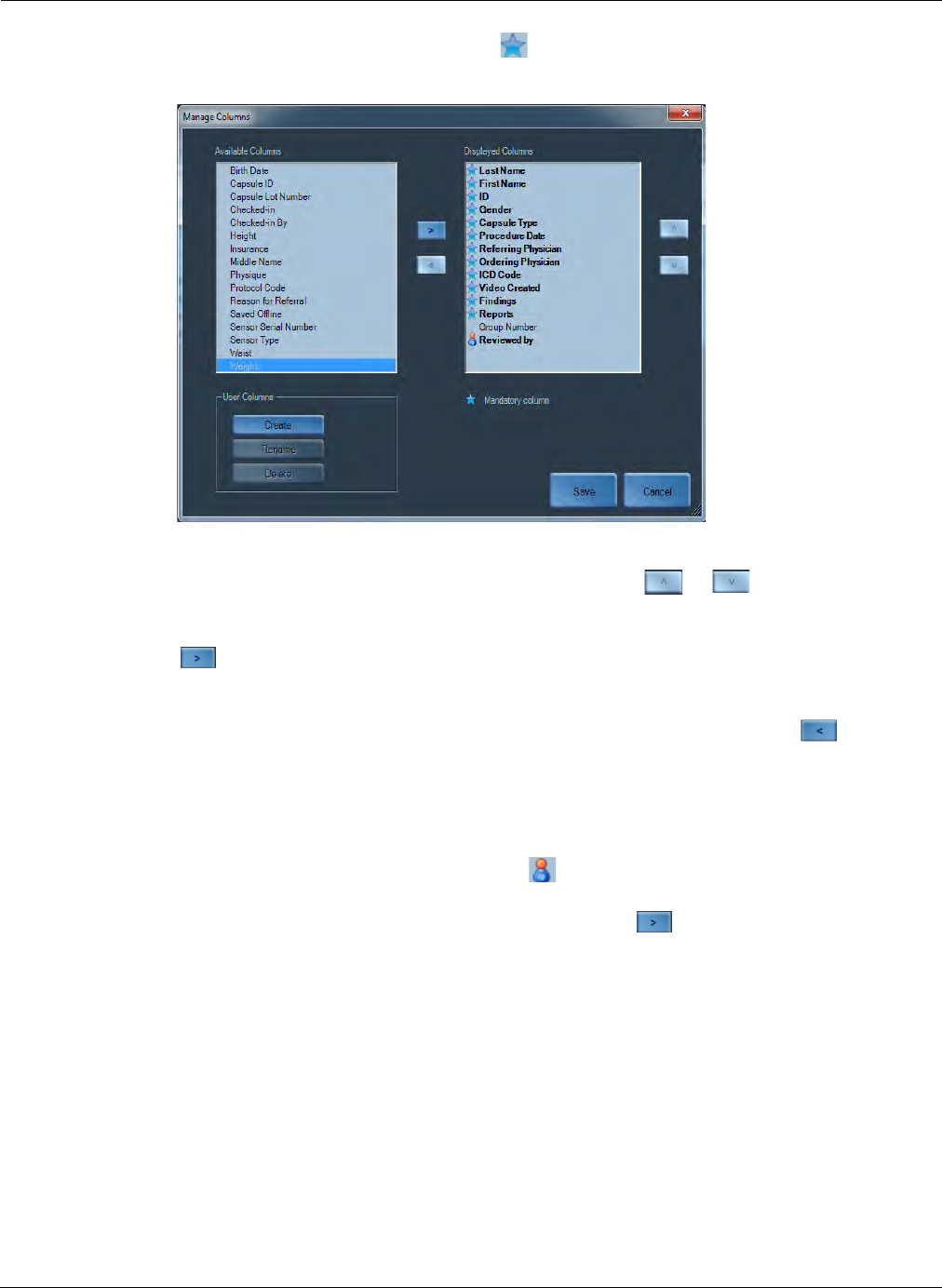
Reviewing and Interpreting RAPID Videos
Loading a Study with the Study Manager 105
Study Manager. The columns with a star icon next to them are fixed and cannot be removed or
renamed. The left is a list of available columns to choose from.
2. To change the left to right order of the displayed columns in the Study Manager screen, select the
column you wish to move in the window on the right and click or .
3. Select any column names that are listed in Available Columns on the left side and click the right
arrow to display them in the Study Manager screen. They will be added to the end of the list
in the window on the right. The position in the list can also be changed.
4. Select any columns that are listed in Displayed Columns and click the left arrow to remove
them from the Study Manager screen.
5. To create a new column, click Create.
6. Type in the name of your new column and click Save. The new column will appear in the
Available Columns list with a user column icon next to it.
7. To display this new column, select it and click the right arrow . It will appear at the bottom of
the Displayed Columns list.
8. To change the name of your column, select it and click Rename.
9. To remove the column from the Study Manager, select it and click Delete.
10. In the Study Manager, the User Column will require manual input of the data. To enter data in your
column, first select the relevant study. Click in the cell of the study in your column. Type in your
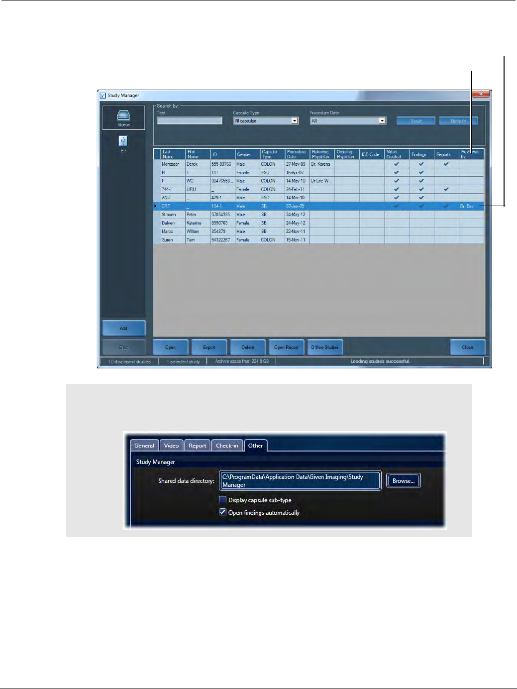
PillCam Capsule Endoscopy
106 Loading a Study with the Study Manager
text in the separate window that opens. When you are done, click OK. In the example below, a user
column named Reviewed by was created.
֠
֠֠
֠Note
Created user columns can be available to other users. Make sure the users
have the same path defined in Shared data directory in the Settings.
Reviewed by column
Manually entered data
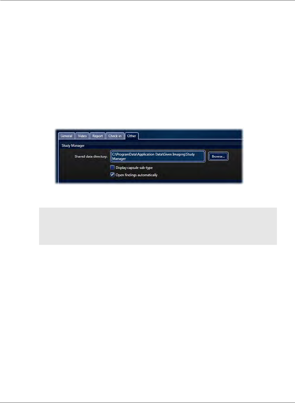
Reviewing and Interpreting RAPID Videos
Using the Study Manager 107
Using the Study Manager
Open
You can open videos and other files in the study from the Study Manager.
To open:
•Study (both the video and the findings): Either double-click the line representing the study or click
the study and then click the Open button at the bottom of the screen. If more than one findings file
is associated with the selected video, a second screen opens prompting you to select the related
findings file you wish to open.
•Video only: Right-click anywhere in the Study Manager screen and select Open Video Only. The
video of the selected study opens. The Settings screen enables further options for the Study
Manager, such as opening findings automatically.
•Report: Click Open Report at the bottom of the screen. The report associated with the selected
study opens as a PDF file.
Export
The Export function allows you to save a study or one of its files in a different place or under a
different name, to reorganize your archives. This includes:
•Burning a study or a file to a CD/DVD
•Saving a study or a file to another archive, such as a USB storage device
•Saving a study or a file to a specific location
•Saving a study as Zip file
֠
֠֠
֠Note
When you open a RAPID video while another video is still open, the RAPID
software automatically closes the previously opened RAPID video. If you
made changes to the findings of the open video, you are asked to save
changes before closing the video.

PillCam Capsule Endoscopy
108 Using the Study Manager
Burning a Study to a CD/DVD
The Study Manager allows you to burn a study or specific files of a study to a disc, with the following
restrictions:
•No multi session disc burning: once a disc is burned, you can not add any other data to it.
•You can burn either the whole study (all the files in the folder) or one single file.
For advanced options, see CD/DVD Burning on page 197.
To save a study to a disc:
1. Insert a blank CD/DVD in the drive.
2. Open the Study Manager by clicking at the top of the screen, or select View Study > Study
Manager from the Home screen.
3. Select a study from the list on the Study Manager to export to CD/DVD.
4. Click Export. The following screen appears.
5. Select either Entire study or Specific files. If you select Specific files, a window opens inside the
screen showing the files for selection.
If you wish to remove all personal patient information, select the check box De-identify at the bottom
of the screen.
The size of the file is displayed below the check box.
֠
֠֠
֠Note
Exporting does not move the study files but creates a copy of them to the
desired location.
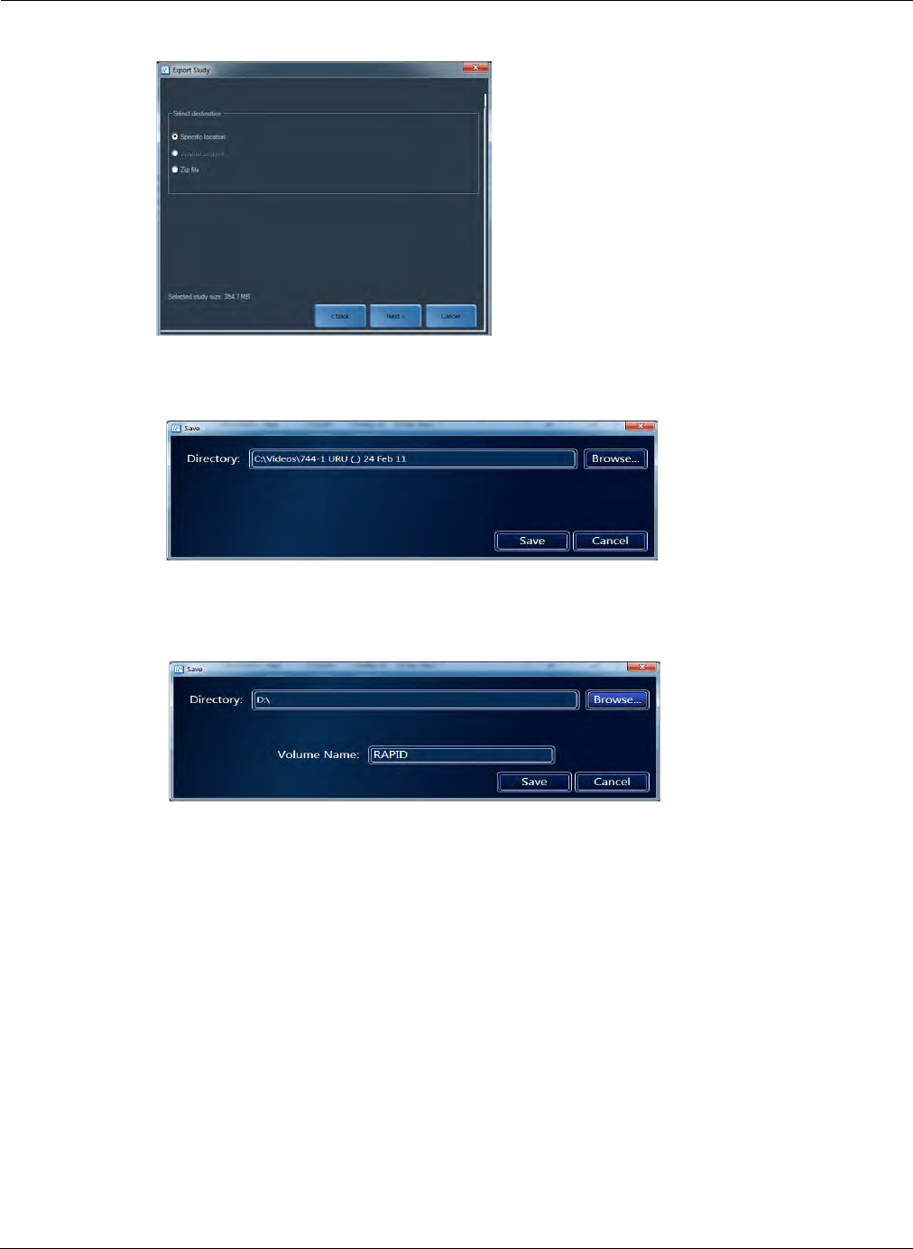
Reviewing and Interpreting RAPID Videos
Using the Study Manager 109
6. Click Next.
7. Select Specific location under Select destination and click Next.
The following screen appears.
8. Browse for the CD/DVD drive on your computer.
9. Type in a volume name for your disc.
10. Click Save. A screen appears showing the progress bar and Exporting study.
When the progress bar is full, the last screen appears with the message File export successful.
11. Click OK. RAPID will eject the CD or DVD when the burning operation is complete.
12. Write the volume name on the disc.
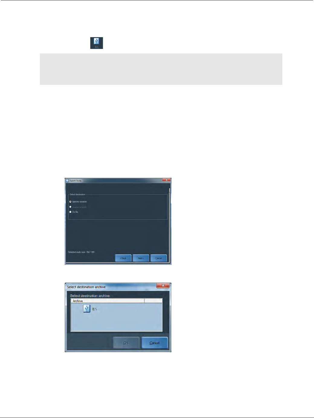
PillCam Capsule Endoscopy
110 Using the Study Manager
Saving a Study to Another Archive
The Study Manager allows you to save a study to another archive, such as a USB storage device. When
you connect a USB storage device to one of the USB ports of the computer, the Study Manager treats it as
an archive and the icon appears on the left side of the screen.
1. Select the relevant study and click Export.
The Export Study screen appears.
2. Select either Entire study or Specific files under Select data to export. If you select Specific files, a
window opens inside the screen showing the files you can select.
3. If you wish to de-identify the video in the study, select the check box De-identify at the bottom of the
screen. Any reports created for this study also will be included in this export so make sure that
sensitive data is already de-identified if needed (see Report Ribbon on page 147).
4. Click Next.
5. Select Another archive under Select destination and click Next.
6. Click the relevant USB icon and click OK.
7. A screen appears showing the progress bar and Exporting study.
8. When the progress bar is full, the last screen appears with the message File export successful.
Click OK.
֠
֠֠
֠Note
USB storage devices used for Raw Data Copy or for transport of RAPID
videos should be labeled as such, stating that it may contain clinical data
and should not be used for other purposes.

Reviewing and Interpreting RAPID Videos
Using the Study Manager 111
Only after saving data on a USB storage device is complete, take special care to correctly unplug the
device. Improper removal of a USB device may cause loss of data and computer instability.
To safely remove a USB storage device:
1. Click the icon on the left side of the screen to select the USB storage device.
2. Click Eject.
3. When the message that you can safely remove the hardware appears, unplug the USB storage
device.
Saving a Study as a Zip File
The Zip function of Study Manager uses WinZip to zip the files, a measure that ensures all files in a
RAPID study stay together for convenient handling such as sending by email.
To zip files:
1. Select the relevant study and click Export.
2. Select either Entire study or Specific files under Select data to export. If you select Specific
files, a window opens inside the screen showing the files for selection.
3. If you wish to de-identify the video in the study, select the check box De-identify at the bottom of
the screen. The report will not be de-identified.
4. Click Next.
5. Select Zip file under Select destination and click Next.
The Save screen appears.
6. If you would like to save to another location click Browse and navigate to the desired location and
click Save.
7. A screen appears showing the progress bar and Saving zip file.
8. When the progress bar is full, the last screen appears with the message Zip study successful.
Click Close.
Delete
The delete function allows you to remove studies entirely or partially from archives.
To delete a study:
1. Select the relevant study and click Delete.
The Delete Files screen appears.
2. If you wish to delete the entire study, select the check box Delete entire study at the top.
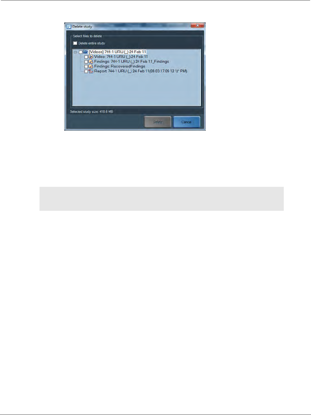
PillCam Capsule Endoscopy
112 Using the Study Manager
3. If you wish to delete specific files, select them and click Delete.
To delete multiple studies:
1. Select the relevant studies by clicking on one and holding the Control button on the keyboard and
selecting another study.
2. Once the studies to be deleted are selected, click the Delete button.
An attention screen appears for confirmation of the deletion.
Offline Studies
The Offline studies screen displays information about studies stored on removable storage media, such as
CDs, DVDs, and some USB storage devices, that have been connected to the computer during use of
RAPID and are currently disconnected from the computer. It allows you to browse for studies in archives
which are currently not connected to the computer.
The offline study list is updated when the removable storage media is disconnected or reconnected. A
reconnected study no longer appears in the Offline Studies list. Once reconnected, the removable media
containing the study is displayed in the left side of the Study Manager screen.
Preexisting studies will appear in the Offline Studies list only after they have been connected and then
disconnected from the computer during use of RAPID.
֠
֠֠
֠Note
Deleting a study in the Study Manager deletes all data including procedure
videos from the source directory. This cannot be undone.

Reviewing and Interpreting RAPID Videos
Using the Study Manager 113
Offline studies information includes the Volume Name and Volume Type or kind of media. Volume
names for CDs and DVDs are allocated at the time the disc is burned. For USB devices, a unique name
(rather than the name designated by the manufacturer) must be allocated before use.
In order to be able to locate a desired study, be sure to mark the disc or other media with its volume
name.
To access an offline study:
1. Find the study in the Offline Studies list.
2. Locate the volume listed.
3. Connect it to the computer.
To save the Offline Studies list, us the Backup/Restore feature for Offline Studies (see Backup/Restore
Offline Studies on page 193).
֠
֠֠
֠Note
DO NOT remove the USB storage device until after any actions performed
with it are completed.
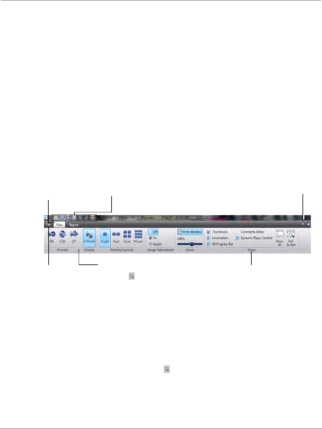
PillCam Capsule Endoscopy
114 Overview of the RAPID Interface
Overview of the RAPID Interface
In the RAPID v8.3 software, a wide band spans the top of the main program window. This ribbon is
comprised of three tabs. Each tab on the ribbon has different buttons and commands that are organized into
ribbon groups. This ribbon replaces the menus and toolbars in previous RAPID versions.
When opening a RAPID video the RAPID user interface displays the View tab. This tab contains many of
the most frequently used commands for viewing a RAPID procedure video.
The ribbon interface is designed to help you quickly find the commands that you need to complete a task.
Commands are organized in logical groups, which are collected together under the View and Report tabs.
When working with study videos, the controls you need for viewing, annotating, and creating reports are
arranged in ribbons, which group similar functions together.
There are three basic components to the ribbon:
•Ribbon Tabs: There are three ribbon tabs, File, View and Report that span across the top. Each one
represents an activity area.
•Ribbon Groups: Each tab has several groups that show related items together.
•Commands: A command is simply a button, a box to enter information, or a menu.
The RAPID interface enables multiple ways to access functions:
•by clicking a button,
•by right-clicking to select an option,
•by using the menu options.
Dialog Box Launchers
Some ribbon groups have a small diagonal arrow in the lower-right corner that when clicked will
display certain options that are not otherwise visible. In the RAPID interface, there are three screen
launchers to show additional options:
•in the View screen, in the Preview ribbon group,
•in the Report screen, in the Configure ribbon group,
•in the Report screen, in the Image Data ribbon group.
Quick Access Toolbar: These
commands are available in all
screen and ribbon views.
Hide the Ribbon
Need more space on your screen?
Click this icon to hide or show the
ribbon.
Ribbon Tabs
Click either tab on the
ribbon to display its
buttons and commands.
Ribbon Groups
Each ribbon tab contains
groups, and each group contains
a set of related commands.
File View
Click the File tab
to enter File view,
where you can
open and save
your RAPID files.
Dialog Box Launchers
If you see this icon next to any
ribbon group label, click it to open a
screen with more options for that
group.
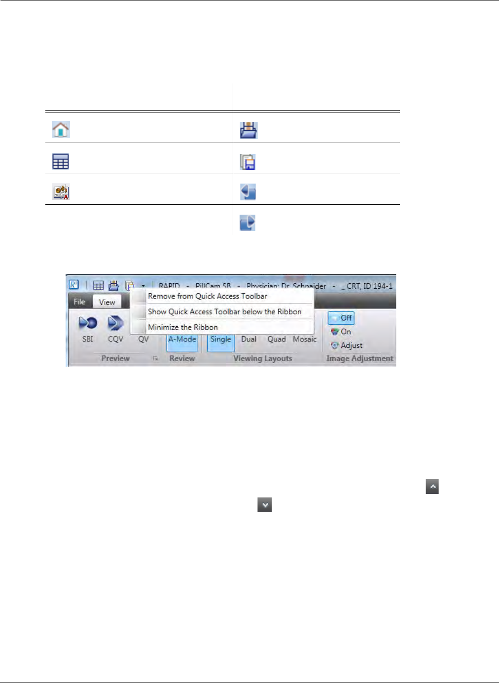
Reviewing and Interpreting RAPID Videos
Overview of the RAPID Interface 115
Quick Access Toolbar
The Quick Access Toolbar is the small area to the upper left of the ribbon that contains a set of
frequently used commands.
By default, the following common commands are displayed in the Quick Access Toolbar.
To remove the command shortcut from the toolbar, right-click the command and select Remove from
Quick Access Toolbar.
When right-clicking the Quick Access Toolbar, there are two other options to select from:
•Show Quick Access Toolbar below the Ribbon: Moves the toolbar below the ribbon. To return
the toolbar above the ribbon, right-click the toolbar again and select Show Quick Access Toolbar
above the Ribbon.
•Minimize the Ribbon: Hides the ribbon temporarily but displays the ribbon tab. To temporarily
show the ribbon click once on the desired tab. Clicking anywhere on the screen will hide the ribbon
again. To restore the ribbon, click on the File, View or Report tabs or right-click the Quick Access
Toolbar and uncheck Minimize the Ribbon. Optionally, click the minimize button on the top
right of part of the ribbon and click it again to restore.
Button Action Button Action
Closes the current study and
returns to the Home screen.
Browses to open a new
video.
Opens the Study Manager. Saves the Findings.
Opens the Atlas. Goes to the previous
image.
Goes to the next image.
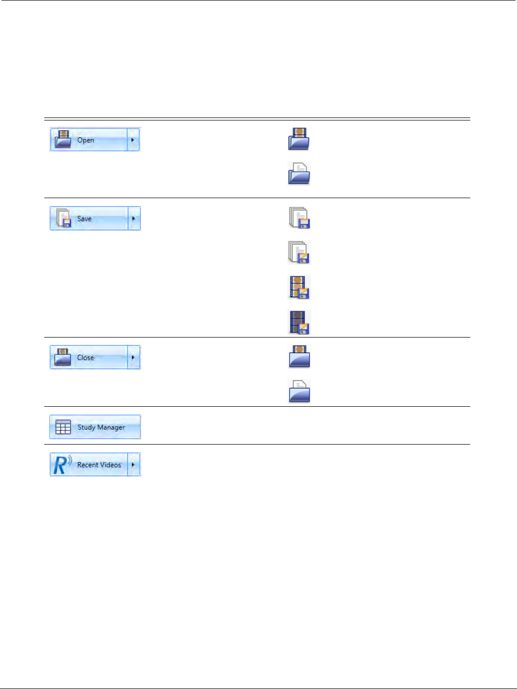
PillCam Capsule Endoscopy
116 Overview of the RAPID Interface
File View
The File menu contains several commands that enable you to open a video or findings, save a video,
connect to the Study Manager, or connect to the RAPID Help.
The table below displays the File menu commands. You can click on the command, or if available a
secondary command appears when hovering the mouse over a command.
Button Menu command Secondary commands
Clicking the Open button
launches the Windows
screen to browse and open
a video. Optionally, hover
over the button to display
secondary commands.
Open Video from selected folder
Open Findings related to the
currently open study
Click to save video or
hover over the button to
display secondary
commands.
Make sure there are no
spaces at the beginning or
end of the file name. If
spaces are included, the
Save option will be grayed
out.
Save Findings
Save Findings As
Save Video As
Save De-Identified Video As
Close video or findings.
Close Video
Close Findings
Click to open the Study
Manager.
Click to load recently
viewed video or hover over
the button to display
recently viewed videos to
choose from.
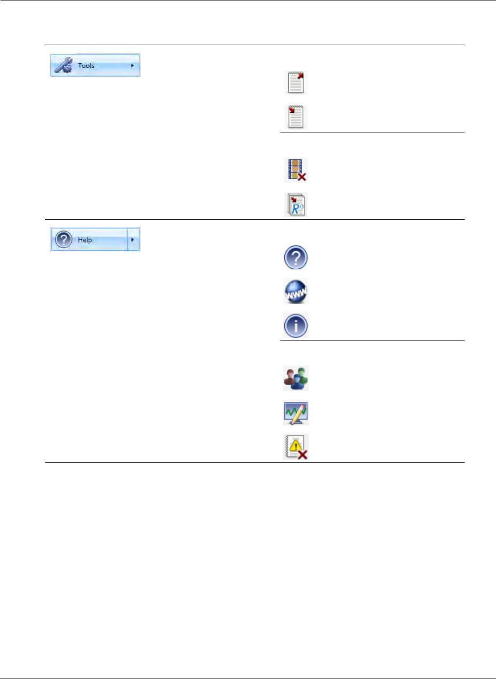
Reviewing and Interpreting RAPID Videos
Overview of the RAPID Interface 117
View Screen
The View screen is used for viewing videos created from PillCam capsule endoscopy procedure. It is
accessed by clicking the View tab.
The View tab in the RAPID software allows for viewing the RAPID video, marking and annotating
thumbnails of interest, and adding any comments to the study.
The View screen enables the following functions to review the patient study:
•Preview and review the patient procedure video using the RAPID video player.
•Use viewing layouts and image adjustment features to enhance or to focus on the video image.
•Annotate thumbnails for later viewing and commenting.
•Designate thumbnails as anatomical landmarks GI tract.
•Use the Localization feature to show the localization track and display the gastric passage times.
Hover over the button to
display secondary
commands.
User Dictionary
Export
Import
Additional Tools
Delete Videos
Import RAPID 4 Reports
Click the Help button to
load the RAPID Online
Help. Hover over the
button to display
secondary commands.
Help
Online Help
Help Center
About
Customer Support
Contact Us
Collect Analysis Files
RAPID Log
Button Menu command Secondary commands
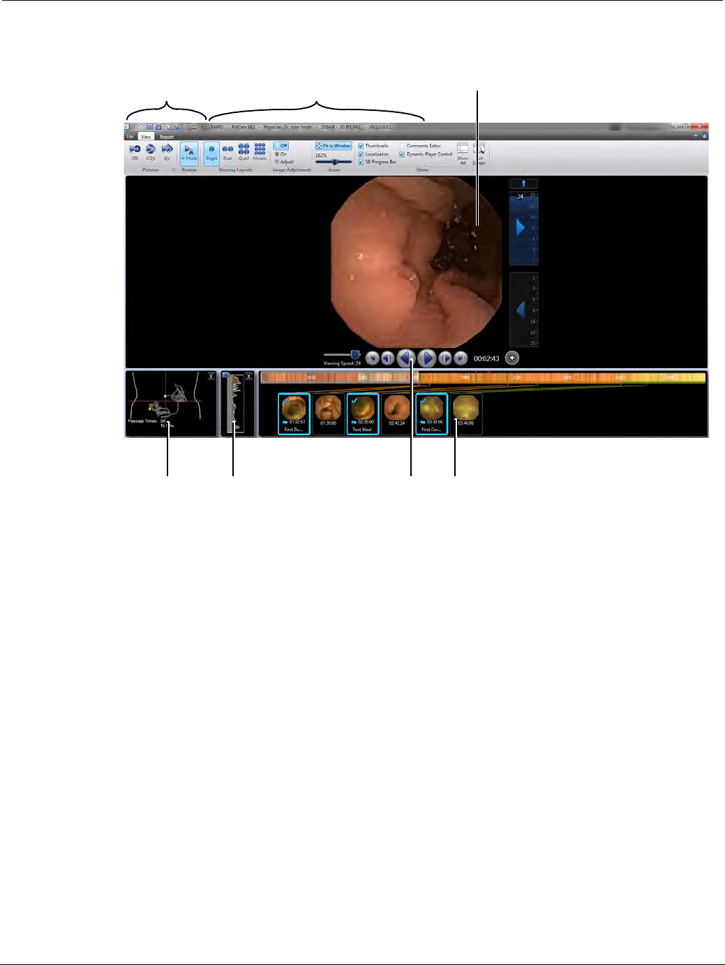
PillCam Capsule Endoscopy
118 Overview of the RAPID Interface
•Display Progress Indicator to show viewing progress of the small bowel
View Ribbon
The View ribbon is divided into several groups enabling many different functions. The buttons and
commands are organized in the following logical groups:
•Preview/Review
•Viewing Layouts
•Image Adjustment
•Zoom
•Show
Preview/Review Groups
Below is a description of the Preview/Review group buttons and the commands that can be performed for
viewing the capsule endoscopy video. This group of buttons enable to preview the video and then to
review and read the complete video using the Viewing Layouts described in the next buttons grouping
below. For a complete explanation on the use of the features described, refer to the cross-reference
provided, if available.
Information about the
open study
Quick Access
Toolbar
Video image
Video play
buttons
Localization Progress indicator Thumbnails
Reviewing and Interpreting RAPID Videos
Overview of the RAPID Interface 119
Preview Group buttons
Button Function
Collage view for COLON 2 procedures only: In the Collage view
relevant parts from selected images of interest are cropped and arranged in a
matrix to provide an overview of the most interesting image elements from the
study video. This layout contains 8 rows and the maximum number of the
columns that can fit on the display (dependent on resolution). Use the
navigation buttons or the scroll wheel on the mouse to view the next matrix.
When hovering on an image in the collage, the complete image with both the
previous and next images will be displayed on the left side of the screen.Click
an image to select that image and view the enlarged image on the left, without
hovering. A blue border appears around the selected image. To return to
hovering mode, click the image with the blue border again.
The Collage View must be enabled in the Settings (see Research Tools
Section on page 175).
Collage view is not intended for diagnostic review and should not
be used as a substitute for reviewing the video in one of the other
viewing layouts.
SBI mode: Sets the Suspected Blood Indicator (SBI) previewing mode to
show marked images suspected of containing blood for fast review in
sequence. SBI image locations are marked on the color bar (see SBI View on
page 129).
QuickView mode: Sets the QuickView previewing mode to show only
significant images that may be of interest in the video stream to provide fast
previewing and landmarking of a video. The number of images to be
considered images of interest can be set as percentage of the video by the
user. This provides for fast previewing and landmarking of a small bowel video.
The RAPID scans all images and scores them according to the possible level
of significance. Then according to the percentage level set by the user it
displays a short video to provide an overview of the case prior to full review
(see QuickView on page 125).
The QuickView feature is not intended to detect pathology in lieu
of a physician and should not be used as a substitute for
reviewing the entire video.
Complementary QuickView mode: Sets the Complementary QuickView
previewing mode to show the complementary part of the video (that is,
everything that was not included in QuickView) (see Complementary
QuickView on page 126). Only a review of both QuickView and
Complementary QuickView is equivalent to a full video read.
The Complementary QuickView feature is not intended to detect
pathology in lieu of a physician and should not be used as a
substitute for reviewing the entire video. Review of both
QuickView and Complementary QuickView is required for full
video review.
PillCam Capsule Endoscopy
120 Overview of the RAPID Interface
Review Group Buttons
Viewing Layout Group
This ribbon group contains tools for customizing the video display from one video head: Single, Dual,
Quad, and Mosaic view. The tools maximize reading efficiency by simultaneously displaying consecutive
images in one easily viewed image.
Button Function
Standard Review mode: This button sets the standard review mode for
PillCam SB 3 (AFR), PillCam COLON 2, PillCam ESO 2, and PillCam
ESO 3/UGI procedure videos.
Display rate can be set by the user by moving the viewing speed slider.
A-mode Review mode: This button sets the review mode to show the A-
mode (Automatic) video created for all PillCam SB 2 as well as for all
PillCam SB 3 with PillCam recorder DR2 procedure videos.
M-mode Review mode: This button sets the review mode to show the M-
mode (Manual) video created sequentially from all captured images for
PillCam SB 2 as well as for all PillCam SB 3 with PillCam recorder DR2
procedure videos if creation of M-mode video is enabled in Settings.
By default, creation of manual mode video is disabled during download.
To enable manual mode, see Video Creation Section on page 174.
֠
֠֠
֠Note
Dual and Quad views are available only when viewing the video from one
video head (that is, from one end of the capsule).
Button Function
Single view: Shows the video in Single view, one single frame on the
screen.
Dual view: Shows the video in Dual view, which displays two
consecutive frames side by side at the same time. Frames 1 and 2,
then frames 3 and 4, then frames 5 and 6, etc. This is different than
dual-head capsule videos that display a single frame from each of the
two capsule heads simultaneously side by side.
Quad view: Shows the video in Quad View, which displays four
consecutive frames. The order of the images is presented in a
clockwise fashion.
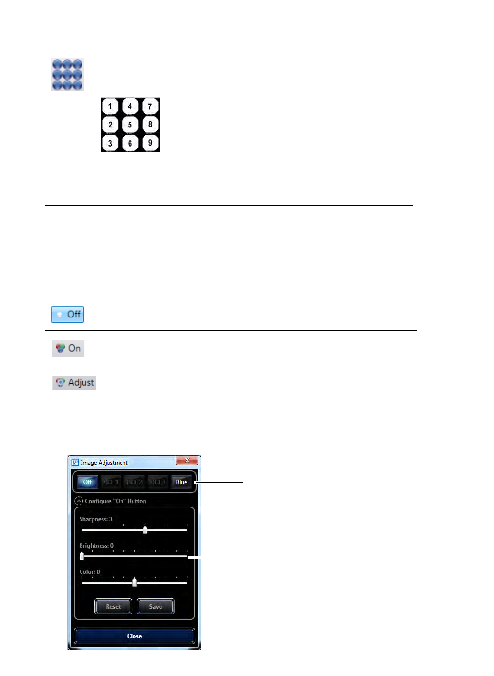
Reviewing and Interpreting RAPID Videos
Overview of the RAPID Interface 121
Image Adjustment Group
This ribbon group contains the controls for preset or custom adjustment of the displayed video image.
This includes controls adjusting for sharpness, brightness, color, and FICE or Blue-processed image.
Image Adjustment provides an alternative image view via a single click using proprietary technology
with enhanced sharpness and contrast. Sharpness, Color, and Brightness controls enable customizing
of the image to user preferences. When clicking the Adjust button the following screen appears:
Mosaic view: Shows the video in Mosaic view, which displays 18
consecutive frames on the screen. The order of the images is from top
to bottom for each column, moving from left to right.
The time indicator shows the time for the top left image in the display.
Mosaic view is not intended for diagnostic review and
should not be used as a substitute for reviewing the video in
one of the other layouts.
Button Function
Turns Image Adjustment off.
Turns Image Adjustment on.
Opens the Image Adjustment screen.
Button Function
Click on the buttons labeled
FICE or Blue for pre-set image
enhancements.
Use the sliders to adjust the
sharpness, brightness and
color of the image.
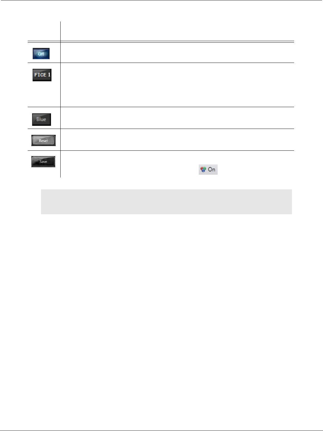
PillCam Capsule Endoscopy
122 Overview of the RAPID Interface
Button Action
No image adjustment.
FICE 1, FICE 2, or FICE 3. FICE viewing tool aids the reader in observing tissue
surface characteristics and vascularity and by visually enhancing suspected
structures.
All FICE settings are available when viewing SB videos; only FICE 1 is available
when reviewing a COLON 2 video.
Blue Mode provides another view of the mucosa that may assist in the
interpretation process by rendering a bluish image to enhance contrast.
Clicking this button applies the factory presets for image adjustment:
Sharpness: 3; Brightness: 0; Color: 0.
Clicking this button AFTER you have made your own adjustments using the sliders
preserves your setting for the next time you click .
֠
֠֠
֠Note
The image adjustment tools have not been cleared by FDA for polyp
diagnosis purposes.
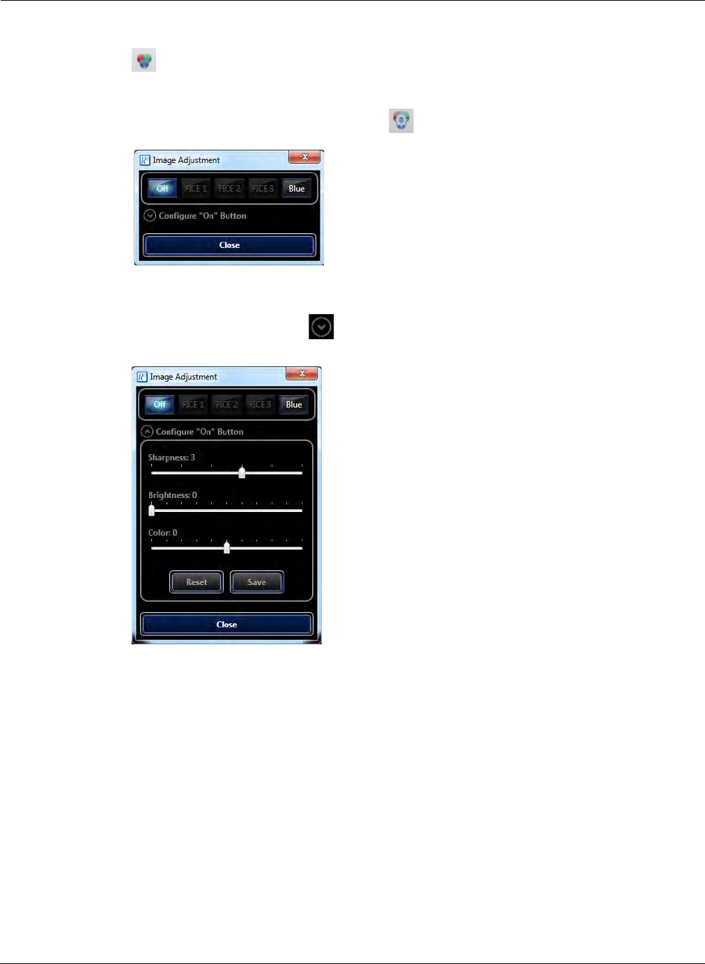
Reviewing and Interpreting RAPID Videos
Overview of the RAPID Interface 123
To adjust the video image appearance:
1. Click in the ribbon to turn on Image Adjustment to the factory presets, or, if you saved your
preferences earlier, to your own presets.
2. If you wish to adjust the image otherwise, click in the ribbon. The following screen appears:
3. Select any of the active buttons FICE 1, FICE 2, FICE 3 or Blue for these preset image adjusters.
4. To adjust the image yourself, click Configure "On" Button. The screen expands.
5. The "On" state means that image adjustment is applied to the image. The expanded screen shows
the settings currently in effect.
6. Use the Sharpness, Brightness and Color sliders to get the desired result, as shown by the video
image of the open video that provides a preview of the changes you are making.
7. Click Save to save your settings. The next time you turn on the Image Adjustment, these settings
are applied.
8. If you wish to return to the factory preset, click Reset.
9. Click Close to close the Image Adjustment screen.
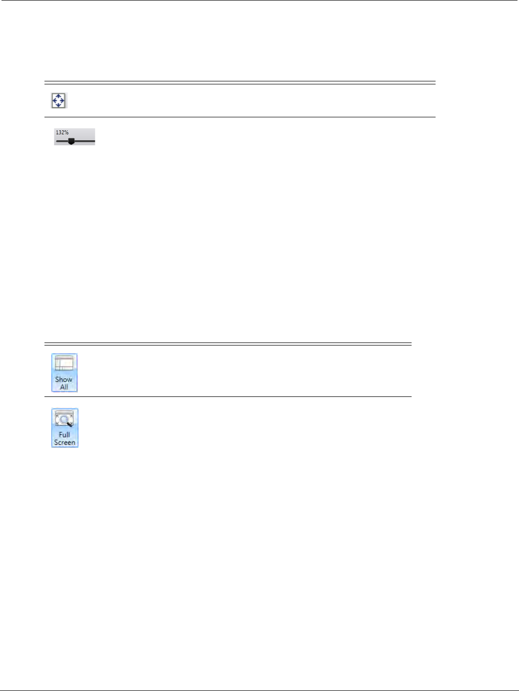
PillCam Capsule Endoscopy
124 Overview of the RAPID Interface
Zoom Group
This ribbon group contains the controls for displaying the video image at different zoom levels.
Show Group
This ribbon group enables the display or hiding of video viewing features. Using the check boxes next to
the features you can enable or disable the following:
•Show Thumbnails
•Show Localization
•Show SB Progress bar (SB videos only)
•Show Comments Editor
•Dynamic Player Control
Button Function
Fits the image size to the window.
Changes the zoom of the video view by sliding left or right and displays
the current zoom in percentage.
Button Function
Click to show all features in the Show group. Clicking again will only
show the features that were previously enabled.
Shows the video in full screen on the computer screen, without any of
the other elements of the View screen, including the ribbons. To return
to normal view, press ESC. Pressing F11 on the keyboard has the
same functionality.

Reviewing and Interpreting RAPID Videos
Using RAPID to View a Video 125
Using RAPID to View a Video
To view a video, you can open a video from Study Manager by clicking View Study, and then
selecting Study Manager on the RAPID home screen. Open a selected study by double-clicking it or
click once and then click the Open button. The Study Manager provides a list of the videos available
for viewing. The Study Manager organizes the studies in a way that enables easy access and
management of studies, sorting, and searching by different criteria such as patient name and procedure
dates. Refer to Using the Study Manager on page 107 for more information.
To view a video, you can also click the File menu on the RAPID interface and browse for your study in
the file system.
Reading a Capsule Endoscopy Video
Before loading a RAPID Capsule Endoscopy video to the RAPID software it is important to review the
patient history prior to reading the video in order to:
•Consider all possible indications as per the patient history.
•Prepare for identification of relevant pathology as per the possible indications identified.
To read PillCam studies efficiently, the reading process is broken into 3 components:
•Preview: Scan the capsule endoscopy video using QuickView mode to establish a preliminary
interpretation. This can be followed by a Complementary QuickView scan to perform a complete
review of the video. You may also augment your preview by previewing in SBI mode to quickly
scan the bleeding-suspicious images.
•Review: Review the complete capsule endoscopy video in normal viewing mode.
•Report: Complete a Patient Report that summarized the interpretation of the study. Refer to
Creating a PillCam Capsule Endoscopy Report on page 146.
QuickView
Use the QuickView feature to perform a fast preview of the video. QuickView makes RAPID video
review more efficient by displaying only images that may be of interest in the video stream to provide
fast previewing and land marking of a small bowel video. The number of images to be considered
images of interest can be set as percentage of the video by the user. The RAPID scans all images and
scores them according to the possible level of significance. Then according to the percentage level set
by the user it displays a short video to provide an overview of the case prior to full review. The
recommended viewing speed is 7-10 frames per second using Single View layout.
Scan the entire video in QuickView mode to:
•Establish approximate landmarks for the first gastric, first duodenal, and first cecal images.
•Capture thumbnails of images of interest.
•QuickView may be used to review gastric and colon images.
•The absence of a particular frame from the QuickView display should not be interpreted to mean
that no abnormal finding is present in that frame.
•SBI images are also included in the QuickView.
֠
֠֠
֠Note
The QuickView feature is not intended to detect pathology in lieu of a
physician and should not be used as a substitute for reviewing the entire
video.
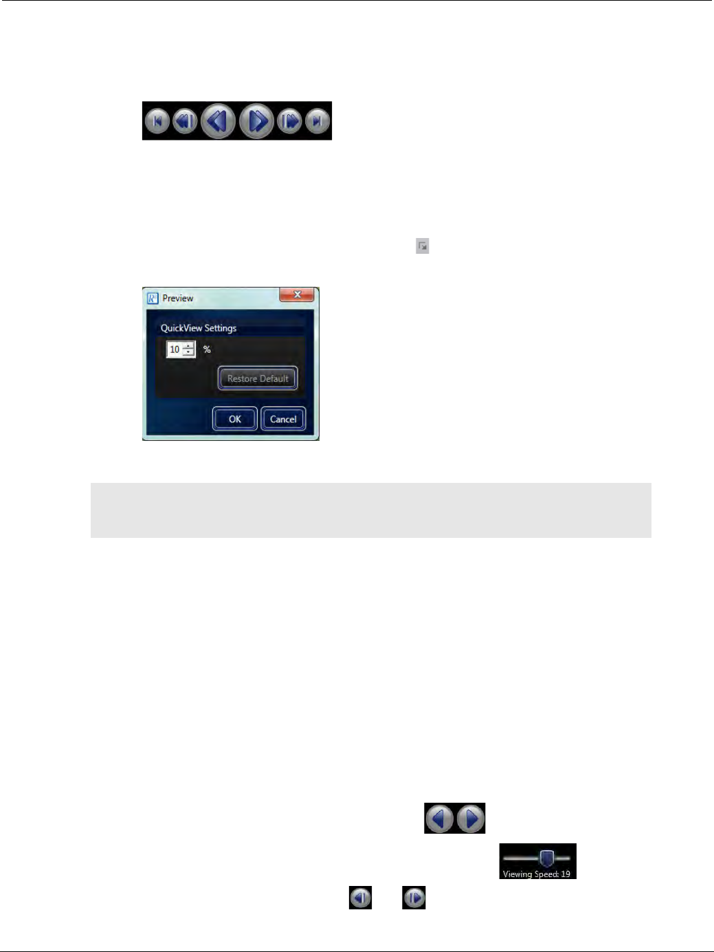
PillCam Capsule Endoscopy
126 Using RAPID to View a Video
•The QuickView by no means replaces a thorough viewing of the entire video by a qualified physician.
•Using the mouse scroll wheel in this mode also displays each frame of this mode one by one.
•In the QuickView mode, the video navigation buttons change to the following:
•The user can determine the number of images of the RAPID video to be viewed in the QuickView
mode, by changing the percentage rate of images to be shown and hence the sampling rate, which is by
default 10%.
To change the QuickView sampling rate:
1. From the View screen, click the dialog box launcher ( ) of the Preview ribbon group. The following
screen appears:
2. Type in the desired sampling rate (between 2% and 80%) for the QuickView and click OK to save it.
Complementary QuickView
After viewing the video in QuickView mode, click the CQV (Complementary QuickView) button to view
rest of the video not shown in QuickView mode. In Complementary QuickView mode (CQV) RAPID
displays only the images that are not included in the QuickView mode. Playing a video from start to end in
QV mode and then in CQV mode results in viewing all video images.
Viewing a Video
If not using QuickView and Complementary QuickView combination to view a RAPID video, you can use
the View button to scan the video from start to finish if you are using a PillCam SB 3, PillCam COLON 2,
or PillCam ESO/UGI capsule. If you are viewing a PillCam SB 2 video, you can select the A-mode
(default) or M-mode button to set your viewing mode. In all modes, you can move through a video by
using the navigation buttons below the video image:
•Play forward or backward by clicking the play buttons .
•Speed up or down the video display using the viewing speed slider .
•View the next or previous image by using and .
֠
֠֠
֠Note
When viewing a video in CQV mode, you cannot change the QuickView
sampling rate.
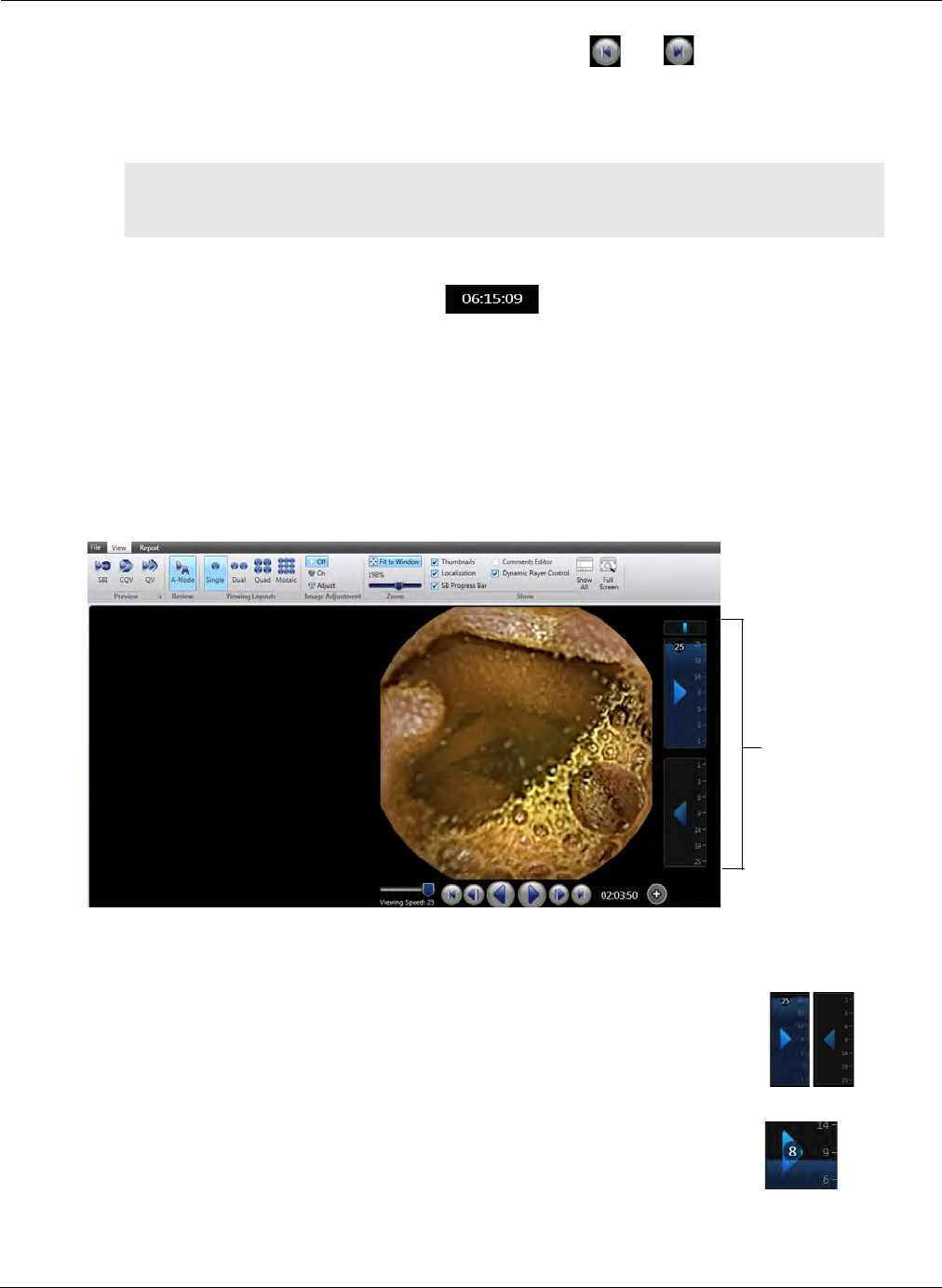
Reviewing and Interpreting RAPID Videos
Using RAPID to View a Video 127
•View the last image or first image of the video by using and .
•Clicking and holding the slider on the time bar and dragging it to move the video forwards or
backwards.
•Clicking the time bar moves the video image to that specific location.
To go to a specific frame of the video:
1. Select numbers of the time indications .
2. Type the desired time unit in the format HH:MM:SS.
3. Press ENTER. The video automatically progresses to the exact video image of the time specified.
Dynamic Player Control
The Dynamic Player Control in RAPID allows you to control the speed at which you view the video,
using your computer mouse.
Use the controls as follows:
֠
֠֠
֠Note
After the Pause button is pressed, the mouse wheel can be used to scroll the
video forward or backward.
• Click anywhere in the rectangle around the backward or forward arrow
to play the video in the required direction. Click again to stop the video.
• While the video is playing, move your mouse cursor UP and DOWN to
control the speed at which the video plays. When you stop the video, a
number appears showing the rate at which you are viewing the frames.
Dynamic
Player Control
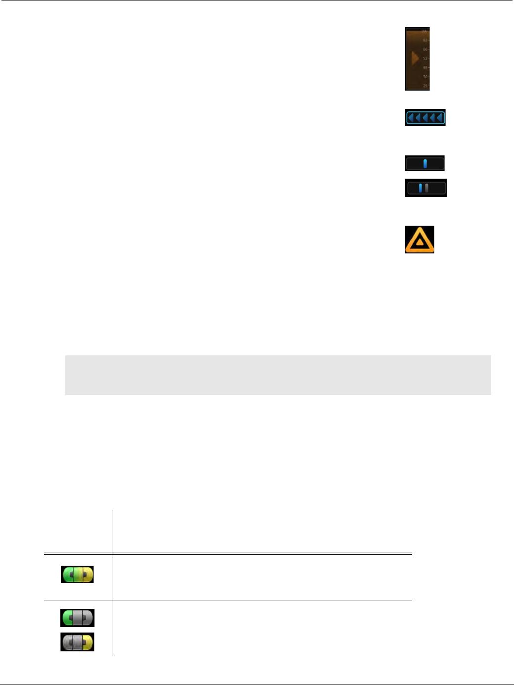
PillCam Capsule Endoscopy
128 Using RAPID to View a Video
Dual Head View
PillCam ESO/UGI and PillCam COLON capsules have two video heads and by default, two images are
displayed: one for each head of the capsule. Note that two images displayed thus side-by-side is not
DualView: the two side-by-side images are from different video heads.
The capsule icon as shown below is both the control and the indicator for the dual head video display
mode. The icon consists of three buttons: left (green if active), right (yellow if active) and the middle
(activates both displays green and yellow).
• The Dynamic Player Control includes a Turbo Mode function which
allows speeding up viewing a segment of the video. To view the video
in Turbo Mode, while the video is playing, click and hold the mouse
button. The playing speed accelerates and the rectangle changes to
orange.
• The orientation indicator appears briefly above the backward/forward
arrows to show the viewing direction when you switch between the
backward or forward arrows.
• The orientation indicator appears above the backward/forward arrows
when you pause viewing, and shows your current viewing location.
Use the mouse wheel to scroll backward/forward in the video. Click the
gray location indicator to go back to the previous location, and
continue viewing from that point.
• The Area of rapid change alert is available in COLON 2 videos only.
The Approaching area of rapid change notification appears when
you approach a segment of the video where the capsule travels at a
higher speed. This notification is followed by the Area of rapid
change alert. When this alert appears, you can use the Dynamic
Player Controls to adjust viewing speed.
This option is available when the Enable Reviewer Alert option is
enabled under Tools > Settings.
֠
֠֠
֠Note
The Reviewer Alert functionality is only available for COLON 2 videos compiled
with RAPID v8.3.
Capsule
Icon Display
Default view for COLON and ESO/UGI videos: images of both
heads are displayed. Click either the far left side or the far right of
the icon to view one single video.
View only the images from one head. Click the other side to view
the video from the other head. Click the middle of the icon to
return to viewing both videos.
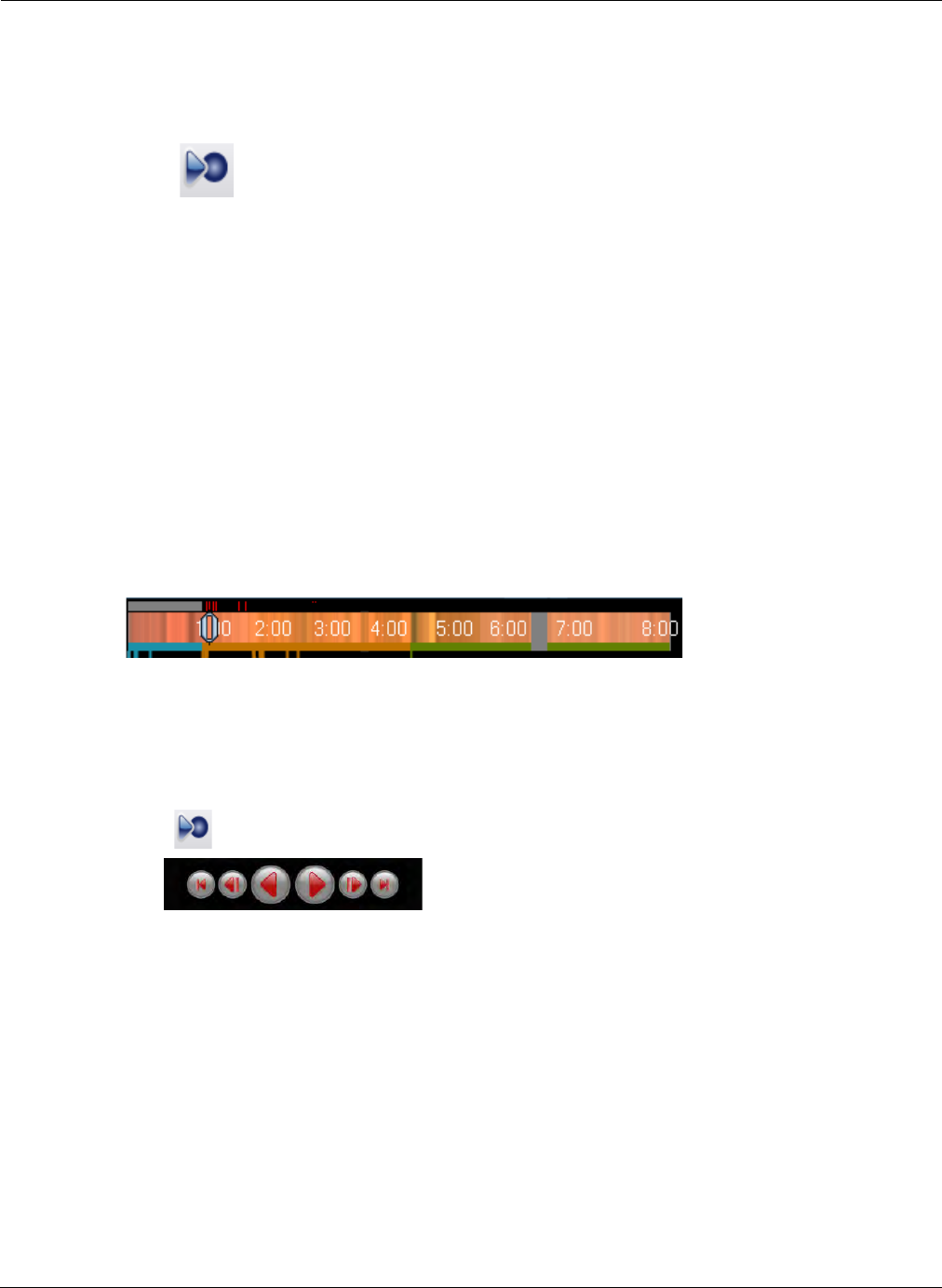
Reviewing and Interpreting RAPID Videos
Using RAPID to View a Video 129
When typing thumbnail comments in PillCam ESO/UGI and PillCam COLON videos, the comments
appear green for thumbnails captured from the left represented capsule video head and yellow for
thumbnails captured from the right represented capsule video head.
SBI View
The Suspected Blood Indicator (SBI) feature automatically marks images suspected of containing blood
and provides the ability to review them in sequence. SBI image locations are marked on the Color Bar.
In the SBI mode, RAPID shows the SBI marked images. The video control buttons turn red and you can
play the SBI-images-only back and forth by the controls. SBI images are also included in the
QuickView.
This viewing mode feature is available only:
•After the first landmark is entered for the first duodenal image or any later landmark.
•If suspected images are found.
•The RAPID video is a PillCam SB video.
The Suspected Blood Indicator (SBI) feature is a guide to areas of suspected fresh GI bleeding. The SBI
display feature becomes available only from the first duodenal image on. Hence the SBI feature is
available only after labeling the first image of the Duodenum.
When SBI is activated, red ticks appear above the time bar and they indicate the location of SBI images.
SBI lines are not displayed in the area before the first duodenal image, which becomes gray.
To display the SBI images:
1. Identify and label the first image of the duodenum (see Using Localization and Landmarks on
page 137).
2. Click . The video navigation buttons turn red.
3. Use the video navigation buttons to view the SBI images.
Time Bar/Color Bar
The Time bar/Color bar displays:
•The total length of the video and the average color for easy anatomical segmentation of the
corresponding image. The unit of time is hours, minutes, and seconds.
•The slider moves along the time bar as the video is played. You can also use the slider to move the
video forwards or backwards.
•Moving your mouse over the time bar displays the time at the position of the cursor. If the mouse
stays at a location on the time bar for longer (about 2 seconds), the image at that location is
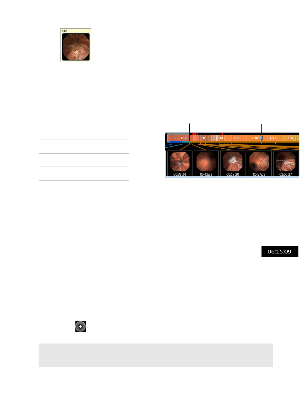
PillCam Capsule Endoscopy
130 Using RAPID to View a Video
displayed in a small window, with the time indication of that image at the top. If the video is in dual
head view (ESO/UGI, COLON), two images appear, one for each head.
•You can move the video display to a certain location by clicking that location on the color bar.
•SBI indicators (small red ticks) appear in the top part of the Time bar/Color bar (see SBI View on
page 129). The area before the landmarked first duodenal image is gray.
•GI divisions are colored in a narrow strip below the Color Bar according to defined Landmarks. The
regions have specific colors.
•Lines in region-specific colors connect the location on the time bar and the thumbnails.
•Before defining the landmarks, the narrow strip below the Color Bar is gray.
Time Indication
The time indication box displays the time of the frame in hours, minutes, and seconds
after the capsule starts acquiring images in the procedure.
Creating and Annotating Thumbnails
Thumbnails are images captured from the video. Some images are automatically created, such as those
that are marked during real-time viewing with PillCam recorder DR3. These thumbnails have the default
annotation of Thumbnail marked in recorder. When reviewing the video, the physician may capture any
image of interest for further scrutiny. The images appear at the bottom of the screen. Double-click on the
video image in the center of the screen to designate it as a thumbnail. Alternatively, click the Capture
Thumbnail button located next to the video player buttons to add an image that appears on the screen
to the thumbnails.
Each thumbnail appears in the Thumbnail section.
Color Region of GI
Blue Esophagus
Turquoise Stomach
Orange Small intestine
Green Colon
(large intestine)
֠
֠֠
֠Note
The thumbnails and associated comments are saved in the Findings file
(see Working with Findings on page 143).
GI data display Time Bar slider
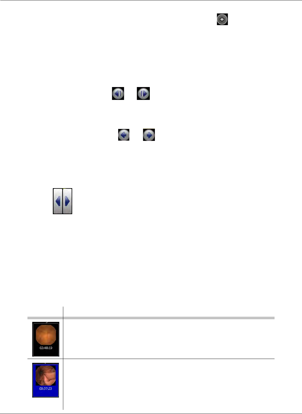
Reviewing and Interpreting RAPID Videos
Using RAPID to View a Video 131
To create a thumbnail:
Double-click the video display, or click the Capture Thumbnail button .
To view a thumbnail in the video display:
Double-click the thumbnail.
The RAPID automatically displays the desired image on the screen, the cursor in the Time Bar jumps to
the thumbnail location in the video. The thumbnail in the thumbnail section will be highlighted with a
blue filled outline.
To view the adjacent images:
•In the View screen, use the and buttons to view the adjacent images, or roll the mouse
wheel forward or backward.
•In the Report screen, roll the mouse wheel forward or backward to view adjacent images of the
video to capture these as new thumbnails.
•In the Report screen, use the and buttons to view the adjacent thumbnails.
To scroll through the thumbnails:
If there are more thumbnails than fit on the screen, RAPID allows you to scroll through the thumbnails
by several alternative modes:
•An arrow appears on either side of the thumbnail section. Click and hold either of the arrows to
scroll left or right.
•When you hover with your cursor in the thumbnail section, a scrollbar appears on the bottom
allowing you to drag the bar to sideways to scroll through the thumbnails.
•Using your mouse, click and hold on the thumbnail section and drag the thumbnails to either side
to scroll.
Thumbnails can also be annotated, saved, exported as images, deleted or selected for inclusion in the
capsule endoscopy report.
Thumbnail Status
The thumbnails are displayed on the bottom of the View screen. Visual appearance designates different
statuses:
Thumbnail Description
This is a thumbnail without any visual effects.
Dark blue background means that the thumbnail is selected so operations can be
performed on it.
For multiple selections hold down the CTRL button on the keyboard and click the
desired thumbnails with the mouse. For multiple selection of consecutive thumbnails,
hold down the SHIFT key and select the first and last desired thumbnail.
All available actions (displayed by right-clicking on a thumbnail) will operate on all
selected thumbnails.
PillCam Capsule Endoscopy
132 Using RAPID to View a Video
Thumbnail Comments
To add a comment to a thumbnail, use one of the following methods:
•Click the check box Comments Editor in the ribbon of the View screen.
•Right-click the thumbnail when in the View or Report screen and select Edit Thumbnail Comment.
•Hover over the thumbnails and click in the top right corner of the thumbnail to open the
thumbnail editor.
Comments Editor
The Comments Editor at the left side of the screen enables the user to type in their comments about a
specific thumbnail or about the whole study.
Editing Tools
When typing comments in the comments editor the following two tools are available:
•GI Dictionary: simplifies efficient report creation via a pre-populated and customizable database of
terms and phrases. RAPID software preserves the GI Dictionary customizations from previous
versions during software upgrade.
The external blue border means that the thumbnail is selected for inclusion in the
report.
The internal border means that this thumbnail is the currently displayed video image.
Mouse-over a thumbnail without effects gives it a light blue background.
Also, a checkbox becomes available in the top left corner for selecting the thumbnail
for the report, as well as the edit button in the top right corner.
֠
֠֠
֠Note
You can toggle to and from right-to-left writing, as supported by Windows
convention by pressing the ALT and SHIFT keys simultaneously.
Thumbnail Description
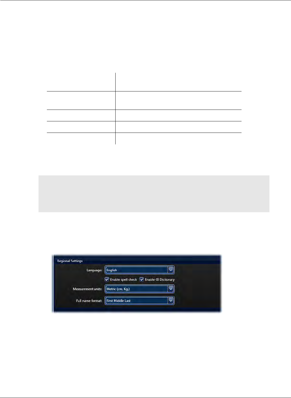
Reviewing and Interpreting RAPID Videos
Using RAPID to View a Video 133
a. Type a part of a word anywhere during text editing on the screen.
b. The dictionary is context sensitive and shows the closest approximations of words starting
with the same letters.
c. If the matching word exists, the cursor highlights the word. Press SHIFT + ENTER to enter
the complete word without further typing, or double-click it in the GI Dictionary.
d. All keyboard commands for the user dictionary:
•Spell checker: if the spell checker is activated, a red wavy line appears below any word not
recognized by RAPID. To select one of the suggested corrections, right-click and select the desired
option.
To enable or disable the GI Dictionary or the Spell Checker:
1. From the Home screen, select Tools > Settings.
2. On the General tab, under the Regional Settings, select the check box next to the feature.
Shift +
Up Arrow/Down Arrow
Move the selection cursor inside the dictionary
up/down
Shift + Enter Inserts the current selected dictionary phrase
to the text box.
Control + R Shift Align the text to right side
Control + L Shift Align the text to the left side
ESC Temporarily close the GI Dictionary
֠
֠֠
֠Note
If you write more than 256 characters in the text box of a thumbnail
comment, the final printout of the thumbnail comment may be split between
two pages. Any thumbnail after that will start on a new page, leaving the
space after the split comment blank.
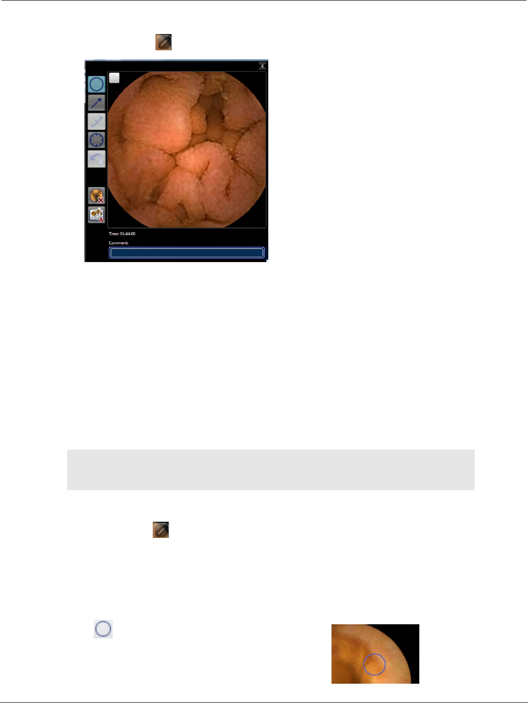
PillCam Capsule Endoscopy
134 Using RAPID to View a Video
Thumbnail Editor
Clicking the edit button in the top right corner of a thumbnail loads the Thumbnail Editor.
Buttons on the left of the thumbnail editor are tools to affect markings on the thumbnail (see Markings
Buttons Group on page 152) that also appear in the ribbon of the Report screen.
At the bottom, you can type your comments into the Comment box. Alternatively, use the comments editor
to type your comments for the thumbnail.
The Thumbnail Comments box is also located on the left side of the screen in the View screen if checked
in the Show ribbon group. It is displayed automatically in the Report screen.
If a thumbnail is selected to be included in the patient report, any comments entered for the thumbnail will
appear below it in the report.
Marking Tools
You can add marks to a thumbnail to point out or emphasize certain areas of interest.
To access marking tools:
•View screen: click in the top right corner of a thumbnail. The thumbnail appears enlarged with all
the marking tools at its left side.
•Report screen: all the marking tools are in the Markings ribbon group.
Mark Circle
Creates a circle around an area of interest:
1. Click .
2. Place cursor over area of interest.
֠
֠֠
֠Note
Marks on a thumbnail are saved together with the thumbnail and appear as
such in the report and on the thumbnail itself.
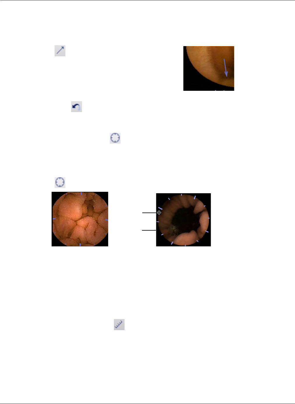
Reviewing and Interpreting RAPID Videos
Using RAPID to View a Video 135
3. Click and drag without releasing to create a larger circle to include area of interest.
Mark Arrow
Creates an arrow pointing to an area of interest:
1. Click .
2. Place cursor to the side of the area of interest.
3. Click and drag without releasing toward area of
interest. The arrow points to region of interest.
Undo Mark
Clears the last mark created. Click repeatedly to successively clear all marks.
Circumference Scale
(For ESO/UGI procedures only) RAPID provides an optional circumference scale for estimating the
circumferential involvement of esophageal varices in the Report screen. The circumference scale
displays 12 equivalent ticks on the image periphery.
1. Click in the ribbon of the Report screen.
2. Point the cursor and click anywhere on the circle to rotate the scale to facilitate estimation of the
circumferential involvement of a finding.
3. You can rotate the grid as follows:
a. Move the cursor over the periphery of the image.
The cursor changes into a 4-arrow cursor.
b. Click and drag the scale around to match it to the varices you want to measure.
Polyp Size Estimation
COLON 2 procedures only: Tool for estimating the size of a suspected polyp. To use this feature,
activate it as follows.
To activate Polyp Size Estimation:
1. From the Home screen, select Tools > Settings. The Settings screen appears.
Cursor
changes
shape
Rotated
scale
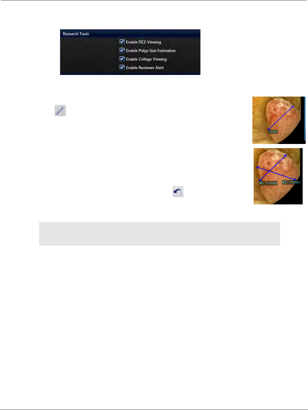
PillCam Capsule Endoscopy
136 Using RAPID to View a Video
2. Select the Video tab and in the Research Tools section, click the checkbox next to Enable Polyp Size
Estimation.
3. Click Apply or OK.
To use Polyp Size Estimation
1. Click in the ribbon.
2. Place cursor at one end of a polyp.
3. Click and drag (without releasing) to create a double pointed arrow from one
end to the other end of the polyp for which you wish to estimate the size.
4. The size estimate appears at the end where you release the mouse button. If
you make more than one size estimate for the same image, the estimates are
counted: #1, # 2, etc.
5. To remove a marking, click the Undo Mark button . The last marking
disappears. Click repeatedly to successively clear all marks.
֠
֠֠
֠Note
The Polyp Size Estimation measurement is only an estimate. Use discretion
when making diagnostic or treatment decisions.

Reviewing and Interpreting RAPID Videos
Using RAPID to View a Video 137
Using Localization and Landmarks
Landmarks
Landmarking an image labels it as one of the anatomical landmarks along the GI tract. A different
default landmark menu is displayed for each RAPID video type, as follows:
To create a landmark:
Right-click on video image you identify as the landmark and select the correct landmark label.
If, for the video you are viewing, you wish to enter a landmark other than the default landmarks, select
Capture Other Landmarks. This option displays all the landmarks for selection.
Video type Landmarks
ESO/UGI video First esophageal image
Z-Line image
First gastric image
SB video First gastric image
First duodenal image
First Ileocecal image
First cecal image
COLON video First cecal image
Last cecal image
Hepatic flexure image
Splenic flexure image
First rectal image
Last rectal image

PillCam Capsule Endoscopy
138 Using RAPID to View a Video
Suggested Flexure Landmarks for COLON Videos
This feature is available (active) only if a PillCam sensor array was used during the procedure. If a
PillCam sensor belt was used, this option is inactive and grayed out.
After you have marked the first cecal image, RAPID can suggest the Colon Flexure landmarks.
1. Right-click anywhere on the video images and select Suggest Colon Flexure Landmarks.
Two new thumbnails appear in the thumbnail section with the captures: ?Hepatic flexure? and
?Splenic flexure?
2. Double-click the suggested flexure thumbnails for review.
3. To accept the suggested flexure landmarks, right-click the thumbnails and select the appropriate
landmark option.
Suggested Landmarks for PillCam SB 3 Videos
To use this feature to suggest the SB 3 landmarks:
1. Right-click anywhere on the video images and select Suggest Small Bowel Landmarks.
Two new thumbnails appear in the thumbnail section with the captures: ?First Duodenal Image? and
?First Cecal Image?
2. Double-click the suggested thumbnails for review.
3. To accept the suggested landmarks, right-click the thumbnails and select the appropriate landmark
option.
Localization
Localization provides the approximate location and track of the PillCam capsule in the GI tract via a two-
dimensional representation. The Localization feature is available for SB and COLON videos.
When at least one landmarked thumbnail has been captured and created for the first image of one of the
anatomical landmarks along the GI tract, the localization feature is enabled, and:
•A sketch of the estimated track of the capsule in the GI tract in the abdomen area is displayed in gray.
•A small white circle with a black dot on the capsule track indicates the estimated position of the
current frame being viewed.
֠
֠֠
֠Note
Localization is only available for procedures performed with a sensor array.
The Colon Location Diagram is available for COLON 2 videos even if
performed with the sensor belt, but the localization feature will not appear.
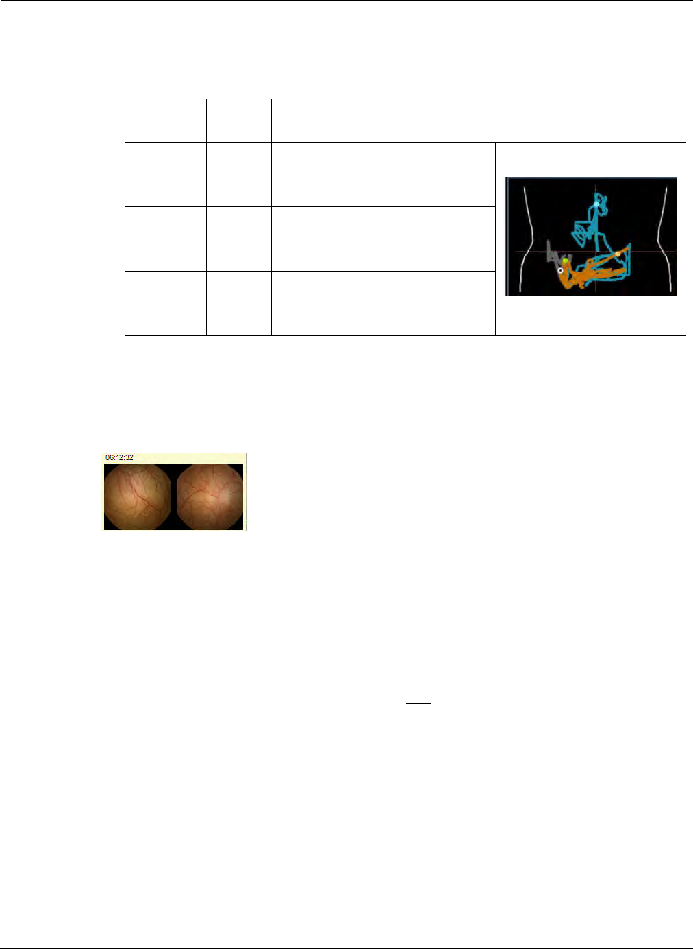
Reviewing and Interpreting RAPID Videos
Using RAPID to View a Video 139
•The capsule track is segmented into the GI tract segments according to user landmarking: stomach,
small intestine, large intestine. The part of the GI tract that is already viewed becomes colored
according to the same color codes as on the lower part of the time bar, as follows:
•Defined landmarks on the capsule track are seen as small circles of the same color as the
underlying GI region.
You can move the video display to a certain location by clicking that location on the localization track.
Hovering the mouse over a point on the localization track for 2 seconds shows the images at that
location in a separate window. The time indication of these images appears at the top.
If instead of the localization track, a small sketch of the abdomen appears with a red X in the upper left
corner, this means that the Localization feature is not enabled due to the detection of a faulty sensor.
When viewing an ESO/UGI video, the PillCam logo appears instead of the localization track.
PillCam Progress Indicator
The PillCam Progress Indicator appears only:
•for SB videos,
•after you have landmarked the first duodenal image and the first cecal image,
•in automatic (A) mode for SB 2 and SB 3 (AFR) videos.
The PillCam Progress Indicator graphically presents the progress of reviewing the small bowel. The
slider on the left side shows an estimate of the percentage of the small bowel viewed.
Section
of GI tract Color Explanation
Gastric Blue Indicates the stomach. The
entrance to the stomach is
marked by a blue dot.
Small
bowel
Orange Indicates the small bowel. The
entrance to the small bowel is
marked by a green dot.
Cecal Green Indicates the large intestine. The
entrance to the large intestine is
marked by a yellow dot.
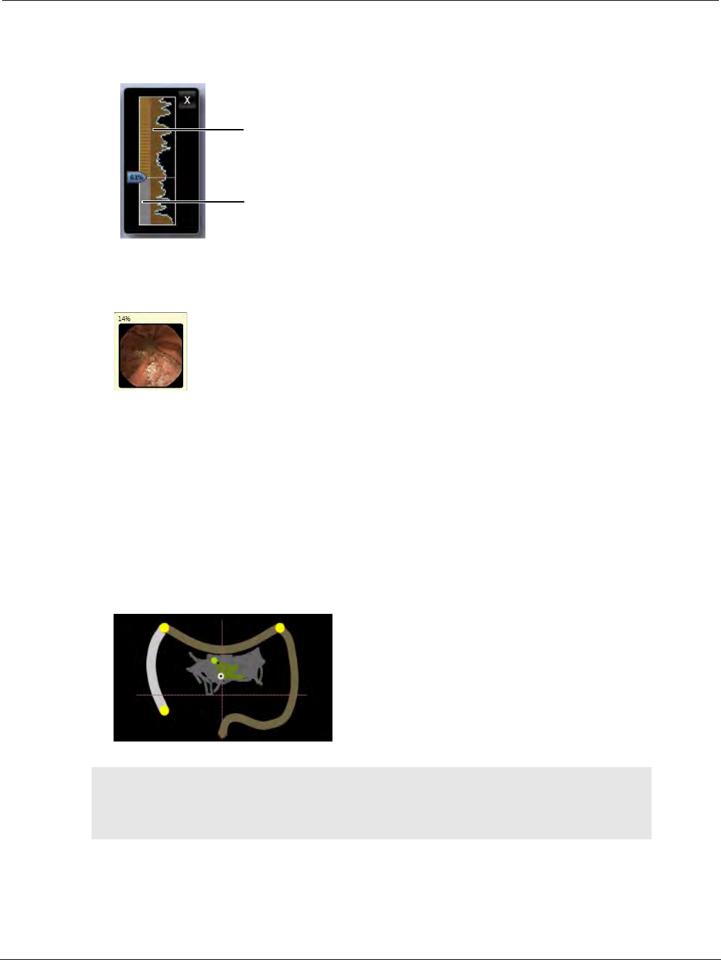
PillCam Capsule Endoscopy
140 Using RAPID to View a Video
The right side graph represents an estimate of the redundancy or similarity of the images captured by the
capsule as it moves through the small bowel (and may indicate where the capsule moved slowly through the
bowel).
You can move the video display to a certain location by clicking that location on the Progress Indicator.
Hovering the mouse anywhere on the Progress Indicator loads an image from that location in a pop up
window. The top of the window shows the progress of the SB reviewed in percentage.
Colon Location Diagram
The Colon Location Diagram, a three-sided localization diagram, represents the three colon sections:
ascending (or right) colon (on the left side), transverse colon (on the upper side), and descending (or left)
colon (on the right side). This feature is available for PillCam COLON videos of procedures performed
with a sensor array as well as a sensor belt in addition to the localization track. It displays the colon
segment containing the displayed image is by highlighting the corresponding section in the diagram.
The Colon Location Diagram becomes active after you landmark the first cecal image. Once you have
landmarked the Hepatic and Splenic Flexures, either independently by identifying these landmarks or by
accepting the suggested landmarks in RAPID if the procedure was performed with a sensor array, the
section containing the currently displayed images lights up on the Colon Location Diagram.
֠
֠֠
֠Note
Colon Location Diagram function is only an aid for the physician, and the
accuracy is dependent on the physician’s ability to identify the four
anatomical images that separate the right, transverse, and left colons.
Level of image
similarity
Reviewing progress
indicator
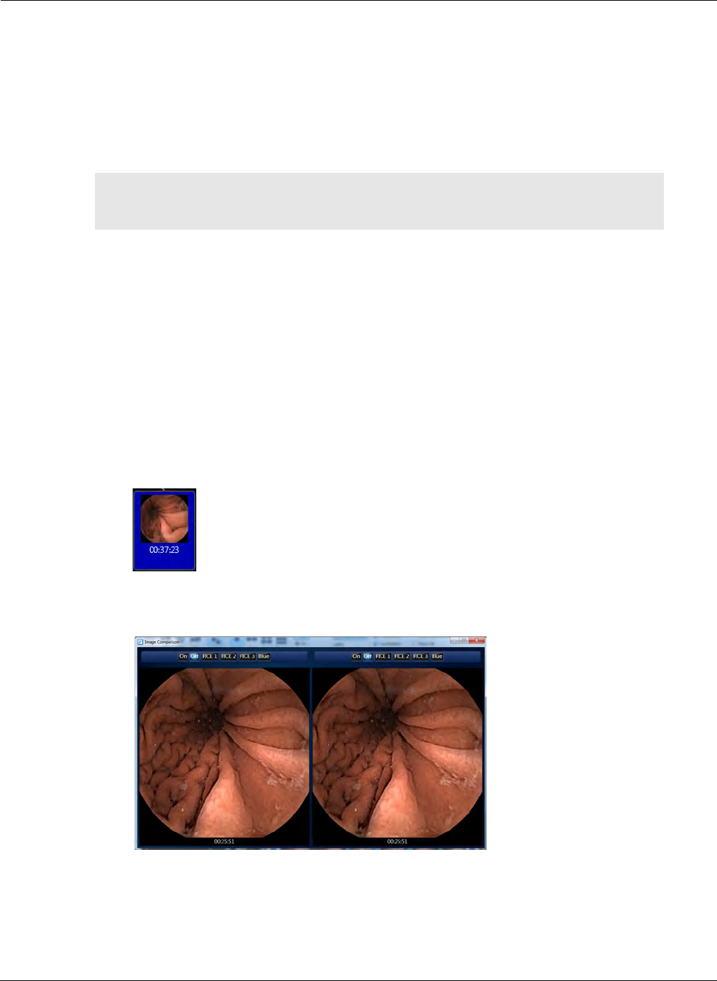
Reviewing and Interpreting RAPID Videos
Using RAPID to View a Video 141
Passage Times
Passage time is the time the capsule spent in a given region of the GI tract. Calculation of passage
times is enabled only after you segment the video from the GI tract by labeling the first images of the
different sections of the GI tract. When you identify and label the first and last images of the gastric,
SB, or cecal section of the GI tract, the RAPID software calculates the relevant passage times based on
the time tags of the landmarked thumbnails.
The RAPID software displays this data at the bottom of the Localization Display section.
Comparing Thumbnails
There are three scenarios to compare images in RAPID v8.3:
•Atlas comparison: Compare a RAPID video thumbnail to Atlas images (see Comparing Video
Images to Atlas Images on page 154).
•One thumbnail comparison: Compare a thumbnail to itself in different display modes (i.e. FICE
or Blue mode).
•Two thumbnail comparison: Compare two different thumbnails in the same video.
To compare a thumbnail to itself (One frame comparison):
1. Click the thumbnail you wish to examine.
A dark blue background appears around the thumbnail.
2. Right-click the thumbnail and select Compare Thumbnails.
A separate window appears, showing the same thumbnail duplicated, side by side.
֠
֠֠
֠Note
The estimation of the passage times is based solely on the physician’s
identification of the first image of each section of the GI tract.
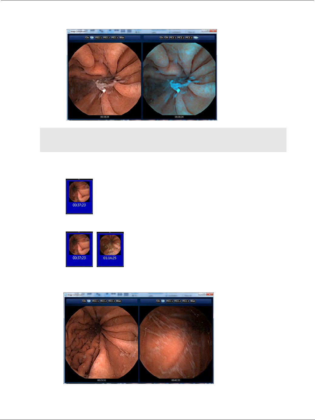
PillCam Capsule Endoscopy
142 Using RAPID to View a Video
3. Use the Image Adjustment tools at the top of the window of one of the thumbnails to compare the two
images.
.
To compare two images in the same video:
1. Click the first thumbnail you wish to use for comparison.
2. Hold down the CTRL key on your keyboard and select the second thumbnail.
3. While still holding down the CTRL key, right-click either one of the selected thumbnails and select
Compare Thumbnails. A separate window appears, showing both thumbnails side by side.
֠
֠֠
֠Note
You can scroll through the video by moving your mouse wheel in either
direction. Both sides will move together.

Reviewing and Interpreting RAPID Videos
RAPID Video Files 143
RAPID Video Files
A RAPID study is the collection of files associated with a specific PillCam Capsule Endoscopy
procedure. A RAPID study is contained in a folder that contains the RAPID video file.
The *.gvi file and the RAPID study name consists of several elements separated by spaces. The
components are derived from Patient Check-in information transferred from the PillCam recorder
during the video creation:
•Patient’s last name
•Patient’s middle initial
•Patient’s first name
•Patient’s ID number in parentheses
•Examination date in the following format: (DD MMM YYYY)
For example: the RAPID folder with the name
Doe F. John (12345) 21 Feb 2008, which resides in E:\videos, contains the file named Doe F. John
(12345) 21 Feb 2008.gvi.
You may save a RAPID folder and a RAPID video to a removable media (USB storage device or disc)
for one of the following reasons:
•To back up a RAPID study.
•To copy a RAPID study from the hard disk in order to free space on the computer.
•To move a copy of a RAPID study onto another computer or to another physician.
To save a RAPID study to a CD/DVD, see Burning a Study to a CD/DVD on page 108.
To save a RAPID study to a USB storage device, see Saving a Study to Another Archive on page 110.
Working with Findings
A findings file contains:
•All the thumbnails with their comments or other annotations (see Creating and Annotating
Thumbnails on page 130).
•A link to the check-in information.
•All the study comments (see Editing Tools on page 132).
•A link to the report, if created.
֠
֠֠
֠Note
• You can scroll through the video by moving your mouse wheel in either
direction, for each thumbnail separately.
• If other applications on your computer use keyboard shortcuts, they may
be activated when you use the shortcuts in RAPID.

PillCam Capsule Endoscopy
144 Working with Findings
Saving Your Findings
It is possible to save your findings. You can open and view this file with the RAPID video on any
computer with the RAPID software installed.
To save findings in new location:
1. Select File > Save > Save Findings As.
The Save Findings As screen appears.
2. Select a location for saving:
•If you intend saving to a disc, insert a formatted writable disc into the appropriate drive and select it
from the screen.
•If you intend saving on a USB device, plug in the USB device and select the USB device.
•The default location is the RAPID folder of the currently displayed video.
3. Click Save.
The findings file is saved. The name of the associated RAPID directory, the associated RAPID video
file and the findings are saved.
To save findings to the last saved findings file:
Click the button in the Quick Access toolbar located at the top of the RAPID screen, or click Save
Findings from the File menu.
The findings file is saved to the same location and under the same name.
֠
֠֠
֠Note
Save the findings frequently to avoid accidental loss of data.
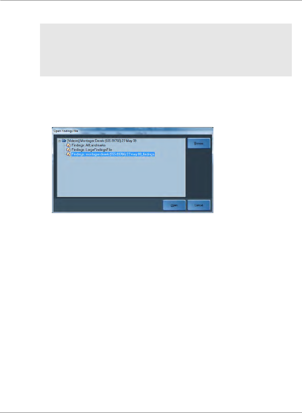
Reviewing and Interpreting RAPID Videos
Working with Findings 145
Opening a Findings File
To open a findings file:
1. Select File > Open and then select Open Findings. If a findings file is open, RAPID asks is you
want to save the current findings file. The Open Findings File window appears, showing the
folder of the current video. A findings file can also be opened using the Study Manager (see Open
on page 107).
2. Select the findings file you wish to open.
To open a findings file stored in another location, navigate to the desired location by clicking
Browse.
3. Click Open.
Once the findings file is loaded, the saved thumbnails with their annotations appear in the
Thumbnail section. If the findings contain landmarks, the following note appears: Calculation of
passage times for the esophagus, stomach, small bowel and colon is based solely on your selection
of the first image of the esophagus, stomach, duodenum, cecum and the last image of the rectum.
4. Click OK to continue.
֠
֠֠
֠Note
You may load only a findings file associated with the currently displayed video. If
you attempt to load a findings file that is not associated with the loaded RAPID
video, an error message appears notifying you of the mismatch.
If you load another findings file associated with the same video, the open
findings file closes. A message appears asking you to save the open findings file
before closing it.
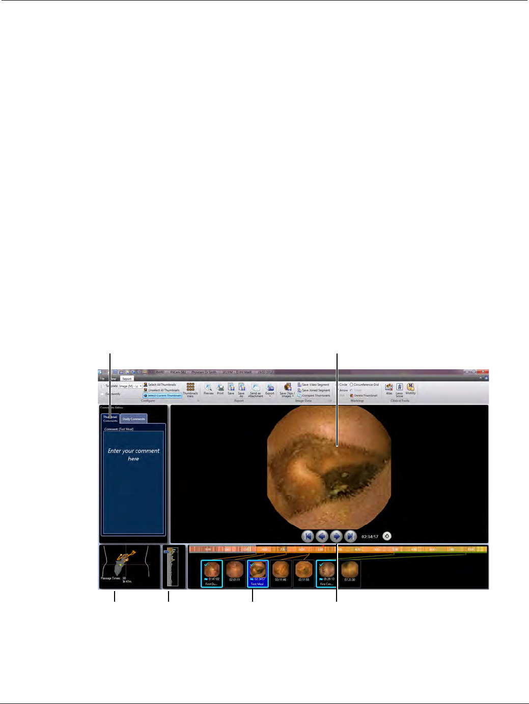
PillCam Capsule Endoscopy
146 Creating a PillCam Capsule Endoscopy Report
Creating a PillCam Capsule Endoscopy Report
Overview
After viewing the capsule endoscopy video and creating the findings, an interpretation of the study can be
summarized in a patient report. This phase should be performed after the Preview and Review phases.
The data created during the Review phase is now stored in the corresponding video folder. The RAPID
software's Report ribbon enables the creation of reports and exporting of video clips and images to another
directory or to a CD for later use.
The Report ribbon enables the following functions to prepare the patient report and complement the patient
study:
•Add additional thumbnails and study comments.
•Add markings (circles and arrows around lesions for emphasis and size estimates) to all thumbnails.
•Add comments to all thumbnails.
•Save images and create video clips for later use in presentations or referrals.
•Use the Atlas or Lewis Score clinical tools in the Report ribbon for diagnostic comparison or
evaluation using a scoring index.
•Generate a patient report that can also be printed and emailed.
•Export the results or patient summary.
Selected thumbnail
Thumbnail
navigation buttons
Comments Editor:
thumbnail or entire study
Localization Progress indicator Thumbnails
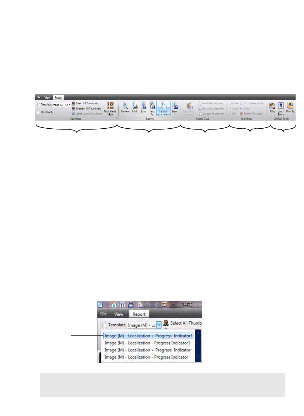
Reviewing and Interpreting RAPID Videos
Creating a PillCam Capsule Endoscopy Report 147
Report Ribbon
The Report ribbon is divided into several groups enabling many different functions. The buttons and
commands are organized in the following logical groups:
•Configure
•Report
•Image Data
•Markings
•Clinical Tools
In this chapter the groups will be explained in a different sequence than how it appears in the software in
order to closely match the proper workflow that is used when interpreting the data.
Configure Buttons Group
The Configure group enables the selection of templates for report generation, the ability to de-identify
patient information, to select/deselect all thumbnails, and view an enlarged view of captured
thumbnails.
Template Selection for Report
The template drop-down list shows 4 templates that can be selected before generating a report. The 4
templates to be shown in the drop-down list can be configured from a pre-loaded library of 12 report
templates of different variations. The templates differ from each other in the inclusion or exclusion of
the Localization track, the SB Progress Indicator associated with each thumbnail and the size of
thumbnails displayed in the final report. It is at the discretion of the user to configure which templates
to use in the workflow. Only 4 of the 12 reports will be shown in the drop-down list in the Report
ribbon. The selected report template will be used for each particular case. To configure the templates,
see Report Templates Section on page 177.
֠
֠֠
֠Note
Upgrading the RAPID software from v7 to v8.3 will replace all templates and
their configuration.
Configure group Report group Image Data
group
Marking
group
Clinical Tools
group
Template will
appear on
Report ribbon
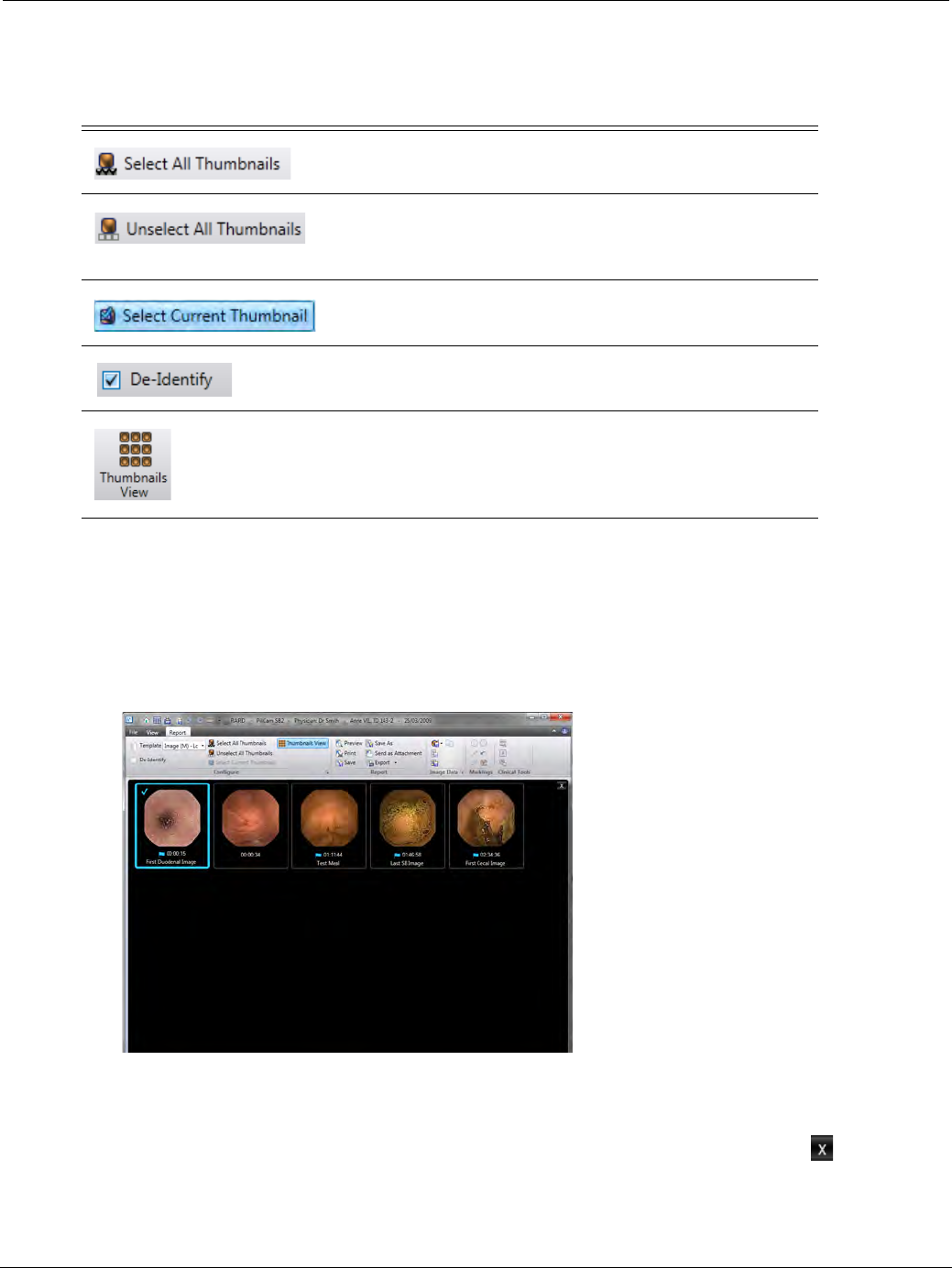
PillCam Capsule Endoscopy
148 Creating a PillCam Capsule Endoscopy Report
Other Commands in the Configure Buttons Group
Thumbnails View
For easier viewing of captured thumbnails, RAPID includes a screen that displays an enlarged view of the
thumbnails. You can use this screen to review and select thumbnails for inclusion in the report.
To view a screen with magnified thumbnails:
1. In the Report tab, select the Thumbnails View button.
An enlarged view of the thumbnails appears. Thumbnails that were selected in the regular view are
also selected in this view. You can select or unselect thumbnails as required.
2. To return to the previous screen, click the Thumbnails View button again or click the button in the
upper right corner of the screen.
Button Function
Select all thumbnails for inclusion in the capsule
endoscopy report or when saving clips and images.
Unselect all thumbnails to exclude them from the
capsule endoscopy report or when saving clips and
images.
Select current thumbnail for inclusion in the capsule
endoscopy report or when saving clips and images.
Anonymize patient information in the printed or
exported capsule endoscopy report.
Displays an enlarged view of captured thumbnails.
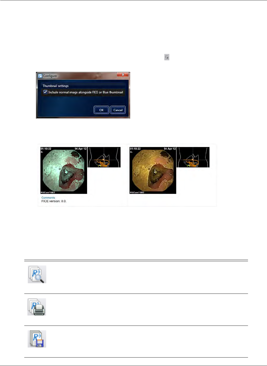
Reviewing and Interpreting RAPID Videos
Creating a PillCam Capsule Endoscopy Report 149
Configure Dialog Box
Allows defining thumbnail settings. When using the FICE or Blue Mode options when viewing
thumbnails, you can include a “normal” thumbnail image alongside the FICE/Blue thumbnail when
creating a report.
To include a normal image alongside the FICE or Blue thumbnail in the Report:
1. From the Report screen, click the dialog box launcher ( ) of the Configure ribbon group. The
following screen appears:
2. Select the Include normal image alongside FICE or Blue thumbnail checkbox and click OK.
When you generate a report, you will see both images, as in the example that follows.
Report Buttons Group
Below is a description of the Report button group buttons and the commands that can be performed
for managing the capsule endoscopy reports:
Button Function
Print Preview
Preview the capsule endoscopy report before printing. Clicking
this button opens a print preview page of the report. The
buttons on top of the print preview screen also enable to print,
save, save as, or email the report as an attachment.
Print
Select the printer, number of copies, page range, and other
printing options before printing. A version of the report is also
saved after printing.
Save
Save the report with a date and time stamp in the current
location with the current file name.
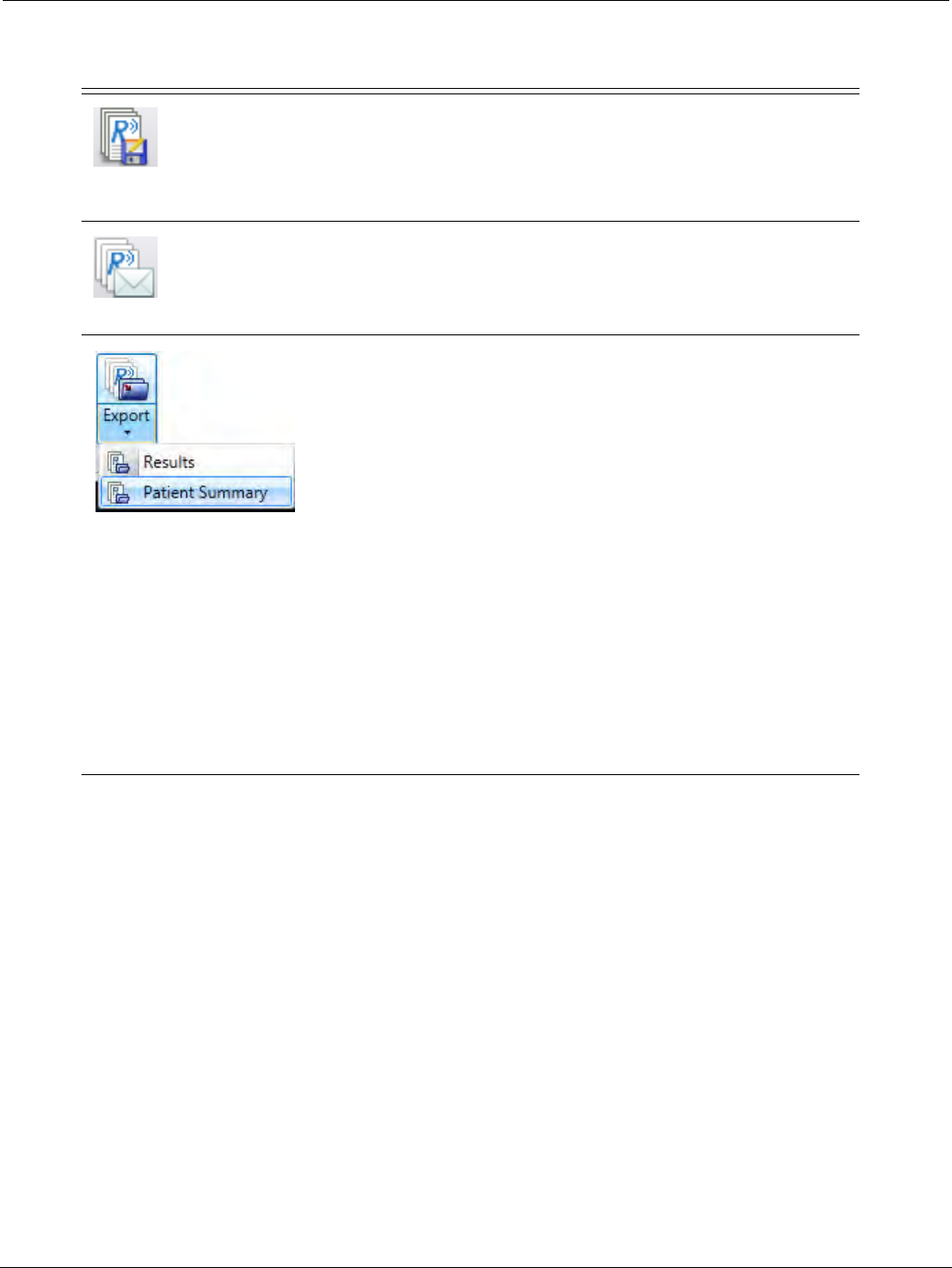
PillCam Capsule Endoscopy
150 Creating a PillCam Capsule Endoscopy Report
Save As
Save the report in the user selected location with the selected
name.
Use only alphanumeric characters in the file name. Make sure
there are no spaces at the beginning or end of the file name. If
spaces are included, the Save option will be grayed out.
Send as Attachment
Send a copy of the capsule endoscopy report in an email
message as an attachment, using your default email program.
The Export button drops down to display the following export
options:
• Export the Results of the capsule endoscopy to a folder in a
directory defined in Settings, for use by a hospital
information system. This folder can contain thumbnail
images and markings, video clips, PDF report, and an XML
file containing the report data, including the path to these
files. This option enables hospital administrators to later
import the files with related attachments from this
predefined directory. To enable this data sharing, the
structure of the input and output of the files must be defined
for the RAPID software. Consult the RAPID v8.3 IT Guide
for more information.
• Export a Patient Summary of the capsule endoscopy to a
folder in a directory defined in Settings. This folder can
contain thumbnail images, PDF report, and video clips that
can be played in Windows Media Player. This option
enables the patient to keep the procedure data archived or
to use as the summary for a physician referral.
Button Function
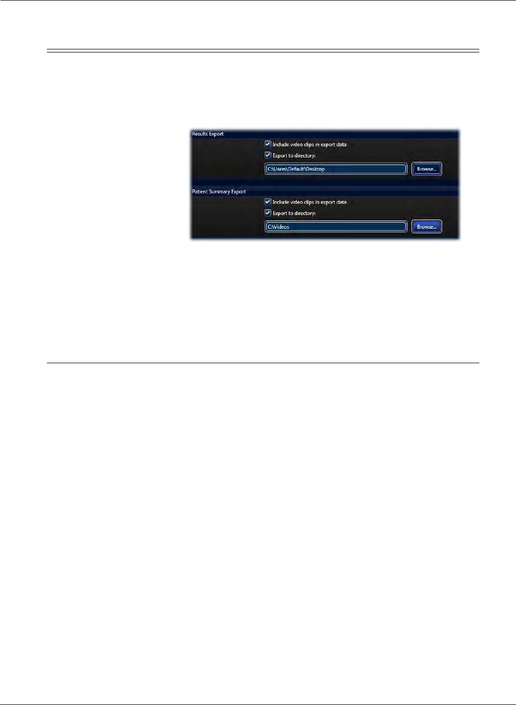
Reviewing and Interpreting RAPID Videos
Creating a PillCam Capsule Endoscopy Report 151
For both export options above, the data can be exported to a
drive directory (which is pre-defined in Settings) or to a CD.
Simply check the box Export to directory and type the
location to where to export. This can be done for both the
Results Export and Patient Summary Export.
If the Export to directory is left unchecked, a screen
prompting to select a location to export will be displayed each
time you export. When exporting the results or patient
summary, a copy of the report is also saved locally on the
computer.
The checkbox Include video clips in export data will enable
saving clips and images during export.
Button Function
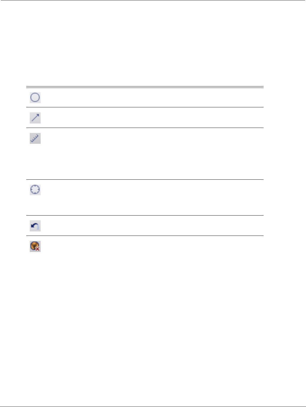
PillCam Capsule Endoscopy
152 Creating a PillCam Capsule Endoscopy Report
Markings Buttons Group
This ribbon group contains tools for marking the selected thumbnails. When viewing a procedure video,
the Thumbnail Editor enables marking and annotating of selected thumbnails. The Markings buttons
group in the Report tab enables deleting a thumbnail and marking already created thumbnails during the
Preview and Review stages. Refer to (Marking Tools on page 134) for complete instructions on marking
thumbnails.
Clinical Tools Buttons Group
The Clinical Tools button group provides access the two diagnostic tools, the RAPID Atlas and the Lewis
Score that are available in RAPID.
Button Function
Creates a circle around an area of interest.
Creates arrow pointing to area of interest.
Enabled only for COLON 2 procedures: Polyp Size Estimation (PSE)
tool: Tool for estimating the size of a suspected polyp after boundaries are
marked, and displays the results on the thumbnail next to the polyp. Polyp
Size Estimation is a tool for research purposes only and the
values generated should not be used in making diagnostic or
treatment decisions.
For ESO/UGI procedures only: Activates Circumference Scale: Displays
circular grid to estimate the circumferential involvement of suspected
pathology such as esophageal varices (see Circumference Scale on
page 135).
Undoes last graphical marking made on the thumbnail.
Deletes selected thumbnail.
Reviewing and Interpreting RAPID Videos
RAPID Atlas 153
RAPID Atlas
RAPID Atlas enables side-by-side comparisons of an image in a video to validated reference images
with Atlas reference images. It provides a database of Capsule Endoscopy images from case studies.
Reference images are searchable by Capsule Endoscopy Structured Terminology (CEST), by
Diagnosis or by Lewis Score terms.
There are three ways to reach the RAPID Atlas:
•Home screen: Click Tools > Atlas.
•View screen: Right-click any thumbnail or video image and select Open Atlas.
Current image from
the video
Atlas images
Categories
of the Atlas
images
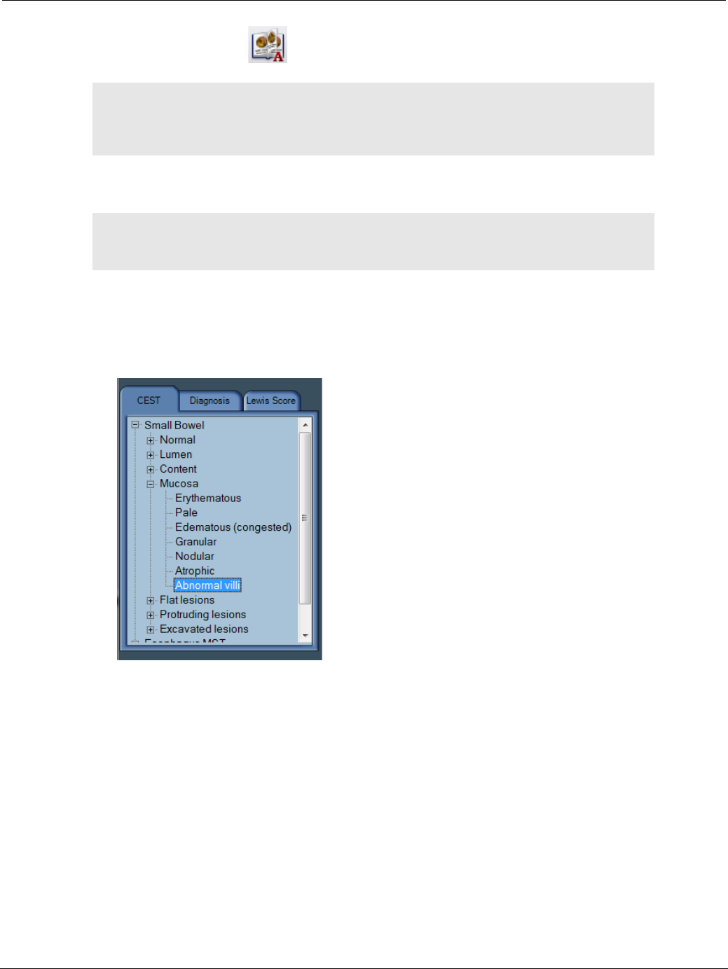
PillCam Capsule Endoscopy
154 RAPID Atlas
•Report screen: Click the button on the Report ribbon.
If there are more than six images available in the RAPID Atlas under a certain category, a scroll bar appears
at the bottom of the screen to allow you to scroll to the rest of the images.
Comparing Video Images to Atlas Images
To compare an image from the current video (Current image) with any of the images from the RAPID
Atlas, select CEST, Diagnosis or Lewis Score tabs to display images in that category.
֠
֠֠
֠Note
You can activate the RAPID Atlas regardless of whether or not a video is
open. If there is no video open, the space for the Current Image in the top
left corner is left blank.
֠
֠֠
֠Note
The RAPID Atlas by no means replaces careful diagnosis from a
trained physician.
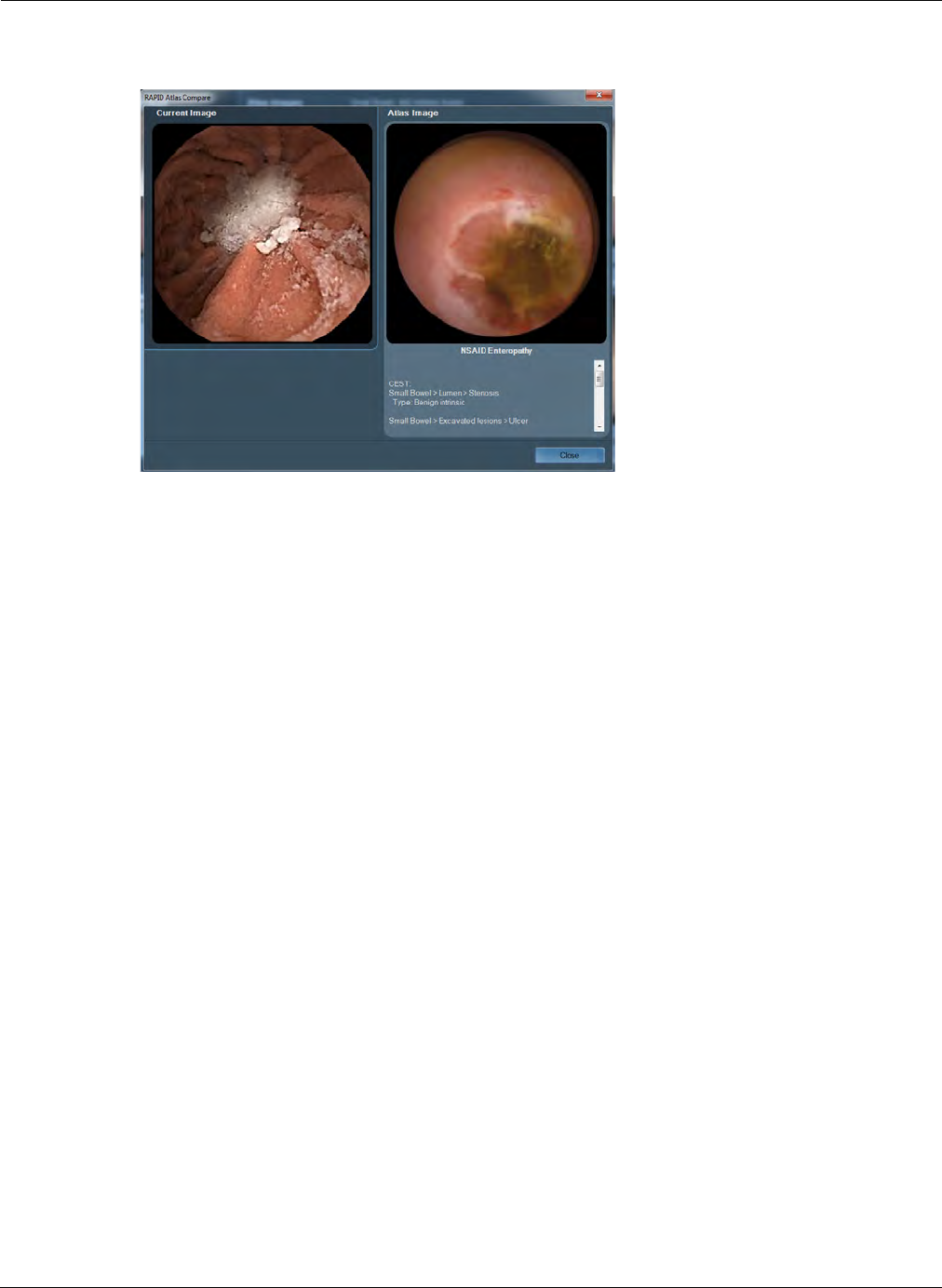
Reviewing and Interpreting RAPID Videos
Lewis Score 155
Select from the images that appear on the right side of the screen by clicking on the image. A
comparison screen with the current image and the Atlas provided image appears.
Both images appear enlarged, side by side. The Current Image on the left and the image from the
RAPID Atlas on the right, including all the comments, case history and any additional available
information on the Atlas image. If there is more text than available space on the screen, a scroll bar
appears next to the text.
Atlas Image Export
You can export images from the Atlas to your computer as follows:
1. Right-click an image on the Atlas screen and select Export. The Export Atlas Image screen
appears.
2. Browse for the location on your computer where you wish to save the image, if desired rename the
image, and click Save.
Lewis Score
The Lewis Score is an aid to diagnosis that provides an approximate measure of degree and extent of
mucosal damage based on direct visual imaging of the small intestine not provided by current
methodologies. The index is based on quantitative and qualitative descriptors relating to three
endoscopic variables: villous edema, ulceration, and stenosis. Combined with other clinical parameters
(symptoms, patient and family history, previous diagnostic tests, lab values), the score could provide a
threshold for differential diagnosis and be used to monitor the progress of treatment.
•An automated scoring system provides greater standardization of disease activity assessment.
•Provides an additional point of evaluation to assist in determining appropriate patient management.
•The Lewis Score facilitates communication and standardization for assessing disease states before,
during, and after treatment.
•Monitoring therapy effectiveness with a standardized score has clinical value.
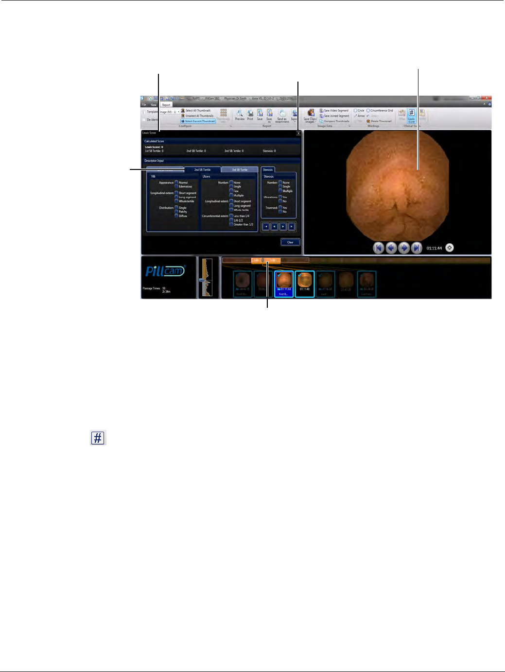
PillCam Capsule Endoscopy
156 Lewis Score
When clicking the Lewis Score button, a scoring index is displayed and the currently selected thumbnail is
shown on the right side.
The tool is used for calculating the Lewis Score and is enabled only when all of the following conditions are
met:
•The video is a PillCam SB video.
•Thumbnail images of the small bowel have been created.
•The first duodenal and first cecal images are marked as landmarks.
To use the Lewis score:
1. Click in the ribbon of the Report screen.
The Lewis Score screen appears.
• The current thumbnail is visible at the right side of the screen.
• The Lewis score (which appears at the top of the screen) and the panel entitled Stenosis (next to
the thumbnail), relate to the entire small bowel.
• The left of the screen is divided into three tabs, relating to video tertiles. A tertile is one third of the
small bowel as calculated by the transit time of the PillCam capsule in the small bowel. The 1st SB
Tertile is the proximal third, the 2nd SB Tertile is the middle third, and the 3rd SB Tertile is the
distal third of the small bowel.
2. Select the tertile you are referring to by clicking the appropriate tab. The selected tertile on the color
bar and the thumbnails selected within it are in color, whereas the rest of the color bar and thumbnails
are darkened.
3. Under Villi, select the:
• Degree of Appearance
Lewis score Selected thumbnailClose Lewis score screen and
return to the Report screen
Current tertile
Current tertile

Reviewing and Interpreting RAPID Videos
Lewis Score 157
• Length of segment for Longitudinal extent
• Correct Distribution
4. Under Ulcers, select the:
• Appropriate Number
• Length of segment for Longitudinal extent
• Correct range for Circumferential extent
5. Select the next tertile and repeat.
6. Under Stenosis, select:
• The appropriate Number
•Yes or No for Ulcerations
•Yes or No for Traversed
The Lewis Score at the top of the screen updates automatically.
Lewis Score Glossary:
Term Meaning
Appearance (of villi) Edema where width of villi is equal or greater than height
of villi. Evaluated villi in mucosa distinct and separated
from an ulcer rather than contiguous to a mucosal break.
Circumferential Extent Portion of the entire image involved by the lesion, based
on the entire lesion including its collar. This value is
entered for the largest ulcer in the tertile.
Few (ulcers) 2–7 lesions.
Lewis Score A capsule endoscopy scoring index for small intestinal
mucosal disease activity (Aliment Pharmacol Ther 27,
146-154).
Long segment Equal to 11–50% of a tertile.
Multiple (ulcers) 8 or more lesions.
Short segment Less than or equal to 10% of a tertile.
Tertile One third of small bowel as calculated by the transit time
of the PillCam capsule in the small bowel.
• 1st SB Tertile is the proximal third of the small bowel.
• 2nd SB Tertile is the middle third of the small bowel.
• 3rd SB Tertile is the distal third of the small bowel.
Whole segment Greater than 50% of a tertile.
Ulceration Mucosal break with white or yellow base surrounded by a
red or pink collar.

PillCam Capsule Endoscopy
158 Generating a Report
Generating a Report
1. Click on the checkbox on the top left of the thumbnail to include it in the report.
• To unselect the thumbnail, click the checkbox again.
• Optionally, use the Select Current Thumbnail button to select or unselect the thumbnail.
• To select all the images, click Select All Thumbnails button.
• To unselect all images, click Unselect All Thumbnails button.
2. Check De-Identify to exclude any patient information from the report.
3. Select the desired template from the list in the ribbon.
4. If you have selected to request electronic signature feature option (see Electronic Signature on
page 178), enter the Windows login password.
5. If you have marked thumbnails in FICE or Blue Mode, you can create a report that includes these
thumbnails alongside the normal thumbnail. To do this, click the dialog box launcher ( ) of the
Configure ribbon group and select the Include normal image alongside FICE or Blue thumbnail
checkbox. Click OK to save the settings.
֠
֠֠
֠Note
Any disclosure of images taken by the PillCam Capsule Endoscopy System
without the patient’s consent might violate the patient’s privacy rights. To
export findings without patient information, click the De-Identify checkbox in
the ribbon.
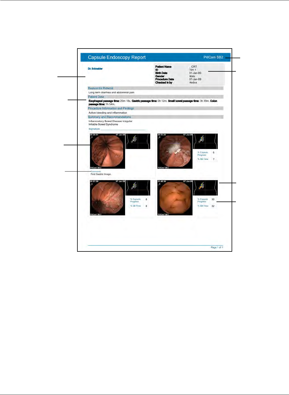
Reviewing and Interpreting RAPID Videos
Generating a Report 159
A report, like the one displayed below is generated and can be saved, printed or sent as an email
attachment. The report template Image (M) + Localization + Progress Indicator is used to generate
the below layout.
Sample report:
Image Data Buttons Group
The Image Data ribbon group contains advanced tools for working with images, video clips and
segments. Short RAPID video segments can be created and combined from different points in the
procedure video. Images can be saved in JPEG format for viewing later. Additionally, video clips
centered on selected images can be generated for the patient to be viewed in Windows Media Player.
Capsule type
Physician
name, clinic
info and logo
are located
here
Patient and
procedure
information
Thumbnail
Localization
Thumbnail
comment
SB progress
indication
Passage times
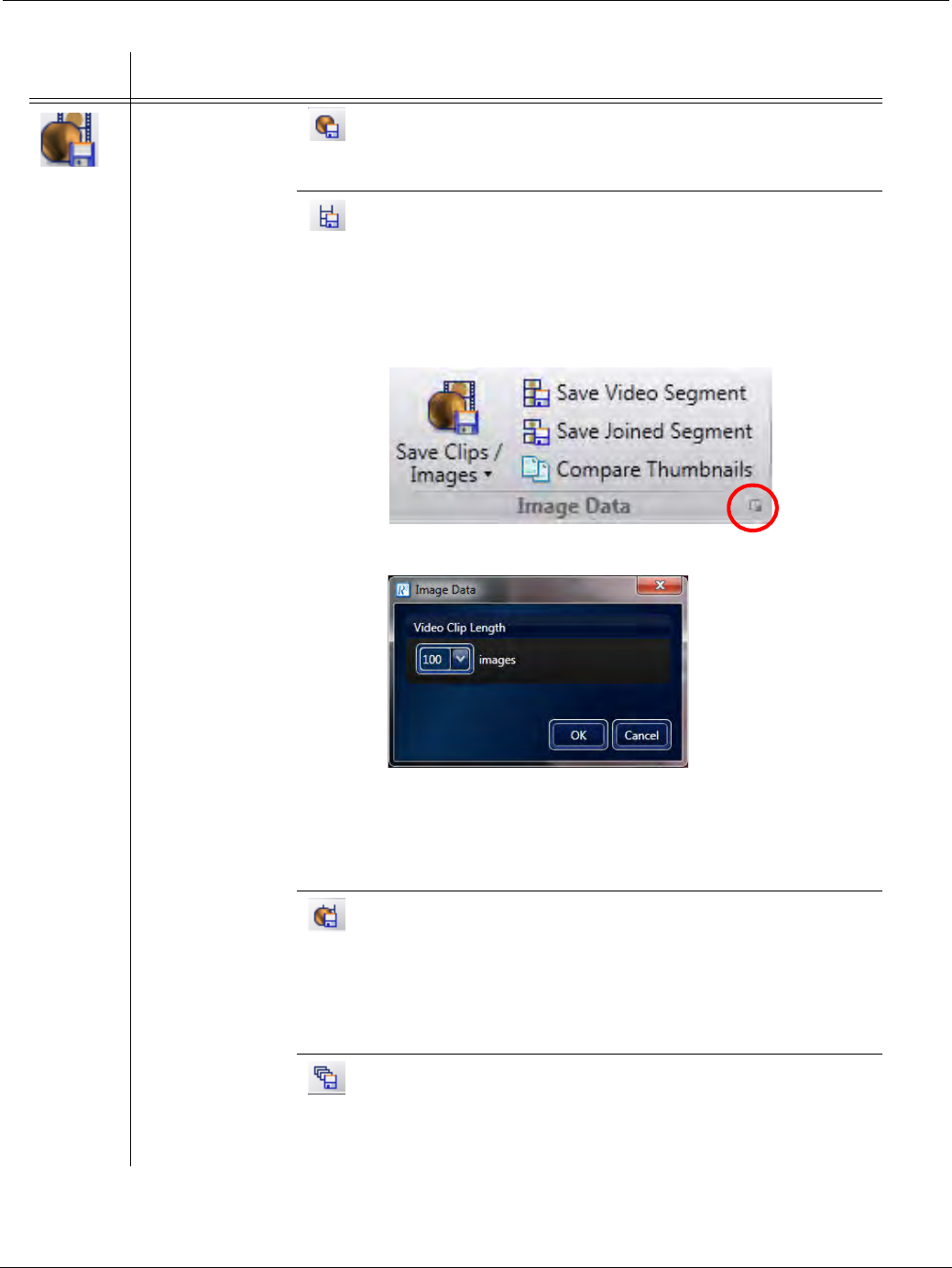
PillCam Capsule Endoscopy
160 Generating a Report
Button Function
Save
Clips/
Images
Create an image
from a thumbnail
or a video clip
from a selection
of thumbnails.
Click the Save
Clips / Images
button and select
from the different
options in the
drop-down list,
and then choose
the directory to
save the file(s).
Images
Only
Creates (a) jpeg(s) of the selected thumbnail(s) and saves
to a folder.
Video
Clips
Only
Creates mpeg video clip(s) surrounding the selected
thumbnail, created from 50 before and 50 after the selected
thumbnail. Video clips can be viewed in Windows Media
Player. The number of images used in creating a video clip
from a selected thumbnail is determined in Image Data
settings. Click the dialog launcher on the bottom right of the
Image Data button group.
This will launch the Image Data screen.
The default length of clips is 100 images. Change and
select the number of desired images for each thumbnail
(50, 100 or 150) and click OK. If upgrading from RAPID v7
and previous setting was 200 images for each thumbnail
then need to redefine it in this screen.
Video
Clips
and
Images
Creates jpeg(s) as well as mpeg clip(s) of the selected
thumbnail. Using this feature will create a jpeg image of
each selected thumbnail and a video clip surrounding the
selected thumbnail. The number of images used in creating
a video clip from a selected thumbnail is determined in
Image Data settings described above. The images and
clips are saved to a selected directory.
Create
Video
Clip
Creates a video clip from the first selected thumbnail to the
last selected thumbnail. This feature is enabled only if at
least two thumbnails are selected. Video clips can be
viewed in Windows Media Player.
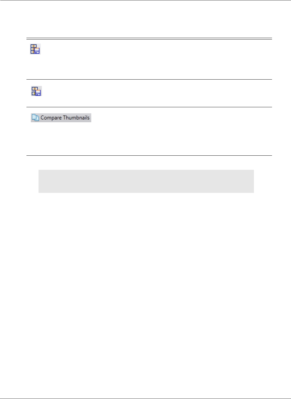
Reviewing and Interpreting RAPID Videos
Generating a Report 161
Button Function
Save Video Segment
Creates a short RAPID video segment from the first thumbnail to
the last thumbnail selected. Video can only be viewed with RAPID
software. The segment is saved in a folder with a copy of the
findings file. Feature is enabled only if at least two thumbnails are
selected.
Save Joined Segments
Joins RAPID video segments created from selected thumbnails.
Video can only be viewed with RAPID software. The segment is
saved in a folder with a copy of the findings file.
Enables comparing of a thumbnail to itself in different display
modes (i.e. FICE or Blue mode) or comparing of two different
thumbnails in the video.
Refer to Comparing Thumbnails on page 141 for more
information.
֠
֠֠
֠Note
To export video clips and images without patient information, click the
De-Identify checkbox in the ribbon.

PillCam Capsule Endoscopy
162 Generating a Report

Setup Requirements 163
Appendix A1
Installing RAPID Software
Setup Requirements
Set up your office to accommodate the new PillCam Capsule Endoscopy System. Review the following
workstation specifications:
Four electrical outlets are required to connect the following components: workstation computer,
monitor, printer, and one cradle. Each additional cradle requires an additional outlet.
RAPID Installation
Before installing RAPID on your computer, it is recommended to be aware of the following
prerequisites and other information:
•Before installation, it is highly recommended to format the hard disk in NTFS format, if not already
done.
•You need at least 1.5 GB free space on your computer to install RAPID.
•For high resolution monitors, RAPID v8.3 offers the capability to resize and maximize the video
view.
֠
֠֠
֠Note
• You may use a Given approved power strip.
• You may connect your monitor directly to the workstation using a DVI
cable (the DVI-VGA adaptor is not required).
!
Caution
Do not connect any component of the PillCam Capsule Endoscopy System to
the same outlet together with any appliance or device that has a high power
requirement (refrigerators, generators, devices with motors, etc.). When
setting up the system, make sure that the total power requirements for all of
the devices connected to a single outlet or circuit do not exceed the rated limit
for that circuit. If you are not sure of the rated limit, please consult your
maintenance department or an electrician.
Do not use a KVM (Keyboard, Video, Mouse) Switch with the PillCam
Capsule Endoscopy System.
֠
֠֠
֠Note
Extra space is needed for air circulation and cable connectors behind the
workstation.

PillCam Capsule Endoscopy
164 Main Endoscopy System Components
•The registration window will open during the first time RAPID is opened and not at the end of the
installation process.
•It is highly recommended to install anti-virus software on the computer running RAPID.
Main Endoscopy System Components
Following is a list of items which you need to connect in order to set up the PillCam Capsule Endoscopy
System:
•RAPID workstation
•Monitor
•Keyboard
•Mouse
•Printer
•PillCam recorder with cradle
Connecting the Components
֠
֠֠
֠Note
At the end of the RAPID installation process, restart your computer.
!
Warning
The RAPID workstation has either an automatic or a manual voltage select
switch. In case the workstation has a manual switch:
• Verify that the workstation’s voltage is set according to the local voltage
prior to connecting the RAPID workstation to the wall outlet.
• If the voltage is not set according to the local voltage, do not connect the
system. Call Given Customer Support.
!
Caution
Voltage mismatch will damage the RAPID workstation.
֠
֠֠
֠Note
For workstations use the monitor's native screen resolution according to the
monitor user manual.

Installing RAPID Software
Connecting the PillCam Recorder Cradle 165
Connecting the PillCam Recorder Cradle
You can connect the cradle only to the USB 2.0 ports that are side by side in a separate slot on the back
panel of the Workstation. If you are not using a Given Workstation, use a USB hub for connecting
more than one cradle to your computer.
Starting RAPID for the First Time
Before using the RAPID software, you must configure and personalize it.
1. For the RAPID workstation, log in as follows:
•Single-user mode: Log in with the username rapid and leave the password blank.
•Multi-user configuration: Log in with your username and password (created for you by the
administrator).
.
2. Click on the desktop.
When you run the software for the first time, a license agreement in English (US) appears on your
screen, which is the legally binding version. A translation in other languages is provided.
֠
֠֠
֠Note
If you use more than one cradle, make sure each one is connected to a
different power outlet.
֠
֠֠
֠Note
RAPID installed not on a Workstation and RAPID Reader do not require
login.
֠
֠֠
֠Note
RAPID workstation only: The Computer Locked screen appears
whenever the workstation is idle for more than half an hour. Only two users
can unlock it:
• The user who was logged in when the RAPID workstation went into
Autolock, by entering the appropriate password, or
• The system administrator.
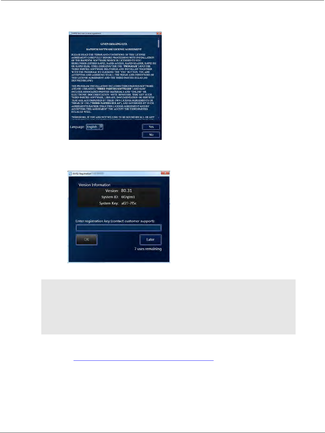
PillCam Capsule Endoscopy
166 Starting RAPID for the First Time
3. Read license agreement and click Yes to accept it. You may select another language to read it, but you
must reselect English to accept the license agreement.
Unrestricted use of RAPID requires registration via the Given Imaging registration center. You must
supply requested information to obtain the Registration Key. The registration screen appears at the end
of the installation process:
4. Obtain a Registration Key via the Given Imaging registration center online or by phone:
•Online: https://portal.givenimaging.com/RapidRegistration
•By phone: Call your local Given Imaging customer support center.
5. Be ready to provide the Given Imaging registration center with the following information:
• System ID (from the registration screen)
• System Key (from the registration screen)
• RAPID DVD serial number (supplied with the DVD)
֠
֠֠
֠Note
Keep the registration window open until you finish the registration. Each time
you open the registration window, a new System Key appears and any
Registration Key based on a previous System Key will not be accepted.
If you click Later, you can open and use the RAPID software, but after seven
uses without registering, you must first perform registration in order to use
RAPID.
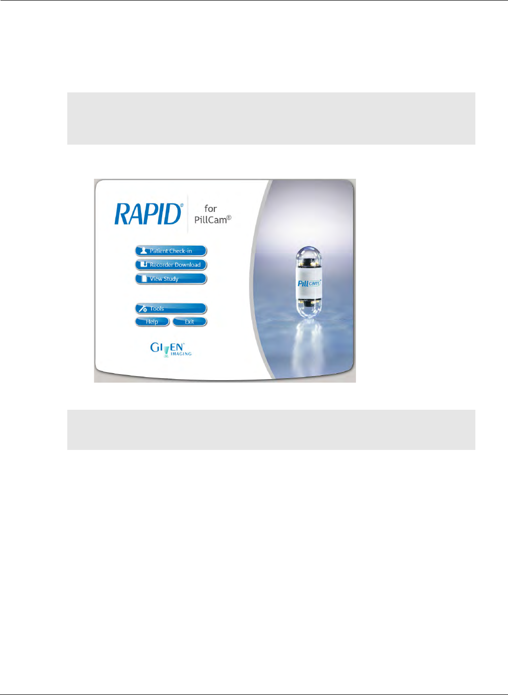
Installing RAPID Software
Starting RAPID for the First Time 167
• Your customer ID
6. Enter the Registration Key received from the Given Imaging registration center using ONLY lower
case letters and numbers.
7. Click OK.
8. The Home screen appears:
֠
֠֠
֠Note
If you do not register during installation, the next six times you open RAPID,
you will be asked to register. After seven uses without registering, you
cannot use RAPID without first performing registration.
֠
֠֠
֠Note
It is recommended to restart your computer at least once a month to ensure
optimal performance.

PillCam Capsule Endoscopy
168 Starting RAPID for the First Time
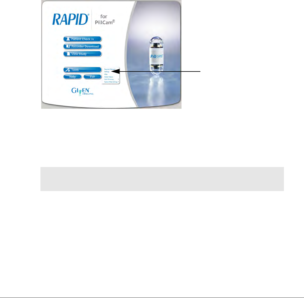
Single or Multi-user Setting 169
Appendix A2
Configuring RAPID Software
Single or Multi-user Setting
When RAPID is installed in a network environment, you can limit the access to RAPID to a group of
authorized users. The system administrator must first set up a network group with all the authorized
users. Once this is done, you must set up RAPID to enable only the users from this network group to
use RAPID.
To enable access to other RAPID users:
1. From the Home screen, select Tools > Settings.
The Settings screen appears.
2. Under the General tab, in the Permissions section, check RAPID users must belong to this
network group: and type in the network group.
3. Click Apply to accept the new settings or click Cancel to close the Settings screen without making
any changes. Clicking OK will accept the new settings and close the Settings screen.
RAPID Workstation Configuration
The RAPID workstation configuration is a stand-alone configuration on a dedicated workstation that
cannot be connected to a network environment. The RAPID workstation supports multi-user
configuration. This allows you to control access to the workstation and to make sure that only
authorized personnel use the appropriate files on the workstation.
By default, the RAPID workstation is configured for a single user with the username rapid and a blank
password. You can continue to use the RAPID workstation this way, or you can use the multi-user
feature. To do so, the administrator (person who sets up and maintains your facility's PCs) creates
additional users, each with a unique username and password, and defines the appropriate access for
them.
֠
֠֠
֠Note
Multi-user setting enables the correct electronic signature for each user
login.
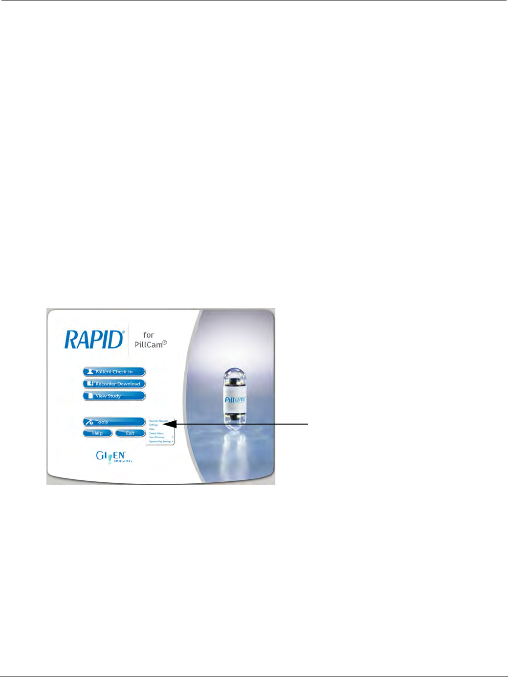
PillCam Capsule Endoscopy
170 RAPID Settings
A user can change the password (usually what the administrator creates is a temporary password) by
pressing Ctrl+Alt+Delete and clicking Change Password.
For Dell T3500 workstations, the administrator logs in with the username administrator and password
administrator.
RAPID Settings
Before you can start using the PillCam Capsule Endoscopy System, you should configure the settings in
RAPID. These include:
•Information about users (those people authorized to use RAPID)
•The names of medical personnel involved in PillCam procedures, so these appear on the report
•Preferences and customization of CE (capsule endoscopy) reports
•...and more
These settings affect all PillCam procedures performed with your RAPID. For example, the medical
personnel names you enter during configuration are available for selection at patient check-in performed on
your PC or workstation. If you set units of measurement to centimeters and kilograms, all patient data is
shown that way.
To reach the Settings screen, select Tools > Settings from the Home screen:
The RAPID software settings are divided by tabs that categorize the settings options into the following
divisions:
•General
•Video
•Report
•Check-in
•Other
Each tab is also sub-divided into separate sections. In the explanations on settings below some of the
information may also be available in the corresponding chapter of the feature. For instance, the Regimen
format section describes in brief the two options for printing a regimen however the Regimen Manager
section displays the printout examples for comparison.
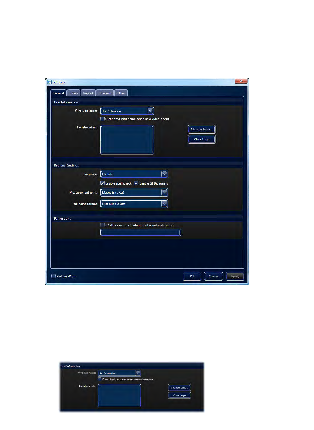
Configuring RAPID Software
RAPID Settings 171
General Tab
From the Home screen, select Tools > Settings. The Settings screen appears showing the General
tab.
After making the changes to the settings, click Apply to accept the new settings or click Cancel to close
the Settings screen without accepting any changes. Clicking OK will accept the new settings and close
the Settings screen.
User Information Section
The physician name, facility name and address (where the procedure was performed) and the facility
logo entered during configuration show on the endoscopy report.
To define user information:
1. From the Home screen, select Tools > Settings. The Settings screen appears.
2. In the User Information section, type in the Physician Name.
If you do not enter the physician name here, no physician name appears on the CE report.
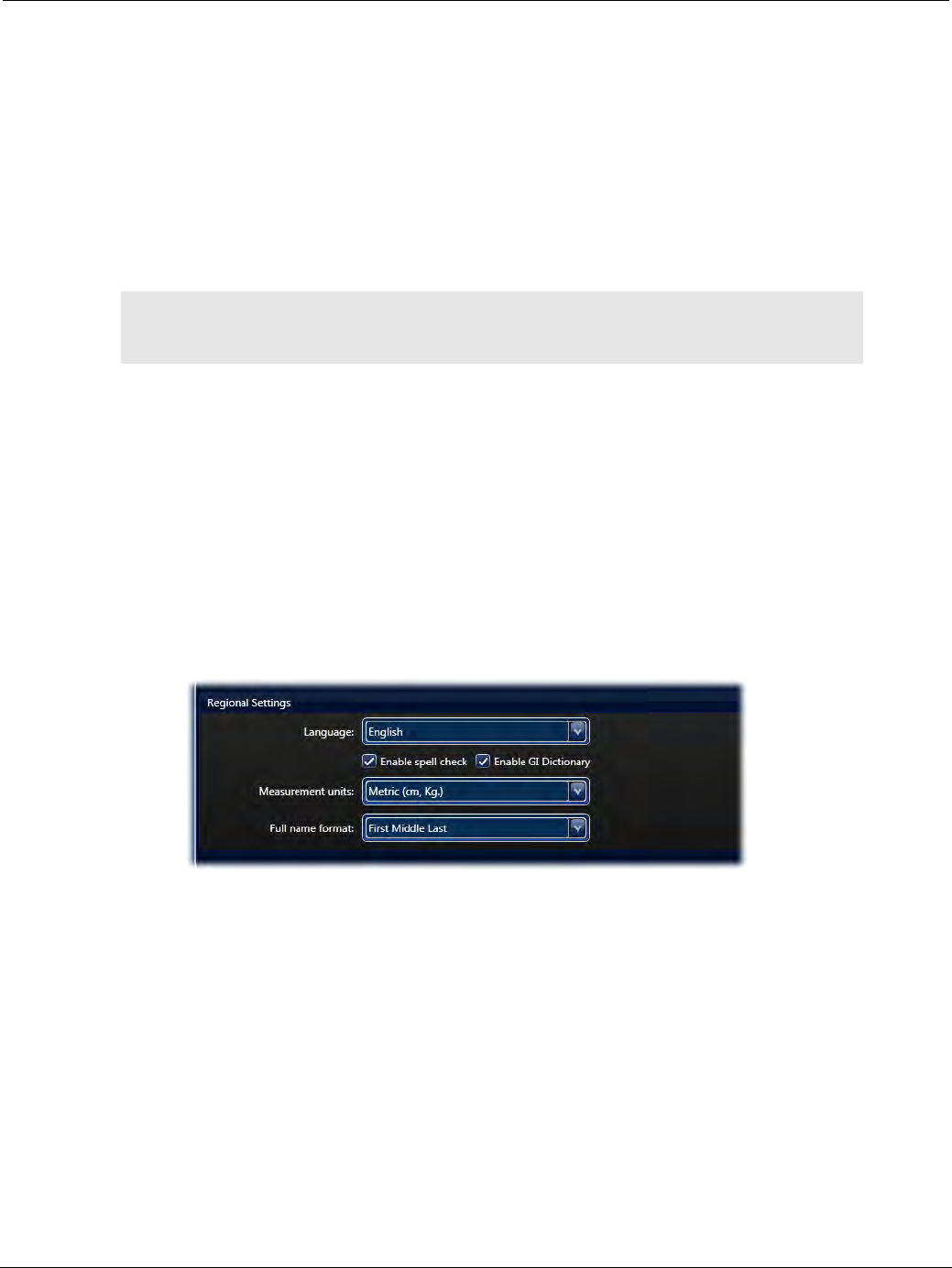
PillCam Capsule Endoscopy
172 RAPID Settings
3. If more than one physician uses this installation of RAPID, select Clear physician name when new
video opens. If selected, you can manually enter the physician name for each video when needed.
4. In Facility details, type in the facility information (for example, address and telephone number).
5. To add your facility’s logo to reports, click Change Logo.
a. Navigate to the logo file (jpeg image) you previously saved on your computer and select the
file.
b. Click Open to set the file as the report logo.
c. To clear the selected logo and prevent any logo from appearing on the report, click Clear Logo.
6. Click Apply to accept the new settings or click Cancel to close the Settings screen without making
any changes. Clicking OK will accept the new settings and close the Settings screen.
Regional Settings Section
By default, the RAPID software is set to English and uses metric units of measure. You may change these
settings.
To change the language:
1. From the Home screen, select Tools > Settings. The Settings screen appears.
2. Under the General tab, in the Regional Settings section, open the Language list and select the
desired language.
3. Enable spell check is selected by default. Un-check to disable spell check.
4. Enable GI Dictionary is selected by default. Un-check to disable the GI Dictionary.
5. Open the Full name format list and select the desired name format.
6. Click Apply to accept the new settings or click Cancel to close the Settings screen without making
any changes. Clicking OK will accept the new settings and close the Settings screen.
To change the unit of measure:
1. From the Home screen, select Tools > Settings. The Settings screen appears.
2. Under the General tab, in the Regional Settings section, open the Measurement units list and select
the desired measurement system.
֠
֠֠
֠Note
The Logo used in the report is copied to a RAPID directory. The original is
not affected.
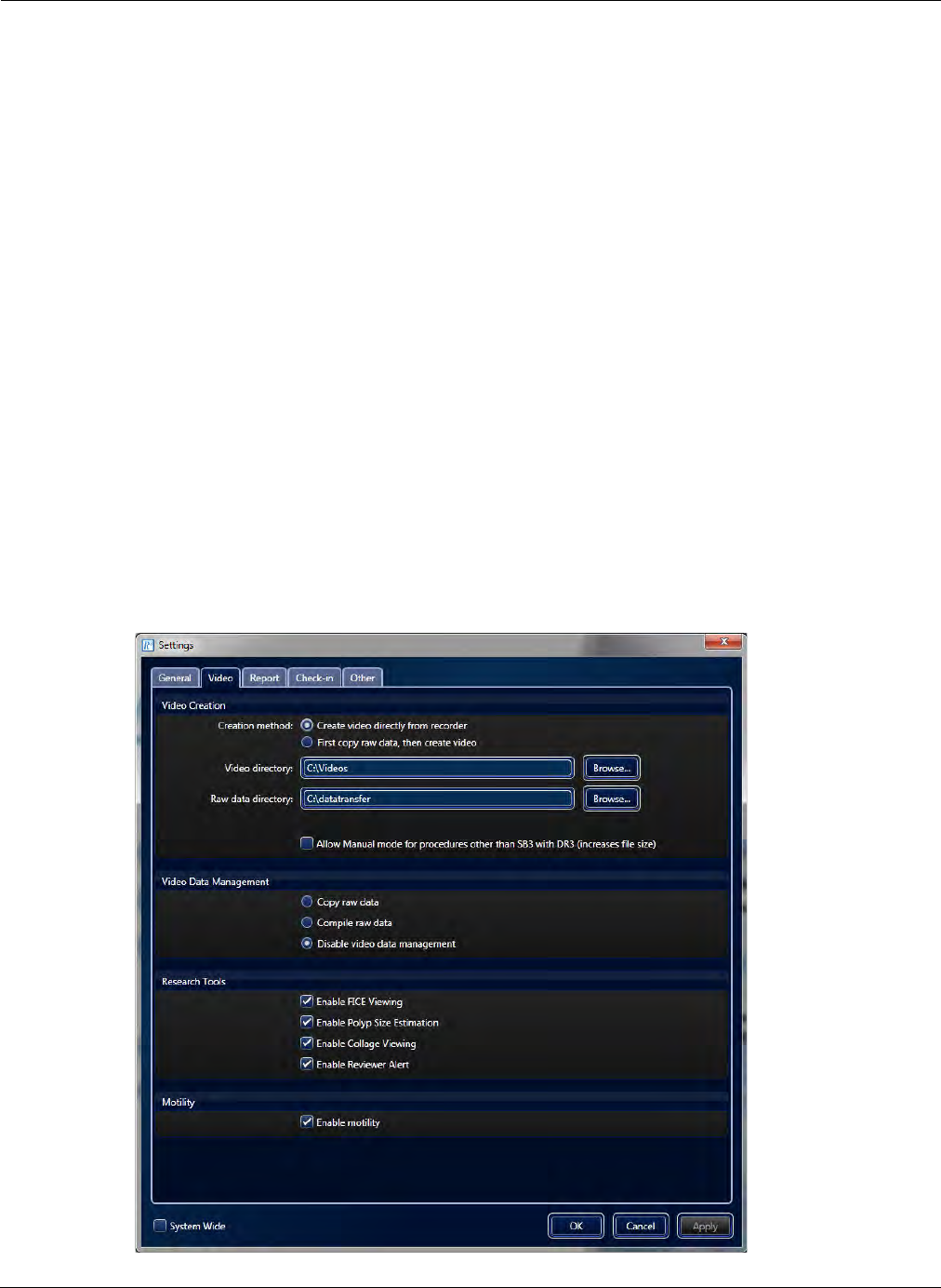
Configuring RAPID Software
RAPID Settings 173
3. Click Apply to accept the new settings or click Cancel to close the Settings screen without
making any changes. Clicking OK will accept the new settings and close the Settings screen.
Permissions Section
Enables the system administrator to specify a list of local or domain user groups that are allowed to
open RAPID.
To specify a user group for RAPID:
1. From the Home screen, select Tools > Settings. The Settings screen appears.
2. Under the General tab, in the Permissions section, type in the user group you wish to allow
access to RAPID.
The domain must be entered in this format:
<domain name>\<group name>;<local group name>;<next group>
This feature is enabled only if System Wide check-box is selected.
Video Tab
From the Home screen, select Tools > Settings. Then select the Video tab.
After making the changes to the settings, click Apply to accept the new settings or click Cancel to close
the Settings screen without accepting any changes. Clicking OK will accept the new settings and close
the Settings screen.

PillCam Capsule Endoscopy
174 RAPID Settings
Video Creation Section
You can copy the video data from the PillCam recorder to the computer, a USB storage device, or a DVD
without compilation into a video.
To determine video creation method:
1. From the Home screen, select Tools > Settings.
2. Select the Video tab.
3. In Video Creation, under Creation method select either Create video directly from recorder or
First copy raw data, then create video.
4. You can change the following directories by typing the desired location or clicking Browse to select a
folder:
•Video directory: destination location for created videos
•Raw data directory: destination location for copying raw from the PillCam recorder
5. You can also enable the following feature with the video by selecting the check box Allow Manual
mode for procedures other than SB3 with DR3 (increases file size).
Video Data Management Section
To enable video data management:
1. From the Home screen, select Tools > Settings.
2. Select the Video tab.
3. Under Video Data Management, Disable video data management is selected by default.
4. Select:
•Copy raw data: copy data ONLY. This takes less time, so the PillCam recorder is freed up sooner.
The copied data can later be processed to create a video.
•Compile raw data: process for video creation.
5. Click OK to accept the new settings or click Cancel to close the Settings screen without making any
changes.
֠
֠֠
֠Note
Select First copy raw data, then create video to quickly release the
PillCam recorder after the raw data is copied without waiting for full video
compilation.
You must manually clear raw data files because this video creation method
can rapidly fill up your hard drive.

Configuring RAPID Software
RAPID Settings 175
Research Tools Section
To enable video research tools:
1. From the Home screen, select Tools > Settings.
2. Select the Video tab.
3. Under Research Tools, select:
•Enable FICE Viewing (for SB, ESO 3/UGI and COLON 2 videos only)
•Enable Polyp Size Estimation (for COLON 2 videos only)
•Enable Collage Viewing (for COLON 2 videos only)
•Enable Reviewer Alert (for COLON 2 videos compiled with RAPID v8.3 only)
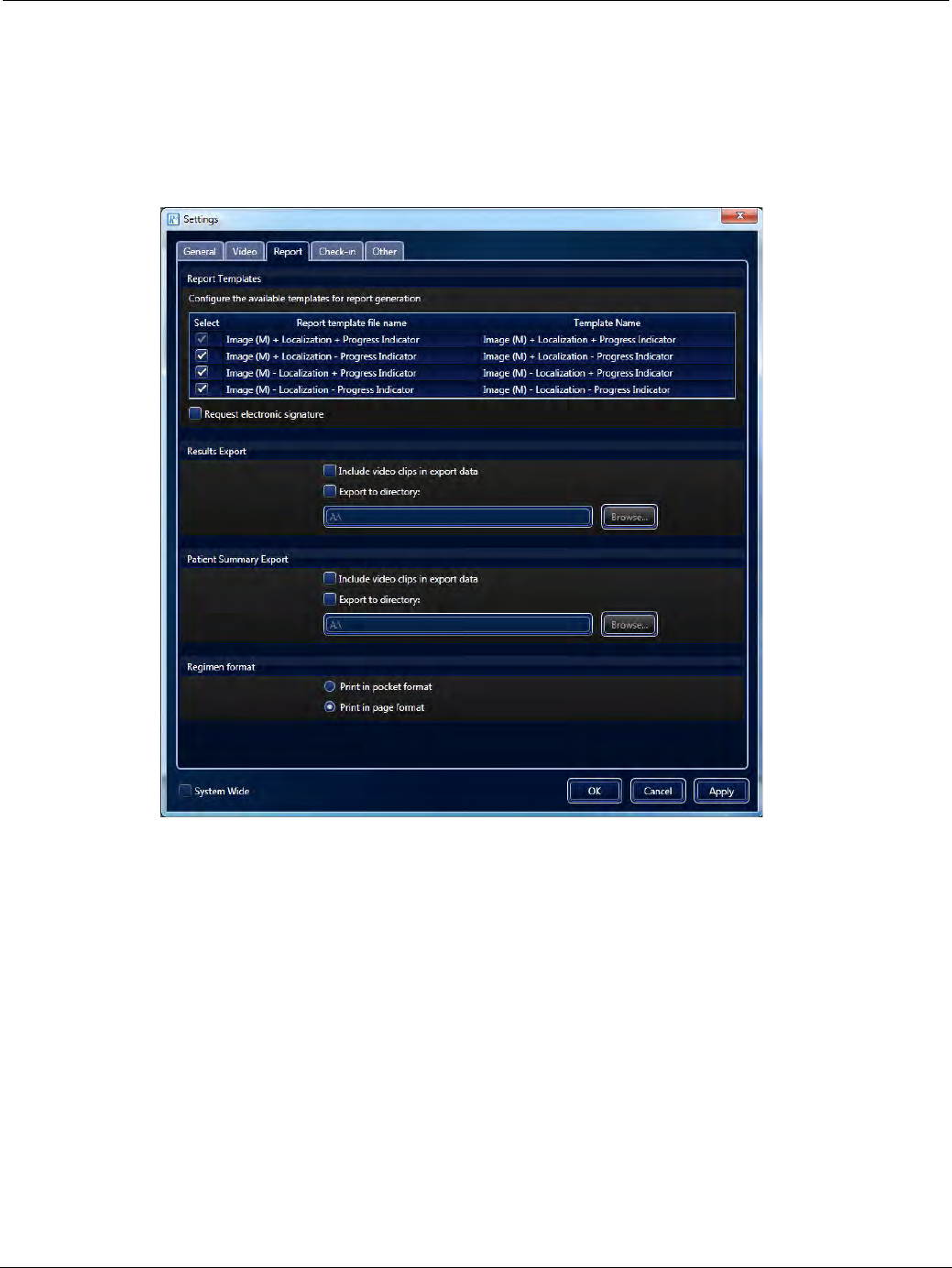
PillCam Capsule Endoscopy
176 RAPID Settings
Report Tab
From the Home screen, select Tools > Settings. Then select the Report tab.
After making the changes to the settings, click Apply to accept the new settings or click Cancel to close the
Settings screen without accepting any changes. Clicking OK will accept the new settings and close the
Settings screen.
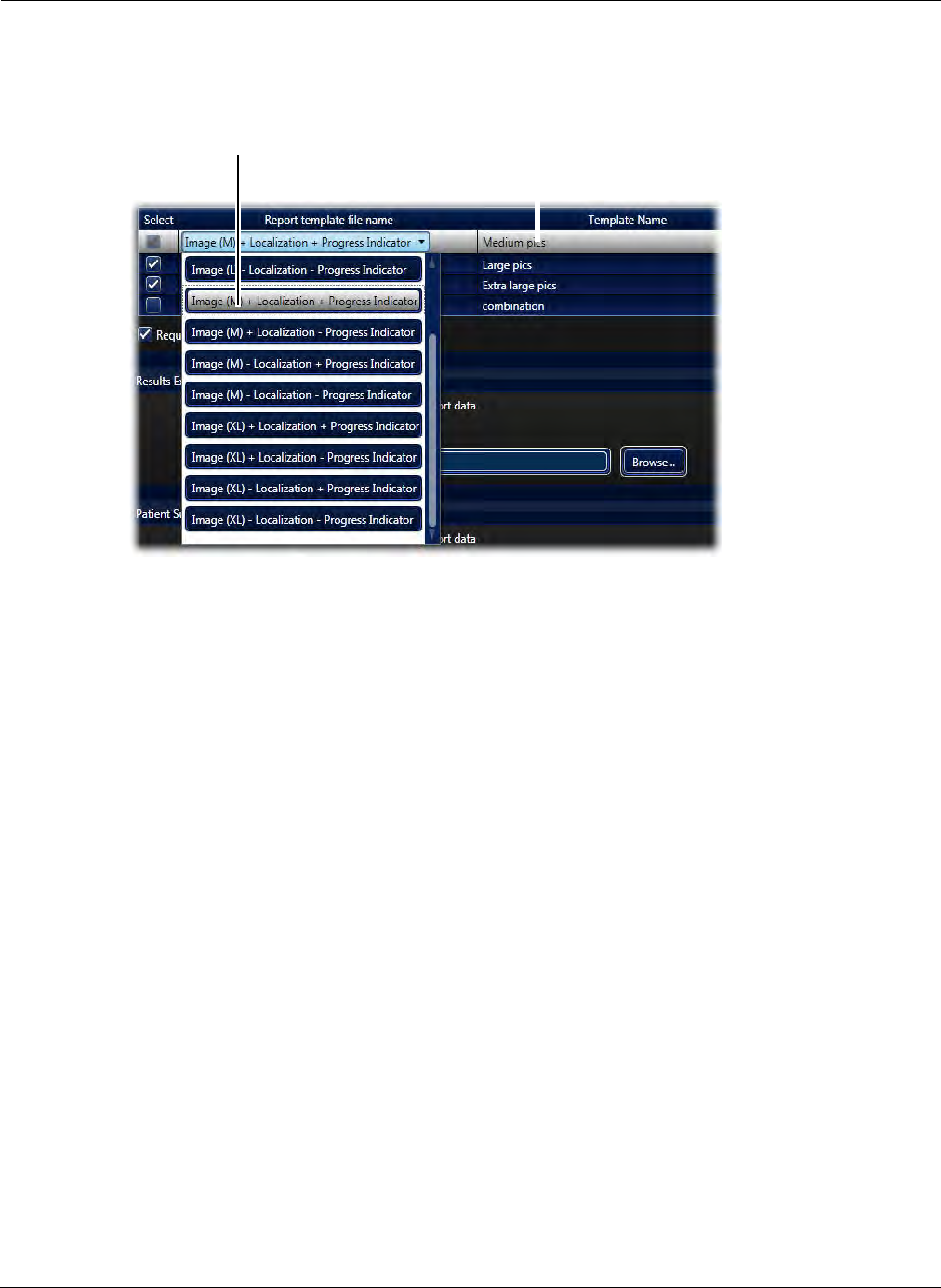
Configuring RAPID Software
RAPID Settings 177
Report Templates Section
To configure which of the pre-loaded templates will appear ready to use in the drop-down list in the
Report ribbon, go to Home screen > Settings > Report tab.
Every template file name contains a variation of the features that determine the layout of the patient
report. The abbreviations of features in the filename are explained below:
•Localization: Include localization of the GI tract with an indication of the progress of the capsule
during the time that the thumbnail was taken. This is available only for SB and COLON procedures
performed with sensor array.
•Progress Indicator: Displays both the capsule progress and the time elapsed in the Small Bowel in
percent (%) format.
•Image (L): Displays large thumbnails on the report printout. Note that this will use up additional
paper when printing the report.
•Image (XL): Displays extra-large thumbnails on the report printout. Note that this will use up
additional paper when printing the report.
The templates are defined by either an inclusion (+) or exclusion (-) of the features described above. So
for example, the template file name Localization + Progress Indicator + Image (M) would generate a
report displaying the localization of GI tract with the progress indicator next to the medium-sized
thumbnails. The report printout sample on page 159 is based on this template.
As another example, the template Image (L) will display only large thumbnails, without localization of
the GI tract and without progress indicator.
After selecting the desired templates in Settings, the default template name in the drop-down list would
be the template file name. The template name can be edited by clicking once in the template name field
Select templates for
Report layout
Template name that will
appear on Report ribbon
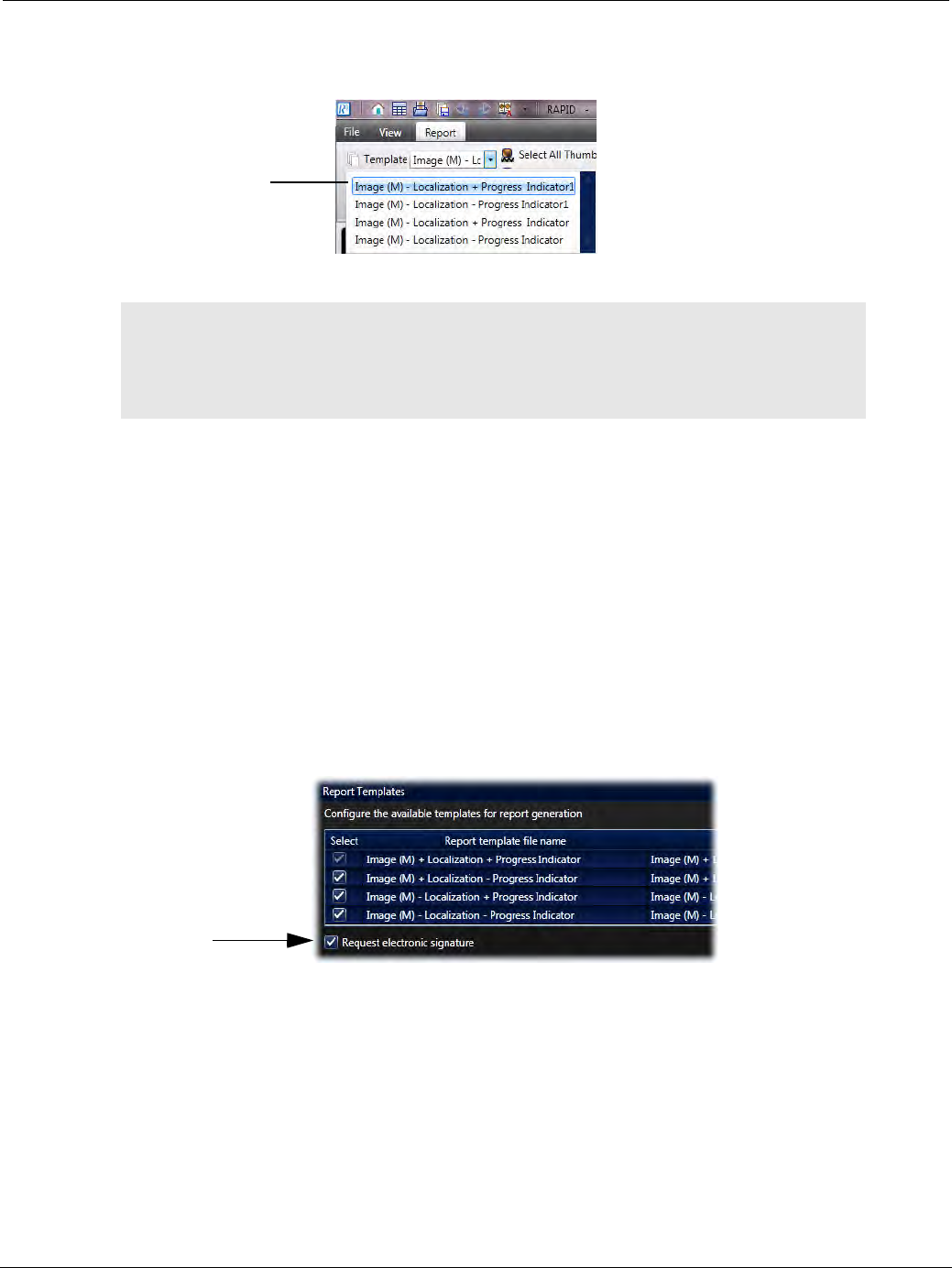
PillCam Capsule Endoscopy
178 RAPID Settings
and editing the name. Click Apply or OK to save the settings. The four templates selected and edited will
appear in the Report tab drop-down list.
Electronic Signature
An electronic signature is a way to allow authorized personnel to approve and validate a document without
having to physically print it out and sign it manually. With this feature you may produce a signed capsule
endoscopy report with the signing physician's name appearing in the signature field, where the electronic
signature feature is the only way to achieve this, thus signifying signature of the document. Electronic
signatures are widely accepted as legally valid. By default, the electronic signature feature is disabled.
To activate use of electronic signature in Settings:
1. From the Home screen, select Tools > Settings. The Settings screen appears.
2. Select the Report tab.
3. In the Report Templates section, select Request electronic signature and click Apply or OK.
֠
֠֠
֠Note
From the four templates in the settings screen, the templates can be
unselected so as not to display in the RAPID software by clicking the
checkbox on the left side of the template file name in the Settings screen. By
default, one template must be selected.
Template name
will appear on
Report ribbon
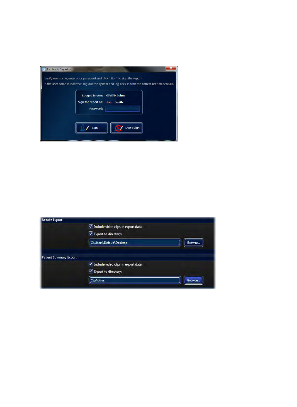
Configuring RAPID Software
RAPID Settings 179
Use of the Electronic Signature
Once the electronic signature is activated, the signature will be displayed when creating or printing a
report. The signature will appear as electronically signed by: your first and last name, as it appears in
your user profile (in Windows).
As a result, whenever a report is saved, printed, exported or sent as an attachment by email, the
following screen appears.
Enter your login password and click Sign.
Results Export and Patient Summary Sections
For both Results and Patient Summary Exports, the data can be exported to a drive directory (which
is pre-defined in Settings) or to a CD. Simply check the box Export to directory and type the location
to where to export. This can be done for both the Results Export and Patient Summary Export.
The checkbox Include video clips in export data will enable saving clips and images during export.
If Export to directory is left unchecked a screen pops up prompting you to select a location to export
each time you export. When exporting the results or patient summary, a copy is also saved locally on the
computer.
Regimen Format Section
The Regimen Manager enables two convenient print layout options for the post-capsule ingestion
instructions. The desired default layout can be configured selecting either pocket format or page
format (see Print Layout of the Post-Capsule Ingestion Instructions on page 190).
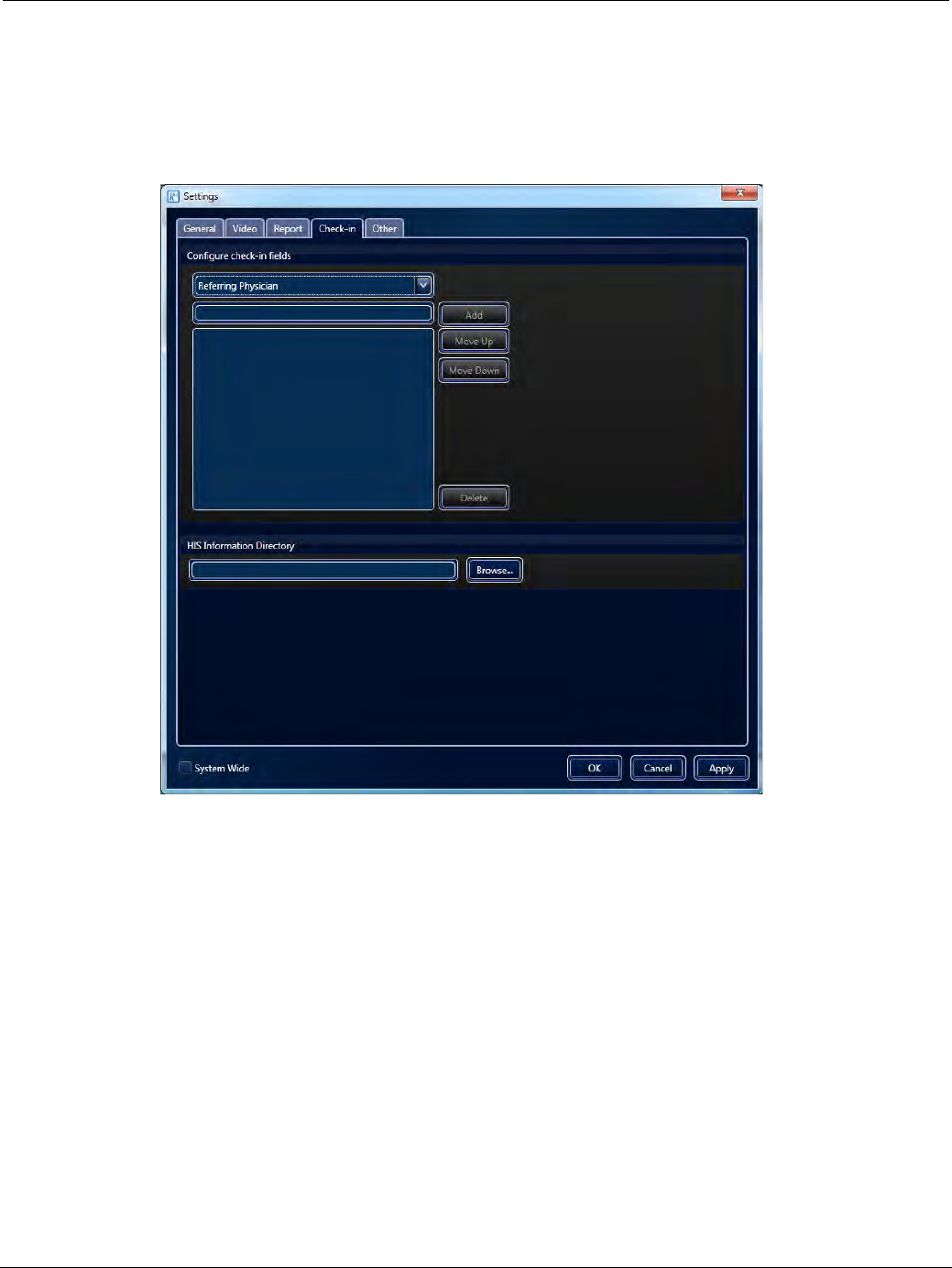
PillCam Capsule Endoscopy
180 RAPID Settings
Check-in Tab
From the Home screen, select Tools > Settings. Then select the Check-in tab.
After making the changes to the settings, click Apply to accept the new settings or click Cancel to close the
Settings screen without accepting any changes. Clicking OK will accept the new settings and close the
Settings screen.
Configure Check-in Fields Section
To add frequently used Check-in fields such as physician name and ICD Code:
1. Select a Check-in field from the list at the top of the screen and enter the corresponding values for this
field.
2. Click Add.
In the window below the text box you can see all the values already entered for this check-in field.
3. Use the Move Up and Move Down buttons to organize the defined values.
HIS Information Directory Section
If a HIS system is available, type the desired location from which to import patient data for Check-in or
click Browse to navigate to the desired folder.
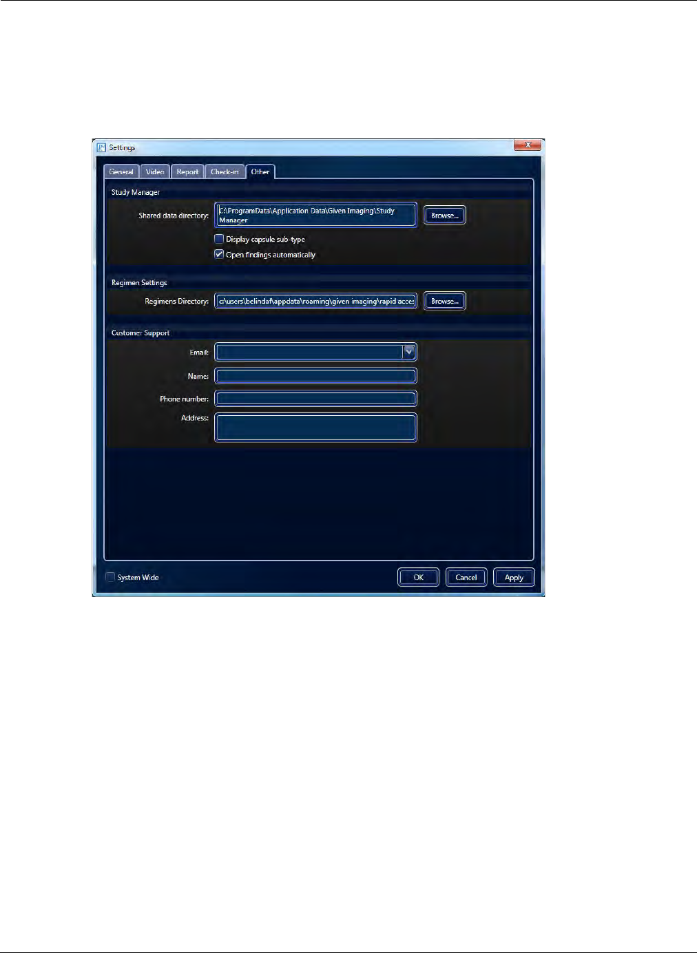
Configuring RAPID Software
RAPID Settings 181
Other Tab
From the Home screen, select Tools > Settings. Then select the Other tab.
After making the changes to the settings, click Apply to accept the new settings or click Cancel to close
the Settings screen without accepting any changes. Clicking OK will accept the new settings and close
the Settings screen.
Study Manager Section
Created user columns in the Study Manager are available for use by other users. In the Shared data
directory, type the desired location of the shared path or click Browse to navigate to it.
Display capsule sub-type: Also shows the sub-type in the Capsule Type column of the Study
Manager.
Open findings automatically: When opening a video from the Study Manager the findings file will
open as well (if available).
Regimen Settings Section
Displays the Regimen Settings location configured for your system. If this needs to be changed, type
the location of or click Browse to navigate to the desired folder.
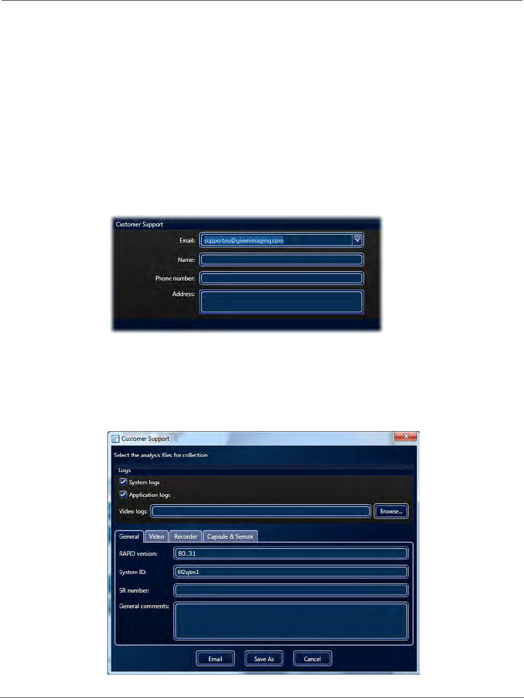
PillCam Capsule Endoscopy
182 RAPID Settings
Customer Support Section
This section explains how to configure the RAPID software to allow the collection of performance data
for analysis by the Given Imaging support department. RAPID allows for a one-click extraction and
sending of RAPID log files for convenient customer support access. This enables easier tracking and
background information on the RAPID software being used in the event of a malfunction. In order to
enable this feature, you must first enter the email address for your regional Given Imaging Customer
Support.
To enter Customer Support details:
1. From the Home screen, select Tools > Settings. This opens the Settings screen.
2. Select the Other tab.
3. Under Customer Support, click the Email list and select your regional customer support center. If it is
not listed, you can type in the email address manually.
To extract and send RAPID log files:
In the event that you are requested to send the log files to Given Imaging Customer Support the steps
below should be followed:
1. From the Home screen, click Help > Customer Support > Collect Analysis Files.
The following screen appears.

Configuring RAPID Software
Regimen Manager 183
2. Click the General, Video, Recorder, Capsule & Sensor tabs and enter the information in the
editable fields. If there is a video that needs to be sent as well, click the Browse button in Video
logs and navigate to the video.
3. Click the Email button to send the data to Given Imaging Customer Support.
Regimen Manager
The Regimen Manager is a RAPID software tool for generating pre-capsule ingestion regimen
instructions to give to the patient before the day of the procedure and post-capsule ingestion regimen
instructions for the patient to follow during the capsule endoscopy procedure. The Regimen Manager
allows the creation and maintenance of a library of different instruction sets appropriate for different
circumstances to choose from. The post-capsule ingestion regimen instruction set is selected during
Check-in from the regimen library. The instruction set is uploaded to the PillCam recorder DR3 and is
used for creating timely alerts to the patient during the procedure. Currently only procedures using the
PillCam recorder DR3 with PillCam COLON 2 capsule utilize regimen instructions during the
procedure.
This section gives an overview of the Regimen Manager tool as well as the following:
•Using the Regimen Manager on page 183
•Pre-Ingestion Patient Instructions on page 185
•Post-Ingestion Patient Instructions on page 187
•Printing the Patient Instructions on page 190
•Print Layout of the Post-Capsule Ingestion Instructions on page 190
Using the Regimen Manager
To access the Regimen Manager from the Home screen select Tools > Regimen Manager. The
Regimen Manager comes pre-loaded with several pre- and post-capsule ingestion regimens for you to
use. Any regimen to be used must be in an approved status by the user. None of the pre-loaded
regimens in the library are in an approved status. Any regimen in the library to be valid and selectable
in the menu must be reviewed and made approved by a physician. You can approve a regimen only
when it is opened and from within the opened regimen screen.
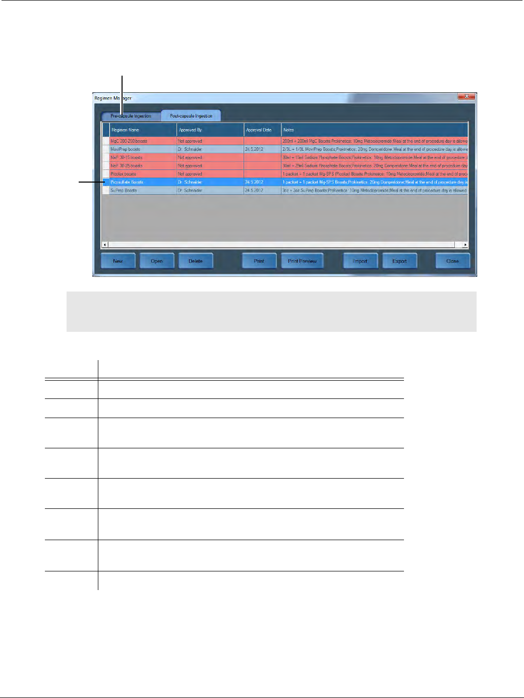
PillCam Capsule Endoscopy
184 Regimen Manager
Open a regimen by double clicking it or selecting it and clicking the Open button.
The buttons on the bottom of the Regimen Manager screen perform the following:
֠
֠֠
֠Note
If upgrading the RAPID software to version 8.3 all previously approved
regimens will require approval again.
Button Action
New Creates a new regimen to add to the regimen library.
Open Opens the selected regimen.
Delete Deletes the selected regimen. All regimens, including the pre-loaded
regimens can be deleted.
Print Prints the selected regimen. This button becomes available only after a
regimen is approved.
Print
Preview
Displays a preview of the selected regimen before printing. This button
becomes available only after a regimen is approved.
Import Imports a regimen from a selected directory. The default directory is
designated in Settings.
Export Exports a regimen to a selected directory. The default directory is
designated in Settings.
Close Closes the Regimen Manager.
Select pre-capsule ingestion
or post-capsule ingestion
regimens
Approved
Regimen
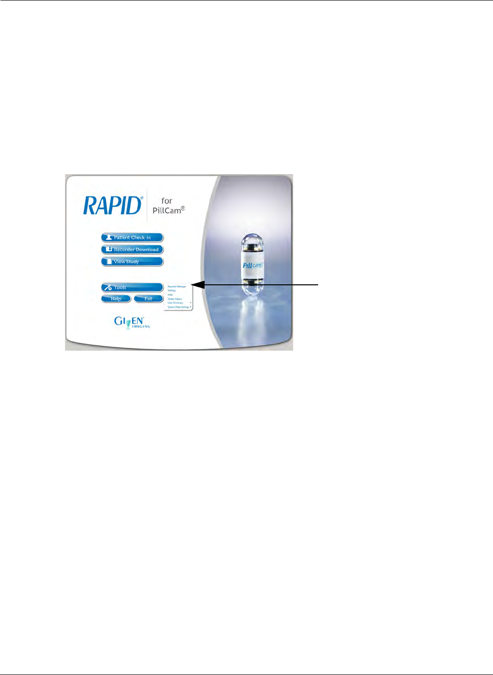
Configuring RAPID Software
Regimen Manager 185
Pre-Ingestion Patient Instructions
A completely clean colon is essential for a successful examination. Therefore, the patient must
carefully adhere to the preparation procedure for cleaning the GI tract. The Regimen Manager can be
used to create instructions for the patient to follow before the planned PillCam COLON capsule
endoscopy procedure. The instructions identify the type of medications and liquid intake that must be
taken in order to prepare the colon prior to the procedure. These preparation instructions can be edited
and printed out for the patient beforehand. There could be different instructions appropriate to different
circumstances or available medications. Pre-capsule ingestion patient instruction sets can be created
and edited to maintain a library of valid usable regimen instruction sets.
To edit and save pre-capsule ingestion regimen instructions:
1. From the Home screen, select Tools > Regimen Manager.
The Regimen Manager screen appears.
2. Select the Pre-capsule Ingestion tab. The Regimen screen appears displaying all available pre-
ingestion regimens.
3. Open a regimen by double clicking it or selecting it and clicking Open.
The Notes field on the top right allows the typing of information for the specific regimen. It will
not appear on the printout.
4. To add an instruction to the regimen click Add Instruction and designate the appropriate start and
end day and time of the instruction using the drop-down lists in the adjacent columns. When typing
the instructions in Instructions for Patient a pop up screen appears if the text is too long.
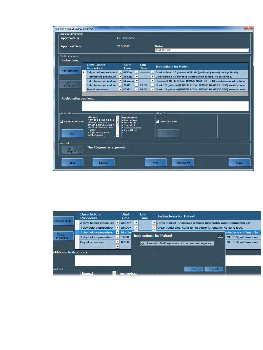
PillCam Capsule Endoscopy
186 Regimen Manager
5. Type any other information in the Additional Instructions field that will appear on the printout for
that specific regimen.
6. The regimens contain editable instructions in the Instructions for Patient column. Clicking the
instruction will prompt an editor. Information represented in brackets [ ] is generic and is intended for
completion by local staff with consideration to local names of the solution. Make any changes and
click OK to save the updated instruction.
7. If you wish to include your instructions for a liquid diet, check the Clear liquid diet check box. This
will display the list of Allowed and Not Allowed food or drink items. To modify these lists, click the
Edit button. Make necessary edits and then click Save and Close.
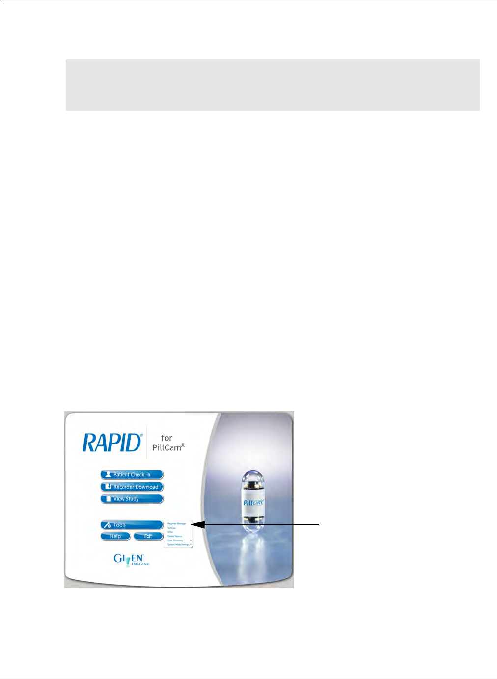
Configuring RAPID Software
Regimen Manager 187
8. If you wish to include your instructions for a fiber diet, check the Low fiber diet check box. This
will display the list of Allowed and Not Allowed food or drink items. To modify these lists, click
the Edit button. Make necessary edits and then click Save and Close.
9. If you want to print this regimen later after saving it, click Approve Regimen.
10. When you are finished, click Save. This adds the regimen you created to the library of regimens
available.
Post-Ingestion Patient Instructions
In order to ensure a successful procedure after capsule ingestion, the patient must carefully adhere to
the personalized Post-Capsule Ingestion Instructions. The post capsule ingestion regimen instruction
uploaded to the PillCam recorder DR3 during check-in enables the patient to be alerted during the
procedure as to specific medications or liquids that must be ingested at the specified times. Once a
regimen is selected from the regimen library during the patient check-in stage (before capsule
ingestion) the schedule of instructions are programmed into the PillCam recorder for that particular
procedure in a numbered sequence.
Using a tactile and sound alert, the PillCam recorder will notify the patient of a scheduled event by
displaying the event number on the LCD screen. The patient needs to refer to this number on the Post-
Ingestion Patient Instructions printout given by the medical staff. The following section explains how
to edit the Post-Ingestion Patient Instruction. Approved regimens can be selected during the patient
check-in stage and can be printed (see Performing Patient Check-in on page 43).
To edit and save a post-ingestion regimen:
1. From the Home screen, select Tools > Regimen Manager.
2. Select the Post-capsule Ingestion tab. The Post-capsule Ingestion Regimen screen appears
displaying all the post-ingestion regimens available.
3. Open a regimen by double clicking it or select it and click Open.
֠
֠֠
֠Note
The Liquid Diet and Fiber Diet instructions are shared across all pre- capsule
ingestion regimens. Any changes to the instructions will apply to all the pre-
capsule ingestion regimens.
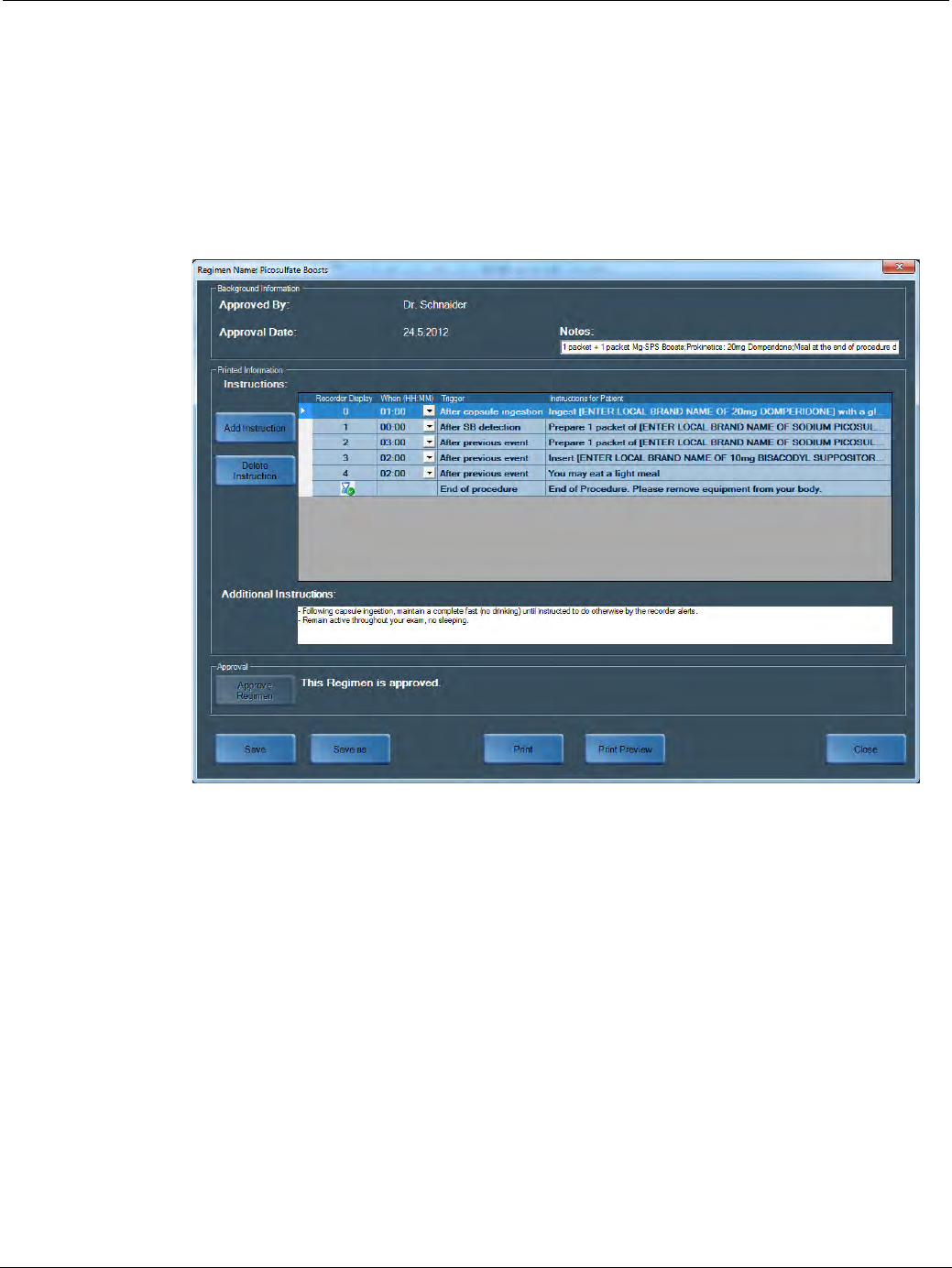
PillCam Capsule Endoscopy
188 Regimen Manager
The Notes field on the top right allows the typing of information for the specific regimen. It will not
appear on the printout.
4. To add an instruction to the regimen click Add instruction and select from the list the appropriate
timing for the instruction. When typing the instructions in the Instructions for Patient a pop-up
screen appears if the text is too long.
5. Type any other information in the Additional Instructions field that will appear on the printout for
that specific regimen.
6. The Recorder Display column on the left lists the event numbers that will be displayed on the PillCam
recorder LCD screen along with the corresponding alert.
7. The Trigger column on the right of the timing column indicates that the event will activate the recorder
instruction. This can be delayed by using the drop-down list on the left of each trigger.
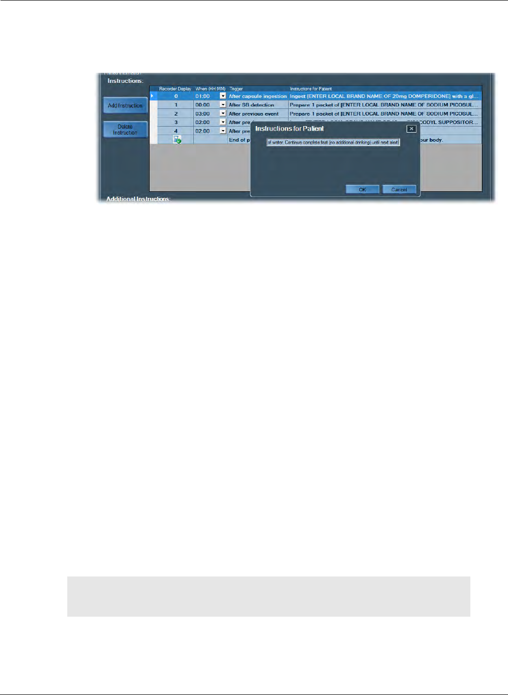
Configuring RAPID Software
Regimen Manager 189
8. The regimens contain editable instructions in the Instructions for Patient column. Clicking the
instruction will prompt an editor. Information represented in brackets [ ] is generic and is intended
for completion by local staff with consideration to local names of the solution. Make any changes
and click OK to save the updated instruction.
9. Any other important information can be entered in the Additional Instructions box. This
information will appear in the patient printout.
10. When you are finished, click Save. This adds the regimen you created to the library of regimens
available.
11. By default, the post-capsule ingestion regimen includes at least the following alerts and
instructions:
•Alert 0: An instruction #0 (based on time passed from capsule ingestion) to take prokinetics to
facilitate passage of the PillCam COLON 2 capsule to the small bowel. Threshold of time
passed from capsule ingestion for alert 0 can be modified in the regimen manager for a specific
regimen.
•Alert 1: An instruction #1 for taking laxative after detection by the PillCam recorder of PillCam
COLON 2 passage into the small bowel. It coincides with the activation of the AFR mode in the
PillCam COLON 2 capsule and the appearance of the AFR status icon in the right corner of the
status line at the top of the PillCam recorder DR3 display. The appearance of alert 1 can be
designated to appear either an additional 0 minutes or 15 minutes after original tentative alert 1
timing. Alert 0 will not occur if alert 1 was raised before it.
• The End of Procedure (EOP) alert occurs when no more capsule images are received by the
PillCam recorder or enough time has elapsed in the procedure. This may occur several minutes
after capsule excretion or after the capsule battery was depleted without excretion in a long
procedure.
In general, additional alerts and instructions can be inserted between alert 1 and the EOP alert to
reflect the physician's preference for post-ingestion instructions in a PillCam COLON 2 procedure.
These instructions can be entered and edited, saved and approved through the Regimen Manager
and be made ready to be selected in the Check-in for a PillCam COLON 2 procedure.
֠
֠֠
֠Note
Alert times are designated by the PillCam recorder and the RAPID
software. In rare cases, short delays may occur.

PillCam Capsule Endoscopy
190 Regimen Manager
Printing the Patient Instructions
You may need to print both types of Patient Instructions for the patient:
•The pre-capsule ingestion instructions need to be handed to the patient 1 or 2 days prior to the
ingestion and are needed for proper preparation of the patient for the procedure.
•The post-capsule ingestion instructions need to be printed and handed to the patient after capsule
ingestion. The reminder numbers on the screen of the PillCam recorder DR3correspond to numbered
instructions in the post capsule ingestion patient instructions but the instruction is not detailed on the
PillCam recorder screen. You must print the post-capsule ingestion patient instructions for the patient
to allow the association of the alerted number with the instruction details.
To print the Pre-capsule Ingestion Regimen Instructions:
1. Select the Pre-capsule Ingestion tab. The relevant screen appears displaying all the regimens or
patient instruction templates available.
2. Select your item and click Print.
To print the Post-capsule Ingestion Regimen Instructions:
•Click Print Regimen during the patient Check-in stage (see Performing Patient Check-in on page 43),
the patient's details are included in the printout.
•You can also print out the personalized post-capsule ingestion instructions after completing the check-
in by going to the Study Manager, right-clicking the patient details and selecting the Print Preview
Regimen option.
•You can also print out the post-capsule ingestion instructions (without the patient's details) via the
Regimen Manager, as follows:
a. From the Home screen, select Tools > Regimen Manager.
b. Select either the Post-capsule Ingestion tab or the Pre-capsule Ingestion tab. The relevant
screen appears displaying all the regimens or patient instruction templates available.
c. Select your item and click Print.
Print Layout of the Post-Capsule Ingestion Instructions
The Regimen Manager enables two convenient print layout options for the post-capsule ingestion
instructions. The desired default layout can be configured in the RAPID settings, by clicking the Report
tab and selecting either pocket format or page format.
֠
֠֠
֠Note
RAPID will print the regimen only if the regimen you selected is approved
by a physician (This Regimen is approved. appears next to the Approve
Regimen button at the bottom).
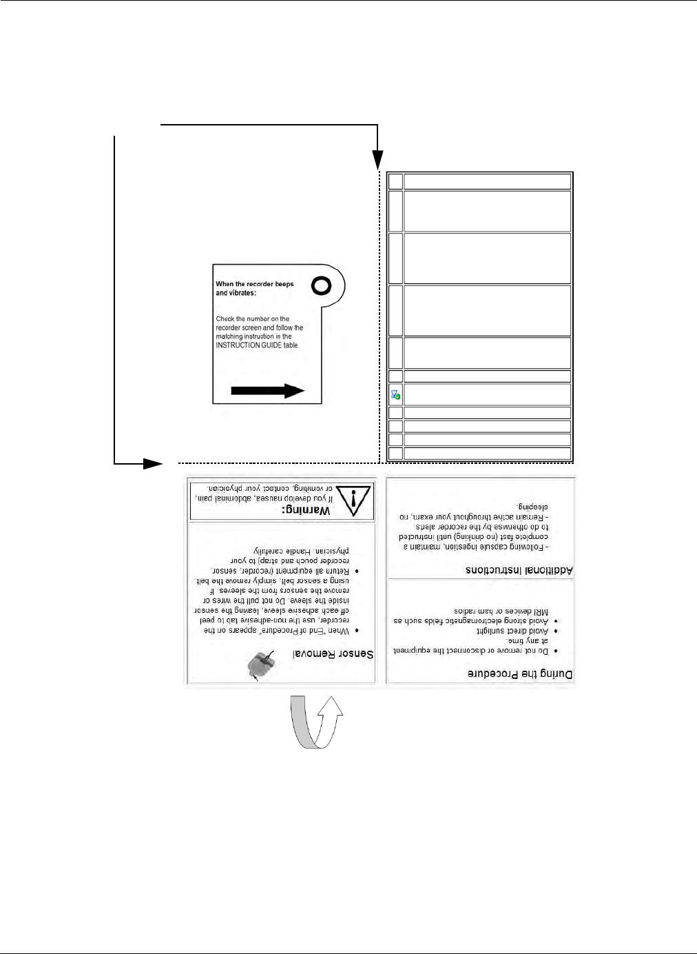
Configuring RAPID Software
Regimen Manager 191
Print in Pocket Format
When enabled, the post-capsule ingestion instructions will be printed in a convenient pocket format
with fold lines for folding to an easy to carry size (quarter fold). The pocket format will show the
Instructions guide on one side and important contact information on the other.
,QVWUXFWLRQV'XULQJ<RXU3LOO&DP
3URFHGXUH
3DWLHQWQDPHBBBBBBBBBBBBBBBBBBBB
3DWLHQW,'BBBBBBBBBBBBBBBBBBBB
3URFHGXUHGDWHBBBBBBBBBBBBBBBBBBBB
BBBBBBBBBBBBBBBBBBBBBBBBBBBBBBBBBBBBBB
BBBBBBBBBBBBBBBBBBBBBBBBBBBBBBBBBBBBBB
,16758&7,21*8,'(
,QJHVW>(17(5/2&$/%5$1'1$0(2)
PJ'203(5,'21(@ZLWKDJODVVRI
ZDWHU&RQWLQXHFRPSOHWHIDVWQRDGGLWLRQDO
GULQNLQJXQWLOQH[WDOHUW
'LOXWH>(17(5/2&$/%5$1'1$0(2)
PO62',803+263+$7(@LQDJODVVRI
ZDWHUDQGGULQNLW'ULQNDWOHDVWOLWHURI
ZDWHURYHUWKHQH[WKRXU<RXPD\UHVXPH
GULQNLQJDOOFOHDUOLTXLGVIUHHO\
'LOXWH>(17(5/2&$/%5$1'1$0(2)
PO62',803+263+$7(@LQDJODVVRI
ZDWHUDQGGULQNLW'ULQNDWOHDVWOLWHURI
ZDWHURYHUWKHQH[WKRXU<RXPD\FRQWLQXH
GULQNLQJDOOFOHDUOLTXLGVIUHHO\
,QVHUW>(17(5/2&$/%5$1'1$0(2)
PJ%,6$&2'</683326,725<@
DFFRUGLQJWRLQVWUXFWLRQVLQSDFNDJHLQVHUW
<RXPD\HDWDOLJKWPHDO
(QGRI3URFHGXUH3OHDVHUHPRYH
HTXLSPHQWIURP\RXUERG\
$"#"'() %((((
Fold lines
Fold the bottom part of
the printout first
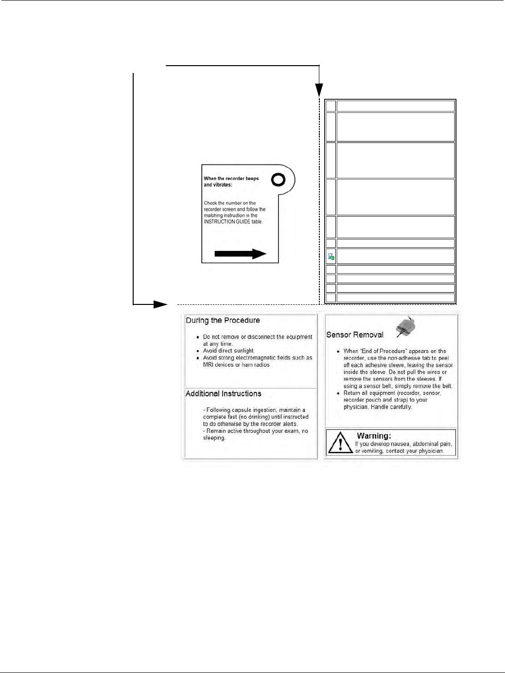
PillCam Capsule Endoscopy
192 Additional Settings
Print in Page Format
When enabled the post-capsule ingestion instructions will be printed on a page layout displaying all
information on one page.
Additional Settings
This section describes advanced configuration features of the RAPID v8.3 software. The section is divided
into the following sections:
•Backup/Restore Offline Studies
•Importing Reports
•Freeing Space on Your Computer
•Backup System Logs
•Advanced CD/DVD Burning
,QVWUXFWLRQV'XULQJ<RXU3LOO&DP
3URFHGXUH
3DWLHQWQDPHBBBBBBBBBBBBBBBBBBBB
3DWLHQW,'BBBBBBBBBBBBBBBBBBBB
3URFHGXUHGDWHBBBBBBBBBBBBBBBBBBBB
BBBBBBBBBBBBBBBBBBBBBBBBBBBBBBBBBBBBBB
BBBBBBBBBBBBBBBBBBBBBBBBBBBBBBBBBBBBBB
,16758&7,21*8,'(
,QJHVW>(17(5/2&$/%5$1'1$0(2)
PJ0(72&/235$0,'(@ZLWKDJODVVRI
ZDWHU&RQWLQXHFRPSOHWHIDVWQRDGGLWLRQDO
GULQNLQJXQWLOQH[WDOHUW
'LOXWH>(17(5/2&$/%5$1'1$0(2)
PO62',803+263+$7(@LQDJODVVRI
ZDWHUDQGGULQNLW'ULQNDWOHDVWOLWHURI
ZDWHURYHUWKHQH[WKRXU<RXPD\UHVXPH
GULQNLQJDOOFOHDUOLTXLGVIUHHO\
'LOXWH>(17(5/2&$/%5$1'1$0(2)
PO62',803+263+$7(@LQDJODVVRI
ZDWHUDQGGULQNLW'ULQNDWOHDVWOLWHURI
ZDWHURYHUWKHQH[WKRXU<RXPD\FRQWLQXH
GULQNLQJDOOFOHDUOLTXLGVIUHHO\
,QVHUW>(17(5/2&$/%5$1'1$0(2)
PJ%,6$&2'</683326,725<@
DFFRUGLQJWRLQVWUXFWLRQVLQSDFNDJHLQVHUW
<RXPD\HDWDOLJKWPHDO
(QGRI3URFHGXUH3OHDVHUHPRYH
HTXLSPHQWIURP\RXUERG\
$"#"'() %((((
Fold lines
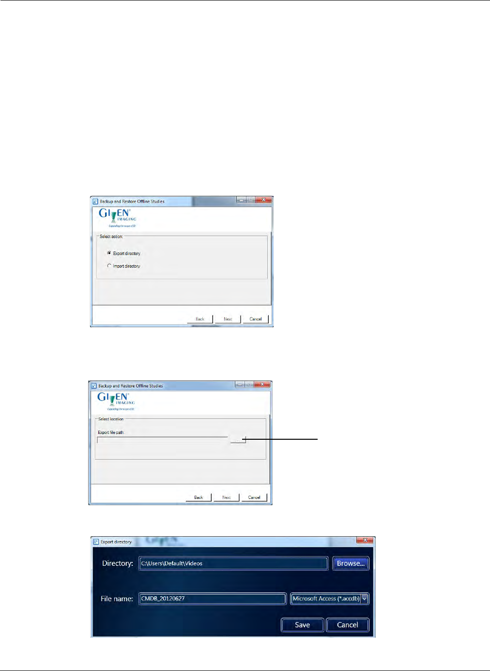
Configuring RAPID Software
Additional Settings 193
Backup/Restore Offline Studies
The RAPID Study Manager displays information about studies stored on removable storage media,
such as CDs, DVDs, and some USB storage devices, that have been connected to the computer during
use of RAPID and are currently disconnected from the computer. After clicking the Offline Studies
button in the Study Manager a list of recently viewed studies which are currently not connected to the
computer is displayed. The offline study list is updated whenever the removable storage media is
disconnected or reconnected.
To back up the list of the Offline Studies to a database file, use the RAPID Backup & Restore Offline
Studies utility.
To backup Offline Studies:
1. You can backup the Study Manager Offline Studies by clicking the Windows Start button and then
selecting Given Imaging > Backup and Restore Offline Studies. The following screen appears.
2. Select Export Directory and click Next. The following screen appears.
3. In the following screen, click the Directory button to launch the Export directory screen.
4. Click the Browse button and navigate to the location to export the backup.
5. Enter the file name of the backup and click Save.
Click to open Export
Directory
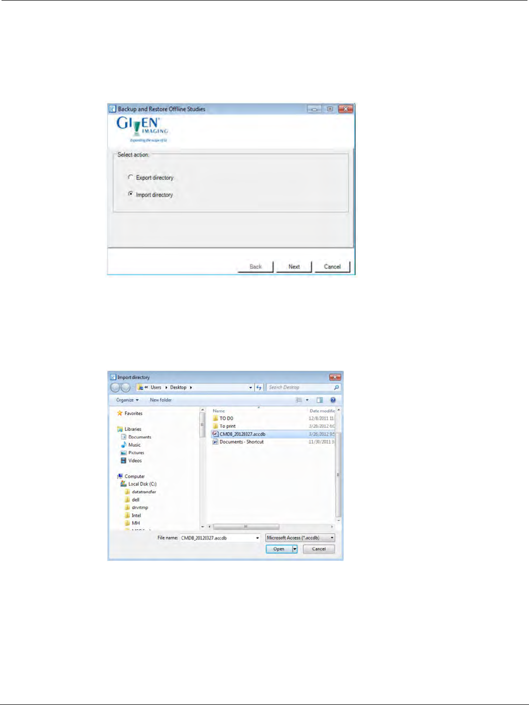
PillCam Capsule Endoscopy
194 Additional Settings
6. When the process is complete, a message appears that it was completed successfully.
Click OK.
To import backup Offline Studies:
1. In order to restore Study Manager Offline Studies, click the Windows Start button > Given Imaging >
Backup and Restore Offline Studies. The following screen appears.
2. Select Import directory and click Next.
3. In the following screen click the Directory button to launch the Import directory screen.
4. Navigate to the database file (with Microsoft Access *accdb file extension) and click the Open button
to load the offline studies to the RAPID Study Manager.

Configuring RAPID Software
Additional Settings 195
Importing Reports
Reports generated by RAPID versions before RAPID 5 are not automatically displayed in the Study
Manager. If you wish to be able to see them in the Study Manager, you must import them by doing the
following:
To import reports:
1. Open the video of the relevant study.
2. Select File > Tools > Import RAPID 4 Reports.
The following message appears: Ensure that the selected reports belong to this study.
3. Click OK.
A Windows browser screen opens at the location of the currently open video, showing the reports
that are in this folder.
4. Select the report(s) you wish to import. If the report is not in this folder, browse to the correct
location and select the report.
5. Click Open. The Reports imported successfully message appears.
6. Click OK and repeat this procedure for all the studies performed before RAPID 5.
Freeing Space on Your Computer
Deleting Videos
If you start to create a video while there is not enough space on your computer, you will be prompted
to empty the Recycle Bin and if more space is needed, to delete RAPID folders.
To free space for new videos, delete RAPID studies from your hard disk or from E:\Videos.
To delete videos from your hard disk:
1. From the Home screen click Tools > Delete Videos.
֠
֠֠
֠Note
It is recommended to back up the RAPID studies by saving them on
removable media discs (see Burning a Study to a CD/DVD on page 108).
You may then delete them from the computer’s hard disk at any time or
when prompted to do so by the RAPID software.
Failure to back up a RAPID video before deleting will cause the study to be
permanently lost.
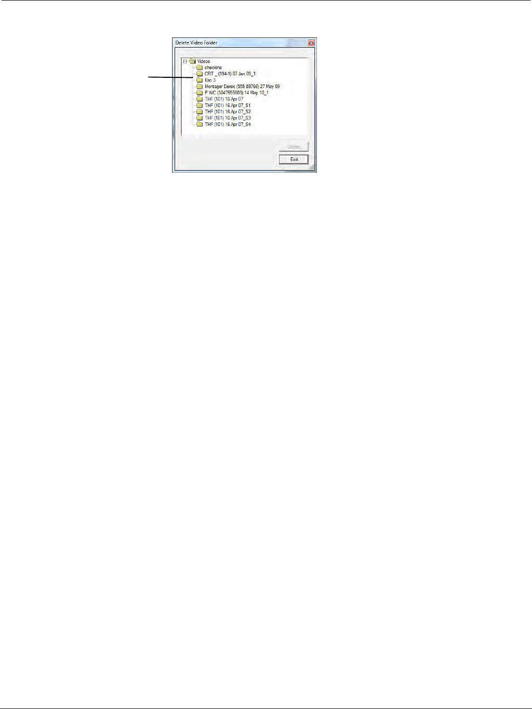
PillCam Capsule Endoscopy
196 Additional Settings
The Delete Video Folder window appears.
2. Select the video folder you wish to delete.
The Delete button becomes available.
3. Click Delete.
The selected RAPID folder and all its contents will be deleted.
4. Empty the Recycle Bin.
Deleting Raw Data Files after Video Creation
When creating videos in default mode, the RAPID software clears previous raw data files it may have
copied to the computer in the video creation process. However, if you select the optional Copy raw data
method for video creation in the Settings screen, raw data files are not automatically deleted after the
video is created. As multiple raw data files can rapidly fill your disk, you must manually clear raw data
files as follows:
To clear raw data files:
1. From the Home screen, click Recorder Download and select the Raw Data Files screen.
2. To confirm that you no longer need a raw data file, you can refer to the Last Use status, and you may
delete files after Successful video creation or data copy.
3. Click the bar displaying the file you wish to delete and click Delete file.
Backup System Logs
The system log file records all system events on the Workstation, such as log in, print, delete, etc. The size
of this file is limited, and when it reaches 80% of its capacity, the following message is displayed
immediately after logging in: The log files need to be backed up. Please notify RAPID Workstation
administrator.
Only the system administrator can back up the system log.
This message appears at every login, until the backup procedure is performed.
Navigate to
folder to
delete.
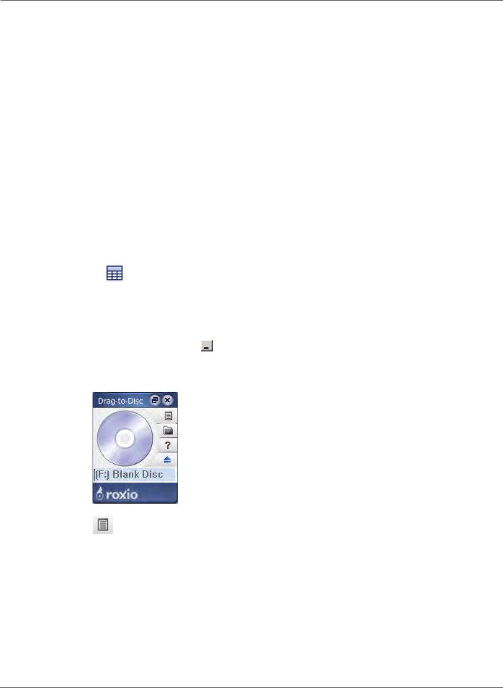
Configuring RAPID Software
Additional Settings 197
CD/DVD Burning
The Export function in the RAPID software allows you to save a study or one of its files in a different
place or under a different name, if needed to reorganize your archives. This includes:
•Saving a study or a file to another archive, such as a USB storage device
•Saving a study or a file to a specific location
•Saving a study as a Zip file
•Burning a study or a file to a CD/DVD
For burning a single study to a disc (no multisession), see Burning a Study to a CD/DVD on page 108.
For burning files from more than one study use the CD burning program on your computer. Given
Workstations have either Roxio Drag-to-Disc 9 or DirectCD 5.
Roxio Drag-to-Disc 9
Burning Procedure
1. Insert a blank disc into the disc drive.
2. If you do not know where the files you wish to burn are located, open the Study Manager by
clicking at the top of the screen, or select View Study > Study Manager from the Home
screen.
3. Move your mouse over the archive where the files reside and a tooltip appears with the location of
the archive on the computer.
4. Minimize RAPID by clicking in the top right corner of the screen.
5. Click the Drag-to-Disc icon on the desktop. The Drag-to-Disc screen appears.
6. Click on the Drag-to-Disc screen and select Format Disc from the pop-up menu. The Format
Options screen appears.
7. Type in the name of your disc in the box next to Enter volume name and click OK. The Disc
Preparation screen appears.
8. As soon as the Disc Preparation screen disappears, double click My Computer on the desktop and
browse for the location of the files you wish to copy to the disc.
9. Click and drag the files you wish to copy to the disc over the Drag-to-Disc screen. A progress bar
appears to indicate the burning process.
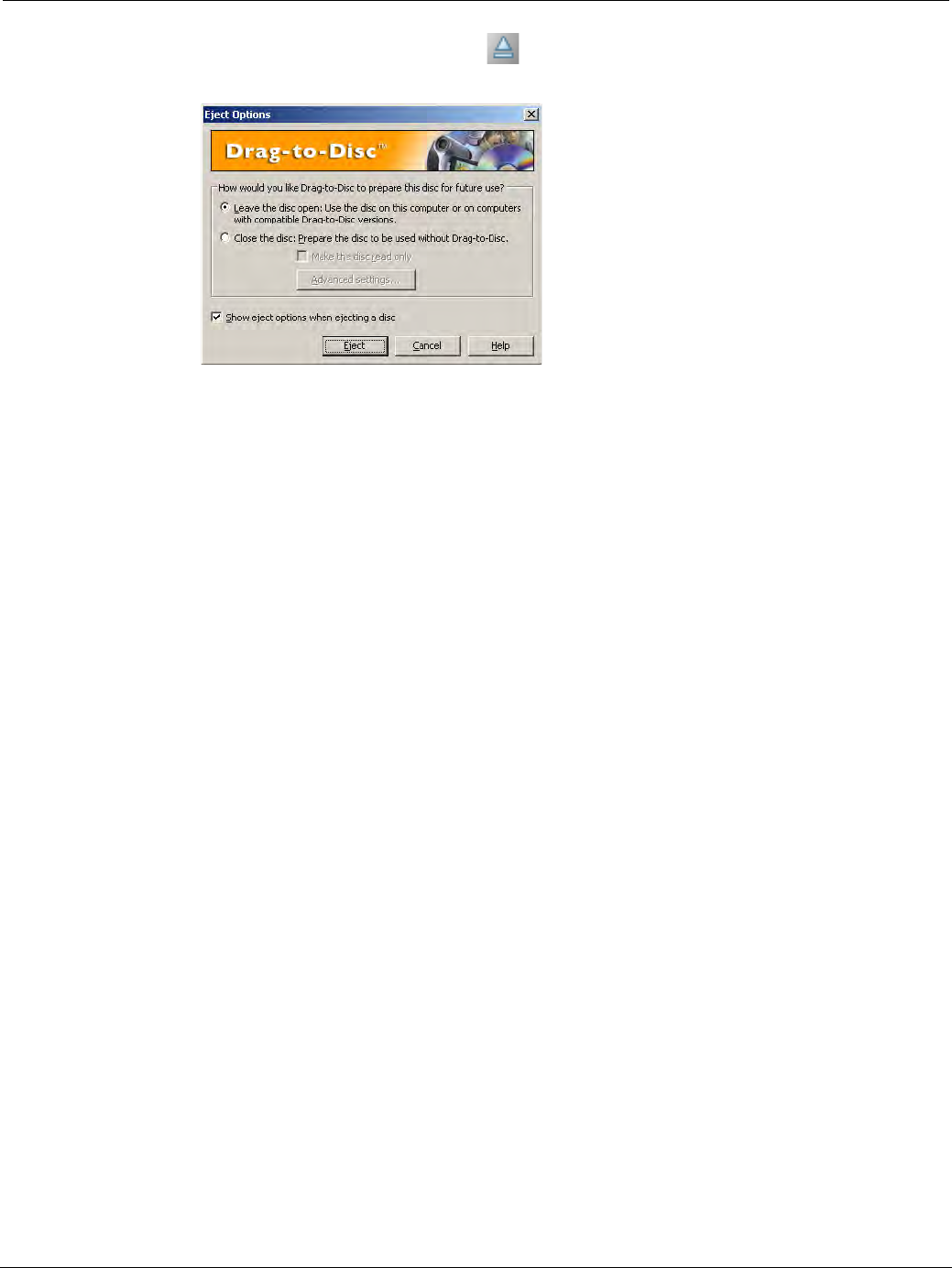
PillCam Capsule Endoscopy
198 Additional Settings
10. Once the progress bar has disappeared, click on the Drag-to-Disc screen to eject the disc. The
following screen appears.
11. If you wish to add files to this disc later (multisession), click the top option and click Eject.
DirectCD 5
To burn a procedure:
1. Insert disc into disc drive.
2. Double click the DirectCD icon on the desktop.
3. In the DirectCD screen, click Format. The Format screen appears.
4. In the Label box, enter a name for your disc limited to 11 characters. This is the volume name in the
Study Manager.
5. Click Start Format.
6. The DirectCD formats the disc.
Once formatting is done the disc behaves like a regular folder and you can save videos, findings and reports
to this folder and the software will burn the CD. You can add more files anytime later.

Configuring RAPID Software
Keyboard Shortcuts 199
Keyboard Shortcuts
Key Operation
Space Play/Pause Video
Left arrow Go to next image
Right arrow Go to previous image
Page-Down Go 10 images forward
Page-Up Go 10 images backwards
Home Go to first image
End Go to last image
Enter Capture Thumbnail
Control + T Show Atlas
Control + M Show Study Manager
Control + N Open Video
Control + S Save Findings
Control + O Open Findings
Control + R Report Preview
Control + P Report Print
Control + I Image Enhancement
Control + Shift + S Set View Layout to Single
Control + Shift + D Set View Layout to Dual
Control + Shift + Q Set View Layout to Quad
Control + Shift + M Set View Layout to Mosaic
Control + Shift + C Set View Layout to Collage
Control + Shift + 1/2/3 Image Enhancement FICE 1/2/3
Control + Shift + B Image Enhancement Blue
Control + Shift + N Image Enhancement Normal
Control + mouse wheel
up/down
Decrease/Increase video Viewing Speed

PillCam Capsule Endoscopy
200 Keyboard Shortcuts
PillCam Recorder Maintenance 201
Appendix A3
PillCam Equipment Maintenance
PillCam Recorder Maintenance
Use only a fully charged (eight or more bars displayed on the battery icon) PillCam recorder. In general,
including first-time use, charging the PillCam recorder is an overnight process and should not be
performed in the vicinity of the patient. When you receive the PillCam recorder after an examination,
charge it immediately until the green LED is lit, and leave it in its cradle.
Important Safety Instructions
!
Warning
Charge the PillCam recorder only with the supplied cradle. Once you have
placed it into the cradle, the PillCam recorder automatically starts charging.
!
Caution
The SD card should never be removed or reinserted when the PillCam
recorder DR3 is ON.
֠
֠֠
֠Disclaimer
The PillCam recorder cradle is a non-medical device, used for charging the
PillCam recorder from Given Imaging Ltd. For full specifications, see System
Specifications on page 217.
֠
֠֠
֠Note
Before using the PillCam recorder cradle, read all instructions on cautionary
markings on the cradle, on the battery, and on the PillCam recorder.
!
Warning
Changes or modifications to this equipment not expressly approved
by the party responsible for compliance (Given Imaging Ltd.) could
void the user’s authority to operate the equipment.
!
Warning
• The cradle is for indoor use only.
• Never charge non-rechargeable batteries.
• All cells containing mercury, cadmium, lithium or lead as electrochemical
substances are subject to special waste disposal requirements.
• This charger is a class A product. In a domestic environment, this
charger may cause radio interference.
PillCam Capsule Endoscopy
202 PillCam Recorder Maintenance
PillCam Recorder DR3
The PillCam recorder DR3 cradle has the following LEDs:
To charge the PillCam recorder:
1. Plug the power cable into the cradle and plug the power cable into the wall outlet.
2. Insert the PillCam recorder into the cradle.
The bottom LED is orange when charging the battery. When the PillCam recorder is fully charged, the
bottom LED turns green.
3. Leave the PillCam recorder in its cradle until the next examination.
֠
֠֠
֠Note
This equipment has been tested and found to comply with the limits for a
Class B digital device, pursuant to part 15 of the FCC Rules. These limits
are designed to provide reasonable protection against harmful interference
in a residential installation. This equipment generates, uses and can radiate
radio frequency energy and, if not installed and used in accordance with the
instructions, may cause harmful interference to radio communications.
However, there is no guarantee that interference will not occur in a
particular installation. If this equipment does cause harmful interference to
radio or television reception, which can be determined by turning the
equipment off and on, the user is encouraged to try to correct the
interference by one or more of the following measures:
• Reorient or relocate the receiving antenna.
• Increase the separation between the equipment and receiver.
• Connect the equipment into an outlet on a circuit different from that to
which the receiver is connected.
• Consult the dealer or an experienced radio/TV technician for help.
LED Status Battery pack is...
Green On ready for use
Yellow/
Orange
On charging
Blinking charging
PillCam Equipment Maintenance
PillCam Recorder Maintenance 203
PillCam Recorder DR2
Charging
Make sure the PillCam recorder is at least 80% charged (eight or more battery bars displayed on the
battery icon) for SB and COLON procedures, and at least 50% charged (five or more bars displayed on
the battery icon) for ESO procedures.
The cradle has the following LEDs:
To charge the PillCam recorder:
1. Plug the power cable into the cradle and plug the power cable into the wall outlet.
All three LEDs turn on for a self-test that takes 5 seconds. After 5 seconds, all LEDs turn off and
the cradle is ready for use.
If during the self-test the red LED blinks, the cradle is faulty. Contact Given Imaging Customer
Support.
2. Insert the PillCam recorder or its Li-Ion battery with its adaptor into the cradle.
All three LEDs of the cradle blink for 4 seconds. When the orange LED is on, charging has started.
As soon as the PillCam recorder or its battery pack are fully charged, the green LED turns on, and
the orange LED turns off.
3. Leave the PillCam recorder in its cradle until the next examination.
LED Status Battery pack is...
Green On ready for use
Orange On charging
Blinking discharging
Red On faulty (malfunctioning)
Adaptor with
battery
Green, orange,
and red LEDs
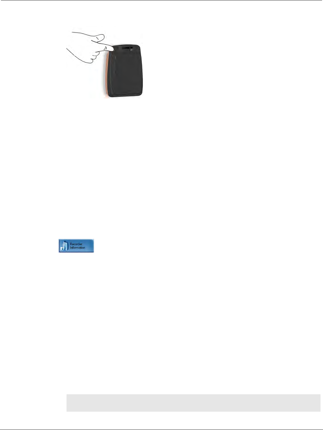
PillCam Capsule Endoscopy
204 PillCam Recorder Maintenance
4. To check the status of the PillCam recorder before an examination, remove it from the cradle and push
the button on the back.
Manual Discharge
If the cradle detects that the battery needs refreshing, it automatically discharges the battery before
recharging it. The orange LED on the cradle blinks during discharging. Discharging is an overnight
process that may take up to 12 hours.
We recommend that you manually discharge the battery every three months, even if it is not used.
To discharge the battery:
1. Open RAPID.
2. Make sure the appropriate battery is inside the PillCam recorder.
3. Insert the PillCam recorder into the cradle.
4. From the Procedures screen, select the relevant PillCam recorder by clicking the Recorder bar.
The buttons on the right side of the screen become available.
5. Click to open the Recorder Information screen.
6. At the bottom of the screen, click Start Discharge.
A message appears: Recorder discharge may take more than 12 hours. Do you wish to continue?
7. Click OK.
While the battery is being discharged, its battery status indicates Discharging:
• In the bottom left corner of the Recorder Information screen.
• In the PillCam recorder DR2 bar in the Recorders screen.
• The orange LED on the cradle blinks.
8. To return to other RAPID functions, click Close.
9. If you need to stop the discharge (also for automatic discharge) while it is in progress, return to the
Recorder Information screen and click Cancel Discharge.
If you stop the automatic discharge process in the middle, the battery LEDs may not indicate the
correct battery status.
֠
֠֠
֠Note
Do not charge or discharge the battery in the vicinity of the patient.
PillCam Equipment Maintenance
PillCam Sensor Cleaning 205
PillCam Sensor Cleaning
This section includes instructions for cleaning the PillCam equipment.
Cleaning the PillCam Sensor Belt
Cleaning the PillCam Sensor Array
For mild cleaning (dirt, sweat), wipe the sensors gently with alcohol wipes (up to 70%). The alcohol
will not remove the adhesive. Use alcohol sparingly and allow the sensor array to dry for 20 minutes.
To remove adhesive from the sensor array (not from the human body), use white benzene.
Alternatively, use one of the following medical adhesive removers to remove adhesive:
•B-508 Secure Solvent
•B-202 Hollister Solvent
•B-206 Detachol Adhesive Remover
Use all precautions as defined by the manufacturer.
Cleaning the Recorder Pouch
To clean the recorder pouch, wipe down all surfaces with alcohol (70% isopropyl or ethyl alcohol)
making sure that all surfaces are exposed to alcohol for at least 1 minute.
֠
֠֠
֠Note
To clean the PillCam equipment surfaces, we recommend using alcohol
wipes (up to 70%). Usage of other classes of disinfectants (Aldehydes,
Oxidizing Agents, Quaternary Ammonium, Chlorine compounds, Iodophor,
Phenolic compounds) may stain or damage the materials.
֠
֠֠
֠Note
Refer to the product insert supplied with your sensor belt for full instructions
on fitting, usage, cleaning, and technical description.
!
Warning
Use white benzene only in a well ventilated area according to all
precautions and instructions on the label.

PillCam Capsule Endoscopy
206 PillCam Sensor Cleaning

RAPID Video 207
Appendix A4
Troubleshooting
RAPID Video
Saving and Opening Videos
Problem Cause Action
Short video • Capsule
• PillCam recorder battery
• PillCam recorder
mishandling
• Contact Customer Support
• Send video on CD/DVD
• Inform capsule lot#
• Do not use the same PillCam
recorder
• Save the raw data locally on
your computer
Gaps • Capsule
• Interference
• Mishandling
• Physiological
Bad image quality • Stripes in video
• Pixilation/confetti
• Dark/red/orange image
Video shorter than
capsule operating time
without either ingestion
phase images or body
exit images
• Capsule
• PillCam recorder battery
• Interference
• Send video on CD/DVD
• Save the raw data locally on
your computer
• Contact Customer Support
No Localization • Malfunction of the sensor
array
• Wrong sensor chosen in
the check-in process
• Do not use the sensor
• Contact Customer Support
Cannot open RAPID
Atlas
Atlas installation incomplete
or incorrect
• Reinstall Atlas
• Save the raw data locally on
your computer
• Contact Customer Support
Cannot view COLON 2
video using RAPID v7.0
COLON 2 video compiled in
RAPID v8.0 cannot be viewed
using RAPID v7.0
Download and use RAPID Reader
v8.0 to view video
Unable to perform
various operations
RAPID computer is “stuck”
typically after a prolonged
period in the ON state without
restarting
It is recommended to restart your
computer at least once a month to
ensure optimal performance
Problem Cause Action
Cannot locate video • Video was not saved in
E:\Videos
• Video was not created
• Incorrect patient’s name
• Search for video in RAPID
Work Directories or in Study
Manager (video creation)
• Contact Customer Support

PillCam Capsule Endoscopy
208 Printer
Printer
CD/DVD
Sensor Array
Cannot locate findings • Findings were not saved
under patient’s folder
• Findings were saved with
the wrong name
• Refer to Saving Findings File
in Saving Your Findings on
page 144
• Contact Customer Support
Study cannot be selected
in Study Manager
Changing an archive location
that appears in the Study
Manager screen, the archive
remains on the archives list
but its study cannot be
selected
Modify the archive location
manually
Cannot open a video
from an archive in Study
Manager and an error
message requests user
to click the refresh button
Archive privileges set with no
access to read or write
Allow Read and Write access to
the archive folder
Problem Cause Action
Cannot print report Printer is turned off Turn printer on
Printer is not set as default
printer
Set printer to Default Printer
Printer has a malfunction Contact Customer Support
Problem Cause Action
Cannot burn CD/DVD CD/DVD is not blank or
compatible with CD/DVD
ROM Contact Customer Support
Wrong burning procedure
Cannot eject CD/DVD A video on the disc is open Close the video and retry
Problem Cause Action
Connector is damaged
• Mishandling
• End of Life Contact Customer Support
Sensor is torn from its
wire
Insulation of the sensor
wire is damaged
Problem Cause Action

Troubleshooting
Sensor Belt 209
Sensor Belt
Capsule
Cradle
PillCam Recorder DR3
Problem Cause Action
Connector is damaged • Mishandling the sensor
belt
• End of Life
• Stop using this sensor
• Contact Customer Support
Insulation of the sensor wire
is damaged
Problem Cause Action
DOA (Dead On Arrival) Capsule failure 1 Send capsule to Given
Imaging Ltd.
2 Open another capsule.
3 If second capsule from 10-
pack is DOA, contact
Customer Support.
Problem Cause Action
Cradle LEDs turn red All LEDs are flashing red 1 Disconnect cradle from
mains power.
2 Reconnect cradle to mains
power.
3 If problem persists, contact
Customer Support.
Cradle orange LED is
blinking, but cradle is not in
discharge mode
Hardware/Software problem
PillCam recorder DR2 cannot
be placed in cradle
Hardware malfunction Contact Customer Support
Problem Cause Action
Capsule LED blinking red Interference
Recorder malfunction
Refer to PillCam recorder DR3 LEDs
Indications
Capsule LED blinking white Capsule not paired yet
SD card errors Wrong or damaged SD
card
Sensor connection Wrong or damaged
sensor
PillCam recorder freezes Possible static
electricity discharge
• Perform Hard Reset on the
PillCam recorder
• Contact Customer Support

PillCam Capsule Endoscopy
210 PillCam Recorder DR2
PillCam Recorder DR2
Cannot initialize PillCam
recorder DR3
Wrong recorder bar is
selected
Select correct recorder bar
No communication between
the PillCam recorder and
RAPID software
Recorder not inserted
into cradle properly
• Clean the contacts with alcohol
wipes
• Firmly insert the recorder into the
cradle
Problem Cause Action
Cannot initialize PillCam
recorder DR2
Wrong recorder bar is
selected
Select correct recorder bar
Computer does not
recognize PillCam
recorder DR2
1 Check USB and power
connection to the cradle.
2 Contact Customer Support.
Cannot create video Wrong recorder bar is
selected
Select PillCam recorder DR2
Recorder bar
Error message is
displayed
Send error message to Customer
Support
Not enough free
space... message is
displayed
Delete PRRs from hard drive
Workstation freezes
during video creation
Contact Customer Support
Capsule LED (right) does not
blink in blue when capsule is
activated (LED is orange) • Capsule failure
• PillCam recorder DR2
failure
1 Return capsule to blister.
2 Activate second capsule.
3 If problem persists, contact
Customer Support.
4 Send malfunctioning capsules
to Given Imaging Ltd.
Capsule LED (right) blinks in
orange once every five
seconds when more than six
hours have passed since
ingestion
• Capsule failure
• PillCam recorder DR2
failure
1 Wait until 8 hours have passed
and take off the sensors.
2 Send video on CD/DVD.
Capsule LED (right) blinks in
orange once every five
seconds when less than six
hours have passed since
ingestion
• Capsule failure
• PillCam recorder DR2
failure
Contact Customer Support
PillCam recorder DR2 shuts
down during the procedure
PillCam recorder DR2
malfunction
1 Turn PillCam recorder DR2 off.
2 Take out the battery pack and
place it back in.
3 Turn PillCam recorder DR2 on.
4 If problem persists, contact
Customer Support
Battery exhausted Replace battery and continue
procedure.
Problem Cause Action

Troubleshooting
Error Messages 211
Error Messages
For LED behavior, see LED Display on page 68. For error messages displayed on the PillCam recorder
screen, see Error Messages on page 73.
Cancelling Data Copy
displays error message that
Video Copy was
unsuccessful
Cancelling the Data Copy
in mid-sequence triggers
Video Copy message
Message Solution
Full name not defined for this
user.
Please contact your IT system
administrator.
In order to enable electronic signatures in the
CE report, the full name field (full name will
appear in electronic signature) must be
defined in the user's relevant Windows
account.
Electronic signature failed.
Please contact your IT system
administrator.
In order to enable electronic signatures in CE
report, the user's relevant Windows account
must be enabled for remote connection login.
Possible reasons for this error message
include:
• Password Expired
• Account Restrictions
• Invalid Logon Hours
• Account Lockout (or expired or disabled)
Password must be changed.
You do not have permission to
access the Regimen Manager.
Please contact your system
administrator.
In order to allow physicians to edit and
approve their regimens, the following must be
done:
• In the Settings screen, under the Other
tab, define an existing folder as the
Regimens Directory.
This folder must be open for read and write
permissions for the relevant users.
Patient Check-in procedure
failed. Reason: Failed to update
recorder software version.
Turn the recorder off and then on before the
next check-in attempt. If the upgrade fails
again, contact customer support.
Problem Cause Action

PillCam Capsule Endoscopy
212 Low Signal
Low Signal
If a low signal is detected during the examination, the following message appears.
A low signal detected during the examination may be due to:
•Improper use of the sensor array/sensor belt
•A defective sensor array/sensor belt
•A PillCam recorder malfunction
If this message is displayed, Contact Customer Support.
Click OK to close the message.
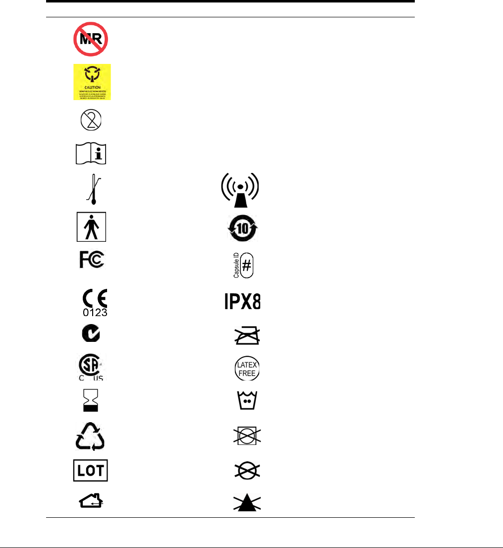
System Labeling 213
Appendix A5
Technical Description
System Labeling
The following table lists the labels attached to various components of the PillCam Capsule Endoscopy
System.
Labeling Explanation
PillCam capsules are MR unsafe.
The PillCam capsule should not be stored and used near any
powerful magnetic fields such as the one created by an MRI.
The PillCam capsule is intended for single use only.
Attention! Consult the documentation provided with the
PillCam Capsule Endoscopy System.
Temperature
limits Non-ionizing radiation
Type BF
equipment RoHs
FCC
compliance Capsule ID
CE mark Ingress protection
C-Tick mark Do not Iron
CSA mark Latex free
Expiration date Machine wash - warm
Recycle Do not tumble dry
Lot number Do not dry clean
Indoor use only Do not use bleach
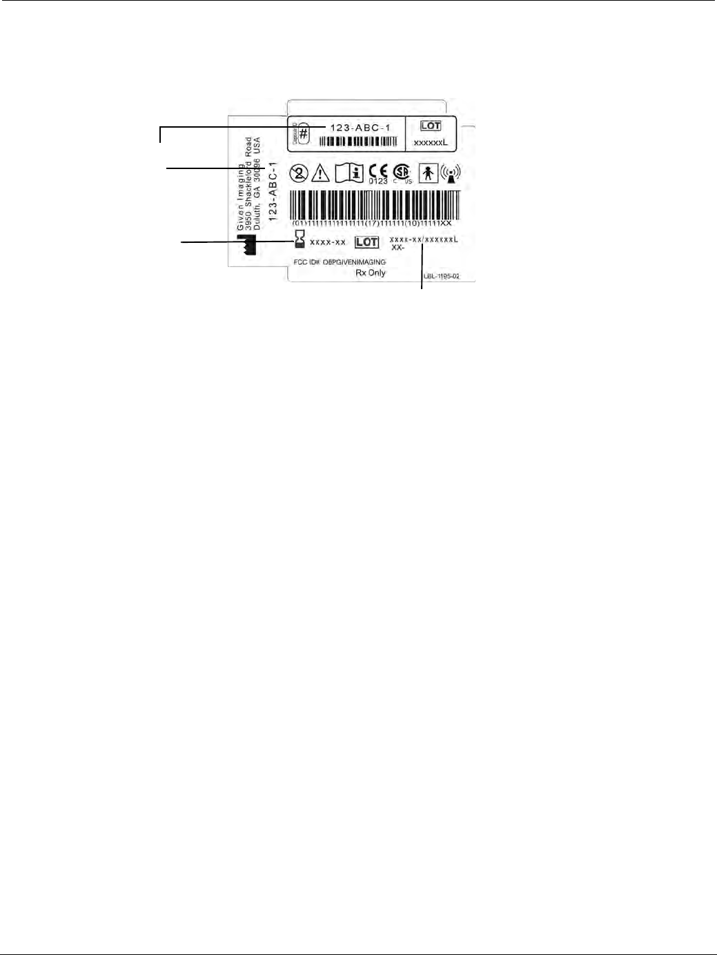
PillCam Capsule Endoscopy
214 Essential Performance
Capsule Labeling
Each box has a label at the bottom as shown below. Each capsule is marked with the expiration date, lot
number, and a unique capsule ID code.
Essential Performance
PillCam Capsules
ON-Mode
Data transmitting to PillCam recorder is considered to be essential performance of the PillCam capsules.
The PillCam capsules shall transmit data continuously monitored by on-line image display as received by
PillCam recorder.
OFF-Mode
No unintentional transmissions are allowed.
PillCam Recorder DR2 and PillCam Recorder DR3
Data receiving by PillCam recorder is considered to be essential performance of the PillCam recorder DR2
and PillCam recorder DR3.
Warnings
•PillCam Capsule Endoscopy System and its components need special precautions regarding EMC and
need to be installed and put into service according to the EMC information provided in the
accompanying documents.
•Portable and mobile RF communications equipment can affect the PillCam video capsule and the
PillCam recorder.
•PillCam video capsules and PillCam recorder should not be used adjacent to or stacked with other
equipment and that if adjacent or stacked use is necessary, the equipment or system should be observed
to verify normal operation in the configuration in which it will be used.
Lot Number
Expiration
Date
Capsule ID
Code

Technical Description
Warnings 215
•PillCam video capsules and PillCam recorder may be interfered with by other equipment, even if
that other equipment complies with CISPR emission requirements.
•Do not disassemble or modify the battery pack. The battery pack is equipped with built-in safety/
protection features. Should these features be disabled, the battery pack can leak acid, overheat,
emit smoke, burst and/or ignite.
•Do not use or leave the battery pack of the PillCam recorder near a heat source such as a fire or a
heater (+80°C or higher). If the resin separator should be damaged owing to overheating, internal
short-circuiting may occur to the battery pack, possibly leading to acid leakage, smoke emission,
bursting and/or ignition of the battery pack.
•Do not immerse the battery pack in water or seawater and do not allow it to get wet. Otherwise, the
protective features in it can be damaged, it can be charged with extremely high current and voltage,
abnormal chemical reactions may occur in it, possibly leading to acid leakage, smoke emission,
bursting and/or ignition.
•Do not recharge the battery pack near fire or in extremely hot weather. Otherwise, hot temperatures
can trigger its built-in protective features, inhibiting recharging, or can damage the built-in
protective features, causing it to be charged with an extremely high current and voltage and, as a
result, abnormal chemical reactions can occur in it, possibly leading to acid leakage, overheating,
smoke emission, bursting and/or ignition.
•To recharge the battery pack, use the PillCam recorder cradle and observe the recharging
conditions. A recharging operation under non-conforming recharging conditions (higher
temperature and larger voltage/current than specified, modified battery charger, etc.) can cause the
battery pack to be overcharged, or charged with extremely high current, abnormal chemical
reaction can occur in it, possibly leading to acid leakage, overheating, smoke emission, bursting
and/or ignition.
•Do not pierce the battery pack with a nail or other sharp objects, strike it with a hammer, or step on
it. Otherwise, the battery pack will become damaged and deformed, internal short-circuiting can
occur, possibly leading to acid leakage, overheating, smoke emission, bursting and/or ignition.
•Do not strike or throw the battery pack. The impact might cause leakage, overheating, smoke
emission, bursting and/or ignition. Also, if the protective feature in it becomes damaged, it could
become charged with an extremely high current and voltage, abnormal chemical reactions can
occur, which can lead to acid leakage, overheating smoke emission, bursting and/or ignition.
•Do not use an apparently damaged or deformed battery pack. Otherwise, acid leakage, overheating,
smoke emission, bursting and/or ignition of the battery pack may occur.
•If the battery pack leaks and the electrolyte gets into the eyes, do not rub them. Instead, rinse the
eyes with clean running water and immediately seek medical attention. Otherwise, eye injury may
result.
•If recharging operation fails to complete even when a specified recharging time has elapsed,
immediately stop further recharging. Otherwise, acid leakage, overheating, smoke emission,
bursting and/or ignition can occur.
•Do not put the battery pack into a microwave oven or pressurized container. Rapid heating or
disrupted sealing can lead to acid leakage, overheating, smoke emission, bursting and/or ignition.
•If the battery pack leaks or gives off a bad odor, remove it from any exposed flame. Otherwise, the
leaking electrolyte may catch fire and the battery pack may emit smoke, burst or ignite.
•If the battery pack gives off an odor, generates heat, becomes discolored or deformed, or in any
way appears abnormal during use, recharging or storage, immediately remove it from the

PillCam Capsule Endoscopy
216 Cautions
equipment or cradle and stop using it. Otherwise, the problematic battery pack can develop acid
leakage, overheating, smoke emission, bursting and/or ignition.
•The use of accessories, transducers and cables other than those supplied or approved by
Given Imaging Ltd. as replacement parts for internal PillCam recorder components, may result in
increased emissions or decreased immunity of the PillCam Capsule Endoscopy System.
Cautions
•Do not use or subject the battery pack to intense sunlight or hot temperatures such as in a car in hot
weather. Otherwise, acid leakage, overheating and/or smoke emission can occur. Also, its guaranteed
performance will be lost and/or its service life will be shortened.
•The battery pack incorporates built-in safety devices. Do not use it in a location where static electricity
(greater than the manufacturer’s guarantee) may be present. Otherwise, the safety devices can be
damaged, possibly leading to acid leakage, overheating, smoke emission, bursting and/or ignition.
•The guaranteed recharging temperature range is 0°C to +45°C. A recharging operation outside this
temperature range can lead to acid leakage and/or overheating of the battery pack and may cause
damage to it.
•If acid leaking from the battery pack comes into contact with your skin or clothing, immediately wash
it away with running water. Otherwise, skin inflammation can occur.
•For recharging procedures, refer to Charging on page 66.
•If you find rust, a bad odor, overheating and/or other irregularities when using the battery pack for the
first time, return it to your supplier or vendor.

Technical Description
System Specifications 217
System Specifications
PillCam SB 2 Capsule
֠
֠֠
֠Note
• Specifications are subject to change without prior notice and without any
obligation to users on the part of the manufacturer.
• Specifications are rounded to the appropriate decimal place.
Properties
Physical Dimensions Length: 26.3 mm
Diameter: 11.4 mm
Weight 2.9 g
Material Biocompatible plastic
Optical Illumination 4 white light emitting diodes
# of imaging heads 1
Field of view 156° (Optical field of view at 4.5 mm
from top cover per ISO-8600-3)
130° (Optical field of view from
entrance pupil per FDA Method)
Effective visibility Distance: 3 cm
Min. detectable object At least 0.1 mm
Frequency 434.1 MHz
Band width 1.6 MHz
Modulation MSK
ERP [nW] 16
Operational Frame rate either 2 or 4 fps (two capsule versions)
Operating time > 8 hours
Chemical safety Resistant to dissolution in pH=2 to
pH=8
Battery type Silver Oxide batteries
Operating
temperature
20–40°C
Storage temperature 0–40°C

PillCam Capsule Endoscopy
218 System Specifications
PillCam SB 3 Capsule
Properties
Physical Dimensions Length: 26.2 mm
Diameter: 11.4 mm
Weight 3.0 g
Material Biocompatible plastic
Optical Illumination 4 white light emitting diodes
# of imaging heads 1
Field of view 156° ISO-8600-3
Effective visibility 156° (Optical field of view at 4.5 mm from
top cover per ISO-8600-3)
136° (Optical field of view from entrance
pupil per FDA Method)
Min. detectable object At least 0.07 mm
Frequency 434.1 MHz
Band width 3.2 MHz @ 2.7 Mbps;
6.5 MHz @ 5.4 Mbps
Modulation MSK
ERP [nW] ~20
Operational Frame rate 2 fps or 2–6 fps
Operating time > 8 hours
Chemical safety Resistant to dissolution in pH=2 to pH=8
Battery type Silver Oxide batteries, Mercury Free
Operating
temperature
20–40°C
Storage temperature 0–25°C
Downlink
communication
Operating frequency 13.6 MHz
Receiver Bandwidth + 150 kHz

Technical Description
System Specifications 219
PillCam UGI Capsule
Properties
Physical Dimensions Length: 32.3 mm+0.5 mm
Diameter: 11.6 mm
Weight 2.9 g + 0.1 g
Material Biocompatible plastic
Optical Illumination 4 white light emitting diodes on each side
# of optical heads 2
Field of view 172° ISO-8600-3
Effective visibility Distance: 3 cm
Min. detectable object At least 0.1 mm
Frequency 434.1 MHz
Band width 9.7 MHz
Modulation MSK
ERP [nW] 8
Operational Frame rate 18–35 fps
Operating time 90 min
Chemical safety Resistant to dissolution in pH=2 to pH=8
Battery type Mercury Free Silver Oxide batteries
Operating temperature 20–40°C
Storage temperature 0–25°C

PillCam Capsule Endoscopy
220 System Specifications
PillCam COLON 2 Capsule
Properties
Physical Dimensions Length: 32.3 mm+0.5 mm
Diameter: 11.6 mm
Weight 2.9 g + 0.1 g
Material Biocompatible plastic
Optical # of optical heads 2
Illumination 4 white light emitting diodes on each side
Field of view 172° ISO-8600-3
Effective visibility Distance: 3 cm
Min. detectable object At least 0.1 mm
Operational Operating time Minimum of 10 hours
Chemical safety Resistant to dissolution in pH=2 to pH=8
Battery type Silver Oxide batteries
Operating temperature 20–40°C
Band width 3.2 MHz @ 2.7 Mbps;
9.7 MHz @ 8.1 Mbps
Storage temperature 0–25°C
Uplink
communication
Operating frequency 434.1 MHz
Frame rate 4–35 fps
Data rate 2.7 Mbps and 8.1 Mbps
Modulation type MSK/Digital data
Effective radiated power -44.56 dBm
Downlink
communication
Operating frequency 13.6 MHz
Receiver Bandwidth + 150 kHz

Technical Description
System Specifications 221
Sensor Array PillCam Recorder DR2
Sensor Array PillCam Recorder DR3
Versions: SB, COLON, ESO
Reception antenna Sensor size Diameter 40 mm
Color Black
Material Plastic
SB = COLON sensor
array
8 sensor elements
ESO sensor array 3 sensor elements
Properties
Reception antenna # of sensor elements 3 or 8 sensors
Sensor size Diameter: 40 mm
Color Black
Material Polyurethane, Teflon
Antennas wire
material Coax wire
Transmission antenna Antenna structure Loop antenna
Size 1.9 m
Color Black
Material Polyurethane, Teflon

PillCam Capsule Endoscopy
222 System Specifications
PillCam Recorder DR2/DR2C
Cradle PillCam Recorder DR2
Properties
Physical Software Proprietary firmware
Recording capacity PillCam recorder DR2: @ 2fps for 10 hours
PillCam recorder DR2C: @ 4fps for 10
hours
Weight 500 g, including battery pack
Operational Power 6–10 V DC, 100–250 mA
Battery type Internal, Li-Ion, 7.2 V, 4400 mAH
Battery Pack weight 200 g
Operating temp. 0–40°C
Storage temp. 0–55°C
Storage and Operating
humidity
Up to 85%
Storage and Operating
pressure
790–520 mmHg
Properties
Weight 890 g
Size (without battery inserted) 14 [D] x 165 [W] x 97 [H] mm
Color Black
Power mains range 100 to 240 V

Technical Description
System Specifications 223
PillCam Recorder DR3
Properties
Physical Software Proprietary firmware
Recording capacity Up to 15 hours @ LCD OFF
Weight 500 g, including battery pack
Operational Power 3.5–4.2 V DC, 0.15–0.5 A
Battery type Internal, Li-Ion, 3.8 V typical,
8800 mAH
Operating temp. 0–40°C
Storage temp. 0–55°C
Storage and Operating
humidity
Up to 85%
Storage and Operating
pressure
790–520 mmHg
Receiver (Rx) Operating frequency 434.1 MHz
Bandwidth of the
receiving section in this
band
10 MHz
Transmitter Operating frequency 13.6 MHz
Frequency band ISM
Modulation type Linear Chirp
Type of modulated
signal
Digital data
Effective radiated
power
-27.4 dBm

PillCam Capsule Endoscopy
224 System Specifications
PillCam Recorder DR3 SDHC Memory Card
Cradle PillCam Recorder DR3
DC Power Supply
Properties
Dimensions 24 mm x 32 mm x 2 mm
Weight 2.5 g
Capacity >16 GB
Rating Class 6: 40X or higher,
6 MB/sec minimum data transfer rate
Storage temp -40°C–85°C
Security Built-in write-protect switch prevents accidental data
loss
Compatibility SDHC host devices; not compatible with standard SD-
enabled devices/readers
File format FAT 32
Properties
Weight 250 g
Operating temp 0–45°C
Color White & Black
Power mains range Input Voltage: Maximum 5.25 V, Min 4.75 V
Input Current: Maximum 4 A, Min 100 mA
Properties
Weight 300 g
Input connector 3 pole AC inlet IEC320-C14C
Input Voltage 90–246 VAC
Output voltage 5 V DC, 5 A
Protections Short circuit/Over load/Over voltage/Over temp.

Technical Description
Guidance and Manufacturer's Declarations 225
RAPID for PillCam Software
Guidance and Manufacturer's Declarations
PillCam Capsules
Software RAPID proprietary, version 8
Languages English/French/German/Italian/Spanish/Portuguese/
Dutch/Swedish/Finnish/Danish/Chinese-Mandarin/
Korean/Russian/Greek
Data export JPEG Images, (MPEG) Video clips, grml (Given
proprietary) files, PDF Reports, generic XML-format
Capsule Endoscopy report data
Displayed data Single and multi images, Timebar, Colorbar with region
specific color and other diagnostic data
Event marker Annotated thumbnails
Viewing rate 5–80 fps
Viewing Modes Single, Dual, Quad, Mosaic, and Collage view, Dual-
head view (ESO/UGI and COLON)
Run Modes View, Automatic, QuickView, SBI
Guidance and manufacturer’s declaration - electronic emissions
The PillCam capsules are intended for use in the electromagnetic environment specified below.
The customer or the user of the PillCam capsules should assure that it is used in such an
environment.
Emissions test Compliance Electromagnetic environment - guidance
RF emissions
CISPR 11 Group 1
The PillCam capsules use RF energy only for its
internal function. Therefore, its RF emissions are
very low and are not likely to cause any
interference in nearby electronic equipment.
RF emissions
CISPR 11 Class B
The PillCam capsules are suitable for use in all
establishments including domestic
establishments and those directly connected to
the public low-voltage power supply network that
supplies buildings used for domestic purposes.
Harmonic emissions
IEC 61000-3-2 Not applicable
Voltage fluctuations/
flicker emissions
IEC 61000-3-3
Not applicable

PillCam Capsule Endoscopy
226 Guidance and Manufacturer's Declarations
Guidance and manufacturer’s declaration - electronic immunity
The PillCam capsules are intended for use in the electromagnetic environment specified below.
The customer or the user of the PillCam capsules should assure that it is used in such an
environment.
Immunity test IEC 60601 test level Compliance
level
Electromagnetic environment -
guidance
Electrostatic
discharge (ESD)
IEC 61000-4-2
±6 kV contact
±8 kV air
±6 kV contact
±8 kV air
Floors should be wood, concrete
or ceramic tile. If floors are
covered with synthetic material,
the relative humidity should be at
least 30%.
Electrical fast
transient/burst
IEC 61000-4-4
±2 kV for power
supply lines
±1 kV for input/output
lines
Not applicable Not applicable
Surge
IEC 61000-4-5
±1 kV line(s) to line(s)
±2 kV line(s) to earth
Not applicable Not applicable
Voltage dips, short
interruptions and
voltage variations on
power supply input
lines
IEC 61000-4-11
<5% UT
(>95% dip in UT)
for 0.5 cycle
40% UT
(60% dip in UT)
for 5 cycles
70% UT
(30% dip in UT)
for 25 cycles
<5% UT
(>95% dip in UT)
for 5 sec
Not applicable Not applicable
Power frequency
(50/60 Hz) magnetic
field
IEC 61000-4-8
3 A/m 3 A/m
Power frequency magnetic fields
should be at levels characteristic
of a typical location in a typical
commercial or hospital
environment.
NOTE: UT is the AC mains voltage prior to application of the test level.

Technical Description
Guidance and Manufacturer's Declarations 227
Guidance and manufacturer’s declaration - electronic immunity
The PillCam capsules are intended for use in the electromagnetic environment specified
below. The customer or the user of the PillCam capsules should assure that it is used in such
an environment.
Immunity test IEC 60601 test
level
Compliance
level
Electromagnetic environment -
guidance
Portable and mobile RF
communications equipment should be
used no closer to any part of a PillCam
capsule, including cables, than the
recommended separation distance
calculated from the equation
applicable to the frequency of the
transmitter.
Recommended separation distance
Conducted RF
IEC 61000-4-6
3 VRMS
150 kHz to
80 MHz
Not applicable Not applicable
Radiated RF
IEC 61000-4-3
3 V/m
80 MHz to
2.5 GHz
3 V/m
d = 1.2 P 80 MHz to 800 MHz
d = 2.3 P 800 MHz to 2.5 GHz
Note 1: At 80 MHz and 800 MHz, the higher frequency range applies.
Note 2: These guidelines may not apply in all situations. Electromagnetic propagation is
affected by absorption and reflection from structures, objects and people.
Note 3: P is the maximum output power rating of the transmitter in watts (W) according to the
transmitter manufacturer and d is the recommended separation distance in meters
(m).
Note 4: Field strengths from fixed RF transmitters, as determined by an electromagnetic site
surveya, should be less than the compliance level in each frequency rangeb.
Note 5: Interference may occur in the vicinity of equipment marked with the following
symbol:
a Field strengths from fixed transmitters, such as base stations for radio (cellular/cordless) telephones
and land mobile radios, amateur radio, AM and FM radio broadcast and TV broadcast cannot be
predicted theoretically with accuracy. To assess the electromagnetic environment due to fixed RF
transmitters, an electromagnetic site survey should be considered. If the measured field strength in
the location in which the PillCam capsules are used exceeds the applicable RF compliance level
above, the PillCam capsules should be observed to verify normal operation. If abnormal
performance is observed, additional measures may be necessary, such as re-orienting or relocating
the PillCam capsules.
b Over the frequency range 150 kHz to 80 MHz, field strengths should be less than 3 V/m.

PillCam Capsule Endoscopy
228 Guidance and Manufacturer's Declarations
Recommended separation distances between portable and mobile RF communications
equipment and the PillCam capsules
The PillCam capsules are intended for use in an electromagnetic environment in which radiated
RF disturbances are controlled. The customer or the user of the PillCam capsules can help
prevent electromagnetic interference by maintaining a minimum distance between portable and
mobile RF communications equipment (transmitters) and the PillCam capsules as
recommended below, according to the maximum output power of the communications
equipment.
Rated maximum
output power of
transmitter [W]
Separation distance according to frequency of transmitter [m]
150 kHz to 80 MHz
d = 1.2 P
80 MHz to 800 MHz
d = 1.2 P
800 MHz to 2,5 GHz
d = 2.3 P
0.01 Not applicable 0.12 0.23
0.1 Not applicable 0.38 0.73
1 Not applicable 1.2 2.3
10 Not applicable 3.8 7.3
100 Not applicable 12 23
For transmitters rated at a maximum output power not listed above, the recommended
separation distance d in meters (m) can be determined using the equation applicable to the
frequency of the transmitter, where P is the maximum output power rating of the transmitter in
watts (W) according to the transmitter manufacturer.
Note 1: At 80 MHz and 800 MHz, the separation distance for the higher frequency range
applies.
Note 2: These guidelines may not apply in all situations. Electromagnetic propagation is
affected by absorption and reflection from structures, objects and people.

Technical Description
Guidance and Manufacturer's Declarations 229
PillCam Recorder DR2/DR2C
Guidance and manufacturer’s declaration - electronic emissions
The PillCam recorder DR2 is intended for use in the electromagnetic environment specified
below. The customer or the user of the PillCam recorder DR2 should assure that it is used in
such an environment.
Emissions test Compliance Electromagnetic environment - guidance
RF emissions
CISPR 11 Group 1
The PillCam recorder DR2 uses RF energy only
for its internal function. Therefore, its RF
emissions are very low and are not likely to
cause any interference in nearby electronic
equipment.
RF emissions
CISPR 11 Class B
The PillCam recorder DR2 is suitable for use in
all establishments including domestic
establishments and those directly connected to
the public low-voltage power supply network that
supplies buildings used for domestic purposes.
Harmonic emissions
IEC 61000-3-2 Class A
Voltage fluctuations/
flicker emissions
IEC 61000-3-3
Complies
Guidance and manufacturer’s declaration - electronic immunity
The PillCam recorder DR2 is intended for use in the electromagnetic environment specified
below. The customer or the user of the PillCam recorder DR2 should assure that it is used in
such an environment.
Immunity test IEC 60601 test level Compliance level Electromagnetic
environment - guidance
Electrostatic
discharge
(ESD)
IEC 61000-4-2
±6 kV contact
±8 kV air
±6 kV contact
±8 kV air
Floors should be wood,
concrete or ceramic tile. If
floors are covered with
synthetic material, the
relative humidity should be
at least 30%.
Electrical fast
transient/burst
IEC 61000-4-4
±2 kV for power supply
lines
±1 kV for input/output
lines
±2 kV for power supply
lines
±1 kV for input/output
lines
Mains power quality should
be that of a typical
commercial or hospital
environment.
Surge
IEC 61000-4-5
±1 kV line(s) to line(s)
±2 kV line(s) to earth
±1 kV line(s) to line(s)
±2 kV line(s) to earth
Mains power quality should
be that of a typical
commercial or hospital
environment.

PillCam Capsule Endoscopy
230 Guidance and Manufacturer's Declarations
Voltage dips,
short
interruptions
and voltage
variations on
power supply
input lines
IEC 61000-4-11
<5% UT
(>95% dip in UT)
for 0.5 cycle
40% UT
(60% dip in UT)
for 5 cycles
70% UT
(30% dip in UT)
for 25 cycles
<5% UT
(>95% dip in UT)
for 5 sec
<5% UT
(>95% dip in UT)
for 0.5 cycle
40% UT
(60% dip in UT)
for 5 cycles
70% UT
(30% dip in UT)
for 25 cycles
<5% UT
(>95% dip in UT)
for 5 sec
Mains power quality should
be that of a typical
commercial or hospital
environment. If the user of
the PillCam recorder DR2
requires continued
operation during power
mains interruptions, it is
recommended that the
PillCam recorder DR2 be
powered from an
uninterruptible power
supply or a battery.
Power
frequency (50/
60 Hz)
magnetic field
IEC 61000-4-8
3 A/m 3 A/m
Power frequency magnetic
fields should be at levels
characteristic of a typical
location in a typical
commercial or hospital
environment.
NOTE: UT is the AC mains voltage prior to application of the test level.
Guidance and manufacturer’s declaration - electronic immunity
The PillCam recorder DR2 is intended for use in the electromagnetic environment specified
below. The customer or the user of the PillCam recorder DR2 should assure that it is used in
such an environment.
Immunity test IEC 60601 test
level
Compliance
level
Electromagnetic environment -
guidance
Portable and mobile RF
communications equipment should be
used no closer to any part of PillCam
recorder DR2, including cables, than
the recommended separation distance
calculated from the equation
applicable to the frequency of the
transmitter.
Recommended separation distance
Conducted RF
IEC 61000-4-6
3 VRMS
150 kHz to
80 MHz
3Vms
d = 1.2 P
Radiated RF
IEC 61000-4-3
3 V/m
80 MHz to
2.5 GHz
3 V/m d = 1.2 P 80 MHz to 800 MHz range
d = 2.3 P 800 MHz to 2.5 GHz
range
Guidance and manufacturer’s declaration - electronic immunity

Technical Description
Guidance and Manufacturer's Declarations 231
Note 1: At 80 MHz and 800 MHz, the higher frequency range applies.
Note 2: These guidelines may not apply in all situations. Electromagnetic propagation is
affected by absorption and reflection from structures, objects and people.
Note 3: P is the maximum output power rating of the transmitter in watts (W) according to the
transmitter manufacturer and d is the recommended separation distance in meters
(m).
Note 4: Field strengths from fixed RF transmitters, as determined by an electromagnetic site
surveya, should be less than the compliance level in each frequency rangeb.
Note 5: Interference may occur in the vicinity of equipment marked with the following
symbol:
a Field strengths from fixed transmitters, such as base stations for radio (cellular/cordless) telephones
and land mobile radios, amateur radio, AM and FM radio broadcast and TV broadcast cannot be
predicted theoretically with accuracy. To assess the electromagnetic environment due to fixed RF
transmitters, an electromagnetic site survey should be considered. If the measured field strength in
the location in which the PillCam recorder DR2 is used exceeds the applicable RF compliance level
above, the PillCam recorder DR2 should be observed to verify normal operation. If abnormal
performance is observed, additional measures may be necessary, such as re-orienting or relocating
the PillCam recorder DR2.
b Over the frequency range 150 kHz to 80 MHz, field strengths should be less than 3 V/m.
Recommended separation distances between portable and mobile RF communications
equipment and the PillCam recorder DR2
The PillCam recorder DR2 is intended for use in an electromagnetic environment in which
radiated RF disturbances are controlled. The customer or the user of the PillCam recorder DR2
can help prevent electromagnetic interference by maintaining a minimum distance between
portable and mobile RF communications equipment (transmitters) and the PillCam recorder
DR2 as recommended below, according to the maximum output power of the communications
equipment.
Rated maximum
output power of
transmitter [W]
Separation distance according to frequency of transmitter [m]
150 kHz to 80 MHz
d = 1.2 P
80 MHz to 800 MHz
d = 1.2 P
800 MHz to 2,5 GHz
d = 2.3 P
0.01 Not applicable 0.12 0.23
0.1 Not applicable 0.38 0.73
1 Not applicable 1.2 2.3
10 Not applicable 3.8 7.3
100 Not applicable 12 23
For transmitters rated at a maximum output power not listed above, the recommended
separation distance d in meters (m) can be determined using the equation applicable to the
frequency of the transmitter, where P is the maximum output power rating of the transmitter in
watts (W) according to the transmitter manufacturer.
NOTE 1: At 80 MHz and 800 MHz, the separation distance for the higher frequency range applies.
NOTE 2: These guidelines may not apply in all situations. Electromagnetic propagation is affected by
absorption and reflection from structures, objects and people.
Guidance and manufacturer’s declaration - electronic immunity

PillCam Capsule Endoscopy
232 Guidance and Manufacturer's Declarations
PillCam Recorder DR3
Guidance and manufacturer’s declaration - electromagnetic emissions
The PillCam recorder DR3 is intended for use in the electromagnetic environment specified
below. The customer or the user of the PillCam recorder DR3 should assure that it is used in
such an environment.
Emissions test Compliance Electromagnetic environment - guidance
RF emissions
CISPR 11 Group 1
The PillCam recorder DR3 uses RF energy only
for its internal function. Therefore, its RF
emissions are very low and are not likely to
cause any interference in nearby electronic
equipment.
RF emissions
CISPR 11 Class B
The PillCam recorder DR3 is suitable for use in
all establishments, including domestic
establishments and those directly connected to
the public low-voltage power supply network that
supplies buildings used for domestic purposes.
Harmonic emissions
IEC 61000-3-2 N/A
Voltage fluctuations/
flicker emissions
IEC 61000-3-3
N/A
Guidance and manufacturer’s declaration - electromagnetic immunity for all equipment
and systems
The PillCam recorder DR3 is intended for use in the electromagnetic environment specified
below. The customer or the user of the PillCam recorder DR3 should assure that it is used in
such an environment.
Immunity test IEC 60601 test level Compliance level Electromagnetic
environment - guidance
Electrostatic
discharge
(ESD)
IEC 61000-4-2
6 kV contact
8 kV air
6 kV contact
8 kV air
Floors should be wood,
concrete or ceramic tile. If
floors are covered with
synthetic material, the
relative humidity should be
at least 30%.
Electrical fast
transient/burst
IEC 61000-4-4
2 kV for power supply
lines
1 kV for input/output
lines
N/A
Mains power quality should
be that of a typical
commercial or hospital
environment.
Surge
IEC 61000-4-5
1 kV line to line
2 kV line to earth
N/A
Mains power quality should
be that of a typical
commercial or hospital
environment.

Technical Description
Guidance and Manufacturer's Declarations 233
Voltage dips,
short
interruptions
and voltage
variations on
power supply
input lines
IEC 61000-4-11
<5% UT
(>95% dip in UT)
for 0.5 cycle
40% UT
(60% dip in UT)
for 5 cycles
70% UT
(30% dip in UT)
for 25 cycles
<5% UT
(>95% dip in UT)
for 5 sec
N/A
Mains power quality should
be that of a typical
commercial or hospital
environment. If the user of
the PillCam recorder DR3
requires continued
operation during power
mains interruptions, it is
recommended that the
PillCam recorder DR3 be
powered from an
uninterruptible power
supply or a battery.
Power
frequency (50/
60 Hz)
magnetic field
IEC 61000-4-8
3 A/m 3 A/m
Power frequency magnetic
fields should be at levels
characteristic of a typical
location in a typical
commercial or hospital
environment.
NOTE: UT is the AC mains voltage prior to application of the test level.
Guidance and manufacturer’s declaration - electromagnetic immunity for all equipment
and systems

PillCam Capsule Endoscopy
234 Guidance and Manufacturer's Declarations
Guidance and manufacturer’s declaration - electromagnetic immunity
The PillCam recorder DR3 is intended for use in the electromagnetic environment specified
below. The customer or the user of the PillCam recorder DR3 should assure that it is used in
such an environment.
Immunity test IEC 60601 test
level
Compliance
level
Electromagnetic environment -
guidance
Portable and mobile RF
communications equipment should be
used no closer to any part of PillCam
recorder DR3, including cables, than
the recommended separation distance
calculated from the equation
applicable to the frequency of the
transmitter.
Conducted RF
IEC 61000-4-6
3Vrms
150 kHz to
80 MHz
3Vrms
150 kHz to
80 MHz
Recommended separation distance:
d = 1.2 P
Radiated RF
IEC 61000-4-3
3 V/m
80 MHz to
2.5 GHz
3 V/m
Recommended separation distance:
d = 1.2 P 80 MHz to 800 MHz range
d = 2.3 P 800 MHz to 2500 MHz
range
Note 1: At 80 MHz and 800 MHz, the higher frequency range applies.
Note 2: These guidelines may not apply in all situations. Electromagnetic propagation is
affected by absorption and reflection from structures, objects and people.
Note 3: P is the maximum output power rating of the transmitter in watts (W) according to the
transmitter manufacturer and d is the recommended separation distance in meters
(m).
Note 4: Field strengths from fixed RF transmitters, as determined by an electromagnetic site
surveya, should be less than the compliance level in each frequency rangeb.
Note 5: Interference may occur in the vicinity of equipment marked with the following symbol:
a Field strengths from fixed transmitters, such as base stations for radio (cellular/cordless) telephones
and land mobile radios, amateur radio, AM and FM radio broadcast and TV broadcast cannot be
predicted theoretically with accuracy. To assess the electromagnetic environment due to fixed RF
transmitters, an electromagnetic site survey should be considered. If the measured field strength in
the location in which the PillCam recorder DR3 is used exceeds the applicable RF compliance level
above, the PillCam recorder DR3 should be observed to verify normal operation. If abnormal
performance is observed, additional measures may be necessary, such as re-orienting or relocating
the PillCam recorder DR3.
b Over the frequency range 150 kHz to 80 MHz, field strengths should be less than 3 V/m.

Technical Description
Guidance and Manufacturer's Declarations 235
Recommended separation distances between portable and mobile RF communications
equipment and the PillCam recorder DR3
The PillCam recorder DR3 is intended for use in an electromagnetic environment in which
radiated RF disturbances are controlled. The customer or the user of the PillCam recorder DR3
can help prevent electromagnetic interference by maintaining a minimum distance between
portable and mobile RF communications equipment (transmitters) and the PillCam recorder
DR3 as recommended below, according to the maximum output power of the communications
equipment.
Rated maximum
output power of
transmitter [W]
Separation distance according to frequency of transmitter [m]
150 kHz to 80 MHz
d = 1.2 P
80 MHz to 800 MHz
d = 1.2 P
800 MHz to 2,5 GHz
d = 2.3 P
0.01 0.12 0.12 0.23
0.1 0.38 0.38 0.73
1 1.2 1.2 2.3
10 3.8 3.8 7.3
100 12 12 23
For transmitters rated at a maximum output power not listed above, the recommended
separation distance d in meters (m) can be determined using the equation applicable to the
frequency of the transmitter, where P is the maximum output power rating of the transmitter in
watts (W) according to the transmitter manufacturer.
Note 1: At 80 MHz and 800 MHz, the separation distance for the higher frequency range
applies.
Note 2: These guidelines may not apply in all situations. Electromagnetic propagation is
affected by absorption and reflection from structures, objects and people.

PillCam Capsule Endoscopy
236 Guidance and Manufacturer's Declarations

Index 237
Index
A
abdominal pain ............................................ 37
Acknowledge button .................................... 68
action buttons, Study Manager .................... 102
adaptive frame rate (AFR) ............................ 30
adhesive sleeve ............................................ 52
AFR mode ......................... 69, 86, 120, 139
archive, add ................................................. 99
Archives ..................................................... 99
archives, options ........................................ 100
Atlas ........................................................... 34
Automatic shutdown .................................... 74
B
battery-operated ........................................... 31
Blue .......................................................... 121
C
capsule
troubleshooting .................................... 209
Capsule ID .................................................. 47
capsule ID ................................................... 81
Capsule ingestion ........................................ 82
CD ............................................................ 107
CD/DVD burning, Study Manager .............. 108
charging, PillCam recorder DR2 ................... 75
circumference scale ................................... 135
Clear Liquid Diet ......................................... 40
Clips/Images ............................................. 160
COLON 2 Sensor Locations ......................... 56
Colon Location Diagram ................... 138, 140
commands
Image Adjustment group ...................... 121
Image Data group ................................ 159
Markings group ................................... 152
Preview group ..................................... 118
Report group ....................................... 149
Review group ...................................... 120
Show group ......................................... 124
Viewing Layout group ......................... 120
Zoom group ........................................ 124
Comments Editor ....................................... 132
compare thumbnails ................................... 141
Components, PillCam Capsule Endoscopy System
29
Configure dialog box ................................. 149
CQV (Complementary QuickView) ............ 126
cradle
troubleshooting .................................... 209
cradle, DR3 ................................................. 66
create a thumbnail ...................................... 131
creating a video ........................................... 89
Customer Support ........................................ 34
D
Delay First Instruction ........................... 47, 72
delete a study ............................................. 111
delete multiple studies ................................ 112
Delete Videos .............................................. 34
Dialog Box Launchers ................................ 114
downlink loop .............................................. 53
downlink loop, adjust ................................... 53
Download .................................................... 89
Dual Head View ........................................ 128
Dual view .................................................. 120
DVD .................................................. 91, 107
Dynamic player control ..................... 124, 127
E
Error Messages
RAPID ................................................ 211
ESO Sensor Locations .................................. 54
expiration date, capsule ................................ 57
F
FICE ................................................ 121, 122
findings ..................................................... 143
findings, open ............................................ 144
findings, save ............................................. 144
G
GI Dictionary ............................................ 132
H
Help ............................................................ 34
Help Center ................................................. 34
HIS Import .................................................. 45
Home screen ................................................ 32
How to ........................................................ 34
I
Identify recorder .......................................... 39

238 Index
Image Adjustment ............................. 121, 123
image data, restore ....................................... 31
image data, transmission ............................... 31
image date, receive ....................................... 31
Indications for Use ......................................... 3
Initialization ................................................ 63
L
landmark, create ......................................... 137
Landmarks ........................................ 130, 137
laxative ....................................................... 40
LEDs, PillCam recorder DR3 ........................ 70
Lewis Score ............................................... 155
Localization ............................................... 138
loop ............................................................. 52
M
manage columns ........................................ 104
Manual Data Entry ....................................... 46
manual discharge ....................................... 204
Mark Arrow ............................................... 135
Mark Circle ............................................... 134
marking tools ............................................. 134
markings, on thumbnails ............................. 134
menu commands
File ..................................................... 116
metal objects ................................................ 31
Mosaic view .............................................. 121
MRI device .................................................. 37
MRI device .................................................. 42
N
nausea ......................................................... 37
navigation buttons ........................................ 69
O
Offline studies ........................................... 112
One thumbnail comparison ......................... 141
P
Pairing ................................................. 63, 80
pairing, before capsule ingestion ................... 81
Pairing, during patient check-in ..................... 80
passage times ............................................. 141
Patient Check-In .......................................... 32
Patient check-in ........................................... 43
Patient Check-In wizard ............................... 45
Patient Guidelines, general ........................... 42
Patient Instructions, creating ......................... 40
Patient, preparing ......................................... 37
PillCam Capsule Endoscopy ......................... 29
PillCam Capsule Endoscopy, Process ............ 29
PillCam Capsules ........................................ 29
PillCam Progress Indicator ......................... 139
PillCam Recorder DR2
troubleshooting .................................... 210
PillCam recorder DR2 .................................. 31
Troubleshooting .................................. 210
PillCam recorder DR2, charging ................... 75
PillCam recorder DR2, software update ......... 44
PillCam Recorder DR3
troubleshooting .................................... 209
PillCam recorder DR3 .................................. 31
PillCam recorder DR3, charging ................... 66
PillCam recorder DR3, LEDs ....................... 70
PillCam recorder DR3, pairing ..................... 80
PillCam recorder, initiation .......................... 63
PillCam recorder, positioning ....................... 59
PillCam recorder, preparing .......................... 63
PillCam sensor array, applying ..................... 52
PillCam sensor belt, applying ....................... 51
PillCam sensors ........................................... 32
Polyp size estimation ................................. 135
Post-ingestion Instructions ........................... 41
Pre-ingestion Instruction Handouts ............... 40
Q
Quad View ................................................ 120
Quick Access Toolbar ................................ 115
QuickView ................................................ 125
R
RAPID Atlas ............................................. 153
RAPID for PillCam software ........................ 32
raw data ...................................................... 89
real-time viewer, activate ............................. 68
Real-time viewing ....................................... 63
Real-time viewing, activate .......................... 69
Real-time viewing, PillCam recorder DR3 ..... 85
Reason for Referral, check-in ....................... 48
Recorder bar ......................................... 39, 90
recorder belt ................................................ 58
Recorder Download .............................. 32, 90
recorder pouch ............................................. 57
recorders update, automatic .......................... 44
Regimen Manager ....................................... 34
Regimen reminder ................................ 64, 66
regimen, patient check-in ............................. 47
report template, select ................................ 147
report, create ............................................. 146
ribbon ....................................................... 114
ribbon groups ............................................ 114

Index 239
ribbon tabs ................................................ 114
S
SB Sensor Locations .................................... 55
SBI View .................................................. 129
SD card ....................................................... 64
search, Study Manager ............................... 103
Sensor array
troubleshooting .................................... 208
sensor array ................................................. 32
sensor belt ................................................... 32
troubleshooting .................................... 209
Sensor Location Guide ................................. 53
Settings ....................................................... 34
Single view ............................................... 120
skin ............................................................ 52
spell checker ............................................. 133
status bar ................................................... 101
studies ............................................... 97, 100
Study Manager ............................................ 97
Study Manager, access ................................. 97
study, options ............................................ 101
Suggest SB Landmarks .............................. 138
suggest the Colon Flexure landmarks .......... 138
T
thumbnail comments .................................. 132
Thumbnail Editor ...................................... 134
Thumbnail settings .................................... 149
thumbnail, create ....................................... 131
thumbnail, view ......................................... 131
thumbnails ................................................ 130
thumbnails, scroll through .......................... 131
thumbnails, status ...................................... 131
two thumbnails comparison ........................ 142
U
Undo Mark ................................................ 135
update patient details ................................... 50
update, PillCam recorders ............................ 44
USB storage device ..................................... 91
use cycles, DR2 ........................................... 74
User Column ............................................. 105
User Dictionary ........................................... 34
V
video creation, batch .................................... 92
video segment ........................................... 161
view a video .............................................. 125
View Study ................................................. 32
vomiting ..................................................... 37
X
X-ray .......................................................... 37
Z
Zip ............................................................ 107
Zip, study .................................................. 111

240 Index