10 14 15 MRI Arthroscopy Knee Syllabus
2015-10-14
: Pdf 10 14 15 Mri Arthroscopy Knee Syllabus 10_14_15_MRI_Arthroscopy_Knee_Syllabus 10 2015 pdf
Open the PDF directly: View PDF ![]() .
.
Page Count: 72
10/13/2015
1
Disclosures
Cree Gaskin:
•Thieme Med Pub
–Book Royalties
•Oxford Univ Press
–Book Royalties
Mark Miller:
•Elsevier/LWW
–Book Royalties
•MRC
–Founder/Director
Acknowledgments
Cree Gaskin:
•Some images courtesy of:
Mark W. Anderson, M.D.
Mark Miller:
•Some images from:
•Miller et al. Sports Medicine Conditions –
Return to Play. Wolters Kluwer
10/13/2015
2
Overview
•Introduction
–Anatomy of the ACL
–MRI of the ACL
• Case 1: ACL & “Bone Bruise”
•Case 2: Pedi ACL
•Case 3: Revision ACL
•Case 4: ALL Augmentation
•Conclusion
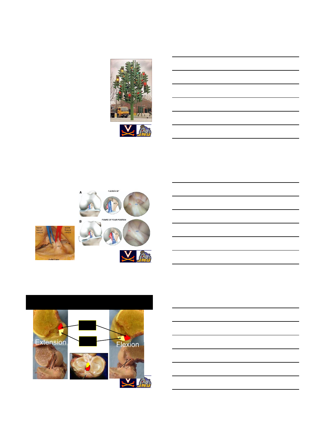
Knee—ACL
Anatomy and Biomechanics
Tibia => LFC
•33 mm x 11 mm
•2 Bundles:
–AM (tight in flexion)
–PL (tight in
extension)
•Middle Geniculate A.
PL
bundle
AM
bundle
Flexion
AM
PL
Femoral Insertion Alignment Changes With Knee
Flexion
Extension
PL
AM PL
AM
10/13/2015
3
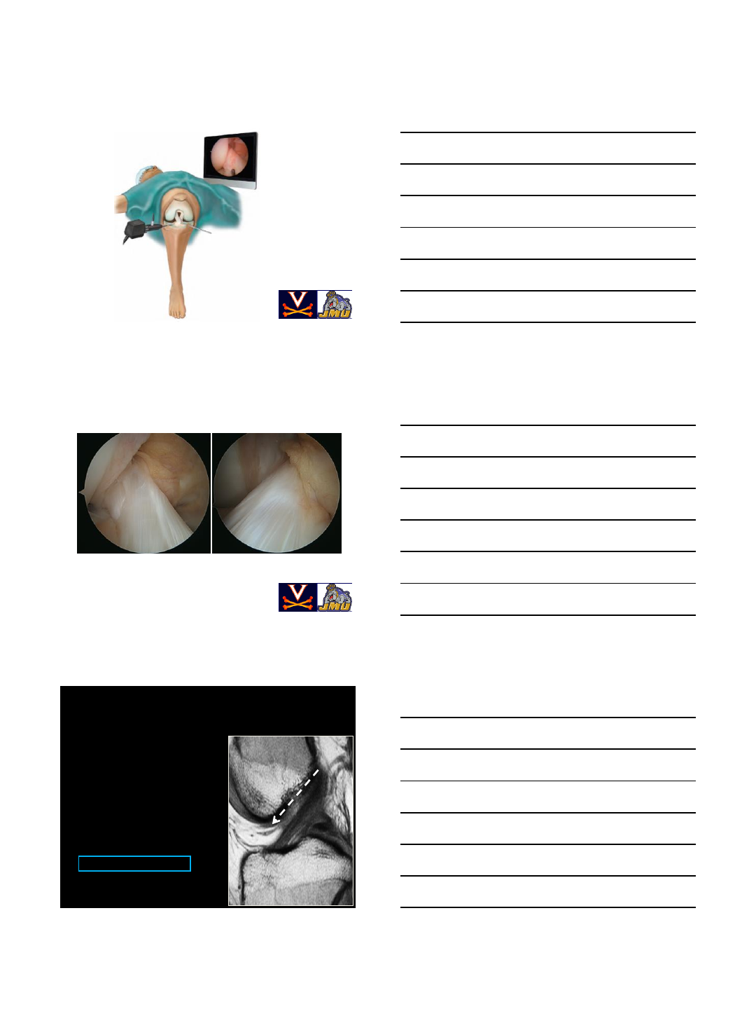
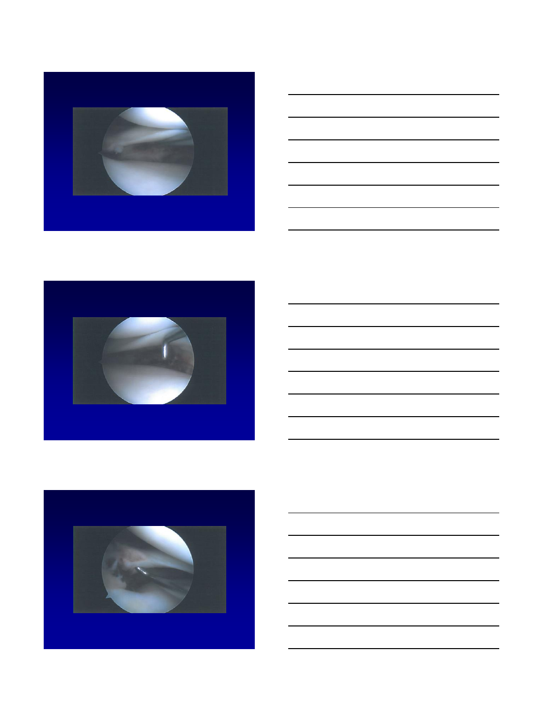
ACL Arthroscopy
ACL
ACL
ACL Arthroscopy
View from Anterolateral Portal View from Anteromedial Portal
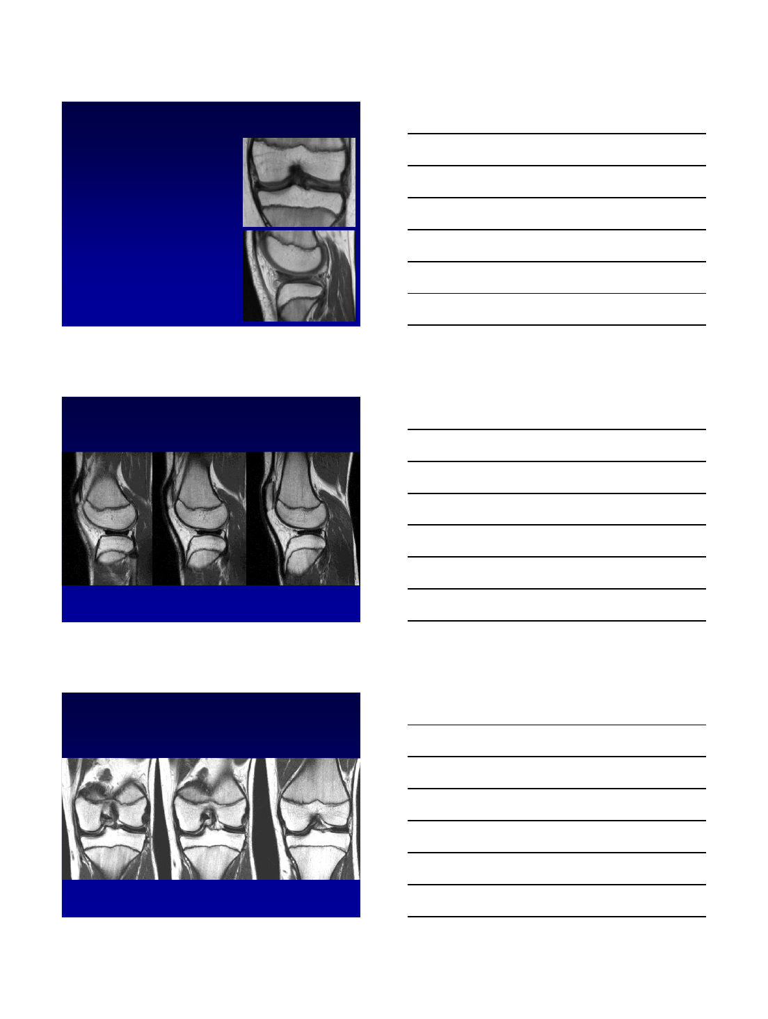
Normal ACL
•Sagittal
–Taut
–Parallel
•intercondylar roof
(aka - Blumenstaat’s line)
•Signal intensity
–Low / intermediate
–Striated
Evaluate in all planes
10/13/2015
4
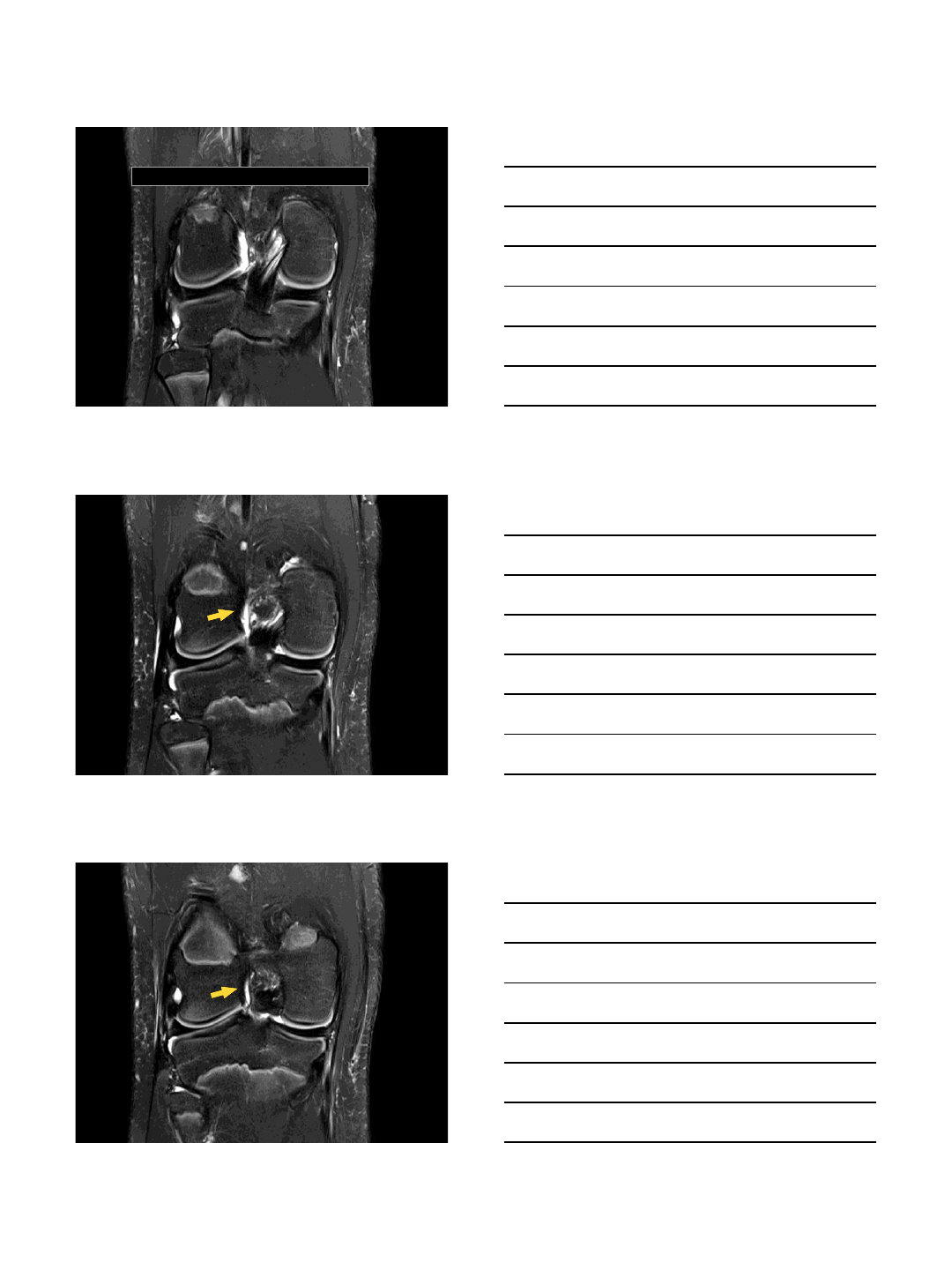
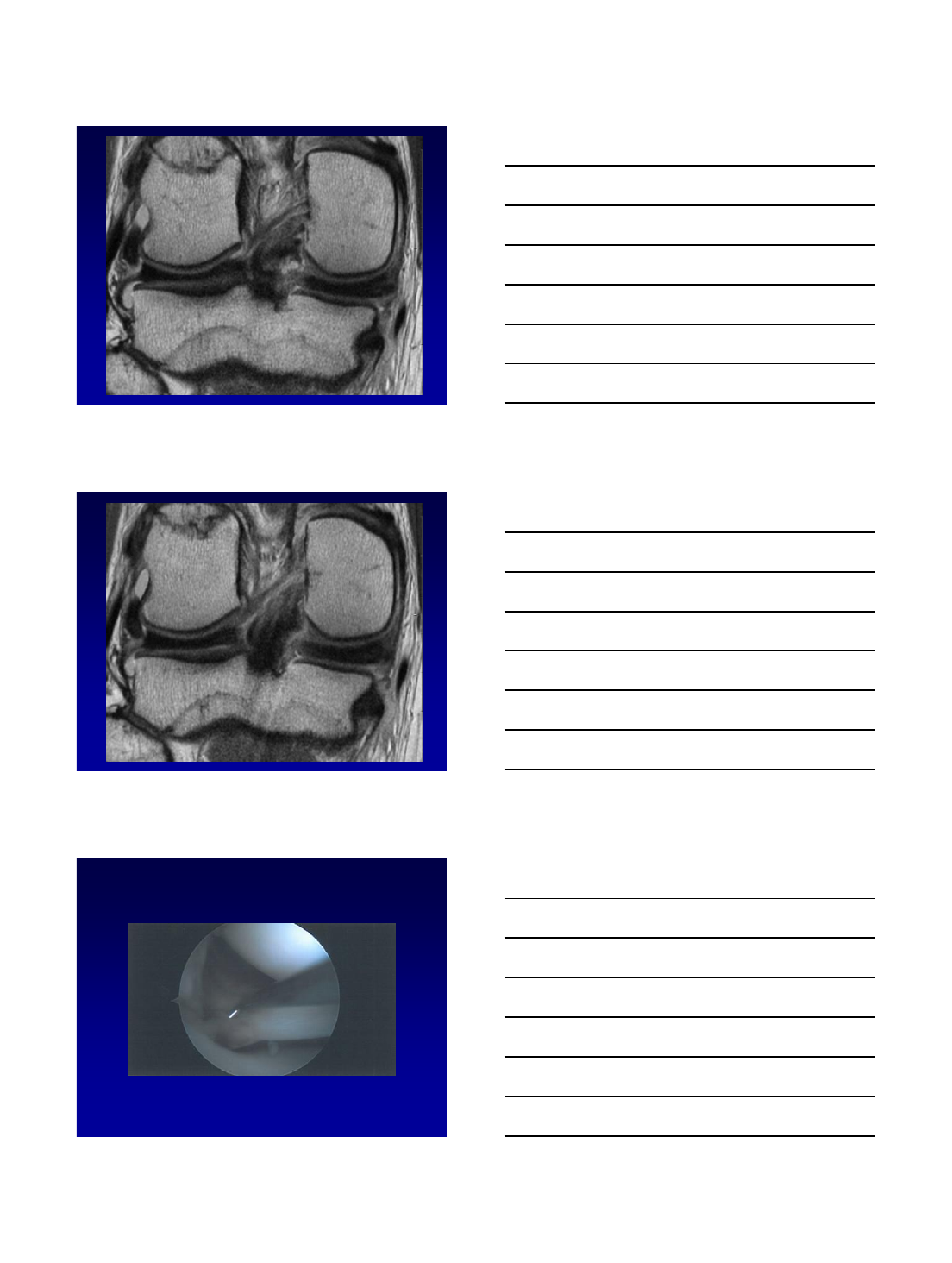
Coronal
10/13/2015
5
AM
PL
AM
PL
PL
AM
10/13/2015
6
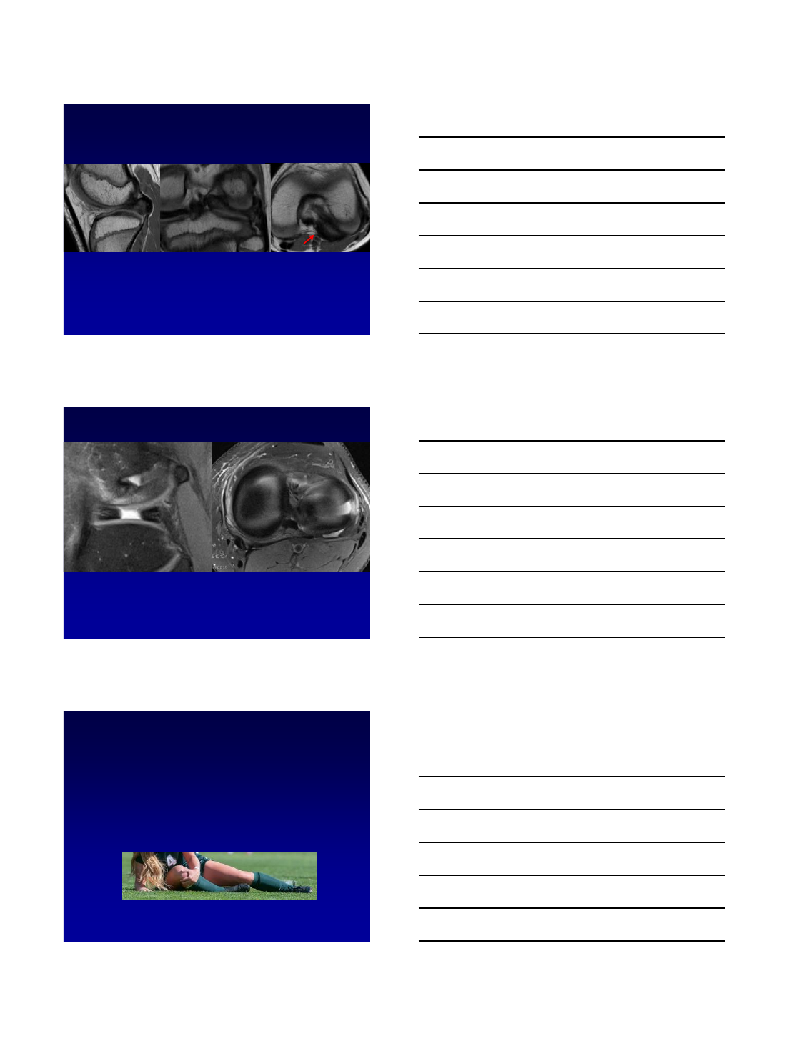
AM
Different patient:
ACL may blend with anterior
horn lateral meniscus
10/13/2015
7
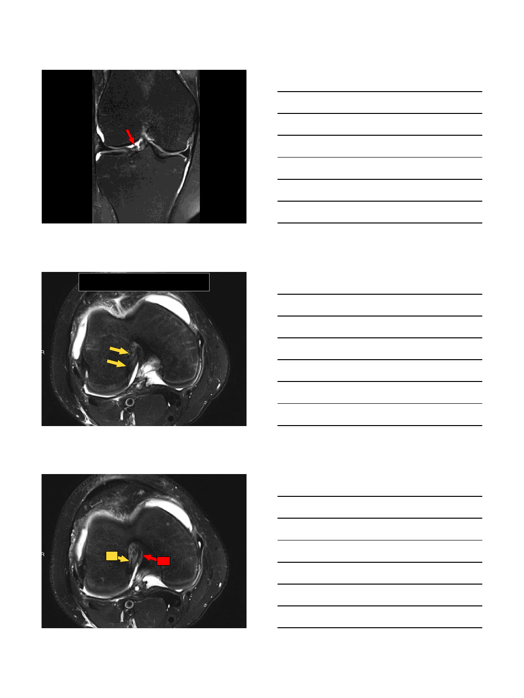
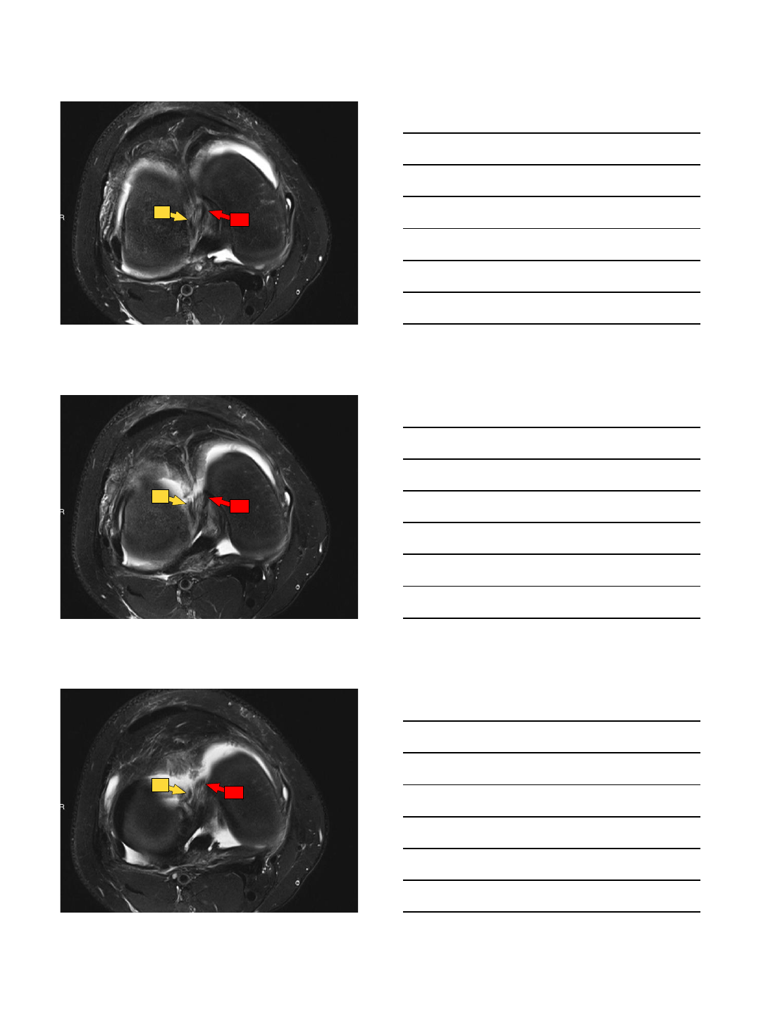
Axial:
Best for proximal to mid-portion of ligament
PL AM
10/13/2015
8
PL AM
PL AM
PL AM
10/13/2015
9
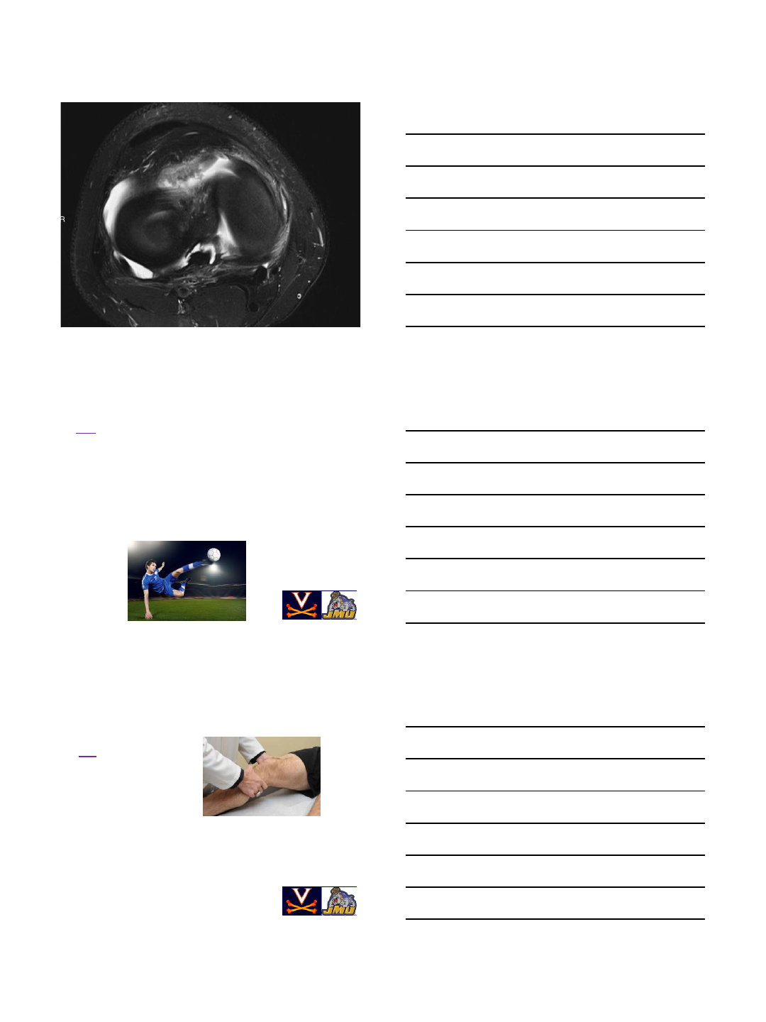
HPI
•17 yo M soccer player s/p non-contact pivoting
injury to L knee during game 8 weeks prior
•Attempted to continue playing but had 2 recurrent
pivoting episodes, most recently 5 days prior to
presentation
•L knee swelling, pain, and locking with
incomplete extension on most recent episode
Case 1
PE
•(+) effusion
•ROM 15-90
•(+) Lachman
•(+) lateral joint line tenderness
No XRs provided, brought in MRI from OSH
performed after last instability episode
Case 1
10/13/2015
10
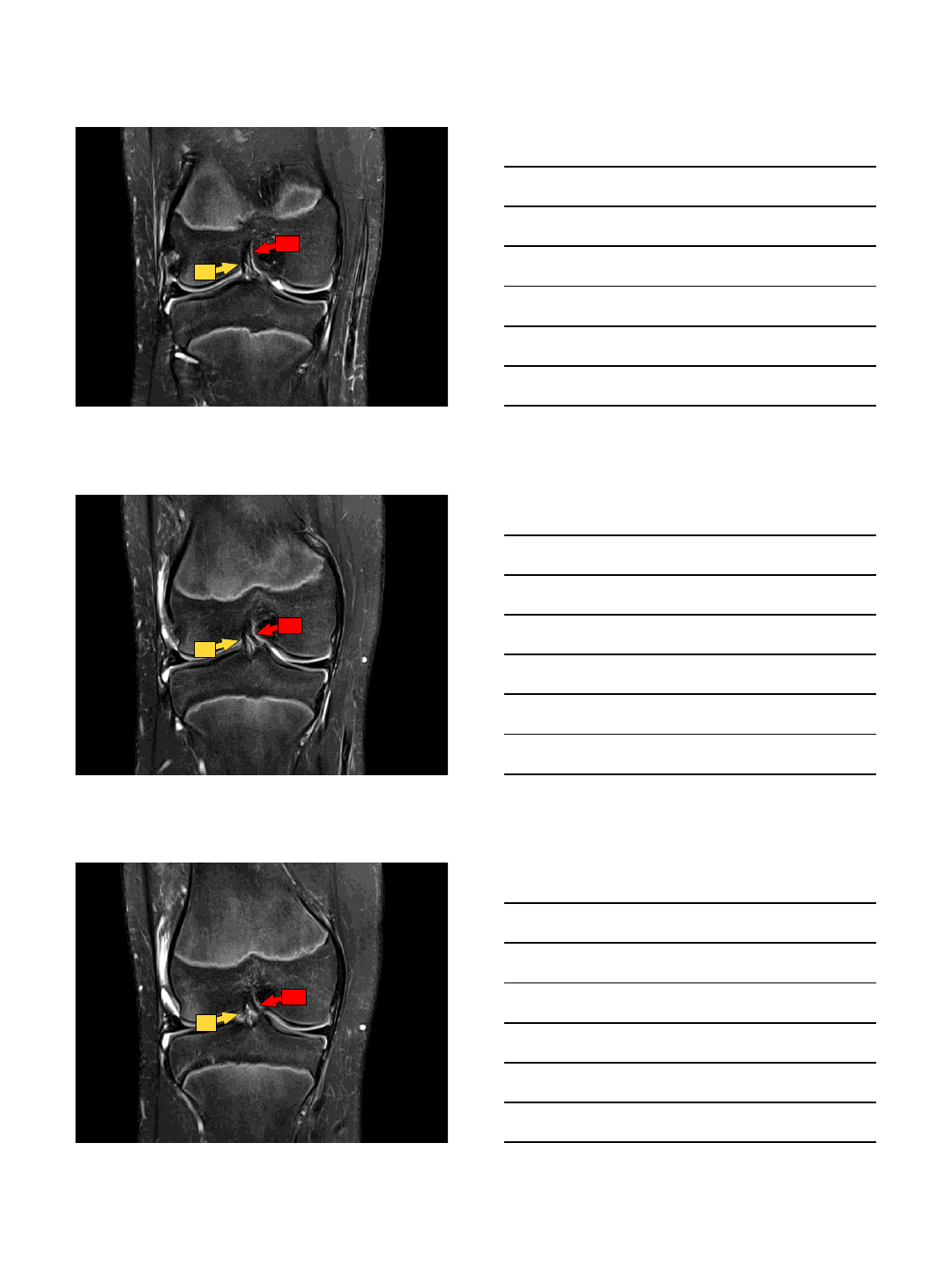
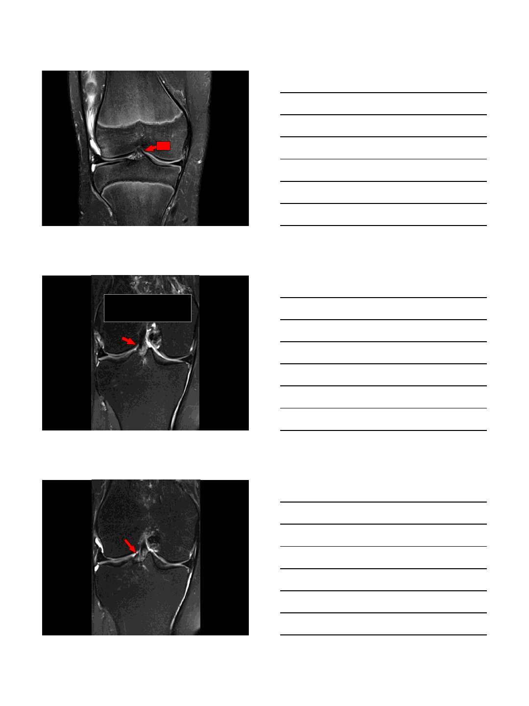
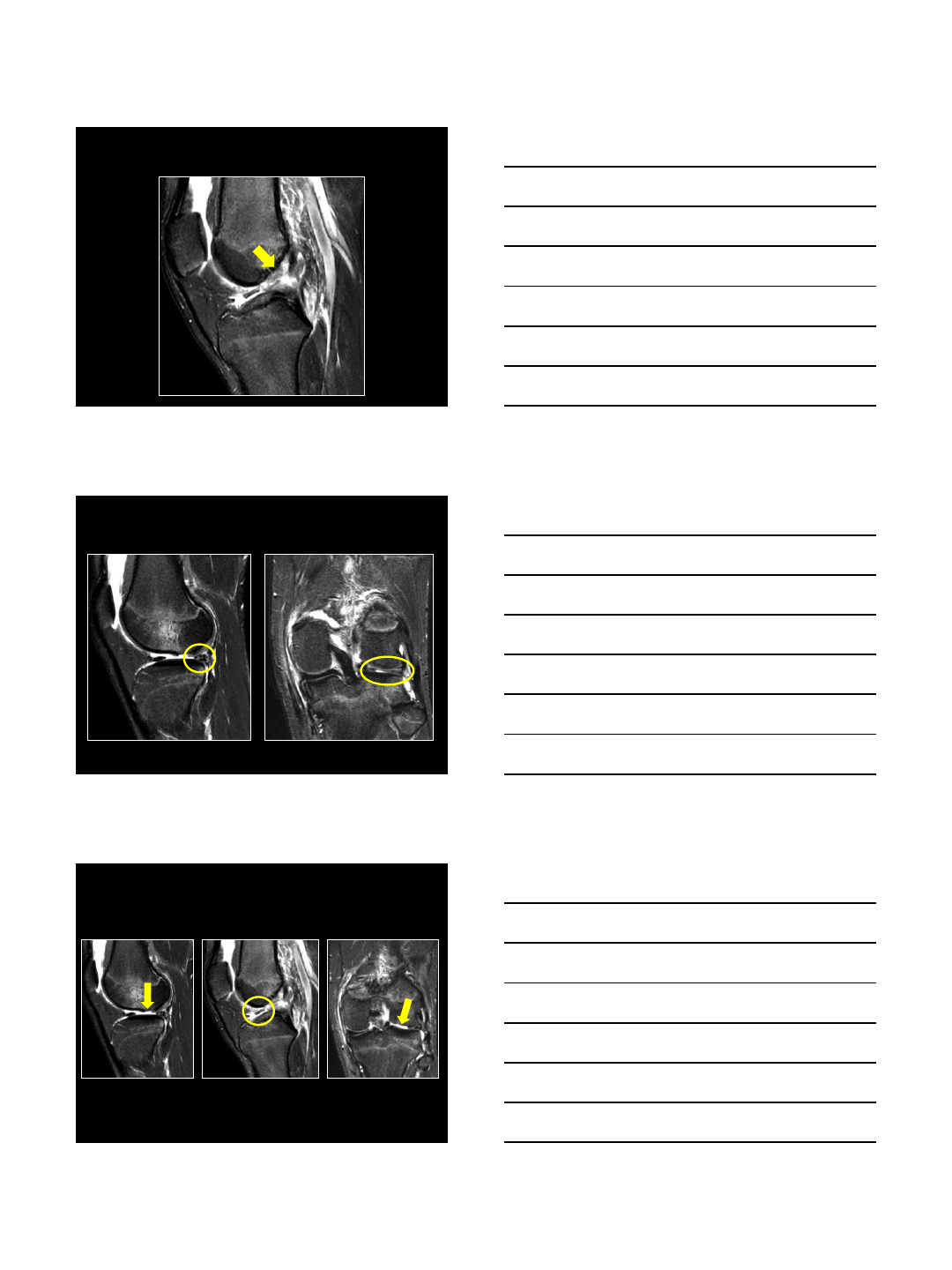
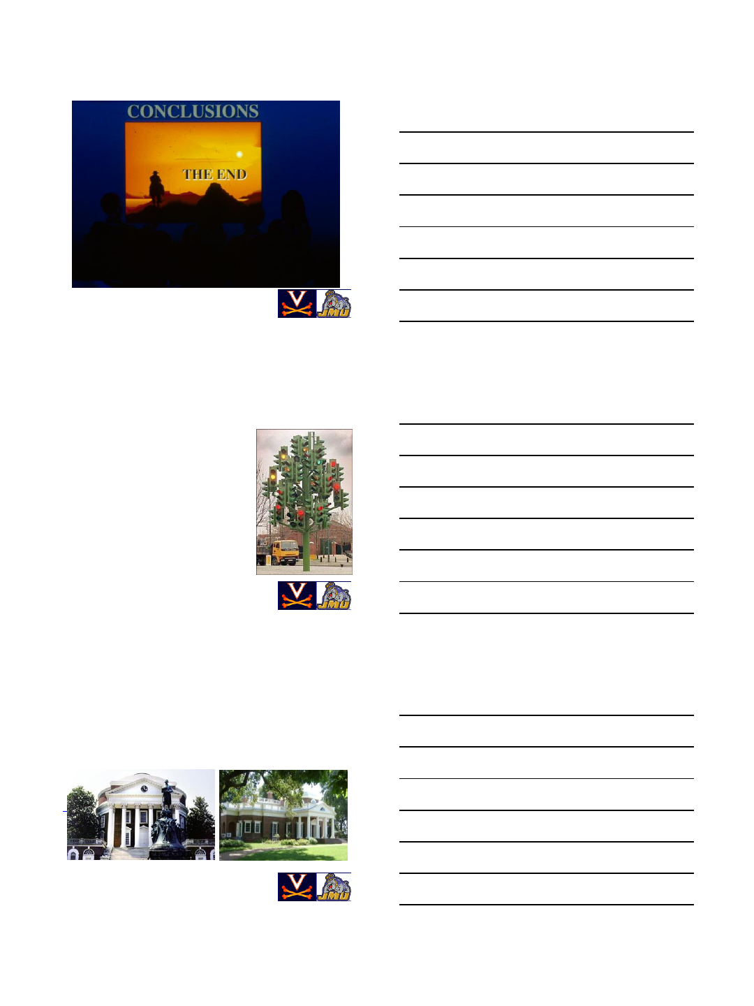
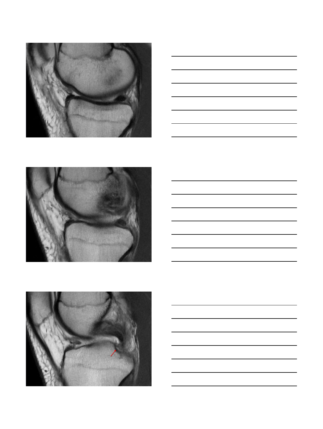
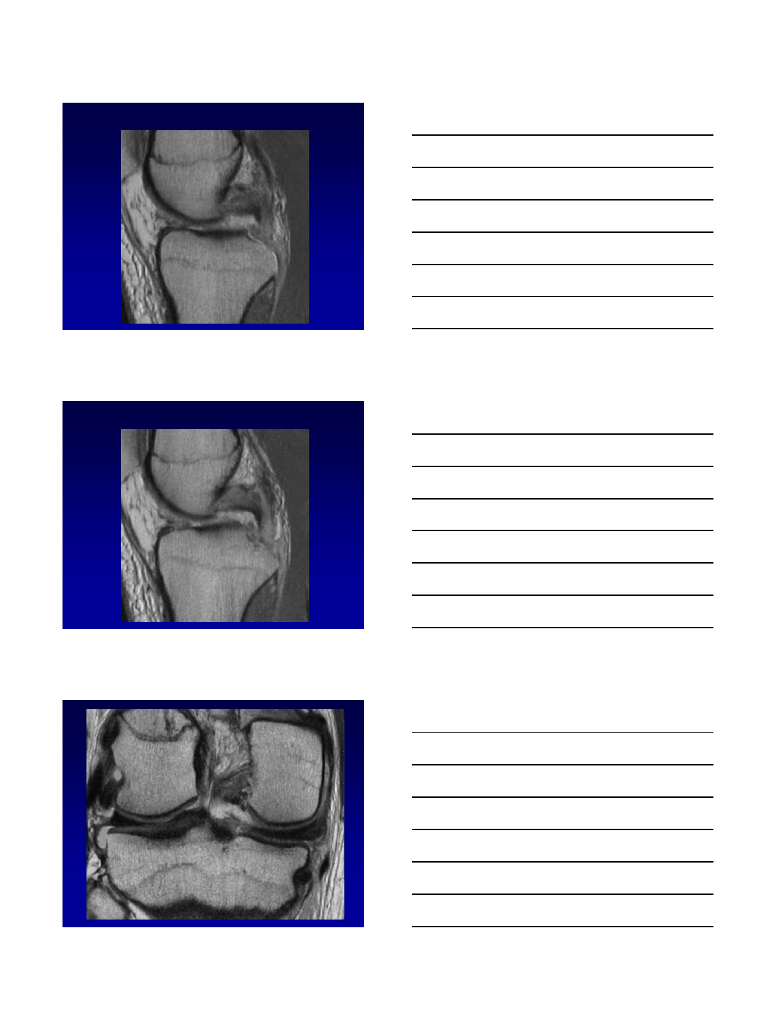
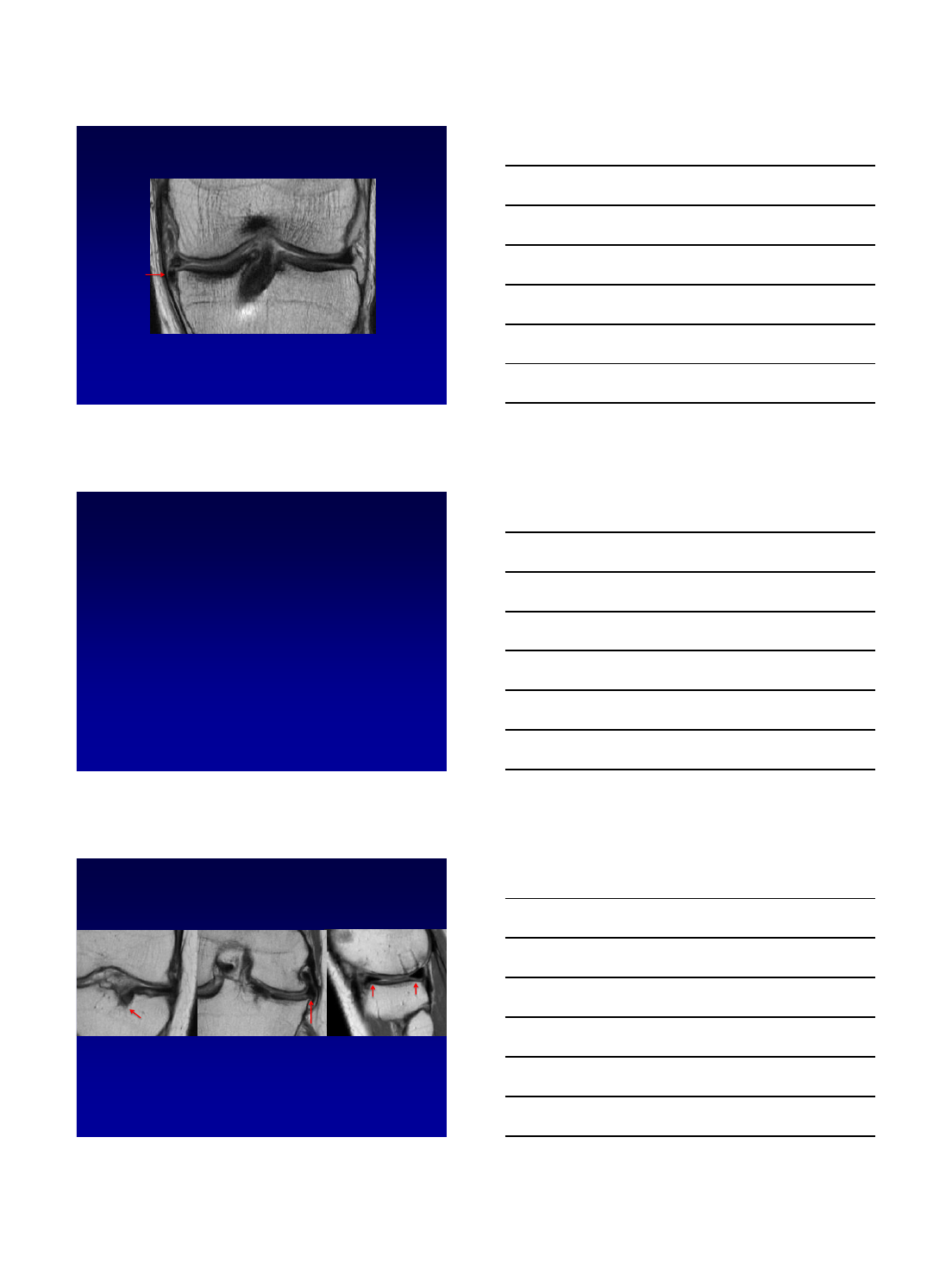
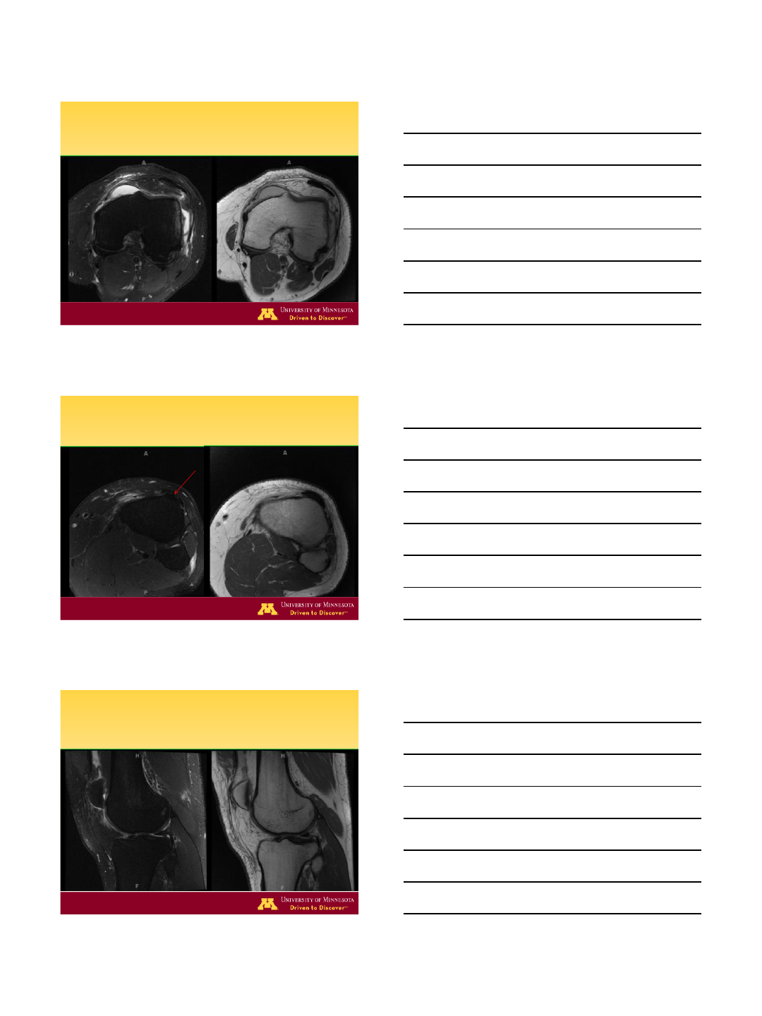
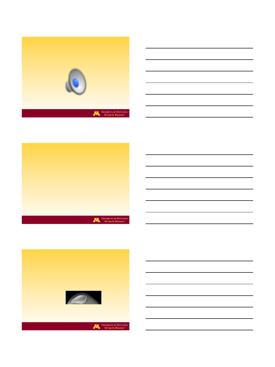
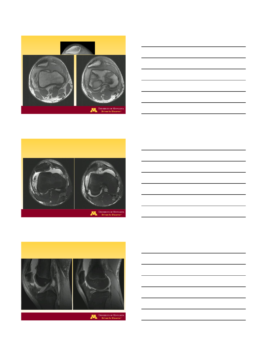
MRI –ACL Tear
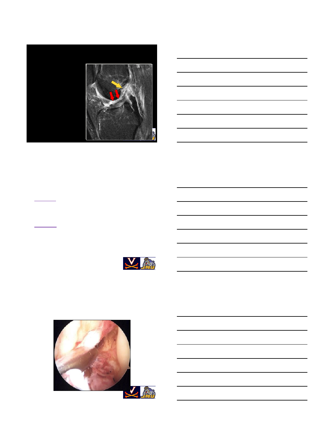
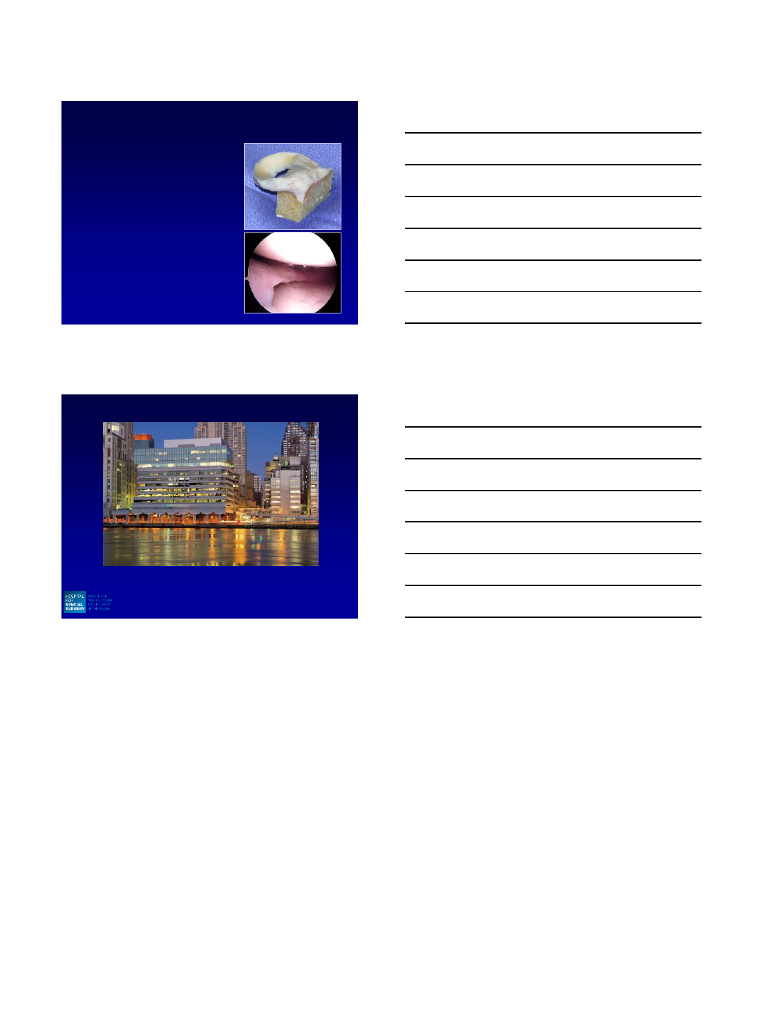
MRI –Lateral Meniscus Tear
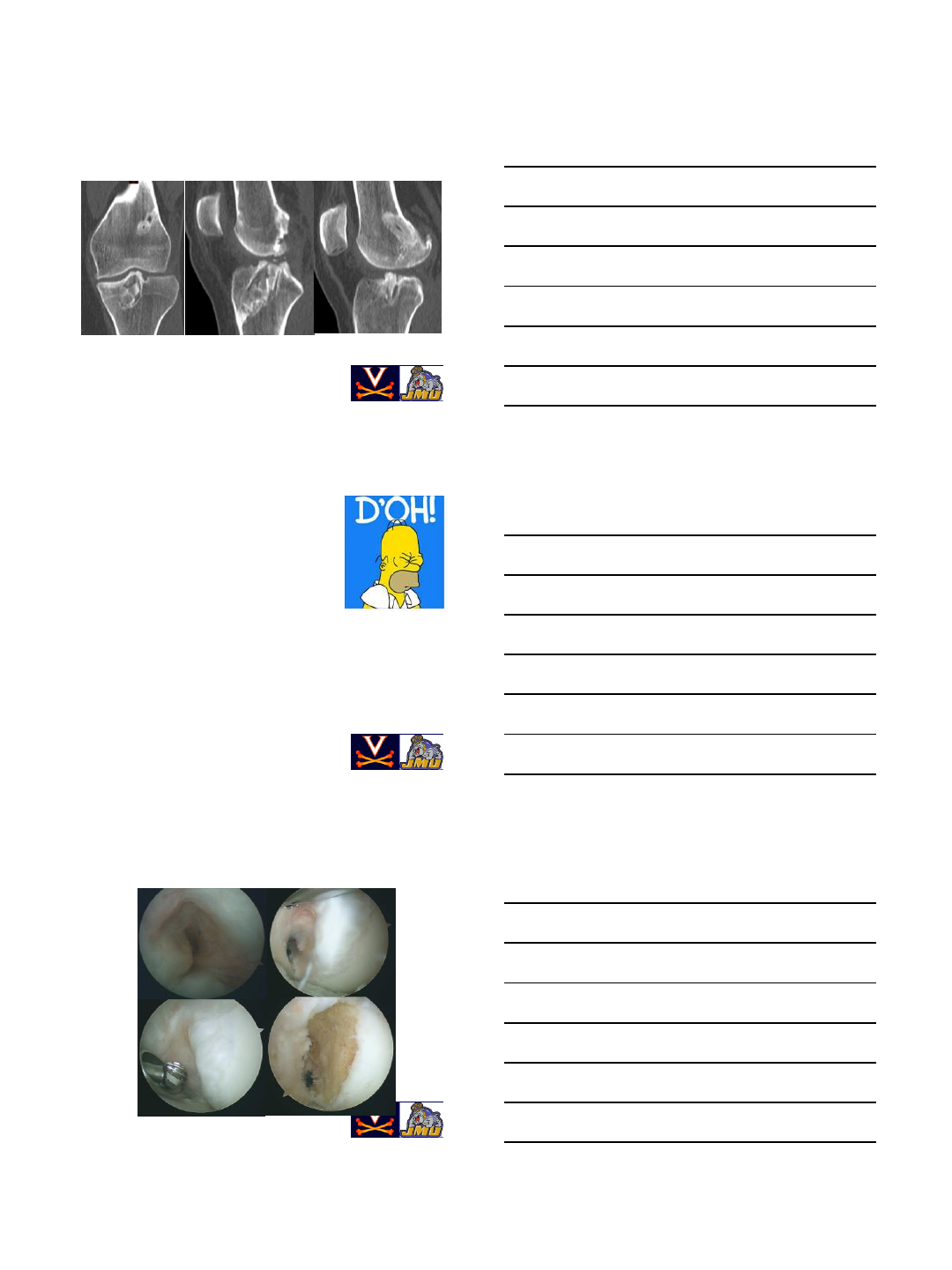
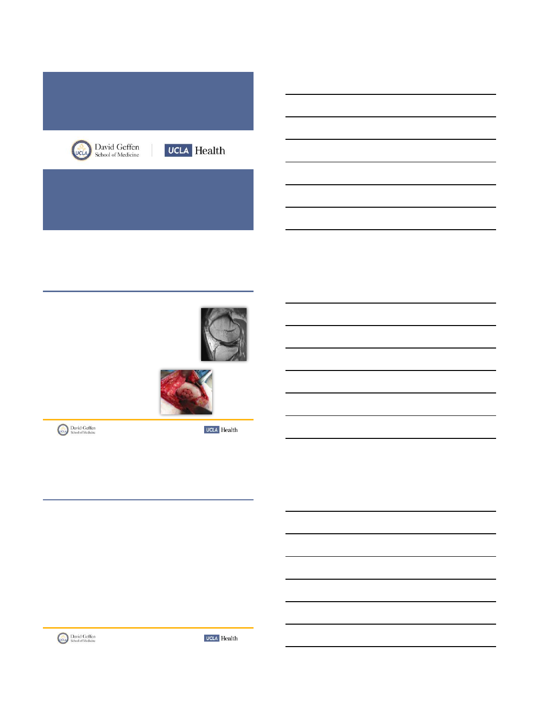
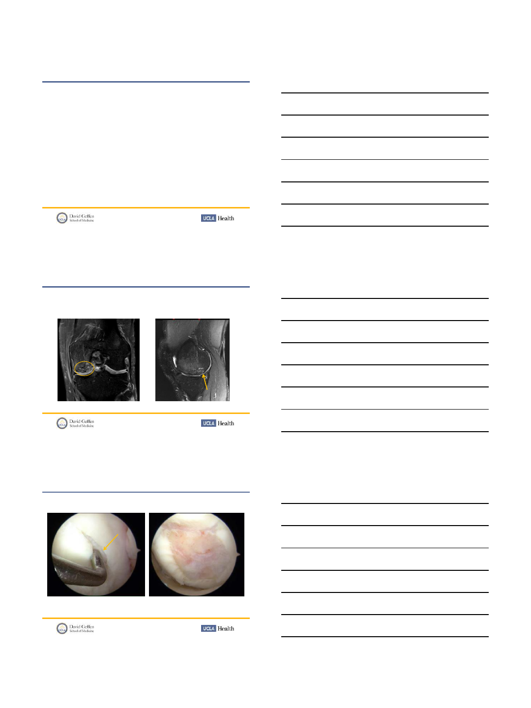
MRI –Chondral Defect/Loose Body
10/13/2015
11
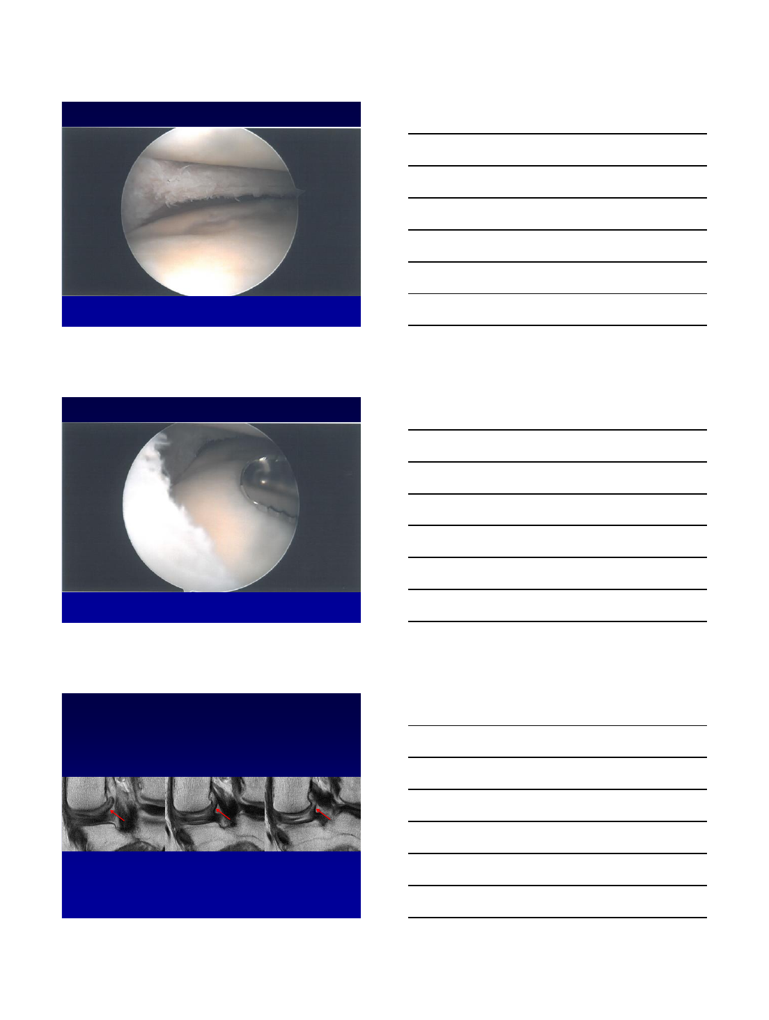
ACL: Complete Tear
Primary signs
•edematous mass
• “empty notch”
•irregular, horiz contour
•focal disruption
Diagnosis
•Left knee ACL tear
•Left knee complex bucket-handle lateral meniscus tear
•Left knee lateral femoral condyle chondral fracture
with loose body
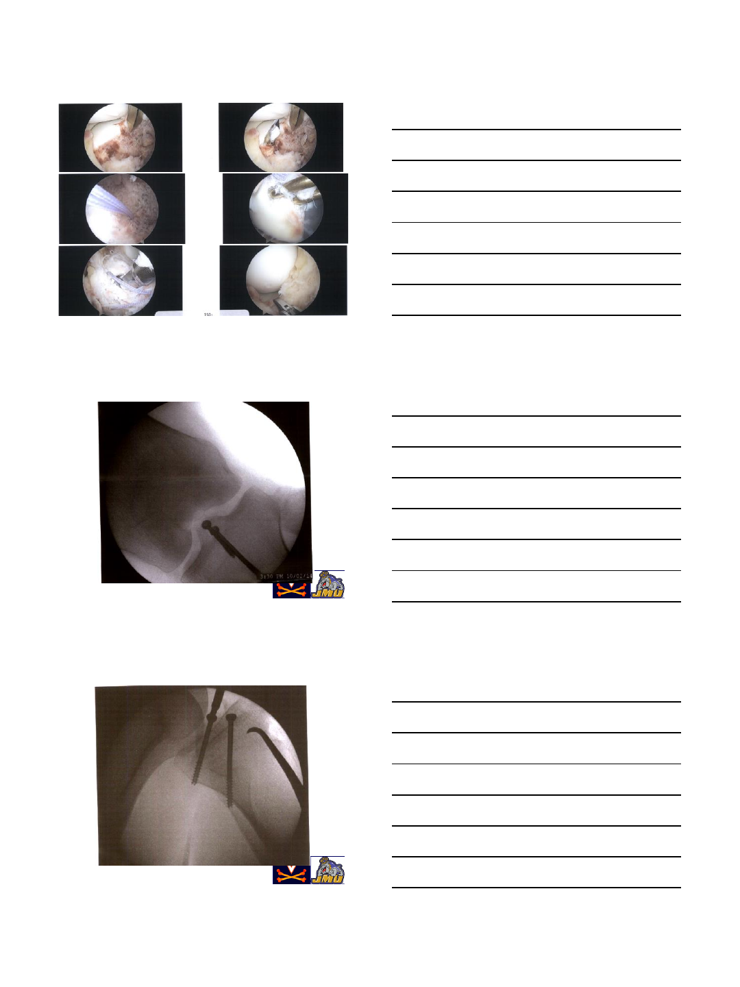
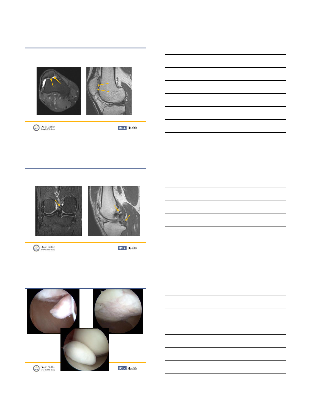
Procedure
•Left knee ACL reconstruction with B-PT-B autograft
•Left knee PLM
•Left knee removal of loose body
•Left knee OATS to LFC
Case 1
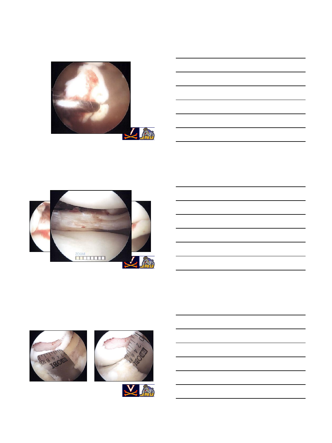
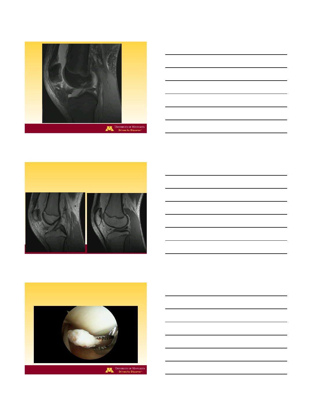
ACL Tear
10/13/2015
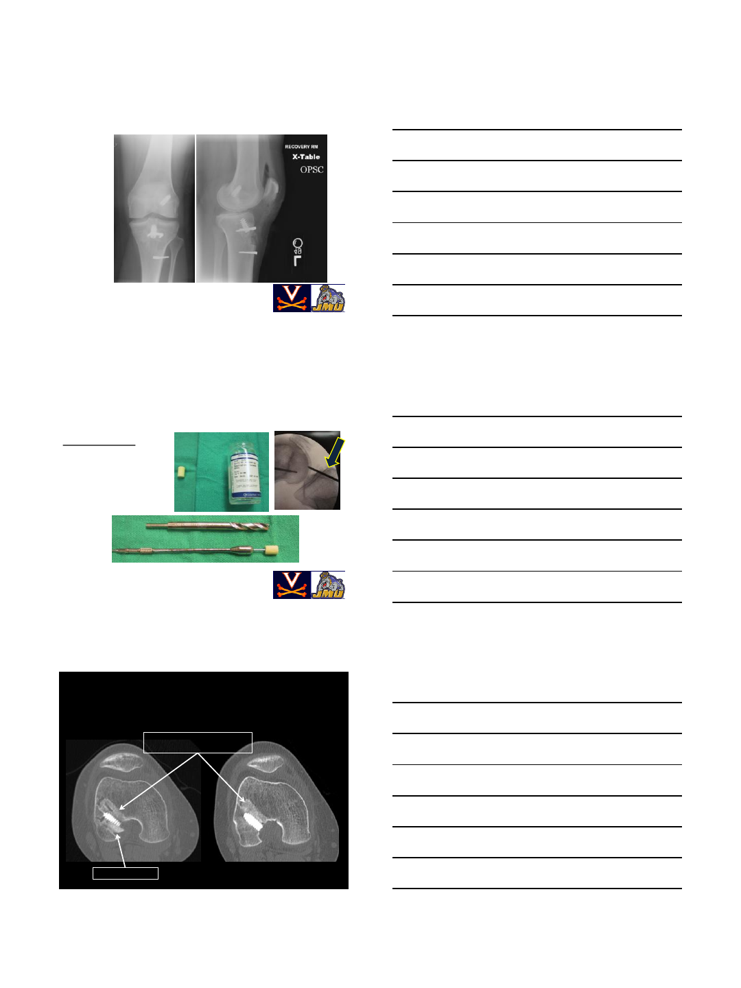
12
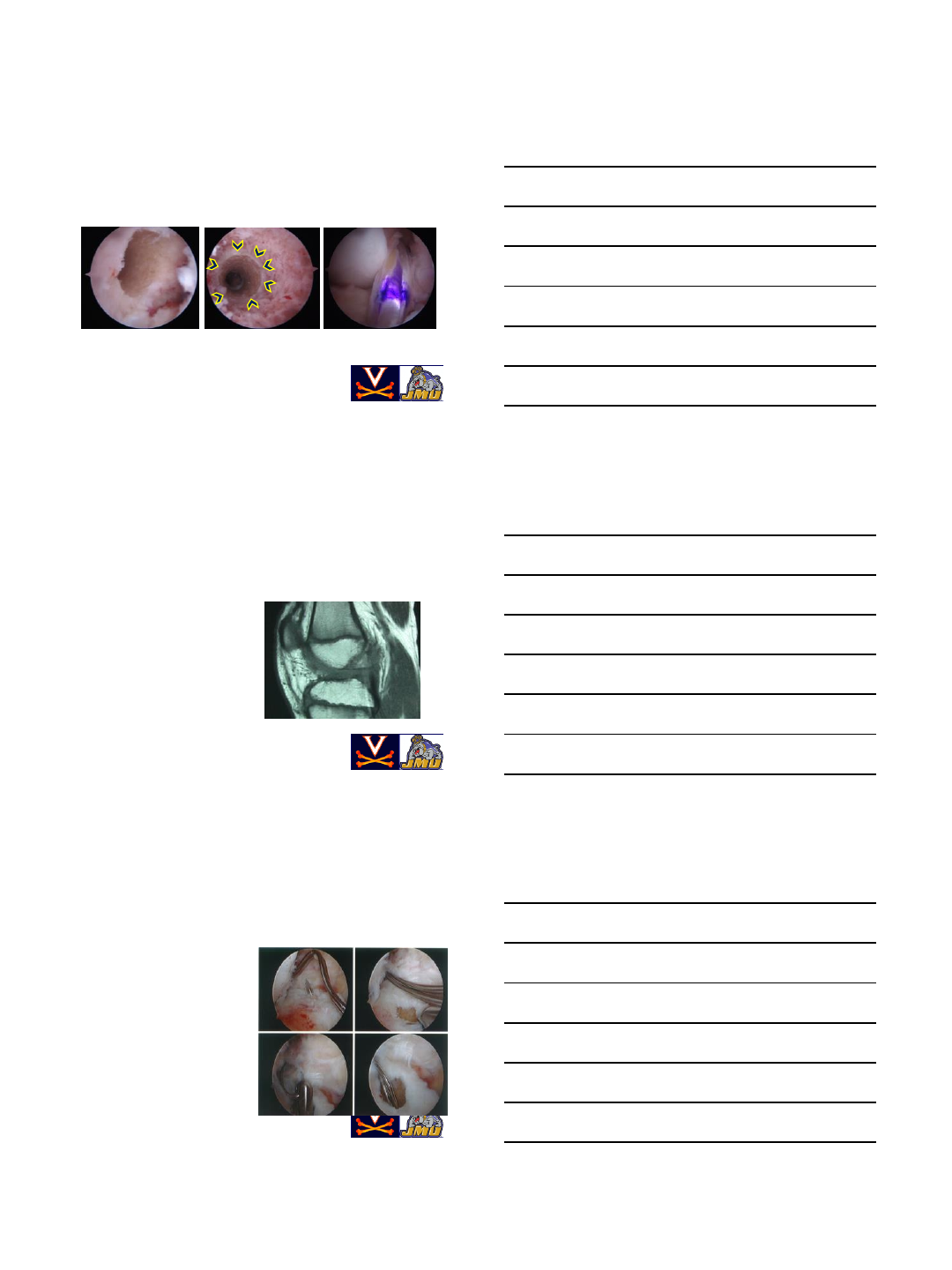
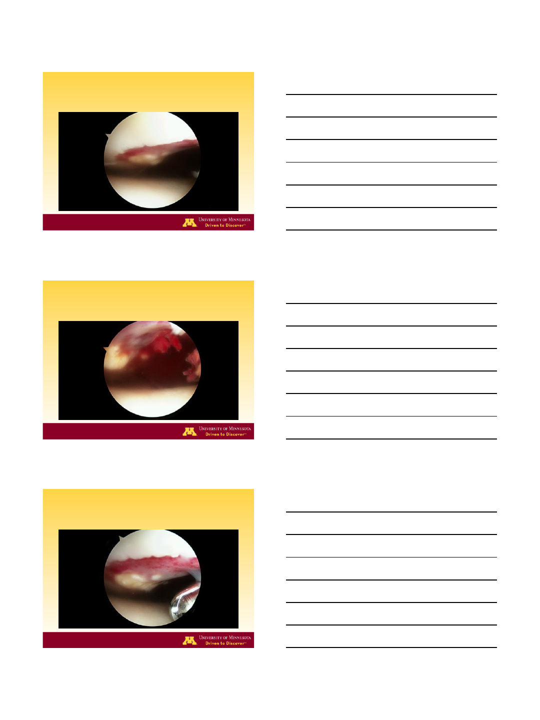
Removal of Loose Body
Lateral Meniscus Tear
Chondral Defect

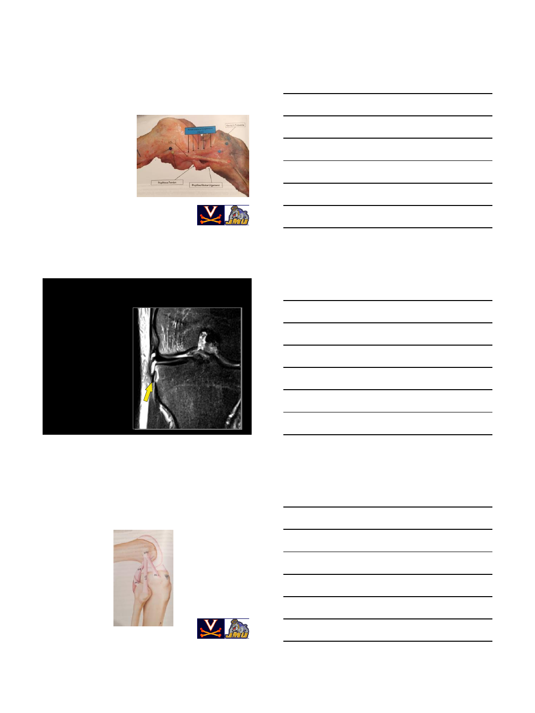
10/13/2015
13
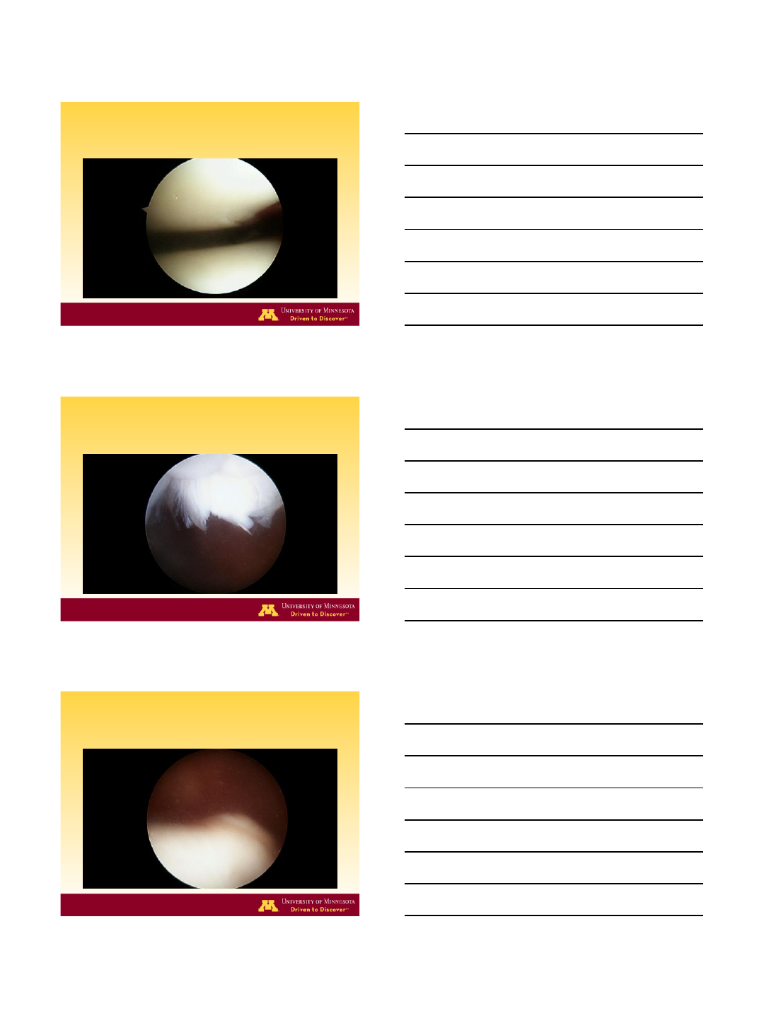
ACL Graft
OATS Plugs
HPI
•14 yo boy S/P attempted ACL eminence
repair one year prior @ Outside Hospital.
•Recurrent instability
•2+ Lachman
•+ Pivot Shift
Case 2
CT
1 yr
earlier

10/13/2015
14
MRI Findings
New MRI: failed repair
ACL Reconstruction
Lateral
Bifurcate
Ridge
ACL
Laxity
ACL Guide Pin Placement
Femoral: Below Physis (All-Epiphyseal)
Tibial: Trans Physeal--Verticla
10/13/2015
15
Tibial
Physis
Case 2: Pediatric ACL
Over-the-Top
Case Example
•11 year old boy with
pivoting injury
(football)
• Up to 12” growth
remaining
•Failed non-operative
management
(recurrent instability)
Pedi ACL Case
•7-mm Central
Vertical Tibial Tunnel
•18-gauge looped
wire passed from
“over the top” and
out the Tibial Tunnel
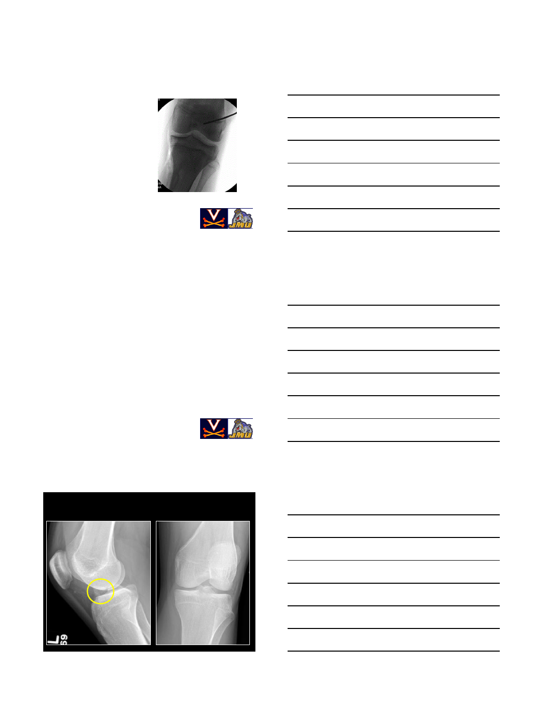
10/13/2015
16
Pedi ACL Case
• ACL Hamstring Graft Placed “Over the Top”
Pedi ACL Case
•Post-Operative Films
4 Year
Follow-up
10/13/2015
17
Adult Reconstruction
•Trans-Tibial
–Less disruption of
femoral physis
–Non-anatomic
•Independent Drilling
–Substantial risk to
the physis*
*Nelson J, Miller MD; JBJS-A 2011; 18:93 e53: 1-4
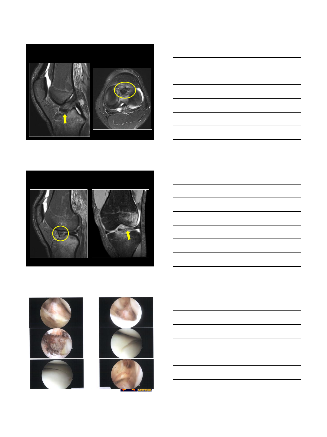
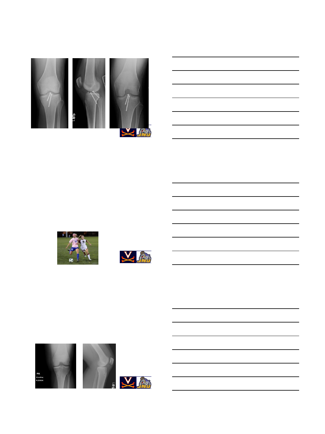
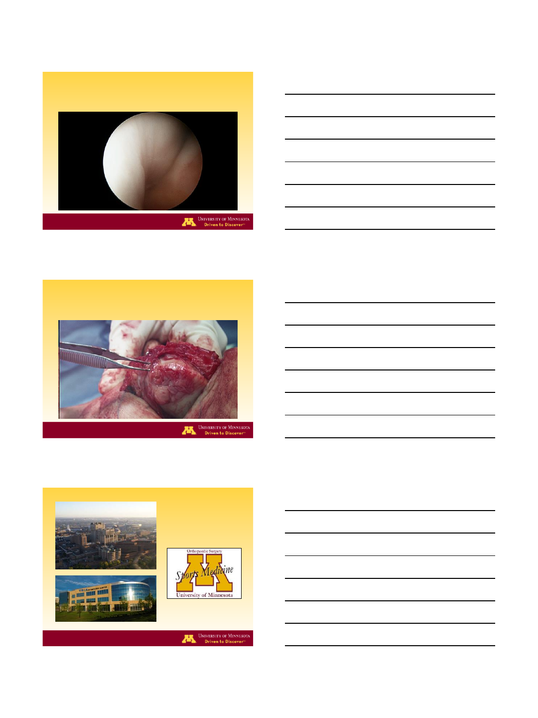
ACL Avulsion Repair
EMcD 2481546
HPI: 19M with left knee pain, had a non-
contact injury playing flag football and
planted his foot on 9/12/14. He heard a pop
and had immediate pain and swelling.
Evaluated at ED, pain with WBAT, using
crutches
Exam: 10° Loss of Extension;
2+ Lachman, + Pivot
XR: tibial spine avulsion
10/13/2015
18
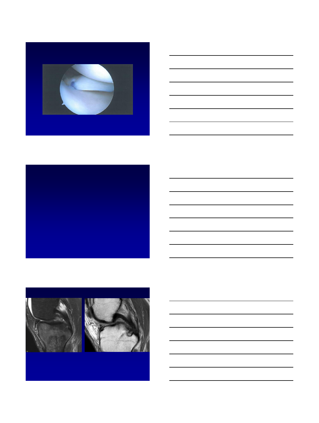
MRI: tibial spine avulsion
MRI: avulsion/AHLM root tear
10/13/2015
19
10/13/2015
20
Case 3
Revision ACL Case
•19yo Female S/P L ACL Revision x2
–First ACL at age 15 BPTB T-T Allograft
•Failed at 10 months—Soccer “injury”
–Second ACL at age 16 BPTB T-T Allograft (again)
•Failed again at 10 months—Soccer “injury”
–Hardware removal and allograft bone grafting
Revision ACL Case (Continued)
•On Presentation (4 months S/P bone grafting),
patient (and parents) complained of recurrent
instability and requested a second opinion
•Radiographs suggested tunnel osteolysis and
vertical femoral tunnel placement
10/13/2015
21
CT
Tibial Tunnel
18mm
Femoral Tunnel
ACL Case 1 (Continued)
•Labs:
–Knee Aspirate: 600 WBC, Gram Stain -, No Growth
–Systemic Labs: WBC, ESR, CRP all Normal
ACL Case 1
ARS Question 1
•What would you do next?
–A. One Stage Revision ACL with Allograft
–B. One Stage Revision ACL with Autograft
–C. Two Stage Revision ACL—Allograft Bone Graft
–D. Two Stage Revision ACL—Autograft Bone Graft
10/13/2015
22
ACL Case 1 Management
•Planned 2 stage Revision
•Tibia
–Overdrilling (up to 18mm)
– ICBG (“Sandwich” Technique)
ACL Case 1 Management
•Planned 2 stage Revision
•Femur
–Overdrilling (10 mm)
–Allograft Cloward Dowel
•Transtibial placement after
tibial overdrilling
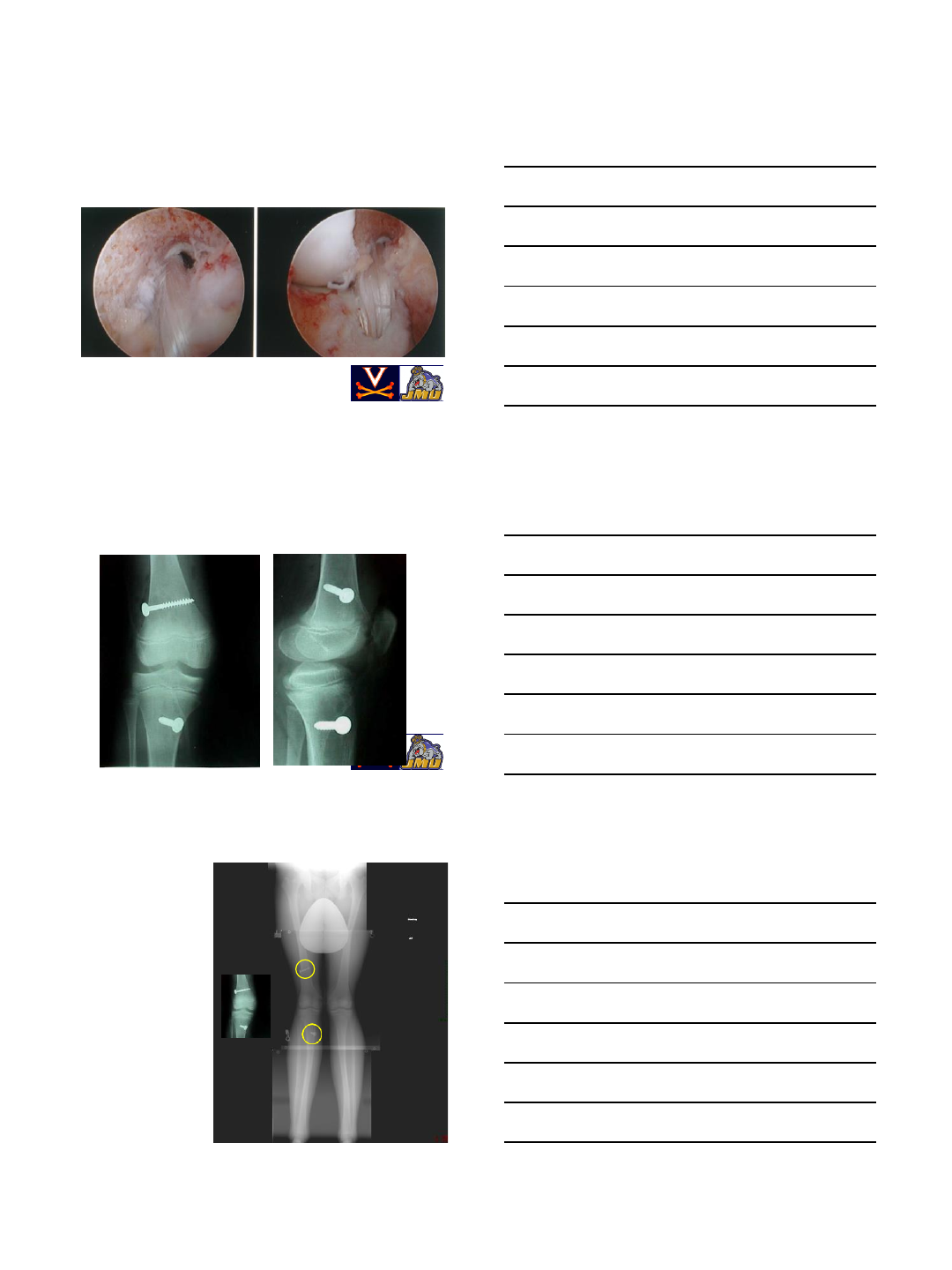
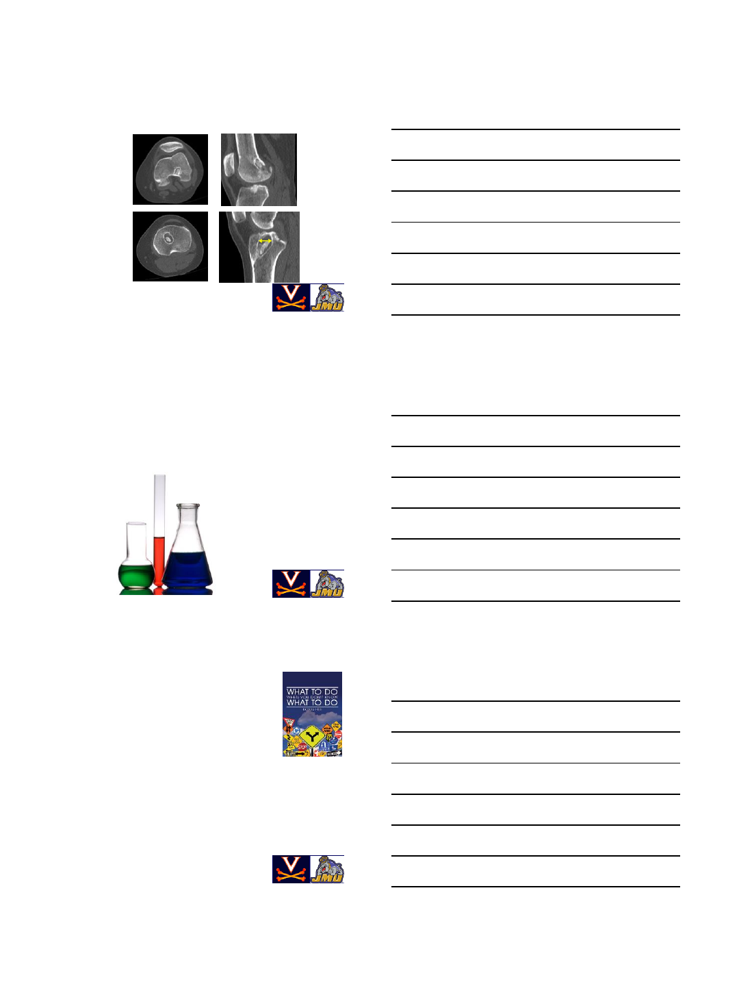
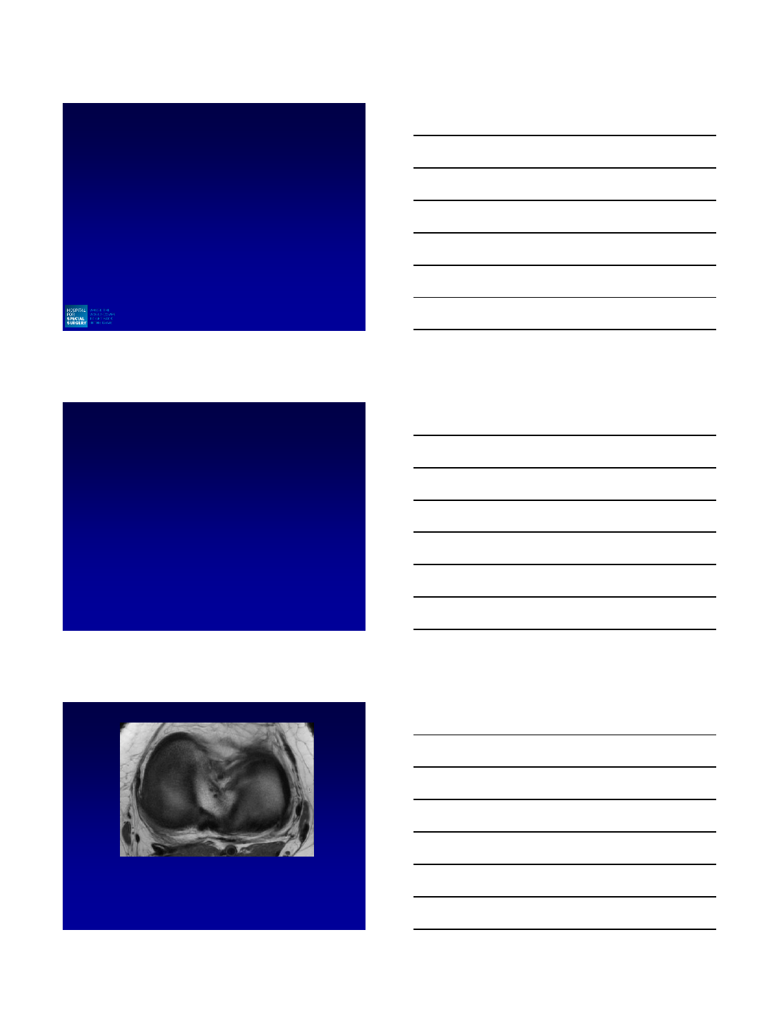
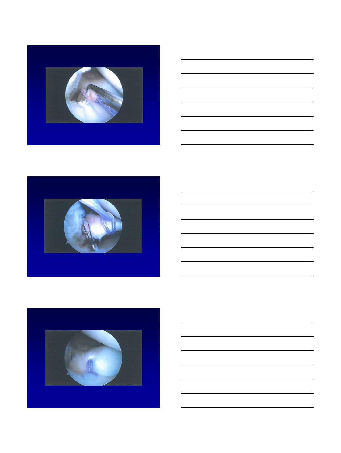
Revision ACL—Stage 1
10/13/2015
24
2nd Stage: ACL reconstruction
2nd Stage: ACL reconstruction
2nd Stage: ACL reconstruction
BPTB Autograft
10/13/2015
25
2nd Stage: ACL reconstruction
Revision ACL
Plug Technique
Cloward Plugs—
Allograft Dowels used
to fill in defects from
hardware removal or
tunnel osteolysis
Battaglia T, Miller MD; Arthroscopy 2005; 21:767
Guide wire placed In old tibial
tunnel. Drilled to 10 mm, 10 mm
Dowel Placed, New Tunnel Placed
Behind Old Tunnel
Patellar bone graft
Bone dowel filling tunnel from
first ACL reconstruction
CT: 5mos post ACL single stage
revision with bone dowel
10/13/2015
26
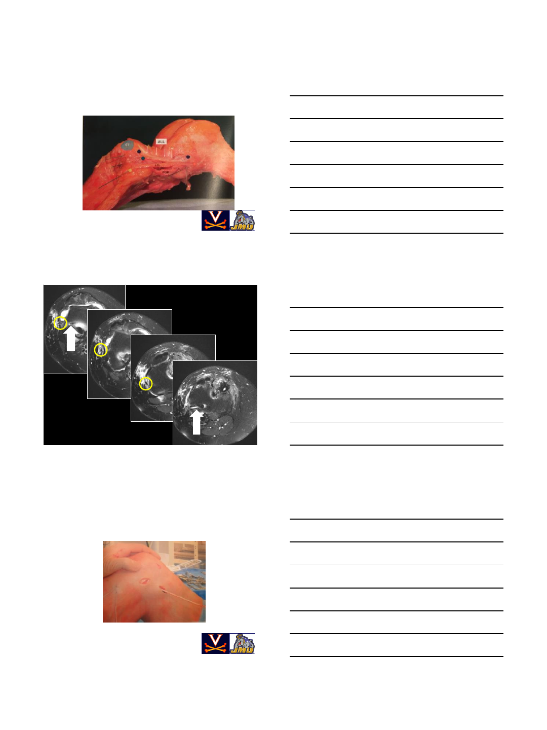
Anterolateral Ligament (ALL)
•Controversial
• Not really “New”
• What is it’s role?
•Europeans (Claes and
others) recommend
Repair/Reconstruction
in Patients with:
1. Segond Fx’s
2. Huge Pivots
3. Revisions
ACL: Complete Tear
Secondary signs
•bone contusions
•“deep notch”
•Segond fracture
10/13/2015
27
ALL reconstruction
ALL Reconstruction
10/13/2015
1
MRI –Arthroscopy Correlations of the
Knee: Menisci
Gabrielle P. Konin, MD
Assistant Professor of Radiology
Hospital for Special Surgery
Weill Cornell Medical College
New York, NY
Robert G. Marx, MD, MSC, FRCSC
Professor of Orthopedic Surgery
Hospital for Special Surgery
Weill Cornell Medical College
New York, NY
Disclosures
Robert G Marx:
Books and copyrights:
–Marx, RG (Editor). Revision ACL Reconstruction: Indications and
Technique. Springer. 273 pages. New York, 2013.
–Marx RG, Myklebust G, Boyle B. The ACL Solution: Prevention
and Recovery from Sports’ Most Devastating Knee Injury. Demos
Health. 174 pages. New York, 2012.
Journal Editorship:
–Deputy Editor for Sports Medicine, The Journal of Bone & Joint
Surgery
–Associate Editor for Evidence Based Orthopedics, The Journal of
Bone & Joint Surgery
–Senior Associate Editor, The HSS Journal
Gabrielle Konin: No relevant financial disclosures.
•Semilunar (C–shaped)
–Medial is more C-shaped and larger and lateral more rounded and smaller
•Divided into anterior and posterior horns and body
•Wedge shaped with biconcavity
10/13/2015
2
Medial Meniscus
•Posterior horn is larger than
anterior horn
•Non-mobile –more firmly
attached to the joint capsule
•Meniscofemoral and
meniscotibial (coronary)
ligaments
Lateral Meniscus
•Anterior = Posterior Horn
•Fibers of ACL extend into anterior horn
•Posterior root attaches anterior to PCL
•Meniscofemoral ligaments –Humphrey
& Wrisberg
•Popliteomeniscal fascicles
•Fascicles (2-3)
–Meniscocapsular extension around
popliteal hiatus
–Anteroinferior: body LM to
musculotendinous portion of popliteus
–forms floor of hiatus
–Posterosuperior: post horn LM to
popliteus tendon
–forms roof of popliteal hiatus
–If ruptured, can render the LM
hypermobile; pain and locking
Lateral Meniscus
10/13/2015
3
Discoid meniscus
•Watanabe classification: Complete,
Incomplete* and Wrisberg variants
•Non-tapering of apex of meniscus
•Radial diameter > 13 mm
•Increased height >2mm than opp meniscus
•Predisposes to degeneration and tear
•Pain, clicking, mechanical locking
Discoid meniscus
Discoid meniscus
10/13/2015
4
8 year-old girl with posterior knee pain & clicking for a few years
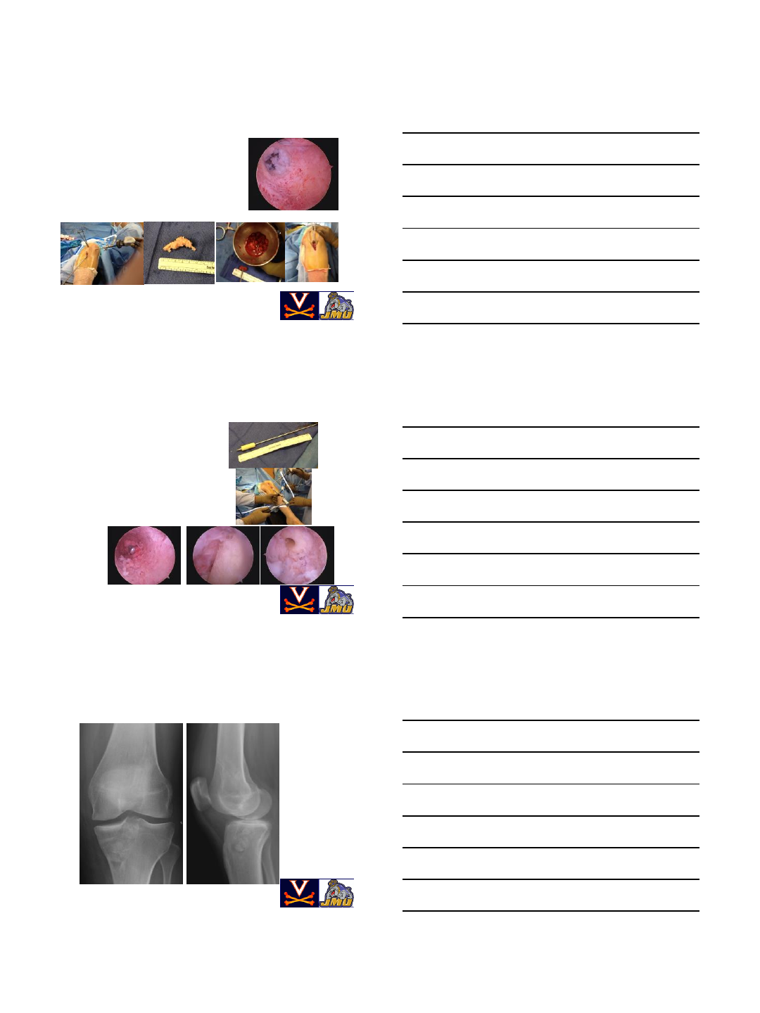
23 year old man s/p snowboarding injury
•Slight compression of the bone plate and overlying cartilage with
subchondral edema and chondral shear anteriorly
Radial tear
16 year-old girl with medial knee pain
following soccer injury
10/13/2015
5

10/13/2015
6
Radial Tear at Tibial Root
Radial Tear at Tibial Root

10/13/2015
7
Radial Tear at Tibial Root
Radial Tear at Tibial Root
Radial Tear at Tibial Root
10/13/2015
8
Radial Tear at Tibial Root
Radial Tear at Tibial Root
10/13/2015
9
10/13/2015
10

10/13/2015
11
10/13/2015
12
10/13/2015
13
47 year-old female with medial knee pain. Prior
history of meniscal root re-attachment.
Radial tear
• Radial split at the post horn root junction MM. “Ghost sign”
•Subacute subchondral medial plateau fracture with mild bone plate
depression and focal area of devitalized bone
One year later

10/13/2015
14
•Measured from outer meniscal edge to proximal tibial margin
•Medial > 3 mm. Lateral > 1mm
•Meniscal extrusion is 4 times more common medially
Meniscal extrusion
Bucket handle
•Circumferential longitudinal vertical tear
w/ displacement of free internal fragment
into intercondylar notch
•MM > LM
•MRI Signs
–Double PCL
–Double delta (lateral)
–Large AH
–Fragment in notch
–Absent bow tie
–Disproportionate horns
Double PCL
Double delta
Large AH
48 year-old man with medial knee pain.
Twisting injury a few months ago, heard a
“crack”.

10/13/2015
15
Bucket handle
10/13/2015
16
Flap tear with displacement
10/13/2015
17
Flap tear with displacement
•Important to recognize because gutters can be difficult to visualize
at arthroscopy
35 year-old woman with history of subtotal
lateral meniscectomy and subsequent
meniscal allograft.
•The allograft bone slot is incorporated
•Mild extrusion of the body segment
•Satisfactory position of the horns
•No meniscal split
Meniscal Allograft Transplantation
10/13/2015
18
Meniscal Allograft Transplantation
Why implant an allograft?
• “Arthroprotection”
–Decrease contact stress on articular cartilage
•Pain relief
•Restore normal / near normal kinematics
Thank you
10/12/2015
1
MRI -Arthroscopy
Correlations: Cartilage
Frank Petrigliano, MD
Assistant Professor
UCLA Department of Orthopaedic
Surgery
Benjamin Levine, MD
Assistant Professor
UCLA Department of Radiology
Disclosures
•Frank A. Petrigliano, MD:
•Speaker - Biomet
•Research Support –Musculoskeletal Transplant Foundation
•Honoraria –Musculoskeletal Transplant Foundation
•Committee Member –AOSSM Research Committee
•Benjamin D. Levine, MD:
•None
10/12/2015
2
Imaging Hyaline Cartilage
Quantitative MRI Techniques
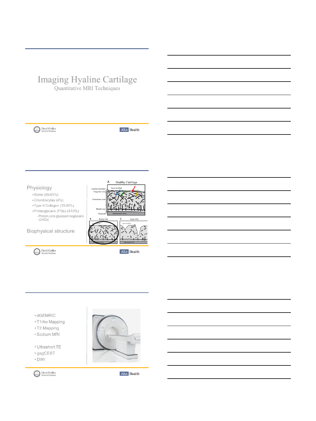
Hyaline Cartilage
Physiology
Water (65-85%)
Chondrocytes (4%)
Type II Collagen (15-20%)
Proteoglycans (PGs) (3-10%)
•Protein core glycosaminoglycans
(GAGs)
Biophysical structure
Matzat SJ et al. Quant Imaging Med Surg 2013;3(3): 162-174
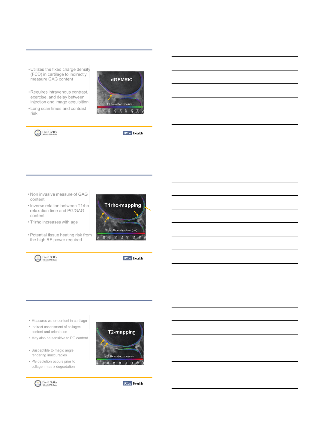
Quantitative MRI of Cartilage
•dGEMRIC
•T1rho Mapping
•T2 Mapping
•Sodium MRI
•Ultrashort TE
•gagCEST
•DWI
10/12/2015
3
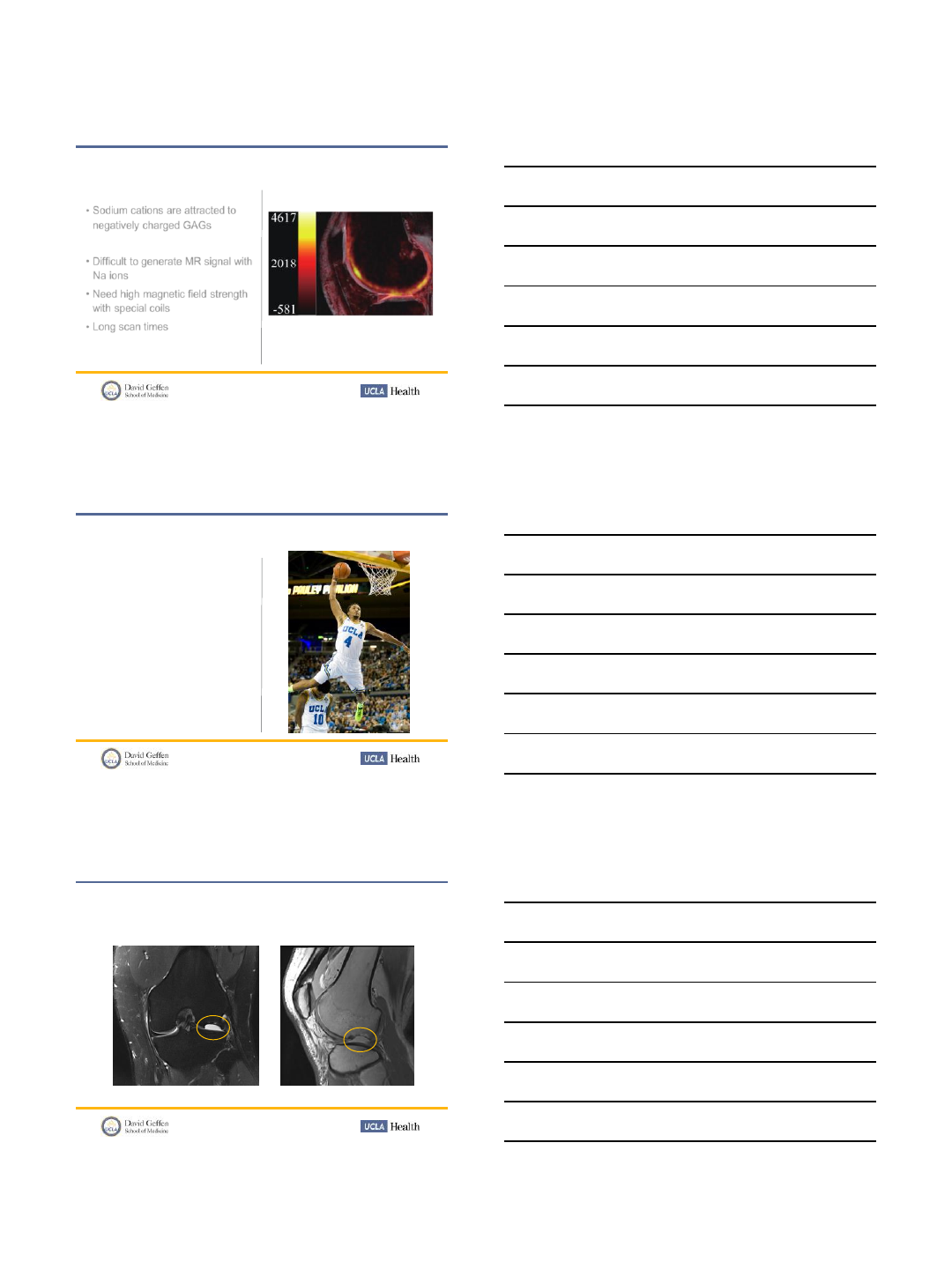
dGEMRIC
•Utilizes the fixed charge density
(FCD) in cartilage to indirectly
measure GAG content
•Requires intravenous contrast,
exercise, and delay between
injection and image acquisition
•Long scan times and contrast
risk
Bittersohl B, et al. Invest Radiol 2010;45:538-42.
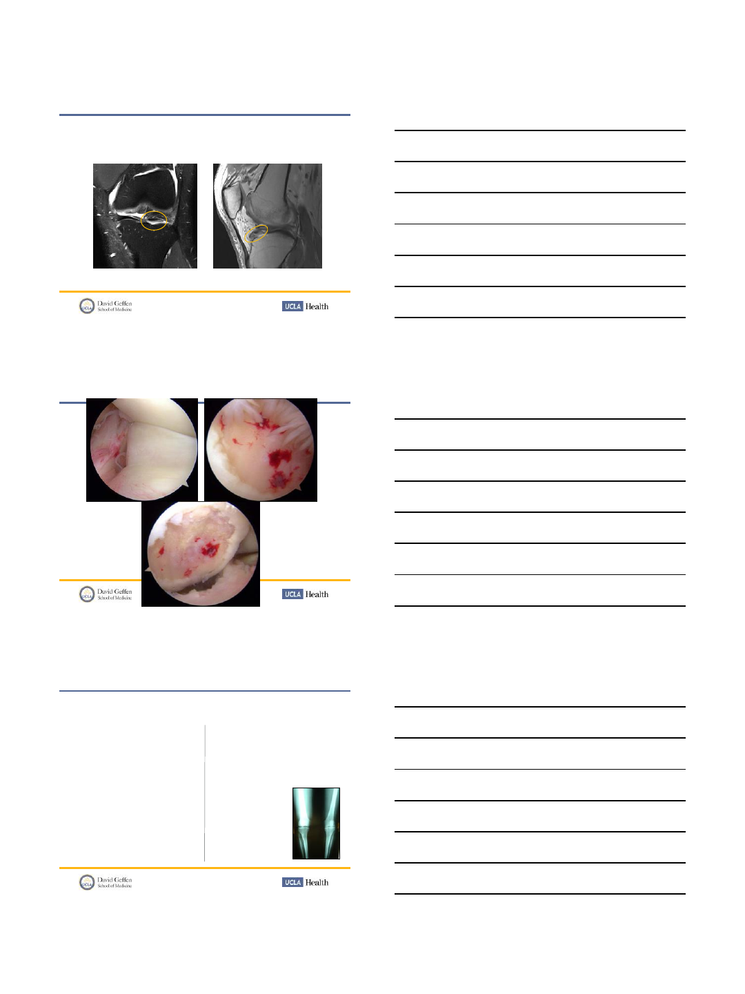
T1rho Mapping
•Non invasive measure of GAG
content
•Inverse relation between T1rho
relaxation time and PG/GAG
content
•T1rho increases with age
•Potential tissue heating risk from
the high RF power required
Matzat SJ et al. Quant Imaging Med Surg 2013;3(3):162-174
T2 Mapping
•Measures water content in cartilage
•Indirect assessment of collagen
content and orientation
•May also be sensitive to PG content
•Susceptible to magic angle,
rendering inaccuracies
•PG depletion occurs prior to
collagen matrix degradation
Mosher TJ, et al. Semin Musculoskelet Radiol 2004;8:355-68.
10/12/2015
4
Sodium MRI
•Sodium cations are attracted to
negatively charged GAGs
•Difficult to generate MR signal with
Na ions
•Need high magnetic field strength
with special coils
•Long scan times
Matzat SJ et al. Quant Imaging Med Surg 2013;3(3):162-174
Case #1
•30-year-old man with a
acute on chronic left knee
pain following an
traumatic basketball injury
•PE
•Substantial effusion, TTP
LJTL
•ROM 20 –90 degrees
•Stable ligament exam
•Neutral alignment
MRI
10/12/2015
5
MRI
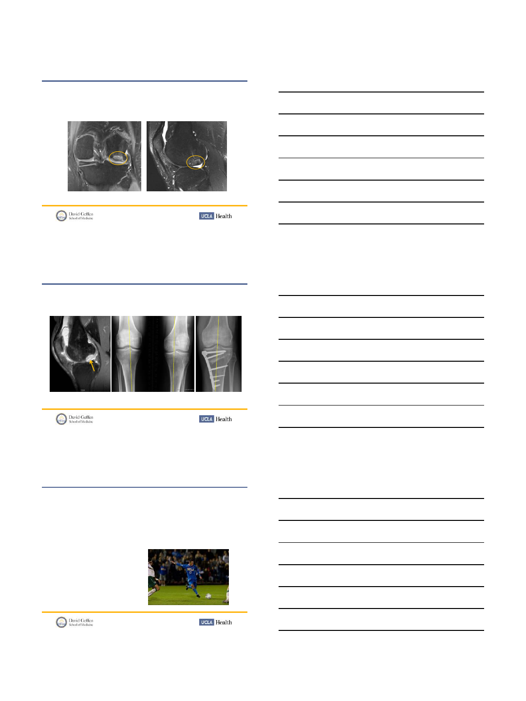
Treatment?
•Lesion Qualities
•Etiology
•Trauma
•AVN
•OCD
•Location
•Grade
•Size
•Character
•Chondral vs. Osteochondral
•Patient Qualities
•Age
•Demand (High v. Low)
•BMI (>30)
•Expectations
•Alignment
•Meniscal Status
•Knee Stability
10/12/2015
6
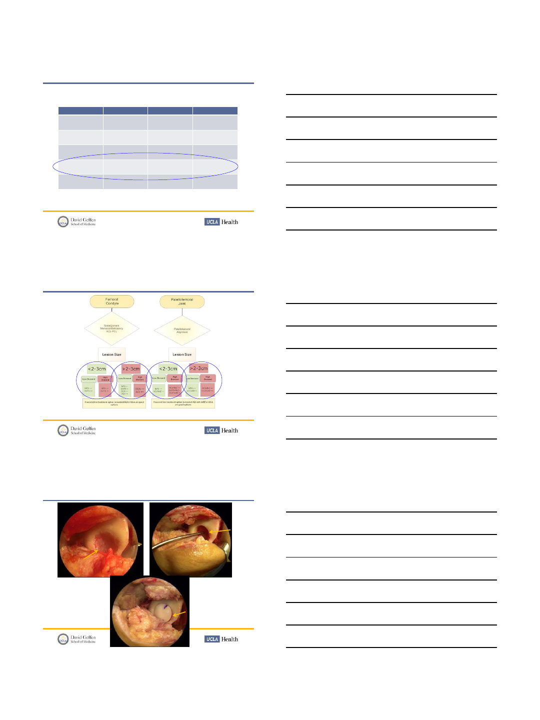
Current Treatment Options in the USA
Treatment
Repair Tissue
Fill
Durability
Marrow
Stimulation
Fibrocartilage
Partial
2
-3 Years
Autologous
OATS
Hyaline Cartilage
Near total
3
-5 Years
ACI
Hyaline
-like
Fibrocartilage
Partial to
near
total
2
-5 Years
Osteochondral
Allograft
Hyaline Cartilage
Near total
5
-10 Years
Particulated
Juvenile Allograft
Hyaline
-like
Fibrocartilage
Partial to
near
total
UNKNOWN
Brian J. Cole et al. J Bone Joint Surg Am 2009;91:1778-
1790
Persistent lateral
compartment pain
despite adequate
rehabilitation
10/12/2015
7
Post-op MRI
ACB
Companion Case
Case #2
•24-year-old male with a chief complaint of chronic knee
pain. History of recurrent patellar dislocations. Pt
complains of pain and intermittent buckling/catching of
his right knee as well as clicking of his right knee.
•PE
•0-130
•Stable knee exam
•+ Patellar grind
•Lateral patellar tilt
•Positive J sign
•TT-TG = 15 mm
10/12/2015
8
MRI
MRI

10/12/2015
9
Case # 3
•30-year-old male with ACL, PMM, MFC OCD treated
initially with ACLR, partial meniscectomy, and
microfracture and subsequent debridement one year
later.
•Pain with running and pivoting localized to medial
compartment of knee
•PE:
•ROM 0-135
•TTP MFC
•Stable Knee exam
MRI
10/12/2015
10
MRI 2013
10/12/2015
11
Post-op Course
•Did well for about a year
•Moderate medial knee pain and catching/clicking
sensation
•PE:
•Trace effusion
•0-130
•TTP over the MFC with click on flexion
•Stable ligament exam
•Repeat MRI
MRI 2015
10/12/2015
12
Keys to successful cartilage surgery
•Understand the basic physiology of cartilage repair &
healing with each approach
•Clarify relevant diagnoses stringent indications
•Manage patient expectations
•Attention to surgical detail and rehabilitation
•Do the surgery with which you are most comfortable
10/14/2015
1
Patellofemoral Instability
Jutta Ellerman, MD
Associate Professor
Department of Radiology
University of Minnesota
Marc Tompkins, MD
Assistant Professor
Department of Orthopaedic Surgery
University of Minnesota/TRIA Orthopaedic Center
We have no conflicts to declare.
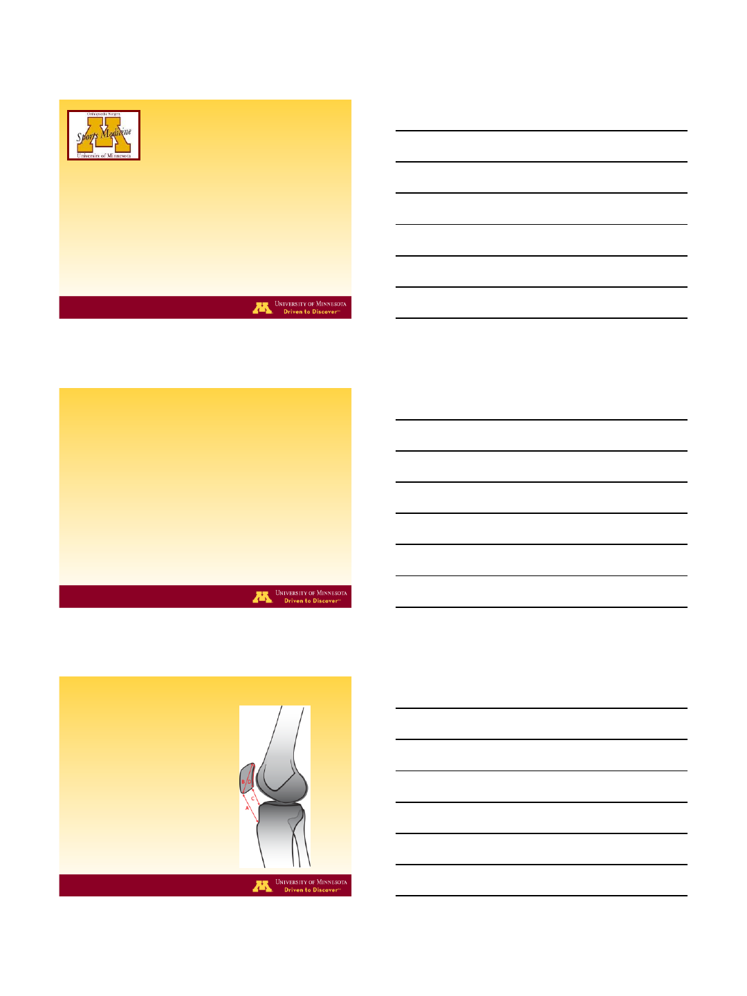
Patella Height
•Caton Deschamps Index
–C/D
•Insall Salvati Ratio
–A/B
10/14/2015
2
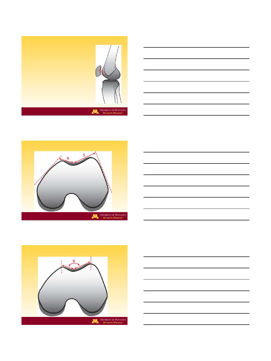
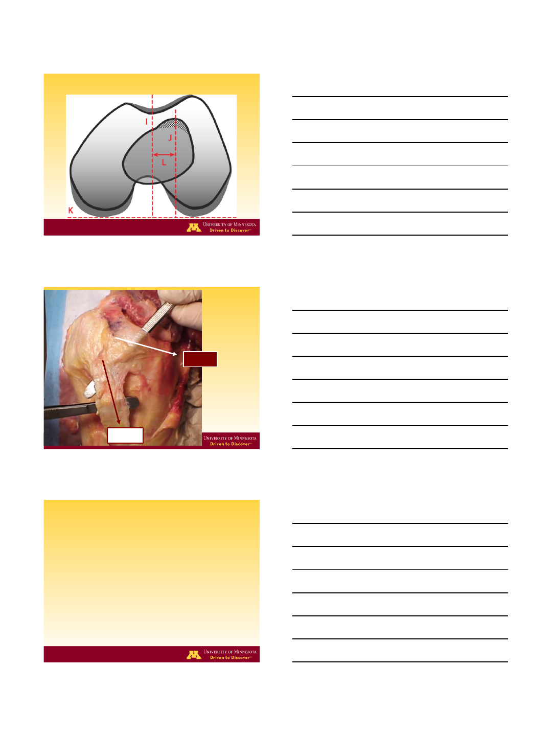
Patella Height :
Patella Trochlear Index (PTI)
•E/D
Facet Asymmetry (Medial/Lateral)
Sulcus Angle
10/14/2015
3
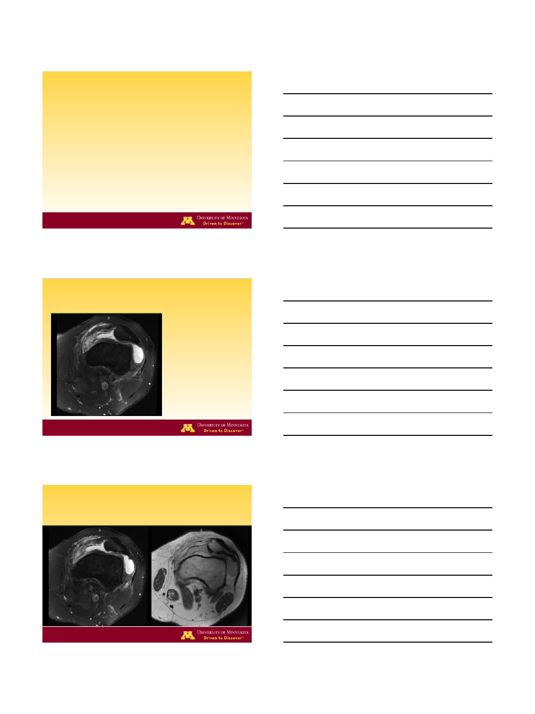
Tibial Tubercle Trochlear Groove Distance
MPFL
MPTL
MEDIAL
Patella
Courtesy Liza
Arendt, MD
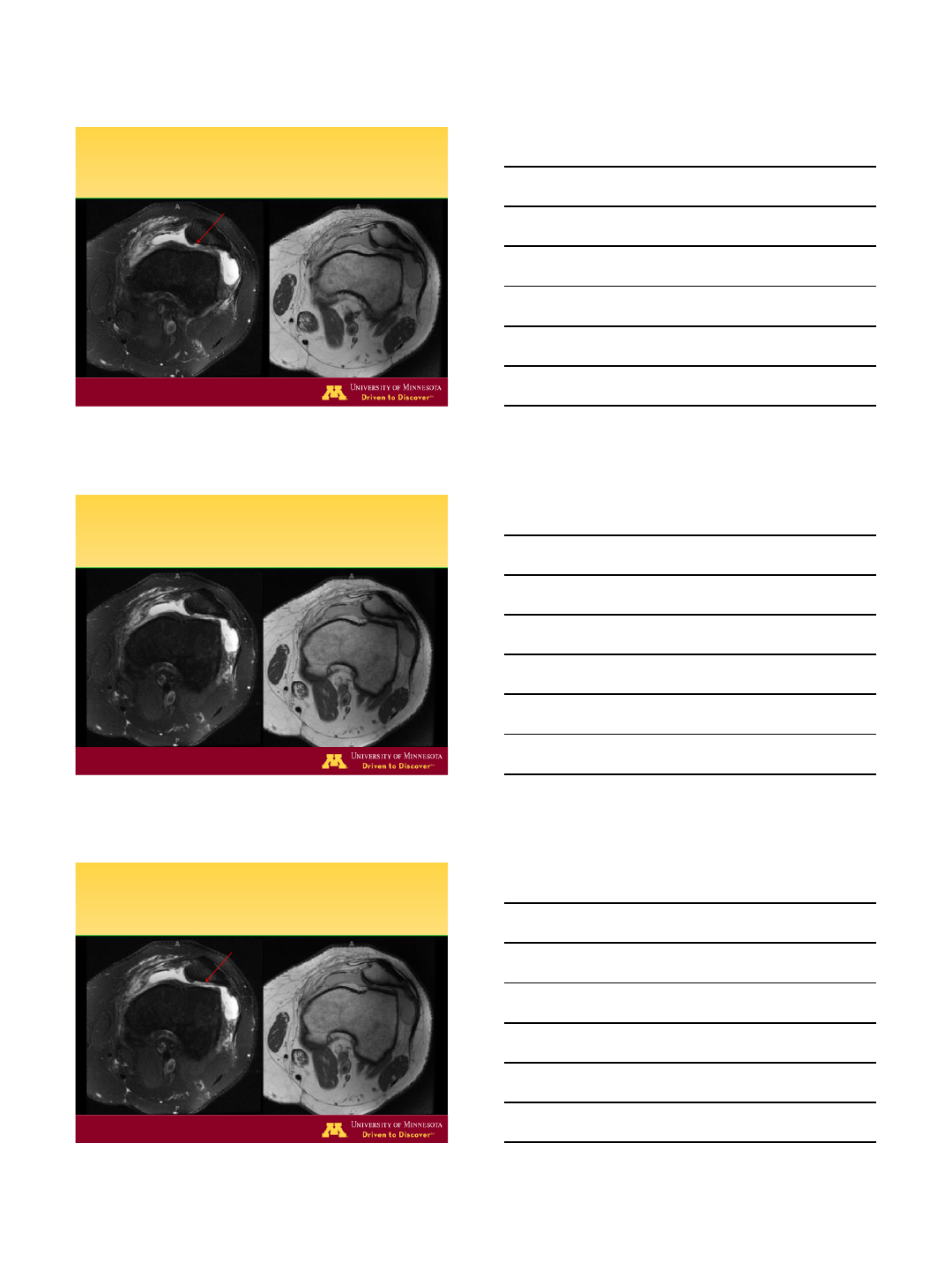
Case 1
•HX:
•18 y/o offensive lineman for high school
football team
•First injured 2 y/a
–Valgus force & patellar dislocation
•Reinjured playing football
–Valgus force and re-dislocation
10/14/2015
4
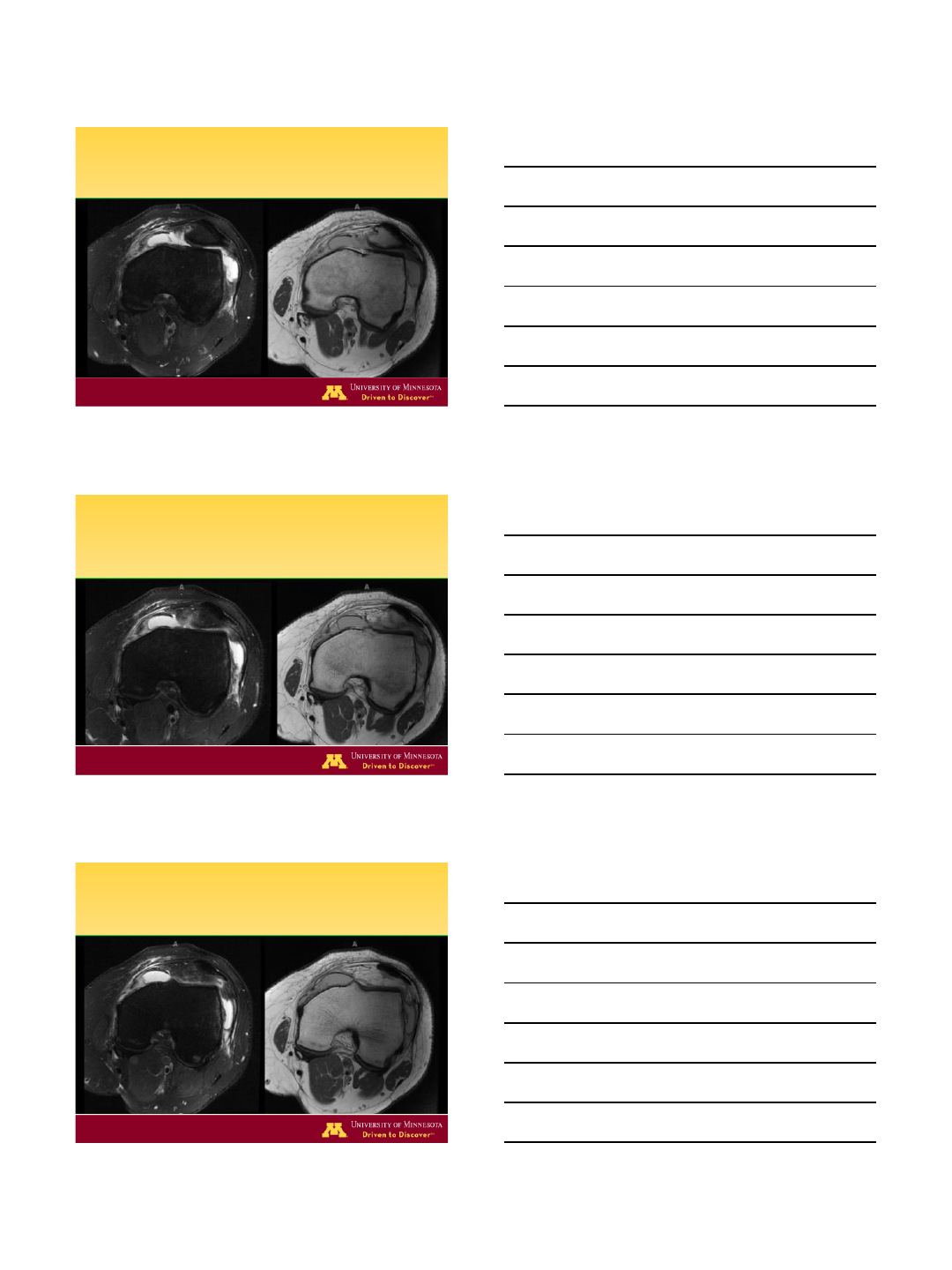
Case 1
•Exam:
•2-3Q lateral patellar translation with soft
endpoint
•Patellar apprehension
•Medial patellar TTP
•Mild J sign
•Tight lateral retinaculum
•
10/14/2015
5
10/14/2015
6
10/14/2015
7

10/14/2015
8
10/14/2015
9
Case 2
•Hx/Exam:
•15 y/o M
•Non contact injury following spin move
playing football
•First injury to the knee
•+ effusion, global patellar tenderness, &
patellar apprehension
10/14/2015
10
10/14/2015
11
10/14/2015
12
10/14/2015
13
10/14/2015
14
Thank You
