Corail Surgical Technique 1
2013-06-12
: Pdf Corail Surgical Technique 1 Corail_Surgical_Technique_1 6 2013 pdf
Open the PDF directly: View PDF ![]() .
.
Page Count: 20
SURGICAL TECHNIQUE
CORAIL®
HIP SYSTEM

THE SCIENCE OF SIMPLICITY
With 1,000,000 stems provided for patients
worldwide1 and two and a half decades of
clinical success, the CORAIL® Total Hip System
now has the most extensive experience
with a hydroxyapatite (HA) coated stem.
Combining basic design features,
including shape, surface finish and
extensive hydroxyapatite coating, with a
simple compaction broach-only surgical
technique, the CORAIL Total Hip System
has demonstrated reproducible results and
long-term biomechanical joint restoration.
Advancing science, enhancements were
made to CORAIL to provide solutions
for orthopaedic surgeons treating
today’s higher-demand patients.
Enhancements to the
CORAIL include:
• Neck geometry designed
for maximum range of
motion
• High offset option to
treat increased femoral
offset patients
• Coxa vara stem option
to treat varus neck
angled patients
US Surgeon Team
James Caillouette, MD
Charles R. Clark, MD
Mark Froimson, MD
Jonathan; Garino, MD
William Lanzer, MD
Joel Matta, MD
Sam Sydney, MD
2 DePuy Synthes Joint Reconstruction CORAIL® Hip System Surgical Technique

Simple Surgical Technique: Reproducible surgical results with minimal instrumentation,
broach-only technique
Compaction Broaching Technique: Preservation of endosteal blood supply and
cancellous bone structures
Dual Offset Options: Accommodates a variety of patient anatomies to restore hip
biomechanics
Two and a Half-Decade Clinical Success: Trust for the surgeon and for the patient
7
6
3
1
2
4
5
1
2
3
4
5
6
7
Three offset options to restore hip biomechanics
Tapered neck geometry and Articul/eze® taper designed to
increase range of motion
Low-profile lateral shoulder design enables easy insertion in
reduced incision techniques, including the anterior approach.
Available in collared or non-collared options
Step geometry converts hoop stresses to compressive loads
Vertical/horizontal grooves provide rotational and axial stability
Proprietary HA coating
Surgical Technique CORAIL® Hip System DePuy Synthes Joint Reconstruction 3

Preoperative planning goals
1. Determine preoperative leg length discrepancy
2. Assess acetabular component size and placement
3. Determine femoral component, size, position and fit
4. Assess femoral offset
PREOPERATIVE PLANNING
The CORAIL stem may be implanted using any of the
contemporary less invasive approaches as well as the
traditional surgical techniques for total hip arthroplasty.
The goal of any technique selected is adequate
visualization of both the acetabulum and the proximal
femur so that a direct view down the femoral canal can
be gained and the entire rim and depth of the
acetabulum visualized.
Preoperative planning enables the surgeon to prepare for
the case and anticipate situations that may arise during
surgery. A thorough preoperative plan incorporates
elements from the patient’s history, physical examination
and radiographic analysis.
4 DePuy Synthes Joint Reconstruction CORAIL® Hip System Surgical Technique

Figure A
Radiographs
The first step in accurate templating is obtaining high-
quality radiographs using a standardized protocol with
known magnification. Use magnification markers
attached to the patient leg at the level of the greater
trochanter to verify magnification.
The CORAIL Total Hip System incorporates 20%
magnification.
Obtain an anterior/posterior (A/P) view of the pelvis with
both extremities in 15 degrees of internal rotation to
position the head and neck parallel to the coronal plane.
A direct lateral radiograph should also be obtained and
used to determine femoral fixation.
Determination of leg length discrepancy
Perform a clinical evaluation in conjunction with a
radiographic analysis to determine preoperative leg length
discrepancy and use both to determine intraoperative leg
length management.
To estimate leg length discrepancy radiographically, draw
a reference line through the bottom of the ischium (Figure
A). Measure the distance from the lesser trochanter
landmark to the reference line on each side. The
difference between the two is the radiographic leg length
discrepancy. Clinical examination should help determine
the actual leg length irregularity.
The tip of the greater trochanter may be used as an
alternative reference mark in conjunction with the lines
through the obturator foramina.
Surgical Technique CORAIL® Hip System DePuy Synthes Joint Reconstruction 5

Acetabular cup size and position
Most sizing determinations are made using the A/P
radiograph of the hip. Determine the optimal position for
the acetabular component and estimate the size using the
Pinnacle® Acetabular Cup System template overlays. The
acetabular teardrop can be referenced as the interior
margin of the acetabular reconstruction.
The goal in cementless acetabular fixation is to optimize
position and bone contact. Once this is determined, mark
the intended center of rotation of the bearing surface on
the A/P radiograph (Figure B).
Cementless femoral component selection
The CORAIL stem is designed to seat in cancellous bone,
and cortical contact should be avoided when templating.
Select the appropriate template size that is smaller than
the cortex in the proximal femur. The femoral template
should be in line with the long axis of the femur and the
neck resection line drawn at the point where the selected
stem provides the desired amount of leg length.
The vertical distance between the planned center of
rotation of the acetabular component and the center of
rotation of the femoral head constitutes the distance the
leg length will be adjusted.
The level of neck osteotomy depends on the stem size and
the desired leg length, with the goal of using a non-skirted
modular head to optimize range of motion prior to
prosthetic impingement. To help properly position the
template on the lateral radiograph, estimate the distance
between the tip of the greater trochanter and the lateral
shoulder of the prosthesis using the A/P radiograph (Figure
C).
Verify that the stem size chosen in the A/P plane also fits
in the lateral plane. The lateral radiograph of a properly
sized CORAIL implant will not exhibit cortical contact.
+4 Head Center
0 Head Center
54mm
Figure B Cup center of rotation
Figure C Head center of rotation
Preoperative Planning
6 DePuy Synthes Joint Reconstruction CORAIL® Hip System Surgical Technique

Offset requirements
The CORAIL Total Hip System implants are available with
standard, high offset and varus options for all stem body
sizes (except 6 and 8). Through templating and
intraoperative trialing, determine which option restores
proper offset by matching the cup’s center of rotation
with the desired head center of rotation (Figure D).
Figure D Head center of rotation
Cup center of rotation
Surgical Technique CORAIL® Hip System DePuy Synthes Joint Reconstruction 7
SURGICAL TECHNIQUE
1
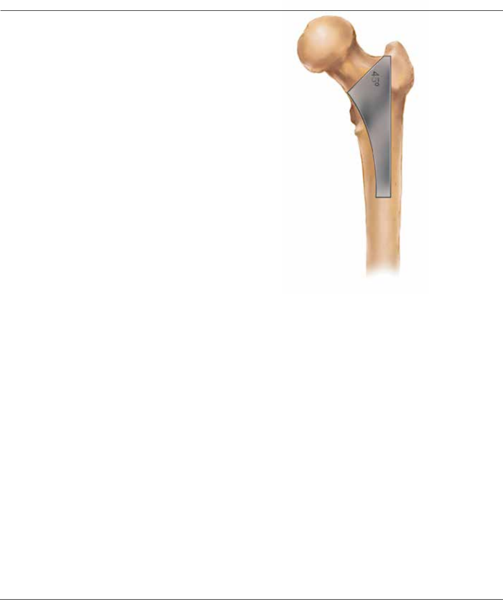
Neck Osteotomy
The level of the neck resection is determined during
preoperative templating. The cut will be approximately 1
cm above the lesser trochanter. Center the resection
guide along the neutral axis of the femur and mark the
resection line. Perform the osteotomy, taking care to
maintain the correct angle (Figure 1).
Figure 1
8 DePuy Synthes Joint Reconstruction CORAIL® Hip System Surgical Technique
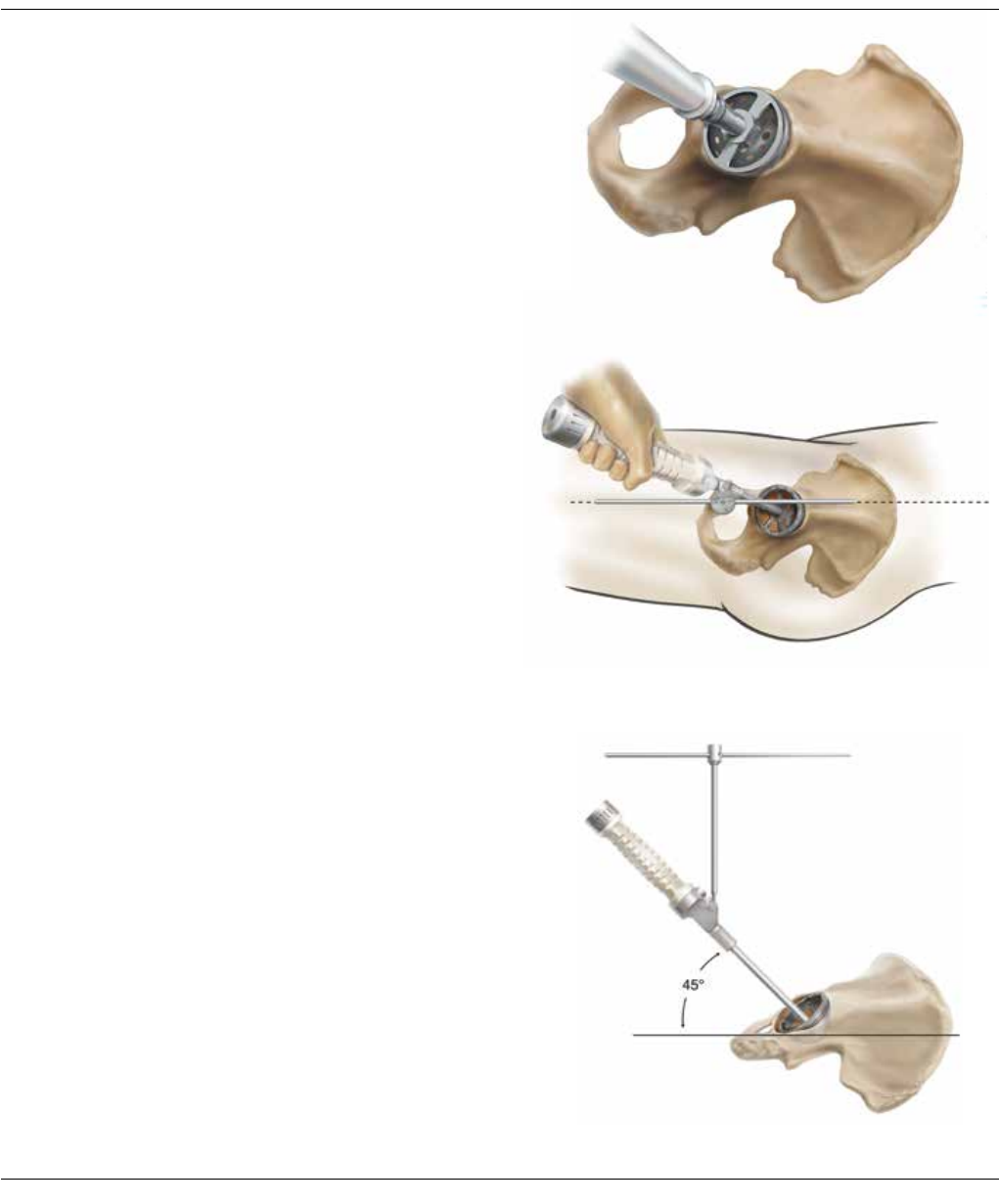
Figure 2B
Figure 2C
2
Reaming and alignment
Make sure that the acetabulum is fully exposed and
remove soft tissue from the acetabular rim.
Progressively ream the acetabulum until bleeding
subchondral bone is reached and a hemispherical dome is
achieved (Figure 2A).
Using the cup impactor, place a trial cup sizer into the
reamed acetabulum and assess its position and cortical
bone contact.
The inferior rim of the trial cup should typically be level
with the bottom of the teardrop. The trial cup angle of
orientation should match that recorded during
preoperative templating, which is normally 45 degrees of
lateral opening (abduction) and 15–30 degrees of
anteversion. Confirm this using the external alignment
instrumentation (Figures 2B and 2C).
Remove the cup impactor from the trial shell and place
the desired liner trial into the cup trial.
Figure 2A
Surgical Technique CORAIL® Hip System DePuy Synthes Joint Reconstruction 9
3
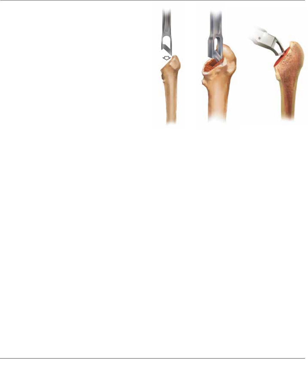
Metaphyseal preparation (optional)
The version osteotome can be used to remove a wedge of
cancellous bone, creating a starting cavity for broach
insertion. The osteotome can be positioned in a neutral or
anteverted fashion, depending on patient anatomy.
A modular osteotome may also be used to accomodate
multiple approaches to the hip (Figure 3).
Surgical Technique
Figure 3
10 DePuy Synthes Joint Reconstruction CORAIL® Hip System Surgical Technique
4
Femoral canal preparation
The CORAIL broach is available with several broach
handle options depending on the surgical approach
(Figures 4A, 4B, 4C); dual-offset handle also available, but
not shown. Select the appropriate handle for the surgical
approach.
Beginning with the smallest CORAIL compaction broach
attached to the selected broach handle, progressively
enlarge the metaphyseal cavity by compacting and
shaping the cancellous bone until the level of the neck
resection is reached. Broaching should continue until
complete stability is achieved with the last size broach
used without reaching cortical contact in the femoral
canal, ensuring cancellous bone preservation. The size of
each broach is the same as the corresponding implant
without HA (hydroxyapatite) coating (155 microns).
If you impact a broach and it does not fully seat in the
canal, it is recommended to go back to the previous size
broach and re-establish the broach envelope of cancellous
bone to accept the smaller size implant. The CORAIL
implant’s design allows you to go back to the smaller size.
5
Calcar Preparation (Optional)
Place the calcar planer onto the broach stud and mill the
calcar to the broach face, allowing the implant collar (if
used) to seat flush against the calcar. Make certain the
calcar planer is rotating before engaging calcar to prevent
the planer from binding on the calcar.
Posterior Approach
Figure 4A
Posterolateral/
Anterolateral Approach
Figure 4B
Anterior Approach
Figure 4C
Surgical Technique CORAIL® Hip System DePuy Synthes Joint Reconstruction 11
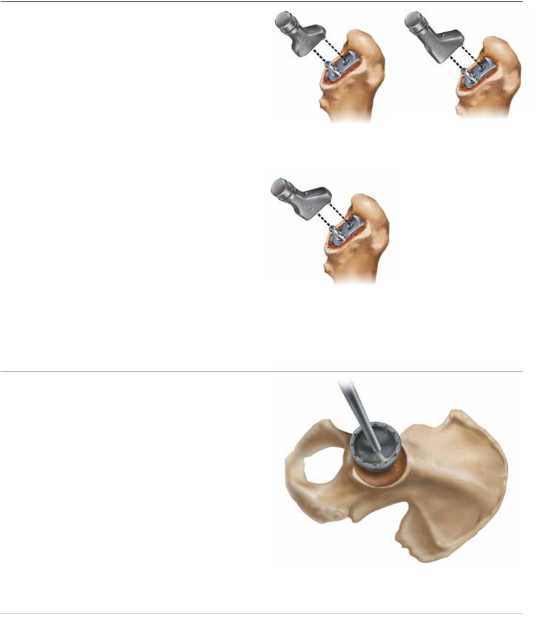
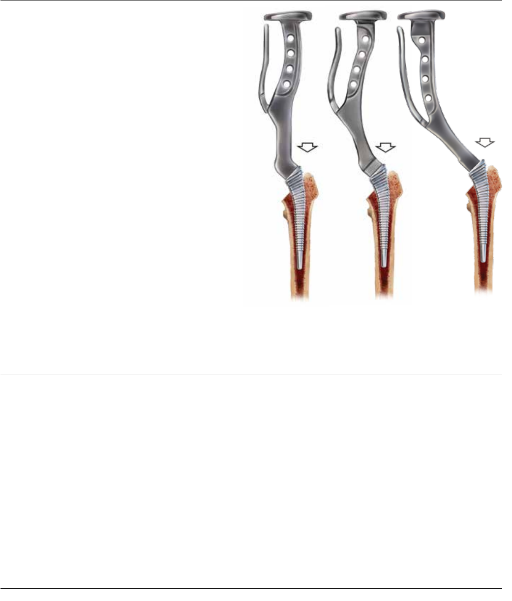
Standard offset collared/collarless (KA/KS)
Figure 5A
6
Trial Reduction
Trial neck segments and trial modular heads are available
to assess proper component position, joint stability,
range-of-motion and leg length (Figures 5A, 5B and 5C).
The CORAIL is available in three stem options, a standard
collarless/collared stem, a high offset collarless stem, and
a coxa vara collared stem and offers the appropriate neck
segment to match up with the stem option.
With the CORAIL broach in situ, attach one of the three
neck segment options. Perform a trial reduction with a +5
Articul/eze head trial to allow for one up or down
adjustment in neck length without using a skirted femoral
head (see stem specifications chart in back of the
technique for adjustment measurements). Reduce the hip
and assess stability through a full range of motion, and
check for impingement. Leg length and offset may be
adjusted by varying the neck length with the appropriate
femoral head. Alternatively, leg length may be reduced
with a lower neck cut and advancing the broach or
alternatively driving the broach and repeating the calcar
milling.
7
Acetabular Shell Insertion
Remove the trial acetabular components and implant the
desired acetabular shell (Figure 6). Take care to ensure cup
orientation mimics the orientation of the trial component.
Insert a trial liner into the shell implant.
High offset collarless (KHO)
Figure 5B
Coxa vara collared (KLA)
Figure 5C
Figure 6
Surgical Technique
12 DePuy Synthes Joint Reconstruction CORAIL® Hip System Surgical Technique
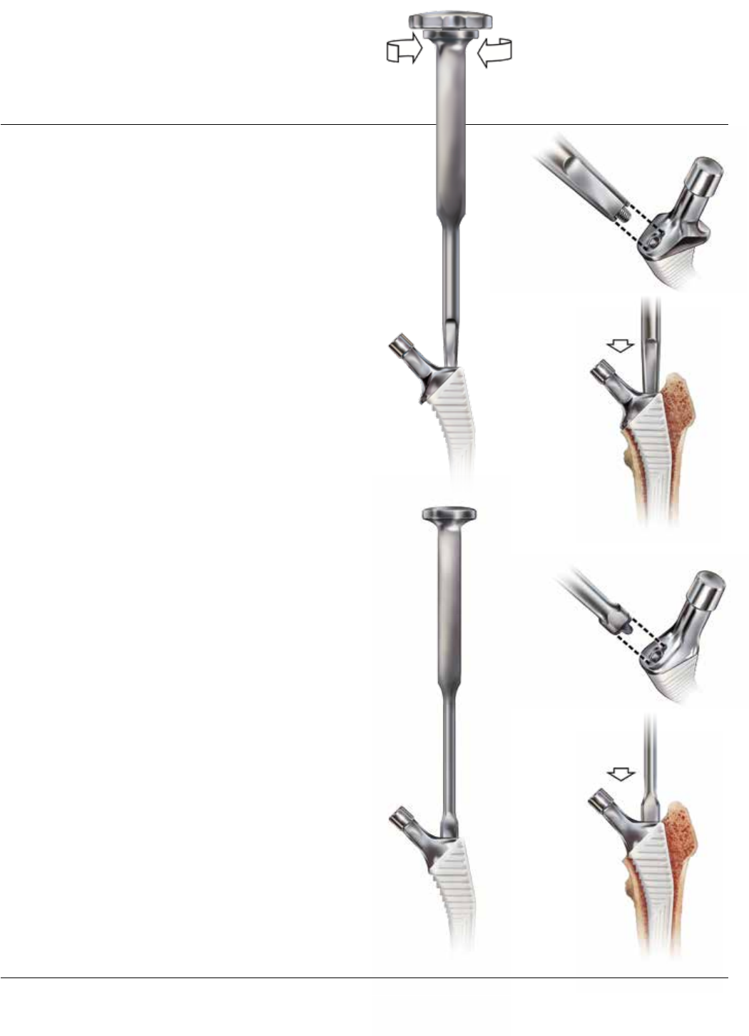
Figure 7B
8
Femoral Component Insertion
CORAIL Total Hip System implants can be inserted with
either a threaded retaining inserter or a non-threaded
inserter. Both inserters provide rotational control during
stem implantation.
A new modular inserter system further enables multiple
approaches (see ordering information).
Prior to using either inserter, the CORAIL stem
should be inserted by hand into the femoral canal
with 1.5 to 2.0 cm of HA showing above the
resection.
If the retaining inserter is chosen, verify that it is
assembled with the inserter shaft threaded into the
inserter handle (Figure 7A). Ensure the tines on the
inserter are aligned with the recesses of the inserter
platform on the top of the implant (Figure 7B). Fully
engage the threads of the inserter into the implant to
ensure the inserter is securely attached to the implant.
If the non-retaining inserter is chosen, introduce stem by
hand into femoral canal (Figure 8A). Ensure the tines of
the inserter are aligned with the recesses of the inserter
platform on the top of the implant (Figure 8B).
With the taper protected by the cover, gently introduce
the implant and impact it in the central axis of the femur,
to the level of the HA coating (or the collar) (Figures 7C
and 8C). With the prostheses in situ, remove the taper
cover and add the trial head and acetabular trial liner to
assess implant stability and leg length.
Figure 7A
Figure 8A
Figure 8B
Figure 7C
Figure 8C
Surgical Technique CORAIL® Hip System DePuy Synthes Joint Reconstruction 13
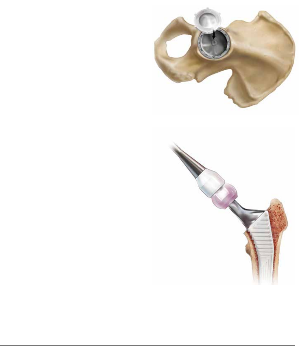
9
Acetabular Insert Implantation
Following the final trial reduction, remove the trial
acetabular liner and insert the appropriate acetabular liner
(Figure 9).
Figure 10
10
Femoral Head Impaction
Irrigate, clean and dry the prosthesis to ensure the taper is
free of debris. Place the appropriate femoral head onto
the taper and lightly tap using the head impactor before
reducing the hip (Figure 10).
Figure 9
Surgical Technique
14 DePuy Synthes Joint Reconstruction CORAIL® Hip System Surgical Technique

5 years post-op
Surgical Technique CORAIL® Hip System DePuy Synthes Joint Reconstruction 15

C
D
A
B
E
CORAIL® AMT STEM SPECIFICATIONS
Standard Offset - Collarless/Collared
Size Stem Length
(mm) A
Offset
(mm) B
Neck Length
(mm) C
Neck Shaft
Angle D
Leg Adjustment
Length
(mm
) E
6* 93 30.8 33.8 135º 34
895 38.0 38.5 135º 36
9110 38.5 38.5 135º 36
10 120 39.5 38.5 135º 36
11 125 40.0 38.5 135º 36
12 130 41.0 38.5 135º 36
13 135 41.5 38.5 135º 36
14 140 42.0 38.5 135º 36
15 145 43.0 38.5 135º 36
16 150 43.5 38.5 135º 36
18 160 44.5 38.5 135º 36
20 170 45.5 38.5 135º 36
* The size 6 is available in collarless only.
High Offset - Collarless
Size Stem Length
(mm) A
Offset
(mm) B
Neck Length
(mm) C
Neck Shaft
Angle D
Leg Adjustment
Length
(mm
) E
9110 45.5 43.2 135º 36
10 120 46.5 43.2 135º 36
11 125 47.0 43.2 135º 36
12 130 48.0 43.2 135º 36
13 135 48.5 43.2 135º 36
14 140 49.0 43.2 135º 36
15 145 50.0 43.2 135º 36
16 150 50.5 43.2 135º 36
18 160 51.5 43.2 135º 36
20 170 52.5 43.2 135º 36
Coxa Vara Offset - Collared
Size Stem Length
(mm) A
Offset
(mm) B
Neck Length
(mm) C
Neck Shaft
Angle D
Leg Adjustment
Length
(mm
) E
9110 45.5 40.3 125º 31
10 120 46.5 40.3 125º 31
11 125 47.0 40.3 125º 31
12 130 48.0 40.3 125º 31
13 135 48.5 40.3 125º 31
14 140 49.0 40.3 125º 31
15 145 50.0 40.3 125º 31
16 150 50.5 40.3 125º 31
18 160 51.5 40.3 125º 31
20 170 52.5 40.3 125º 31
Note: All measurements are based on a 28 mm +5.0 Articul/eze head, which is the middle length of non-skirted
femoral heads
16 DePuy Synthes Joint Reconstruction CORAIL® Hip System Surgical Technique

ORDERING INFORMATION
IMPLANTS
INSTRUMENTS
CORAIL AMT Broach Case†
L20440 Neck Resection Guide
L20408 Broach Size 8
L20409 Broach Size 9
L20410 Broach Size 10
L20411 Broach Size 11
L20412 Broach Size 12
L20413 Broach Size 13
L20414 Broach Size 14
L20415 Broach Size 15
L20416 Broach Size 16
L20418 Broach Size 18
L20420 Broach Size 20
L20431 CORAIL Standard Offset Neck Segment
L20432 CORAIL Coxa Vara Neck Segment
L20433 CORAIL High Offset Neck Segment
9522-11-500 CORAIL AMT Curved Handle
9522-10-500F CORAIL AMT Straight Broach Handle
9522-12-500F CORAIL AMT Extra Curved Handle
2002-31-000 Anteversion Osteotome
2570-04-100 Calcar Planer-Small
2665-99-000 Broach Case Complete
Standard Collarless
Cat. No. Size
L20106 6
3L92507 8
3L92509 9
3L92510 10
3L92511 11
3L92512 12
3L92513 13
3L92514 14
3L92515 15
3L92516 16
3L92518 18
3L92520 20
Standard Collared
Cat. No. Size
3L92498 8
3L92499 9
3L92500 10
3L92501 11
3L92502 12
3L92503 13
3L92504 14
3L92505 15
3L92506 16
3L92508 18
3L92521 20
High Offset Collarless
Cat. No. Size
L20309 9
L20310 10
L20311 11
L20312 12
L20313 13
L20314 14
L20315 15
L20316 16
L20318 18
L20320 20
Coxa Vara Collared
Cat. No. Size
3L93709 9
3L93710 10
3L93711 11
3L93712 12
3L93713 13
3L93714 14
3L93715 15
3L93716 16
3L93718 18
3L93720 20
CORAIL AMT Core Case Complete
2354-10-000 Canal Probe
53-0360 T-Handle
2570-05-000 Retaining Implant Inserter
2570-05-100 Standard Implant Inserter
2001-65-000 Head Impactor
2530-81-000 28 mm Articul/eze +1.5 mm Trial Head
2530-82-000 28 mm Articul/eze +5.0 mm Trial Head
2530-83-000 28 mm Articul/eze +8.5 mm Trial Head
2530-84-000 28 mm Articul/eze +12.0 mm Trial Head
2530-85-000 28 mm Articul/eze +15.5 mm Trial Head
2665-99-003 Core Case Complete
†
Note: For size 6 instrumentation and implant ordering information, see the CORAIL Size 6
surgical technique - EO-75, available from your DePuy Synthes Joint Reconstruction Sales
Consultant.
X-Ray Templates
2665-01-500 Collarless X-Ray Template
2665-02-500 Collared X-Ray Template
2665-03-500 Size 6 X-Ray Template
Surgical Technique CORAIL® Hip System DePuy Synthes Joint Reconstruction 17

TSS Femoral Core Case 1
2598-07-400 Base
2598-07-411 Tray
2598-07-410 Lid
2800-88-511 SE Set (includes all instruments)
2598-07-460 Universal Stem Inserter Handle
Trial Heads – 2 Sets per Case
*accommodates up through size 44 mm
2598-07-570 Retaining Stem Inserter (2 pcs)
2598-07-530 Modular Box Osteotome
Any two of the below handles accommodated:
2570-00-000 SUMMIT® Universal Broach Handle
2598-07-540 Long Posterior Broach Handle
2598-07-550 Extra Curved Broach Handle
2598-07-350 Anterior Dual Offset Broach Handle – Left
2598-07-360 Anterior Dual Offset Broach Handle – Right
9522-10-500F CORAIL AMT Straight Broach Handle
9522-11-500 CORAIL AMT Curved Broach Handle
2598-07-470 CORAIL/TRI-LOCK® Posterior Stem
Inserter Shaft
2598-07-480 SUMMIT Posterior Stem Inserter Shaft
2598-07-435 Bullet Tip Stem Inserter Shaft
2598-07-430 Standard Straight Stem Inserter Shaft
2598-07-440 CORAIL/TRI-LOCK Anterior Stem
Inserter Shaft
2598-07-450 SUMMIT Anterior Stem Inserter Shaft
TSS Femoral Core Case 2
2598-07-421 Base
2598-07-422 Lid
2800-88-512 SE Set (includes all instruments)
2354-10-000 Muller Awl Reamer with Hudson End
2001-42-000 T-Handle
2001-80-501 IM Initiator Sized
9400-80-007 Shielded Calcar Planer
85-3927 Femoral Rasp
9400-80-001 Canal Finder
2001-65-000 Femoral/Humeral Head Impactor
2001-66-000 Replacement Tip Femoral Head Impactor
Instruments
Ordering Information
18 DePuy Synthes Joint Reconstruction CORAIL® Hip System Surgical Technique

ESSENTIAL PRODUCT INFORMATION
Important
This Essential Product Information sheet does not include
all of the information necessary for selection and use of a
device. Please see full labeling for all necessary
information.
Indications
The CORAIL AMT Hip Prosthesis is intended for use in
total hip arthroplasty and is intended for pressfit
(uncemented) use. Total hip arthroplasty is intended to
provide increased patient mobility and reduce pain by
replacing the damaged hip joint articulation in patients
where there is evidence of sufficient sound bone to seat
and support the components. Total hip replacement is
indicated in the following conditions:
1. A severely painful and/or disabled joint from
osteoarthritis, traumatic arthritis, rheumatoid arthritis,
or congenital hip dysplasia.
2. Avascular necrosis of the femoral head.
3. Acute traumatic fracture of the femoral head or neck.
4. Failed previous hip surgery including joint
reconstruction, internal fixation, arthrodesis,
hemiarthroplasty, or total hip replacement.
5. Certain cases of ankylosis.
HA-coated stems are indicated for cementless use only.
Contraindications
The following conditions are contraindications for total or
hemi-hip replacement:
1. Active local or systemic infection.
2. Loss of musculature, neuromuscular compromise or
vascular deficiency in the affected limb rendering the
procedure unjustified.
3. Poor bone quality, such as osteoporosis, where, in the
surgeon’s opinion, there could be considerable
migration of the prosthesis or a significant chance of
fracture of the femoral shaft, considerable migration
of the prosthesis or a significant chance of fracture of
the femoral shaft and/or the lack of adequate bone to
support the implant(s).
4. Charcot’s or Paget’s disease.
5. For hemi-hip arthroplasty, any pathological condition
of the acetabulum, such as distorted acetabuli with
irregularities, protrusion acetabuli (arthrokatadysis), or
migrating acetabuli, that would preclude the use of
the natural acetabulum as an appropriate articular
surface for the hemi-hip prosthesis.
Warnings and Precautions
• HA coated implants must not be implanted with
cement
• When changing the head on a femoral stem which is
still in place, it is essential to use a metal-metal
interface
• For some stems, head offset ‘Warning’ notices are
visible on labels to limit the maximum offset used for
the head. For the CORAIL Revision stem, the maximum
offset used for the head is limited to 13mm.
Adverse Events
The following are the most frequent adverse events after
hip arthroplasty: change in position of the components,
loosening of components, wear or fracture of
components, dislocation, infection, peripheral
neuropathies, tissue reaction.
CORAIL AMT HIP
Surgical Technique CORAIL® Hip System DePuy Synthes Joint Reconstruction 19

References
1. Data on file at DePuy Orthopaedics, Inc.
US agent
DePuy Orthopaedics, Inc.
700 Orthopaedic Drive
Warsaw, IN 46582
USA
T. +1 (800) 366-8143
www.depuysynthes.com
Manufactured by
DePuy France SAS
7 Allee Irene Joliot Curie
69801 St. Priest Cedex
France
© DePuy Synthes Joint Reconstruction, a division of DOI 2013.
0612-82-501 (Rev. 7) 2.5M 05/13
Limited Warranty and Disclaimer: DePuy Synthes Joint Reconstruction products are sold with a limited warranty to the original purchaser against defects
in workmanship and materials. Any other express or implied warranties, including warranties of merchantability or fitness, are hereby disclaimed.
WARNING: In the USA, this product has labeling limitations. See package insert for complete information.
CAUTION: USA Law restricts these devices to sale by or on the order of a physician.
Not all products are currently available in all markets.
