Craniofacial Trauma Case Study MXCSCraniofacial J6819B
2015-06-08
: Pdf Mxcscraniofacialtraumaj6819B MXCSCraniofacialTraumaJ6819B 6 2015 pdf
Open the PDF directly: View PDF ![]() .
.
Page Count: 4

Craniofacial Trauma. Resorbable
and titanium plating application.
Case Study
Patient Profile
A 36-year-old male was involved in a motorcycle accident.
Patient suffered basilar skull fracture, multiple facial
fractures, head injuries and soft tissue injuries of the
cervical spine. Patient was not wearing a helmet at the
time of the accident.
Treatment
Patient was treated with closed reduction of a right condyle
fracture and nasal bone fractures. Arch bars were adapted
to the maxillary and mandibular arches and secured with
stainless steel wire. The mandible was then brought into
occlusion and wired. The nasal bones were then molded
into midline alignment, and a stent was placed over the top.
One week later an open reduction and internal fixation of
the right orbital floor blowout and right zygoma/malar
fracture was performed. A 2.1 mm taper Fisher burr was
used to make a hole in the thickest portion of the malar
buttress. The zygoma was manipulated until the fracture was
anatomically reduced. A 1.5 mm Synthes orbital rim plate
was placed across the zygomatico-frontal suture. A 2.0 mm
plate was placed across the zygomatic buttress. A 2.0 mm
4-hole plate was placed on the piriform rim. With the
zygoma secured, attention was given to the orbital floor.
A 1.5 mm Resorbable Contourable Mesh Plate (50 mm x
50 mm, 0.5 mm thick) was heated in the hot water bath,
contoured over the thin comminuted orbital floor, and
secured with one 1.5 mm screw. The resorbable mesh
provided contourability and adequate support for the globe.
Prior to closing, a forced duction test was performed on the
inferior rectus muscle. It was noted that the patient had full
range of motion of the right globe.
Synthes CMF Craniofacial Trauma
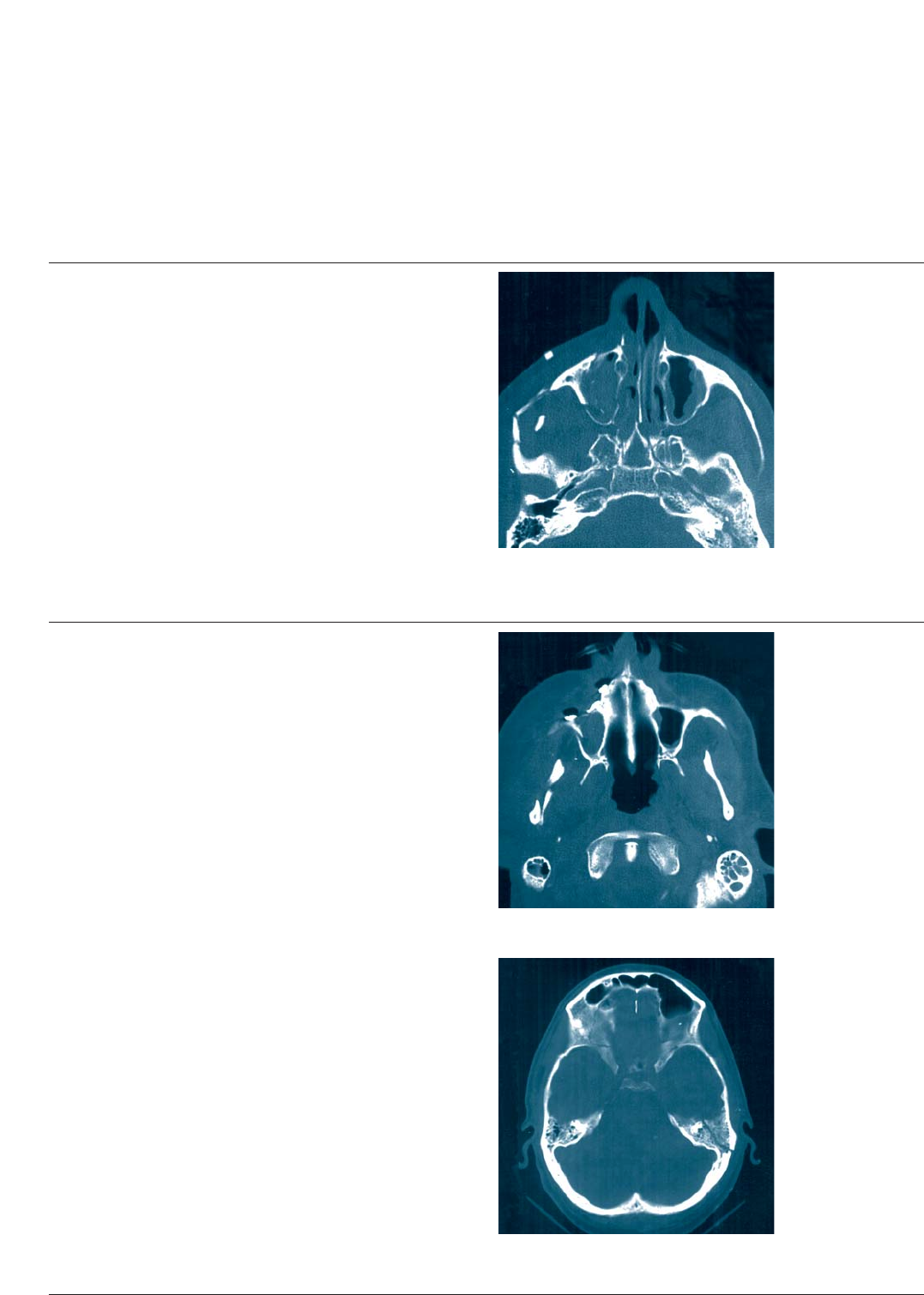
Craniofacial Trauma
Preoperative radiograph
Immediate postoperative radiograph
Immediate postoperative radiograph
Postoperative Management
Patient was kept in IMF for two weeks due to the mandible
fracture, then progressed to elastics and physical therapy.
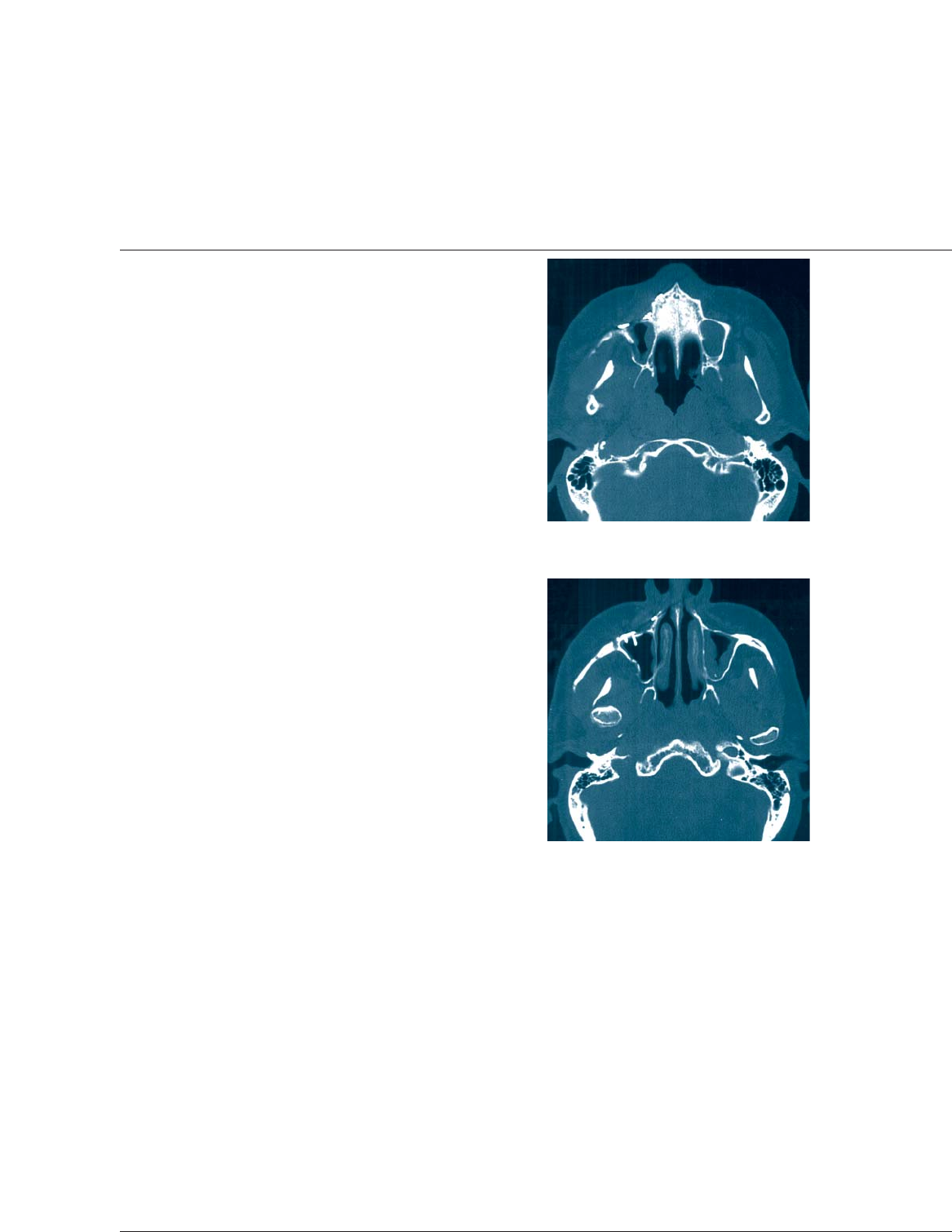
Approximately 3 months postoperative the patient
experienced conductive hearing loss. A CT of the temporal
bones was performed. A fracture of the left temporal bone
appears to extend through the middle ear. The fracture line
is considerably less distinct than seen on the immediate
postoperative scan. No obvious consequence is noted to
explain the patient’s conductive hearing loss.
Synthes CMF
3-month postoperative radiograph
3-month postoperative radiograph
Results from case studies are not predictive of results in other cases.
Results in other cases may vary.
Craniofacial Trauma
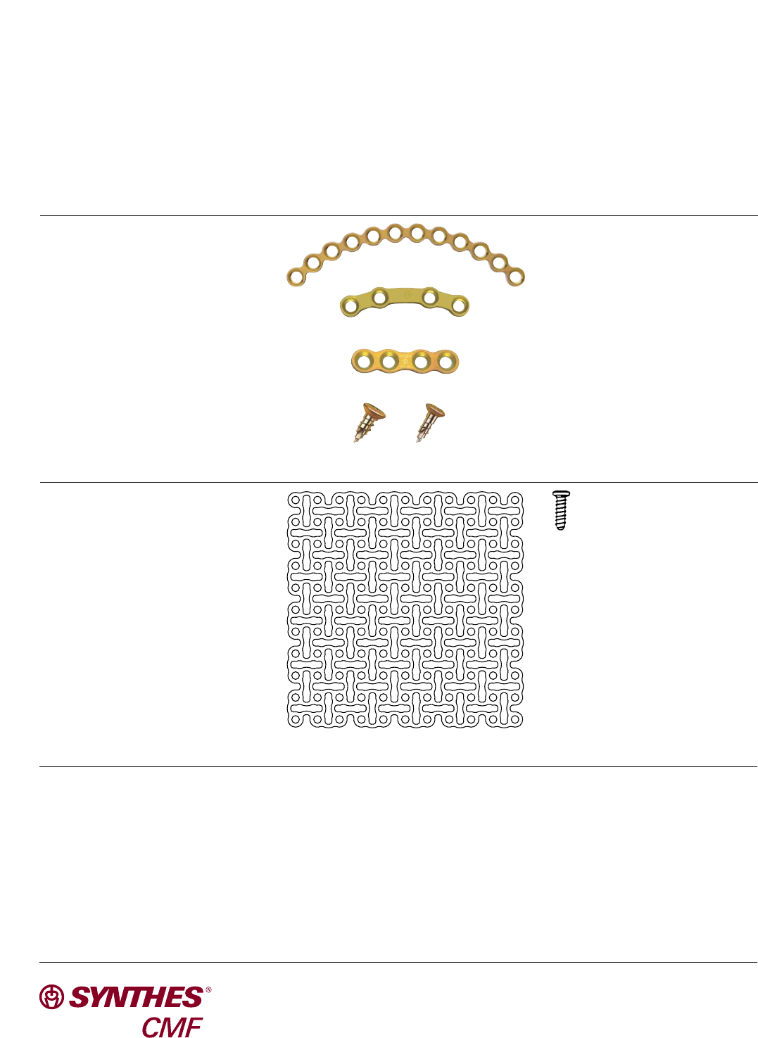
Materials Used
Metal
– 1.5 mm Orbital Rim Plate
– 2.0 mm Tension Band Plate
– 2.0 mm Curved Broad Plate
– 1.5 mm and 2.0 mm Titanium Screws
Surgeon profile
Jeffrey Wheaton, DDS, MD
St. Vincent’s Hospital
Santa Fe, New Mexico
Synthes CMF
1302 Wrights Lane East
West Chester, PA 19380
Telephone: (610) 719-5000
To order: (800) 523-0322
Fax: (610) 251-9056
Synthes (Canada) Ltd.
2566 Meadowpine Boulevard
Mississauga, Ontario L5N 6P9
Telephone: (905) 567-0440
To order: (800) 668-1119
Fax: (905) 567-3185
© 2006 Synthes, Inc. or its affiliates. All rights reserved. Synthes is a trademark of Synthes, Inc. or its affiliates. Printed in U.S.A. 6/07 J6819-B
www.synthes.com
Biomaterials
– 1.5 mm Resorbable Contourable
Mesh
– 1.5 mm Resorbable Cortex Screw
