Pcl Technique Guide
Pcl Technique Guide pcl_technique_guide pcl_technique_guide 3 2013 pdf 258413772373414384
Pcl Technique Guide pcl_technique_guide pcl_technique_guide 2 2013 pdfdoc 258413772373414384 3:
Pcl Technique Guide pcl_technique_guide pcl_technique_guide 3 2013 pdfdoc 258413772373414384 3:
2013-02-24
: Pdf Pcl Technique Guide pcl_technique_guide 2 2013 pdf
Open the PDF directly: View PDF ![]() .
.
Page Count: 8
Technique
Guide
Michael A. Rauh, MD
e opinions expressed are those of Dr. Rauh
and are not necessarily those of Stryker
PCL Technique
VersiTomic®
VersiTomic PCL Technique
Michael Rauh, M.D. is an Orthopaedic Sports Medicine specialist at the University at Bualo. His surgical cases
focus on the latest techniques in arthroscopic surgery of the knee, shoulder, and elbow, as well as, general orthopaedic
reconstructive surgery, and trauma surgery. During his Sports Medicine Fellowship at the renowned Cleveland
Clinic, Dr. Rauh worked with the Cleveland Browns, Indians, and Cavaliers. While in Bualo, Dr. Rauh serves
as the medical director and team physician for the Bualo Bandits of the National Lacrosse League. Dr. Rauh is
the Clinical Assistant Professor of Orthopaedic Surgery for the School of Medicine and Biomedical Sciences,
University at Buffalo, where he teaches orthopaedic sports medicine fellows, residents, medical students, and
conducts research.
Introduction
Michael Rauh, M.D
Orthopaedic Sports Medicine specialist at the University at Bualo and
Clinical Assistant Professor of Orthopaedic Surgery, School of Medicine and Biomedical Sciences, University at Bualo
Surgeons should consider anatomy, scientic evidence and their own experience when deciding
to perform this surgery as well as considering single versus double bundle PCL reconstructions.
Arthroscopic Evaluation and Debridement
1. Care is taken to identify the anatomic insertion points of the PCL on both the femur and
tibial surfaces.
2. e PCL remnant on the medial wall of the intercondylar notch is debrided arthroscopically.
Note the respective PCL footprints, especially if choosing to perform double bundle
reconstructions.
3. Creation of a posteromedial portal is usually helpful. is can be accomplished with spinal
needle localization and visualization with a 70° arthroscope through the notch superior to
the ACL. A posteromedial cannula is oen useful to aid in later insertion and removal of
instruments and arthroscope.
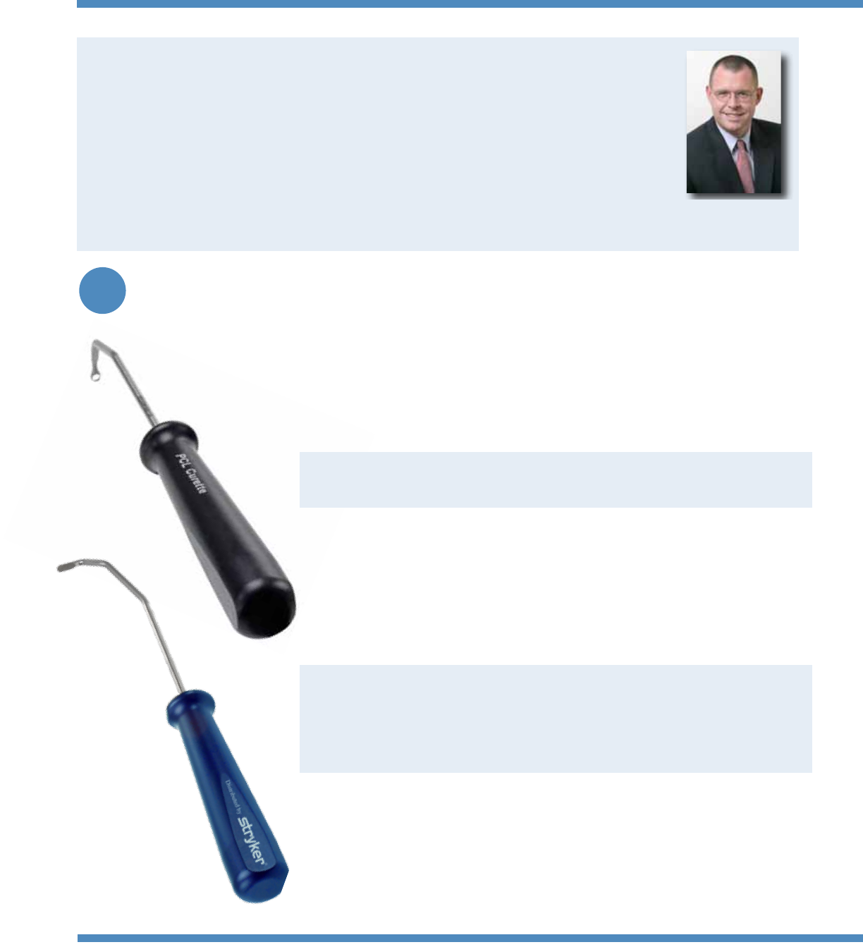
Locate tibial PCL footprint and begin to remove the remaining PCL bers using a combination
of shaver, Stryker PCL Curette, and Stryker PCL Liberator/Rasp.
Stryker PCL Liberator/Rasp has 3 functions:
•Liberatorwithataperedtipforelevatingsofttissue
•Rasptoassistinremovaloftissue/bone
•epinholelocatedjustabovetheraspcanserveasapinprotectorwhilereamingthetibialtunnel
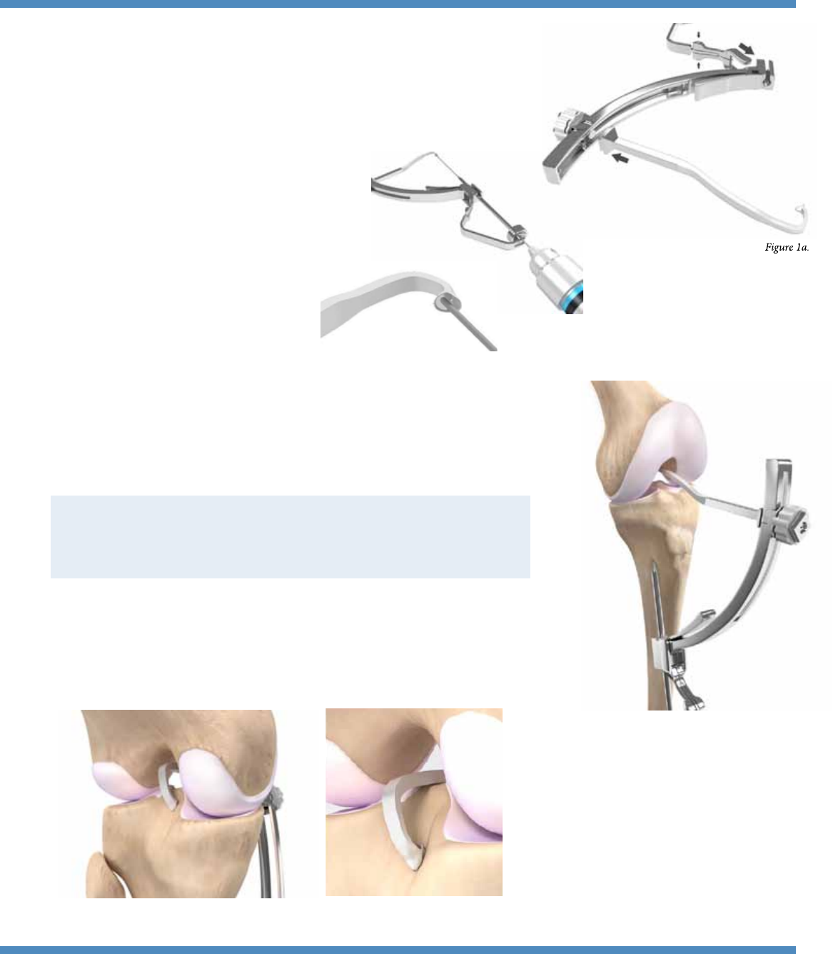
Unscrew the knob until it is in its nal unlock position, press in the button and slide tibial
armintoplace.Adjustangleandtightendowntheknobtothelockposition.Squeezesides
of Backstopper and slide over distal aspect of tibial spine into slots.
Pre-chuck the 2.4mm guide pin up to the back of the
Backstopper to provide a positive stop when drilling.
is is done to assist in preventing over drilling of the
tibial pin.
Ensure the pin meets the PCL capture cup.
S
Tibial Guide Assembly
a
b
Tibial Tunnel Guide Placement
1. A 30° arthroscope is placed into the PM portal.
2.InserttheStrykerPCLTibialArmintothejointthroughtheAMportal.
Tips:
•CentralorAMportalhelpstoavoidcondyles
•Removebulletentirelywheninsertingtheguide
•Holdupside-downasbringingitintoincision,theniparoundonceinsidejoint
e guide is designed to accommodate two PCL tibial tunnel insertion sites
(based on surgeon preference and specic to each patient’s individual anatomy).
•PosteriorViewA:withinthenativePCLfootprint
•PosteriorViewB:approximately14mmdownthebackofthetibia(when
bottomed out against tibial plateau)
Figure 2.
Figure 1b.
Posterior View A Posterior View B
c
Figure 1c.
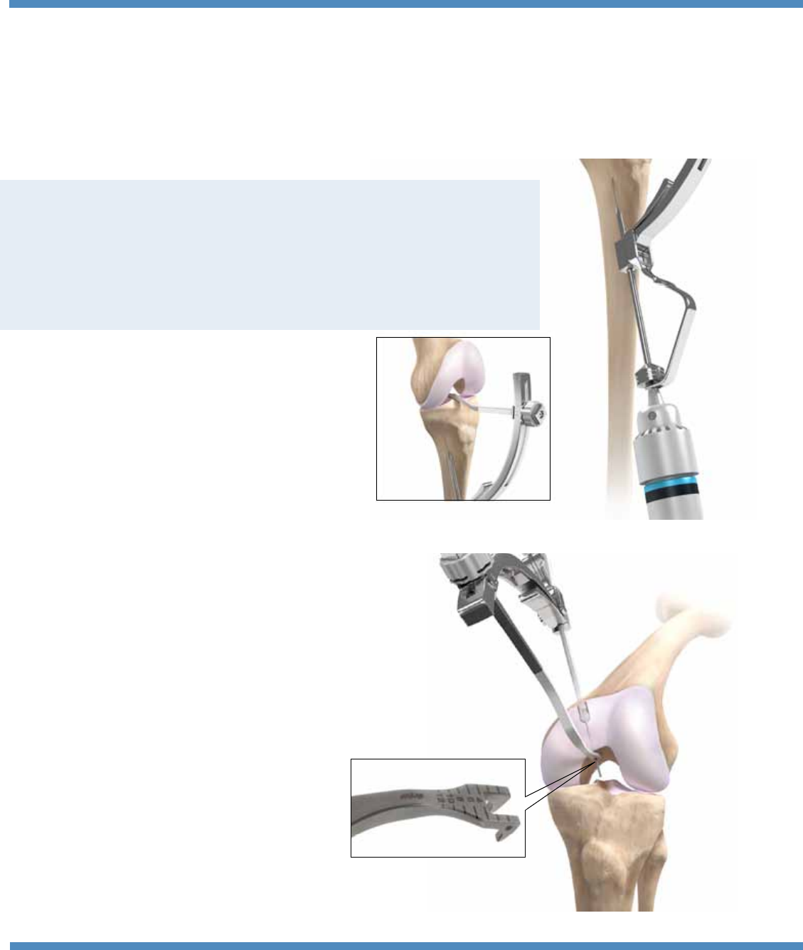
Tibial Tunnel
Figure 3.
S
Femoral Tunnel – Outside-In
Usethepre-chucked2.4drilltipguidepintodrilltibialtunnel.BesuretoipdowntheBackstopperanddrilluntiltheJacob’sChuckhitstheBackstopper,
which oocurs when the 2.4 guide pin tip should be inside the PCL capture cup. Fluro may be used to monitor/evaluate pin. Drilling should occur
with visualization through the PM portal.
Pin Protector
e PCL Tibial Arm is designed to be a pin protector. Tips to prevent advancement of the
guide pin through the PCL capture cup window:
•RemovetheBulletandBackstopper
•Dropyourhandtocapturetheguidepininthe‘cup’oftheguide,awayfromthe‘window’
•Alternatively,youcanusetheholeintheLiberator/Raspasapinprotector.
epositionoftheguidepinisveriedusinguoroscopy.Drilltibial
tunnelusingappropriatesizedStrykerVersiTomicCannulatedDrill.
Completion of the tibial tunnel drilling may be done by hand reaming.
Fortheoutside-intechnique,thePCLFemoralArmisattachedtothetibialspine
andinsertedintothejoint.Laserlinesonthefemoralarmhelptomeasure
distance from the articular cartilage. Care is taken to ensure anatomic tunnel
placement whether performing single or double bundle reconstructions.
Make a small incision and advance the guide bolt through medial soft tissues and
secure to bone.
Drill the pin from outside – in and advance the reamer over the guide pin.
Figure 4.

Graft Passage and Fixation
Use a grasper, 18 gauge wire, or surgical wire to pass a suture loop through the tibial tunnel. Use the suture manipula-
tororprobetopullthesuturethroughjointspaceandouttheanterolateralportal.Usetheeyeletofthefemoralpinto
pass suture through femoral tunnel. Use passing suture to pass the graft through the tibial tunnel and femoral tunnels.
GraftfixationtechniquesandimplantsaresurgeonandpatientspecificbaseduponimplantIFU,patientanatomy,and
surgeon preference.

Notes:

Notes:

PART NUMBER DESCRIPTION
234-020-181 Tibial Drill Guide Spine
234-020-182 Tibial Drill Guide Bolt
234-020-126 PCL Tibial Arm
234-020-127 PCL Femoral Arm
234-020-128 PCL Backstopper
234-020-131 PCL Liberator/Rasp
234-020-132 PCL Curette
234-040-050 5.0mmVersiTomicCannulatedDrill
234-040-055 5.5mmVersiTomicCannulatedDrill
234-040-060 6.0mmVersiTomicCannulatedDrill
234-040-065 6.5mmVersiTomicCannulatedDrill
234-040-070 7.0mmVersiTomicCannulatedDrill
234-040-075 7.5mmVersiTomicCannulatedDrill
234-040-080 8.0mmVersiTomicCannulatedDrill
234-040-085 8.5mmVersiTomicCannulatedDrill
234-040-090 9.0mmVersiTomicCannulatedDrill
234-040-095 9.5mmVersiTomicCannulatedDrill
234-040-100 10.0mmVersiTomicCannulatedDrill
234-040-105 10.5mmVersiTomicCannulatedDrill
234-040-110 11.0mmVersiTomicCannulatedDrill
234-040-115 11.5mmVersiTomicCannulatedDrill
234-020-148 5mm Femoral Reamer 3-Fluted
234-020-028 5.5mm Femoral Reamer 3-Fluted
234-020-062 6mm Femoral Reamer 3-Fluted
234-020-029 6.5mm Femoral Reamer 3-Fluted
234-020-061 7mm Femoral Reamer 3-Fluted
234-020-030 7.5mm Femoral Reamer 3-Fluted
234-020-008 8mm Femoral Reamer 3-Fluted
234-020-031 8.5mm Femoral Reamer 3-Fluted
234-020-009 9mm Femoral Reamer 3-Fluted
234-020-032 9.5mm Femoral Reamer 3-Fluted
234-020-010 10mm Femoral Reamer 3-Fluted
234-020-033 10.5mm Femoral Reamer 3-Fluted
234-020-011 11mm Femoral Reamer 3-Fluted
234-020-034 11.5mm Femoral Reamer 3-Fluted
234-020-078 12mm Femoral Reamer 3-Fluted
Asurgeonmustalwaysrelyonhisorherownprofessionalclinicaljudgmentwhendecidingwhethertouseaparticular
product when treating a particular patient. Stryker does not dispense medical advice and recommends that surgeons
be trained in the use of any particular product before using it in surgery.
e information presented is intended to demonstrate the breadth of Stryker product oerings. A surgeon must
always refer to the package insert, product label and/or instructions for use before using any Stryker product. Products
maynotbeavailableinallmarketsbecauseproductavailabilityissubjecttotheregulatoryand/ormedicalpractices
inindividualmarkets.PleasecontactyourStrykerrepresentativeifyouhavequestionsabouttheavailabilityof
Stryker products in your area.
Stryker Corporation or its divisions or other corporate aliated entities own, use or have applied for the following
trademarksorservicemarks:Stryker,VersiTomic.Allothertrademarksaretrademarksoftheirrespective
owners or holders.
LiteratureNumber:LJPVTPCL-BRRev.1
MS/GS 01/12
Copyright © 2012 Stryker
Printed in USA
325 Corporate Drive
Mahwah,NJ07430
t: 201 831 5000
www.stryker.com
