Zimmer Periarticular Distal Femoral Locking Plate Surgical Technique
2016-04-04
: Pdf Zimmer Periarticular Distal Femoral Locking Plate Surgical Technique Zimmer_Periarticular_Distal_Femoral_Locking_Plate_Surgical_Technique 4 2016 pdf
Open the PDF directly: View PDF ![]() .
.
Page Count: 20
Zimmer®
Periarticular
Distal Femoral
Locking Plate
Surgical Technique
The Science of the Landscape

1
Zimmer Periarticular Distal Femoral Locking Plate
Surgical Technique
Developed in conjunction with
Stephen K. Benirschke, M.D.
Professor, Department of
Orthopaedics and Sports Medicine
University of Washington
Harborview Medical Center
Seattle, Washington
Paul J. Duwelius, M.D.
Adjunct Associate Professor
Orthopaedics
Oregon Health Sciences University
Clinical Attending
St. Vincent Hospital & Medical Center
Portland, Oregon
James A. Goulet, M.D.
Professor and Director
Section of Orthopaedic Trauma
Department of Orthopaedic Surgery
The University of Michigan Hospitals
Ann Arbor, Michigan
David A. Templeman, M.D.
Associate Professor
Orthopaedic Surgery
University of Minnesota
Staff, Hennepin County Medical Center
Minneapolis, Minnesota
Robert A. Winquist, M.D.
Clinical Professor,
Department of Orthopaedics
University of Washington
Orthopaedic Surgeon
Swedish Hospital and Medical Center
Seattle, Washington
Table of Contents
Introduction 2
Locking Screw Technology 2
Locking Plate Technology 2
Distal Femur Plate Indications 2
Fracture Classification 2
Plate Features 3
Surgical Technique 4
Required Instrumentation 4
Preoperative Preparation 4
Fracture Reduction 4
Plate Positioning 5
Screw Trajectory 9
Condylar Fixation 10
Shaft Fixation 12
Wound Closure 14
Postoperative Treatment 14
Implant Removal 14
Surgical Pearls 14
Instruments and Implants 15
Order Information 16

2Zimmer Periarticular Distal Femoral Locking Plate
Introduction
The Zimmer Periarticular Locking
Plate System combines locking screw
technology with periarticular plates to
create fixed-angle constructs for use in
comminuted fractures or where deficient
bone stock or poor bone quality is
encountered. The fixed-angle plate/
screw device can be used in osteopenic
bone and other areas where traditional
screw fixation may be compromised.
The Periarticular Locking Plates will
accommodate standard screws, as well
as locking screws with threaded heads.
When necessary, interfragmentary
compression can be achieved with
lag screws.
Cannulated screws and instruments
allow provisional fixation with guide pins
in the metaphysis. This helps ensure
that the threaded locking screw heads
align properly with the threaded
plate holes.
All plate configurations contain locking
screw holes in the plate head, and
alternating locking and compression
screw slots in the shaft.
Three types of locking screws are
available with the system:
• 5.5mm cannulated locking screws
for use in the plate head
• 5.5mm cannulated conical screws for
use in the plate head
• 4.5mm noncannulated locking screws
for use in the plate shaft
Locking Screw Technology
The heads of the locking screws contain
male threads while the holes in the
plates contain female threads. This
allows the screw head to be threaded
into the plate hole, locking the screw
into the plate. This technical innovation
provides the ability to create a fixed-
angle construct while using familiar
plating techniques.
Locking Plate Technology
By using locking screws in a bone plate,
a fixed-angle construct is created. In
osteopenic bone or fractures with
multiple fragments, secure bone
purchase with conventional screws may
be compromised. Locking screws do
not rely on bone/plate compression to
resist patient load, but function similarly
to multiple small angled blade plates.
In osteopenic bone or comminuted
fractures, the ability to lock screws into
a fixed-angle construct is imperative.
By combining locking screw holes with
compression screw slots in the shaft,
the plate can be used as both a locking
device and a fracture compression
device. If compression is desired, it
must be achieved first by inserting the
standard screws in the compression
screw slots before inserting any
locking screws.
Indications
The Periarticular Locking Plate System is
indicated for temporary internal fixation
and stabilization of osteotomies and
fractures, including:
• Comminuted fractures
• Supracondylar fractures
• Intra-articular and extra-articular
condylar fractures
• Fractures in osteopenic bone
• Nonunions
• Malunions
Fracture Classification
Refer to OTA Fracture and Dislocation
Compendium
, or the Schatzker
classification for more information.
3
Zimmer Periarticular Distal Femoral Locking Plate
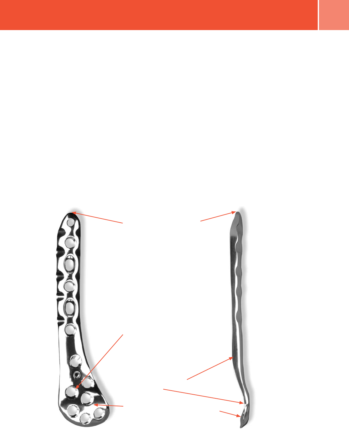
Fig. 1 Zimmer Periarticular Distal Femoral Locking Plate features.
Plate Features
• Anatomically contoured plates
are precontoured to create a fit
that requires little or no additional
bending and helps with metaphyseal/
diaphyseal reduction
• Threaded holes create a 95 degree
fixed angle between the plate head
and the locking screws to allow screw
placement that is parallel to the
joint line
• The central locking screw hole in plate
head provides initial reduction of the
plate to the condyles
Thick-to-thin plate profiles make the plates
autocontourable
The anatomical shape of the head of the
plate matches the shape of the distal femur
Multiple locking holes in the plate head
allow placement of the screws to capture
fragments
The plate shaft design allows for a
minimally invasive technique with
submuscular passage of the plate
• The low profile plate facilitates
fixation without impinging
on soft tissue
• Plates are available in a variety of
sizes and lengths, from 6 to 18 holes,
left and right
• Dual-compression slots will
accommodate periarticular screws or
conventional stainless steel screws
and allow bi-directional compression
• The last diaphyseal plate hole is
designed to accomodate the tension
device (00-4817-000-05)
The locking plate design does not
require compression between the plate
and bone to accommodate loading.
Therefore, purchase of the screws in
the bone can be achieved with a thread
profile that is shallower than that of
traditional screws. The shallow thread
profile, in turn, allows for screws with
a large core diameter to accommodate
loading with improved bending and
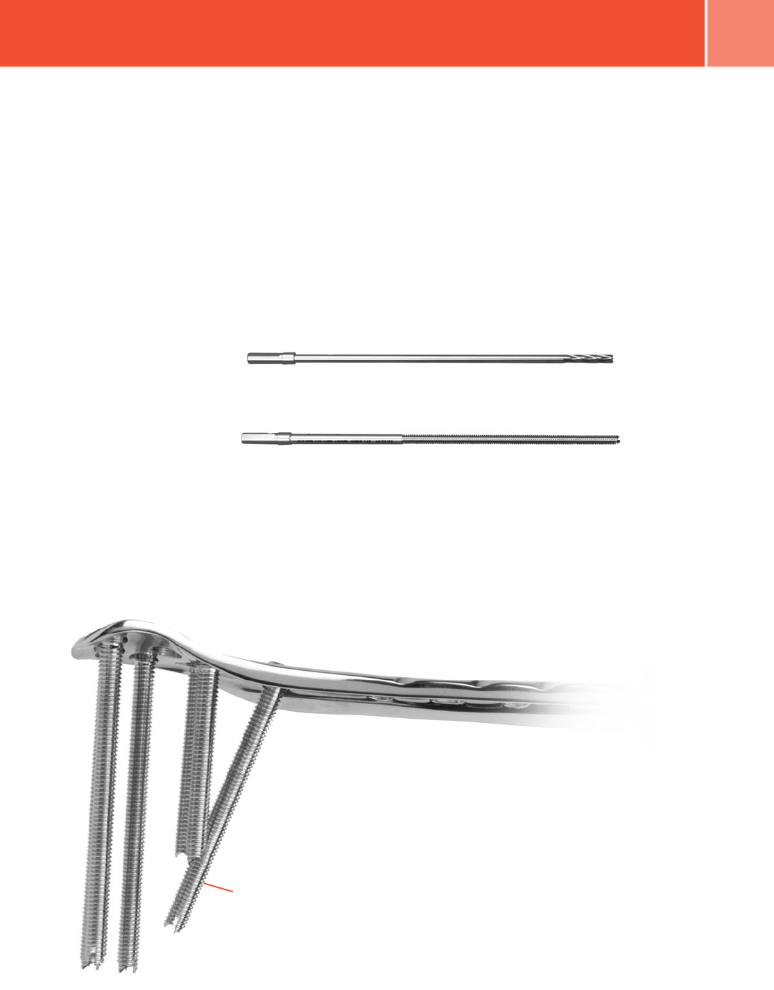
shear strength (Fig. 1).
4Zimmer Periarticular Distal Femoral Locking Plate
Surgical Technique
Required Instrumentation
The following sets may be required for
application of the 5.5mm Periarticular
Locking Proximal Tibia Plates:
• Standard Screw Set
• Basic Instrument Set
• Basic Forcep Set
• 5.5mm/4.5mm Locking Screw and
Instrument Set
• Periarticular Distal Femoral Locking
Plate and Standard Jig Set
• Linear Bone Clamps
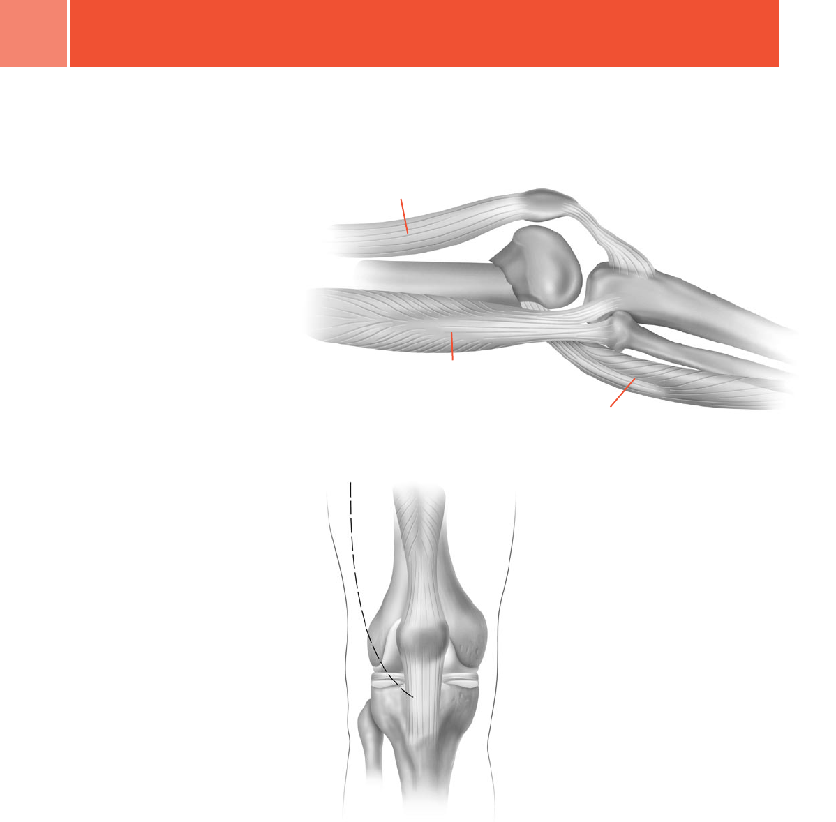
Preoperative Preparation
After assessing the fracture
radiographically and preparing a
preoperative plan, place the patient in
the supine position on a radiolucent
table. Be sure that the fluoroscope can
be positioned to visualize the distal
femur in both the lateral and anterior/
posterior (A/P) views (Figs. 2 & 3).
Fracture Reduction
It is imperative that accurate reduction
of the fracture be obtained prior to and
maintained during application of the
distal lateral femoral locking plate.
An external fixator or distractor can
serve as preliminary fixation. This will
make operative reduction easier, and
the device can be used as a
tool intraoperatively.
Before locking screws are placed in any
fragment, length, rotation, varus-valgus
and recurvatum correction should
be achieved.
The Plate Reduction Instrument is
designed to aid in minor varus-valgus
and translation corrections prior to
screw placement.
Fig. 2
Fig. 3
After radiographic verification of
preliminary reduction of the fracture, use
the preferred approach and technique to
expose the distal lateral femur.
Reduce the intra-articular fragments
using linear bone clamps or Kirschner
wires to temporarily hold the reduction.
For a Hoffa fracture, reduce the posterior
articular fragment and stabilize it with
K-wires inserted from anterior
to posterior.
Use lag screws to secure the intra-
articular fragments. To help avoid
inserting the lag screws where they will
interfere with the plate placement, hold
the plate on the bone in its approximate
position. Then insert the lag screws
as needed.
Use 3.5mm cortical screws, 4.0mm
cancellous screws, HerbertTM or Herbert/
Whipple® screws for fixation of a
posterior articular Hoffa fragment. Insert
the screws from anterior to posterior,
and where applicable countersink the
heads below the level of the
articular cartilage.
Quadriceps
Hamstrings
Gastrocnemius
5
Zimmer Periarticular Distal Femoral Locking Plate
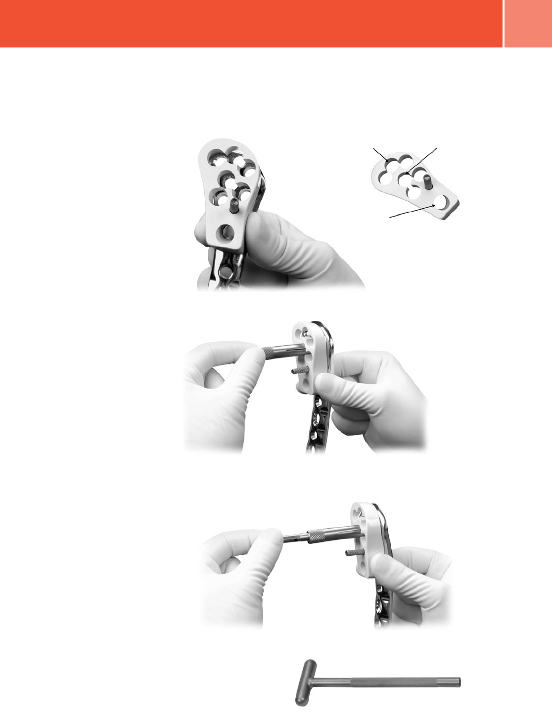
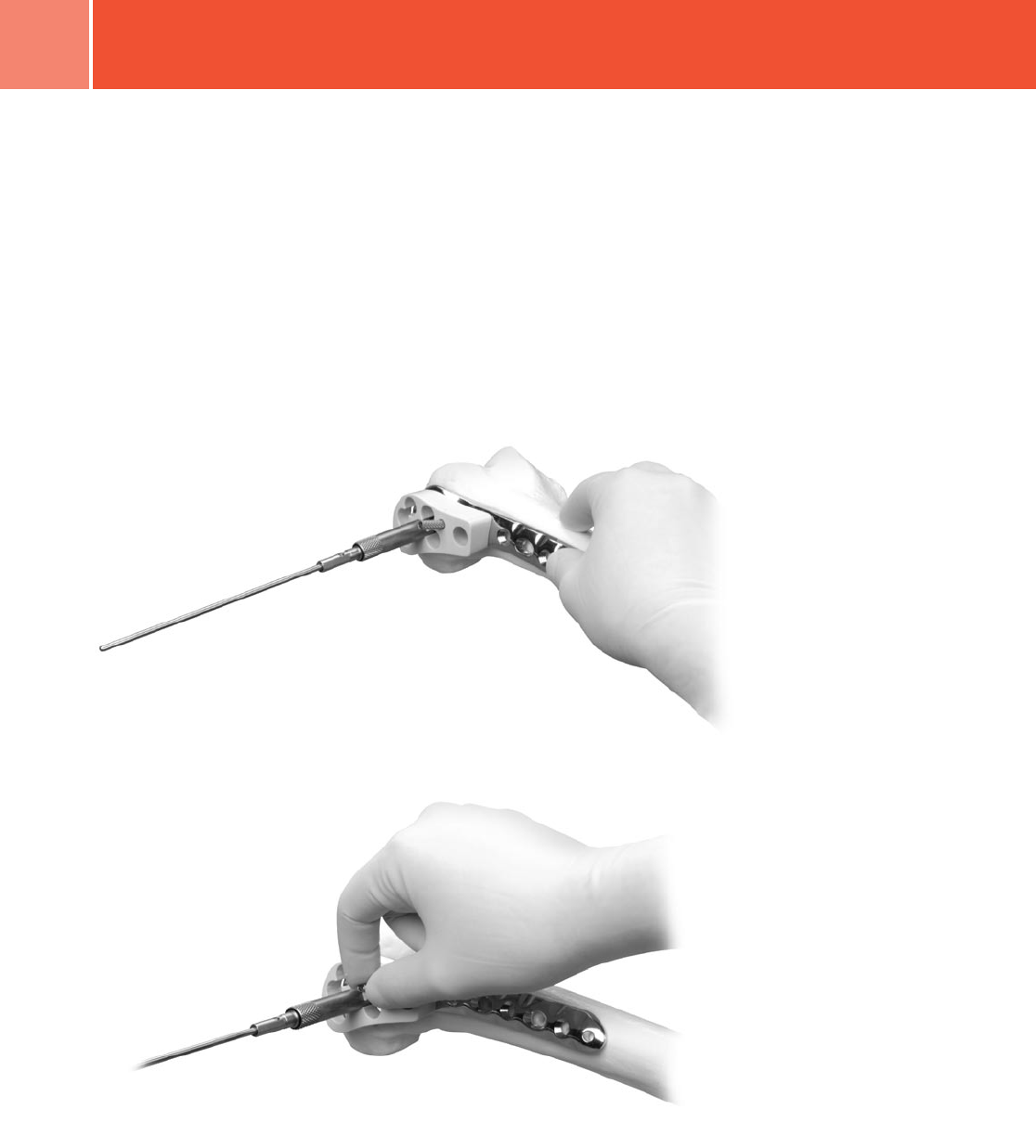
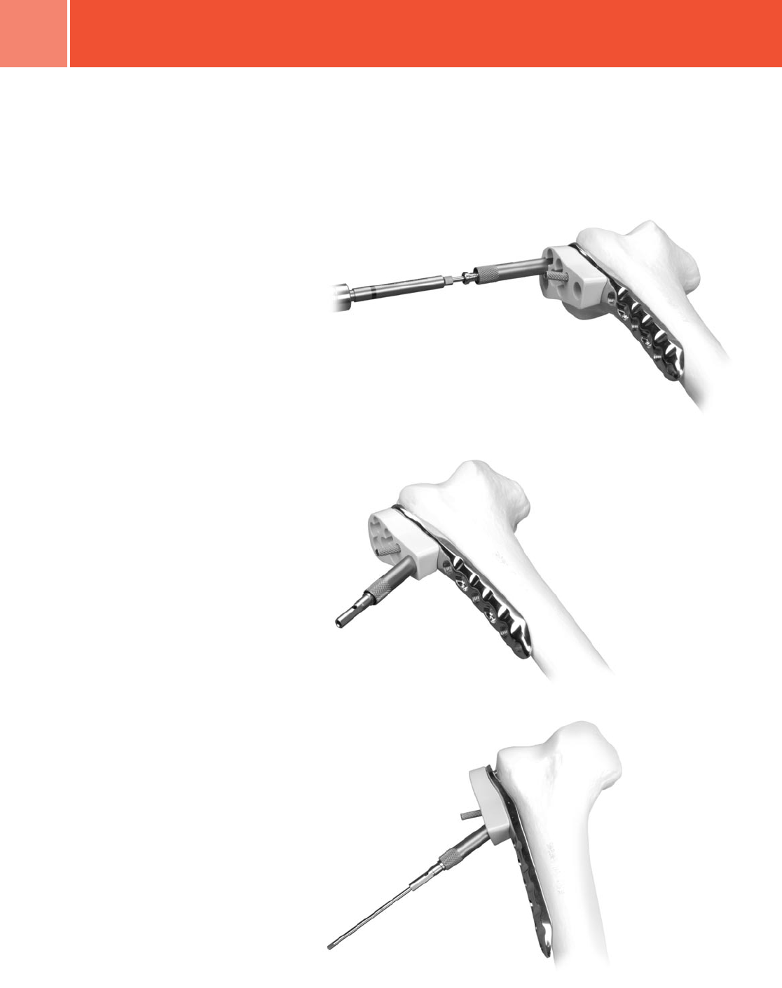
Plate Positioning
Hold the Metaphyseal Jig on the selected
plate (Fig. 4). Insert the 5.5mm Standard
Jig Sleeve into the CENTRAL hole of the
Jig/plate (Fig. 5.) and thread the 3.2mm
Standard Cannula into the plate hole
(Fig. 6). DO NOT tighten the set screw.
NOTE: Attaching the Metaphyseal Jig
to the plate using the set screw at this
time may cause or result in improper
placement of the plate on the bone.
NOTE: The Cannula Inserter may be used
to tighten the cannula if necessary
(Fig. 6A).
Fig. 5
Fig. 6A
Fig. 4
Central Distal Hole
Strut Screw Hole
Central Hole
Fig. 6
6Zimmer Periarticular Distal Femoral Locking Plate
Use this construct to place the initial
3.2mm Drill Tip Guide Wire in the
metaphysis (Fig. 7). Check plate
placement – visually and fluoroscopically
to ensure that the plate is positioned
correctly on the metaphysis of the bone.
If placement is appropriate, hold the Jig
on the plate and finger tighten or use the
3.5mm Screwdriver to tighten the
set screw (Fig. 8).
NOTE: The position of the plate on the
bone must be verified because of the
tendency to place the proximal end of
the plate too far anterior on the femoral
shaft. This placement can cause the
locking screws to be placed at a tangent
and can result in insufficient
holding strength.
Because the femoral shaft may not be
aligned with the distal fragment, the
plate head should be used to determine
the appropriate placement of the plate.
The plate head should conform to the
shape of the intact or reconstructed
condyles. This will determine the
alignment of the shaft.
Fig. 7
Fig. 8
NOTE: The Metaphyseal Jig and
Standard Cannulas MUST be used to
ensure that the screws align properly
with the threaded plate holes. Failure to
use the Metaphyseal Jig and Standard
Cannulas may result in cross-threading
or improper seating of the screws.
7
Zimmer Periarticular Distal Femoral Locking Plate
NOTE: It is easier to thread the cannulas
into the plate before placing the plate on
the bone.
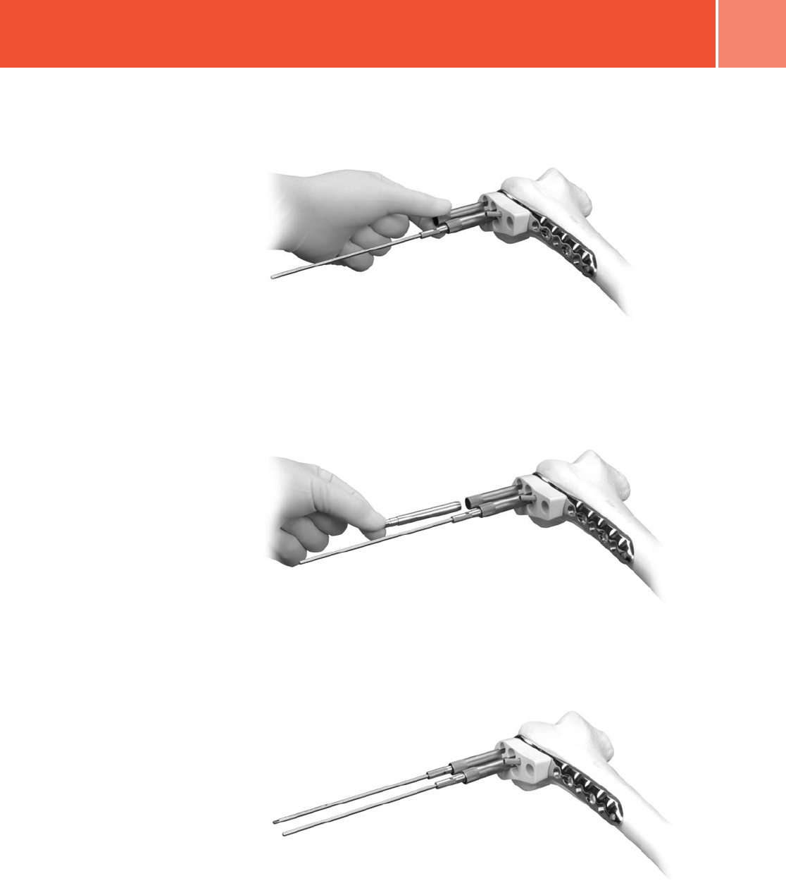
Once the plate is properly positioned,
insert the Jig Sleeve into the most
CENTRAL DISTAL locking hole in the plate
head (Fig. 9). Thread the 3.2mm Guide
Wire Cannula through the sleeve and into
the plate hole (Fig. 10).
WARNING: Do not contour or bend
the plate at or near a threaded hole,
as doing so may deform the threaded
hole and cause incompatibility with the
Locking Screw.
Insert a 3.2mm Drill Tip Guide Wire
through the cannula until the tip engages
the medial cortical wall (Fig. 11). Be sure
that the wire remains parallel to both
axes. Use the fluoroscope to confirm the
wire position in both the A/P and lateral
planes. Adjust the wire location
if necessary.
Fig. 9
Fig. 11
Fig. 10
8Zimmer Periarticular Distal Femoral Locking Plate
Black Ring
Fig. 14
Fig. 15
Fig. 12
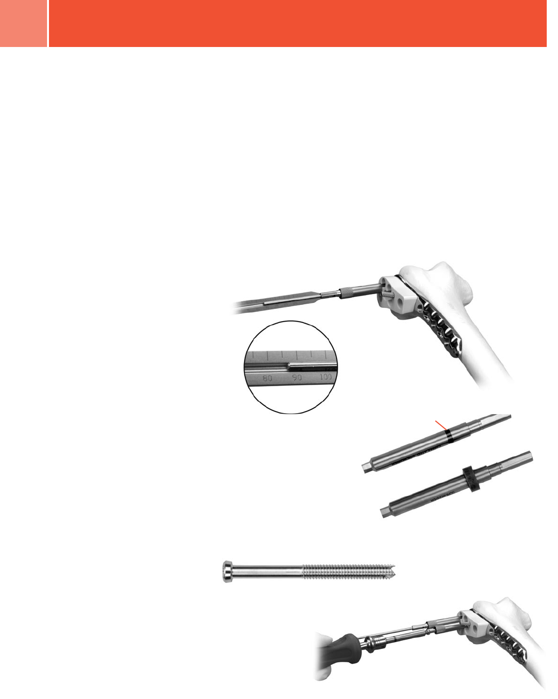
Thread a 3.2mm Standard Cannula into
one of the most proximal holes in the
plate shaft. Insert a 3.2mm Drill Tip
Guide Wire though the Cannula. Again,
check plate and bone position both
visually and fluoroscopically to ensure
proper fracture reduction and
plate placement.
Once the plate is placed appropriately
and properly aligned, slide the 5.5mm
Cannulated Screw Depth Gauge over the
guide wire in the CENTRAL plate hole
until it contacts the top of the cannula.
Read proper screw length from the
guide (Fig. 12).
Screw length measurement:
Metaphyseal Screws – the length
measurement for screws in the
metaphyseal region of the plate is
measured line-to-line – from the base
of the screw head to the tip of the screw.
Placement of the tip of the Guide Wire
will determine placement of the tip
of the screw.
Diaphyseal Screws – the length
measurement for screws in the
diaphyseal region of the plate is also
measured line-to-line – from the base
of the screw head to the tip of the screw.
In order to achieve full cortical purchase
with these screws, it is recommended
that 5mm be added to the screw length
measurement to allow for the self-
tapping flutes.
The Zimmer Periarticular Distal Femoral
Locking Plate is designed to be placed
slightly anteriorly on the distal femoral
condyles. In order to achieve an
accurate lateral x-ray or c-arm image,
it will be necessary to externally
rotate the affected limb 20-30˚. As in
distal targeting of intramedullary nails,
visualization of “round holes” from the
cannulas will ensure a true lateral image.
In other words, the x-ray beam must be
in line with the axis of the cannulas.
Fig. 13
NOTE: Slide the Screwdriver Stop Ring
onto the screwdriver shaft and place it
at the level of the black ring etched on
the driver shaft (Fig. 13). When the Blue
Stop Ring hits the top of the Jig Sleeve,
power insertion must stop. Screws must
be seated by hand. The Screwdriver Stop
Ring is intended to be a visual cue to
stop power insertion of locking screws.
Remove the Guide Wire Cannula and
use the 5.0mm Hex-head Cannulated
Screwdriver to insert a 70mm Long
5.5mm Conical Screw (Fig. 14) into
the CENTRAL plate hole to secure the
plate, or if preferred, use a linear bone
clamp or Plate Reduction Instrument
for provisional fixation. Observe
placement of the plate head and use the
fluoroscope to confirm that it is in the
desired location (Fig. 15).
9
Zimmer Periarticular Distal Femoral Locking Plate
Fig. 16
NOTE: Insertion of a screw longer than
70mm may cause interference with
other screws.
NOTE: A screwdriver shaft can be
used to loosely insert the screw under
power, but the final seating MUST be
accomplished by hand to avoid cross-
threading of the screws in the plate
holes or failure of the screw or driver.
NOTE: If lag screw fixation is necessary
for any fragment, the lag screw must be
inserted before inserting locking screws
into that fragment.
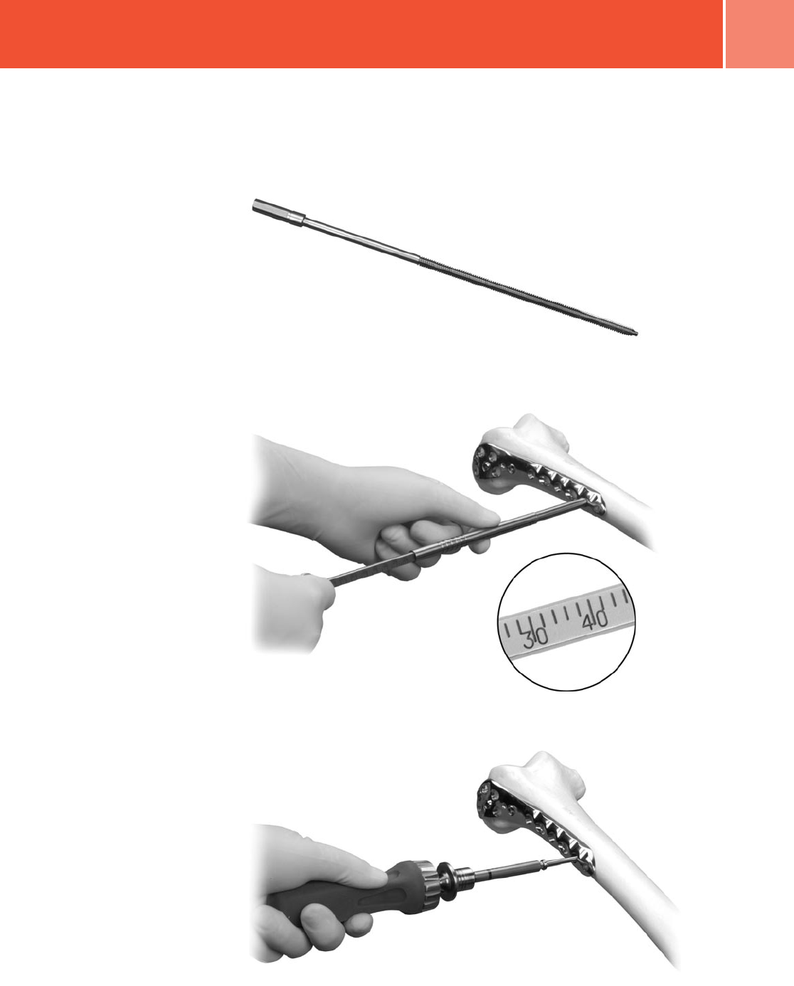
Strut Screw
Predrilling and tapping are typically not
necessary as the flutes of the screws
are self-drilling and self-tapping. If the
bone is dense, the lateral cortex can be
predrilled and tapped. If desired, use
the 4.7mm Cannulated Drill and 5.5mm
Cannulated Tap (Fig. 16) for a
5.5mm screw.
Screw Trajectory
10 Zimmer Periarticular Distal Femoral Locking Plate
Fig. 17
Fig. 19
Fig. 18
Condylar Fixation
For additional condylar fixation, slide the
5.5mm Cannulated Screw Depth Gauge
over the guide wire in the CENTRAL
DISTAL locking hole in the plate head
until it contacts the top of the cannula.
Read the proper screw length from the
guide, remove the 3.2mm Guide Wire
Cannula and use the 5.0mm Hex-head
Screwdriver to insert the appropriate
length 5.5mm Conical or Locking Screw
over the guide wire and into the
bone (Fig. 17).
Follow the same procedure for each
additional 5.5mm Cannulated Locking
Screw to be inserted into the
metaphyseal portion of the plate. Be sure
that all screws are securely tightened.
Next, insert the Jig Sleeve into the STRUT
screw hole (Fig. 18). Thread a 3.2mm
Guide Wire Cannula into the plate hole
and insert a 3.2mm Drill Tip Guide Wire
(Fig. 19). Again, carefully position the
tip of the guide wire; it will indicate the
position of the tip of the screw once it is
inserted into the plate.
Slide the 5.5mm Cannulated Screw
Depth Gauge over the guide wire in the
STRUT screw hole in the plate until it
contacts the top of the cannula. Read
the proper screw length from the guide.
Insert the appropriate length 5.5mm
Conical or Locking Screw over the guide
wire and into the bone.

11
Zimmer Periarticular Distal Femoral Locking Plate
Remove the 3.2mm Guide Wire Cannula
(Fig. 20) and use the 5.0mm Hex-
head Cannulated Driver to insert the
appropriate length 5.5mm Conical or
Locking Screw over the guide wire and
into the bone (Fig. 21). A screwdriver
shaft can be used to loosely insert the
screw under power, but final seating
MUST be accomplished by hand to
avoid cross-threading of the screws in
the plate holes or failures of the screw
or driver. Once adequate fixation is
achieved, if necessary or desired, remove
the Conical Screw from the CENTRAL
plate hole and replace it with a
Locking Screw.
NOTE: If the plate shifts during screw
insertion, all the pins and screws must
be removed and reinserted for the
screws to lock properly to the plate.
NOTE: If a plate screw impinges on
one of the intra-articular lag screws,
the lag screw must be removed and
repositioned.
Loosen the set screw and remove the
Metaphyseal Jig (Fig. 22).
Fig. 20
Fig. 22
Fig. 21
12 Zimmer Periarticular Distal Femoral Locking Plate
Fig. 25
Fig. 24
Shaft Fixation
Reduce the plate to the shaft. Confirm
rotation of the extremity by clinical
examination. Check the alignment of the
shaft with A/P and lateral fluoroscopic
views. The shaft portion of the plate can
be compressed to the bone by either
inserting a nonlocking screw through the
most proximal shaft compression slot or
by using the Plate Reduction Instrument
to hold the plate against the bone while
inserting a locking screw. If preferred,
a linear bone clamp can be used.
The Plate Reduction Instrument can be
used for:
• MINOR varus-valgus adjustment (<5°)
• Translational adjustments
• Stabilization of plate orientation with
respect to the bone during insertion
of the first screws
• Alignment of segmental fragments
When used without the MIS Jig
To use the Plate Reduction Instrument,
make a stab incision at the desired
location. Insert the 5.5mm/4.5mm
Percutaneous Sleeve and Trocar through
soft tissues ensuring that contact is
made with the surface of the plate
at the desired location. Remove the
Trocar. Insert the Plate Reduction Sleeve
through the Percutaneous Sleeve and
thread it into the plate. Thread the
Reduction Spin Knob all the way onto
the Shaft of the Reduction Instrument.
Next insert the Reduction Instrument
through the Reduction Sleeve and into
the bone fragment by hand or under
power. Rotating the Spin Knob clockwise
will cause it to contact the top of the
Reduction Sleeve and in turn, draw
the plate and bone together. Monitor
progress using C-Arm images. Stop when
desired reduction is achieved.
Once reduction is achieved, and it is
appropriate, the plate may be loaded in
tension using the Tension Device
[00-4817-005-00].
NOTE: In comminuted fractures, it may
not always be possible or desirable
to achieve anatomic reduction of the
fracture.
Insert standard 4.5mm cortical (Fig. 23)
screws through the compression slots in
the plate as desired. If both locking and
nonlocking screws will be used in the
shaft, the nonlocking screws must
be inserted first.
Predrill both cortices with the drill bit.
Measure for screw length using the
depth gauge. Then select and insert
the appropriate length 4.5mm Cortical
Screws using the Large Hex Screwdriver.
To insert 4.5mm Locking Screws, thread
the 3.7mm Standard Cannula (Black
Ring) into the desired locking hole
(Fig. 24). Use the 3.7mm Standard Drill
through the cannula to drill (Fig. 25).
Use the fluoroscope to confirm the drill
position in both the A/P and lateral
planes. Then remove the cannula.
Fig. 23
13
Zimmer Periarticular Distal Femoral Locking Plate
Fig. 26
Fig. 27
Fig. 28
Tapping is typically not necessary as the
flutes of the screws are self-tapping. If
the bone is dense, the lateral cortex can
be tapped. If desired, use the 4.5mm
Screw Tap (Fig. 26) to tap for the
4.5mm screw.
Insert the 4.5mm Locking Screw Depth
Gauge (Fig. 27) into the screw hole until
the tip of the gauge bottoms out in the
hole. Read the proper screw length from
the gauge at the point where the gauge
meets the surface of the plate.
Use the 5.0mm Hex-head Driver to insert
the 4.5mm Locking Screw (Fig. 28). A
screwdriver shaft can be used to loosely
insert the screw under power, but the
final seating MUST be accomplished
by hand to avoid cross-threading of the
screws in the plate holes or failure of
the screw or driver.
Follow the same procedure for each
additional 4.5mm Locking Screw. Be sure
that all screws are securely tightened.
Make a final check of the limb alignment
and fracture reduction. Then make sure
that all shaft locking screws are
securely tightened.
Securely tighten the distal locking screws
again by hand before closing.

14 Zimmer Periarticular Distal Femoral Locking Plate
Wound Closure
Use the appropriate method for surgical
closure of the incision.
Postoperative Treatment
Postoperative treatment with locking
plates does not differ from conventional
open reduction internal fixation (ORIF)
procedures.
Implant Removal
To remove locking screws, use the Large
Hexagonal screwdriver, 5.0mm Hex to
first unlock all screws from the plate and
then remove the screws completely. DO
NOT use the forward captive screwdrivers
for screw removal.
Please refer to the package insert
for product information, including
contraindications, warnings, and
precautionary information.
Surgical Pearls
Depending upon the screw position in
the plate, the screw head may not be
flush with the plate surface. If unsure
that the screw is seated, loosen screw
and retighten.
If the locking screw is difficult to insert
or stops advancing before locking to the
plate, remove the screw and pre-drill with
the appropriate drill bit. Then reinsert
the screw. (This condition may be caused
by very dense or thick cortical bone.)
Flexion/extension of the distal femoral
fragment may be achieved using the
Plate Reduction Instrument as a joystick.
Bumps or other devices may be used
under the distal femoral metaphyseal
area to help reduce the fracture in the
lateral view.
Varus/valgus can be checked using the
C-arm and a cord or long guide wire
from the femoral head to the center of
the ankle joint on antero-posterior view.
Use the C-Arm over the knee joint to
check that the cord or guide wire passes
10mm medially of the center of the knee
joint. Minor adjustment to varus/valgus
reduction can be achieved using the
Plate Reduction Instrument.
A distractor or large external fixator may
also be useful in gaining reduction.
Cleaning of the cannulated instruments
is necessary for proper function. The
cleaning stylet can clear debris in the
cannulations and prevent binding of the
instruments. The cleaning brush should
be used postoperatively.
15
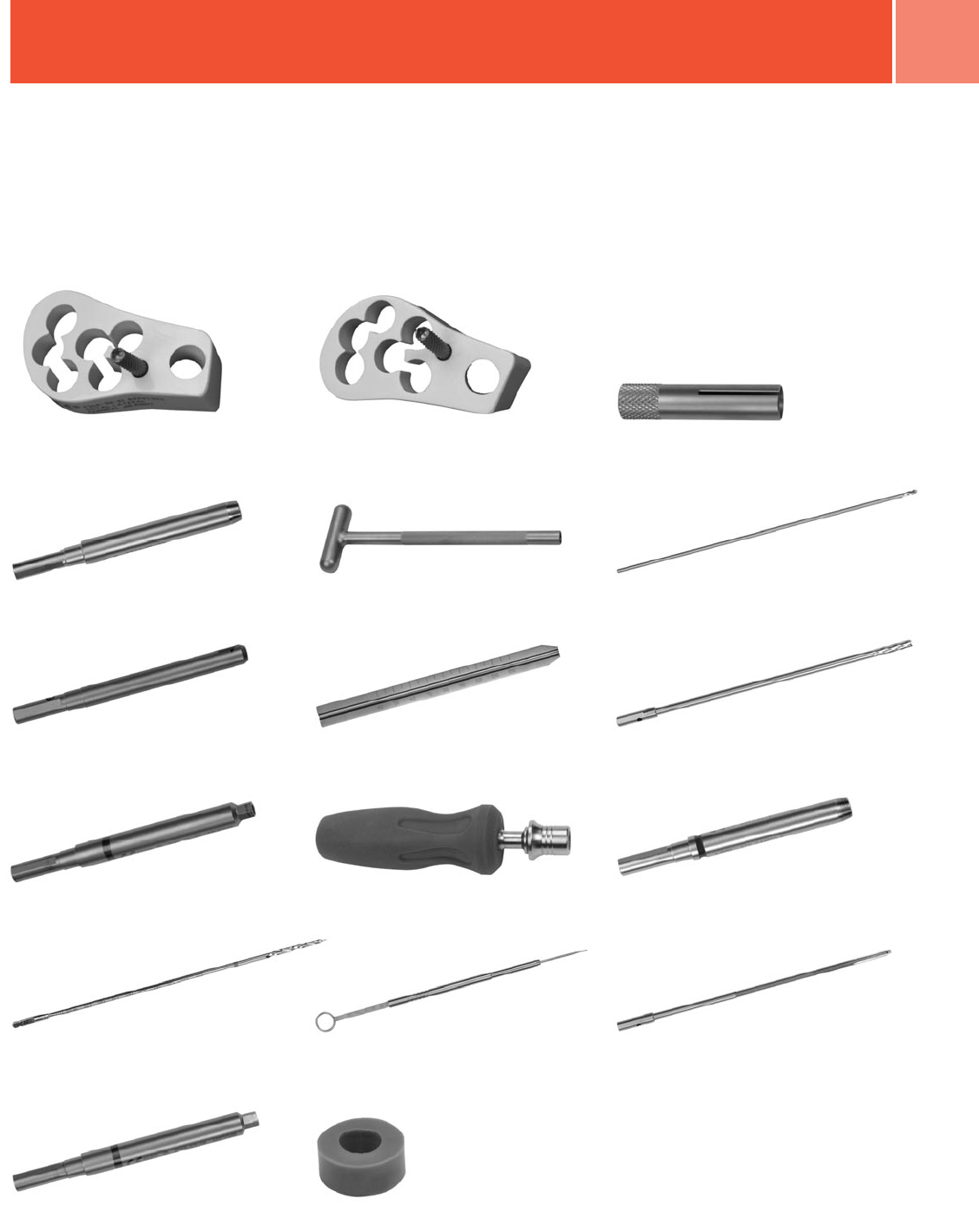
Zimmer Periarticular Distal Femoral Locking Plate
Instruments and
Implants
Distal Lateral Femoral Plate Jig, Right
00-2360-090-01
5.5mm/4.5mm Standard Jig Sleeve
00-2360-090-04
3.7mm Standard Cannula
00-2360-020-37
3.2mm Standard Cannula
00-2360-021-32
3.2mm Standard Drill Tip Guide Wire
00-2360-033-32
4.5mm Locking Screw Standard
Depth Gauge 00-2360-040-45
5.5mm Cannulated Locking Screw
Depth Gauge 00-2360-041-55
4.5mm Locking Screw Tap
00-2360-053-45
5.0mm Hex Std Screwdriver
00-2360-065-50
5.0mm Hex Std Cannulated
Screwdriver 00-2360-066-50
4.7mm Std Cannulated Drill
00-2360-071-47
Cannula Inserter 00-2360-088-00
Guide Wire Inserter 00-2360-085-00
Modular Handle 00-2360-186-00
3.7mm Std Drill 00-2360-225-37
5.0mm Screwdriver Stop Ring
00-2360-065-05
Distal Lateral Femoral Plate Jig, Left
00-2360-090-02

16 Zimmer Periarticular Distal Femoral Locking Plate
Order Information
Distal Femoral Plate Standard Jig Set - Set #00-2360-000-11
Prod. No. Description
00-2360-090-01 Distal Lateral Femoral Plate Jig, Right
00-2360-090-02 Distal Lateral Femoral Plate Jig, Left
00-2358-010-05 Dist Lat Fem Plate/Jig Case
00-2360-093-03 Standard Jig Set Screw 2 ea.
Distal Lateral Femoral Locking Plate Set - Set #00-2357-000-13
Prod. No. Description
00-2357-102-06 Distal Lateral Femoral Locking Plate, 6 Hole,
159mm Lng, Left
00-2357-102-10 Distal Lateral Femoral Locking Plate, 10 Hole,
223mm Lng, Left
00-2357-102-14 Distal Lateral Femoral Locking Plate, 14 Hole,
286mm Lng, Left
00-2357-102-18 Distal Lateral Femoral Locking Plate, 18 Hole,
349mm Lng, Left
00-2357-102-20 Distal Lateral Femoral Locking Plate, 20 Hole,
368mm Lng, Left
00-2357-101-06 Distal Lateral Femoral Locking Plate, 6 Hole,
159mm Lng, Right
00-2357-101-10 Distal Lateral Femoral Locking Plate, 10 Hole,
223mm Lng, Right
00-2357-101-14 Distal Lateral Femoral Locking Plate, 14 Hole,
286mm Lng, Right
00-2357-101-18 Distal Lateral Femoral Locking Plate, 18 Hole,
349mm Lng, Right
00-2357-101-20 Distal Lateral Femoral Locking Plate, 20 Hole,
368mm Lng, Right
Also Available:
47-2357-101-22 Distal Lateral Femoral Locking Plate, 22 Hole, 400mm
Lng, Right, Sterile Only
47-2357-102-22 Distal Lateral Femoral Locking Plate, 22 Hole, 400mm
Lng, Left, Sterile Only
5.5mm/4.5mm Periarticular Locking Instrument Set - Set #00-2360-000-01
Prod. No. Description
00-1147-073-00 Cleaning Stylet
00-1147-078-00 Cleaning Brush
00-2358-035-05 5.5mm/4.5mm Periarticular Locking Screw/
Instrument Case
00-2360-012-01 5.5mm Plate Reduction Instrument
00-2360-012-02 5.5mm Plate Reduction Sleeve
00-2360-012-03 Plate Reduction Spin Knob
00-2360-020-37 3.7mm Standard Cannula
00-2360-021-32 3.2mm Standard Cannula
00-2360-033-32 3.2mm Standard Drill Tip Guide Wire
00-2360-040-45 4.5mm Locking Screw Standard Depth Gauge
00-2360-041-55 5.5mm Cannulated Locking Screw Depth Gauge
00-2360-053-45 4.5mm Locking Screw Tap
00-2360-054-55 5.5mm Cannulated Locking Screw Tap
00-2360-065-05 5.0mm Screwdriver Stop Ring
00-2360-065-50 5.0mm Hex Std Screwdriver
00-2360-066-50 5.0mm Hex Std Cannulated Screwdriver
00-2360-071-47 4.7mm Std Cannulated Drill
00-2360-085-00 Guide Wire Inserter
00-2360-186-00 Modular Handle
00-2360-088-00 Cannula Inserter
00-2360-090-04 5.5mm/4.5mm Standard Jig Sleeve
00-2360-225-37 3.7mm Std Drill
00-4812-045-00 Large Hex Screwdriver
5.5mm/4.5mm Locking Screw Set - Set #00-2359-000-01
Prod. No. Description
00-2359-030-55 5.5mm Cannulated Locking Screw 30mm Lng
00-2359-035-55 5.5mm Cannulated Locking Screw 35mm Lng
00-2359-040-55 5.5mm Cannulated Locking Screw 40mm Lng
00-2359-045-55 5.5mm Cannulated Locking Screw 45mm Lng
00-2359-050-55 5.5mm Cannulated Locking Screw 50mm Lng
00-2359-055-55 5.5mm Cannulated Locking Screw 55mm Lng
00-2359-060-55 5.5mm Cannulated Locking Screw 60mm Lng
00-2359-065-55 5.5mm Cannulated Locking Screw 65mm Lng
00-2359-070-55 5.5mm Cannulated Locking Screw 70mm Lng
00-2359-075-55 5.5mm Cannulated Locking Screw 75mm Lng
00-2359-080-55 5.5mm Cannulated Locking Screw 80mm Lng
00-2359-085-55 5.5mm Cannulated Locking Screw 85mm Lng
00-2359-090-55 5.5mm Cannulated Locking Screw 90mm Lng
00-2359-095-55 5.5mm Cannulated Locking Screw 95mm Lng
00-2359-100-55 5.5mm Cannulated Locking Screw 100mm Lng
00-2359-050-56 5.5mm Cannulated Conical Screw 50mm Lng
00-2359-055-56 5.5mm Cannulated Conical Screw 55mm Lng
00-2359-060-56 5.5mm Cannulated Conical Screw 60mm Lng
00-2359-065-56 5.5mm Cannulated Conical Screw 65mm Lng
00-2359-070-56 5.5mm Cannulated Conical Screw 70mm Lng
00-2359-075-56 5.5mm Cannulated Conical Screw 75mm Lng
00-2359-080-56 5.5mm Cannulated Conical Screw 80mm Lng
00-2359-085-56 5.5mm Cannulated Conical Screw 85mm Lng
00-2359-090-56 5.5mm Cannulated Conical Screw 90mm Lng
00-2359-012-45 4.5mm Locking Screw 12mm Lng
00-2359-014-45 4.5mm Locking Screw 14mm Lng
00-2359-016-45 4.5mm Locking Screw 16mm Lng
00-2359-018-45 4.5mm Locking Screw 18mm Lng
00-2359-020-45 4.5mm Locking Screw 20mm Lng
00-2359-022-45 4.5mm Locking Screw 22mm Lng
00-2359-024-45 4.5mm Locking Screw 24mm Lng
00-2359-026-45 4.5mm Locking Screw 26mm Lng
00-2359-028-45 4.5mm Locking Screw 28mm Lng
00-2359-030-45 4.5mm Locking Screw 30mm Lng
00-2359-032-45 4.5mm Locking Screw 32mm Lng
00-2359-034-45 4.5mm Locking Screw 34mm Lng
00-2359-036-45 4.5mm Locking Screw 36mm Lng
00-2359-038-45 4.5mm Locking Screw 38mm Lng
00-2359-040-45 4.5mm Locking Screw 40mm Lng
00-2359-042-45 4.5mm Locking Screw 42mm Lng
00-2359-044-45 4.5mm Locking Screw 44mm Lng
00-2359-046-45 4.5mm Locking Screw 46mm Lng
00-2359-048-45 4.5mm Locking Screw 48mm Lng
00-2359-050-45 4.5mm Locking Screw 50mm Lng
00-2359-055-45 4.5mm Locking Screw 55mm Lng
00-2359-060-45 4.5mm Locking Screw 60mm Lng
00-2359-065-45 4.5mm Locking Screw 65mm Lng
00-2359-070-45 4.5mm Locking Screw 70mm Lng
5.5mm/4.5mm Periarticular Locking Instrument Set - Set #00-2360-000-01
Prod. No. Description
Also Available:
47-2360-080-05 Torque Limiting Attachment

Contact your Zimmer representative or visit us at www.zimmer.com
97-2347-044-00 Rev. 2 5ML Printed in USA ©2005,2007 Zimmer, Inc.
+H124972347044001/$070427R2D07$
