X‐RAY RADIATION SAFETY Operator Training Manual Bruker
User Manual: Pdf bruker radiation safety manual
Open the PDF directly: View PDF ![]() .
.
Page Count: 21
OperatorTrainingManual
X‐RAYRADIATIONSAFETY
030.0011.03.2
For Sales & Service Contact
2650 E. 40th Ave. • Denver, CO 80205
Phone 303-320-4764 • Fax 303-322-7242
1-800-833-7958
www.geotechenv.com

Radiation Safety Manual
030.0011.03.2 2 of 21
1. Training Objectives ............................................................................................................... 3
2. What is Radiation? ................................................................................................................ 3
3. Radiation Terminology .......................................................................................................... 3
4. Types of Radiation ................................................................................................................ 5
4.1. Non-ionizing Radiation ................................................................................................ 5
4.2. Ionizing Radiation ........................................................................................................ 5
4.3. Penetration .................................................................................................................. 6
5. Units for Measuring Radiation .............................................................................................. 6
5.1. Rad (Radiation Absorbed Dose) .................................................................................. 6
5.2. Rem ............................................................................................................................. 6
5.3. Dose and Dose Rate..................................................................................................... 7
6. Significant Doses ................................................................................................................... 7
7. Biological Effects of Radiation ............................................................................................... 8
7.1. Cell Sensitivity.............................................................................................................. 8
7.2. Acute and Chronic Doses of Radiation ........................................................................ 8
7.3. Biological Damage Factors......................................................................................... 10
8. Putting Risks in Perspective ................................................................................................ 10
8.1. Risk Comparison ........................................................................................................ 10
8.2. Radiation Dose Limits ................................................................................................ 11
8.2.1. Declared Pregnant Worker ............................................................................... 12
9. Measuring Radiation ........................................................................................................... 12
9.1. Dosimeters ................................................................................................................ 13
9.2. Survey Meters ........................................................................................................... 13
10. Exposure Reduction (ALARA) .............................................................................................. 15
11. Production of X-Ray Radiation ............................................................................................ 17
12. Radiation Exposure Potential .............................................................................................. 19
13. Rights and Responsibilities ................................................................................................. 20

Radiation Safety Manual
030.0011.03.2 3 of 21
1. Training Objectives
Describe occupation radiation worker rights and responsibilities
Describe Ionizing Radiation
Describe the nature and properties of X-Ray radiation and its associated hazards
Describe how X-Rays are produced
Describe the biological effects of exposure to X-Rays
Describe the X-Ray exposure potential of the Bruker Handheld XRF Analyzer
Identify and describe personnel monitoring devices
Identify and describe radiation survey instruments
Describe the principles of radiation protection and ALARA
Describe the designed safety features in the Bruker Handheld XRF Analyzer
Describe the proper operating procedure for the Bruker Handheld XRF Analyzer
Identify failure of designed safety features or other unusual conditions
Describe XRF Analyzer user responsibilities
Describe the Federal Regulatory Dose Limits
List the common sense rules for safely operating the Bruker Handheld XRF Analyzer
The Bruker Handheld XRF Analyzer user’s training consists of this manual, the User Guide, the Basic
Operation Training Video, and the Radiation Safety Video. A PowerPoint presentation and the
instructor may also supplement these training materials. Proper training is vital for compliance, safe
operation, and understanding of the responsibilities of the user of handheld XRF analyzers. Some local
regulatory agencies require that training be documented and a demonstration of sufficient knowledge
through an examination be performed.
Bruker recommends that local regulatory requirements in regards to training be determined,
understood, and followed.
2. What is Radiation?
The term radiation is used with all forms of energy—light, X-rays, radar, microwaves, and more. For
the purpose of this manual, however, radiation refers to invisible waves or particles of energy from
radioactive sources or X-ray tubes. High levels of radiation may pose a danger to living tissue because
it has the potential to damage and/or alter the chemical structure of cells. This could result in various
levels of illness (mild to severe).
The user of a Bruker XRF analyzer must understand the nature of radiation and how to safely use XRF
analyzers.
3. Radiation Terminology
Before examining the subject of radiation in more detail, there are several important terms to be
reviewed and understood.
Bremsstrahlung: The X-rays or “braking” radiation produced by the deceleration of electrons, namely
in an X-ray tube.
Characteristic X-rays: X-rays emitted from electrons during electron shell transfers.
Radiation Safety Manual
030.0011.03.2 4 of 21
Fail-Safe Design: One in which all failures of indicator or safety components that can reasonably be
anticipated cause the equipment to fail in a mode such that personnel are safe from exposure to
radiation. For example, if the red lamp indicating “X-RAY ON” fails, the production of X-rays would be
prevented.
Ion: An atom that has lost or gained an electron.
Ion Pair: A free electron and positively charged atom.
Ionization: The process of removing electrons from the shells of neutral atoms.
Ionizing Radiation: Radiation that has enough energy to remove electrons from neutral atoms.
Isotope: Atoms of the same element that have a different number of neutrons in the nucleus.
Non-ionizing Radiation: Radiation that does not have enough energy to remove electrons from
neutral atoms.
Normal Operation: Operation under conditions suitable for collecting data as recommended by
manufacturer, including shielding and barriers.
Primary Beam: Ionizing radiation from an X-ray tube that is directed through an aperture in the
radiation source housing for use in conducting X-ray fluorescence measurements.
Radiation: The energy in transit in form of electromagnetic waves or particles.
Radiation Generating Machine: A device that generates X-rays by accelerating electrons, which strike
an anode.
Radiation Source: An X-ray tube or radioactive isotope.
Radiation Source Housing: That portion of an X-ray fluorescence (XRF) system, which contains the X-
ray tube or radioactive isotope.
Radioactive Material: Any material or substance that has unstable atoms, which are emitting
radiation.
System Barrier: That portion of an area, which clearly defines the transition from a controlled area to
a radiation area and provides the necessary shielding to limit the dose rate in the controlled area
during normal operation.
X-ray Generator: That portion of an X-ray system that provides the accelerating voltage and current
for the X-ray tube.
X-ray System: Apparatus for generating and using ionizing radiation, including all X-ray accessory
apparatus, such as accelerating voltage and current for the X-ray tube and any needed shielding.
Radiation Safety Manual
030.0011.03.2 5 of 21
4. Types of Radiation
As stated earlier, radiation consists of invisible waves or particles of energy that could have a health
effect on humans if received in too large a quantity. There are two distinct types of radiation: non-
ionizing and ionizing.
4.1. Non-ionizing Radiation
Non-ionizing radiation does not have the energy needed to ionize an atom (i.e. to remove
electrons from neutral atoms). Sources of non-ionizing radiation include light, microwaves,
power lines, and radar. Although this type of radiation can cause biological damage, like
sunburn, it is generally considered less hazardous than ionizing radiation.
4.2. Ionizing Radiation
Ionizing radiation has enough energy to remove electrons from neutral atoms in a process
called ionization. An atom having either a
positive or negative charge is an ion. A free
electron is also an ion. Ionizing radiation is
of concern due to its potential to alter the
chemical structure of living cells. These
changes can alter or impair the normal
functions of a cell. Sufficient amounts of
ionizing radiation can cause hair loss, blood
changes, and varying degrees of illness.
These levels are approximately 1,000 times
higher than levels that the public or workers
are permitted to receive.
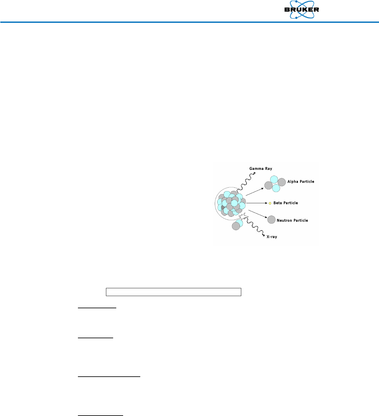
The four basic types of ionizing radiation are emitted from different parts of an atom, as
shown in the image to the right.
NOTE: Bruker handheld XRF devices only emit X-rays.
Alpha Particles have a large mass, consisting of two protons and two neutrons, and a positive
charge. They ionize by stripping away electrons (-) from other atoms with its positive (+)
charge, and are generally only considered a radiation hazard if ingested or inhaled.
Beta Particles are high-energy, high-speed electrons or positrons which form ionizing
radiation also known as beta rays. They ionize other atoms by stripping electrons out of their
orbits with their negative charge, and are primarily a radiation hazard only to the skin and
eyes.
Gamma Rays and X-rays are electromagnetic waves or photons of pure energy that have no
mass or electrical charge. They ionize atoms by interacting with electrons, and are best
shielded by use of dense materials, such as concrete, lead, or steel. Bruker handheld devices
produce X-rays.
Neutron Particles are ejected from the nucleus of an atom during the normal operation of a
nuclear reactor or particle accelerator, as well as the natural decay process of some
Radiation Safety Manual
030.0011.03.2 6 of 21
radioactive elements. They can split atoms by colliding with their nuclei, forming two or more
unstable atoms and cause ionization as they try to become stable. They are best shielded by
materials with a high hydrogen content (water, concrete or plastic).
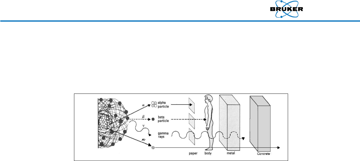
4.3. Penetration
The penetrating power for each of the four basic radiations varies significantly, as shown
below.
5. Units for Measuring Radiation
The absorption of radiation into the body, or anything else, depends upon two things: the type of
radiation involved and the amount of radiation energy received. Internationally, the units for
measuring radiation are the Gray and Sievert; in the USA, the units are the rad and rem.
5.1. Rad (Radiation Absorbed Dose)
A rad is:
A unit for measuring the amount of radiation energy absorbed by a material (i.e.,
dose)
Defined for any material (e.g., 100 ergs/gm)
Applied to all types of radiation
Not related to biological effects of radiation in the body
1 rad = 1000 millirad (mrad)
o The Gray (Gy) is the System International (SI) unit for absorbed energy
o 1 rad = 0.01 Gray (Gy) and 1 Gray = 100 rad
5.2. Rem
Actual biological damage depends upon the concentration as well as the amount of radiation
energy deposited in the body. The rem is used to quantify overall doses of radiation, their
ability to cause damage, and their dose equivalence (see below).
A rem is:
Is a unit for measuring dose equivalence
Is the most commonly used unit of radiation exposure measure
A term that pertains directly to humans
Takes into account the energy absorbed (dose); the quality of radiation; the
biological effect of different types of radiation in the body and any other factor.
Radiation Safety Manual
030.0011.03.2 7 of 21
For gamma and X-ray radiation all of these factors are unity so that for these
purposes a rad and a rem are equal.
1 rem = 1000 millirem (mrem)
o Sievert is the SI unit for dose equivalence
o 1 rem = 0.01 Sievert (Sv) and 1Sv = 100 rem
5.3. Dose and Dose Rate
Dose is the amount of radiation you receive during any exposure.
Dose Rate is the rate at which you receive the dose.
Example:
Dose rate = dose/time = mrem/hr
Dose = dose rate x time = mrem
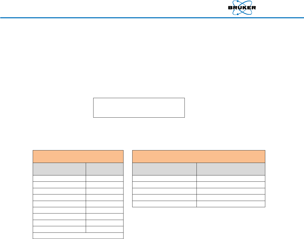
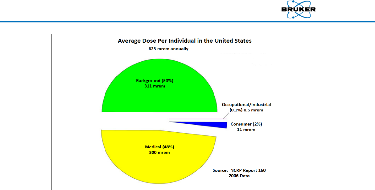
6. Significant Doses
NCRP Report No. 160, Ionizing Radiation Exposure of the Population of the United States, published in
2006, summaries the effective dose per individual in the U.S. population as 625 mrem annually. This
number has increased over the previous value of 360 mrem primarily due to the increased radiation
exposure from medical procedures.
Typical Radiation Doses from Selected
Sources (Annual)*
Average Occupational Doses
Exposure Source
mrem per year
Occupation
Exposure
(mrem per year)
Background (50%)
311
Airline flight crewmember
1000
Medical (48%)
300
Nuclear power plant worker
700
Consumer (2%)
13
Grand central station worker
120
Occupational (0.1%)
0.5
Medical personnel
70
Round trip US by air
5
DOE/DOE contractors
44
Building materials
3.6
Worldwide fallout
<1
Natural gas range
0.2
Smoke detectors
0.0001
* Based on 2006 U.S. data only
Radiation Safety Manual
030.0011.03.2 8 of 21
As previously stated, the general public is exposed daily to small amounts of radiation. However, there
are four major groups of people that have been exposed in the past to significant levels of radiation.
Because of this we know much about ionizing radiation and its biological effects on the body. The
earliest radiation workers, such as radiologists, received large doses of radiation before biological
effects were recognized. Since then, safety standards have been developed to protect such
employees.
The more than 100,000 people who survived the atomic bombs dropped on Hiroshima and Nagasaki,
those involved in accidents like Chernobyl, and those who have received radiation therapy for cancer
are examples of large groups that have received significant doses of radiation.
7. Biological Effects of Radiation
7.1. Cell Sensitivity
The human body is composed of billions of living cells. Groups of these cells make up tissues,
which in turn make up the body’s organs. Some cells are more resistant to viruses, poisons,
and physical damage than others. Rapidly dividing cells are the most sensitive cells, which is
why exposure to a fetus is so carefully controlled. Radiation damage may depend on both
resistance and level of activity during exposure.
7.2. Acute and Chronic Doses of Radiation
All radiation, if received in sufficient quantities, can damage living tissue. The key lies in how
much and how quickly a radiation dose is received. Doses of radiation fall into one of two
categories: acute or chronic.

Radiation Safety Manual
030.0011.03.2 9 of 21
Acute Dose
An acute dose is a large dose of radiation received in a short period of time that results in
physical reactions due to massive cell damage (acute effects). The body can't replace or
repair cells fast enough to undo the damage right away, so the individual may remain ill for a
long period of time. Acute doses of radiation can result in reduced blood count and hair loss.
Recorded whole body doses of 100 – 250 mSv (10 - 25 rem) have resulted only in slight blood
changes with no other apparent effects.
Radiation Sickness
Radiation sickness may occur at acute doses greater than 1 Sv (100 rem.) Radiation therapy
patients often experience it as a side effect of high-level exposures to singular areas.
Radiation sickness may cause nausea (from cell damage to the intestinal lining), and
additional symptoms such as fatigue, vomiting, increased temperature, and reduced white
blood cell count.
Acute Dose to the Whole Body
Recovery from an acute dose to the whole body may require a number of months. Whole
body doses of 5 Sv (500 rem) or more may result in damage too great for the body to recover.
Example: 30 firefighters at the Chernobyl facility lost their lives as a result of
severe burns and acute radiation doses exceeding 8 Sv (800 rem.)
Only extreme cases (as mentioned above) result in doses so high that recovery is unlikely.
Acute Dose to Part of the Body
Acute dose to a part of the body most commonly occur in industry (use of X-ray machines),
and often involve exposure of extremities (hand, fingers, etc.). Sufficient radiation doses may
result in loss of the exposed body part. The prevention of acute doses to part of the body is
one of the most important reasons for proper training of personnel.
Chronic Dose
A chronic dose is a small amount of radiation received continually over a long period of time,
such as the dose of radiation we receive from natural background sources every day.
Chronic Dose vs. Acute
The body tolerates chronic doses better than acute doses because only a small number of
cells need repair at any one time. Also, since radical physical changes do not occur as with
acute doses, the body has more time to replace dead or non-working cells with new ones.
Genetic Effects
Genetic effects involve changes in chromosomes or direct irradiation of the fetus. Effects can
be somatic (cancer, tumors, etc.) and may be heritable (passed on to offspring).
Somatic Effects
Somatic effects apply directly to the person exposed, where damage has occurred to the
genetic material of a cell that could eventually change it to a cancer cell. It should be noted
that the chance of this occurring at occupational doses is very low.
Heritable Effects
This effect applies to the offspring of the individual exposed, where damage has occurred to
genetic material that doesn't affect the person exposed, but will be passed on to offspring.

Radiation Safety Manual
030.0011.03.2 10 of 21
To date, only plants and animals have exhibited signs of heritable effects from radiation. This
data includes the 77,000 children born to the survivors of Hiroshima and Nagasaki. The
studies performed followed three generations, which included these children, their children,
and their grandchildren.
7.3. Biological Damage Factors
Biological damage factors are those factors that directly determine how much damage living
tissue receives from radiation exposure, including:
Total dose: the larger the dose, the greater the biological effects
Dose rate: the faster the dose is received, the less time for the cell to repair
Type of radiation: the more energy deposited the greater the effect
Area exposed: the more body area exposed, the greater the biological effects
Cell sensitivity: rapidly dividing cells (e.g., eyes) are the most vulnerable
Individual sensitivity: some individuals are more sensitive than others
Individuals sensitive to ionizing radiation:
Developing embryo/fetus is the most sensitive
Children are the second most vulnerable
The elderly are more sensitive than middle-aged adults
Young to middle-aged adults are the least sensitive
Bruker analyzers, if used in accordance with manufacturer’s instructions, do not pose any
significant threat of exposure to the operator. Because an embryo/fetus is most susceptible
to ionizing radiation, special rules have been developed for pregnant workers. See
Section 8.2.1.
8. Putting Risks in Perspective
Acceptance of any risk is a very personal matter and requires that a person make informed judgments,
weighing benefits against potential hazards.
8.1. Risk Comparison
The following summarizes the risks of radiation exposure:
The risks of low levels of radiation exposure are still unknown.
Since ionizing radiation can damage chromosomes of a cell, incomplete repair may
result in the development of cancerous cells.
There have been no observed increases of cancer among individuals exposed to
occupational levels of ionizing radiation.
Using other occupational risks and hazards as guidelines, nearly all scientific studies
have concluded the risks of occupational radiation doses are acceptable by
comparison.

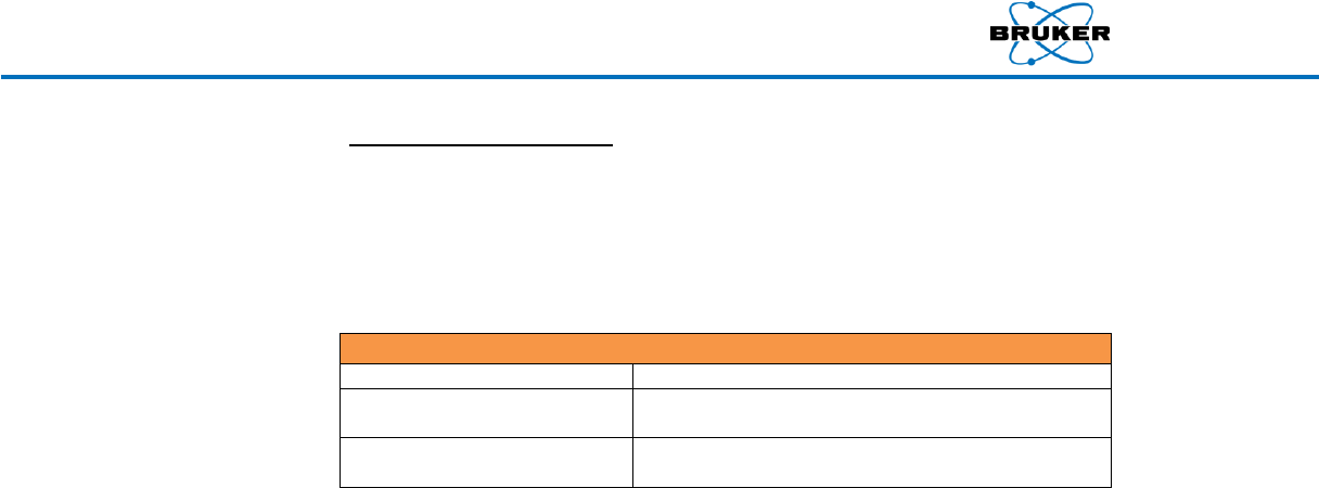
Radiation Safety Manual
030.0011.03.2 11 of 21
Average Estimated Days Lost
By Industrial Occupations
Average Lifetime Estimated Days Lost
Due to Daily Activities
Occupation*
Estimated
Days Lost
Activity*
Estimated
Days Lost
Mining/Quarrying
328
Cigarette smoking
2250
Construction
302
25% Overweight
1100
Agriculture
277
Accidents (all types)
435
Transportation/Utilities
164
Alcohol consumption (U.S. avg.)
365
5 rem radiation dose per yr for 30 years
150
Driving a motor vehicle
207
All industry
74
Medical X-rays (U.S. avg.)
6
Government
55
1 rem Occupational Exposure
1
Service
47
1 rem per year for 30 years
30
Manufacturing
43
* Note: based on US data only
Trade
30
The comparison of health and industrial risks illustrates the fact that no matter what you do
there is always some associated risk. For every risk there is some benefit, so you as the
worker must weigh these risks and determine if the risk is worth the benefit. Exposure to
ionizing radiation is a consequence of the regular use of many beneficial materials, services,
and products. By learning to respect and work safely around radiation, we can effectively
manage our exposure.
8.2. Radiation Dose Limits
To minimize risks from the potential biological effects of radiation, regulatory agencies and
authoritative bodies have established radiation dose limits for occupational workers. These
limits apply to those working under the provisions of a specific license or registration.
In general, the larger the area of the body that is exposed, the greater the biological effects
for a given dose. Extremities are less sensitive than internal organs because they do not
contain critical organs. That is why the annual dose limit for extremities is higher than for a
whole body exposure that irradiates the internal organs.
Your employer may have additional guidelines and set administrative control levels. Each
employee should be aware of such additional requirements to do their job safely and
efficiently. The limits described below have been developed based on information and
guidance from the International Commission on Radiological Protection (ICRP-1990), the
Biological Effects of Ionizing Radiation (BEIR) Committee, the US Environmental Protection
Agency (EPA), and the National Council of Radiation Protection (NCRP). For an XRF analyzer
using an X-ray tube as the source, any requirement on dose limits for the operators would be
established by the appropriate regulatory agency.
Annual Occupational Dose Limits
Exposed Area
International
U.S.
Whole Body
20 mSv*
5 rem
Extremities
500 mSv
50 rem
Organs or Tissue
(Excluding lens of the eye and skin)
500 mSv
50 rem
Lens of the Eye
150 mSv
15 rem
*Averaged over 5 years
Radiation Safety Manual
030.0011.03.2 12 of 21
8.2.1. Declared Pregnant Worker
A female radiation worker may inform her supervisor of her pregnancy, in
writing, at which time she becomes a Declared Pregnant Worker. The employer
should then provide the option of a mutually agreeable assignment of work
tasks, without loss of pay or promotional opportunity, such that further
radiation exposure will not exceed the dose limits as shown in the following
table for the declared pregnant worker.
Radiation Limits for Visitors, Public, and Pregnant Workers
International and US Limit
1 mSv (100 mrem) per year
Pregnant Worker
(International Limit)
2 mSv (200 mrem) to abdomen during remainder of
gestation period after declaration
Pregnant Worker (US Limit)
Declared Pregnant Worker (embryo / fetus) - 0.5 rem / 9
months ( 0.05 rem / month)
The radiation produced by the hand-held XRF analyzer from the primary beam
is low energy X-Rays (4 to 50 keV) in a narrow collimated beam. The radiation
exposure to the user is primarily from scattered radiation from the objects
being analyzed and a small amount of radiation that passes through the
housing. When correctly using the XRF Analyzer, its engineered safety features
ensure radiation exposure will be significantly less than the annual limits.
9. Measuring Radiation
Because we cannot detect radiation through our senses, special devices may be required in some
jurisdictions for personnel operating an XRF Analyzer to monitor and record the operator’s exposure.
These devices are commonly referred to as dosimeters, and the use of them for monitoring is called
dosimetry.
The following information may apply to personnel using hand-held XRF analyzers in jurisdictions where
dosimetry is required:
Wear an appropriate dosimeter that can record low energy photon (X-ray) radiation.
Dosimeters wear period of three months may be used, subject to local regulation.
Each dosimeter will be assigned to a particular person and is not to be used by anyone
else.
Whole body dosimetry should be worn on the upper portion of your body between the
neck and waist.
Extremity dosimeters should be worn on the fingers or wrist closest to the XRF analyzer
and is most importantly used on the hand not holding the analyzer.
Do not intentionally expose a dosimeter to the primary beam.
Do not expose you dosimeter to radiation outside of work (e.g., medical facilities and
dentist offices).
Do not put you dosimeter in checked or hand carried luggage when traveling through
airports.
If your dosimeter is damaged or lost, notify your supervisor and/or RSO.
Bruker recommends that local regulatory requirements in regards to occupational radiation
monitoring be determined, understood, and followed.

Radiation Safety Manual
030.0011.03.2 13 of 21
9.1. Dosimeters
While there is variation between dosimeters, and from one type to another, most dosimeters
operate in a similar way. Exposure to ionizing radiation is absorbed by a material contained
within the dosimeter and, when processed, provides a measured dose. Regulators require
that processing of dosimeters be performed by a company that is NVLAP accredited.
Monitoring radiation exposure with dosimeters provides an indication of the working habits
and working conditions of the XRF user and may be a way to identify whether the XRF
analyzer is being properly used. Remember: a dosimeter does not protect you against
radiation exposure; it is simply a passive device that measures the amount of radiation
exposure received where the dosimeter was worn. The figure below shows various types of
dosimeters available.
Whole Body Dosimeter
A Thermoluminescent Device (TLD) or Optically Simulated Luminescence (OSL) whole-body
dosimeter may be used to measure both shallow and deep penetrating radiation doses. It is
normally worn between the neck and waist. The measured dose recorded by this device may
be used as an individual's legal occupational exposure.
Finger Ring
A finger ring is a TLD in the shape of a ring, which is worn by workers to measure the radiation
exposure to the extremities. The measured dose recorded by this device may be used as the
worker's legal occupational extremity exposure.
Wrist Dosimeter
A wrist dosimeter is a whole-body type dosimeter that is designed to be worm on the wrist
similarly to a wrist watch. Processing of the dose takes into account where the dosimeter is
worn and measures the radiation exposure to the extremities. The measured dose recorded
by this device may be used as the worker's legal occupational extremity exposure.
9.2. Survey Meters
Some jurisdictions require the measurement of radiation emitted or scattered from handheld
analyzers by the use of a survey meter, which detects radiation in real time. Survey meters

Radiation Safety Manual
030.0011.03.2 14 of 21
generally consist of a detector and a read-out display. Commonly used survey meters are the
ionization chamber, Geiger-Mueller (GM) tube, and photomultiplier tube scintillation
detector. It is important to select a suitable instrument that is capable of monitoring the
type, energy, and intensity of ionizing radiation produced by the hand-held XRF analyzer. The
hand-held XRF analyzer produces low energy X-ray ionizing radiation. The energy of the X-rays
produced by the analyzer will be between 4 keV and 50 keV, with the average energy near 20
to 30 keV, and most of the X-ray energy being less than this due to effects of scattering which
significantly reduces the X-ray energy.
The GM tube instrument has the advantage of being economical and sensitive to low levels of
radiation. This instrument is good at identifying and isolating hot spots. However, an
instrument using a GM tube detector usually does not do well in providing accurate dose rate
measurements, unless specifically designed to do so. In such cases, a specially designed filter
is used to flatten out the energy response.
The ionization chamber is often a preferred instrument. The detector response is relatively
flat across its entire measurement range. The disadvantage is these instruments are often
more expensive and do not read out in the desired or useful measurement ranges. Their large
detector volumes can be challenging because the measurement results are usually affected
by geometry factors and the displayed dose rate is often much less than the actual field at
close distances.
The low energy plastic scintillator dose rate instrument uses a plastic detector that is nearly
tissue equivalent. This type of instrument has the advantage of being sensitive and providing
accurate results. One disadvantage is this type of instrument is typically more expensive than
the other instrument types.
If your jurisdiction requires the use of a survey meter, we recommend that you work with
your preferred instrument provider to identify a suitable survey meter for your application.
Survey instruments are used to provide information to assure that doses are kept ALARA and
to verify the integrity of the XRF Analyzer designed shielding has not been compromised. The
following characteristics should be used to assist in selecting an appropriate instrument.
Radiation type: X-rays
Energy range: 4 to 50 keV
Measurement threshold: 0.01 mem/hr to 200 mrem/hour
Accuracy: ± 10% of reading or better
Consult the meter’s user guide for proper calibration. Remember the instrument should be
calibrated to the type and energy of the radiation being monitored. The manufacturer may
require the use of correction factors to obtain accurate results for the energy of the X-rays
produced by the XRF analyzer.
We recommend that primary beam measurements never be attempted. Our safety
representatives will work with you to provide information about the primary beam should
you need more information than is provided in our manuals.
Radiation Safety Manual
030.0011.03.2 15 of 21
10. Exposure Reduction (ALARA)
While dose limits and administrative control levels already ensure very low radiation doses, it is
possible to reduce these exposures even more. The main goal of the ALARA program is to reduce
ionizing radiation doses to a level that is As Low As Reasonably Achievable (ALARA). ALARA is designed
to prevent unnecessary exposures to employees, the public, and to protect the environment. It is the
responsibility of all workers, managers, and safety personnel alike to ensure that radiation doses are
maintained ALARA.
There are three basic practices to maintain external radiation ALARA: Time, Distance, and Shielding.
Time
The first method of reducing exposure is to limit the amount of time spent in a radioactive
area. Generally, the shorter the time, the lesser the amount of exposure.
The effect of time on radiation could be stated as
Dose = Dose Rate x Time
Example: If 1 hour of time in an area results in 1 mSv (100 mrem) of
radiation, then 1/2 an hour results in 0.5 mSv (50 mrem), and 1/4 an
hour would result in 0.25 mSv (25 mrem), and so on.
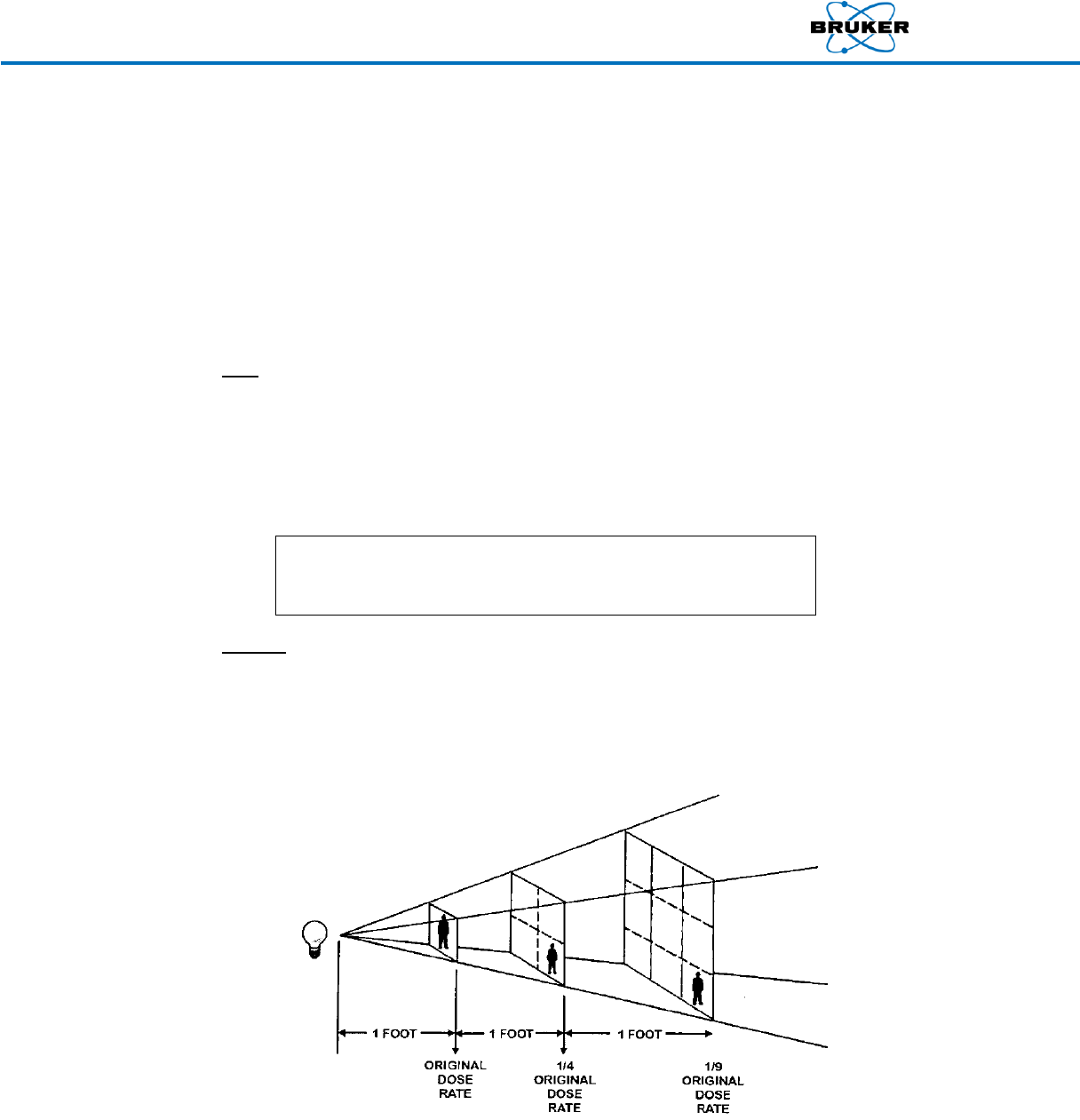
Distance
The second method for reducing exposure is by maintaining the maximum possible distance
from the radiation source to the operator or member of the public. The principle of distance
is that the exposure rate is reduced as the distance from the source is increased. The greater
the distance, the amount of radiation received is reduced. This method can best be
expressed by the Inverse Square Law.
Radiation Safety Manual
030.0011.03.2 16 of 21
The inverse square law states that doubling the distance from a point source reduces the dose
rate (intensity) to 1/4 of the original. Tripling the distance reduces the dose rate to 1/9 of its
original value. Expressed mathematically:
I
D
D
C2
2
2
1
Where:
C is the intensity (dose rate) of the radiation source
D1 is the distance at which C was measured
D2 is the distance from the source
I is the new level of intensity at distance D2 from the source
The inverse square law does not apply to sources of greater than a 10:1 (distance: source size)
ratio, or to the radiation fields produced from multiple sources.
Shielding
The third, and perhaps most important, method of reducing exposure is shielding. Shielding is
generally considered to be the most effective method of reducing radiation exposure, and
consists of using a material to absorb or scatter the radiation emitted from a source before it
reaches an individual.
As stated earlier, different materials are more effective against certain types of radiation than
others. The shielding ability of a material also depends on its density, or the weight of a
material per unit of volume.
Example: A cubic foot of lead is heavier than the same volume of
concrete, and so it would also be a better shield.
Although shielding may provide the best protection from radiation exposure, there are still
several precautions to keep in mind when using handheld XRF devices:
Persons outside the shadow cast by the shield are not necessarily 100% protected. Note:
All persons not directly involved in operating the XRF should be kept at least three feet
away.
A wall or partition may not be a safe shield for persons on the other side.
Scattered radiation may bounce around corners and reach nearby individuals, whether or
not they are directly in line with the test location.
WARNING: To avoid inadvertent exposure to others, the
operator should ensure that there is no one on the other
side of the wall or barrier when using an XRF analyzer.
Radiation Safety Manual
030.0011.03.2 17 of 21
11. Production of X-Ray Radiation
X-rays are produced by two separate processes:
Bremsstrahlung: continuous energy spectrum, process = acceleration of electron
Characteristic: single energy line, process = electron shell transition
Bremsstrahlung is the German term for "braking" and was originally used to describe the unknown
penetrating radiation (X-rays) released when high-speed electrons were stopped by sudden impact with a
metal target. X-ray tubes are designed to use this process to create X-rays.
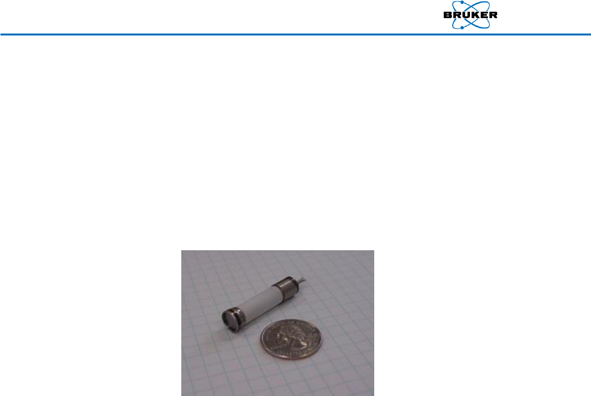
A modern miniature industrial X-ray tube consists of a ceramic container that is under vacuum. The
major components of a miniature X-Ray tube is the cathode and anode housed in a vacuum ceramic
tube. High voltage bias is applied between the anode (+) and the cathode (-). The picture below is
representative of the type of X-Ray tube used in the hand-held XRF analyzer.
A current passed through a miniature coiled tungsten filament raises the temperature of the wire to
approximately 1000°K, causing the thermionic emission of electrons. Typical currents of 5 to 100 µ
amps of current are used depending on the type of analysis being conducted.
The electrons emitted from the tungsten filament passes through the vacuum of the tube and are
accelerated as they are attracted to the positive charge of the anode. The large voltage potential of 40
to 50 keV transfers a large amount of energy to the electrons.
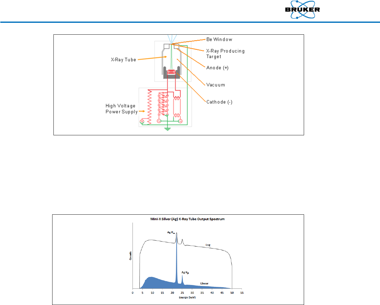
The electrons having gained a huge amount of kinetic energy impact the X-ray producing target. The
targets are usually made of Rhodium (Rh) or Silver (Ag) in the X-ray tubes used for handheld XRF
analyzers. The impaction of electrons on the target knocks a lower orbit electron from the target
atoms lower shell creating a void. A higher shell electron moves to fill the void, releasing its extra
energy in the form of an X-ray photon. This produces the X-rays required to conduct XRF analysis. The
figure below provides a diagram of a typical X-ray tube used in our application.
Radiation Safety Manual
030.0011.03.2 18 of 21
The current applied to the filament changes the intensity of the X-ray by changing the number of
electrons emitted from the miniature coiled tungsten filament. More current means more electrons.
The voltage controls the energy of the X-rays. More voltage means higher energy X-rays.
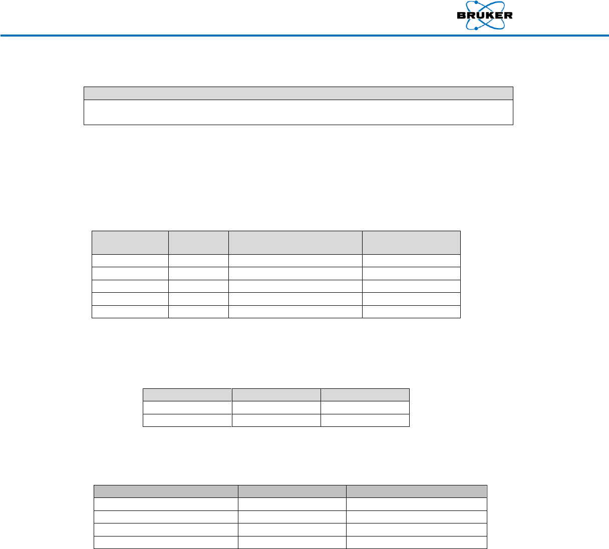
A typical hand-held XRF analyzer X-ray tube will emit x-rays from 8 keV to 50 keV with the maximum
intensity occurring at about one-half the maximum keV. The X-ray spectrum will be distributed across
a continuum of energies. The figure below provides an example of a typical X-ray spectrum.
X-Ray production voltage and current settings in the handheld XRF Analyzer has been programmed to
produce the energy and intensity required to obtain the best analysis results for a particular
application. The user does not make any voltage or current adjustments in operating the XRF analyzer.
Radiation Safety Manual
030.0011.03.2 19 of 21
12. Radiation Exposure Potential
Note
The following section uses the S1 TITAN as an example. The dose rate is typical of handheld XRF
instruments. For specific information on a particular instrument, see the instrument’s User Guide.
The potential for exposure to ionizing radiation is primarily from scattered radiation from the objects
being analyzed and small amounts of radiation from the analyzer’s housing. The potential exists for
exposure to the primary beam if the XRF analyzer is not operated properly, with the greatest potential
being to the operator’s fingers and hands. Table 1 provides the exposure potential of the S1 TITAN
when operated at its maximum voltage setting.
Table 1
Distance (cm)
Rad/hr
Deep Dose
(Rem/hr)
Shallow Dose
(Rem/hr)
Beam Port
4610
3642
5071
5 cm
398
314
438
10 cm
126
99.5
139
30 cm
18
14.2
19.8
100 cm
1.01
0.80
1.11
Table 2 lists the amount of time that it would take to reach the annual limits if the S1 TITAN is
operated with the beam port against a body part at its maximum power setting. The time to
reach these annual limits is quite short.
Table 2
Organ
Eye
Skin
Annual Limit
15 Rem
50 Rem
Time (minutes)
0.17
0.59
Table 3 lists the biological effects and typical dose and time to see the effects from exposure to
the primary beam.
Table 3
Skin Effect
Dose (Rad)
Time (minutes)
Erythema (Redding of Skin)
300
3.9
Dry Desquamation
1000
13.0
Wet Desquamation / Blistering
1500
19.5
Ulceration and Necrosis
3000
39.0
The effects of ionizing radiation exposure to the skin may only appear days or weeks after the
exposure. This is because the radiation damages the developing cells below the skin surface. The
damage cannot be observed until the top layers of the skin slough off.
Erythema is redness of the skin, caused by hyperemia of the capillaries in the lower
layers of the skin.
Dry Desquamation also called skin peeling is the shedding of the outermost membrane
or layer of a skin.

Radiation Safety Manual
030.0011.03.2 20 of 21
Wet desquamation is where the skin thins and then begins to weep because of loss of
integrity of the epithelial barrier and decreased oncotic pressure.
There have been no documented radiation related injuries from hand-held XRF analyzer operations.
The potential exists for injury to the fingers or hands if safety features are disabled and/or proper
operating instructions are not followed. There have been documented injuries which have occurred
from not following proper operating procedures and bypassing safety features on cabinet analytical
systems.
It is important to note that although the annual limits could be reached fairly easily through accidental
exposures through improper use of the XRF analyzer, the skin effects caused by exposures would take
significant more time than might occur through a momentary lapse of proper operation.
13. Rights and Responsibilities
Individuals who work with ionizing radiation producing equipment, such as the handheld XRF analyzer,
have certain rights and responsibilities. These rights and responsibilities are usually specifically defined
by the jurisdiction that the XRF analyzer is registered under.
If you are required to wear a dosimeter, you have the right to be informed of the occupational
exposure you receive. Typically, your employer is required to provide a report of your previous year’s
exposure during the 1st quarter of the next year
Users of handheld XRF analyzers have the responsibility to obey warning labels and follow the
operator training. You are required to follow the rules and regulations governing the operation of a
portable XRF. This is for your protection and the protection of co-workers, clients, and the public.
Typically, you are required to report concerns and violations to your supervisor and regulatory
authorities
Most jurisdictions require that individuals working under their rules and regulations never deliberately
cause a violation of any related rule or regulation. Deliberate violations usually are subject to
enforcement action. Mistakes and unplanned action usually do not apply.
Prior to using the handheld XRF analyzer, carefully read the instrument’s user guide.
As the operator of the handheld XRF analyzer, you are responsible for your safety and the safety of
others. The following are important responsibilities:
Before pulling the trigger, be aware of the direction that the X-rays travel.
Do not place any part of your body (especially the eyes or hands) near the examination area
during measurement.
Do not hold a sample to the window for analysis by hand. Instead, hold the window to the
sample. The infrared (IR) sensor located on the nose is designed to prevent the emission of
X-rays in the absence of an object.
Do not defeat the IR sensor in order to bypass the safety circuit. Defeating this safety
feature could result in unnecessary exposure of the operator. Occasionally, a sample may
not be reflective enough to trigger the IR sensor. In these cases, place a piece of white
paper or other reflective material between the sample and IR sensor.
Use the optional safety shield or benchtop stand accessory for testing small or thin samples
or low-density materials, such as plastic, wood, soil, paper, or ceramics.

Radiation Safety Manual
030.0011.03.2 21 of 21
Wear an appropriate dosimeter if required by a regulatory agency when operating the
analyzer.
Pregnant women should be aware that improper handling or improper use of the
instrument could result in radiation exposure.
The operator is responsible for the security of the analyzer. When in use, the device should
be in the operator's possession at all times.
Do not allow anyone other than trained and certified personnel to operate the analyzer.
Always store the instrument in a secure location when not in use.
If you suspect the analyzer is damaged, remove the battery pack and disconnect all power
sources.
14. Backscatter with Low Density Samples
Be aware that when using a handheld XRF analyzer, some radiation is scattered back towards the
operator. The amount of scatter is dependent on the density of the sample – with low density
samples, such as plastics, scattering more than high density samples, such as metals – and the shape of
the sample – with flat surfaces containing more of the backscatter and curved and irregular surfaces
containing less of the backscatter. The operator must keep hands and eyes away from the analyzer
nosepiece.
Further, it should be noted that low-density (LD) materials, such as plastic, wood, soil, paper, or
ceramic, will not attenuate higher energy X-rays as efficiently as high-density materials, such as metal
alloys. Thus a greater amount of the radiation is transmitted through the sample, which can cause a
higher dose rate to the operator. The operator should keep hands and eyes away from the analyzer
nosepiece. If LD samples are measured frequently, the use of a bench-top stand is recommended to
minimize scattered radiation. If the LD samples are small enough, the Small Sample Safety Shield is
adequate.
