Philips Click For EPIQ Brochure 452296295931 EPIQ7 WHC
User Manual: Philips Click for EPIQ brochure ISUOG 2017
Open the PDF directly: View PDF ![]() .
.
Page Count: 20
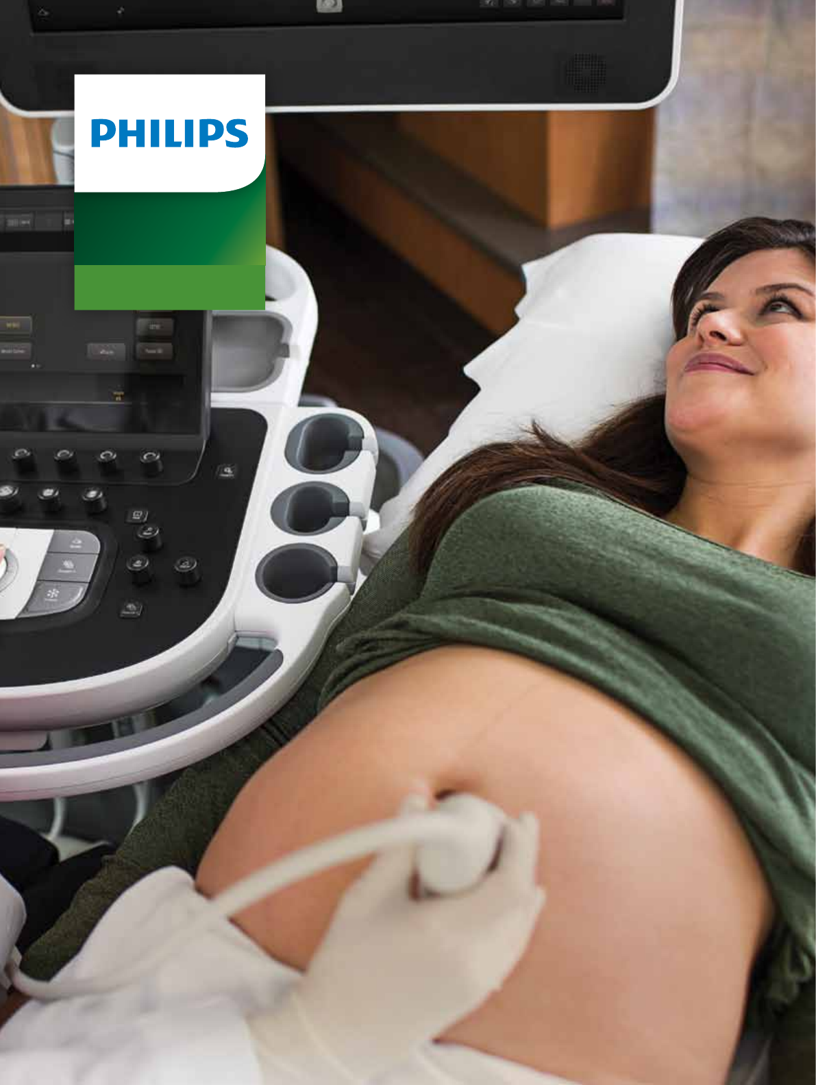
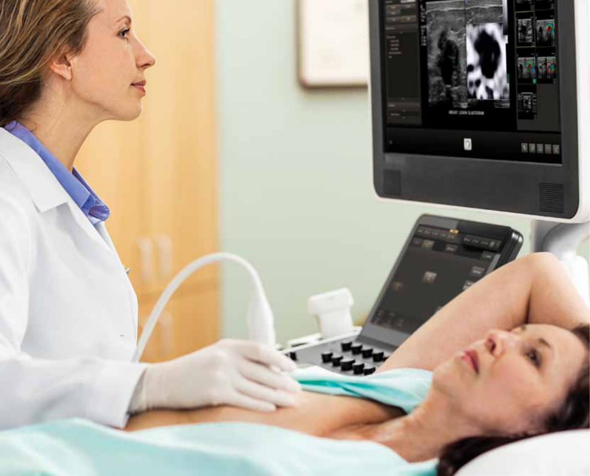
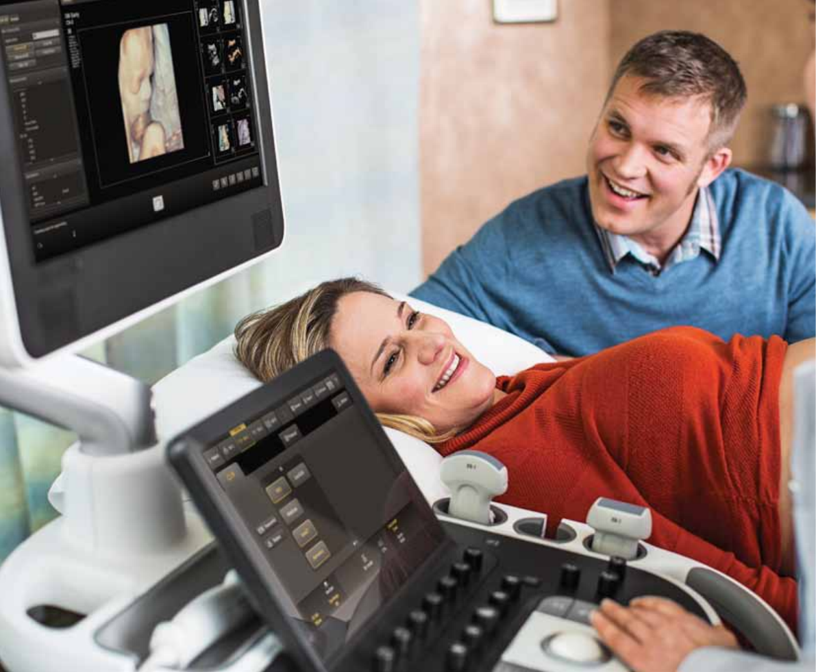
Philips EPIQ 7 ultrasound system for women’s health care
The evolution
of premium ultrasound
EPIQ 7
Ultrasound

2
To help ease the unprecedented strain on hospitals
and healthcare systems, premium ultrasound must
continue to deliver – improved quality, higher accuracy,
and faster and more consistent exams that lead to more
condent diagnoses the rst time, even for technically
dicult patients.
The new challenges
in global health care
3
Key trends in global ultrasound
for women’s health care
It’s our most powerful architecture ever applied
to ultrasound imaging – touching all aspects of
acoustic acquisition and processing, allowing you
to truly experience ultrasound’s evolution to
a more denitive modality.
Supported by our family of proprietary xMATRIX
transducers and solutions for technically dicult
patients for every exam type, this platform oers
our highest level of premium performance.
The evolution of premium
ultrasound for women’s health care
•The need for more denitive image
quality and advanced tools for
all gestational ages and complex
gynecological cases
•More pregnancies in patients
with high BMIs
•The need to improve exam success on
these technically challenging patients
•Higher referral rates with more
complex cases, requiring improvement
in workow eciency
•The need to automate many system
functions to assure ease of use and
consistency of exams between users
•The need for exceptional 3D surface
rendering performance to better
diagnose anomalies
44
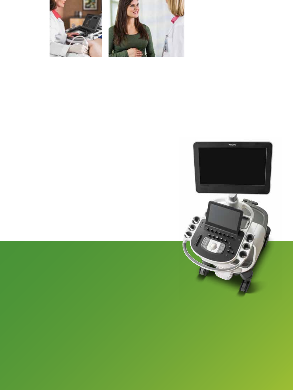
EPIQ 7 is the new direction for premium ultrasound, featuring an
exceptional level of clinical performance to meet the challenges of
today’s most demanding practices and technically dicult-to-image
patients through every gestational age and for gynecology applications.
Performance
More confidence in your diagnosis
even for your most difficult cases
5
Image quality: the numbers tell the story
Comparing EPIQ 7 to conventional premium systems
shows breakthrough advances in imaging performance.*
•Up to 76% increase in penetration
(penetration = ability to scan at depths and maintain
resolution in order to complete the study)
•Up to 213% increase in temporal resolution
(ability to maintain resolution at high frame rates)
Philips nSIGHT Imaging
is a totally new approach
The Philips proprietary nSIGHT Imaging
architecture introduces a totally new
approach to forming ultrasound images.
Unlike conventional systems that form
the image line by line, nSIGHT creates
images with superb resolution down
to the pixel level.
Extraordinary architecture
nSIGHT Imaging incorporates a custom
multi-stage precision beamformer
along with massive parallel processing.
This proprietary architecture captures
an enormous amount of acoustic data
from each transmit operation and
performs digital beam reconstruction
along with mathematically optimized
focal processing to create real-time
images with exceptional resolution
and uniformity.
Creating new realities,
redefining clinical expectations
nSIGHT Imaging goes beyond conventional ultrasound
performance for new levels of denition and clarity.
Conventional
Users must
choose
between
frame rate
and image
quality
nSIGHT
Imaging
More than
doubles the
frame rate
without impact
to image
quality
nSIGHT Imaging
creates superbly focused
images with fewer transmit
operations so you can
experience both highly
detailed ultrasound
images and extraordinary
temporal resolution.
Frame rate Penetration
Conventional
Best resolution
is limited
to transmit
focal zone
nSIGHT
Imaging
Corrects focus
during beam
reconstruction
for superb
uniformity
nSIGHT Imaging
achieves superb uniformity
through coherent beam
reconstruction algorithms
that apply mathematical
focal correction coecients
continually at all depths
of the image.
Conventional
Penetration
limitations
and poor
sensitivity to
weak signals
nSIGHT
Imaging
Superb
penetration
across full
range of
frequencies
nSIGHT Imaging
architecture’s ultra-wide
dynamic range and unique
beam reconstruction
reinforces weak tissue
signals allowing enhanced
penetration at higher
frequencies even on
dicult patients.
Conventional
Penetration icons
option 4
option 5
option 6
Conventional
Penetration icons
option 4
option 5
option 6
Uniformity
* 2013 quantitative engineering study comparing Philips iU22
ultrasound system with EPIQ 7.
6
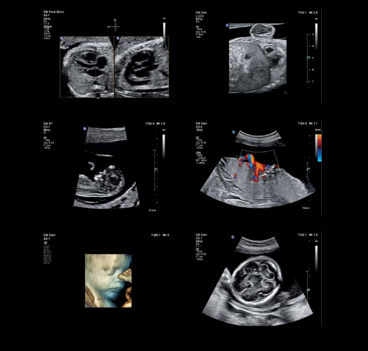
Only with EPIQ: Gather fetal heart
volumes in a two-second acquisition.
xMATRIX is our most leading-edge, versatile
ultrasound transducer technology
No other premium ultrasound system can run the complete
suite of the world’s most innovative ultrasound transducers.
With the touch of a button xMATRIX oers all modes in
a single transducer: 2D, 3D/4D, Live xPlane, Live MPR,
MPR, Doppler, color Doppler, and CPA.
nSIGHT Imaging makes powerful xMATRIX
technology even more so
Achieve ultra-thin 2D slices. Use Live xPlane imaging to create
two full-resolution planes simultaneously, allowing you to
capture twice as much clinical information in the same amount
of time. Acquire near isovoxel resolution to reveal images
from any plane within the volume. Export 3D MPRs in the
X, Y, and Z plane to any PACS system with MPR DICOM Export.
Present superb, real-time 4D volume data in obstetrical exams.
Gather a volume of the fetal heart in as little as a two-second
acquisition compared with the 12-second acquisition time
of conventional volume imaging. Now it’s all possible.
Philips pioneered advanced technologies such as xMATRIX and PureWave.
The revolutionary nSIGHT architecture of EPIQ 7 makes xMATRIX and
PureWave even more powerful.
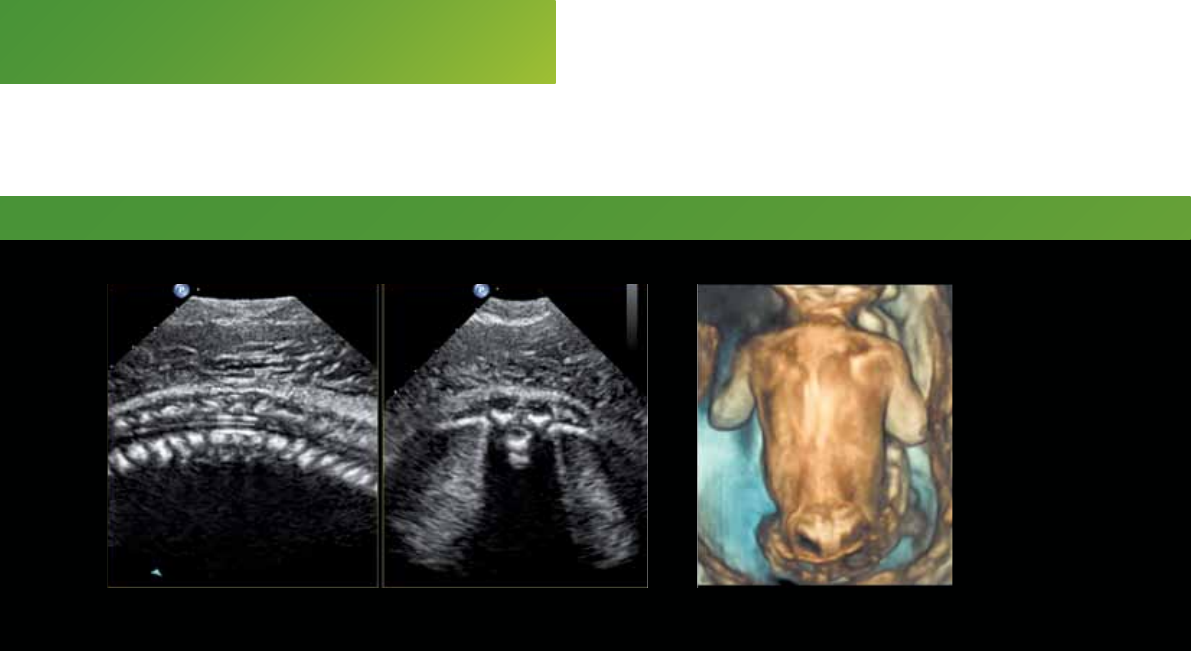
Fetal spine – Live xPlane Spina bifida
Maximize
extreme clinical capabilities
7
26-week gestation 24-week gestation
Bring your most challenging
cases to EPIQ 7 with our
PureWave solutions from
gynecological surveys to the
rst trimester through third
trimester exam.
•C9-2 transducer designed for high-frequency
OB imaging, especially in the rst, second,
and even into the third trimester
•C10-3v transducer ideal for challenging broid
and complex ovarian cases, as well as rst
trimester imaging
•C5-1 transducer suited for the largest abdomens
all the way through the third trimester,
patients with gestational diabetes, or premature
rupture of membranes
•X6-1 xMATRIX transducer excels at diagnostic
requirements that go beyond 2D imaging, bringing
PureWave to a new level that includes live
volume imaging and live imaging in two
planes simultaneously
Greatly enhance the power of the X6-1
transducer for OB and GYN applications
We’re delivering the advances you’ve been asking for,
such as signicant enhancements in X6-1 2D image
quality at shallow depths. You can now implement elevation
compounding on the X6-1 with no frame rate penalty for
enhanced speckle reduction and contrast resolution at all
depths. See dramatic improvements in volume rates across
all 3D/4D modes and applications. Users can now perform
real-time 4D imaging of the fetal heart with the X6-1 with
image quality once reserved for 2D images.
EPIQ 7 nSIGHT
architecture enhances
both the penetration
and image quality of
PureWave transducers.
nSIGHT enhances PureWave imaging for exquisite detail resolution.
88
With a complete family of PureWave transducers, your most dicult diagnoses are now easier.
PureWave crystal technology represents the biggest breakthrough in piezoelectric transducer
material in 40 years. The pure, uniform crystals of PureWave are 85% more ecient than
conventional piezoelectric material, resulting in exceptional performance. This technology
allows for enhanced penetration in dicult patients and for excellent detailed resolution.
PureWave
(x800)
Conventional
(x800)
PureWave crystals have virtually
perfect uniformity for greater
bandwidth and twice the efficiency
of conventional ceramic materials.
The result is excellent imaging and
Doppler performance.
The power of PureWave
to image technically
difficult patients
9
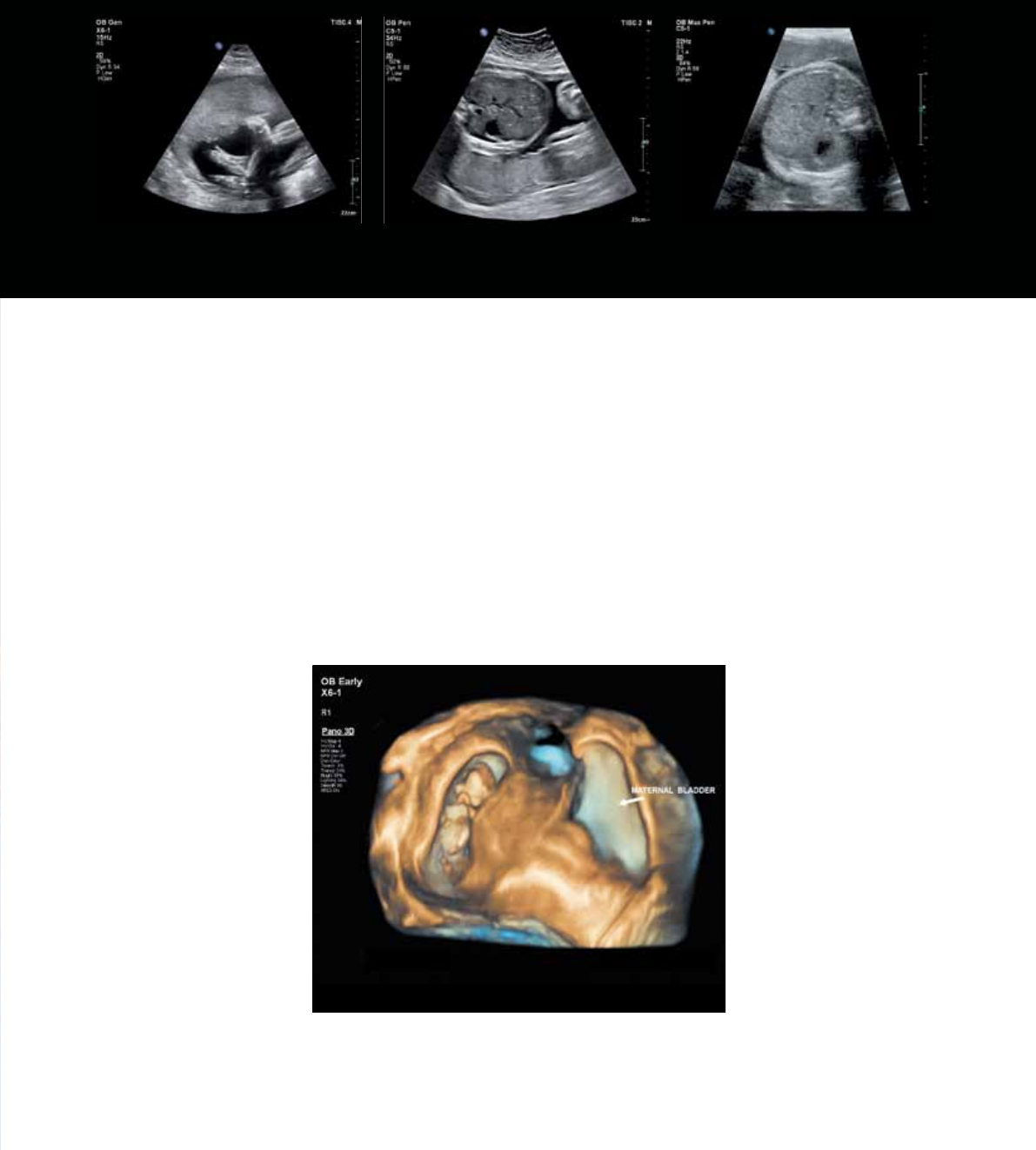
22-week fetus, 3D panoramic image
Exclusive panoramic volume imaging with xMATRIX
Panoramic volume imaging uses Live xPlane imaging
to acquire a calibrated volume over an extended eld of
view. Easily capture, visualize, and quantify 3D panoramic
volumes. For the rst time, you can capture an entire third-
trimester fetus or an entire uterus in one 3D panoramic
volume. Now have exceptional demonstration of the spatial
relationships between structures when a single volume is not
enough to capture the entire Region of Interest, get a global
perspective of the examination area to easily and quickly
identify target structures.
Fetal foot
25-week gestation, BMI = 40
Fetal abdomen, technically
difficult patient, BMI = 80
Fetal abdomen
30-week gestation, BMI = 40.1
10
EPIQ 7 is uniquely designed to support both strain and shear wave
methods of elastography. Highly sensitive strain imaging requires no
external compression and can be used to assess relative tissue stiness
across a variety of applications. Shear wave elastography utilizes unique
pulsing schemes to generate and measure the propagation speed
of shear waves through tissue. This technique produces an absolute
measure of tissue stiness that is helpful in assessing diseases.
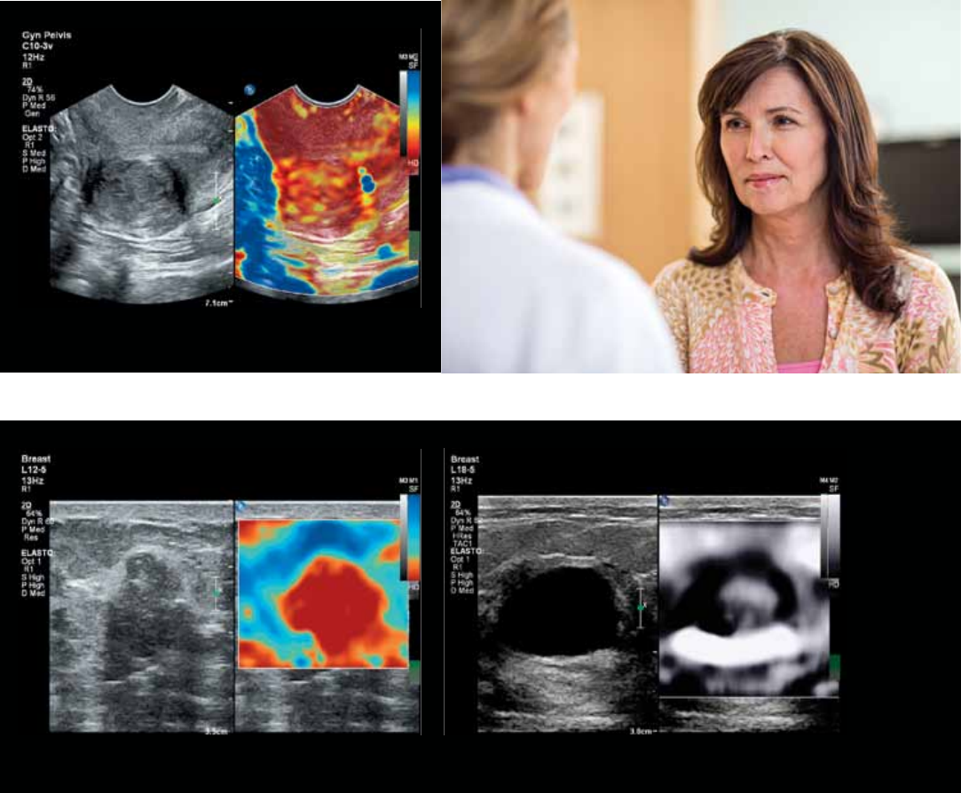
Uniquely designed for elastography –
revealing more denitive information on tissue stiness
11
Breast lesion elastogram
Uterine fibroid
Studies have shown that a combination of sonography and ultrasound elastography,
a technique that enables evaluation of relative tissue stiness, could potentially
reduce unnecessary biopsies.1 EPIQ 7 oers the most sensitive strain elastography
solution in the market for both breast and gynecological applications. No additional
compression required means increased exam consistency and reproducibility.
Breast cyst elastogram/bullseye artifact
Signicant addition to the power of elastography
1 Ferraioli G, et al. Point shear wave elastography method for assessing liver stiffness. World J Gastroenterol 2014 April 28;20(16):4787-4796.
12
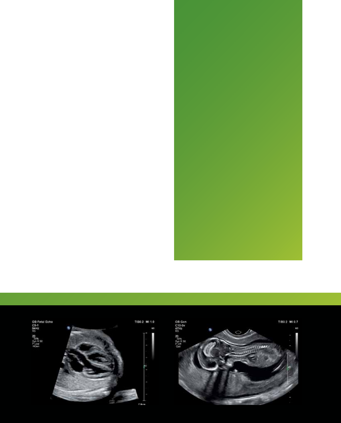
Fetal heart, Live xPlane Fetal diaphragm
Placental cord insertionNuchal translucency
CerebellumFetal face
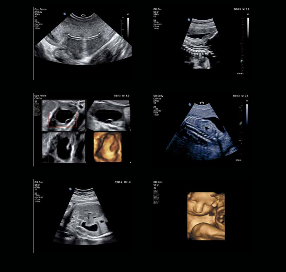
Exceptional images
for a new era
13
Intrauterine device Fetal spine
13-week fetal kidneyOvarian cyst
34-week gestationFetal abdomen
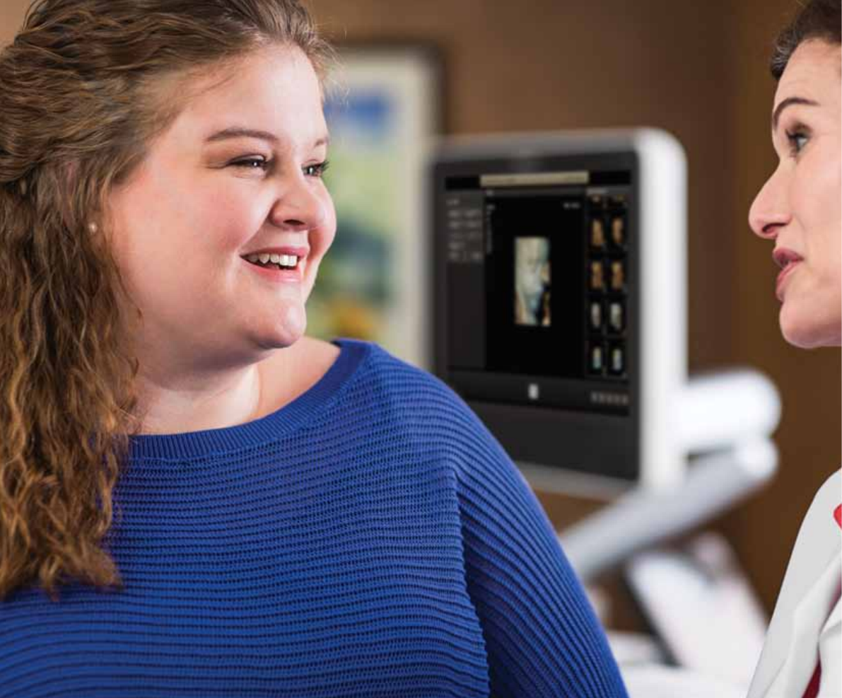
Automation supports
the way you work
This powerful architecture also supports automation designed to aid
your workow and increase your condence in the most challenging
exams, such as rst trimester or fetal heart.
14
15
SmartExam
SmartExam decreases exam time by
30-50%, keystrokes by as many as
300 per exam, and results in a high
level of consistency among users.2
It is fast and easy to customize,
providing consistent annotation,
automatic mode switching, and
missed view alerts to streamline exams.
The result is more time to focus on
your patients, increased condence
in complete studies, less focus
on requirements, less repetitive
motion, less stress, and improved
schedule maintenance and
department eciencies.
Efficient fetal scanning
Ability to create protocols for all
trimesters and specialty exams
such as trisomy 13 and 21.
Real Time iSCAN
Automatically optimizes gain and
TGC to continuously provide an
optimal image in 2D, 3D, or 4D.
2 University of Colorado, Protocols Study, Apr. 2007.
•Solutionsfortechnicallydicult-to-
image patients for every gestational age
and for gynecological exams
•Imagetheentirefetusinone3D
panoramic calibrated volume
•Mostpowerfulsystemwithout
compromise available today among
leading ultrasound manufacturers
16
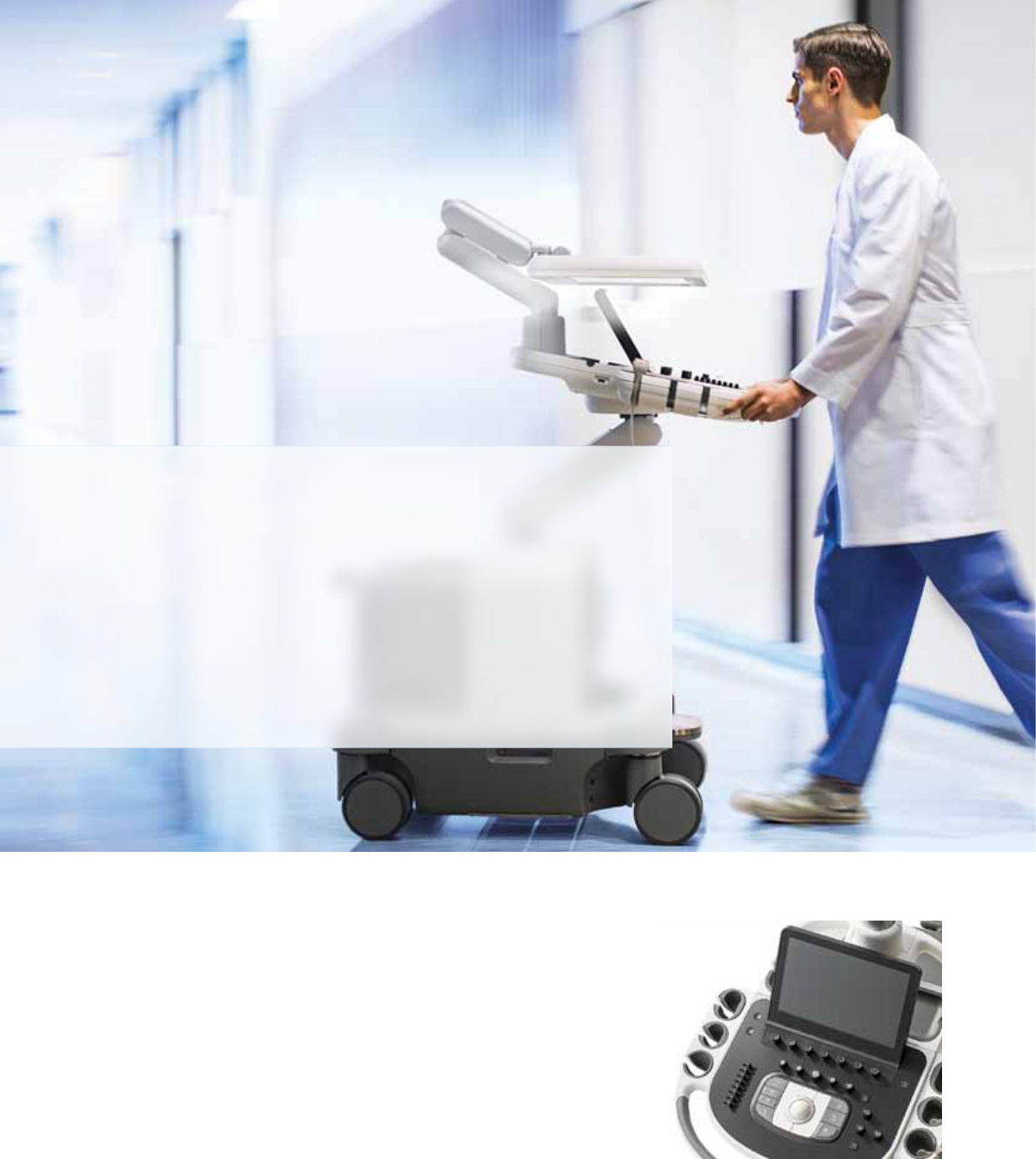
Designedtoreinvent
the user experience
EPIQ 7 has completely reinvented the premium
ultrasound user experience. Ease of use, workow,
ergonomics, portability – we’ve revolutionized how
you interact with an ultrasound system from every
standpoint, and kept it beautifully intuitive.
More than 80% of sonographers experience work-related
pain, and more than 20% of these suer a career-ending
injury.3 The EPIQ 7 tablet-like interface results in dramatic
reduction in reach and button pushes.
Tablet-like touch interface allows
quick navigation to system functions.
Advanced workflow
The design of the platform features
“walk-up usability,” meaning that
users can perform an exam with
minimal training.4 The system oers
the automation to drive eciency
throughout exams with features such
as Real Time iSCAN (AutoSCAN),
which automatically enhances gain
and TGC continuously to provide
excellent images in 2D, 3D, or 4D.
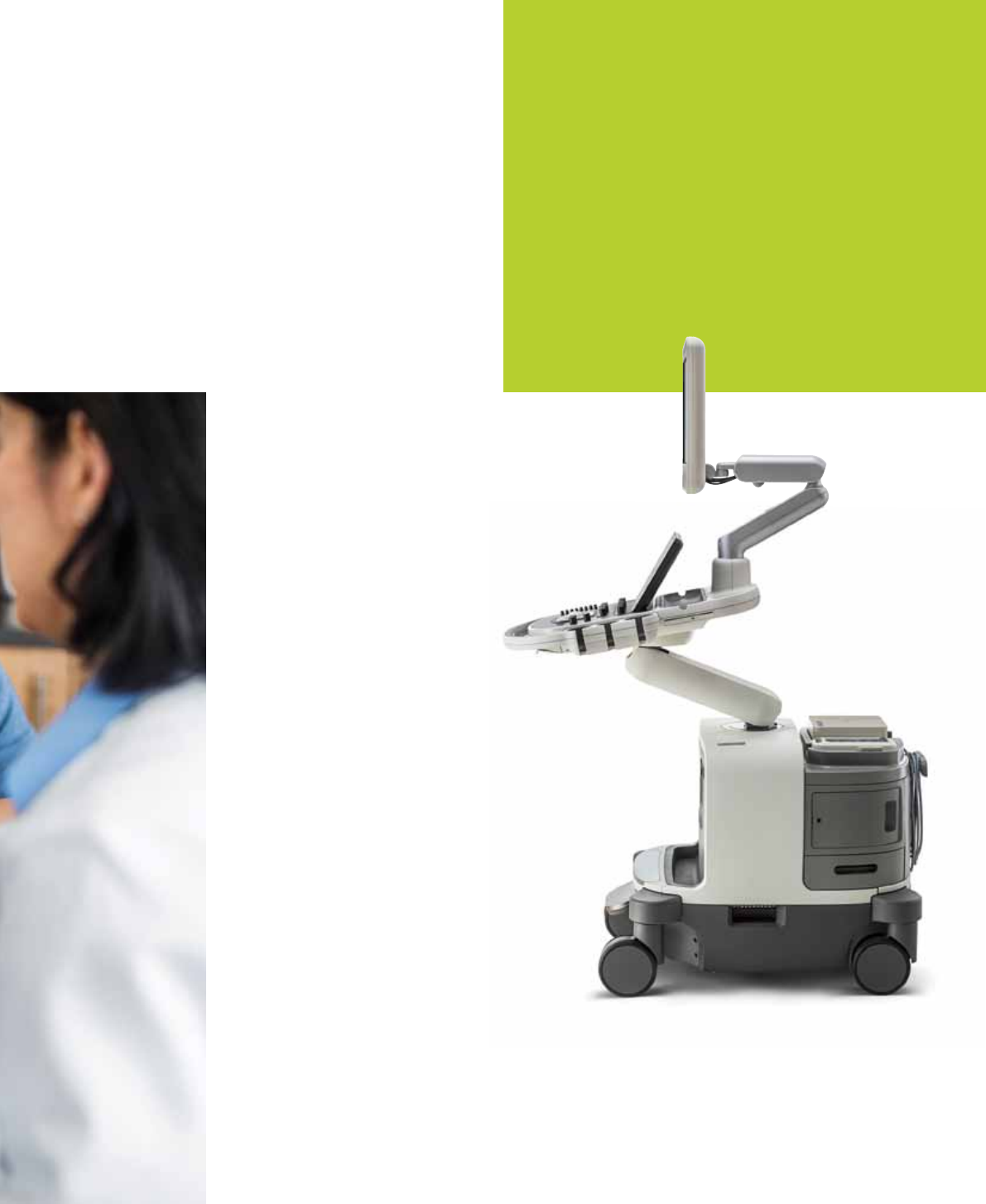
Amazing fit to your environment
At just 104 kg (230 lb), EPIQ 7 is lightest
in its class and 40% lighter than the
heaviest competitive premium system.
Place it in sleep mode, and boot up in
seconds. Exceeds Society of Diagnostic
Medical Sonography ergonomics for
maneuverability by 76% to easily t into
tight spaces. Wireless DICOM further
aids workow.*
17
3 Society of Diagnostic Medical Sonography, Industry Standards for the Prevention
of Musculoskeletal Disorders in Sonography, May 2003.
4 External user study where all users had over 90% success (gold standard in usability)
on their set tasks with no training on EPIQ, Jan 2013.
* Check for availability in your geography.
† 2013 engineering study comparing Philips iU22 ultrasound system with EPIQ.
Library quiet
EPIQ 7 is almost silent when running.
A noise test determined that EPIQ 7
runs at 37-41 dB, which is equivalent
to the sound of a library.
Scanning comfort
Multiple degrees of articulation for both
the control panel and 54.6 cm (21.5 in)
LCD monitor with 720° of freedom allows
for ergonomic alignment, whether sitting,
or standing for scanning comfort.
Easy viewing and ecient use even
in darker scanning environments with
a large 54.6 cm (21.5 in) wide screen
and ambient lighting that provides
subtle visual cues for the keyboard,
OEMS, and transducer ports.
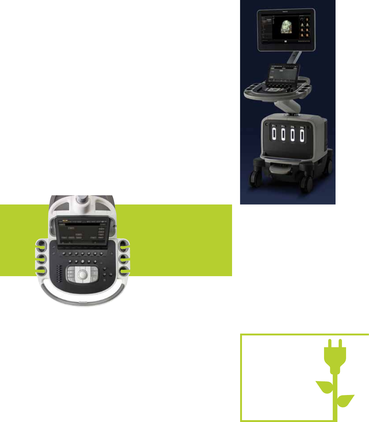
EPIQ 7 is one of the greenest
systems we have ever designed.
It consumes 25% less power than
our legacy premium ultrasound.
25%
less power
EPIQ 7 makes it easy
to be green
Efficiency is built in
Integrated eciency tools address
the expanding demand for greater
throughput and exam consistency.
Active native data
Active native data allows for post-
processing of many exam parameters.
A tablet-like touch interface
allows quick navigation to system
functions and results in dramatic
reduction in reach and button
pushes, with 40% to 80% less
reach and 15% fewer steps†.
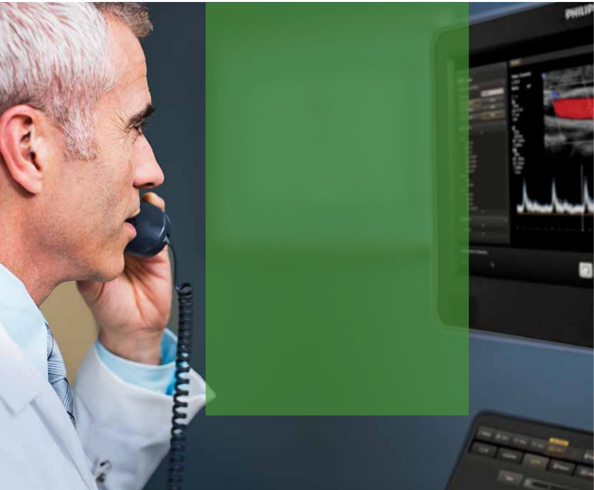
Remote services mean
we’re closer than ever*
Remote desktop
Spend less time on the phone with a Philips “Virtual Visit”
with remote system interaction for fast technical and clinical
troubleshooting and guided scanning options.
iSSL technology
This industry-standard protocol meets global privacy standards
and provides a safe and secure connection to the Philips remote
services network using your existing Internet access point.
Online support request
Enter a support request directly from your EPIQ system for
a fast, convenient communication mechanism that reduces
workflow interruption and keeps you at the system and
focused on your patient.
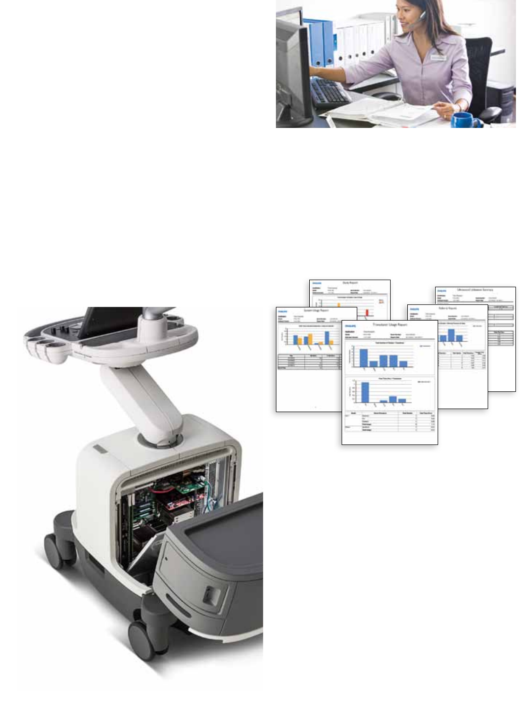
Utilization reports
Data intelligence tools that can help you make informed
decisions to improve workflow, deliver quality patient care,
and decrease the total cost of ownership. This is the only
ultrasound utilization tool that provides individual transducer
usage and the ability to sort by exam type.
Proactive monitoring
Proactive monitoring allows for the detection and repair
of anomalies before they become problems and helps us to
better predict potential failures and proactively act on them.
Increase system availability, optimize workflow, and promote
patient satisfaction by scheduling downtime as opposed
to reacting to an unexpected problem.
18
Advanced support services
are proactive and predictive
We understand your challenges: uncertain economic times, changing
healthcare landscapes, and the impact of healthcare reform. We know
that ecient workows and system uptime are critical success factors
in running an eective healthcare business.
Philips is committed to oering innovative solutions to provide you
with world-class services that move from reactive to proactive and
with predictive service models that provide high system availability
and enhanced workow to help you deliver high-quality patient care.
*Check for availability in your geography.
19
The remote desktop allows Philips
service engineers to gain a live
view of your system’s console for
remote operation, real-time clinical
troubleshooting, and issue resolution.
Exceptional serviceability
Philips oers the only ultrasound utilization
tool that provides individual transducer
usage and the ability to sort by exam type.
The system features superior modular
design for rapid repair, getting your
system up and running quickly.
Intelligent software architecture
Software is easily optimized, maintained, and restored
by the service user without risk to patient data, giving you
peace of mind when dealing with software anomalies and
confidence that your data is safe.
This software architecture takes patient data privacy
to a new level. Patient data is stored on a separate partition
and physical location to provide protection and ease of
removal, providing you total control of your data.
Clinical education solutions
Our comprehensive, clinically relevant courses, programs,
and learning paths are designed to help you improve
operational efficiency and enhance patient care.

© 2015 Koninklijke Philips N.V. All rights are reserved.
Philips Healthcare reserves the right to make changes in specifications
and/or to discontinue any product at any time without notice or
obligation and will not be liable for any consequences resulting from
the use of this publication.
Please visit www.philips.com/EPIQ
Printed in The Netherlands.
4522 991 09261 * APR 2015
