Philips 722024CA Object Moved User Manual Product Brochure Allura Xper X Ray System FD10 10 Fdee1fe9885c4a7d89f6a77c01514f63
User Manual: Philips 722024CA Product Brochure Philips Allura Xper X-ray system FD10-10 Philips - Allura Xper FD10-10 Biplane X-ray system722024CA
Open the PDF directly: View PDF ![]() .
.
Page Count: 1
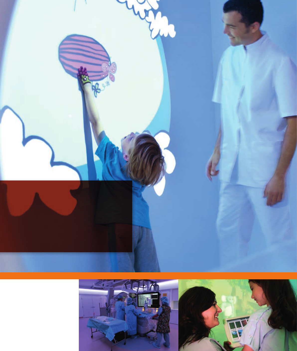
Making the difference with
Philips Live Image Guidance
in treating congenital heart disease
Key bene ts
• Confi dently reduce X-ray dose by 50% without
changing your way of working
• Provide more effective, reproducible treatment with
Philips Live Image Guidance
• Enhance precision, predictability, and confi dence with
advanced pediatric specifi c tools
452299102481.indd 2 19/06/14 11:16

Making the difference with
Philips Live Image Guidance
Asapediatriccardiologist,yourlifeiscommittedto
bringingnewhopetochildren.Asyoustruggleto
understandthecomplexitiesofacongenitalheart
disorder,youneedpediatrictoolsthathelpyouwork
preciselyandeffectively,togiveyourkidsabetterchance
ofalongandhealthylife.
Togetherwemakethedifferenceinthetreatmentof
congenitalheartdiseasetoimprovepatientoutcomes
andsavelives.WithourLiveImageGuidanceweaim
toremovebarrierstosafer,effectiveandreproducible
treatments,deliveringrelevantclinicalvaluewhereit’s
neededmost-atthepointofpatienttreatment.
Intuitive,pediatricspecictools,integratedmulti-
modalityimaging,andpatientinformationarecombined
inaninterventionallaborsurgicalsuite.Withaccess
toenhancedlivevisualizationandnavigationyoucan
condentlydeterminetheoptimalcourseoftreatment
withgreaterprecisionandpredictabilitytohelpimprove
patientoutcomes.WithAlluraClaritywithClarityIQ
youcancondentlyreduceX-raydoseby50%without
changingyourwayofworking,whileotherLiveImage
Guidancetoolshelpminimizecontrast-induced
complicationstopediatricpatients.
Crisp visualizations and fewer contrast-induced
complications
Atthesametime,youcannotsacricecriticalimage
qualityoranatomicinformationthatwouldaffectthe
optimalcourseoftreatment.OurX-rayimages,3D
andmultimodalityLiveImageGuidancedeliverscrisp
visualizationsofthedelicatevasculatureofbabiesand
youngchildren.Italsoprovidesliveimagenavigation
throughsofttissueanatomy,whilereducingtheneed
forcontrastagent.Combined,thesepediatricspecic
toolsofferenhancedprecision,predictability,
andcondence.
Together,weopendoorstonewproceduresand
techniquesthattrulymakeadifferencetothelivesof
childrenandtheirfamilies.
Contents
Arelentlessdrivetowardsmoreeffectiveandsafertechnologiesforchildren 4
Lowerbarriersforminimallyinvasiveinterventions 6
X-ray dose reduction and management solutions lower exposure and extend procedural options
Greaterinsightandcondenceinndingandtreatingtheproblem 10
Advanced interventional tools deliver clinical value where it’s needed most: at the point of patient treatment
Betteruserexperiencetopromoteconsistencyandefciency 16
Unique Ambient Experience concepts reduce anxiety for children and improve clinical workow
Increasedeconomicvalue 22
Service support and education provide a strong return on investment and enhance operational performance
Making the difference with Philips Live Image Guidance | in treating congenital heart disease 3

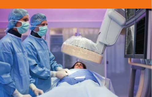
A relentless drive towards
more effective and safer
technologies for children
Congenitalheartproceduresareperformedonarelatively
smallpatientpopulation,buttheycanhaveahugeimpact
onthelivesofchildrenandtheirfamilies.Asamedical
practitionerinthisarea,youdevotemanyyearstotraining
anddevelopingyourskills.AtPhilips,wehavealsodevoted
manydecadesdevelopinginnovationsthatwillmakea
meaningfuldifferenceforcongenitalcardiologyprocedures.
Ourtechnologiesaredesignedtoprovidechildrenwith
effective,age-appropriatecarethathelpsprotectthem
fromunnecessarycontrastmediumanddose.
AlluraClarity
TheAlluraClarityfamilyofX-raysystemswithClarityIQ
technologysetsanewstandardbypushingthe
boundariesofALARA(AsLowAsReasonably
Achievable)imaging.WithAlluraClaritywithClarityIQ
youcancondentlyreduceX-raydoseby50%without
changingyourwayofworking.
Dedicate pediatric solutions
Interventional X-ray
MRI
1989
MRCX-ray
tubeand
SpectraBeam
copperltration
system
1988
Dedicated
Pediatric
protocols
onMR
Dedicated pediatric solutions
Ourpediatricsteamismadeupofprofessionalswhoare
profoundlycommittedtomakingadifferenceinthelivesof
children.TheydrawuponPhilipsrichandvariedexpertise
acrossthecarespectrum–fromadultcardiologyand
neurologytoCT,MR,criticalcareinformatics,ultrasound,
andotherclinicalelds–todevelopnewinnovationsfor
congenitalcardiology.Manyofthesebreakthroughsare
basedonfundamentaltechnologieswehavedevelopedfor
otherareas.Thedramaticdosereductionwehaveachieved
withourClarityIQtechnologyisinpartpossibledueto
theuniquecapabilitiesofourMRCX-raytube.
1900
Introduction
ofthe
world’s1st
commercial
X-raytube
1952
First
commercial
X-rayimage
intensier
1972
Firstdedicated
neonatal
monitorwith
specialized
neonatalECG
andRESP
algorithms
1979
FirstCompact
Multiparameter
Neonatal
Monitor
1980
DoseWise-
rstintegrated
approachto
radiationdose
management
forpediatrics
Patient critical care informatics
2000
Microstream
CO2for
trueairway
respirationon
non-intubated
neonatal
patients
2001
SofToneMR
1st6channelcoil
–combination
ofQuadHead
coiland5
channelPediatric
mattress
Making the difference with Philips Live Image Guidance | in treating congenital heart disease4
Interventional X-ray
MRI
2012
AlluraClarity
X-ray
systemwith
ClarityIQ
technology
reducesdose
by50%for
pediatrics
2010
DoseAware-
realtime
dose
feedbackfor
physicians
andstaff
2007
Introduction
of3DRA
pediatric
3DRA
2006
DedicatedEPX
settingsfor
pediatricson
AlluraXper
X-raysystem
1sttransthoracic
2Dand3D
probe(X7-3)
2002
1stdedicated
TEEpediatric
probe(S7-3t)
2004
Introduction
ofAllura
Xperbiplane
atdetector
system
Ultrasound
2005
FirstAmbient
Experience
installedina
pediatricshospital
atAdvocate
LutheranGeneral
Hospital,Illinois,
USA
Ambient Experience
2007
Dedicated
8channel
Pediatric
Cardiaccoil
2009
Acoustic
Hoodfor
noisedamping
forAchieva
2013
Introduction
ofEPIQ7
ultrasound
system.
Making the difference with Philips Live Image Guidance | in treating congenital heart disease 5
Lower barriers for minimally
invasive interventions
AlluraClarity – What would you choose for
your kids?
PerhapsnowherearethelowdosebenetsofAlluraClarity
moreapplicablethaninpediatrics.Youngpatientsoftenneed
toundergorepeatedcomplexinterventionalprocedures
withanaccumulationofdosage.Therefore,itisextremely
importanttoworkwiththelowestdoseobtainable.
TwocasesfromtheUniversityChildren’sHospitalinZürich,
SwitzerlanddemonstratetheAlluraClarityadvantage.
Reduced dose balloon angioplasty – case 1
Inthecaseofan11-month-oldfemale,sufferingfrom
univentricularheartsyndrome,interventionalballoon-
dilationofaresidualaorticcoarctationafterNorwood
stageIIprocedurewasindicated.Retrogradeballoon
angioplastyofthecoarctationwasperformedandthe
gradienteliminated.
Asthischildmayrequirefurtherinterventioninthefuture,
reducingradiationisofcriticalimportance.Thetotal
cumulativedoseareaproduct(DAP)achievedinthisclinical
situationwasonly1074mGycm2withaframerateof15fps.
Thewholeprocedurelastedjust60minutes.
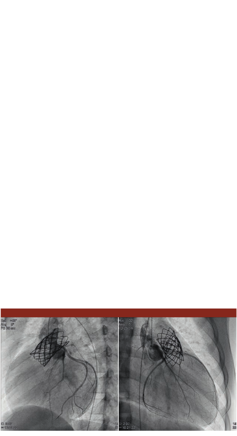
Low dose valve replacement – case 2
Inasecondcase,a10-year-oldboywassufferingfrom
severepulmonarygraftstenosisandmoderateinsufciency.
Duringtheproceduretimeof85minutesaMelody™
valvewasplacedintothisfailinggraftwhileexposingthe
patienttoonly8311mGycm2with15fps.Thisintervention
replacedasurgicalvalvereplacementtohopefullyprovide
thepatientwithenoughcapacitytostayoutoftheORfor
therestofhischildhood.
Conclusion and nal results
ThenewAlluraClaritywithClarityIQtechnologyprovides
equivalentimagequalityat50%lessdose.Thisprovidesthe
exibilitytousethesysteminapersonalizedwayforeach
procedureandpatient.
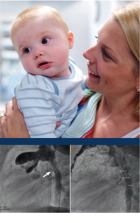
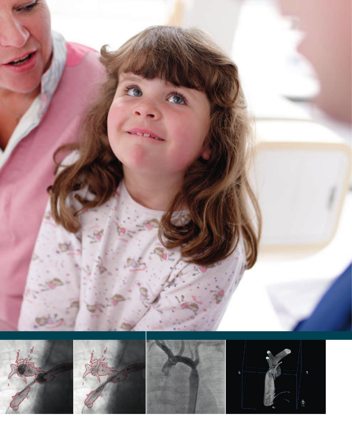
LAO91Cranial1,15fps
Field of view: 15 cm
Angiographyimagedemonstrating
thecoarctationintheaorta(arrow)
LAO 91 Cranial 1, 15 fps,
Field of view: 15 cm
Resultafterdilationofthe
coarctation.Theresultwas
satisfyingwithanaortic
diameteratthelevelofthe
coarctationofalmost7mmand
insignicantresidualsystolic
pressuregradient(2mmHg).
Case 1: Coarctation of aorta
Making the difference with Philips Live Image Guidance | in treating congenital heart disease6
Dose report Case 1 Case 2
Fluoro time [mm:ss] 14:41 12:46
Acquired exposure runs [N] 18 10
Exposure images [N] 3150 1034
Cumulative Frontal Air Kerma [mGy] 36 13
Cumulative Lateral Air Kerma [mGy] 36 8
Cumulative DAP (uoro) [mGycm2] 2924 780
Cumulative DAP (exposure) [mGycm2] 5387 294
TotalCumulativeDAP[mGycm2]8311 1074
“ Especially for pediatric patients it’s extremely important to
achieve the lowest radiation dosage, conserving at the same
time the best required imaging quality, which is necessary to
perform safe and successful interventional procedures in this
specic patient population.”
Prof. Dr. Oliver Kretschmar, University Children’s Hospital, Zürich, Switzerland
LAO 91 Cranial 0, 15 fps
Field of view: 25 cm
Analexposurerunwasmadeto
excludeanycompressionoftheleft
coronaryarteryusingaselective
injection.Theimagesdemonstrate
patentleftcoronaryartery.
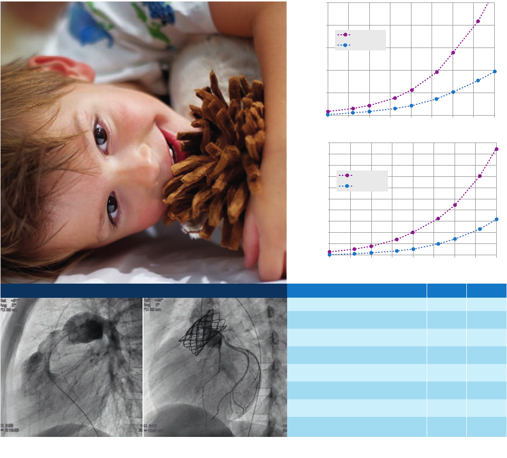
Patient entrance dose rate (mGy/s)
Patient entrance dose (mGy/fr)
Equivalent patient thickness (cm)
Allura Xper
AlluraClarity
0.25
0.2
0.15
0.10
0.05
0
7.55 10 15 17.5 20 22.5 2512.5
Paediatric cardio fluoro default
15 fr/s
Paediatric cardio cine default
15 fr/s
7.55
0.01
0.02
0.03
0.04
0.05
0.06
0.07
0.08
0.09
0.1
0
10 15 17.5 20 22.5 2512.5
Allura Xper
AlluraClarity
Patient entrance dose rate (mGy/s)
Patient entrance dose (mGy/fr)
Equivalent patient thickness (cm)
Equivalent patient thickness (cm)
Allura Xper
AlluraClarity
0.2
0.15
0.10
0.5
0
7.55 10 15 17.5 20 22.5 2512.5
15 fr/s
Paediatric cardio cine default
15 fr/s
7.55
0.01
0.02
0.03
0.04
0.05
0.06
0.07
0.08
0.09
0.1
0
10 15 17.5 20 22.5 2512.5
Allura Xper
AlluraClarity
LAO 91 Cranial 0, 15 fps
Field of view: 25 cm
Angiogramdemonstratingthe
stenosiswithinthepulmonary
valve.Measurementsrevealed
aminimaldiameterof12-13mm
measuredinthemid1/3ofthe
graft,andadistaldiameterof
18-20mmjustbeforethe
pulmonarybifurcation.
Case 2: Melody™ valve implantation
Making the difference with Philips Live Image Guidance | in treating congenital heart disease 7
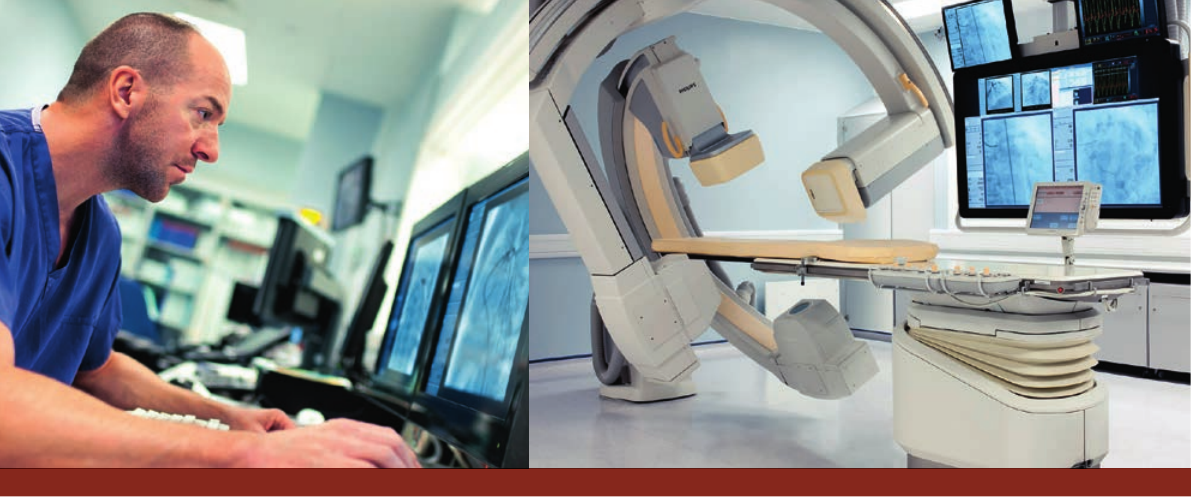
Take advantage of industry-leading image
quality at a fraction of the X-ray dose,
with ClarityIQ technology
AlluraClarity with ClarityIQ technology
AsPhilipsmostpowerfulinterventionalX-raysystemto-
date,AlluraClaritydeliversrelevantclinicalvaluewhereit’s
neededmost–atthepointofpatienttreatment.Clinicians
benetfromhigh-denition,clearvisualizationofeventhe
smallestvessels.
Complexprocedurescanbeperformedwithaccuracy
andcondenceusingthenewAlluraClarityfamilyof
X-raysystems.
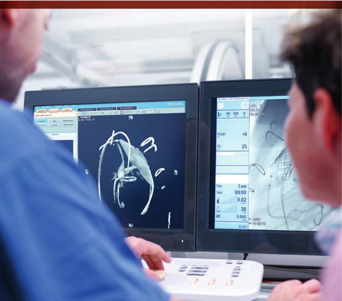
Dramatic results
Inalandmarkcomparativestudy*conductedatRadboud
UniversityHospital,Nijmegen,theNetherlands,ClarityIQ
imagesacquiredusing50%lessdosewereofexcellent
diagnosticqualityforallpatients.
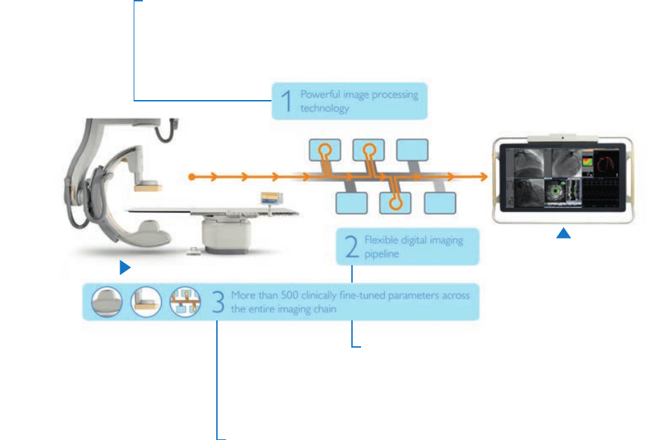
1. Powerful image processing technology
ClarityIQ technology incorporates powerful state-of-the-art image processing technology, developed by Philips Research,
all working in real-time enabled by the latest computing technology:
• Noise and artefact reduction, also on moving structures and objects;
• Image enhancement and edge sharpening;
• Automatic real-time patient and accidental table motion correction on live images.
3. Clinically ne-tuned parameters across the entire imaging chain
With ClarityIQ technology over 500 system parameters are ne-tuned for each
application area; the result of years of Philips clinical leadership. It is now possible to
lter out more X-ray radiation, use smaller focal spot sizes, shorter pulses, thereby
fully utilising the unique capabilities of the Philips MRC X-ray tube.
2. Flexible digital imaging pipeline
ClarityIQ technology utilizes a exible digital imaging pipeline from
tube to display that is tailored for each and every application area
such as Cardio or Neuro. This gives the exibility to select virtually
unlimited application-specic congurations and obtain superb
images at a fraction of the X-ray dose for every intervention.
Fraction of the dose
Industry leading
image quality
*XCY607-130009ClinicalstudyendreportClaritylQ
NijmegenV1.pdf
Making the difference with Philips Live Image Guidance | in treating congenital heart disease8
“ The images are of the same high
quality as we were used to before.
We are so enthusiastic about the
results that we are now acquiring
images with reduced dose.”
Drs. H. Gehlman, Interventional Cardiologist,
Radboud University Hospital
“ With ClarityIQ we are able to get
images at corresponding quality but
with at least a 50% reduction of
radiation dose.”
T.J.F. ten Cate, MD, PhD, UMC Radboud, Nijmegen,
The Netherlands
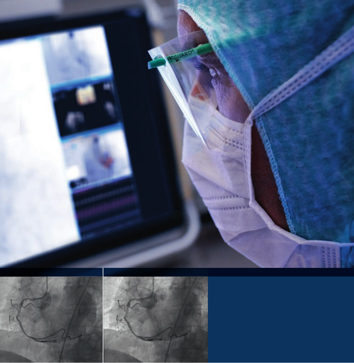
Inthestudy,conductedbetweenSeptemberand
November2012,diagnosticangiographyimagesoftheleft
coronaryarteryof40patientsweremonitored.Thedose
reductionrecordeddecreasedthelikelihoodofsporadic
effectsofradiationforthecathlabpersonnel,while
exposingthepatienttoconsiderablelowerlevelsaswell.
Allura Xper system AlluraClarity system
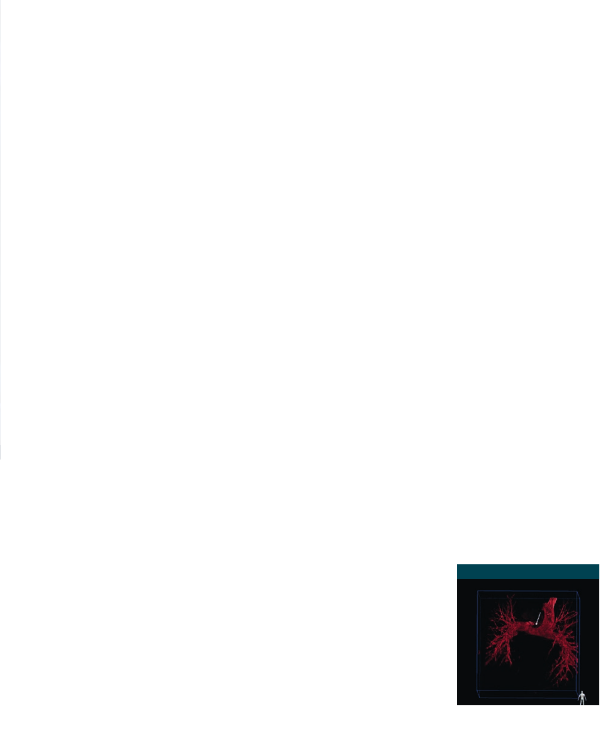
3D-RAreconstructionofthe
pulmonaryarteries.
Enhanced3Dvisualizationandnavigationtechniqueshelp
determinetheoptimalcourseoftreatmentwithgreater
precision.Thisiscriticalinpediatrics,asthevarietyin
patientanatomyandcongenitalheartdefectscanbe
signicant.Theabilitytocaptureandreuse3Ddataas
ameanstoreducetheneedforadditionalcontrastand
X-rayduringaprocedureisimportant.
ThreeinnovativeinterventionaltoolsintheAllurafamily
ofX-raysystemscanhelp.Theymaybeusedalone,orin
acombinedmultimodalitysolution.Intheskilledhandsof
aninterventionalist,thesetoolsmayassistin:
•Improvecondence/efcacyoftheintervention
•Reduceoverallradiationexposure
•Reducecontrastdose
•Reduceproceduretime
Three-dimensional rotational angiography (3D-RA)
3DRAprovidesfast,high-resolution3Dimagesofthe
cardiacanatomyfromanyangulationandrotation.Ithelps
visualizecomplexvascularanomaliestofacilitatedecision
makingfortreatmentstrategies.Ratherthanmultiple2D
views,whichrequiremultiplecontrastinjections,a3DRA
providesacomplete3Doverviewoftheanatomywitha
singleinjection.The3Dimagecanbeinspectedinanyview,
eventhosethatcannotbereachedwithregular
2Dprojections.
Examplesofcongenitalcardiologyapplicationswhere
3D-RAcanbeuseful:
•Diagnosisofthebranchpulmonaryartery–3D-RA
facilitatesadetailedvisualizationofthepulmonary
vasculatureandtheanastomosisthatmayreveala
narrowingnotseenin2D
•PlanningofMelodyvalveimplantation–3DRAgivesa
bettervisualizationofpotentialcompressionofcoronary
arteriesandthelocation/distancebetweentheMelody
valveandthecoronaryarteries
•Treatmentdecisionofaorticcoarctation–3DRAprovides
insightintothecomplexspatialrelationshipbetweenthe
aorticarcanditsbranchingvessels,assistinginthedecision
betweensurgicalrepairorintervention
3D Roadmap
Thistoolfacilitatescomplexinterventionsbyproviding
live3Dimageguidancefornavigatingvascularstructures
anywhereinthebody.
Dynamic3Droadmapoverlaysreal-time2Duoroscopy
imagesanda3Dreconstructionofthevesseltreeacquired
withthe3D-RAfeatureoftheAlluraClarityfamilyofX-ray
systems.Itimprovesvisualizationofanatomicalstructures
anddevicenavigationandreducestheneedforadditional
contrastagentduringnavigationofdevices.
MR/CT Roadmap
ImagingfromapreviouslyacquiredMRorCTscancanbe
reusedandoverlaidbyuoroscopytoguideprocedures.
MR/CTRoadmapletscliniciansfollowtheadvanceofguide
wiresandcathetersreal-time,therebyreducingX-ray
doseandcontrastmediumduringinterventions.Both3D
RoadmapandMR/CTRoadmapautomaticallyadjuststhe3D
imagestogantrychangesandanylateralorlongitudinaltable
movements.Forevenbettervisualization,itoffersenlarged
full-screenmodeand4xdigitalzoom.
3D-RA
Greater insight and
condence in nding and
treating the problem
Making the difference with Philips Live Image Guidance | in treating congenital heart disease12Making the difference with Philips Live Image Guidance | in treating congenital heart disease 9
NexttoAlluraClarityIQtechnology,otherdosereduction
technologiesassistinthemanagementofradiation
exposure.Theyinclude:
DoseWise
DoseWiseisattheheartofourdosereductionefforts.
It'sasetoftechniques,programs,andpracticesthat
ensuresoptimalmagequalitywhileprotectingpeoplein
X-rayenvironments.ItisbasedontheALARÁ(AsLowAs
ReasonablyAchievable)principIe.Witheverynewsystem.
welookathowwecanincorpocatebettershielding
andimproveourX-rayexposureparameterstofurther
reducedose.Thatmeansbeforeyouevenputonalead
æron,Philipshasdonetheirutmosttoprotectyouand
yourpatientsfromunnecessarydose.
Philips DoseAware* family
PhilipsDoseAwarefamilyoffersimmediatefeedbackon
dosetoincreaseradiationawarenessandhelpmanage
occupationalmedicalradiationexposuretophysicians
andstaff.Itprovidesreal-timedosefeedbackinthe
examinationroomtotrackanindividual’sradiation
exposureduringeachshift,aswellasprocedure-baseddata
fordeeperinsightintostaffexposuretrendsandbehavior.
DoseAware Xtend – dedicated room solution
DoseAwareXtendisadedicatedsolutionforroomswith
PhilipsFlexVisionXLdisplay.ItsintegrationwiththeAllura
X-raysystemallowsittoprovidedetailedfeedbackon
scatteredX-raydoselevelsperprocedure.
Pushing the Boundaries of ALARA
Increases radiation awareness
•IdentiesthecumulativeamountofX-raydosereceived
rightaftereachprocedure
•Remindsclinicianstotakesecondaryleadprecautions
•Sendsweeklyormonthlyreportsautomaticallyon
procedure-basedstaffdosetohelpidentifyindividual
exposuretrends
•SendsproceduraldataonX-raydoseinDICOMRDSR
formattoPACSorRIStosimplifydataanalysis
DoseAware – exible solution for different rooms
DoseAwarecanbeusedinanyX-rayroomtoprovide
real-timefeedbackonscatteredX-rayexposuresostaff
canimmediatelyadjusttheirworkinghabitstomanage
radiationexposure.
Advantages of a real-time dosimeter
•Providetheinformationnecessarytomanageindividual
X-raydoseexposure
•ShowwhenandwhereX-raydosewasacquiredtoallow
forappropriateaction
•Checkexposurelevelonthecoloreddisplayinthe
examinationroom
•Archive,report,andanalyzeradiationdatatomaintain
highlevelsofoccupationalsafety
Smart Beam
WithSmartBeam,asthenamesuggests,thedoseis
reducedbytakingamoreintelligentapproachtothe
useofX-rays.Tothisend,theAllurafamilyusesspecial
SpectraBeamltersinuoroandexposurestoremove
unwanted‘soft’radiation,i.e.thoseX-raysthathitthe
patientbutdonothaveenoughenergytoreachthe
imagedetector.
Thesoftradiationisreplacedbyhigherenergyradiation,
whichsignicantlyimprovesimagequality.Alternatively,
itispossibleto‘tradeoff’someofthisimprovedquality
tofurtherreducedose.
*DoseAwaredoesnotreplacethethermo-luminescentdosimeter
(TLD)asalegaldosimeter.
“ DoseAware is one of the most important
new tools available to help reduce
occupational medical radiation exposure
to physicians and staff. It’s a much easier
and practical way to monitor levels than
conventional methods. Creating a better
work environment is not only the right
thing to do but our obligation.”
J. Kiah, MS, RN Lab Manager, Director Cardiac and Vascular
Services, Baptist Cardiac & Vascular Institute, Miami, USA
NexttoAlluraClarityIQtechnology,otherdosereduction
technologiesassistinthemanagementofradiation
exposure.Theyinclude:
DoseWise
DoseWiseisattheheartofourdosereductionefforts.
It'sasetoftechniques,programs,andpracticesthat
ensuresoptimalmagequalitywhileprotectingpeoplein
X-rayenvironments.ItisbasedontheALARÁ(AsLowAs
welookathowwecanincorporatebettershielding
andimproveourX-rayexposureparameterstofurther
reducedose.Thatmeansbeforeyouevenputonalead
æron,Philipshasdonetheirutmosttoprotectyouand
yourpatientsfromunnecessarydose.
Philips DoseAware* family
PhilipsDoseAwarefamilyoffersimmediatefeedbackon
dosetoincreaseradiationawarenessandhelpmanage
occupationalmedicalradiationexposuretophysicians
andstaff.Itprovidesreal-timedosefeedbackinthe
examinationroomtotrackanindividual’sradiation
exposureduringeachshift,aswellasprocedure-baseddata
fordeeperinsightintostaffexposuretrendsandbehavior.
DoseAware Xtend – dedicated room solution
DoseAwareXtendisadedicatedsolutionforroomswith
PhilipsFlexVisionXLdisplay.ItsintegrationwiththeAllura
X-raysystemallowsittoprovidedetailedfeedbackon
scatteredX-raydoselevelsperprocedure.
Pushing the Boundaries of ALARA
Increases radiation awareness
•IdentiesthecumulativeamountofX-raydosereceived
rightaftereachprocedure
•Remindsclinicianstotakesecondaryleadprecautions
•Sendsweeklyormonthlyreportsautomaticallyon
procedure-basedstaffdosetohelpidentifyindividual
exposuretrends
•SendsproceduraldataonX-raydoseinDICOMRDSR
formattoPACSorRIStosimplifydataanalysis
DoseAware – exible solution for different rooms
DoseAwarecanbeusedinanyX-rayroomtoprovide
real-timefeedbackonscatteredX-rayexposuresostaff
canimmediatelyadjusttheirworkinghabitstomanage
radiationexposure.
Advantages of a real-time dosimeter
•Providetheinformationnecessarytomanageindividual
X-raydoseexposure
•ShowwhenandwhereX-raydosewasacquiredtoallow
forappropriateaction
•Checkexposurelevelonthecoloreddisplayinthe
examinationroom
•Archive,report,andanalyzeradiationdatatomaintain
highlevelsofoccupationalsafety
Smart Beam
WithSmartBeam,asthenamesuggests,thedoseis
reducedbytakingamoreintelligentapproachtothe
useofX-rays.Tothisend,theAllurafamilyusesspecial
SpectraBeamltersinuoroandexposurestoremove
unwanted‘soft’radiation,i.e.thoseX-raysthathitthe
patientbutdonothaveenoughenergytoreachthe
imagedetector.
Thesoftradiationisreplacedbyhigherenergyradiation,
whichsignicantlyimprovesimagequality.Alternatively,
itispossibleto‘tradeoff’someofthisimprovedquality
tofurtherreducedose.
*DoseAwaredoesnotreplacethethermo-luminescentdosimeter
(TLD)asalegaldosimeter.
“ DoseAware is one of the most important
new tools available to help reduce
occupational medical radiation exposure
to physicians and staff. It’s a much easier
and practical way to monitor levels than
conventional methods. Creating a better
work environment is not only the right
thing to do but our obligation.”
J. Kiah, MS, RN Lab Manager, Director Cardiac and Vascular
Services, Baptist Cardiac & Vascular Institute, Miami, USA
Reasonably Achievable) principIe. With every new system,
“ The images are of the same high
quality as we were used to before.
We are so enthusiastic about the
results that we are now acquiring
images with reduced dose.”
Drs. H. Gehlman, Interventional Cardiologist,
Radboud University Hospital
“ With ClarityIQ we are able to get
images at corresponding quality but
with at least a 50% reduction of
radiation dose.”
T.J.F. ten Cate, MD, PhD, UMC Radboud, Nijmegen,
The Netherlands
Inthestudy,conductedbetweenSeptemberand
November2012,diagnosticangiographyimagesoftheleft
coronaryarteryof40patientsweremonitored.Thedose
reductionrecordeddecreasedthelikelihoodofsporadic
effectsofradiationforthecathlabpersonnel,while
exposingthepatienttoconsiderablelowerlevelsaswell.
Allura Xper system AlluraClarity system
3D-RAreconstructionofthe
pulmonaryarteries.
Enhanced3Dvisualizationandnavigationtechniqueshelp
determinetheoptimalcourseoftreatmentwithgreater
precision.Thisiscriticalinpediatrics,asthevarietyin
patientanatomyandcongenitalheartdefectscanbe
signicant.Theabilitytocaptureandreuse3Ddataas
ameanstoreducetheneedforadditionalcontrastand
X-rayduringaprocedureisimportant.
ThreeinnovativeinterventionaltoolsintheAllurafamily
ofX-raysystemscanhelp.Theymaybeusedalone,orin
acombinedmultimodalitysolution.Intheskilledhandsof
aninterventionalist,thesetoolsmayassistin:
•Improvecondence/efcacyoftheintervention
•Reduceoverallradiationexposure
•Reducecontrastdose
•Reduceproceduretime
Three-dimensional rotational angiography (3D-RA)
3DRAprovidesfast,high-resolution3Dimagesofthe
cardiacanatomyfromanyangulationandrotation.Ithelps
visualizecomplexvascularanomaliestofacilitatedecision
makingfortreatmentstrategies.Ratherthanmultiple2D
views,whichrequiremultiplecontrastinjections,a3DRA
providesacomplete3Doverviewoftheanatomywitha
singleinjection.The3Dimagecanbeinspectedinanyview,
eventhosethatcannotbereachedwithregular
2Dprojections.
Examplesofcongenitalcardiologyapplicationswhere
3D-RAcanbeuseful:
•Diagnosisofthebranchpulmonaryartery–3D-RA
facilitatesadetailedvisualizationofthepulmonary
vasculatureandtheanastomosisthatmayreveala
narrowingnotseenin2D
•PlanningofMelodyvalveimplantation–3DRAgivesa
bettervisualizationofpotentialcompressionofcoronary
arteriesandthelocation/distancebetweentheMelody
valveandthecoronaryarteries
•Treatmentdecisionofaorticcoarctation–3DRAprovides
insightintothecomplexspatialrelationshipbetweenthe
aorticarcanditsbranchingvessels,assistinginthedecision
betweensurgicalrepairorintervention
3D Roadmap
Thistoolfacilitatescomplexinterventionsbyproviding
live3Dimageguidancefornavigatingvascularstructures
anywhereinthebody.
Dynamic3Droadmapoverlaysreal-time2Duoroscopy
imagesanda3Dreconstructionofthevesseltreeacquired
withthe3D-RAfeatureoftheAlluraClarityfamilyofX-ray
systems.Itimprovesvisualizationofanatomicalstructures
anddevicenavigationandreducestheneedforadditional
contrastagentduringnavigationofdevices.
MR/CT Roadmap
ImagingfromapreviouslyacquiredMRorCTscancanbe
reusedandoverlaidbyuoroscopytoguideprocedures.
MR/CTRoadmapletscliniciansfollowtheadvanceofguide
wiresandcathetersreal-time,therebyreducingX-ray
doseandcontrastmediumduringinterventions.Both3D
RoadmapandMR/CTRoadmapautomaticallyadjuststhe3D
imagestogantrychangesandanylateralorlongitudinaltable
movements.Forevenbettervisualization,itoffersenlarged
full-screenmodeand4xdigitalzoom.
3D-RA
Greater insight and
condence in nding and
treating the problem
Making the difference with Philips Live Image Guidance | in treating congenital heart disease12Making the difference with Philips Live Image Guidance | in treating congenital heart disease 9
Asinglerotationalscanprovides
inputforthe3D-RAreconstruction.
3D-RAreconstructionoftheaorticarc,allowing
youtoplantheoptimaltreatment.
Liveoverlayon3D/MR/CTroadmapimprovesvisualization
ofanatomicalstructuresanddevicenavigationbyre-using
preoperativeCTorMRangiographicimages.
3D-RA: Improved Procedural Planning3D / MR / CT roadmap
Making the difference with Philips Live Image Guidance | in treating congenital heart disease 13
“ I have found that the 3D-RA tool
is very benecial for my pediatric
patients to efciently manage
radiation dose, contrast dye, and
procedure time.”
Dr. Seong-Ho Kim, Chairman,
Department of Pediatrics
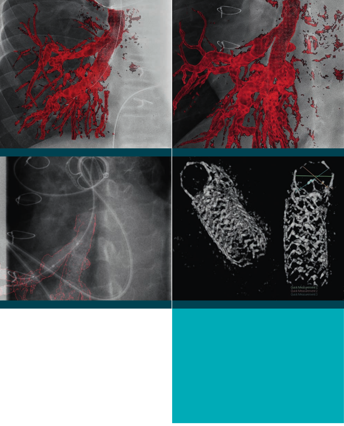
3D Roadmap in AP
3D Roadmap: balloon ination of sidebranch 3D RA of stent after ballooning side branch.
3D roadmap in LAO40/Caud27
3D Roadmap in AP: overlap at bifurcation, inadequate
for intervention
TheostiumofthesidebranchisnotclearlyvisibleintheAllura
3D-RAinAPpositionbecauseofoverlappingvessels.
3D Roadmap in LAO40/Caud27: clear view on bifurcation
ThebifurcationisclearlyvisibleintheAllura3D-RAin
40LAO/27Caudalposition.
3D Roadmap: balloon ination of sidebranch
The3DRoadmapimageshowstheballooninatedinthe
sidebranch.
3D RA of stent after ballooning side branch
Apost-proceduralAllura3D-RAshowsthestentstrutsopenin
thesidebranchtoenableadequatebloodow.
Making the difference with Philips Live Image Guidance | in treating congenital heart disease14

3D-RA and 3D roadmap case study:
Treatment of pulmonary artery stenosis
Catheter based treatment of a complex pulmonary
artery stenosis requires in-depth understanding of the
location and dimension of the stenosis. Using standard
angiograms, requires several X-ray acquisitions to make a
complete evaluation. Dr. Seong-Ho Kim, chairman of the
department of pediatrics at the Sejong General Hospital,
Korea used 3D-RA and 3D roadmap during treatment of
a pediatric patient with a stenotic side branch of the right
pulmonary artery.
Patient
A 13 year-old male patient weighing 43 kg was brought to
the cath lab for a pulmonary artery stenosis intervention.
A side branch of the RPA is stenosed, due to a right
pulmonary artery (RPA) stenting procedure in 2013.
This patient was born with a ventricular septal defect
(VSD) and major aorto-pulmonary collateral arteries
(MAPCA). A Rastelli procedure was performed in 2002
to reconstruct the outfl ow from the right ventricle to the
pulmonary arteries (PA). Several procedures followed in
subsequent years: a reconstruction of the RPA, dilatation
and stenting of the left pulmonary artery (LPA), and
dilatation and stenting of the RPA.
Selection of projection angle
Allura 3D-RA was used to provide a better visualization
of the bifurcation, which helped the physician to plan the
treatment strategy and therapeutic approach. This data set
was reconstructed into a 3D volume rendering. The 3D
volume provided a detailed visualization of the pulmonary
vasculature and bifurcation from any angulation and
rotation, allowing the physician to select the projection
which would allow him to precisely treat the lesion of
interest. The AP projection showed overlapping vessels at
the bifurcation which obscured visibility of the ostium of
the side branch of the RPA. The projection was changed to
40 LAO and 27 Caudal, which provided a clear view of the
bifurcation.
Balloon dilatation
The 3D volume was then overlaid on live fl uoroscopy,
providing a 3D Roadmap. This Roadmap was used to
navigate the guidewire to the correct position and deploy
the balloon, without the need to use additional contrast.
After dilatation of the side branch, a second 3D-RA was
done to check the dilatation result.
Method and materials used
An Allura 3D-RA acquisition was made on the
AlluraClarity FD10/10 biplane X-ray system. The
acquisition protocol was: a 4 sec, 240 degree (120
LAO/120 RAO) rotational angiogram at 30 frames/sec
using a 25 cm fi eld of view. A total contrast volume of
60 cc (2/3 contrast and 1/3 saline) was injected at 12 cc/s
with an injection delay of 1 sec before acquisition. The
contrast was diluted 2/3 contrast to 1/3 saline which
resulted in an actual contrast load of 40 cc.
Conclusion
The view of the stenosis in the bifurcation of the RPA
was obscured in the AP view. Without using Allura
3D-RA, the physician would have had to acquire several
additional 2D angiograms to get a clear view of the
lesion. The projection angle to navigate the guidewire
to the correct side branch was selected based on the
3D-RA model. Only one 3D-RA was required to get
a detailed visualization of the bifurcation and to select
the precise projection angle for treatment, thereby
saving X-ray dose, contrast medium, and time. After
balloon deployment in the side branch, a second 3D-RA
reconstruction clearly showed the stent struts open
at the side branch enabling adequate blood fl ow.
Making the difference with Philips Live Image Guidance | in treating congenital heart disease 15Making the difference with Philips Live Image Guidance | in treating congenital heart disease 15
452299102481.indd 15 19/06/14 16:14
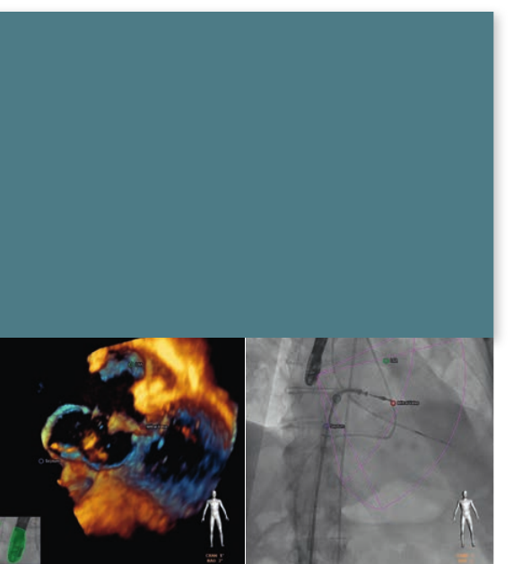
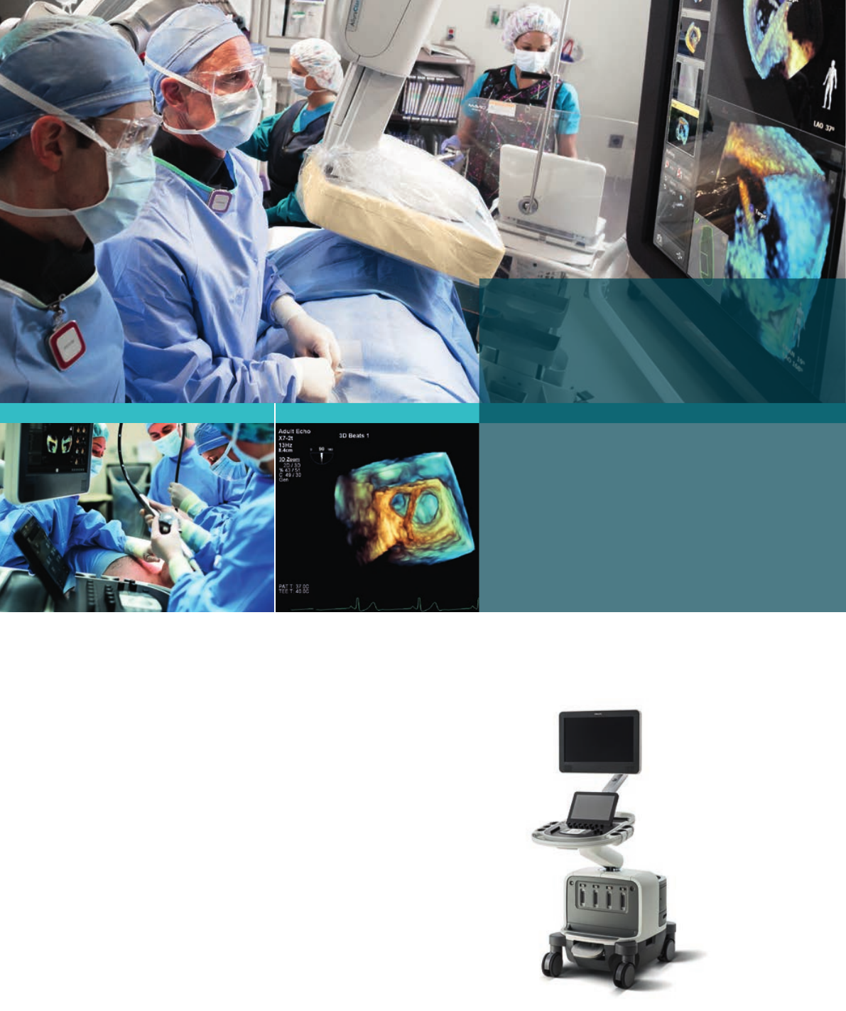
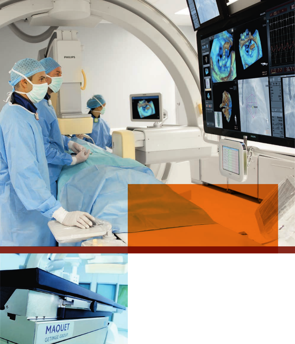
EchoNavigator
Increasingnumbersofpatientswithstructuralheart
disease(SHD)canbetreatedwithcatheter-based
techniques.Oneofthemainchallengesisvisualization;
Live3Dtransesophagealechocardiography(TEE)imaging
providescriticalinsightsintosofttissueanatomy,and
functionandowinformation.Atthesametime,X-rayis
invaluableforvisualizingdevices.Bothimages,however,
arerepresentedseparatelyinadifferentorientationand
sovaluabletimeisspentmentallyaligningthem.
Intuitively combining live X-ray and echo
guidance for Structural Heart Disease repair
Key benets
•Intuitivelycombineslive3DTEEechocardiographyand
uoroscopicimages.BringsTEEechocardiographyand
uoroscopicimagestogether,inreal-time.
•Understandwhereyouareinthe3Dspacemorequickly.
•AnatomicallandmarksinEchoareoverlaidonX-rayfor
guidanceofdevices.
•Directlycontrollableattableside,whichfacilitates
communicationwiththeechooperator.
•Promotesteamworkwithintheheartteaminthelab.
Work with clinical condence by having access to superb quality images, unique live 3D imaging
capabilities and innovative imaging solutions. This will support you in planning, visualization
and Live Image Guidance of even the most challenging procedures.
EchoNavigatortacklesthisissuehead-onbyintuitively
bringinglive3DTEEanduoroscopicimagestogether,
inreal-time,foraquickunderstandingofthe3Dspace.
Imagesfrombothmodalitiesareautomaticallyaligned
bytrackingtheTEEtransducerpositionandorientation
intheX-rayimage.Asaresult,relevantsofttissueanatomy
canbyvisualizedintheX-ray.Markersplacedonthe
softtissuestructureswithintheechoimage,automatically
appearontheX-rayforcontextandguidance.Thisprovides
cleartargetsforcatheternavigation.Theinterventional
operatorcandirectlycontroltheEchoNavigatorattableside,
whichfacilitatescommunicationwiththeechooperator.
Allofthisisdesignedtosimplifynavigation,deviceplacement
andpromotescommunicationwithintheheartteamduring
structuralheartdiseaseprocedures.
Training
OurPeertoPeertrainingprogramsofferaninteractive
programonsitewithexperiencedusers.Thesetraining
programsprovideindepthdetailsonhowtousethe
technologyinclinicalpractice,andprovideyouwith
thecondencetoimplementtheseadvancedimaging
functionalitiesinyourdailyroutineandtotakeyourexpertise
tothenextlevel.WealsoprovideexcellenttrainingofLive3D
TEEandEchoNavigatortohelpdeveloparapidandthorough
understandingofthesebreakthroughSHDtreatments.
“ We’re integrating two separate medical
images and bringing them together in a
way that makes performance of these
interventions more straight-forward.”
Professor John Carroll, MD, Interventional Cardiologist,
University of Colorado, Denver
Making the difference with Philips Live Image Guidance | in treating congenital heart disease16
A new era in premium cardiovascular ultrasound
ThePhilipsEPIQ7ultrasoundsystemincorporatesour
mostpowerfularchitectureeverappliedtoultrasound
imaging–touchingallaspectsofacousticacquisition
andprocessing.Supportedbyourfamilyofproprietary
xMATRIXtransducersandourleading-edgeAnatomical
Intelligence,thisplatformoffersyouaccuratediagnosis,
rst-timeright,whichisfasterandeasiertoperformthan
before.Yougetimprovedclinicalinformationfromeach
scanandahigherlevelofcondence.TheEPIQ7supports
thelive3DTEEforchildrenabove20kg,aswellasthe
minimultiplaneTEEforinfantsabove3.5kg.
Philips EPIQ 7 ultrasound system Live 3D TEE of appendage
• Live3Dtransesophagealechocardiographyallowstheviewing
ofmitralvalves,aorticvalves,interatrialseptum,theleftatrial
appendageandallchambersoftheheartwithuniqueperspectives.
• PhilipsLive3DTEEprovidesreal-time3DandlivexPlaneviews.
Thesetoolsprovidemanybeating-heartviewsthatwerepreviously
seenonlyduringcardiacsurgery.
• Live3DTEEfeaturesprominentlyinrecentprofessionalsociety
recommendationsregardingechocardiographyintrans-catheter
interventions.(EAE/ASE*RecommendationsfortheUseof
EchocardiographyinNewTranscatheterInterventionsforValvular
HeartDisease.)
“ The beauty of good imaging is that
you don’t have to imagine too much”
Professor J Zamorano, Head of Cardiology,
University Hospital Ramon y Cajal, Madrid
3Dzoomedappendageand
pulmonaryvein.
* Journal of the American Society of EchocardiographyVolume 24, Issue 9,
September 2011, Pages 937–965EAE/ASE Recommendations for the Use
of Echocardiography in New Transcatheter Interventions for Valvular Heart
Disease • Jose L. Zamorano et al.
EPIQ7:Designedtoreinventtheuserexperience
Making the difference with Philips Live Image Guidance | in treating congenital heart disease 17
Better user experience
to promote consistency
and efciency
Biplane versatility helps reveal hidden pathologies
Congenitalheartdefectscanbehardtopredictand
visualize,requiringquiteextremeandexoticprojections
tovisualizeeachchild’suniqueanatomy.Achievethe
challengingprojectionviewsyourequirewithPhilips
Allura’suniquelateraldoubleC-arm.Itcanmakesteep
cranial/caudalprojectionstorevealhiddenpathologiesor
missinganatomicalstructures.Itallows,forinstance,afour
chamberviewoftheatrialseptum.Twoviewsarecreated
withasinglecontrastinjection.Bycombininga20x20cm
lateralwitha30x40cmfrontal,yougetfullexibilityfor
imaginginfants,youngchildren,andadultswithoutpanning
tosupportabroadrangeofpatients.
ThefrontalandlateralC-armscanbeindependently
positioned,allowingfullpatientaccessfortheheart
team.Thereisplentyofspaceattheheadsideforthe
anesthesiologistandechocardiologistwithoutinterfering
withtherestoftheteam.TheFD10/10andFD20/10
biplanesystemshaveaverysmallfootprinttoallow
maximaloorclearance.
Biplanevisualizationscanprovideamorecompletepictureofthesituation.Forinstance,on
thefrontalplaneinthiscase,thebranchoftheleftpulmonaryartery(leftimage)appearstoll
completely.However,thelateralimage(right)showsthatonlytheposteriorsegmentislling.
Hybrid Suite
Theinterventionalandsurgicalenvironmentsarerapidly
evolvingasinterventionaltreatmentandsurgeryconverge
inhybridprocedures.Interventionsareperformedfor
increasinglycomplexdiseases,whilesurgicalprocedures
arebecominglessinvasive.Patientpopulationsarealso
becomingbroaderasmorechildrenwithcongenitalheart
diseasesurviveintoadultlife.Inaddition,theavailabilityof
newdevicesexpandsthenumberoftreatmentoptions.
WiththeHybridSuitefromPhilips,afullrangeof
procedurescanbeperformedinasingleroom,virtually
withoutcompromise.Severalkeyinnovationssupport
theheartteamincarryingoutlongandoftencomplex
congenitalheartprocedureswithahighdegreeof
comfortandcondence.UsingtheuniqueFlexMoveor
FlexMoveXL,theceilingmountedC-armcanbeeasily
movedasrequiredanywherearoundthetable–and
thenconvenientlyparkedoutofthewayduringopen
surgery.Thisgivestheteamroomtoworkclosetothe
patientandfreesuptheheadareasotheanesthesiologist/
echocardiologistcanworkwithgreaterease.
Biplane imaging
Making the difference with Philips Live Image Guidance | in treating congenital heart disease18
MAQUET MAGNUS operating table
“ In the hybrid room you feel completely relaxed.
You know every time you can switch from open
surgery to catheter-based, or from catheter-
based to open surgery. So you feel great.”
Prof. Dr. M. Lachat MD, Vascular Surgeon, Zürich University Hospital,
Zürich, Switzerland
MAQUET MAGNUS operating table
TheAllurasystemsaretheonlybiplanesthatcanbeseamlessly
integratedwiththebest-in-classMAQUETsurgicaltablefor
atrulymultifunctionalroomsuitableforconventionalsurgery,
hybridsurgery,orinterventions.Thetwoarecompletely
synchronizedandbenetfromautomaticpositioncontrol(APC),
boluschaseprocedures,and3Dsoftwaretools.Thetablecanbe
outttedwitharadiotranslucenttabletopforendovascularand
hybridprocedures,oramodulartabletopforopensurgery.
Tabletopscanbeeasilyexchangedusingthetransporter,allowing
smoothtransferofpatientsbetweenprocedures.
Making the difference with Philips Live Image Guidance | in treating congenital heart disease 19
For the patientThe Ambient Experience Interventional Suite
Personalizingtheenvironmentallowsthepatientto
becomeapartnerintheirprocedure
“ Children who are nervous when
they arrive are mesmerized by
the pleasant environment, which
often allows examinations to
be completed more quickly.”
Jojanneke de Leeuw, radiology technician,
Jeroen Bosch Hospital, ‘s-Hertogenbosch, The Netherlands
Making the difference with Philips Live Image Guidance | in treating congenital heart disease20
For the staff
CliniciansbenetfromadvancedAmbientExperiencefeatures
thatincreaseefciencyandimproveworkingcomfort
A treatment environment that puts
children at ease
Let’sfaceit,achildisgenerallyunhappyatbeingina
hospital.Theycanfeelapprehensive,nervousandinsome
casesplainscared.Especiallywhentheyhavetoundergo
severalproceduresoveralongerperiod.Youwanttomake
themfeelmoreatease,butthat’sdifculttodowhenthe
surroundingslook,soundandsmellsodifferentfromhome.
Highpatientanxietycanhaveconsequencesforthe
medicalteam.Thiscanalsohaveconsequencesforthe
medicalteam.Clinicalstaffcanbecomefrustratedbecause
examinationstakelongerandthroughputdecreases.It
alsoaffectsmotivationwheneffortstoreassureandrelax
patientsareinvain.
WithAmbientExperiencewecancreateapurposefully
designedhealthcareenvironment(includingtechnology,
spatialdesignandworkow),addressingtheseissues.You
createtheconditionsinwhichchildrenfeelmoreatease,
cliniciansareabletoworkmoreeffectivelyandwhichcan
changethecommunityperception.
For the patient
Dynamiclightingproducesasoft,pleasanteffect.
WithAmbientExperiencechildrencanselectatheme
fromthetouchscreen.ImmediatelythedynamicAmbient
Lightingproducesawarmwelcomingatmosphereinthe
room.Wallsappeartofadeaway.Thematicvideowith
soundprovideapositivedistractionthathelpsthechild
torelax.Thiscalmingatmosphereenhancesinteraction
betweenthechildandstaff.
For your staff
Architecturalenhancementsoptimizeroomdesign.
Intruecollaborativefashion,weworkwithyourin-house
teamtoeffectivelyreduceclutter,recessthecabinetry,
andimprovethelighting.Open,stress-freesurroundings
provideyourstaffwithanuniqueinterventional
environment.Withthesupportofourglobalorganization,
wehelpyoucreateawelcomingandeffectiveenvironment
thatwillrevolutionizethewaymedicalprocedures
areperformed.
Demonstrating value
TheAmbientExperienceInterventionalSuitecan
differentiateyoufromotherhealthcarefacilities.
Thisagshipservicecanenhancetheimageofyour
hospitalintheeyesofallstakeholders.Itsupportsbetter
communicationwiththepatient,andcanprovideamore
efcientworkingenvironmentwiththepotentialto
impactstaffmorale,retention,andrecruitment.Ambient
Experiencedemonstratesyourdedicationtopeople
focusedhealthcare.
AmbientExperienceisapurposelydesignedhealthcare
environment.Witharefreshinglycreativeeye,Ambient
Experienceintegratestechnology,spatialdesign,and
workowimprovementstocreateamorecomfortable,
stress-reducingenvironmentforbothpatientsandstaff.
Making the difference with Philips Live Image Guidance | in treating congenital heart disease 21
Streamlining workow for complex
congenital procedures
Whenworkingtosaveachild’slife,amedicalteam
wantstofocusfullyonwhatneedstobedone.Philips
offersmanyexiblewaystoenhanceworkowandhelp
reducedistractionsandclutterinthecathlaborhybrid
environment.Thesesolutionscontributetoawell-
organized,smoothrunningprocedure.
Everything you need to see
Apediatriccardiologysuiteperformsalargevarietyof
procedures,eachrequiringtheirowntypesofimages
andinformation.TheFlexVisionXLmonitorallowsfor
easyviewingandmagnicationofanyimageinthelab–
fromX-raystopre-acquiredpatientCTandMRscans
–attableside.
Thislarge,high-denitioneight-megapixelLCDscreen
layoutcanbeadaptedontheytophysicianpreferences
andexaminationrequirements–perproceduretypeor
case.Informationfromseveralmulti-modalityimaging
sourcesisintegratedintooneview.Thisallowstheentire
teamtounderstandandshareinformationduringeach
stepoftheprocedure.
TheSuperZoomfeatureclearlybringssmallaspectsof
anatomyanddataintofocuswithoutsacricingresolution.
Withthisfeature,thescreencanbeplacedfurtherfrom
thetable,therebyfreeingupspace.
Zoom in on uoroscopy images without
increasing dose
Pediatricstructurescanbesmallanddifculttovisualize.
OurDualFluorooptionallowsyoutoalsomagnify
liveuoroscopicimagestwotimeswithoutincreasing
radiationdose.Thezoomeduoroscopyimagesare
displayedonthelivemonitor.Thisfeaturecanbeused
onbothfrontalandlateraluoroscopyimages.
Physiomonitoring
PhilipsXperFlexCardio,asmallhemodynamicsystem,
seamlesslyintegrateswiththeAlluraportfolio.Itoptimizes
workowandimprovestheproductivityinthelabthrough
easyexchangeofdata.XperFlexCardioalsoprovides
advancedECGanalysisinthecathlabwithSTmapping
andSTEMI-CA.
Relevant information at hand
Inthisexible,fullyintegratedenvironment,Philips
cardiologyinformatics,toolsandreal-timeinformation
areallwithinreach,makingiteasytocarryoutallaspects
ofprocedures.
Making the difference with Philips Live Image Guidance | in treating congenital heart disease22
Managing cardiology information throughout
the care process
PhilipsCVIS(CardiovascularInformationManagement
System)isauniquesoftwaresolutionthatenhances
theclinicalandnancialperformanceofthecardiology
department.Ituniessilosofdiscretecardiacinformation
collectedatthepointofcareintoasinglepowerful
database.Thiscardiacdatabaseissearchableand
actionabletosupportclinical,qualitative,andbusiness
analysisandreportingneeds.
Multi-modality image management
XceleracardiologyPACSortheenterprise-wide
IntelliSpacePACSsolutionscanhandleimagemanagement,
clinicalanalysis,andreporting.IntelliSpaceisapowerful
toolforadvancedimagereviewandanalysis.Itoffers
theexibilitytodiagnoseandcollaboratevirtually
anywhere.Inaddition,iSitePACSintegratesintoexisting
infrastructure,enablingalldepartmentstorealizeareturn
oninvestmentateachstageofdeployment.
Cath lab workow solution
XperIMandXperFlexCardiofeatureintuitive
innovationsthatcreateamoreefcientworkowin
thecathlabdepartment,includingdirectaccessto
acquiredimages,physiomonitoring,scheduling,inventory
managementreporting,andstatistics.
Philipsiscommittedtoworkingcloselytogetherwith
customers.Thepurchaseofanewinterventionalsuite
andinterventionaltoolsdemonstratesalong-term
commitmenttopatientsandpersonnel.Weoffera
comprehensiveportfolioofserviceoptions,training
programsforstaffanddelivernancingoptionstohelp
optimizetheeconomicvalueacrossthetotallife-cycle
ofthesystems.
RightFit Service agreements
Frompremiumserviceplanstostandardservicesupport,
thereisaRightFitServiceAgreementtosuiteveryneed.
Theperfectmixofon-demandsupport,immediateparts
andservice,andcomprehensivebusinesssolutionshelps
hospitalsdeliverqualitypatientcare.
Exceptional support is a priority
ThePhilipsglobalservicenetworkisreadytoassistwhen
necessary.Fieldengineers,remoteservicetechnicians,and
frontlinecall-agentssupportallsystemswithaworld-
classservicesnetwork.Thegoal:helpcustomerssucceed
ineveryphaseofsystemownership,fromplanningto
start-up,throughpeakusageandrenewal.
ARemoteServicesoptionprovidesadvancedsystem
troubleshootingthathelpslessendowntimeevenfurther.
Equipmentremainsreliablethroughremotesystem
diagnosesandfastrepair.
Education and training
Regulartrainingiscriticaltohelpthemedicalstaffto
bettermasterclinicalprocedures,getmoreoutofthe
equipment,andbuildprofessionalandclinicalskills.
Better-trainedstaffimprovestheoveralllevelofservice
andoperation.
Increased economic value
Asanexample,PhilipshasestablishedPeer-to-
Peertrainingprogramsforphysiciansinterestedin
learningaboutEchoNavigator.Participantsvisitpeers
inthehospital,whoareexpertintheclinicaluseof
EchoNavigator,andreceiveacomprehensivetwo-day
programcombiningclassroomtutorials,hands-on
training.Theyworkcloselywiththeseexperienced
usersduringrealcases.
Moreknowledgeablepersonnelhaveahigherchance
ofobtainingaccurateresults.
Financial options are plenty
PhilipsMedicalCapitalmakesitsurprisinglyeasyto
arrangenancingtoaddresscapitalbudgetrequirements,
manageincreasingpatientvolumeandachieveclinical
superiority.PhilipsProPluscombinesequipment,service,
andnancinginonesimpleofferingtohelp:
Enhances patient care
Making the difference with Philips Live Image Guidance | in treating congenital heart disease24

•Stretchcapitalbudgetandenhancemonthlycashow
•Lock-inequipmentandservicecostsforuptoveyears
•Avoidunexpectedequipmentormaintenancecosts
•Simplifynancialmanagementandreduceadministrativeburdens
•Gettheequipmentnecessarytodeliverahighlevelofqualitycare
•Enhanceorganization’sreputationandprotpotential
OurLiveImageGuidancesolutionsstandattheforefrontof
interventionalcare.Forplanning,diagnosis,andtherapy,theysupport
theskillsofclinicalprofessionalslikeyou.
Makingthedifferencewhereitreallymatters.
Appeals to patients and staff
Making the difference with Philips Live Image Guidance | in treating congenital heart disease 25

©2014 Koninklijke Philips Electronics N.V.
All rights reserved.
This document is not intended for use in the United States.
Document order number: 452299102481 *JUNE 2014
www.philips.com/congenitalheartdisease
