Philips 795117 User Manual Product Brochure Epiq 7 – DS Advance Ultrasound System 851a5ec38df646f69064a77c01494b99
795117 Cardiovascular Brochure EPIQ7_CV_ Epiq 7 â DS Advance Ultrasound system795117
User Manual: Philips 795117 Product Brochure Philips Epiq 7 – DS Advance Ultrasound system Philips - Epiq 7 â DS Advance Refurbished Ultrasound Machine795117
Open the PDF directly: View PDF ![]() .
.
Page Count: 20
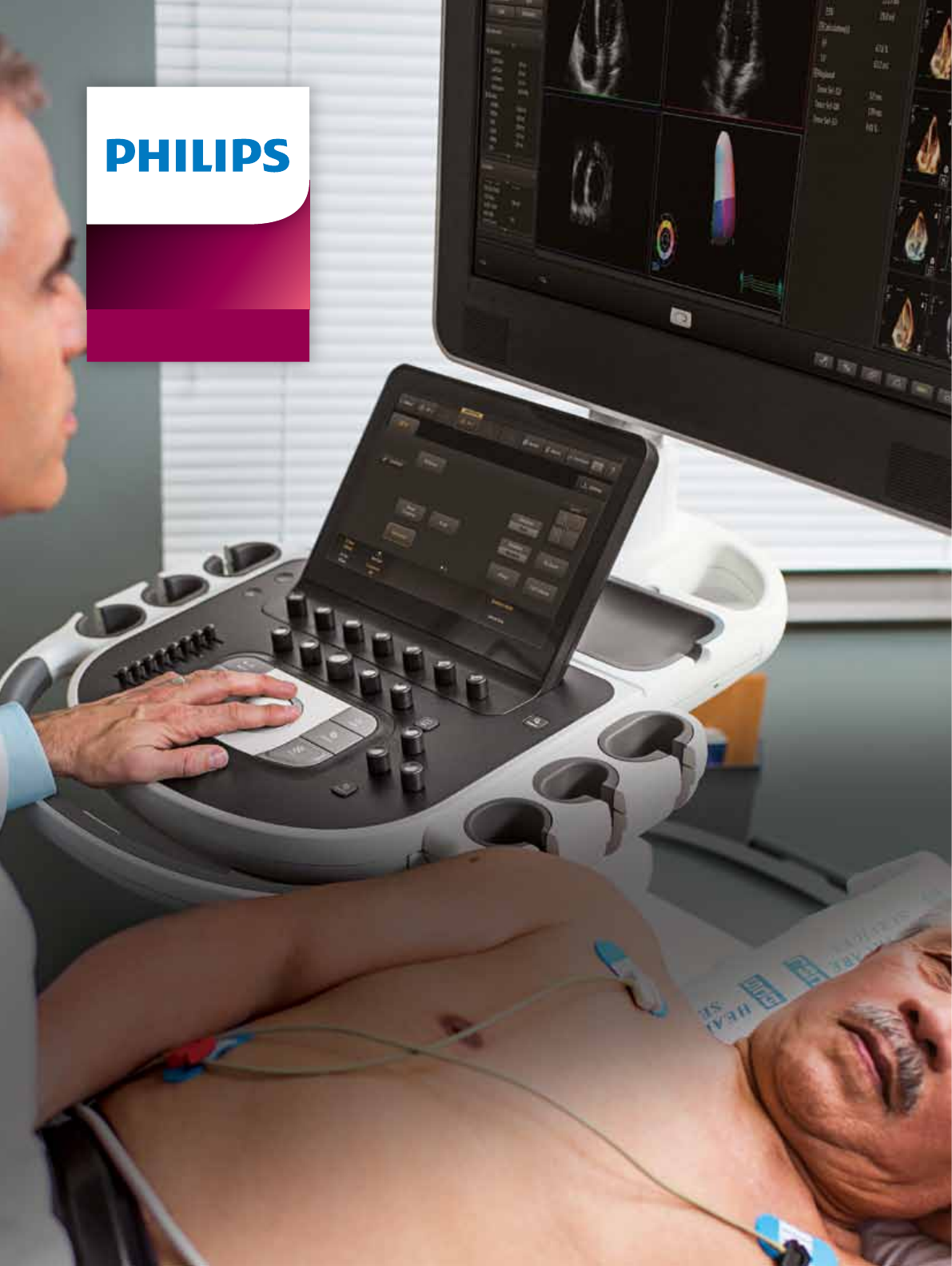
Philips EPIQ 7 ultrasound system
The evolution of
premium cardiovascular ultrasound
Ultrasound
EPIQ 7

2
Unprecedented advances in premium ultrasound
performance can help address the strains on
overburdened hospitals and healthcare systems,
which are continually being challenged to provide
a higher quality of care cost-effectively. The goal
is quick and accurate diagnosis the first time and
in less time.
Premium ultrasound users today demand improved
clinical information from each scan, and faster and
more consistent exams that are easier to perform
and allow for a high level of confidence, even for
technically difficult patients.
The new challenges
in global healthcare

3
Key trends in global ultrasound
It’s our most powerful architecture ever applied to
ultrasound imaging – touching all aspects of acoustic
acquisition and processing, allowing you to truly
experience ultrasound’s evolution to a more definitive
modality. Supported by our family of proprietary
xMATRIX transducers and our leading-edge Anatomical
Intelligence, this platform offers our highest level of
premium performance.
•There is a continued search for
aordable healthcare solutions in
order to deliver more for less with
high-quality patient care.
•Echocardiography is the imaging
mode of choice and exam volumes
continue to increase every year.
•With echocardiography gaining
prominence as a point-of-care
tool (such as in the emergency
department), increasing numbers
of patients are being referred to
cardiologists for further investigation.
The evolution of premium
cardiovascular ultrasound

44
EPIQ 7 is the new direction for premium ultrasound, featuring
an exceptional level of clinical performance to meet the
challenges of today’s most demanding practices.
Performance
More confidence in your diagnoses
even for your most difficult cases

5
Image quality: the numbers tell the story
Comparing EPIQ 7 to conventional premium systems shows
breakthrough advances in imaging performance.*
•Up to 30% increase in penetration
(penetration = ability to scan at depths and maintain
resolution in order to complete the study)
•Up to 15% increase in axial resolution
(increased resolution throughout the depth of image)
all while maintaining frame rates
Creating new realities,
redefining clinical expectations
nSIGHT Imaging goes beyond conventional ultrasound
performance for new levels of definition and clarity.
Conventional
Users must
choose
between
frame rate
and image
quality
nSIGHT
Imaging
More than
doubles the
frame rate
without impact
to image
quality
nSIGHT Imaging
creates superbly focused
images with fewer transmit
operations so you can
experience both highly
detailed ultrasound
images and extraordinary
temporal resolution.
Frame rate Penetration
Conventional
Best resolution
is limited
to transmit
focal zone
nSIGHT
Imaging
Corrects focus
during beam
reconstruction
for superb
uniformity
nSIGHT Imaging
achieves superb uniformity
through coherent beam
reconstruction algorithms
that apply mathematical
focal correction coecients
continually at all depths
of the image.
Conventional
Penetration
limitations
and poor
sensitivity to
weak signals
nSIGHT
Imaging
Superb
penetration
across full
range of
frequencies
nSIGHT Imaging
architecture’s ultra-wide
dynamic range and unique
beam reconstruction
reinforces weak tissue
signals allowing enhanced
penetration at higher
frequencies even on
dicult patients.
Conventional
Penetration icons
option 4
option 5
option 6
Conventional
Penetration icons
option 4
option 5
option 6
Uniformity
Philips nSIGHT Imaging
is a totally new approach
The Philips proprietary nSIGHT Imaging
architecture introduces a totally new
approach to forming ultrasound images.
Unlike conventional systems that form
the image line by line, nSIGHT creates
images with superb resolution down
to the pixel level.
Extraordinary architecture
nSIGHT Imaging incorporates a custom
multi-stage precision beamformer
along with massive parallel processing.
This proprietary architecture captures
an enormous amount of acoustic data
from each transmit operation and
performs digital beam reconstruction
along with mathematically optimized
focal processing to create real-time
images with exceptional resolution
and uniformity.
* 2013 quantitative engineering study comparing Philips iE33 ultrasound
system with EPIQ 7. Dependant upon transducer, application, and TSI.

6
Philips pioneered advanced technologies such as xMATRIX
and PureWave. The revolutionary nSIGHT architecture of EPIQ 7
makes xMATRIX and PureWave even more powerful.
xMATRIX is our most leading-
edge, versatile ultrasound
transducer technology
No other premium ultrasound system
can run the complete suite of the
world’s most innovative ultrasound
transducers. With the touch of a button,
xMATRIX oers all modes in a single
transducer: 2D, M-mode, color Doppler,
Doppler, iRotate, Live xPlane, Live 3D,
Live 3D Zoom, and Live 3D Full Volume.
nSIGHT Imaging makes
powerful xMATRIX technology
even more so
Use Live xPlane imaging to create two
full-resolution planes simultaneously,
allowing you to capture twice as much
clinical information in the same amount
of time. Acquire near isovoxel resolution
to reveal images from any plane within
the volume. Now it’s all possible.
2D and Live 3D image clarity with the X5-1 and X7-2 transducers.
Maximize
extreme clinical capabilities

7
nSIGHT Imaging strengthens
the power of PureWave to image
technically difficult patients
PureWave crystal technology
represents the biggest breakthrough
in piezoelectric transducer material
in 40 years. The pure, uniform crystals
of PureWave are 85% more ecient
than conventional piezoelectric
material, resulting in exceptional
performance. This technology allows
for improved penetration in dicult
Additional PureWave transducer solutions for abdominal and fetal echo.
C5-1 C9-2
Leading-edge xMATRIX transducers for cardiology also include X7-2t for TEE applications.
patients with a single transducer
for excellent detailed resolution.
All xMATRIX transducers incorporate
PureWave technology.
PureWave oers the solution when
imaging technically dicult patients
in a wider range of applications
on a cardiology platform, such as
the PureWave C5-1 and C9-2 for
dicult-to-image abdominal and
fetal echo patients.

8
Advanced workflow
The design of the platform features
“walk-up usability,” meaning that
users can perform an exam with
minimal training.2 The system oers
the automation to drive eciency
throughout exams with features such
as Real Time iSCAN (AutoSCAN),
which automatically optimizes gain
and TGC continuously to provide
excellent images in 2D, 3D, or 4D.
Amazingly portable
At just 104 kg (230 lb), EPIQ 7 is lightest
in its class and 40% lighter than the
heaviest competitive premium system.
Easily transport EPIQ 7 on both carpet
and tile oors. Place it in sleep mode,
move it, and boot up in seconds. The
monitor folds down to reduce overall
system height for transport, and the
integrated cable hooks and catch tray
are ideal for portable studies. Wireless
DICOM further aids workow.†
Tablet-like touch interface allows
quick navigation to system functions
with 80% less reach and 15% fewer
steps to complete an exam.
Designed to reinvent
the user experience
EPIQ 7 has completely reinvented the premium ultrasound
user experience. Ease of use, workflow, ergonomics,
portability – we’ve revolutionized how you interact with
an ultrasound system from every standpoint, and kept it
beautifully intuitive.
More than 80% of sonographers experience work-related
pain, and more than 20% of these suffer a career-ending
injury.1 The EPIQ tablet-like interface results in dramatic
reduction in reach and button pushes, with 80% less
reach and 15% fewer steps.*

9
1 Society of Diagnostic Medical Sonography, Industry Standards for the Prevention
of Musculoskeletal Disorders in Sonography, May 2003.
2 External user study where all users had over 90% success (gold standard in usability
on their set tasks with no training on EPIQ, Jan 2013.
3 University of Colorado, Protocols Study, Apr. 2007.
4 Auto Doppler Clinical Study, Dec. 2011.
* Engineering study comparing Philips iE33 ultrasound system with EPIQ 7.
† Check for availability in your geography.
Easy viewing and efficient use even
in darker scanning environments with
a large 54.6 cm (21.5 in) wide screen
and ambient lighting that provides
subtle visual cues for the keyboard,
OEMS, and transducer ports. Four
transducer ports decrease the amount
of plug/unplugging required during
a day of scanning.
Library quiet
EPIQ 7 is almost silent when running.
A noise test determined that EPIQ 7
runs at 37-41 dB, which is equivalent
to the sound of a library. This is
extremely welcome in small
scanning/examination rooms.
Scanning comfort
Multiple degrees of articulation for both
the control panel and 54.6 cm (21.5 in)
LCD monitor with 720° of freedom
allows for ergonomic alignment for
scanning comfort, whether sitting
or standing.
SmartExam
SmartExam decreases exam time
by 30-50%, keystrokes by as many
as 300 per exam, and results in a high
level of consistency among users.3 It is
fast and easy to customize, providing
consistent annotation, automatic mode
switching, and missed view alerts to
streamline exams.
SmartExam also drives the automation
within Q-Apps, reducing the number
of steps to perform more complex
analysis to a ZeroClick status. The result
is more time to focus on your patients,
increased condence in complete
studies, less focus on requirements,
less repetitive motion, less stress,
and improved schedule maintenance
and department eciencies.
Auto Doppler for vascular imaging
Auto Doppler takes time-consuming
color box positioning and sample
volume placement from ten steps to
three steps and reduces the number
of repetitive button pushes by an
average of 68%.4
Active native data
Active native data allows for post-
processing of many exam parameters
as well as providing the best format
for Q-Apps quantication.
Set-up Wizard
Set-up Wizard allows users to step
up to the system, easily establish user
congurations, and get running quickly.
EPIQ 7 is one of the greenest
systems we have ever designed.
It consumes 25% less power than
our legacy premium ultrasound.
25%
less power
EPIQ 7 makes it easy
to be green
Designed to reinvent
the user experience

10
EPIQ 7 is our most intelligent premium ultrasound system ever, offering a complete
set of easy-to-use quantitative tools to turn reproducible data into information to
guide treatment.
Anatomical Intelligence is the heart of EPIQ 7
More data is available than ever before, requiring tools
for you to simplify and quicken the process of acquiring
reproducible data and turning it into valuable information
for your patients.
At the heart of the powerful EPIQ 7 architecture is our
Philips exclusive Anatomical Intelligence Ultrasound (AIUS),
designed to elevate the ultrasound system from a passive
to an actively adaptive device. With automatic anatomy
Intelligence
turning images into answers
recognition, protocols for automatic functionality, and
proven quantication, exams are easier to perform, more
reproducible, and deliver new levels of clinical information.
Using built-in models to drive exam simplification
With AIUS, libraries of organ model data gathered across
many modalities create a platform where information from
a single exam can be tailored to a patient-specic organ
model or Region of Interest that yields useful information
in less time, with less training, and with less complexity.

11
Automation
Automated Cardiac Motion
QuantificationA.I. (aCMQA.I.) with
ZeroClick technology for adult echo
The ZeroClick technology of the
Automated Cardiac Motion QuanticationA.I.
(aCMQA.I.) uses speckle mechanics to
provide reproducible 2D Global Longitudinal
Strain (GLS) speckle measurements.
A proven EF is also calculated by
using the Auto-ROI that drives the
automation within the aCMQA.I. Q-App.
aCMQA.I. with ZeroClick
technology provides both
EF and GLS from the same
2D images.
*Edit option
a2DQA.I. with ZeroClick for fast, reproducible EF on all your patients.
Automated 2D Cardiac
QuantificationA.I. (a2DQA.I.)
with ZeroClick technology
The ideal tool of every echo lab,
Automated 2D Cardiac QuanticationA.I.
with ZeroClick technology uses AIUS
for an Auto-ROI to drive the Q-App
and provide rapid access to proven
2D EF and volumes. AutoEF is available
during the study and so ts in with
an everyday echo protocol.
Sophisticated modeling adapts certain atlas shapes to
a patient’s individual organ using feature data collected over
hundreds of patients with various conditions. AIUS ranges
from automating repetitive steps to full-blown computer-
driven analysis with minimal user interaction – all using
anatomic intelligence and all providing the results you need.
In fact, many of our tools come with ZeroClick technology,*
which means that, once loaded, the tool does it all for you.
Enhancing the power of xMATRIX TEE
for interventional echo
The EPIQ 7 and Philips Allura Xper X-ray systems create
a powerful combination with the new EchoNavigator feature
for an exceptional level of eciency in the interventional suite.
EchoNavigator digitally links ultrasound and uoroscopy images
using anatomical data. Both active images are displayed and
continuously aligned, even when one image is rotated.

12
The Mitral Valve NavigatorA.I. (MVNA.I.)
The Mitral Valve NavigatorA.I. (MVNA.I.) is designed to take
a Live 3D volume of the mitral valve and turn it into an
easy-to-interpret model in eight guided steps, providing
access to a comprehensive list of MV measurements and
calculations. Internal comparison of MVQ to MVNA.I. tools
measures 89% fewer clicks.5
MVNA.I. saves steps at each part of the process
•Annulus data is acquired with 74% fewer clicks,5 which also
provides leaet tracing with no user interaction.
•MVNA.I. guides the entire process using simple commands
and clear graphics, making this a much easier tool to use
than previous mitral quantication tools.
•Results derived from MVNA.I. can be seen on the screen as
they become available, speeding the process of accessing
required data.
MVNA.I. takes a Live 3D volume of the mitral valve and turns
it into an easy-to-interpret model in just eight guided steps.
Navigation
5 2013 QLAB 9 MVQ and QLAB 10 MVN click comparison internal study.

13
Q-App quantification applications
EPIQ 7 offers a wide variety of sophisticated Q-Apps to quantify ultrasound image
information including our latest AIUS Q-Apps.
Q-Apps Clinical application Benefit
IMT (for vascular) Automatic carotid intima media
thickness measurement
Fast and easy access to IMT data
ROI Echo contrast and color images Extract acoustic measurements from images
Strain Quantification (SQ) Measures the myocardial velocity
from color tissue Doppler
Derive displacement strain and strain rate
CMQ Stress Speckle quantification of stress
echo images
Decrease the subjectivity of stress echo analysis
3DQ View, slice, and display 3D volumes
and measure distance and areas
from 2D MPR views
Bi-plane LV volume, ejection fraction (EF)
and LV mass calculations
3DQA Global LV volumes and timing Measure LV endocardial volumes, stroke volume
(SV), and true 3D ejection fraction (EF) using a
semi-automated border detection in 3D space.
Offers timing assessment for each of 17 minimal
regional volumes and determines a synchronicity
index for all volume segments or a user-selectable
group of volume segments.
AIUS Q-Apps Clinical application Benefit
Automated 2D Cardiac
QuantificationA.I. (a2DQA.I.)
AutoEF for 2D images Fast and reproducible biplane EF
Automated Cardiac Motion
QuantificationA.I. (aCMQA.I.)
Speckle quantification of global
and regional strain data
Both EF and speckle data simultaneously
assist with LV function assessment
Mitral Valve NavigatorA.I. MVNA.I. Takes a Live 3D volume of the
MV and provides qualitative and
quantitative data of the valve
and its surrounding structures
Easy to understand data

14

15
Use the EPIQ 7 multimodality query
retrieve to view DICOM images such
as CT, NM, MRI, iXR, cardiac X-ray,
and ultrasound. Easily compare past
and current studies without the use
of an external reading station and
even review these multimodality
images while live imaging.
iXR integration
Connectivity to EchoNavigator via
our digital network link enhances
communication on modern structural
interventions using 3D TEE. Users
can appreciate anatomy with multiple
views of Live 3D TEE, availability of
virtual echo scanning, and echo target
localization on uoro.
The real-time integration of EchoNavigator
between uoroscopy and Live 3D TEE
provides automatic registration and
tracking – all controlled tableside.
Access to
multimodality
images
EchoNavigator
Multimodality Query Retrieve allows side-by-side comparison
on any DICOM image.
Real time integration of iXR and Live 3D TEE images.

16
New levels
of clinical information
Normal aortic valve Normal mitral valve in dual imaging
Mitral regurgitationAnatomical M-mode of PLAX
Dilated cardiomyopathy using EchoPen preset Mitral and tricuspid regurgitation in Live 3D

17
Common carotid artery bifurcation Posterior tibial veins and artery
Mitral regurgitation Fetal echo – aortic arch
Tissue Doppler PW Fetal echo – four-chamber heart

Remote services mean
we’re closer than ever*
Remote desktop
Spend less time on the phone with a Philips “Virtual Visit”
with remote system interaction for fast technical and clinical
troubleshooting and guided scanning options.
iSSL technology
This industry-standard protocol meets global privacy standards
and provides a safe and secure connection to the Philips remote
services network using your existing Internet access point.
Online support request
Enter a support request directly from your EPIQ system for
a fast, convenient communication mechanism that reduces
workflow interruption and keeps you at the system and
focused on your patient.
Utilization reports
Data intelligence tools that can help you make informed
decisions to improve workflow, deliver quality patient care,
and decrease the total cost of ownership. This is the only
ultrasound utilization tool that provides individual transducer
usage and the ability to sort by exam type.
Proactive monitoring
Proactive monitoring allows for the detection and repair
of anomalies before they become problems and helps us to
better predict potential failures and proactively act on them.
Increase system availability, optimize workflow, and promote
patient satisfaction by scheduling downtime as opposed
to reacting to an unexpected problem.
Advanced support services
are proactive and predictive
We understand your challenges: uncertain economic times, changing
healthcare landscapes, and the impact of healthcare reform. We know
that ecient workows and system uptime are critical success factors
in running an eective healthcare business.
Philips is committed to oering innovative solutions to provide you
with world-class services that move from reactive to proactive and
with predictive service models that provide high system availability
and enhanced workow to help you deliver high-quality patient care.
*Check for availability in your geography.18

19
The remote desktop allows Philips
service engineers to gain a live
view of your system’s console for
remote operation, real-time clinical
troubleshooting, and issue resolution.
Exceptional serviceability
Philips oers the only ultrasound utilization
tool that provides individual transducer
usage and the ability to sort by exam type.
The system features superior modular
design for rapid repair, getting your
system up and running quickly.
Intelligent software architecture
Software is easily optimized, maintained, and restored
by the service user without risk to patient data, giving you
peace of mind when dealing with software anomalies and
confidence that your data is safe.
This software architecture takes patient data privacy
to a new level. Patient data is stored on a separate partition
and physical location to provide protection and ease of
removal, providing you total control of your data.
Clinical education solutions
Our comprehensive, clinically relevant courses, programs,
and learning paths are designed to help you improve
operational efficiency and enhance patient care.

© 2015 Koninklijke Philips N.V. All rights are reserved.
Philips Healthcare reserves the right to make changes in specifications
and/or to discontinue any product at any time without notice or
obligation and will not be liable for any consequences resulting from
the use of this publication.
Please visit www.philips.com/EPIQ
Printed in The Netherlands.
4522 991 09211 * APR 2015
