Philips 882478 User Manual Product Brochure Bright View SPECT/CT Ready System 0f7f7c74320a4494a136a77c0157661e
User Manual: Philips 882478 Product Brochure Philips BrightView SPECT/CT ready system Philips - BrightView X SPECT/CT ready system882478
Open the PDF directly: View PDF ![]() .
.
Page Count: 6
Fits you like no other
BrightView X and XCT specications
The new BrightView X system is a fully featured variable-
angle camera that is eld-upgradeable to BrightView XCT
without any increase in room size or power requirements.
Designed to put patients rst, the system is easy to use, fast
and reliable with exceptional image quality. BrightView X
is differentiated by exclusive CloseUp high resolution
technologies.
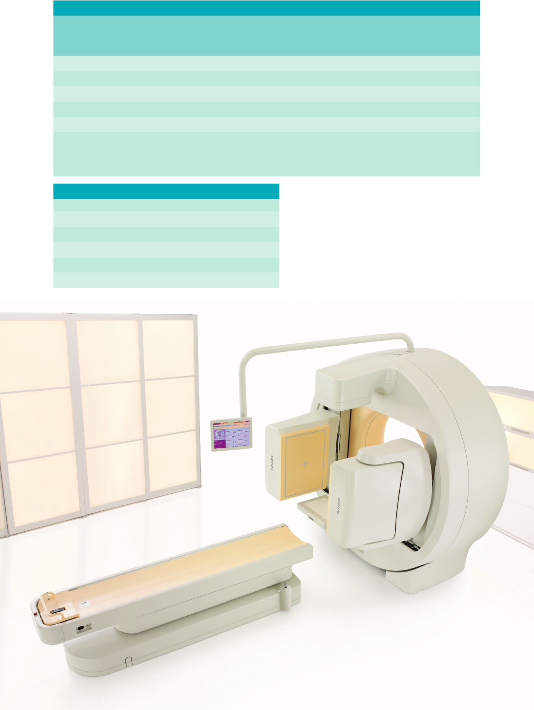
Surprisingly compact given its capabilities, the heart of
BrightView XCT is the unique integration of BrightView
X with advanced Philips at-panel X-ray technology.
Substantial clinical advantages include co-planar SPECT
and CT with no table index between acquisitions in
many cases, exible CT breathing protocols optimized
for localization and attenuation correction, and high
resolution at low CT dose levels.
All of this is possible without changing the way you like
to work or your patients’ comfort. Fully supported
by the resources of Philips services, Philips offers you
comprehensive support and training throughout the life
of your system, giving you knowledge and experience to
make the most of BrightView X and XCT from day one.

BrightView X and XCT specications
Camera characteristics
Gantry dimensions 210 cm H x 212 cm W x 97 cm D
Weight (without collimators) BrightView X: 1936 kg (4260 lb)
BrightView XCT: 2045 kg (4500 lb)
Power requirements 480 VAC, 50A, three phase
Total heat dissipation (typical) BrightView X: 2850 BTU/hr
BrightView XCT: 6831 BTU/hr
Gantry aperture 36" (91.4 cm)
Caudal-cephalic tilt ± 15°
Co-planar CT and SPECT* Axial elds-of-view overlap by 14 cm
Patient table
Dimensions 243 cm L x 47.5 cm W
Pallet type Carbon ber
Thickness 0.38˝ (9.5 mm)
Attenuation <7% @ 140 keV
Pallet dimensions 83.5˝ L x 15˝ W (212 cm x 38.1 cm)
Height (from oor) 26˝ to 40˝ (66.0 cm – 102.8 cm)
Weight capacity 500 lb (227 kg)
Total body
Scan length + UFOV 78.7˝ + 16˝ (200 cm + 40.6 cm)
Scan speed 0.5–75 in/min. (1–190 cm/min.)
Emission tomography
ECT rotation 540°
Angular sampling 1.4° to 90°
Speed of rotation 5.0 rpm
Scan diameter (with LEGP) <4˝ – 29.5˝ (<10.2 cm – 75 cm)
ECT manual rotation speeds 0.5 and 1.33 rpm
Detector relative positions 90° and 180°
Detector
True energy independence Fixed high voltage
Universal ood calibration One ood for all radionuclides
(up to 300 keV)
Non-anger digital detector 1 ADC/PMT
Field of view (rectangular) 16˝ x 21.25˝ (40.6 cm x 53.9 cm)
Crystal thickness 0.375˝ (9.5 mm) or 0.75˝ (19.1 mm)
Photomultiplier tubes 59
Lead shielding 364 keV
Brain reach 2.9˝ (7.4 cm)
Cardiac dead space 0.9˝ (2.4 cm)
Variable magnication 1.0x, 1.46x, 1.85x, 2.19x
Exchanger type Semi-automatic
Collimator storage Cart based up to 11 units
(5 pairs and 1 pinhole)
Collimator types LE, ME, HE, pinhole Note: Specications subject to change. * Applies to BrightView XCT only
JETStream acquisition
Mobile acquisition console 18˝ at LCD monitor, keyboard
and trackball or mouse
Spectrum analyzers 16 (with overlap)
Energy range 56 – 920 keV
Window adjustment 1% to entire energy range
Spectrum display Color-coded, graphical, fully
interactive
Count capacity 32K per channel
Preset count or time 1 ct. to 2 billion cts.,
l sec. to >1,000 min.
Image orientation 0°, 90°, 180°, and 270°
Patient position display 2 sec. to innity, decay-based
persistence or xed refresh
Concurrent imaging Up to 15 simultaneous data sets
from a single acquisition
XCT physical assembly*
Type of detector Digital amorphous silicon,
columnar CsI scintillator
Detector size 30 cm x 40 cm
Detector pixel 0.2 mm x 0.2 mm
Number of elements 3,145,728
Generator output 10 kW, pulsed (2 msec. to continuous)
kVp 120 rotating anode
mA 5 – 80
Max. anode storage capacity 600 kHU
Max. anode cooling rate 1350 HU/sec.
Focal spot 0.4 mm
XCT performance*
Axial eld-of-view 14 cm in a single 360º rotation
Maximum rotation speed 12 seconds for 360º rotation
Maximum axial range 172 cm
Transaxial eld-of-view 47 cm
Spatial resolution > 15 lp/cm @ 10% MTF
Low contrast resolution 5 mm @ 0.5% on 20 cm
CATPHAN phantom with
40 mm slice thickness
Absorption range -1000 to +3000 Hounseld units
Number of slices 140 slices @ 1 mm thickness
Slice thickness Variable from 0.33 mm to 2.0+ mm
Scan or display matrix 256 and 512
Reconstruction time < 1 min for one CT eld-of-view
(for typical patient)*
Extremities 15 mGy +/- 20%
Body (localization) 6.5 mGy +/- 20%
Cardiac (attenuation correction) 0.5 mGy +/- 0.2 mGy
Collimator exchange system and storage CTDIVOL body dose levels (for typical patient)*

Note: Specications subject to change. * Applies to BrightView XCT only
CTDIVOL body dose levels (for typical patient)*
** Astonish – Reconstruction method with 2 iterations and 32 subsets. Values subject to change.
** Planar Astonish – calculated values
1. Specications are NEMA NUI – 2001 method of measurement with a 20% energy window. Values to be used for acceptance testing.
2. Represents calculated values derived from measured 3.8" (9.5 mm) crystal values and/or limited sampling 3/4" (19.1 mm) crystal values.
Values not to be used for acceptance testing.
3. Measured using a pixel size of 9.3 mm.
Note: Specications subject to change.
NEMA1Typical2
Intrinsic spatial resolution
FWHM
FWTM
UFOV
4.3 mm
8.2 mm
CFOV
4.3 mm
8.2 mm
UFOV
4.0 mm
7.8 mm
CFOV
4.0 mm
7.2 mm
Intrinsic energy resolution UFOV
9.8%
UFOV
9.5%
Intrinsic spatial linearity
Absolute
Differential
UFOV
0.6 mm
0.2 mm
CFOV
0.4 mm
0.2 mm
UFOV
0.25 mm
0.04 mm
CFOV
0.13 mm
0.04 mm
Intrinsic ood eld
uniformity3Integral
Differential
UFOV
± 2.5%
± 2.0%
CFOV
± 2.2%
± 1.5%
UFOV
± 2.0%
± 1.4%
CFOV
± 1.7%
± 1.3%
System spatial resolution
(LEHR) @ 10 cm FWHM without scatter
FW TM without scatter
FWHM with scatter
FW TM with scatter
NEMA
7.9 mm
14.8 mm
8.3 mm
17.2 mm
Astonish**
5.8 mm
10.3 mm
5.8 mm
11.0 mm
SPECT reconstructed spatial
resolution (LEHR) without
scatter @ 15 cm radius
Central transaxial
Central axial
Peripheral radial
Peripheral tangential
Peripheral axial
NEMA
10.7 mm
11.3 mm
10.9 mm
9.4 mm
11.2 mm
Astonish*
5.2 mm
5.4 mm
5.0 mm
5.1 mm
5.4 mm
Whole body system spatial
resolution @ 10 cm without
scatter, 10 cm/min scan speed
Parallel LEHR
Perpendicular LEHR
FWHM
8.4 mm
8.3 mm
FWTM
15.5 mm
15.3 mm
Astonish**
5.9 mm
5.9 mm
Astonish**
10.9 mm
10.9 mm
Volume sensitivity per axial
centimeter (LEHR) ± 7%
UFOV
12900 (cts/sec)
(MBq/cm2)
UFOV
12900 (cts/sec)
(MBq/cm2)
Intrinsic spatial resolution
@ 75 Kcps FWHM
FWTM
UFOV
4.8 mm
9.1 mm
UFOV
N/A
N/A
Intrinsic detector count
rate performance
+/- 10%
Output 20% loss
Max count rate
UFOV
300 Kcps
350 Kcps
System sensitivity (LEGP)
± 7%
UFOV
311 cpm/μCi
UFOV
311 cpm/μCi
Multiple window spatial
registration
UFOV
0.8 mm
UFOV
0.6 mm
Detector-detector sensitivity
variation (LEHR,TC-99m)
UFOV
5%
UFOV
1%
BrightView X and XCT detector specications 3/4˝ (19.1 mm) crystal

** Astonish – Reconstruction method with 2 iterations and 32 subsets. Values subject to change.
** Planar Astonish – calculated values
1. Specications are NEMA NUI - 2001 method of measurement with a 20% energy window. Values to be used for acceptance testing.
2. Same test conditions as NEMA specications. Represents average factory test values. Values not to be used for acceptance testing.
3. Measured using a pixel size of 9.3 mm.
Note: Specications subject to change.
NEMA1Typical2
Intrinsic spatial resolution
FWHM
FWTM
UFOV
3.3 mm
6.3 mm
CFOV
3.3 mm
6.3 mm
UFOV
3.2 mm
6.1 mm
CFOV
3.1 mm
6.0 mm
Intrinsic energy resolution UFOV
≤ 9.6%
UFOV
9.2%
Intrinsic spatial linearity
Absolute
Differential
UFOV
0.50 mm
0.10 mm
CFOV
0.35 mm
0.09 mm
UFOV
0.18 mm
0.03 mm
CFOV
0.10 mm
0.03 mm
Intrinsic ood eld
uniformity3Integral
Differential
UFOV
± 2.5%
± 2.0%
CFOV
± 2.2%
± 1.5%
UFOV
± 2.0%
± 1.4%
CFOV
± 1.6%
± 1.2%
System spatial resolution
(LEHR) @ 10 cm FWHM without scatter
FW TM without scatter
FWHM with scatter
FW TM with scatter
NEMA
7.4 mm
14.0 mm
7.8 mm
16.5 mm
Astonish**
5.1 mm
9.0 mm
5.1 mm
9.8 mm
SPECT reconstructed spatial
resolution (LEHR) without
scatter @ 15 cm radius
Central transaxial
Central axial
Peripheral radial
Peripheral tangential
Peripheral axial
NEMA
10.3 mm
10.9 mm
10.5 mm
9.0 mm
10.8 mm
Astonish*
4.3 mm
4.3 mm
3.8 mm
3.7 mm
3.7 mm
Whole body system spatial
resolution @ 10 cm without
scatter, 10 cm/min scan speed
Parallel LEHR
Perpendicular LEHR
FWHM
8.0 mm
7.9 mm
FWTM
14.7 mm
14.5 mm
Astonish**
5.3 mm
5.3 mm
Astonish**
9.7 mm
9.7 mm
Volume sensitivity per axial
centimeter (LEHR) ± 7%
UFOV
11500 (cts/sec)
(MBq/cm2)
UFOV
11500 (cts/sec)
(MBq/cm2)
Intrinsic spatial resolution
@ 75 Kcps FWHM
FWTM
UFOV
3.8 mm
7.3 mm
UFOV
3.6
6.9
Intrinsic detector count
rate performance
+/- 10%
Output 20% loss
Max count rate
UFOV
300 Kcps
350 Kcps
System sensitivity (LEGP)
± 7%
UFOV
277 cpm/μCi
UFOV
277 cpm/μCi
Multiple window spatial
registration
UFOV
0.6 mm
UFOV
0.4 mm
Detector-detector sensitivity
variation (LEHR,TC-99m)
UFOV
5%
UFOV
1%
BrightView X and XCT detector specications 3/8˝ (9.5 mm) crystal
BrightView X and XCT system performance
Type Hole
shape
Size
(mm)
Septa
(mm)
Length
(mm)
Const.
(%) (keV)
Sensitivity
Cpm/μCi
@ 0 cm @ 10 cm
LEGP HEX 1.40 0.180 24.7 Foil 2.1 140 27713.9 8.9
LEHR HEX 1.22 0.152 27 Foil 1.7 140 16813.7 7.4
CHR HEX 2.03 0.152 48 Foil 1.1 140 16514.2 7.8
MEGP HEX 3.40 0.86 58.4 Cast 6.1 300 21225.3 10.9
HEGP HEX 3.81 1.73 58.4 Cast 4.2 364 10635.7 12.1
HEPH ROUND 3.0 25.4 220.0 Cast — —4835— —
4.0 25.4 220.0 Cast — —41395— —
5.0 25.4 220.0 Cast — —42225— —
BrightView X and XCT camera and collimator specications
Septa
penetration
Spatial resolution
system6
Collimators Mass (kg)*
LEGP Low energy general purpose 30
LEHR Low energy high resolution 35
CHR Cardiac high resolution 29
MEGP Medium energy general purpose 88
HEGP High energy general purpose 128
HEPH High energy pinhole 131
* These are the mass of the complete collimator.
1 Sensitivity is for Tc-99m with 20% window, 9.5 mm thick crystal.
2 Gallium-67 with 20% window 93 keV, 184 keV, 300 keV photo peaks.
3 Sensitivity is for I-131.
4 The pinhole collimator is rated for 364 keV and has 25.4 cm
FOV at the crystal.
5 Relative sensitivity is for Tc-99m at 10 cm from the pinhole.
6 For 9.5 mm thick crystal.
Note: Sensitivity numbers are NEMA Class Standards and are ±7%.
Note: Specications subject to change.
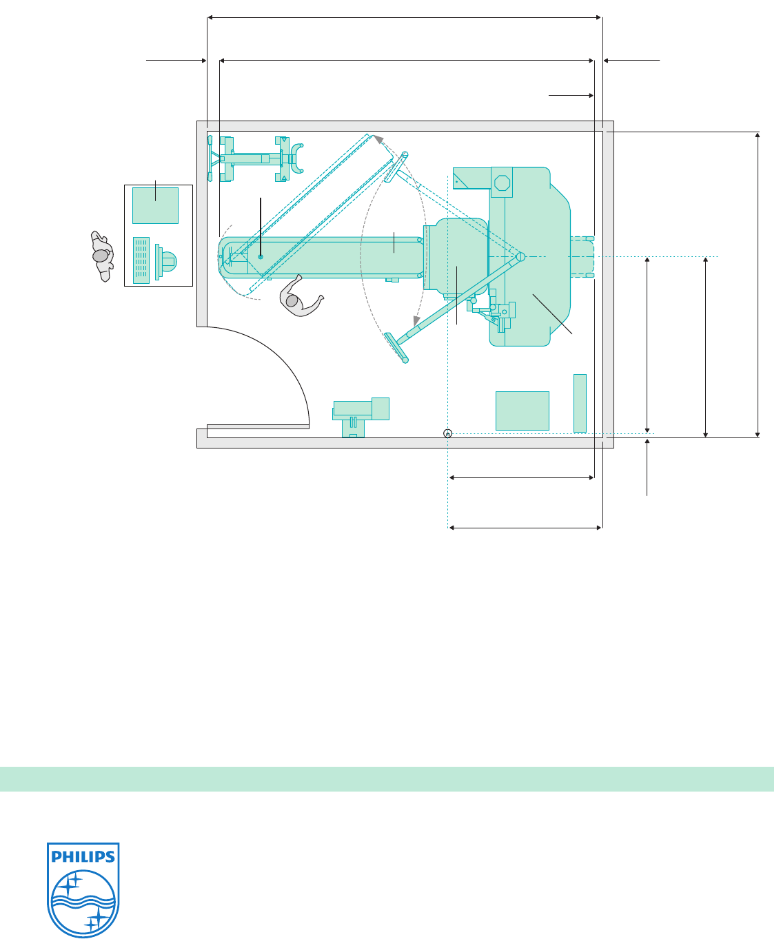
Detectors
Power
generator
Wall-mount
acquisition terminal
Pivot point
Patient
table
UPS
186" (15'6")
4724 mm
176.52"
4484 mm
5.48"
139 mm
End of 2.0 m
Table travel
4"
102 mm
Table
75"
1905 mm
76.8"
1950.7 mm
139" (11'7")
3530 mm
1.8"
45.7 mm
68.92"
1751 mm
72.92"
1852 mm
Source
Detector
Control
Gantry
C
L
C
L
C
L
Customer-provided desktop.
Philips-provided computer,
monitor and keyboard.
Exam Room
BrightView XCT minimum room layout
Environmental requirements for general equipment location
Throughout the SPECT suite, the HVAC system must maintain the
temperature between 16°C (60°F) to 24°C (75°F) with less than 5°C
(10°F) variation per hour. Humidity must be between 20% to 75%.
These requirements are 24 hours per day, 7 days per week.
BrightView X has the same room requirements except the X-ray power generator is not included.
Philips Healthcare is part of Royal Philips Electronics
www.philips.com/healthcare
healthcare@philips.com
Printed in The Netherlands
4522 962 87931 * JUL 2012
© 2012 Koninklijke Philips Electronics N.V.
All rights are reserved.
Philips Healthcare reserves the right to make changes in specications
and/or to discontinue any product at any time without notice or obligation
and will not be liable for any consequences resulting from the use of this
publication.
Please visit www.philips.com/brightviewxct
