Philips 882482 User Manual Product Brochure Bright View SPECT/CT System XCT 24deb4f48194490fb1c3a77c01574e41
User Manual: Philips 882482 Product Brochure Philips BrightView SPECT/CT system XCT Philips - BrightView XCT SPECT/CT system882482
Open the PDF directly: View PDF ![]() .
.
Page Count: 67
Volume 2
Clinical case study collection
Philips BrightView XCT nuclear medicine system
Five key advantages
Cardiology
Oncology
Orthopedics
Infection
Other localization
Case parameters
Contents
4
6
9
33
49
56
66
2
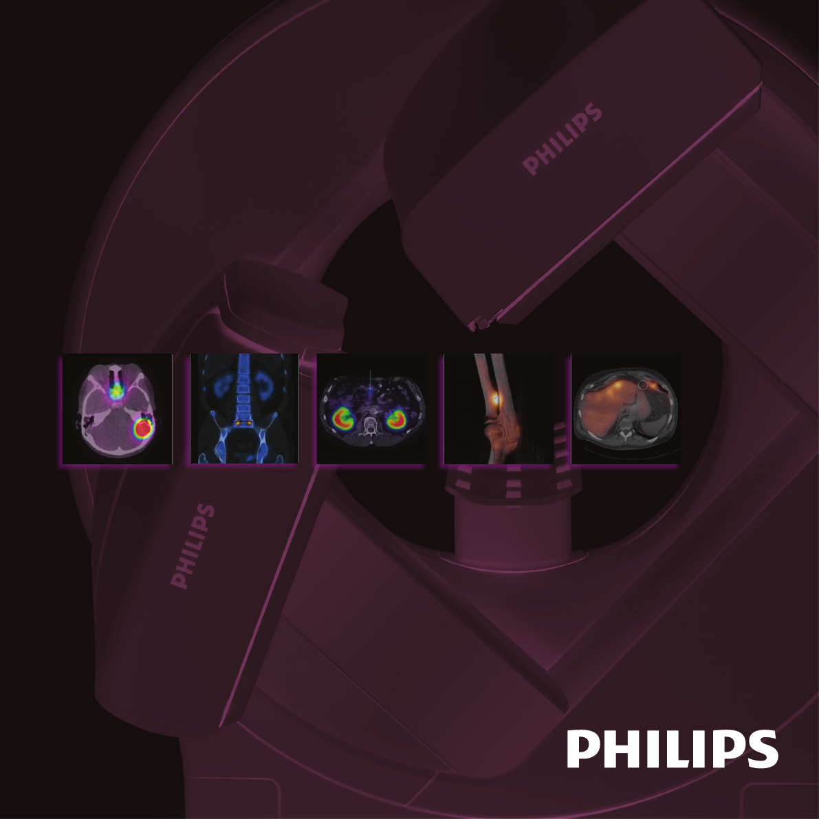
BrightView XCT
A technologically advanced SPECT/CT system
designed entirely for nuclear medicine
At Philips, we are dedicated to providing innovative,
integrated solutions to give you the tools you need
to accurately diagnose abnormalities early in disease
progression. We are tirelessly focused on image quality
and exibility, throughput, and patient care. With that
in mind, we have compiled this second volume of actual
clincial case studies. Philips thanks those customers who
have collaborated with us and contributed their ndings
to this effort.
We hope that you nd this an informative reference
in your quest to provide the best in diagnostic care
for your patients.
4
1 Registration condence with CoPlanar
• No bed index between SPECT and CT, in most cases, for 14 cm axial coverage.
2 Flexible breathing
• Tidal respiration (60 sec) for CT-AC to match SPECT breathing
• Breathhold (12 sec) for localization
3 High resolution – low dose
• Isotropic voxels – high quality CT images when viewed
at any angle
• Sub-mm (0.33 mm) slice thickness for high resolution bone
• Flexible X-ray current (5-80 mA) to t the clinical need
4 Nuclear medicine – tailored workow
• Same capabilities as BrightView SPECT
• Plan SPECT/CT from the p-scope
• Option for in-room CT acquisition control
5 Fits the nuclear medicine space
• Fits in a small nuclear-medicine-sized room
(15'6'' x 11'7''; 4.72 x 3.53 M)
• Low system weight (4500 lb; 2045 kg)
• Separate control room not required
BrightView XCT – Fits you like no other
Five key advantages
Full Iterative Technology (FIT)
Philips introduces Full Iterative Technology (FIT) – the rst hybrid system to provide both iterative
SPECT and CT reconstruction capabilities. The advanced CT reconstruction algorithm improves
CT image quality by reducing noise and improving uniformity. FIT builds on the value of Astonish
SPECT reconstruction, iterative technology that has been proven in practice to improve image
quality and reduce dose. This leading technology provides the necessary foundation for advancing
future developments in iterative CT reconstruction.
55
Filtered Back Projection FIT-Iterative CT Reconstruction
Attenuation correction – SPECT acquisitionAttenuation correction – CT acquisition
Cardiology
Trusted attenuation correction
Condence in the registration accuracy between SPECT and CT
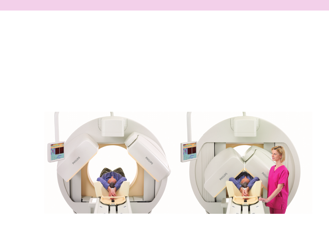
BrightView XCT has several advantages for
cardiac attenuation correction. The gantry
rotation can be set to 60 seconds to allow
for tidal respiration during multiple respiratory
cycles over a single 360° rotation. This technique
blurs the CT to match resolution of the SPECT
image, leading to excellent diaphragm alignment.
The entire heart volume is sampled in a single
14 cm axial eld of view with no stair-step
artifacts as a result of the isotropic resolution.
The CT eld of view overlaps the SPECT
eld of view so that little to no table index
is required between acquisition steps, resulting
in registration condence.
6
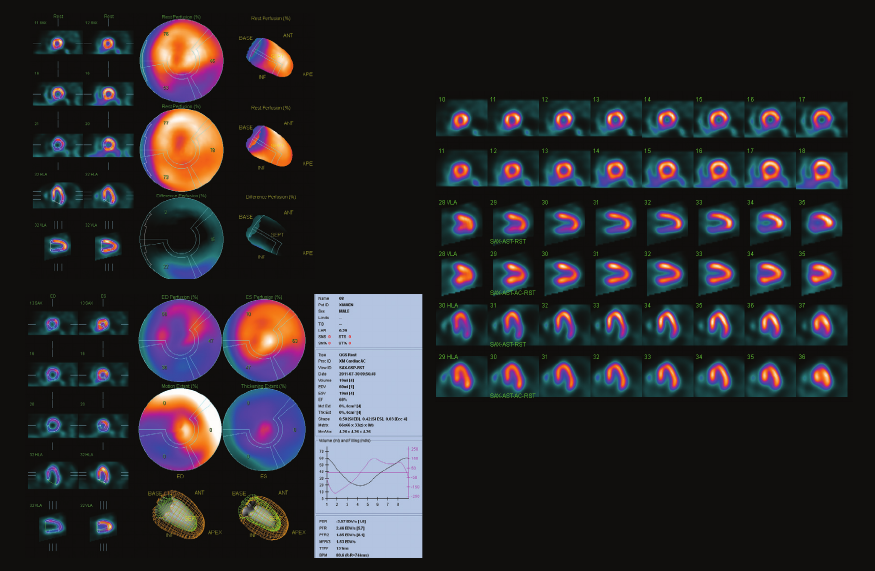
1 Cardiology case study
Patient information
•58-year-oldmale
•Evaluateformyocardialischemia
Procedure
•Tc-99mMIBIcardiacperfusionandfunction
Inferior wall attenuation correction
Courtesy of Xiamen No. 1 Hospital, Xiamen, China
Findings from SPECT/CT study
•Defectininferior-septalwallbutimproved
signicantlywithattenuationcorrection
•Normalejectionfraction
Physician impression of SPECT/CT
•Furtherinvestigationdemonstratedpatient
tobenormal
Top row – no attenuation correction
Bottom row – with attenuation correction
7
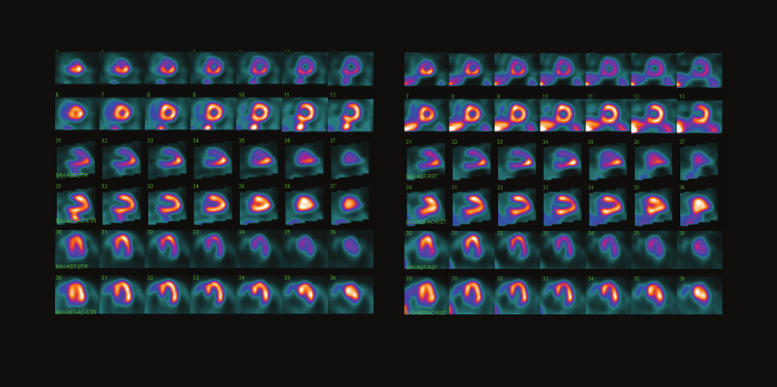
2 Cardiology case study
Patient information
•86-year-oldfemale
•AbnormalstressEKG(posteriorandlateral
hypokinesis);highprobabilityofischemia
Procedure
•Tc-99mMyoviewperfusionandfunction
Anterior wall attenuation correction
Courtesy of Wollongong Nuclear Medicine, New South Wales, Australia
Findings from SPECT/CT study
•Reducedperfusioninanteriorwallwhichnormalized
withCTattenuationcorrection
•Normalstudy:LVEF=68%
Physician impression of SPECT/CT
•SPECT/CTwithattenuationcorrectioncompletely
changedtheinitialhighpatientprobability
tolowprobability
8
Stress Uncorrected and Corrected Rest Uncorrected and Corrected
Image
quality
Radiation dose
(mAs or mGy)
AC Localization Diagnostic
Primary
NM applications
Oncology
Low dose localization
Designed entirely for nuclear medicine
BrightView XCT offers premium CT resolution
at low dose levels – a fraction of a conventional
helical CT.
Flexible breathing protocols during localization
studies allow for a breathhold CT acquisition
to be obtained in as short a time as 12 seconds.
Providing 14 cm of axial coverage in a single
breathhold helps to maintain image resolution
and required anatomic detail.
9
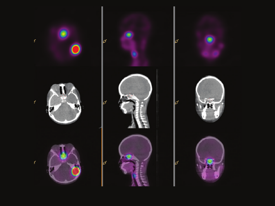
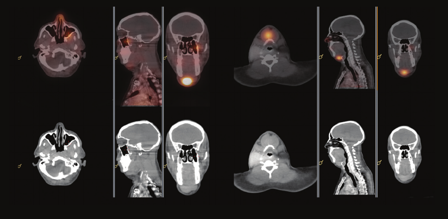
1 Oncology
Patient information
•11-year-oldfemale
•Neuroblastoma,postchemotherapyandtumor
resection;oneyearlater,tumorfoundinleftposterior
cranialfossaonMRI
•Latelytumorgrowingsopre-surgicalMIBGwasordered
Procedure
•Tc-99mMIBGscan
Neuroblastoma
Courtesy of National Center for Child Health and Development, Tokyo, Japan
Findings from SPECT/CT study
•IntracranialMIBGuptakeseenatleftposteriorcranial
fossaandsphenoidalsinus;checkofpreviousMRI
T1-CEfoundsphenoidalsinusenhancedmassaswell
asleftposteriorcranialfossamass
Physician impression of SPECT/CT
•DifculttolocalizeasmalllesionbySPECT-only;
SPECT/CTshowslocationeasily
•Sphenoussinusuptakewasshowncorrectlyandposition
matchedMRIlesion
•Informationofasingleormultiplelesionsisveryimportant
•Biopsyofleftposteriorcraniallesionwasganglioneuroma,
notmalignancy
1 mm isotropic voxels
10
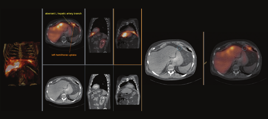
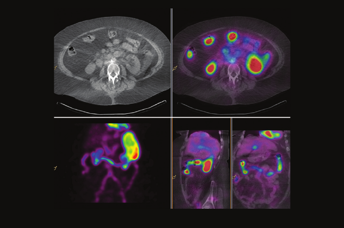
2 Oncology
Patient information
•70-year-oldmale
•Multifocalhepatocellularcancerpresentingfor
MAAmappingandhepatopulmonaryshuntstudy
inpreparationforY-90radioembolization
•Statuspostcoilembolizationofgastroduodenalartery,
rightgastricartery,andsupraduodenalbranchartery
Procedure
•Tc-99mMAA
MAA mapping for radioembolization
Courtesy of University of Washington Medical Center, Seattle, Washington
Findings from SPECT/CT study
•Increaseduptakeinhepaticlobesatknown
hepaticmetastases
•Largeextrahepaticfocusinleftlowerhemothorax
correspondingwithleftcardiophrenicrecessin
inferiormediastinum
•Novisualpulmonaryuptake,estimatedhepatopulmonary
shuntratio4.91%,withinnormallimits
Physician impression of SPECT/CT
•Hepatomediastinalshuntfromaberrantmediastinal
vesselarisingfromdistallefthepaticartery
•InordertoundergosafeY-90radioembolization,
patientwillneedcoilembolizationtoavoiddamage
toleftmediastinum
11
1 mm isotropic voxels
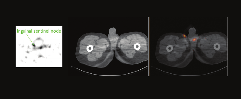
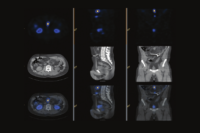
3 Oncology
Patient information
•58-year-oldmale
•Carcinomaofthepenis
•Localizesentinellymphnode
Procedure
•Tc-99mColloid
Carcinoma of the penis
Courtesy of Innsbruck Medical University, Tyrol, Austria
Findings from SPECT/CT study
•Moderateuptakeintherightinguinalregion
Physician impression of SPECT/CT
•SPECT/CTrevealedinguinallymphnode,notvisible
onplanar
•Sentinellymphnodewasremoved;histologyshowed
atrophiclymphnode,nosignofmalignancy
1 mm isotropic voxels
12
4 Oncology
Patient information
•56-year-oldfemale
•PostLu-177DOTA-TATEtherapyevaluation
ofneuroendocrinetumor
Procedure
•Lu-177DOTA-TATE
Post Lu-177 DOTA-TATE ther apy
Courtesy of Innsbruck Medical University, Tyrol, Austria
Findings from SPECT/CT study
•Somatostatinreceptorlesionislocatedinthemusculus
rectusinferior
Physician impression of SPECT/CT
•Patientwasreferredtoaspecializedophthalmologist
1 mm isotropic voxels
13
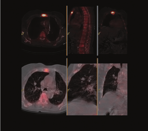
5 Oncology
Patient information
•81-year-oldfemale
•Breastcancer;threeweeksofleftsternal
painwithpositiveregionallymphnodes
Procedure
•Tc-99mHDPbonescan
Incidental pulmonary nodule
Courtesy of Nepean Nuclear Medicine and PET, Sydney, Australia
Findings from SPECT/CT study
•Intenseuptakeinmanubriumconsistentwithrecent
fracturewithevidenceofongoingnewboneformation;
noskeletalmetastases;end-platedegeneration
atL2-3andL4-5
•Incidentalndingofpulmonarynoduleinrightupperlobe
Physician impression of SPECT/CT
•Identiedfractureinsteadofmetastaticdisease
•Incidentalndingofpulmonarynodulewhichwillhelp
forfurthersurveillance
1 mm isotropic voxels
14
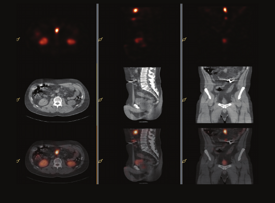
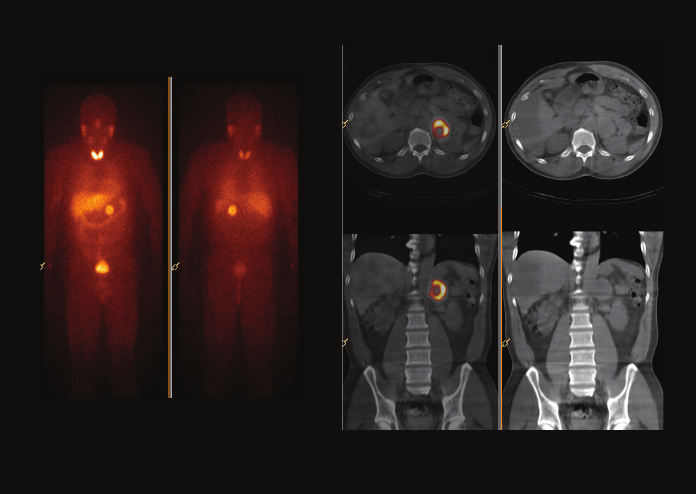
6 Oncology
Patient information
•73-year-oldmale
•Nonfunctioningpancreaticendocrinetumor;
pancreaticoduodenectomywithclearmargins,
negativelymphnodessixyearsago
•MultipleOctreoscans;lasttwoyears–stablefocus
ofuptakeinmidabdomenwithoutndingsonCT,
unclearsignicance
Procedure
•In-111Octreotide
Benign reactive lymph node
Courtesy of University of Washington Medical Center, Seattle, Washington
Findings from SPECT/CT study
•Focusofmidabdominaluptakeagainseen
•SPECT/CTcorrelatespreciselywithsofttissuedensity
locatedwithinthemesentaryposteriortotransverse
colon,8.3cmanteriortoL1vertebralbodyendplate
Physician impression of SPECT/CT
•SPECT/CTdemonstrateduptaketoanormal-sized
mesentericlymphnode;thishadnotbeenpossible
onplanarimages
•Uptakewithinbenignreactivelymphnodesisaknown
falsepositiveinOctreoscans
1 mm isotropic voxels
15
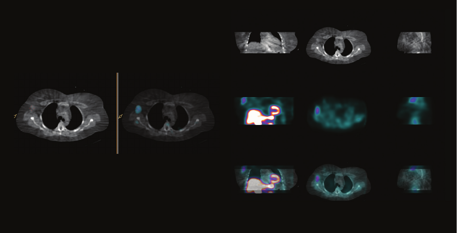
7 Oncology
Patient information
•55-year-oldfemale
•Three-monthhistoryofnonexertionalchest
discomfort,leftarmnumbness,shortnessofbreath
Procedure
•Tc-99mSestamibi
Right breast mass
Courtesy of Fletcher Allen Health Care University, Burlington, Vermont
Findings from SPECT/CT study
•Normalstresstest
•MasswithuptakeofMIBIinaxillarypartofrightbreast
seenonlowdoseCT
Physician impression of SPECT/CT
•Patientunderwentdiagnosticbreastmammography
andultrasoundfollowedbybiopsy
•Inltratingductalcarcinomaonpathology
1 mm isotropic voxels
16
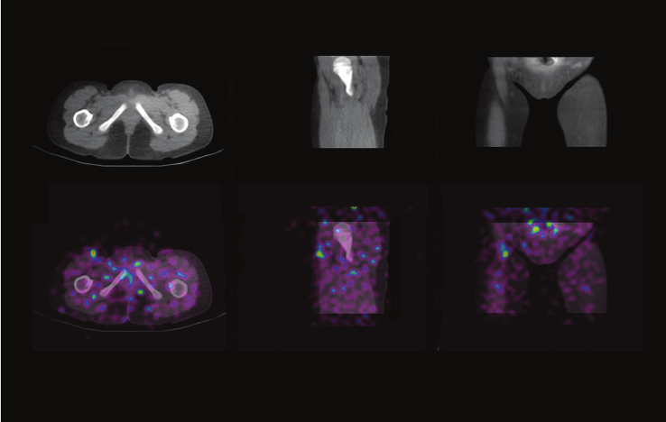
Patient information
•4-year,10-month-oldfemale
•Metastaticneuroblastoma,surgeryunabletond
lymphnodes
•PriorMIBGscanpositiveinrightgroin,decided
torescanafterSPECT/CTinstalled,then
schedulesurgery
Procedure
•I-123MIBG
Findings from SPECT/CT study
•Discretefocusofabnormalactivityinrightgroin;
fusionwithCTindicatesabnormalityisinroot
ofthesartorismuscle,extremelyraresiteofmetastasis
fromaneuroblastoma
Physician Impression of SPECT/CT
•Surgerywithassistanceofgammaprobeallowed
removalofasingleintramuscularmetastasis;SPECT/CT
demonstratedthefocusofMIBGdidnotcorrespond
toalymphnode
8 Oncology
Metastatic neuroblastoma
Courtesy of Clinique Universitaires St-Luc, Brussels, Belgium
1 mm isotropic voxels
17
9 Oncology
Patient information
•85-year-oldmale
•Penilecancer
Procedure
•Tc-99mNanocolloid
Lymphoscintigraphy of the penis
Courtesy of The Harley Street Clinic, London, UK
Findings from SPECT/CT study
•Localizationofsentinelnodesinleftandright
inguinal-femoralregions
•Measurementofdepthfromskinsurfacetaken
toassistsurgeon
Physician impression of SPECT/CT
•Helpedtoassistsurgeoninlocationofsentinellymph
nodespriortobiopsy
•Astonishreconstructionsoftwareassistsgreatlyasthe
SNIdosesareverylow,socountrecoveryreconstruction
assistsinimagequality
1 mm isotropic voxels
18
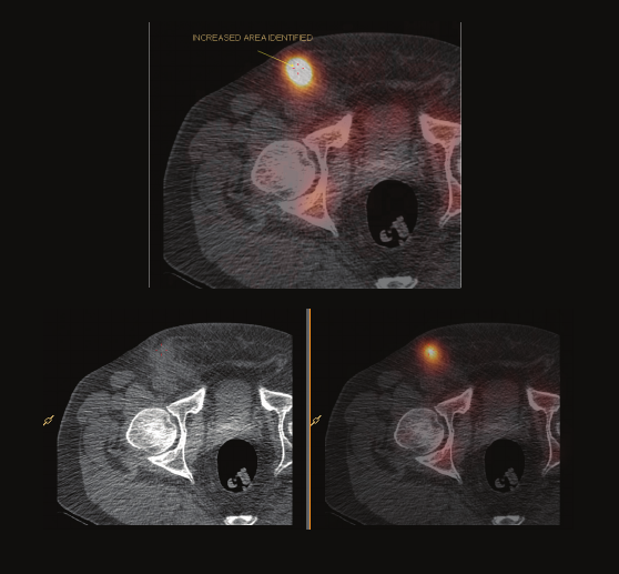
10 Oncology
Sarcoma in pubic symphysis
Courtesy of Sydney X-Ray, Sydney, Australia
Patient information
•Ruleoutosteitispubis
Procedure
•Tc-99mbonescan
Findings from SPECT/CT study
•Earlyanddelayedplanarimageswereconsistent
withasevereosteitispubis
•SPECT/CT,however,clearlydemonstrated
alyticlesioninrightpubicsymphysis
Physician impression of SPECT/CT
•Ratherthancontinuedineffectivetreatmentof
suspectedosteitispubis,abiopsywasperformed
whichconrmedsarcoma;patientthenproceeded
toappropriatetreatment
1 mm isotropic voxels
19
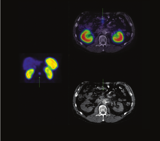
11 Oncology
Patient information
•63-year-oldfemale
•Hyperparathyroidism;bilateraladrenalnodules
onCT,leftsuspiciousformalignancy
•Evaluateforpheochromocytomainoneorboth
adrenalnodules
Procedure
•I-123MIBG
Findings from SPECT/CT study
•Intenseuptakewithin3cmleftadrenalnodule
consistentwithpheochromocytoma
•Milddiffuseuptakeinrightadrenalgland,likelyphysiologic;
secondpheochromocytomacannotbeexcluded
Pheochromocytoma in adrenal nodule
Courtesy of University of Washington Medical Center, Seattle, Washington
Physician impression of SPECT/CT
•Intenseuptakeinleftadrenalregionseenonplanar
imagesbutcouldnotbecondentlylocalizedtoadrenal
gland;SPECT/CTreadilylocalizeduptake
tothesuspiciousnodule
•Givenclinicalsuspicionforbilateralpheochromocytoma,
contrastresolutionofSPECTwasnecessaryto
demonstratenosignicantuptakeinleftadrenalgland
•Successfulleftadrenalectomyanddiscontinuationof
catecholamine-blockingmedicine,conrmingcorrect
ndingofunilateralpheochromocytoma
1 mm isotropic voxels
20
12 Oncology
Calcication of tibial-bular ligament
Courtesy of Washington Hospital Center, Washington DC
Patient information
•63-year-oldmale
•Lungcancer,assessforbonymetastases
Procedure
Tc-99mMDPbonescan
Findings from SPECT/CT study
•Calcicationoflefttibial-bularligament
Physician impression of SPECT/CT
•SPECT/CThelpedpreciselyidentifypost-traumatic
calcicationofaligamentasetiologyofactivity
onbonescan
•Fractureand/ormetastasiswasruledout
21
0.33 mm isotropic voxels
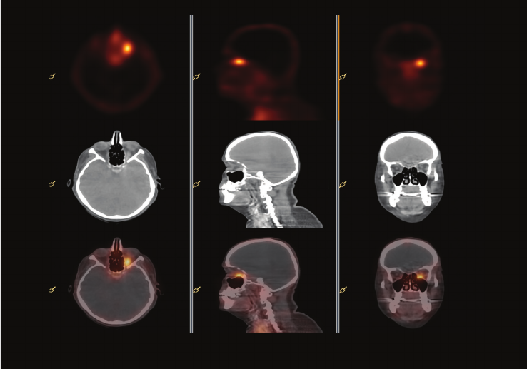
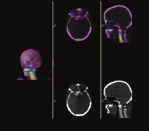
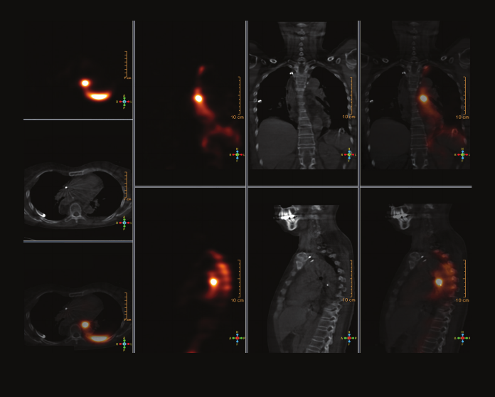
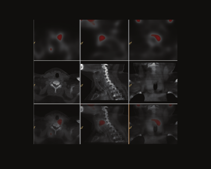
13 Oncology
Patient information
•47-year-oldmale
•Thyroidcancer;evaluateformetastaticdisease
andstaging
Procedure
•I-124scan
Findings from SPECT/CT study
•SPECT/CTHead/Neck–sinuspolyp,submental
lymphnodemetastases
•SPECT/CTChest–macronodularlungmetastases
(known)
Thyroid cancer
Courtesy of Washington Hospital Center, Washington DC
Physician impression of SPECT/CT
•Preciselyidentiedsinuspolypactivityasinammatory
andnotmetastatic,identiedalargesubmentallymph
nodemetastasis,identiedknownmacronodular
pulmonarymetastases
•I-124imagingaspartofdosimetryprovideslow
resolutionSPECTimagesandrequiresproperanatomic
localizationachievedwithSPECT/CT
1 mm isotropic voxels
22
14 Oncology
Patient information
•47-year-oldmale
•Suspectedneuroendocrinetumorafterabdominal
lymphnodebiopsy,unknownprimary;gastroscopy,
rectalprocto-colonoscopy,andendosonocapsule
withoutpathologicndings
Procedure
•In-111Octreotidescan
Findings from SPECT/CT study
•Highintensivefocusonileumloop
•Highintensivefocusinmiddleabdomen,
areaofpathologicallymphadenopathy
Neuroendocrine tumor of ilium
Courtesy of University Hospital of Halle, Halle, Germany
Physician impression of SPECT/CT
•Clearlocalizationofpathologicalsomatostatin-
receptorbindingledtototalresectionofprimary
tumorlocatedinileum
•Pathologyshowedwelldifferentiatedneuroendocrine
carcinomaofileumwithinltrationofmesenterial
fattissue,serosa,lymphaticvessels,andlocoregional
lymphnodemetastasis
•Follow-upisplannedwithOctreoscanandevaluation
forDOTA-TATEtherapy
1 mm isotropic voxels
23
1 mm isotropic voxels
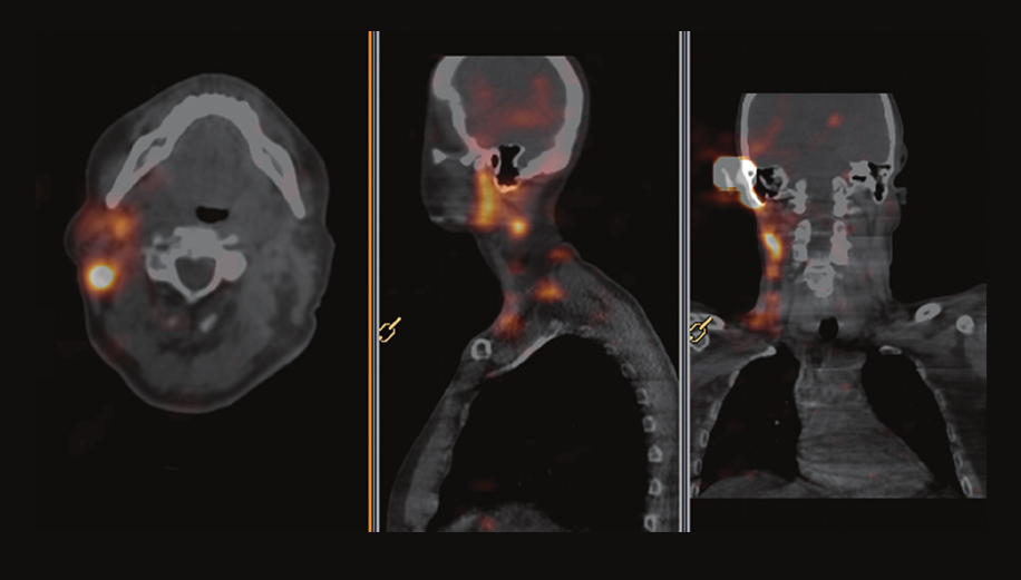
15 Oncology
Melanoma at the right ear
Courtesy of Innsbruck Medical University, Tyrol, Austria
Patient information
•68-year-oldmale
•Excisionofmelanomaatrightear
•Localizesentinellymphnode
Procedure
•Tc-99mColloid
Findings from SPECT/CT study
•Hotspotdorsaltorightjawangleandadditional
hotspotsdownstreamincervicalregion
Physician impression of SPECT/CT
•Sentinelnodedorsaltorightjawwasonlyvisible
onSPECT/CT,notvisibleonplanar
•Sentinellymphnodewasextractedandshowed
nosignofmalignancy
24
16 Oncology
Patient information
•52-year-oldmale
•Leftadrenalmass
Procedure
•I-123MIBG
Pheochromocytoma
Courtesy of Nepean Nuclear Medicine and PET, Sydney, Australia
Findings from SPECT/CT study
•AbnormallyincreasedMIBGuptakeintheleft
adrenalglandsuspiciousofpheochromocytoma
Physician impression of SPECT/CT
•Strongindicationofpheochromocytomawhich
inuencestreatment
25
1 mm isotropic voxels
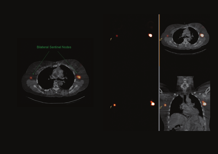
17 Oncology
Bilateral breast cancer
Courtesy of Innsbruck Medical University, Tyrol, Austria
Patient information
•58-year-oldfemale
•Cancerinleftbreast,ductalcarcinomainsitu
rightbreast
•Localizesentinellymphnodes
Procedure
•Tc-99mColloid
Findings from SPECT/CT study
•Sentinellymphnodesinbilateralaxillaryregions;
rightsidenextto4thrib,leftsideintercostalspace
of4thto5thribs
Physician impression of SPECT/CT
•Exactlocalizationofsentinelnodesusingribs
asreference
•Sentinellymphnodeswereresectedandshowed
nosignofmalignancy
1 mm isotropic voxels
26
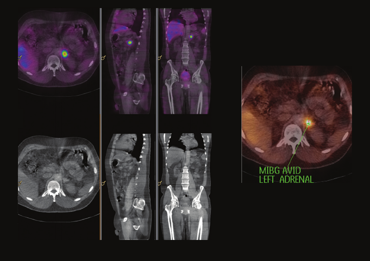
18 Oncology
Patient information
•52-year-oldmale
•LeftadrenalmassonCT;evaluatefor
pheochromocytomaandmetastases
Procedure
•I-123MIBG
Left adrenal mass
Courtesy of Washington Hospital Center, Washington DC
Findings from SPECT/CT study
•Leftadrenalglandpheochromocytomawithcentral
necrosis,noevidenceofmetastaseselsewhere
Physician impression of SPECT/CT
•SPECT/CThelpedpreciselycorrelateI-123MIBG
avidtissuetothemassdescribedbutnotadequately
characterizedbythepriorCTimagesalone
1 mm isotropic voxels
27
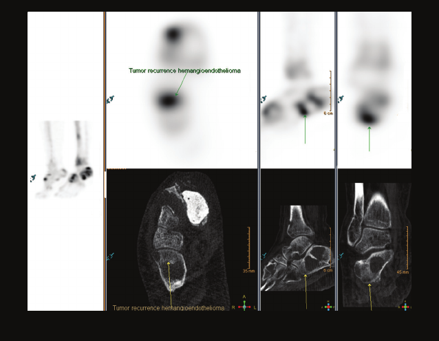
19 Oncology
Hemangioendothelioma
Courtesy of Washington Hospital Center, Washington DC
Patient information
•64-year-oldfemale
•Hemangioendothelioma;posttumorresectioninvolving
proximalrighttibiaandrightmedialcuneiform
Procedure
•Tc-99mMDPbonescan
Findings from SPECT/CT study
•Right-sideddistaltibiaandrightcalcaneum
tumorrecurrence
•Post-surgicalinammatorychangesofproximalright
tibiaandmedialcuneiform
Physician impression of SPECT/CT
•Inthesettingofpost-surgicalchangesandlyticnature
ofthetumor,SPECT/CThelpedcorrectlyidentify
tumorrecurrenceinnewsites;lyticlesionsarenot
greatlyavidwithboneagents
•SPECT/CThelpedidentifyactivity(malignancy)
inperipheryofthelyticlesions
0.33 mm isotropic voxels
28
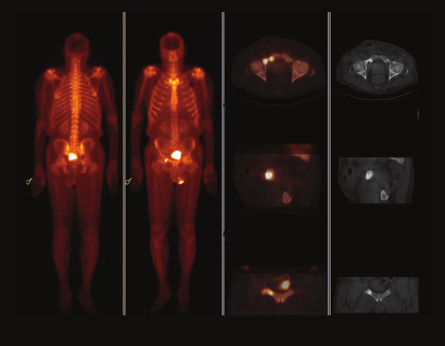
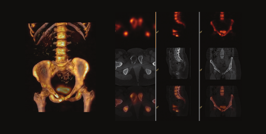
20 Oncology
Patient information
•85-year-oldmale
•Prostatecancer,assessforbonymetastases
Procedure
•Tc-99mMDPbonescan
Sclerotic bony metastases
Courtesy of Washington Hospital Center, Washington DC
Findings from SPECT/CT study
•Rightpubicramusmetastasisextendingintoanterior
aspectofrightacetabulum
Physician impression of SPECT/CT
•Onplanarimages,activityappearstobeinsuperiorlipof
rightacetabulum,commonsitefordegenerativechanges
•SPECT/CThelpedpreciselyidentifylocationtoareasof
scleroticbonymetastases.Managementis100%different
1 mm isotropic voxels
29
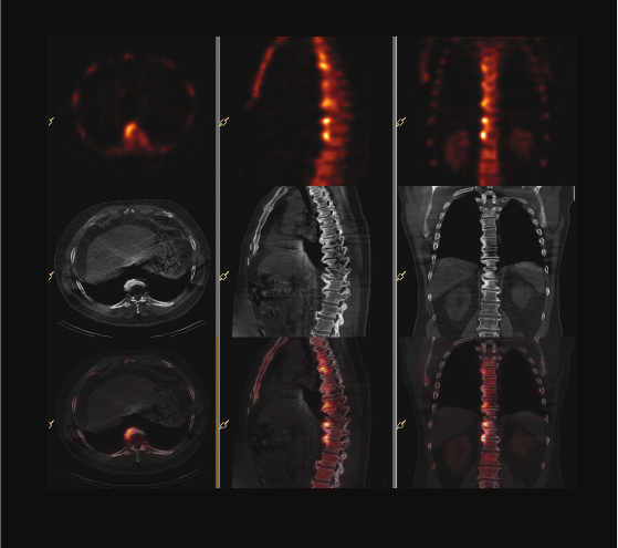
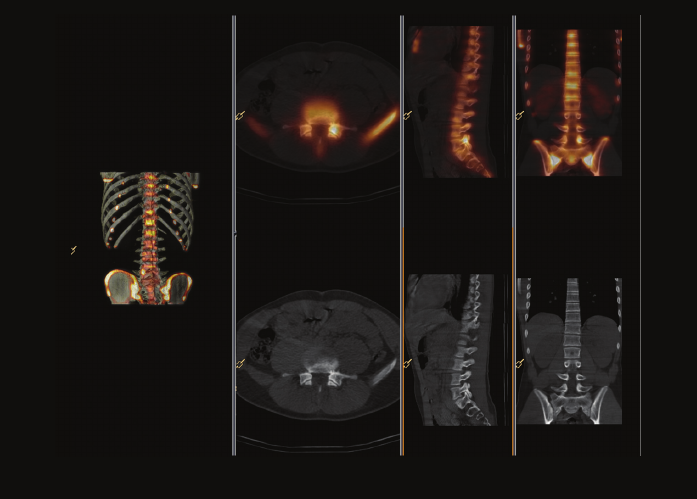
21 Oncology
Multiple degenerative mutations of spine
Courtesy of University Hospital Freiburg, Germany
Patient information
•63-year-oldfemale
•Coloncarcinoma,persistingpaininspine
Procedure
•Tc-99mDPDbonescan
Findings from SPECT/CT study
•Focaluptakeinrightparamedianthoracicspineat5th,
9th,10th,and12ththoracicvertebralbodies;distinct
osteochondrosisinsameregion
•Nofocaluptakeinregionoftheboneislandin
transverseprocessof5ththoracicvertebralbody
Physician impression of SPECT/CT
•Noproofofbonemetastases;multipleobvious
degenerativemutationsinthespine
1 mm isotropic voxels
30
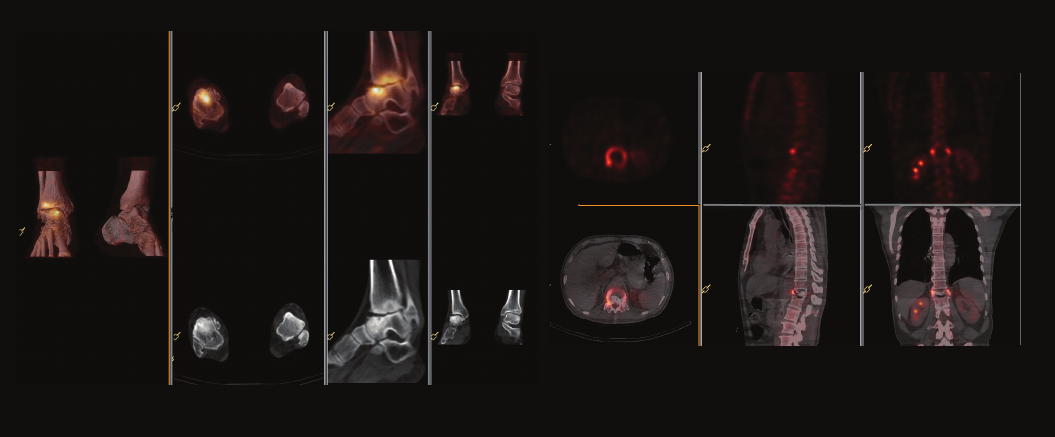
22 Oncology
Patient information
•72-year-oldmale
•Lungcancer;evaluateformetastases
Procedure
•Tc-99mMDPbonescan
Lung cancer evaluation for bone mets
Courtesy of Huadong Hospital, Shanghai, China
Findings from SPECT/CT study
•T12compressionfracture
•Rightanklejointdegeneration
Physician impression of SPECT/CT
•Nometastaseswereidentied;activetreatment
1 mm isotropic voxels
31
Orthopedics
Unique combination of design and technology
High resolution – low CT dose
The high resolution CT images of the BrightView XCT are
a result of the small detector element size (<200 microns).
It has been shown (Optimizing Detector Size in X-ray
Imaging; Kachelrieb & Kalender; IEEE 2005) that signicant
dose reductions can be achieved with such ne sampling.
Our design allows for very high resolution (0.33 mm thick)
CT slices, ideal for extremity bone imaging. Additionally,
high image quality is apparent with data viewed at any
angle, not just the transverse data.
32
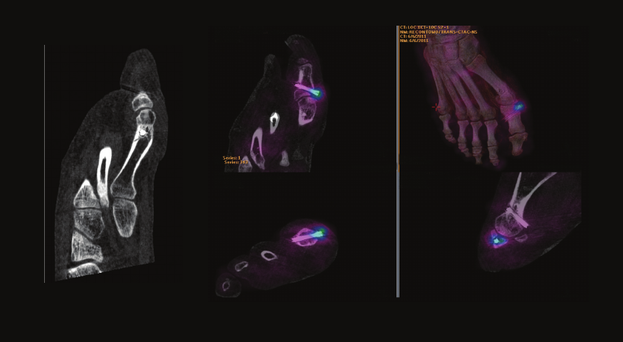
1 Orthopedics
Multiple fractures in Down’s Syndrome patient
Courtesy of Sutherland Nuclear Medicine, Sydney, Australia
Patient information
•44-year-oldfemale
•Down’sSyndrome;injurytoleftfootandankle,
difcultyexplaininglocationandseverityofpain
Procedure
•Tc-99mHDPbonescan
Findings from SPECT/CT study
•Acutefractureofdistalleftbula
•Fractureofanterolaterallipofleftdistalbula
•Injury,possibleincompletefractureofbaseof2nd
metatarsal,bonecontusionofbaseof4thmetatarsal
Physician impression of SPECT/CT
•Provideddetailandclaritytotheextentofinjurythat
planarimagingwasunabletoidentify
•Diagnosisofmutiplefracturesiteshelpedpatient
managementbynecessitatingimmobilizationinapatient
thatwasunabletofullyunderstandandcooperate
0.33 mm isotropic voxels
33
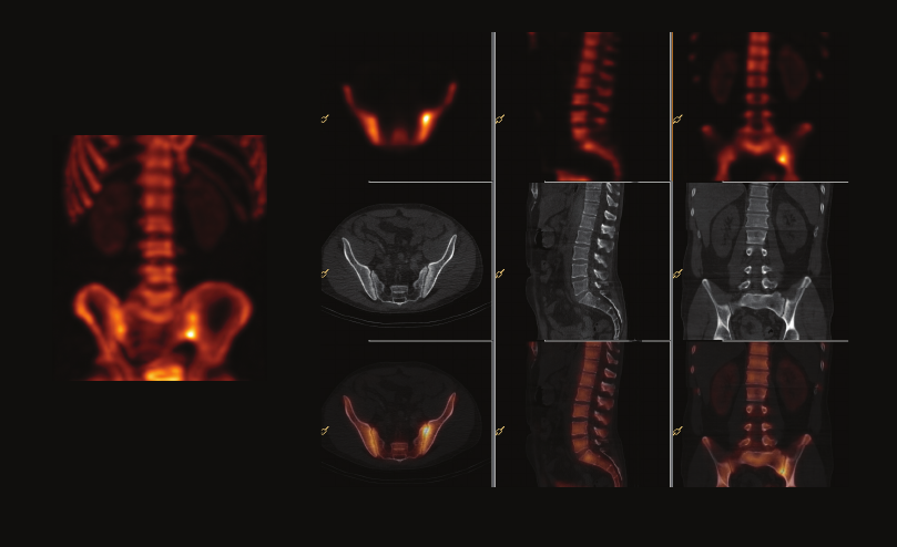
2 Orthopedics
Patient information
•16-year-oldmale
•Suddenonsetbackpaininjuniorprofessional
footballplayer,noimprovementwithphysiotherapy;
MRInormal
Procedure
•Tc-99mMDPbonescan
Early pars stress fracture
Courtesy of Frimley Park Hospital NHS Foundation Trust, Surrey, United Kingdom
Findings from SPECT/CT study
•IncreaseduptakeinL5parsinterarticularis,
normalfacetjoint
Physician impression of SPECT/CT
•Earlystressfracturenotvisualizedonotherimaging
•Criticaldiagnosisinaprofessionalfootballplayer
1 mm isotropic voxels
34
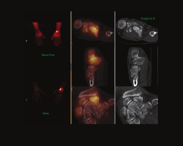
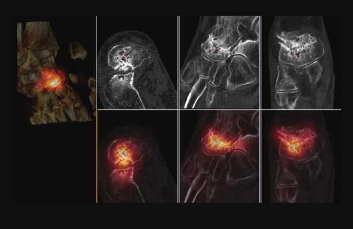
3 Orthopedics
Scaphoid fracture
Courtesy of The Royal Wolverhampton Hospitals NHS Trust, Surrey, United Kingdom
Patient information
•22-year-oldmale
•12weekspostscaphoidfracture,stilltender
Procedure
•Tc-99mMDPbonescan
Findings from SPECT/CT study
•Conrmedscaphoidfracturewithincompleteunion
Physician impression of SPECT/CT
•SPECT/CTprovidedinformationthatallowedmore
aggressiveorthopedictreatmentwithsubsequent
goodoutcome
•Fracturenowhealedandpatientissymptom-free
0.33 mm isotropic voxels
35
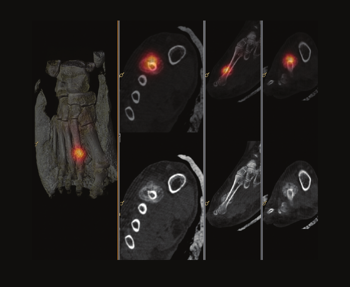
4 Orthopedics
Patient information
•47-year-oldmale
•Rightfootpain
Procedure
•Tc-99mHDPbonescan
Right foot pain
Courtesy of Nepean Nuclear Medicine and PET, Sydney, Australia
Findings from SPECT/CT study
•Activeandosteoblasicprocessatright2ndmetatarsal
shaftconsistentwithrecentfracture
Physician impression of SPECT/CT
•SPECT/CTconrmedfractureandexcludedinfectionand
othercauseswhichledtoappropriatemanagement
0.33 mm isotropic voxels
36
5 Orthopedics
Torus palatini
Courtesy of University of Washington Medical Center, Seattle, Washington
Patient information
•51-year-oldfemale
•Longhistoryofmultiplebonygrowthsinmaxilla,
mandible,hands,rightshoulder,likelyhereditary
multipleexostosis
•Bonygrowthinhardpalatecausingbleeding;
lookformalignanttransformation
Procedure
•Tc-99mMDPbonescan
Findings from SPECT/CT study
•Milduptakeinoropharynxcorrespondingtolarge
growthonCTwithwell-corticatedmarginsandsmall
medullaryspace;hasappearanceoftoruspalatini
•Minimaluptakewithinexostosesinmandible,shoulder,
andcalvarium
Physician impression of SPECT/CT
•CTclariedthehardpalategrowthwasatoruspalatini
ratherthanexostosis
•Concernofmalignanttransformationexcludeddespite
troublingclinicalhistory
1 mm isotropic voxels
37
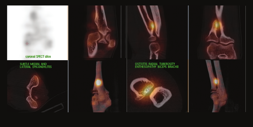
6 Orthopedics
Patient information
•49-year-oldmale
•Chronicelbowpain(mostlyright);
ruleoutepicondylitisradialis
Procedure
Tc-99mMDPbonescan
Findings from SPECT/CT study
•Planarimagesshowhotspotonbilater
proximalforearm,possiblyradius
Biceps enthesopathy
Courtesy of GZA Sint-Augustinus, Antwerp, Belgium
•SPECT/CTofrightelbowaccuratelylocalizes
theintenseuptaketoradialtuberosity,compatible
withbicepsenthesopathy
•Onlysubtleincreaseduptakeatmedialandlateral
epicondyle
Physician impression of SPECT/CT
•Clearlyvisualizesfocalbonelesion–differentdiagnosis
thanorthopedicsurgeonsuspected
•Comparedtoplanarimages(difcultanatomical
interpretation),SPECT/CTgivesmoreaccuratelocalization
ofhotspotandshowsnoevidentstressfracture
0.33 mm isotropic voxels
38
7 Orthopedics
Osteonecrosis
Courtesy of Universitair Ziekenhuis Brussel, Brussels, Belgium
Patient information
•30-year-oldfemale
•Lupusnephretis(underimmunosuppression)
andsicklecelldiseasewithpaininrightknee,
medialaspectofrightfoot,andleftankle
Procedure
•Tc-99mMDPbonescan
Findings from SPECT/CT study
•MDPaccumulationcorrespondingwithaserpigenous
marginofincreaseddensitywhichrunsalongarc-like
radioluscentlesions,characteristicforosteonecrosis
inahealingphase
•Boneinfarctionswithintheepiphysiswithanecrotic
centerofmedullarbonesurroundedbyviablemarrow
andbone
Physician impression of SPECT/CT
•Osteonecrosiswithsignsindicatingrepair;couldbedue
toembolizationofsmallfeedingbloodvesselsrelated
tosicklecelldiseaseorinducedbycorticosteroiduse
0.33 mm isotropic voxels
39
8 Orthopedics
Patient information
•Rightheelpainforafewmonths
Procedure
•Tc-99mbonescan
Calcaneal fracture
Courtesy of Sydney X-Ray, Sydney, Australia
Findings from SPECT/CT study
•Intenseuptakeinrightcalcaneousalongafracture
lineposteriorlyintheCT,whichalsodemonstrated
malalignmentandimpactionofthefracture
Physician impression of SPECT/CT
•Patientwasmanagedappropriatelyforthestress
fractureandorthopedicreviewwasarrangedtoassess
themalalignmentofthefracture
•Nofurtherimagingwasrequired
0.33 mm isotropic voxels
40
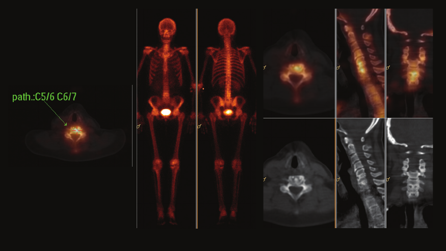
9 Orthopedics
Cervical spine pain
Courtesy of Innsbruck Medical University, Tyrol, Austria
Patient information
•59-year-oldfemale
•Cervicalspinepainx1year;fusionofC5-6
andC6-7in2003
•MRIshowednosignicantclinicalinformation
Procedure
•Tc-99mDPDbonescan
Findings from SPECT/CT study
•Pathologicbonemetabolisminfusionarea
ofC5-6andC6-7
•Easing/relaxationofthe“cage”materialused
intheoperation
Physician impression of SPECT/CT
•SPECT/CTguidedphysiciantoperformaninltration
ofcervicalspineinfusionarea
1 mm isotropic voxels
41
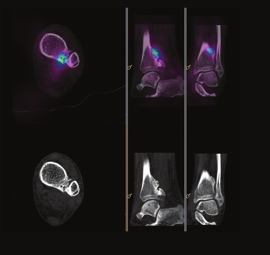
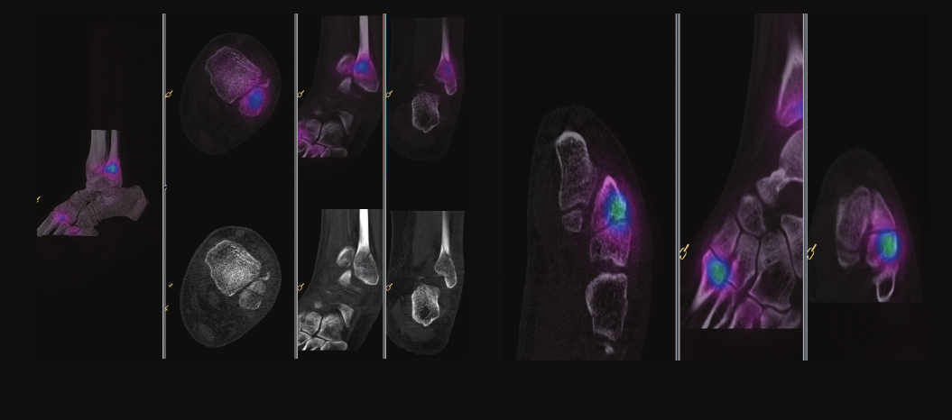
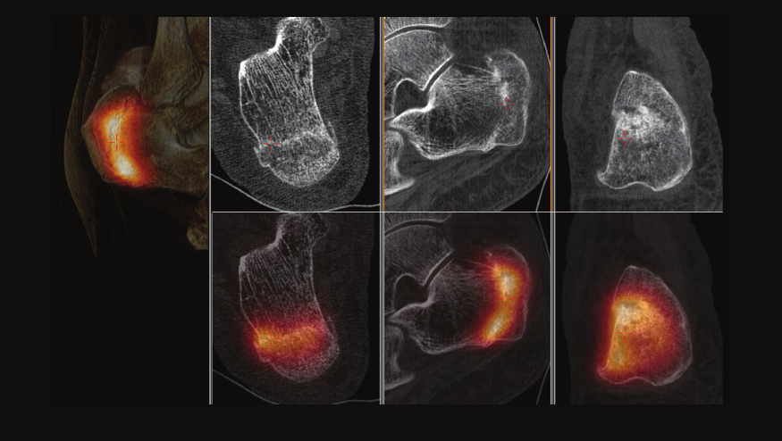
10 Orthopedics
Patient information
•80-year-old
•Severerightanklepainwithsuspectedstressfracture
Procedure
•Tc-99mbonescan
Navicular arthropathy
Courtesy of Sydney X-Ray, Sydney, Australia
Findings from SPECT/CT study
•Markedlyincreasedvascularityanddelayeduptakein
thehindfoot;demonstratedintenseuptakeinright
talonavicularregionwithseveredegenerativechange
onlowdoseCT(subarticularcystformation,joint
narrowingandPeriarticularsclerosis)
Physician impression of SPECT/CT
•Patienttreatedappropriatelyforinammatory
arthropathyofrighttalonavicularjointratherthan
incorrectlyforastressfracturewhichmaynothave
beenappreciatedwithoutSPECT/CT
42
0.33 mm isotropic voxels
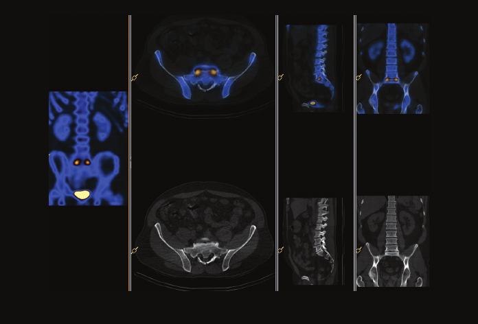
11 Orthopedics
Sacroiliitis
Courtesy of Fletcher Allen Health Care University, Burlington, Vermont
Patient information
•40-year-oldmale
•Chronicbackpainradiatingtolowerextremities,
ruleoutoccultfractures,assessactivediseaseand
guidelevelforfacetinjectionorMBB+/-RFA
•MRIshowedL4-5discdegenerationandfacet
arthropathyL4-5,L5-S1
Procedure
•Tc-99mbonescan
Findings from SPECT/CT study
•Nosignicantuptakeinfacetjointsnordiscogenic
endplatechanges
•IntenseuptakeassociatedwithSIjointsconsistent
withsacroiliitis
Physician impression of SPECT/CT
•Patientreferredfromortho/spineclinic
torheumatologyclinic
43
1 mm isotropic voxels
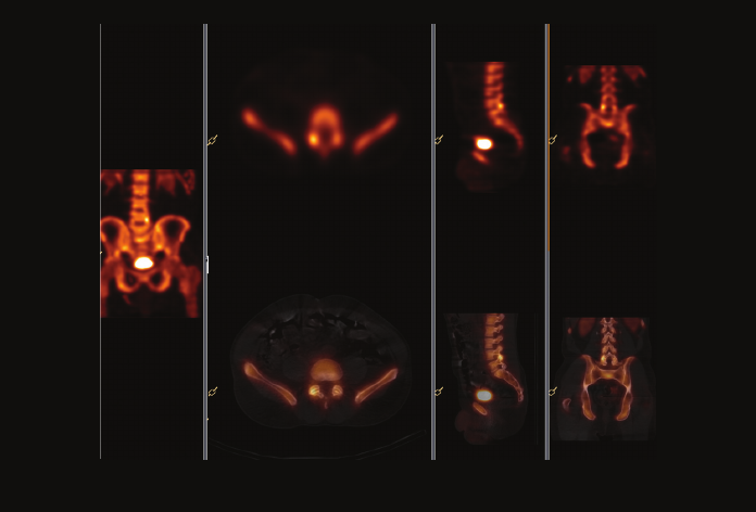
12 Orthopedics
Patient information
•47-year-oldmale
•Lowbackpaininconstructionworker,increasing
throughouttheday
•MRIshowedL3-4,L4-5,L5-S1discdegeneration,
lateralbulgesL3-4,L4-5
•BonescantoguidefacetblocksorMBB+/-RFA
andpossiblefusion
Procedure
•Tc-99mbonescan
Guide facet block or medial branch block
Courtesy of Fletcher Allen Health Care University, Burlington, Vermont
Findings from SPECT/CT study
•PlanaruptakeincreasedL5-S1butcouldbemistaken
forfacetjoints
•SPECT/CTclearlyidentiesincreaseduptake
correspondingtodiscogenicendplatechangesL5-S1
Physician impression of SPECT/CT
•Patientwasofferedmedialbranchblockwith
radiofrequencyablationofmedialbranchifdiagnostic
MBBiseffective
•IfMBBisineffective,L5-S1spinalfusionwillbeoffered
1 mm isotropic voxels
44
13 Orthopedics
Facet joint arthropathy
Courtesy of Washington Hospital Center, Washington DC
Patient information
•61-year-oldmale
•Right-sidedbackpain
Procedure
•Tc-99mMDPbonescan
Findings from SPECT/CT study
•Right-sidedL4-5facetjointarthropathy
•Noevidenceofspondylolysis,spondylolisthesis,
orparsfracture
Physician impression of SPECT/CT
•Demonstrationofinammatoryprocessversusfracture
asetiologyofbackpain
•Guidedreferringphysiciantoconservativemedical
management
1 mm isotropic voxels
45
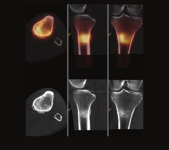
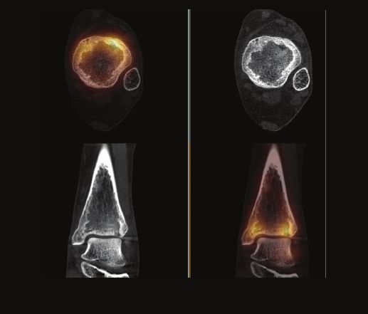
14 Orthopedics
Patient information
•34-year-oldmale
•Six-weekhistoryofleftproximaltibiapain,
queryAVNorosteomyelitis
Procedure
•Tc-99mHDPbonescan
Stress fracture of tibia
Courtesy of Wollongong Nuclear Medicine, New South Wales, Australia
Findings from SPECT/CT study
•IntenseHDPuptakeinleftproximaltibia,associated
withfracturelineseeninthelowdoseCT
•Recentstressfractureofleftproximaltibia
Physician impression of SPECT/CT
•SPECT/CTallowedclearlocalizationanddiagnosis
offracture,rulingoutAVNandosteomyelitis
0.33 mm isotropic voxels
46
15 Orthopedics
Pseudoarthrosis
Courtesy of Sutherland Nuclear Medicine, Sydney, Australia
Patient information
•52-year-oldmale
•Rightshinpain,nocleartrauma
Procedure
•Tc-99mHDPbonescan
Findings from SPECT/CT study
•Exostosisatmedialmarginofrighttibiawithavid
uptakeatitsbase,correlatestoincompletefusion/
pseudoarthrosisratherthanacuteinjury
•Secondfocusofnewboneformationand
correspondingpseudoarthrosis
Physician impression of SPECT/CT
•WithoutSPECT/CT,delayedimageswouldhave
beendiagnosedasstressfractureoftibia
•SPECT/CThelpednditwasanoldinjurywith
incompletefusionandcorrespondingpseudoarthrosis
ratherthanacuteinjury,thereforechanged
patient’streatment
0.33 mm isotropic voxels
47
16 Orthopedics
Patient information
•73-year-oldfemale
•Knownstressfracturesoffemorarelated
tolong-termbiphosphonatetherapy;increasing
lowbackpain
Procedure
•Tc-99mHDPbonescan
Atypical insufciency fractures
Courtesy of Wollongong Nuclear Medicine, New South Wales, Australia
Findings from SPECT/CT study
•Presenceofreactiontoresolvingstress/insufciency
fracturesinlateralcorticalmarginsofbothdistalfemora
•EvidenceofbilateralL5-S1facetjointandL4-5right
facetjointarthropathy
Physician impression of SPECT/CT
•Revealedatypicalinsufciencyfracturesofbothfemora,
secondarytolong-termbiphosphonatetherapy–anew,
recentlydescribed,andcontroversialcondition
0.33 mm isotropic voxels1 mm isotropic voxels
48
Infection
Isotropic voxels
Localization – CT acquisition Localization – SPECT acquisition
High quality images regardless of viewing angle
By acquiring in isotropic voxels, BrightView XCT provides
the same high resolution in all orientations of the CT
images. Coronal and sagittal slices will have the same
resolution as the transverse slices, without the stair-step
artifact common to non-isotropic techniques.
49
1 Infection
Patient information
•60-year-oldmale
•Rightsidepelvicpain;CTshowedmass
onrightside,ruleoutinfectedpelvicgraft
Procedure
•In-111WBCscan
Pelvic graft infection
Courtesy of North Carolina Baptist Hospital, Winston-Salem, North Carolina
Findings from SPECT/CT study
•Increasedactivityinrightgroinadjacenttograft
(femoralarteryanastomosis)correspondingto
edemaandcellulitisinrightgroin,likelyinfection
Physician impression of SPECT/CT
•SPECT/CTshowedtheuptakewasnotover
theboneandconrmedtheCTndings
1 mm isotropic voxels
50
2 Infection
Foot and shin ulcers
Courtesy of Wollongong Nuclear Medicine, New South Wales, Australia
Patient information
•80-year-oldfemale
•Ulcersonrightheelandleftlowershin;
ruleoutosteomyelitis
Procedure
•Tc-99mHDPbonescanandGa-67scan
Findings from SPECT/CT study
•Bonendings–highlysuspiciousforosteomyelitisof
rightcalcaneousinferiorly;mayrepresentperiosteal
reactionofleftshinbutmaybeosteomyelitis
•Galliumndings–mildGalliumuptakeincalcaneum,
faintuptakeinshin
•Combined–doesnotsuggestosteomyelitis
Physician impression of SPECT/CT
•SPECT/CTdemonstratedsuperblocalizationofGallium
distinctfromMDPuptakeregion,excludingosteomyelitis
•Antibioticswerechangedtoreectanon-osseousinfection
0.33 mm isotropic voxels
Gallium scanBone scan
51
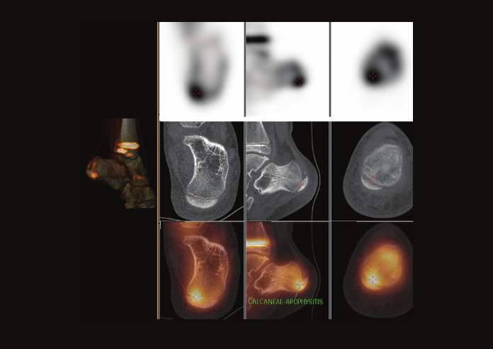
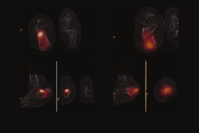
3 Infection
Patient information
•10-year-oldfemale
•Localizedpaininleftcalcaneousmedioposteriorly
withlow-gradefever
Procedure
•Tc-99mHDPbonescan
Apophysitis verses Brodie’s abscess
Courtesy of Nepean Nuclear Medicine and PET, Sydney, Australia
Findings from SPECT/CT study
•Consistentwithleftcalcanealapophysitis
•NoevidenceofBrodie’sabscess
Physician impression of SPECT/CT
•AbletoexcludeBrodie’sabscess,thereforechanged
managementofpatient
0.33 mm isotropic voxels
52
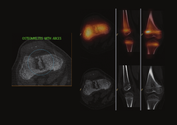
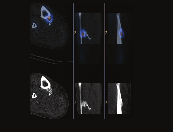
4 Infection
Osteomyelitis with sequester
Courtesy of GZA Sint-Augustinus, Antwerp, Belgium
Patient information
•5-year-oldmale
•Fever,painofdistalthigh,limping;ruleoutosteomyelitis
Procedure
•Tc-99mMDPbonescan
Findings from SPECT/CT study
•Planarimagesshowhyperemiaanddiffuse
increaseduptakeindistalrightfemur
•SPECT/CTconrmsincreaseduptakeofdistalfemoral
growthplatewithacentraldefect;onlowdoseCT,
thereisaclearcentralendomedullaryirregularlesion
suspectforabscessorbonesequester
Physician impression of SPECT/CT
•Classicbonescandiagnosisofosteomyelitis
•SPECT/CTshowedadditionofintra-osseoussequester,
whichrequiresmoreintensiveantibiotictherapyand
follow-up(possiblesurgery)
0.33 mm isotropic voxels
53
5 Infection
Patient information
•30-year-oldfemale
•Persistentpaintwomonthsfollowingsurgery
forhaluxvalgus;ruleoutosteomyelitis
Procedure
•Tc-99mGranuloscint
Findings from SPECT/CT study
•WBCaccumulationinsofttissuesurrounding
headofthescrew
Occult fracture
Courtesy of Universitair Ziekenhuis Brussel, Brussels, Belgium
•Someboneresorptionatproximallevelofthescrews,
noincreasedboneuptakenorinterruptionofthebony
cortex;osteomyelitiswasexcluded
•Straightradioluscentlineatmetaphysisofmetatarsal
bonemarkspresenceofrecentnon-displaced
transcorticaloccultfracture
Physician impression of SPECT/CT
•Thissingleexaminationrulesouttheimportantdiagnosis
ofosteomyelitis,conrmssofttissueinfection,and
demonstratesanoccultfractureassourceofthepain
0.33 mm isotropic voxels
54
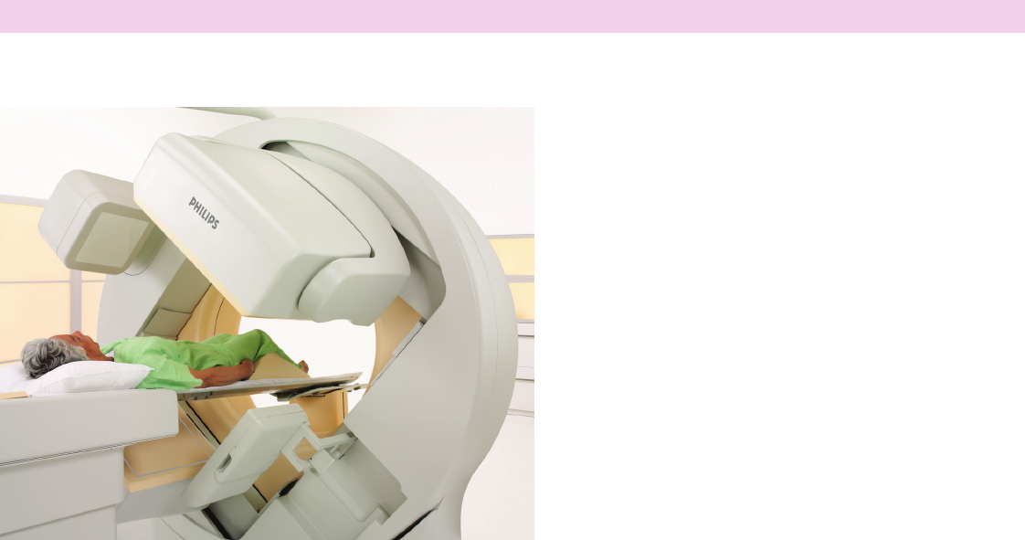
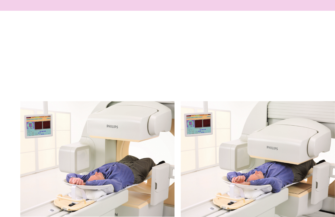
Other localization
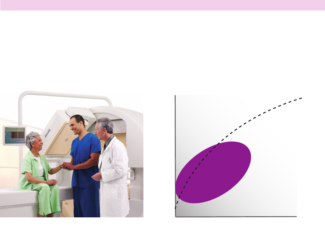
Workow tailored for nuclear medicine
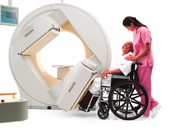
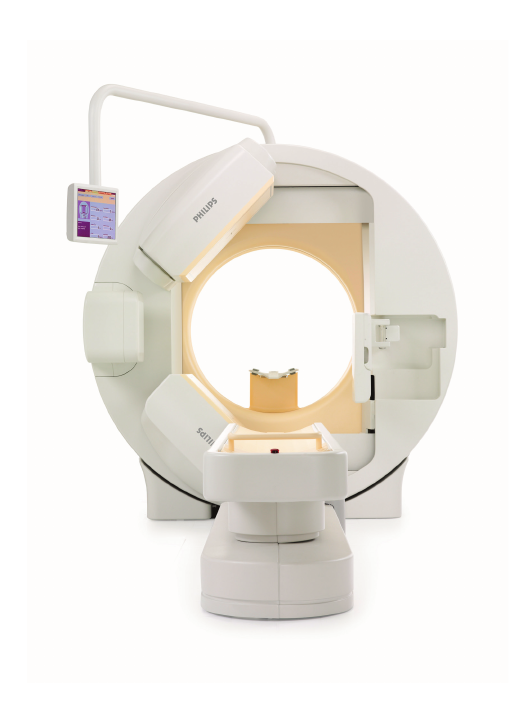
Continue working the way you already do
Having all of the capabilities of the popular BrightView
SPECT camera, BrightView XCT simplies workow
to help improve clinical results and lower lifecyle costs.
The low complexity design is compact, tting
in a standard nuclear-medicine-sized room. An in-room
CT control option allows you to be closer to your patient
and avoid the costs associated with a separate control
room. SPECT/CT planning is done from the nuclear
medicine p-scope, as simple as planning
for a SPECT-only procedure.
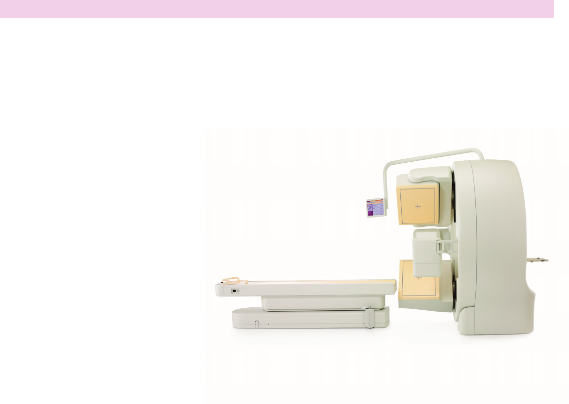
Compact, low complexity design suitable
for a standard nuclear-medicine-sized room
55
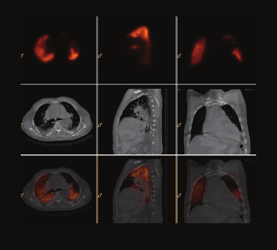
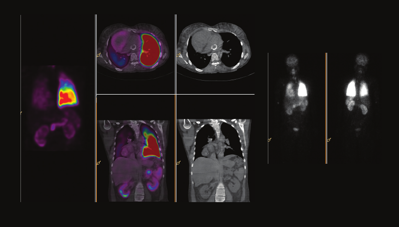
1 Other localization
Patient information
•72-year-oldmale
•Gaspingandchestpain
Procedure
•Tc-99mMAAlungperfusion
Pulmonary embolism
Courtesy of Afliated Hospital of Xuzhou Medical College, Jiangsu, China
Findings from SPECT/CT study
•Signicantdefectinthelingularsegmentofthesuperior
lobeoftheleftlung;pulmonaryembolismshould
beconsidered
Physician impression of SPECT/CT
•BasedontheSPECT/CTndings,thrombolysistherapy
wasrecommended
1 mm isotropic voxels
56
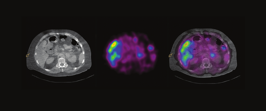
2 Other localization
Biliary leak
Courtesy of North Carolina Baptist Hospital, Winston-Salem, North Carolina
Patient information
•59-year-oldmale
•Abdominalpainpostrecentcholecystectomy;
displacedtubepostsurgery
Procedure
•Tc-99mHIDA
Findings from SPECT/CT study
•Extraluminalactivityextendingfromgallbladderfossa
wasnotedintherightparacolicgutterandpelvis;
positivebiliaryleak
Physician impression of SPECT/CT
•SPECT/CTshowedtheleakandextentoftheleak;
CTonlyshowedleakaroundliver
1 mm isotropic voxels
57
3 Other localization
Lymphatic uid leak
Courtesy of Osaka City University Hospital, Osaka, Japan
Patient information
•59-year-oldfemale
•Esophagealcancer;two-weekleakage
oflymphaticuidafteresophagectomy
Procedure
•Tc-99mHAS-D
Findings from SPECT/CT study
•Uptakeofleakpointwasfoundinmiddleofthe
mediastinum,placedbetweenthebronchusandaorta
Physician impression of SPECT/CT
•Thesurgicalligationofmainlymphductwaseasily
plannedasaresultofaccurateregionalidentication
ofthelymphoidleakpointbySPECT/CT
1 mm isotropic voxels
58
4 Other localization
Hyperparathyroidism
Courtesy of Nepean Nuclear Medicine and PET, Sydney, Australia
Patient information
•46-year-oldfemale
•Hyperparathyroidism;assessforparathyroidadenoma
Procedure
•Tc-99mSestamibi
Findings from SPECT/CT study
•Moderatefocalretentionpresentatsuperoposterior
aspectofleftthyroidlobe
Physician impression of SPECT/CT
•Identiedandlocalizedparathyroidadenomawhich
helpedwithsurgicalplanning
1 mm isotropic voxels
59
5 Other localization
Lung perfusion with unusual anatomy
Courtesy of University of Washington Medical Center, Seattle, Washington
Patient information
•27-year-oldfemale
•Complexcongenitalheartdisease;transpositionof
greatarteries,dexocardia,bilateralsuperiorvenacava
•SinglefunctionalventricleandmultiplepulmonaryAVMs
•Newonsetofpalpitationsanddyspnea;evaluatefor
worseningofR-Lshunt
Procedure
•Tc-99mMAAperfusionandTc-99mDTPAventilation
Findings from SPECT/CT study
•Signicantlydecreasedperfusiontoentirerightlung,
focallymoresevereperfusiondefectinlateralaspect
ofrightupperlobeseenbetteronSPECT/CT
•32%right-to-leftshuntlikelyexplainsthepatient’s
symptoms
Physician impression of SPECT/CT
•SPECT/CTprovidedadditionalanatomicinformation
tobetterunderstandtheperfusioninpatientwith
unusualanatomy
•Planarimageswerechallengingtointerpret
1 mm isotropic voxels
60
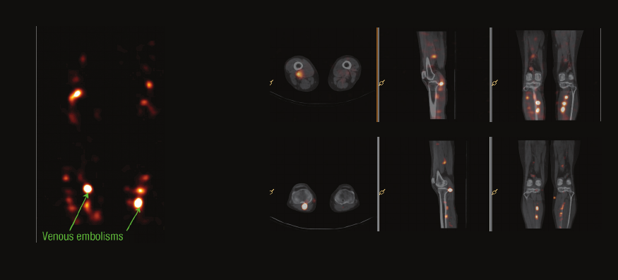
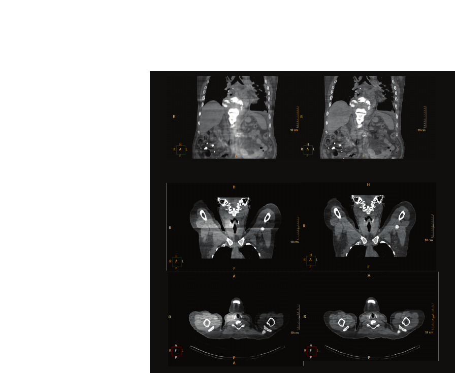
6 Other localization
Venogram
Courtesy of Osaka City University Hospital, Osaka, Japan
Patient information
•74-year-oldfemale
•Illustrateaccurateregionsanddegrees
ofvenousembolismsofthelowerlimb
Procedure
•Tc-99mMAAvenogram
Findings from SPECT/CT study
•Beforewarfarization,manyuptakeswerefoundinboth
lowerlimbsalongtheveinsontheSPECT/CT;found
tobevenousembolisms
•Twoweeksafterwarfarization,feweruptakesofboth
lowerlimbswereillustratedthanbeforethetreatment
Physician impression of SPECT/CT
•Warfarizationtreatmentwasinitiatedasaresultof
accurateregionalidenticationanddegreeofvenous
embolismontheSPECT/CT
•Aftertreatment,thecomparisonSPECT/CTeasily
showedatherapeuticresponsesowarfarizationwas
abletobestopped
1 mm isotropic voxels
Initial
study
Follow-up
study
61
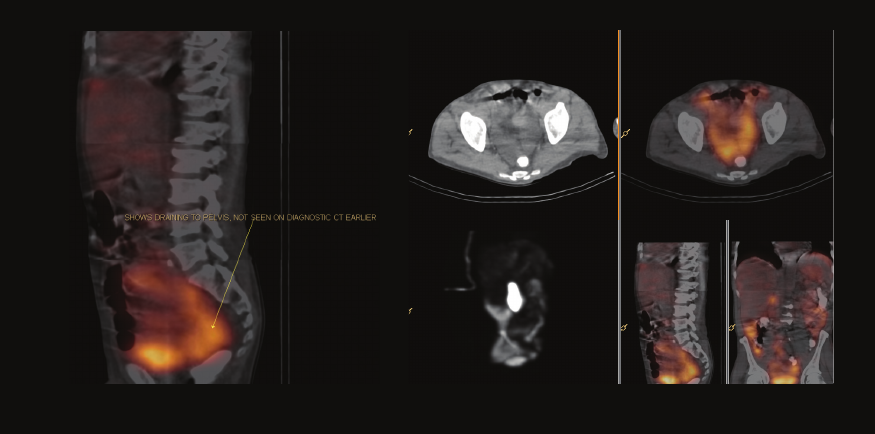
7 Other localization
GI bleeding
Courtesy of Wollongong Nuclear Medicine, New South Wales, Australia
Patient information
•70-year-oldfemale
•GIbloodlossresultinginanemia,requiringblood
transfusions;multipleeffortstoidentifysource
including2xendoscopy
Procedure
•Tc-99mtaggedRBCs
Findings from SPECT/CT study
•Noactivebleedinearlyphase
•At24hours,abnormalactivityinentiretransverse
colon,halfwayalongascendingcolonandentire
descendingcolonpriortosigmoidjunction;
ultrasoundconrmationwasrecommended
Physician impression of SPECT/CT
•SPECT/CTprovidedaspecictargettoallow
subsequentendoscopicconrmationofsourceof
bleeding;endoscopydemonstratedlesioninascending
colonregion(hemangioma/angiodysplasia)
1 mm isotropic voxels
62
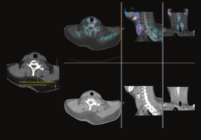
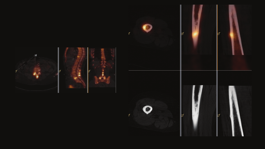
8 Other localization
Hyperparathyroidism
Courtesy of Washington Hospital Center, Washington DC
Patient information
•54-year-oldfemale
•Hyperparathyroidism;identifyparathyroidadenoma
Procedure
•Tc-99mSestamibi
Findings from SPECT/CT study
•ExtrathyroidalMIBIfocusposteromedial
toupperpoleofleftthyroidlobe
•Noevidenceofectopicparathyroidtissue
inmediastinum
Physician impression of SPECT/CT
•SPECT/CThelpedpreciselyidentifyparathyroid
adenomalocation
•Precisesurgicalguidancewasachievedforresection
1 mm isotropic voxels
63

Case study acquisition parameters
Case study CT scan parameters SPECT scan parameters Page
1Cardiology 5mA;60seconds Astonish;4iterations,8subsets,Hanninglter1.0 7
2Cardiology 5mA;60seconds Astonish;4iterations,8subsets,Hanninglter1.0 8
1Oncology 2mA;12seconds Astonish;2iterations,12subsets,Hanninglter1.2 10
2Oncology 30mA;12seconds Astonish;2iterations,16subsets,Hanninglter1.0 11
3Oncology 20mA;24seconds Astonish;2iterations,16subsets,Hanninglter1.2 12
4Oncology 80mA;24seconds Astonish;2iterations,15subsets,Hanninglter1.2 13
5Oncology 20mA;12seconds Astonish;4iterations,8subsets,nolter 14
6Oncology 30mA;12seconds Astonish;3iterations,8subsets,nolter 15
7Oncology 5mA;60seconds MLEM;Butterworth;cutoff0.66,Order5.0,30iterations 16
8Oncology 5mA;12seconds OSEM;Butterworth;cutoff0.50,Order5.0,16iterations,8subsets 17
9Oncology 20mA;12seconds Astonish;3iterations,16subsets,nolter 18
10Oncology 80mA;24seconds Astonish;4iterations,8subsets,nolter 19
11Oncology 20mA;12seconds Astonish;2iterations,16subsets,Hanninglter1.0 20
12Oncology 80mA;24seconds Astonish;2iterations,16subsets,Hanninglter2.0 21
13Oncology 20mA;12seconds Astonish;4iterations,8subsets,Hanninglter2.0 22
14Oncology 20mA;12seconds Astonish;2iterations,16subsets,Hanninglter1.0 23
15Oncology 20mA;24seconds Astonish;4iterations,16subsets,Hanninglter1.2 24
16Oncology 20mA;12seconds Astonish;3iterations,8subsets,nolter 25
64

Case study CT scan parameters SPECT scan parameters Page
1Cardiology 5mA;60seconds Astonish;4iterations,8subsets,Hanninglter1.0 7
2Cardiology 5mA;60seconds Astonish;4iterations,8subsets,Hanninglter1.0 8
1Oncology 2mA;12seconds Astonish;2iterations,12subsets,Hanninglter1.2 10
2Oncology 30mA;12seconds Astonish;2iterations,16subsets,Hanninglter1.0 11
3Oncology 20mA;24seconds Astonish;2iterations,16subsets,Hanninglter1.2 12
4Oncology 80mA;24seconds Astonish;2iterations,15subsets,Hanninglter1.2 13
5Oncology 20mA;12seconds Astonish;4iterations,8subsets,nolter 14
6Oncology 30mA;12seconds Astonish;3iterations,8subsets,nolter 15
7Oncology 5mA;60seconds MLEM;Butterworth;cutoff0.66,Order5.0,30iterations 16
8Oncology 5mA;12seconds OSEM;Butterworth;cutoff0.50,Order5.0,16iterations,8subsets 17
9Oncology 20mA;12seconds Astonish;3iterations,16subsets,nolter 18
10Oncology 80mA;24seconds Astonish;4iterations,8subsets,nolter 19
11Oncology 20mA;12seconds Astonish;2iterations,16subsets,Hanninglter1.0 20
12Oncology 80mA;24seconds Astonish;2iterations,16subsets,Hanninglter2.0 21
13Oncology 20mA;12seconds Astonish;4iterations,8subsets,Hanninglter2.0 22
14Oncology 20mA;12seconds Astonish;2iterations,16subsets,Hanninglter1.0 23
15Oncology 20mA;24seconds Astonish;4iterations,16subsets,Hanninglter1.2 24
16Oncology 20mA;12seconds Astonish;3iterations,8subsets,nolter 25
Case study CT scan parameters SPECT scan parameters Page
17Oncology 20mA;24seconds Astonish;4iterations,16subsets,Hanninglter1.2 26
18Oncology 30mA;12seconds Astonish;6iterations,8subsets,Hanninglter0.95 27
19Oncology 80mA;24seconds Astonish;2iterations,16subsets,Hanninglter2.0 28
20Oncology 20mA;12seconds Astonish;6iterations,8subsets,Hanninglter2.0 29
21Oncology 30mA;12seconds Astonish;4iterations,15subsets,nolter 30
22Oncology Chest:20mA;12seconds
Ankle:80mA;24seconds
Chest:Astonish;4iterations,16subsets,nolter
Ankle:Astonish;4iterations,16subsets,nolter 31
1Orthopedics 80mA;24seconds Astonish;3iterations,8subsets,nolter 33
2Orthopedics 20mA;12seconds Astonish;3iterations,8subsets,nolter 34
3Orthopedics 20mA;12seconds Astonish;4iterations,8subsets,nolter 35
4Orthopedics 80mA;24seconds Astonish;4iterations,8subsets,nolter 36
5Orthopedics 80mA;24seconds Astonish;2iterations,16subsets,Hanninglter1.0 37
6Orthopedics 80mA;24seconds Astonish;3iterations,8subsets,nolter 38
7Orthopedics 80mA;24seconds Astonish;2iterations,32subsets,nolter 39
8Orthopedics 80mA;24seconds Astonish;2iterations,8subsets,nolter 40
9Orthopedics 20mA;24seconds OSEM;Butterworth;cutoff0.60,Order1.0,3iterations,8subsets 41
10Orthopedics 80mA;24seconds Astonish;2iterations,8subsets,nolter 42
11Orthopedics 20mA;12seconds Astonish;4iterations,16subsets,Hanninglter1.3 43
65

Case study CT scan parameters SPECT scan parameters Page
12Orthopedics 20mA;12seconds Astonish;4iterations,16subsets,Hanninglter1.3 44
13Orthopedics 20mA;12seconds Astonish;2iterations,16subsets,Hanninglter2.0 45
14Orthopedics 80mA;24seconds Astonish;3iterations,8subsets,nolter 46
15Orthopedics 80mA;24seconds Astonish;3iterations,8subsets,nolter 47
16Orthopedics 20mA;12seconds Astonish;3iterations,8subsets,nolter 48
1Infection 30mA;12seconds Astonish;3iterations,8subsets,Hanninglter2.0 50
2Infection 80mA;24seconds Astonish;3iterations,8subsets,nolter 51
3Infection 80mA;24seconds Astonish;4iterations,8subsets,nolter 52
4Infection 80mA;24seconds Astonish;3iterations,8subsets,Hanninglter1.5 53
5Infection 80mA;24seconds Astonish;2iterations,32subsets,nolter 54
1Otherlocalization 20mA;12seconds Astonish;3iterations,8subsets,nolter 56
2Otherlocalization 20mA;12seconds Astonish;3iterations,8subsets,Hanninglter2.0 57
3Otherlocalization 20mA;12seconds Astonish;2iterations,16subsets,Hanninglter1.0 58
4Otherlocalization 20mA;12seconds Astonish;4iterations,8subsets,nolter 59
5Otherlocalization 30mA;12seconds Astonish;2iterations,16subsets,Hanninglter1.0 60
6Otherlocalization
Initial:20mA;12seconds;
Follow-up:
30mA;12seconds
Initial&Follow-Up:Astonish;2iterations,
16subsets,Hanninglter1.5 61
7Otherlocalization 80mA;24seconds Astonish;2iterations,8subsets,nolter 62
8Otherlocalization 20mA;12seconds Astonish;6iterations,16subsets,nolter 63
Case study acquisition parameters
continued
66
Please visit www.philips.com/brightviewxct
Philips Healthcare is part of
Royal Philips Electronics
How to reach us
www.philips.com/healthcare
healthcare@philips.com
Asia
+49 7031 463 2254
Europe, Middle East, Africa
+49 7031 463 2254
Latin America
+55 11 2125 0744
North America
+1 425 487 7000
800 285 5585 (toll free, US only)
© 2012 Koninklijke Philips Electronics N.V. All rights are reserved.
PhilipsHealthcarereservestherighttomakechangesinspecicationsand/ortodiscontinueanyproductatanytimewithoutnotice
or obligation and will not be liable for any consequences resulting from the use of this publication.
Printed in The Netherlands.
4522 962 83701 * APR 2012
