Philips This Field Strength Article Black Blood MRI Of HIV Patient With Vasculitis
Descargar este artículo de FieldStrength Black_Blood_MRI_of_HIV_patient_with_vasculitis Diagnóstico por IRM
User Manual: Philips this FieldStrength article Vasculitis on Black Blood imaging
Open the PDF directly: View PDF ![]() .
.
Page Count: 8
FieldStrength MRI magazine User experiences - April 2017
Black Blood imaging helped in suggesting the diagnosis
and choosing the treatment
Black Blood MRI imaging of
HIV patient with brain vasculitis
In a patient with HIV and cardiovascular risk factors, MRI with Black Blood
imaging helped to diagnose brain vasculitis. The same MRI protocol was later
also used to noninvasively conrm treatment response.
www.philips.com/eldstrength
Niloufar Sadeghi MD, PhD, is neuroradiologist at
Erasme Hospital in Brussels, Belgium since 2000.
She completed her PhD in 2010 and has been
recently nominated as professor.
Patient history
A 56-year-old patient presented in the Emergency Room at
Erasme Hospital in Brussels, Belgium, with recurrent left leg
weakness that had been occurring over a period of 24 hours.
The patient was known to have been HIV infected for four years,
but was not treated for this infection. The patient had multiple
cardiovascular risk factors such as obesity, glucose intolerance,
arterial hypertension and hypercholesterolemia. The neurological
examination showed left leg hemiparesis.
MRI examination with Black Blood imaging
After a conventional routine MR imaging examination, the
suspicion of vasculitis arose, therefore we performed an MRI
including Black Blood imaging in a separate session. The
dedicated ExamCard includes diusion, FLAIR, MR angiography
using TOF, and 3D T1 MRA with bolus injection. This ExamCard
also includes Black Blood imaging before and after contrast.
This examination was performed on our Ingenia 3.0T. Black Blood
scan time 4:39 min, acquired voxel size 0.75 x 0.75 x 1.0 mm,
21 slices.
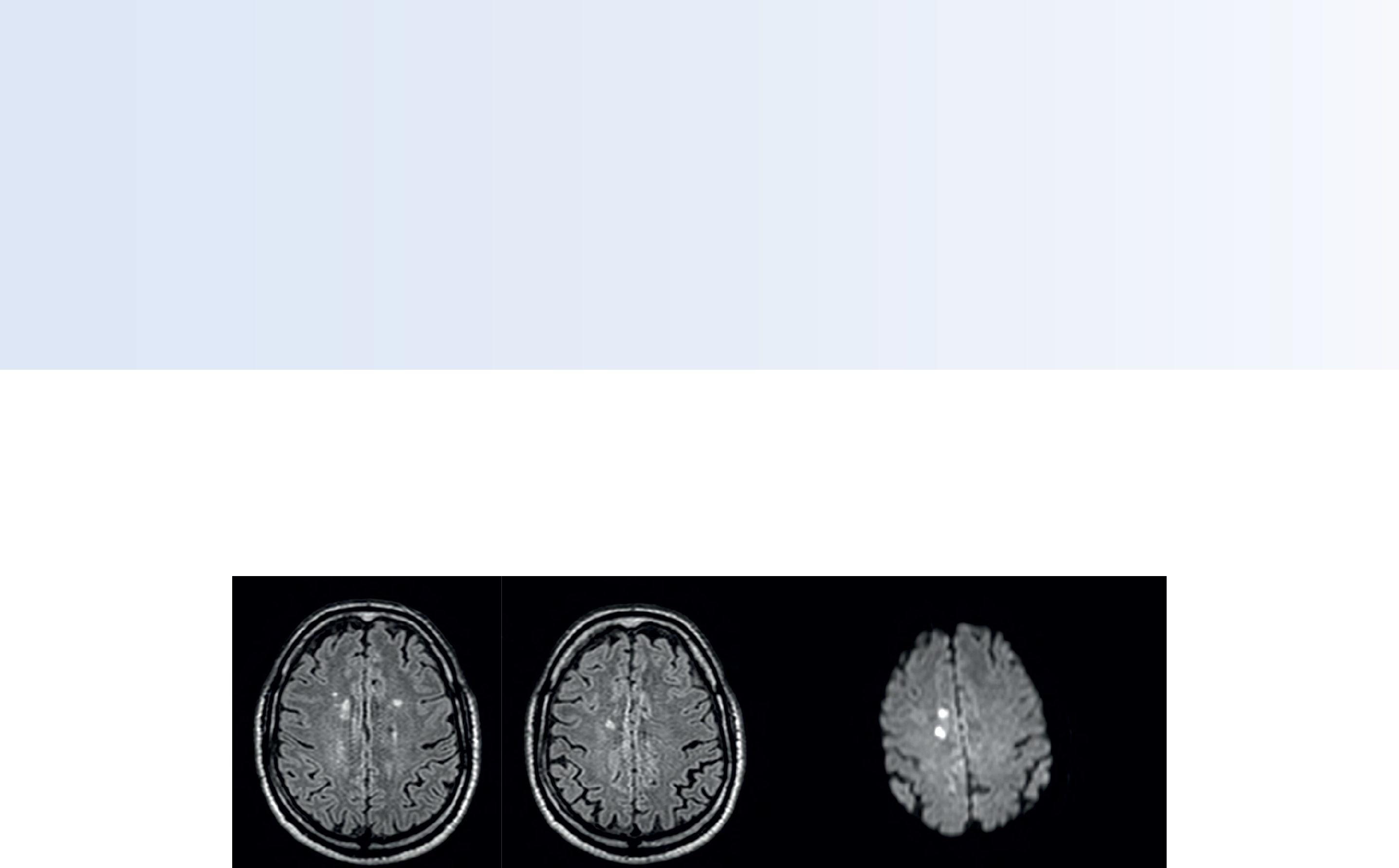
On FLAIR images we can see some nonspecic high signal abnormalities in frontal white matter bilaterally. On DWI we can see
acute ischemic lesions which appear with high signal intensity. Arrows show vessel wall enhancement which appears concentric
and homogeneous in dierent cerebral territories.
FLAIR DWI
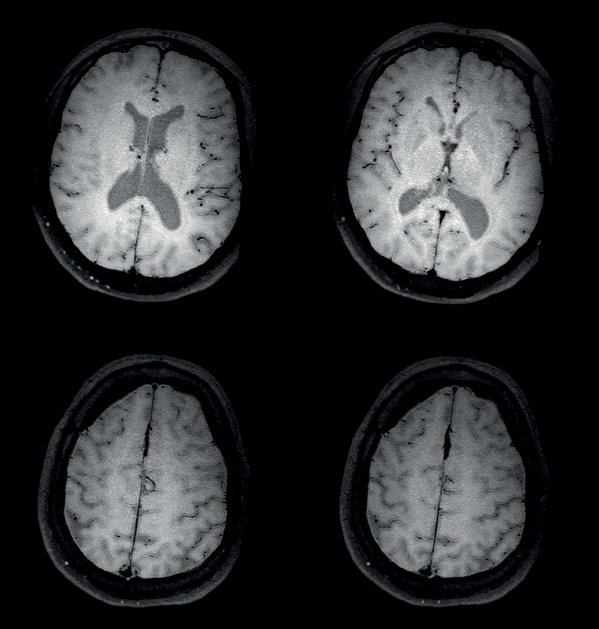
Black Blood imaging pre contrast
Black Blood imaging post contrast
Arrows show vessel wall enhancement which appears concentric and homogeneous in dierent cerebral territories.

Discussion of ndings
On the routine MR sequences that we did, we could see acute
ischemic lesions. We see them very well on the diusion images,
where acute ischemic lesions usually appear with high signal
intensity and restricted diusion. However, the etiology of these
lesions cannot be derived from these images.
An area of restricted diusion was seen in the anterior cerebral
artery territory and we concluded it was an ischemic lesion. On
MR angiography we can just see if there is stenosis or vessel
occlusion, but it does not provide us information on the etiology
of this kind of lesion.
So, we decided to perform Black Blood imaging. The presence
and the pattern of vessel wall enhancement on Black Blood
imaging, can help us to determine the etiology of the lesion.
Many studies have shown that Black Blood imaging can help
dierentiate vasculitis from other causes of vasculopathy, such as
atherosclerosis, with a high specicity [1-3]. In an atherosclerotic
lesion, vessel wall thickening and enhancement are usually eccentric,
while in vasculitis the wall thickening and enhancement are usually
concentric, homogenous, and in a long portion of the vessel.
Furthermore, this imaging can also be used for the follow-up
of patients whenever their treatment is installed in order to
determine the ecacy of a particular treatment.
In this case the Black Blood imaging helped us to suggest the
diagnosis of HIV-related brain vasculitis.
Impact of Black Blood imaging for this
patient
With the multiple cardiovascular risk factors this patient
had, such as glucose intolerance, arterial hypertension and
hypocholesteremia, his lesions could be atherosclerotic lesions or
vasculitis, conditions which require dierent treatment. Especially
in this patient with HIV infection causing the vasculitis, treatment
of the two conditions is dierent.
The results of MRI with Black Blood imaging, helped to choose the
preferred treatment for this patient, which was based on antiviral
medication rather than an antiaggregant or anticoagulation
treatment which is usually given to patients with risk of ischemia
based on atherosclerotic lesions.
One month after beginning the antiviral treatment, the same
MRI examination was repeated and again 8 months after the
beginning of treatment. On follow-up images, we see the
enhancements have almost disappeared.
So in case of this patient, the MRI exam with Black Blood imaging
helped us to give the patient the appropriate treatment and also
allowed us to noninvasively conrm the treatment response.
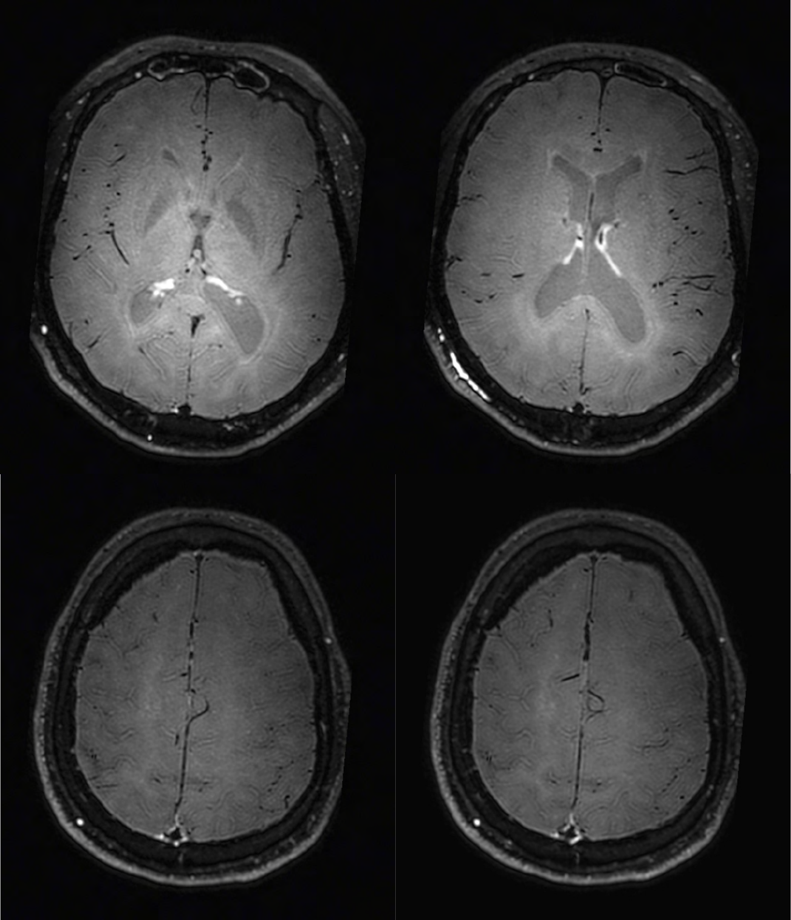
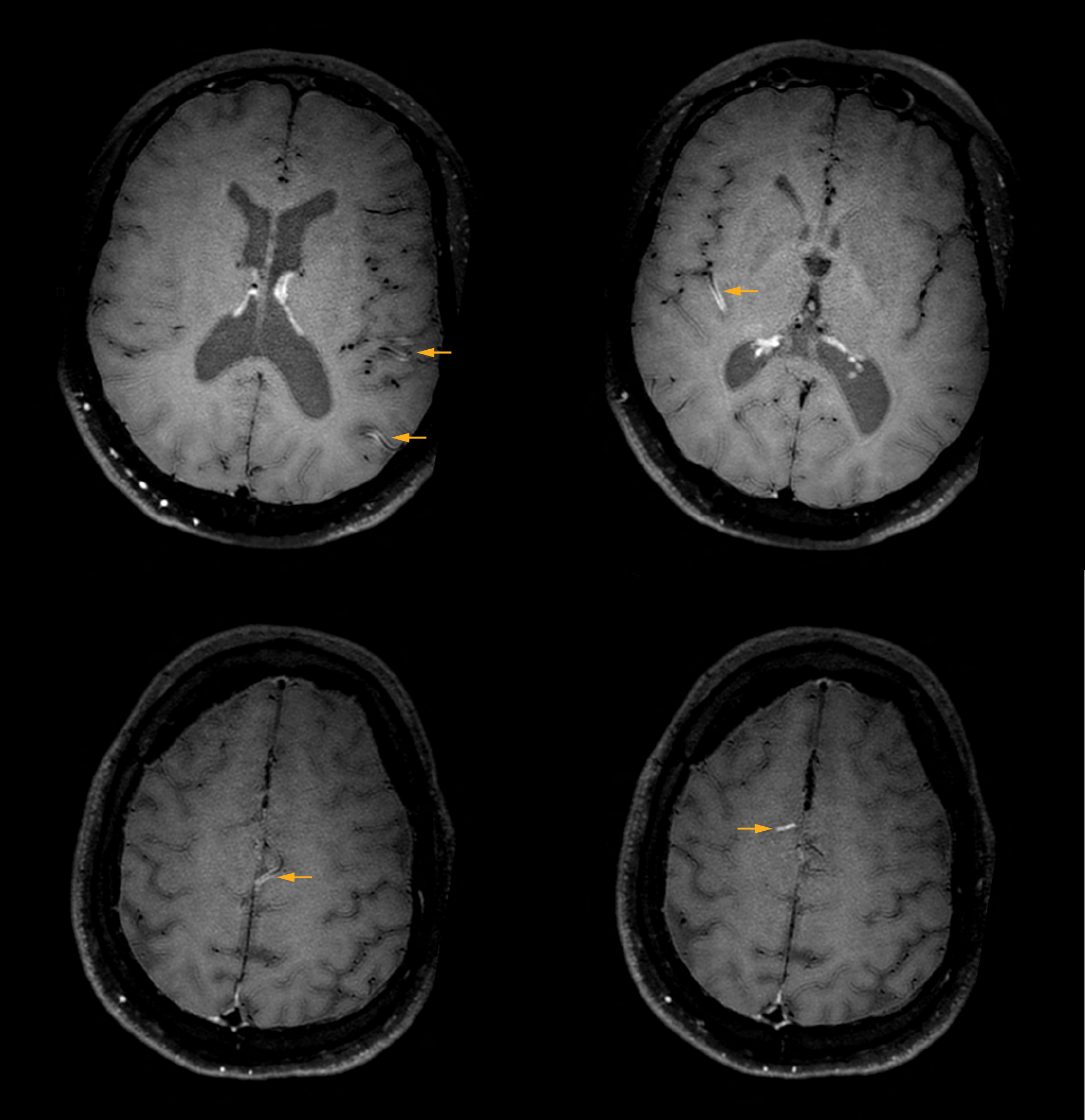
Black Blood imaging after one month
After one month of treatment, post-contrast Black Blood images at the exact same levels as in the gure above show disappearance of
the vessel wall enhancements which were seen on the previous examination.
Black Blood imaging post contrast

References
1. Swartz RH, Bhuta SS, Farb RI, Agid R, Willinsky RA, Terbrugge KG, et al. Intracranial
arterial wall imaging using high-resolution 3-tesla contrast-enhanced MRI.
Neurology. 2009 Feb 17;72(7):627–34.
2. Obusez EC, Hui F, Hajj-Ali RA, Cerejo R, Calabrese LH, Hammad T, et al. High-
resolution MRI vessel wall imaging: spatial and temporal patterns of reversible
cerebral vasoconstriction syndrome and central nervous system vasculitis. AJNR Am
J Neuroradiol. 2014 Aug;35(8):1527–32.
3. Mossa-Basha M, Hwang WD, De Havenon A, Hippe D, Balu N, Becker KJ, et al.
Multicontrast high-resolution vessel wall magnetic resonance imaging and its value
in dierentiating intracranial vasculopathic processes. Stroke J Cereb Circ. 2015
Jun;46(6):1567–73.
Black Blood imaging
Philips Black Blood imaging is 3D brain imaging with reduced
intraluminal blood signal1 over the complete imaging volume
in the brain.
It helps you to better dierentiate intraluminal blood signal
from other signal, which can enhance diagnostic condence.
The Black Blood sequence allows
• fast2, isotropic 3D imaging
• higher spatial resolution3
• reformatting in any plane without loss of resolution
1. Compared to our 3D T1W scan without MSE prepulse
2. Compared to our 2D double inversion recovery methods with same full brain
coverage
3. Compared to our 2D double inversion recovery methods with same brain
coverage and scan time
Results from case studies are not predictive of results in other cases. Results in
other cases may vary.
The importance of Black Blood imaging
Black Blood imaging can help us to noninvasively visualize vessel
wall thickening and enhancement patterns that occur in vasculitis,
and help us distinguish it from atherosclerotic lesions. Imaging
techniques such as time-of-ight (TOF) MR angiography are not
very sensitive or specic for this kind of lesions. Other possible
diagnostic methods are intra-arterial angiography or brain
biopsies which are both invasive.
Recommendations for using Black Blood
imaging
We do not perform this examination with Black Blood imaging
on all patients with ischemic lesions in the brain, because in
most patients the lesion origin is embolic or atherosclerotic. We
typically use it in young patients (less than 60 years old) or those
patients without cardiovascular risk factors. We nd it important
to use Black Blood imaging in such cases, because treatment is
dierent for a patient with vasculitis.
Cheron J, Wyndham-Thomas C, Sadeghi N, Naeije G. Response of Human
Immunodeficiency Virus-Associated Cerebral Angiitis to the Combined Antiretroviral
Therapy. Front. Neurol., 13 March 2017, doi.org/10.3389/fneur.2017.00095
4.

© 2017 Koninklijke Philips N.V. All rights reserved.
Specications are subject to change without notice.
Trademarks are the property of Koninklijke Philips N.V.
(Royal Philips) or their respective owners.
www.philips.com
How to reach us
Please visit www.philips.com/healthcare
healthcare@philips.com
Subscribe to FieldStrength
Our periodic FieldStrength MRI newsletter provides you articles on latest trends and insights,
MRI best practices, clinical cases, application tips and more. Subscribe now to receive our free
FieldStrength MRI newsletter via e-mail.
Stay in touch with Philips MRI
Related information
• Philips methods for brain MRI ›
• ExamCards for Ingenia 3.0T ›
• White papers related to Ingenia ›
• MRI Clinical Case map ›
More from FieldStrength
• MRI in emergency department for fast, condent decisions –
St. Joseph’s Hospital and Medical Center, USA ›
• Advanced methods for neuro MR can improve eciency and
condence – Phoenix Children’s Hospital, USA ›
• Sherbrooke researchers investigate DWI and fMRI – Sherbrooke
University, Canada ›
• UVM brain MRI protocols upgraded with latest methods –
University of Vermont Medical Center, USA ›
Subscribe now
