Philips IGTD88901 User Manual Quick Guide: Iliac Venous Compression 16 5 224 Guide Rebrand
IGTD86700 Quick guide: Iliac venous compression Iliac%20Vein%20Compression%20Quick%20Guide Philips Volcano - Visions PV .018 Digital IVUS catheterIGTD86700
User Manual: Philips IGTD88901 Quick guide: iliac venous compression Philips Volcano - Visions PV .035 Digital IVUS catheterIGTD88901
Open the PDF directly: View PDF ![]() .
.
Page Count: 2
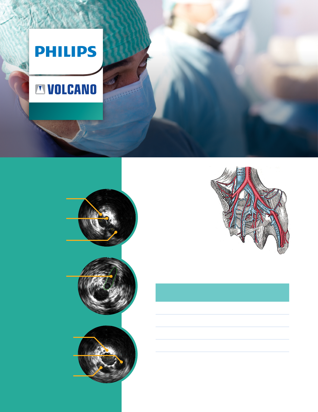
Quick reference
Venous
segment
Approximate
length
Approximate
diameter
Inferior Vena Cava 140 mm 23.4 mm
Common Iliac Vein 60 mm 16.0 mm
External Iliac Vein 130 mm 14.0 mm
Common Femoral Vein 60 mm 12.0 mm
Typical IVUS
imagery
Venous
anatomy
Approximate venous
segment dimensions
Ouriel K, Greenberg RK, Green RM, et al. A volumetric index for the quantification
of deep venous thrombosis. J Vasc Surg 1999;30:1060-6.
Raju S, Davis BS. Anomalous features of iliac vein stenosis that affect diagnosis and
treatment. J Vasc Surg: Venous and Lym Dis 2014;2:260-7.
Iliac venous
compression
Venous applications
Left iliac vein stent
Right iliac vein
Right iliac artery
Area of
compressed
left Iliac vein
Right iliac artery
Left iliac vein
Right iliac vein
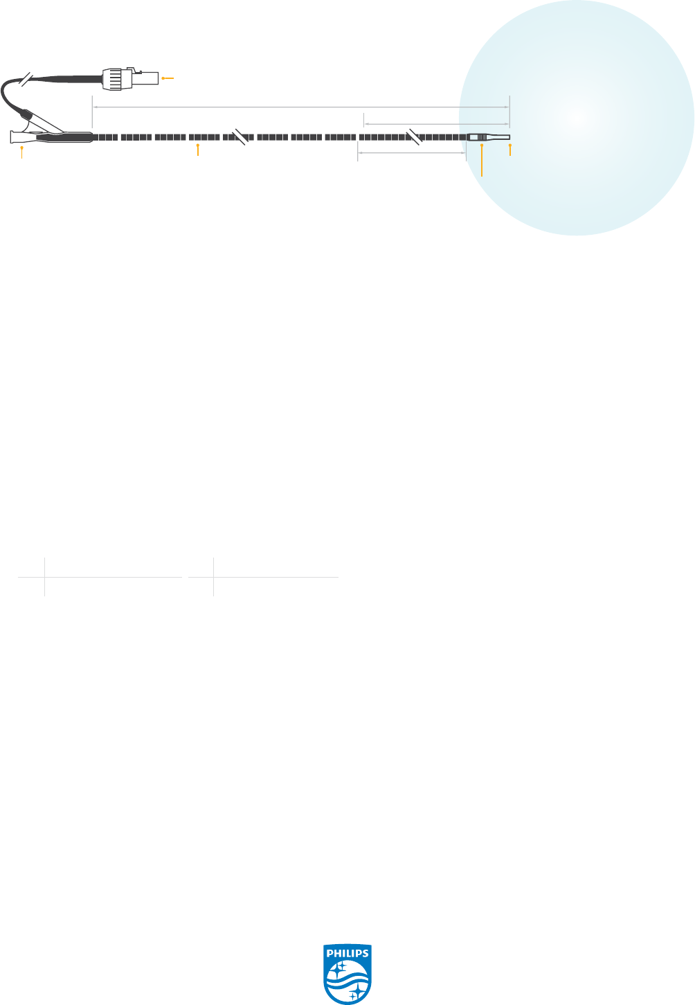
Actual Size of
Field of View
(60mm)
Guide wire exit port
≤ 0.038" (0.97 mm)
90 cm working length
PIM connector
Y - connector
25 RO markers,
1 cm apart
7 F
8.2 F
transducer
Tip O.D.
≤
0.055”
30 cm GlyDx hydrophilic coating
© 2017 Koninklijke Philips N.V. All rights reserved.
Trademarks are the property of Koninklijke Philips
N.V. or their respective owners.
D000140310/A
Philips Volcano
3721 Valley Centre Drive, Suite 500
San Diego, CA 92130 USA
www.volcanocorp.com
1. Femoral or popliteal venous access
• Patient is therapeutically heparinized if not already
anticoagulated
• Insert a sheath through percutaneous or open access
site via standard interventional technique
2. Wire placement
• Advance an 0.035” guide wire to the area of interest
• An angled guide catheter may be used to facilitate
placement
3. Perform venogram of leg from access point to the level
of the diaphragm
• Place guide catheter in the cranial portion of the
femoral vein, just below the trochanter to image
the CFV, EIV, and CIV
• Advance guide catheter to EIV or CIV to image the IVC
4. Advancement of IVUS catheter
• Replace sheath with 9Fr introducer sheath
(8.5Fr minimum)
• Prepare the PV.035 IVUS catheter by ushing the guide
wire lumen, and then wipe down the entire working
length with sterile heparinized normal saline
• Connect the IVUS catheter to the imaging system’s
Patient Interface Module (PIM) as described in the
imaging system Operator’s Manual. Verify that the
device is imaging.
• Advance the IVUS catheter to the supra-renal IVC over
operator’s choice of 0.035” guide wire (wire exchanges
can be made through the IVUS catheter
at the Y-connector)
5. IVUS pullback for branch identication starting with
the renal veins
• Look for areas of narrowing due to compression or
hyperechoic scar from post-thrombotic change
• Evaluate the vein for any evidence of webbing
or spurs
• Measure cross-sectional area of narrowing, as well
as that of either side of narrowing; measure minimum
and maximum diameters at these points. Measure the
length of narrowed section using the Visions PV .035
IVUS catheter’s radiopaque or inked markers.
• Physician evaluates patient’s condition. Physician
decides whether it is medically necessary to intervene
and whether to proceed with venoplasty, venous
stenting or some other type of intervention.
• Venoplasty and venous stenting are illustrated in steps
6-7 of the workow. Post-intervention workow is
illustrated in steps 8-10. If no intervention is necessary,
proceed to Step 10.
6. Exchange the IVUS catheter for the physician’s choice
of angioplasty balloon, and treat the aected venous
segment(s)
7. Deploy the physician’s choice of stent to cover the area
requiring treatment
8. Perform post intervention IVUS assessment to assess
adequacy of stent apposition and proper dilation of the
venous segment(s)
9. Replace IVUS catheter with guide catheter and perform
nal venogram
10. Remove wire and sheath per standard interventional
procedure
CFV Common Fermoral Vein CIV Common Illiac Vein
EIV External Illiac Vein IVC Inferior Vena Cava
Visions PV .035 digital IVUS catheter
Workow
These workflow instructions were developed in consultation with Paul Gagne, MD (a paid Philips Volcano consultant) and are intended to serve as a general
reference guide for incorporating the use of IVUS into the diagnosis, and when medically necessary, the treatment and post intervention assessment of iliac venous
compression. They are not intended to replace the instructions for use of any medical device used in the procedure or the physician’s own workflow based upon
his/her medical experience and judgment.
