Philips NOCTN442 Object Moved User Manual Product Brochure Intelli Site Ultra Fast Scanner Digital Pathology A2f7bf2e78b04769bce2a77c014e99d0
User Manual: Philips NOCTN442 Product Brochure Philips IntelliSite Ultra Fast Scanner Digital pathology scanner Philips - Escáner ultrarrápido IntelliSiteNOCTN442
Open the PDF directly: View PDF ![]() .
.
Page Count: 1
IntelliSite Pathology Solution
Designed to meet the
needs of high volume
laboratories and integrated
pathology networks
Philips IntelliSite Pathology Solution IVD for Diagnosis.
The Philips IntellSite Pathology Solution IVD is based on the Philips Pathology Solution Technology Platform
which comprises the Philips IntelliSite Pathology Solution Image Management System (IMS) and the Philips
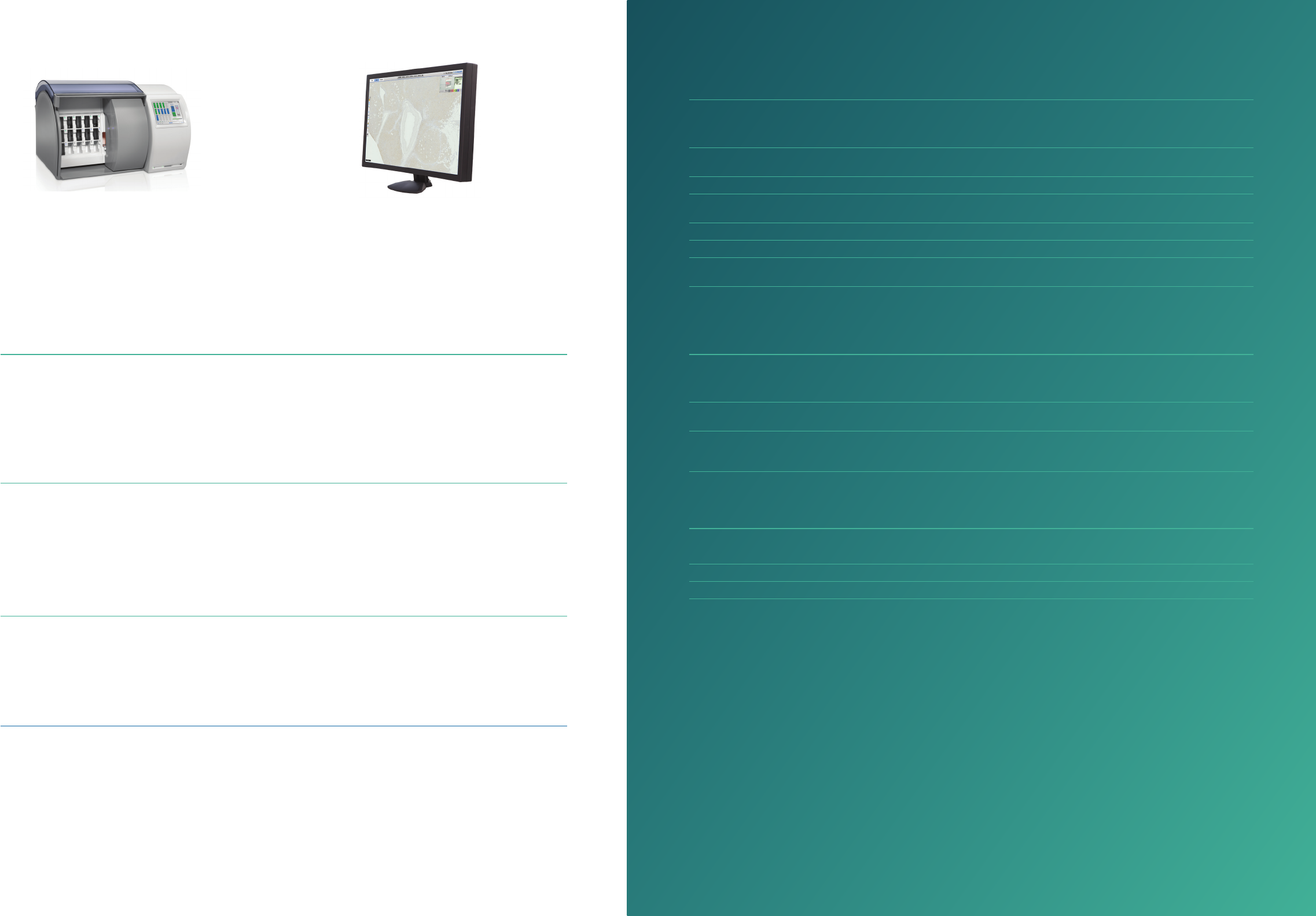
IntelliSite Pathology Solution Ultra Fast Scanner (UFS).
The Philips IntelliSite Pathology Solution IVD is intended as an aid to the pathologist to view, review and
diagnose digital images of stained slides. The pathologist needs to ensure the validity of the diagnosis by
employing the appropriate morphological studies and controls. The Philips IntelliSite Pathology Solution is an
automated digital slide creation, management, viewing and analysis system that can be used for Diagnosis in
clinical histopathology laboratories.
In Vitro Diagnostic Medical Device
IVD
Philips IntelliSite Pathology
Solution Image Management
System (IMS) 2.4
Philips IntelliSite Pathology
Solution Ultra Fast Scanner
(UFS) 1.6
Workow driven
Ease of use
With a 2-step “Load & Scan” operation; scanning starts
automatically, saving time, technician training simplied
Continuous processing
Add/remove slides without interrupting the scanning process
Full barcode integration
Data import from LIS for slide & case association
Workow / smart features
• Image alignment automatically aligns multiple serial tissue
sections for synchronized panning and zooming
• Tissue detection automatically suggests bookmarked area for
single click slide navigation
• Smart navigation via a combination of intuitive workow
oriented shortcuts keys and novel clickless panning for
comfortable navigation of images
High performance
High throughput
• A storage capacity of 300 slides
• 60 seconds per slide at 40x equivalent (15 x 15 mm scan area)
• High image quality through continuous autofocus
Ease of use
• Auto tissue detection; eliminating extra steps, saving
valuable time
• Automated “walk away” scanning
Speed
The IMS aims to improve eciency and eectiveness of pathology
labs to get pathologists through cases as fast as possible. Case
centric work list helps organize workload
• Fast workow navigation for next slide and case
• Advanced navigation tools incl. magnier zoom and bookmarks
• Performance design for handling > 1,000,000 cases
• Performance and capacity based storage architecture for
enhanced viewing performance
Collaboration made easy
Enhanced tools for interaction and remote viewing with the aim to
improve information sharing and simplify connectivity.
• Single, unied case list over multiple locations
• Simple case sharing via secure web link
• Simultaneous viewing with real-time collaboration
• Non-intrusive notications with single click access to a shared
session directly from the image viewer
Seamless Integration
Seamless integration with workow and information systems.
LIS can remain the central system to drive the workow for case
dispatching and reporting.
• Role-based access with secure user login
• Customizable bi-directional LIS connectivity and
communication
Technical specications
Philips IntelliSite Pathology Solution Ultra Fast Scanner
Slide capacity 300 slides (15 racks each hold
20 slides)
Slide rack Winlab LS-20/Winlab LSM-20,
Sakura 4768 20-slide basket
Total handling and
imaging time per slide
60 seconds at 40x equivalent
(15x15 mm scan area)
Barcode support DataMatrix (recommended),
Code 39, Code 128
Scanning method TDI line scanning Operating temperature 10 to 35º (for performance)
Microscope objective Olympus, NA of 0.75
Plan Apo
Relative humidity (no
condensation)
20 - 80% (for performance)
Focus method Continuous auto focus Dimensions of scanner 993 x 656 x 587mm (LxWxH)
Pixel size/resolution 0.25 µm/pixel Weight of scanner 129 kg
UFS output format iSyntax Philips proprietary le format with
either RAW or iSyntax compression
Power supply 110-230 VAC, 50/60 Hz, 150 Watts
Compliance to
standards
EN 61010-2-101:2002, EN 61326-2:2006,
CAN/CSA-C22.2 No 61010-2-101.04, UL
61010-2-101, FCC Part 15
UFS connectivity ports USB2.0, with 2 x RJ45 connectors,
Ethernet cable for 10GB and/or
1G/100MB
Philips IntelliSite Pathology Solution Image Management System Viewer – minimum hardware requirements
CPU Dual-core @3GHz Operating system Any operating system supporting a
browser with Microsoft Silverlight® 5
RAM 3GB of physical RAM memory Other software A PDF reader
(e.g. Adobe Acrobat Reader)
Monitor Resolution: 1600 x 1200
Size: 21”
Brightness: 300 cd/m2
Connectivity 100Mbit or 1Gbit Ethernet
connection to internet/intranet
Browser Internet browser supporting Microsoft
Silverlight® 5
Philips IntelliSite Pathology Solution Image Management System Application Server & storage – options
Storage capacity Flexible and extendable storage congurations from Terabytes to Petabytes
Conguration options Single and multi site
LIS interface HTTP/XML, or HL7 via LIS broker

© 2014 Koninklijke Philips N.V. All rights reserved.
Specications are subject to change without notice.
Trademarks are the property of Koninklijke Philips N.V.
(Royal Philips) or their respective owners.
www.philips.com
Printed in the Netherlands
www.philips.com
www.philips.com/digitalpathology
4522 207 28231 * DEC 2014 ENG
How to reach us:
Headquarters / Benelux / Denmark
Veenpluis 4-6, 5684 PC Best,
The Netherlands
Phone: +31 6 552 39 040
Email: cees.smit@philips.com
DACH (Germany, Switzerland, Austria)
Phone: +49 4121 2611501
Email: christian.tank@philips.com
Southern Europe (France, Spain, Portugal, Italy)
Phone: +33 6 13494071
Email: franck.schoens@philips.com
UK, Ireland & Scandinavia
Phone: + 44 7824 474270
Email: andy.humes@philips.com
South East Asia
620A, Lorong 1 Toa PayohBuilding TP3, Level 3
Singapore 319762
Phone: +65 688 24 714
Email: kelvin.pow@philips.com
United Arab Emirates, Saudi Arabia, Qatar & Lebanon
Choueiri Group Building -
Dubai Knowledge village, Al Sufouh 2, Dubai United Arab
Emirates
Phone: +971 55 10003719
Email: ramy.helal@philips.com
Manufacturing address:
Philips Electronics Nederland BV
Philips Digital Pathology Solutions
Veenpluis 4-6, 5684 PC Best,
The Netherlands
In Vitro Diagnostic Medical Device
