Philips NOCTN502 User Manual Digital Diagnost C50 Product Overview 452299116101 V7
NOCTN502 Product Brochure Philips DigitalDiagnost C50 Ceiling mounted digital X-ray system 1db0b9400af140a3b23ea77c014ae8ef Philips - DigitalDiagnost C50 Ceiling mounted digital X-ray systemNOCTN502
User Manual: Philips NOCTN502 DigitalDiagnost C50 Product Overview DigitalDiagnost C50 Ceiling mounted digital X-ray systemNOCTN502
Open the PDF directly: View PDF ![]() .
.
Page Count: 3

Radiography solutions
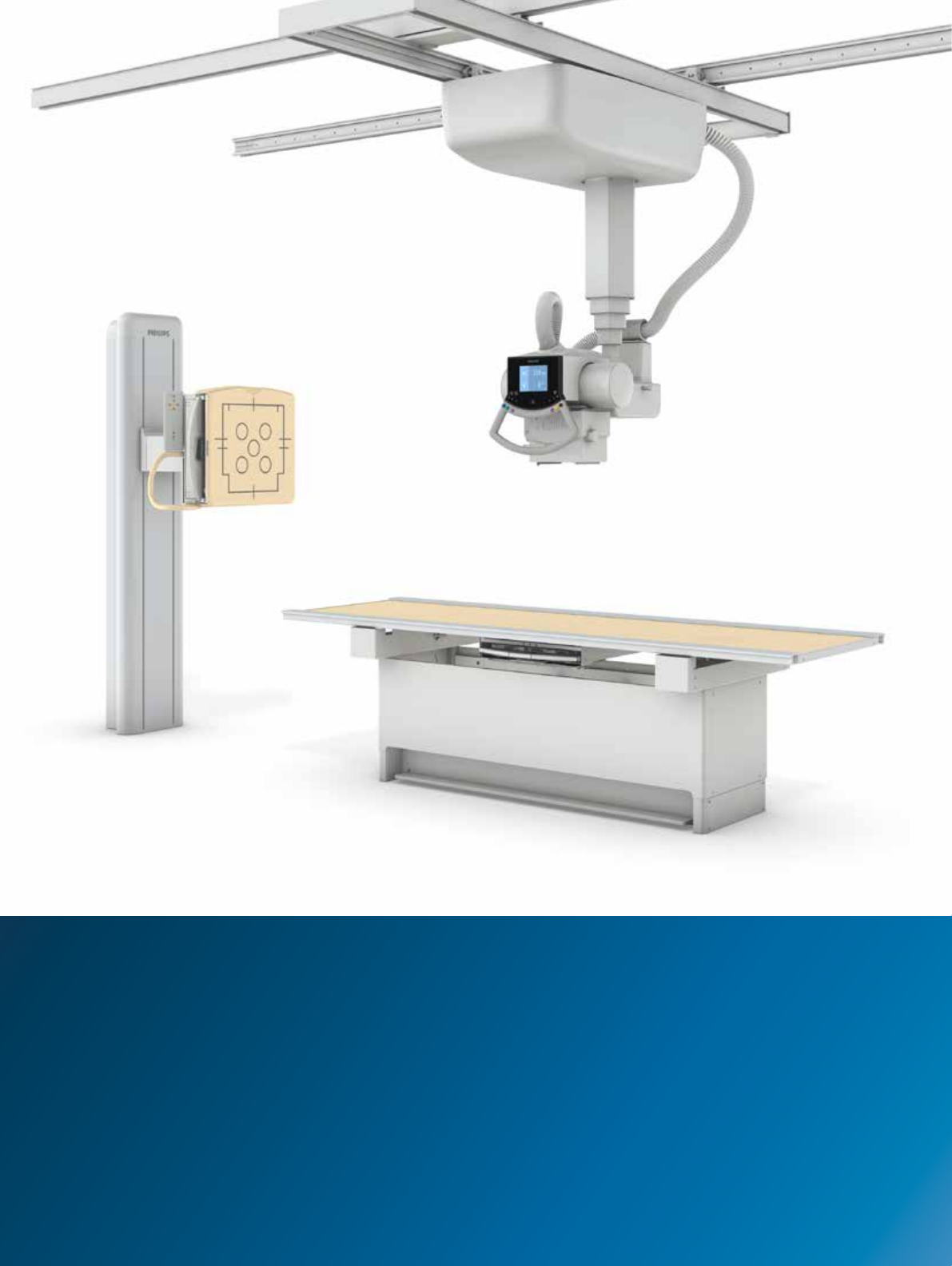
DigitalDiagnost C50
Pair performance with
ease-of-ownership
Philips DigitalDiagnost C50 ceiling mounted digital X-ray system
Key advantages
• Cost-eective ceiling
suspended system
with xed or wireless
detector
• Broad application range
includes image stitching
• Proven Philips quality
and support
Philips DigitalDiagnost C50 is a performance oriented
ceiling mounted digital X-ray system that delivers
diagnostic quality images for fast, ecient exams.
The DigitalDiagnost C50 oers you the kind of
versatility necessary to address a broad range of clinical
applications, while doing so in a cost-eective manner.
Choose either a xed or wireless detector conguration
and benet from motorized auto-tracking, a robust digital
workow, and UNIQUE image processing.
Make smart use of your resources
with this versatile system
Elegant movements
The ceiling suspended X-ray tube moves smoothly
and manually into position. The vertical stand detector
slides up/down and tilts -20° - 90° for easy multiple
angle exams. Optional standard stitching speeds the
full body imaging process.
DigitalDiagnost C50 uses the latest generation of
robust wireless portable/xed detectors and cutting
edge image processing techniques to reinforce
diagnostic condence.
Choose a room that ts
The DigitalDiagnost C50 is available in three
room congurations to best suit your institutional
requirements.
• Value Room – A wireless portable detector is paired
with a xed height or height adjustable table to oer
versatility and free exposures for a wide variety of
exam types
• Chest Room (Fixed) – This xed detector and trolley
conguration is perfect for high volume specialty
chest and multi-purpose applications
• Chest Room (Wireless) – Swap the xed detector
for a wireless portable one and you broaden system
capabilities
Strong workow continuity
The Eleva workstation enhances your workow via
pre-settings and customized user proles. Images are
available on screen just six seconds after acquisition.
And it takes only three clicks to complete an
examination. This high degree of automation supports
streamlined patient scheduling and throughput.
The system is fully DICOM compatible and easily
connects to your PACS for IT infrastructure integration.
Consistent diagnostic quality
Proprietary UNIQUE image processing software
delivers consistently uniform clinical image quality for
all anatomic regions by automatically adjusting the
balance between heavily exposed and barely exposed
areas. UNIQUE harmonizes contrast to enhance faint
details, helping to ensure you experience consistent
results.
Quality from a brand you can trust
DigitalDiagnost C50 is an example of proven
technology from a leading manufacturer of high-end
medical imaging equipment*. The system reects
decades of company experience in digital X-ray
development and enjoys the benets of Philips
customer service. It’s well built and easy to own.
DigitalDiagnost C50 extends your imaging capabilities in an aordable
way. You get Philips brand quality with diagnostic functions that cover
all your general clinical application needs. Enjoy a high degree of system
automation with the Eleva user interface and workstation. Special
automatic exposure control (AEC) permits short, accurate exposures for
eective dose management. And consistent, diagnostic quality is possible
with Philips UNIQUE multi-resolution image processing software.
It’s a smart investment.
Advantages
The DigitalDiagnost C50 has something for everyone:
• For the patient: Quick and easy imaging exam, eortless experience, and excellent results
• For the radiologist: Fast, digital image quality for condent diagnoses and seamless PACS integration
• For the technologist: Simple system operation with intuitive user interface for a patient-focused workow
• For the radiology director: Cost-eective ceiling mounted digital X-ray system from a recognized industry leader
* 2013 Global Medical Imaging Equipment Market Outlook - Positioning for the Future ; Frost&Sullivan; NC8B-54, June 2013
2 3

How to reach us
Please visit www.philips.com/healthcare
healthcare@philips.com
How to reach us
Please visit www.philips.com/healthcare
healthcare@philips.com
© 2016 Koninklijke Philips N.V. All rights reserved.
Specications are subject to change without notice.
Trademarks are the property of Koninklijke Philips N.V.
(Royal Philips) or their respective owners.
4522 991 16101 * FEB 2016
X-ray generation
Generator 50 kW 65 kW
High-voltage generator The converter generator generates high voltage equivalent to DC voltage
Mains voltage 380V/400 V / 480 V (±10%); 50 Hz or 60 Hz, 3-phase
Max. mains resistance at 400 V 0.3 Ohm 0.2 Ohm
Max. mains current at 400 V 112A 134A
Nominal power (IEC) 50 kW 65 kW
Max. tube voltage 150 kV 150 kV
Max. tube current (at 80 kV) 630 mA 800 mA
mAs product 0.4 mAs to 850 mAs 0.4 mAs to 850 mAs
Exposure times 1ms to 4s 1ms to 4s
Tube High power X-ray tube RO1750 SRO 33100
Two focal spots 0.6 and 1.2 0.6 and 1.2
Maximum power: with focal spot 0.6
with focal spot 1.2
21 kW
60 kW
33 kW
100 kW
Anode angle 13° 13°
Maximum tube voltage 150 kV 150 kV
Anode heat storage capacity 220 kJ (300 kHU) 220 kJ (300 kHU)
Assembly heat capacity 1500 kJ (2202.4 kHU) 1500 kJ (2202.4 kHU)
Continuous anode input power 250 W 200 W
Minimum anode speed 3000-3600 revolutions/minute 9000-10800 revolutions/minute
Fixed detector
Type Digital GOS (Gadox) at detector
Detector size 43 cm x 43 cm (17"x 17")
Active area 42.5 cm x 42.5 cm (16.7" x 16.7")
Image matrix size 2,869 pixel x 2,874 pixel
Detector pixels 8.3 Megapixel
Pixel size 148 μm
Image resolution up to 3.4 Lp/mm
Weight 2.1 kg
Wireless Portable detector
Type Digital wireless at detector
Scintillator Gadox
Detector size 35 cm x 43 cm (approx 14" x 17")
Active Area 34.1 cm x 42.4 cm (approx 13.4" x 16.7")
Image matrix size 2476 x 3072 pixels
Active pixel area 2456 x 3052 pixels
Pixel size 139 μm
Detector pixels 7.6 Megapixels
A/D conversion (bits) 16 bits
Weight (incl. battery) 3.6 kg approx. (7.93 lbs)
Ceiling Suspension CS
Column 110 cm (44")
Ceiling height at source image distance
2.70 m to 3.00 m (8' 8.3" to 10' 5.9")
Movements
Travel range X axis(Longitudinal) 3.05/4.05m (10'/13'3.4")
Travel range Y axis(Lateral) 1.8m (5' 11")
Travel range Z axis(Vertical) 1.5m (4' 11")
Collimator
Type Manual, with light eld indicator
Angle of aperture and rotation ±45°
Timer switch 30 s (should be congurable)
Table
Choice of height adjustable table, xed height table and
trolleyWallstand: vertical stand with xed detector unit and
vertical stand with wireless portable detector unit
Eleva workspot computer
Hard disk 500 Gb total; 203 Gb for the image data
(equivalent to approximately 11548 images)
RAM storage capacity 4 Gb
DigitalDiagnost C50 is not available for sale in North America
