Digdiag12 VR 130 DICOM Digital Diagnost R1.2
User Manual: VR 130
Open the PDF directly: View PDF ![]() .
.
Page Count: 23

© Copyright Philips Medical Systems Nederland B.V. 2001
All rights reserved
Philips Medical Systems
DICOM Conformance Statement
Digital Diagnost
R 1.2
Document Number 4512 130 91942
10 October 2001

Page ii DICOM Conformance Statement 4512 130 91942 - 10 Oct 01
All rights reserved
© Philips Medical Systems Nederland B.V. 2001
Philips Medical Systems Nederland B.V.
Medical Imaging IT, Interoperability
Building QV-282
P.O. Box 10.000
5680 DA Best
The Netherlands
Tel.: +31 40 2763079
Fax.: +31 40 2764263
email: dicom@philips.com
Internet: http://www.medical.philips.com/dicomcs/

4512 130 91942 - 10 Oct 01 DICOM Conformance Statement Page iii
Table of Contents
All rights reserved
© Philips Medical Systems Nederland B.V. 2001
1Introduction . . . . . . . . . . . . . . . . . . . . . . . . . . . . . . . . . . . . . . . . . . . . . . . 1
1.1 Scope and field of application . . . . . . . . . . . . . . . . . . . . . . . . . . . . . . . 1
1.2 Intended audience . . . . . . . . . . . . . . . . . . . . . . . . . . . . . . . . . . . . . . . . . 1
1.3 Contents and structure . . . . . . . . . . . . . . . . . . . . . . . . . . . . . . . . . . . . . 1
1.4 Used definitions, terms and abbreviations . . . . . . . . . . . . . . . . . . . . . . 1
1.5 References . . . . . . . . . . . . . . . . . . . . . . . . . . . . . . . . . . . . . . . . . . . . . . 1
1.6 Important note to the reader . . . . . . . . . . . . . . . . . . . . . . . . . . . . . . . . . 2
1.7 General Acronyms and Abbreviations. . . . . . . . . . . . . . . . . . . . . . . . . 3
2Implementation model . . . . . . . . . . . . . . . . . . . . . . . . . . . . . . . . . . . . . . . 4
2.1 Application Data Flow Diagram . . . . . . . . . . . . . . . . . . . . . . . . . . . . . . 4
2.2 Functional definition of Application Entities . . . . . . . . . . . . . . . . . . . . 4
2.3 Sequencing of Real World Activities . . . . . . . . . . . . . . . . . . . . . . . . . . 4
3AE Specifications . . . . . . . . . . . . . . . . . . . . . . . . . . . . . . . . . . . . . . . . . . . 5
3.1 AE Digital Diagnost DICOM Export Specification . . . . . . . . . . . . . . . 5
3.1.1 Association Establishment Policies . . . . . . . . . . . . . . . . . . . . . . . . . 5
3.1.2 Association Initiation Policy . . . . . . . . . . . . . . . . . . . . . . . . . . . . . . 6
3.1.3 Association Acceptance Policy . . . . . . . . . . . . . . . . . . . . . . . . . . . . 14
4Communication Profiles . . . . . . . . . . . . . . . . . . . . . . . . . . . . . . . . . . . . . 15
4.1 TCP/IP Stack . . . . . . . . . . . . . . . . . . . . . . . . . . . . . . . . . . . . . . . . . . . . 15
4.1.1 Physical Media Support . . . . . . . . . . . . . . . . . . . . . . . . . . . . . . . . . . 15
5Configuration . . . . . . . . . . . . . . . . . . . . . . . . . . . . . . . . . . . . . . . . . . . . . . 16
5.1 AE Title/Presentation Address mapping . . . . . . . . . . . . . . . . . . . . . . . 16
5.1.1 Local AE Titles and Presentation Addresses . . . . . . . . . . . . . . . . . . 16
5.1.2 Remote AE Titles and Presentation Addresses . . . . . . . . . . . . . . . . 16
5.2 Configurable parameters . . . . . . . . . . . . . . . . . . . . . . . . . . . . . . . . . . . . 16
5.3 Export Filter . . . . . . . . . . . . . . . . . . . . . . . . . . . . . . . . . . . . . . . . . . . . . 16
5.4 Configurable Attributes . . . . . . . . . . . . . . . . . . . . . . . . . . . . . . . . . . . . 16
5.4.1 Bits stored . . . . . . . . . . . . . . . . . . . . . . . . . . . . . . . . . . . . . . . . . . . . 17
5.4.2 Photometric Interpretation . . . . . . . . . . . . . . . . . . . . . . . . . . . . . . . . 17
5.4.3 Modes . . . . . . . . . . . . . . . . . . . . . . . . . . . . . . . . . . . . . . . . . . . . . . . . 17
6Support of Extended Character Sets . . . . . . . . . . . . . . . . . . . . . . . . . . . 19

Page iv DICOM Conformance Statement 4512 130 91942 - 10 Oct 01
All rights reserved
© Philips Medical Systems Nederland B.V. 2001

4512 130 91942 - 10 Oct 01 DICOM Conformance Statement Page 1 of 19
Introduction
All rights reserved
© Philips Medical Systems Nederland B.V. 2001
1 Introduction
This chapter provides general information about the purpose, scope and contents of this Con-
formance Statement.
1.1 Scope and field of application
The scope of this DICOM Conformance Statement is to facilitate data exchange with equip-
ment of Philips Medical Systems. This document specifies the compliance to the DICOM
standard (formally called the NEMA PS 3.X standards). It contains a short description of the
applications involved and provides technical information about the data exchange capabilities
of the equipment. The main elements describing these capabilities are: the supported DICOM
Service Object Pair (SOP) Classes, Roles, Information Object Definitions (IOD) and Transfer
Syntaxes.
The field of application is the integration of the Philips Medical Systems equipment into an
environment of medical devices.
This Conformance Statement should be read in conjunction with the DICOM standard and its
addenda [DICOM].
1.2 Intended audience
This Conformance Statement is intended for:
• (potential) customers,
•system integrators of medical equipment,
•marketing staff interested in system functionality,
•software designers implementing DICOM interfaces.
It is assumed that the reader is familiar with the DICOM standard.
1.3 Contents and structure
The DICOM Conformance Statement is contained in chapter 2 through 7 and follows the con-
tents and structuring requirements of DICOM PS 3.2.
1.4 Used definitions, terms and abbreviations
DICOM definitions, terms and abbreviations are used throughout this Conformance Statement.
For a description of these, see NEMA PS 3.3 and PS 3.4.
The word Philips in this document refers to Philips Medical Systems.
1.5 References
[DICOM] The Digital Imaging and Communications in Medicine (DICOM) standard:
NEMA PS 3.X 2000
National Electrical Manufacturers Association (NEMA) Publication Sales
1300 N. 17th Street, Suite 1847
Rosslyn, Va. 22209, United States of America

Page 2 of 19 DICOM Conformance Statement 4512 130 91942 - 10 Oct 01
Introduction
All rights reserved
© Philips Medical Systems Nederland B.V. 2001
1.6 Important note to the reader
This Conformance Statement by itself does not guarantee successful interoperability of Philips
equipment with non-Philips equipment. The user (or user’s agent) should be aware of the fol-
lowing issues:
•Interoperability
Interoperability refers to the ability of application functions, distributed over two or more
systems, to work successfully together. The integration of medical devices into a networked
environment may require application functions that are not specified within the scope of
DICOM. Consequently, using only the information provided by this Conformance State-
ment does not guarantee interoperability of Philips equipment with non-Philips equipment.
It is the user’s responsibility to analyse thoroughly the application requirements and to
specify a solution that integrates Philips equipment with non-Philips equipment.
•Validation
Philips equipment has been carefully tested to assure that the actual implementation of the
DICOM interface corresponds with this Conformance Statement.
Where Philips equipment is linked to non-Philips equipment, the first step is to compare the
relevant Conformance Statements. If the Conformance Statements indicate that successful
information exchange should be possible, additional validation tests will be necessary to
ensure the functionality, performance, accuracy and stability of image and image related
data. It is the responsibility of the user (or user’s agent) to specify the appropriate test suite
and to carry out the additional validation tests.
•New versions of the DICOM Standard
The DICOM Standard will evolve in future to meet the user’s growing requirements and to
incorporate new features and technologies. Philips is actively involved in this evolution and
plans to adapt its equipment to future versions of the DICOM Standard. In order to do so,
Philips reserves the right to make changes to its products or to discontinue its delivery.
The user should ensure that any non-Philips provider linking to Philips equipment, also
adapts to future versions of the DICOM Standard. If not, the incorporation of DICOM
enhancements into Philips equipment may lead to loss of connectivity (in case of network-
ing) and incompatibility (in case of media).

4512 130 91942 - 10 Oct 01 DICOM Conformance Statement Page 3 of 19
Introduction
All rights reserved
© Philips Medical Systems Nederland B.V. 2001
1.7 General Acronyms and Abbreviations.
The following acronyms and abbreviations are used in the document.
•ACC American College of Cardiology
•AE Application Entity
•ACR American College of Radiology
•ANSI American National Standard Institute
• BOT Basic Offset Table
•CD-R CD Recordable
•CD-M CD Medical
•DCI Digital Cardio Imaging
•DCR Dynamic Cardio Review
•DICOM Digital Imaging and Communication in Medicine
•DIMSE DICOM Message Service Element
•DIMSE-C DICOM Message Service Element-Composite
•DIMSE-N DICOM Message Service Element-Normalized
•ELE Explicit VR Little Endian
•EBE Explicit VR Big Endian
•FSC File Set Creator
•GUI Graphic User Interface
•HIS Hospital Information System
•HL7 Health Level Seven
•ILE Implicit VR Little Endian
•IOD Information Object Definition
•ISIS Information System - Imaging System
•NEMA National Electrical Manufacturers Association
• PDU Protocol Data Unit
•RIS Radiology Information System
•RWA Real World Activity
•SC Secondary Capture
•SCM Study Component Management
•SCP Service Class Provider
•SCU Service Class User
•SOP Service Object Pair
•TCP/IP Transmission Control Protocol/Internet protocol
•UID Unique Identifier
•WLM Worklist Management
Page 4 of 19 DICOM Conformance Statement 4512 130 91942 - 10 Oct 01
Implementation model
All rights reserved
© Philips Medical Systems Nederland B.V. 2001
2 Implementation model
The Digital Diagnost R1.2 of Philips Medical Systems is an Computed Radiography (CR)
image generating system. It contains an Export function based on the DICOM Image Storage
to transfer image data from the Digital Diagnost system to a remote system. This DICOM
Export function is described in this document.
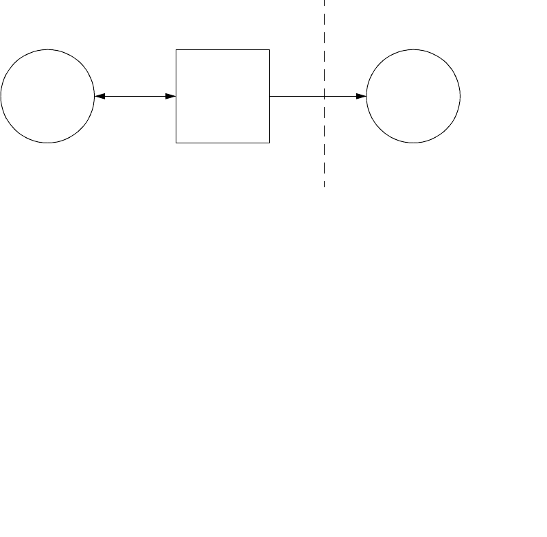
2.1 Application Data Flow Diagram
The Digital Diagnost DICOM Export transfers a Digital Diagnost image to a remote DICOM
system. This is activated by an operator request or automatically if the system is configured to
do so. A remote destination is selected from the user interface, followed by the selection of the
image to be transferred.
Post-processed image data can be transferred (pixel value between 0 and 30,000) as an
instance of the DICOM Computed Radiography IOD. The images transferred are intended for
viewing purpose and VOI LUT transformation (grayscale transformation) only.
The Digital Diagnost DICOM Export behaves as a single Application Entity. The related
Implementation Model is shown in Figure2-1 on page4.
2.2 Functional definition of Application Entities
The Digital Diagnost DICOM Export application entity acts as a Service Class User (SCU) of
the Storage Service Class. After invoking it will open an association to the remote system. For
each image to be transported a retrieve action from the internal Digital Diagnost storage will
take place followed by the conversion to a DICOM message to be transferred to the remote
system.
2.3 Sequencing of Real World Activities
Not applicable.
Figure 2-1: Digital Diagnost DICOM Export Implementation Model
DICOM
Local Remote
Standard Interface
DICOM
Image
Export
Request
Store
Image
Remotely
Digital
Diagnost
DICOM
Export

4512 130 91942 - 10 Oct 01 DICOM Conformance Statement Page 5 of 19
AE Specifications
All rights reserved
© Philips Medical Systems Nederland B.V. 2001
3 AE Specifications
Digital Diagnost DICOM Export acts as a single Application Entity.
3.1 AE Digital Diagnost DICOM Export Specification
The Digital Diagnost Export Application Entity provides Standard Extended Conformance to
the following DICOM 3.0 SOP class as an SCU:
The Digital Diagnost Export Application Entity does not support DICOM 3.0 SOP Classes as
SCP.
3.1.1 Association Establishment Policies
3.1.1.1 General
The Digital Diagnost export will offer a fixed maximum PDU size of 16K = 16384 bytes on
the associations initiated by the application itself.
3.1.1.2 Number of Associations
The Digital Diagnost export will attempt to establish one association at a time.
3.1.1.3 Asynchronous Nature
The Digital Diagnost export does not support asynchronous operations and will not perform
asynchronous window negotiation.
3.1.1.4 Implementation Identifying Information
The Implementation Class UID is: “1.3.46.670589.26.1.2“.
The implementation version name is: “DigiDiagnost1.2”.
Table 3-1: Supported SOP class by the Digital Diagnost Export AE as SCU
SOP class Name UID
Computed Radiography Image Storage 1.2.840.10008.5.1.4.1.1.1

Page 6 of 19 DICOM Conformance Statement 4512 130 91942 - 10 Oct 01
AE Specifications
All rights reserved
© Philips Medical Systems Nederland B.V. 2001
3.1.2 Association Initiation Policy
The Digital Diagnost export initiates associations as a result of only one Real-World activity
and is described below.
3.1.2.1 The Digital Diagnost DICOM Image Export Request
3.1.2.1.1 Associated Real-World Activity
The DICOM Image Export Request can be done on the following ways:
•the operator requests via the User Interface the export of the selected Digital Diagnost
image to a remote system,
•the generation of a new Digital Diagnost image will result in an automatic export of that
image when the system is configured in automatic export mode.
For each export request a new association is set-up, then the transfer of the image is started.
The association is released when the transfer is ended. The transferred image will not be
deleted from the system.
In case of unsuccessful transfer with special response status conditions (e.g. Store SCP down),
a new attempt will be done automatically every 20 seconds. These queued export requests can
be aborted by the operator.
3.1.2.1.2 Proposed Presentation Contexts
The Digital Diagnost export will propose the following presentation contexts:
3.1.2.1.3 SOP Specific Conformance to Storage SOP Classes
The Digital Diagnost provides standard conformance to the Storage Service Class.
The status of the C-STORE Response (Success, Refused, Error, Warning) will be displayed
via the user interface.
Extended negotiation is not supported.
Table 3-3 lists the applied optional and extended modules and attributes of the CR IOD. Con-
ditional attributes Patient Orientation (type 2C), Image Date (type 2C), Image Time (type 2C),
Specific Character Set (type 1C) are always present.
Table 3-2: Proposed Presentation Contexts for the Digital Diagnost Export
Presentation Context table
Abstract Syntax Transfer Syntax
Role Extended
Negotiation
Name UID Name List UID List
Computed Radiography
Image Storage 1.2.840.10008.5
.1.4.1.1.1 ILE
ELE
EBE
1.2.840.10008.1.2
1.2.840.10008.1.2.1
1.2.840.10008.1.2.2
SCU None

4512 130 91942 - 10 Oct 01 DICOM Conformance Statement Page 7 of 19
AE Specifications
All rights reserved
© Philips Medical Systems Nederland B.V. 2001
Table 3-3: Applied optional Modules and Attributes of the applied CR IOD
IE Module Optional Attributes Conditional Attributes
Patient Patient Other Patient’s ID, Ethnic Group,
Patient Comments -
Study General Study Study Description, Physician(s)
of Record -
Patient Study Additional Patient’s History -
Series General Series Series Date, Series Time, Opera-
tor’s Name, Protocol Name Laterality.
CR Series Filter Type, Focal Spot(s), Plate
Type, Focal Spot(s)
Equipment General Equipment Institution Name, Station Name,
Institutional Department Name,
Manufacturer’s Model name,
Device Serial Number, Software
Version(s), Date of Last Calibra-
tion, Time of Last Calibration
-
Image General Image Image Type, Acquisition
Number, Image Comments Image Date, Image Type, Patient
Orientation
Image Pixel - -
X-Ray Acquisition Image Area Dose Product, Grid,
Imager Pixel Spacing -
XRF Tomagraphy
Acquisition Tomo Angle, Tomo Time, Scan
Options -
CR Image KVP, Distance Source to Detec-
tor, Exposure Time, Exposure,
Sensitivity, Generator Power,
Collimator/Grid Name, Process-
ing Function, Postprocessing
Function
-
VOI LUT Window Center Window Width
SOP Common -Specific Character Set

Page 8 of 19 DICOM Conformance Statement 4512 130 91942 - 10 Oct 01
AE Specifications
All rights reserved
© Philips Medical Systems Nederland B.V. 2001
The modules selected from the CR Image IOD module table of DICOM 3.0 and the extended
modules are given in the table below.
The details of these applied modules are given in the tables below. The list of possible attribute
values are given (if applicable). The situation that an attribute is present conditionally/option-
ally or that an attribute may contain a zero length value, is indicated too.
The greyed attributes are the attributes from which the contents are received from the RIS via
the EasyLink.
Table 3-4: Applied Modules in the Extended CR IOD
IE Module Reference
Patient Patient MTable3-5
Study General Study MTable3-6
Patient Study UTable3-7
Series General Series MTable3-8
CR Series MTable3-9
Equipment General Equipment MTable3-10
Image General Image MTable3-11
Image Pixel MTable3-12
CR Image MTable3-13
Private MTable3-14
X-Ray Acquisition MTable3-15
XRF Tomagraphy Acquisition CTable 3-16
VOI LUT UTable3-17
SOP Common MTable3-18
Table 3-5: Computed Radiography Image Storage SOP Class - Patient Module
Attribute Name Tag Note
Patient's Name 0010,0010 Received from RIS or filled in by Operator or
is empty.
Patient ID 0010,0020 Received from RIS or filled in by Operator or
is empty.
Patient's Birth Date 0010,0030 Received from RIS or filled in by Operator or
is empty.

4512 130 91942 - 10 Oct 01 DICOM Conformance Statement Page 9 of 19
AE Specifications
All rights reserved
© Philips Medical Systems Nederland B.V. 2001
Patient's Sex 0010,0040 Received from RIS or filled in by Operator or
is empty.
Applied value(s): F, M, O
Other Patient IDs 0010,1000 Received from RIS or filled by operator or is
empty.
Ethnic Group 0010,2160 Received from RIS or filled by operator or is
empty.
Patient Comments 0010,4000 Received from RIS or filled by operator or is
empty.
Table 3-6: Computed Radiography Image Storage SOP Class - General Study Module
Attribute Name Tag Note
Physician(s) of Record 0008,1048 Received from RIS or filled by operator or is
empty.
Study Date 0008,0020 First Image Date of an acquisition sequence of
a Study.
Study Time 0008,0030 First Image Time of an acquisition sequence
of a Study.
Accession Number 0008,0050 Received from RIS or filled by operator or is
empty.
Referring Physician's Name 0008,0090 Received from RIS or filled by operator or is
empty..
Study Description 0008,1030
Additional Patient’s History 0010,21B0 Received from RIS or filled by operator or is
empty.
Study Instance UID 0020,000D Received from RIS or generated by Digital
Diagnost.
Study ID 0020,0010 Digital Diagnost generated.
Table 3-5: Computed Radiography Image Storage SOP Class - Patient Module (Continued)
Attribute Name Tag Note

Page 10 of 19 DICOM Conformance Statement 4512 130 91942 - 10 Oct 01
AE Specifications
All rights reserved
© Philips Medical Systems Nederland B.V. 2001
Table 3-8: Computed Radiography Image Storage SOP Class - C-STORE-RQ - Patient Study Module
Table 3-7: Computed Radiography Image Storage SOP Class - General Series Module
Attribute Name Tag Note
Series Date 0008,0021 Identical to Study Date.
Series Time 0008,0031
Modality 0008,0060 Applied value(s): CR
Operator's name 0008,1070 Filled in by operator or is empty.
Protocol Name 0018,1030 Received from RIS of filled by operator or is
empty.
Series Instance UID 0020,000E Digital Diagnost generated, derived from
Series Number.
Series Number 0020,0011 Digital Diagnost generated.
Laterality 0020,0060 Always Empty
Attribute Name Tag Note
Additional Patient History 0010,21B0
Table 3-9: Computed Radiography Image Storage SOP Class - CR Series Module
Attribute Name Tag Note
Body Part Examined 0018,0015 Applied value(s): ABDOMEN, ANKLE,
BREAST, CHEST, CLAVICLE, COCCYX,
CSPINE, ELBOW, EXTREMITY, FOOT,
HAND, HIP, KNEE, LSPINE, PELVIS,
SHOULDER, SKULL, SSPINE, TSPINE
Filter Type 0018,1160
Focal Spot(s) 0018,1190
Plate Type 0018,1260
View Position 0018,5101 Applied value(s): AP, LL, LLD, LLO, PA,
RL, RLD, RLO

4512 130 91942 - 10 Oct 01 DICOM Conformance Statement Page 11 of 19
AE Specifications
All rights reserved
© Philips Medical Systems Nederland B.V. 2001
Table 3-10: Computed Radiography Image Storage SOP Class - General Equipment Module
Attribute Name Tag Note
Manufacturer 0008,0070 Applied value(s): Philips Medical Systems
Institution Name 0008,0080
Station Name 0008,1010
Institutional Department Name 0008,1040
Manufacturer's Model Name 0008,1090 Applied value(s): digital DIAGNOST
Device Serial Number 0018,1000
Software Version(s) 0018,1020
Date of Last Calibration 0018,1200
Time of Last Calibration 0018,1201
Table 3-11: Computed Radiography Image Storage SOP Class - General Image Module
Attribute Name Tag Note
Image Type 0008,0008 Applied Value(s): DERIVED\ PRIMARY
Content Date 0008,0023
Content Time 0008,0033
Acquisition Number 0020,0012
Instance Number 0020,0013 Is composed of Digital Diagnost acquisition
and post number.
Patient Orientation 0020,0020 Applied Value(s): A, F, H, L, P, R
Image Comments 0020,4000 Text from annotationtool included.
Table 3-12: Computed Radiography Image Storage SOP Class - Image Pixel Module
Attribute Name Tag Note
Samples per Pixel 0028,0002 Applied value(s): 1
Photometric Interpretation 0028,0004 Applied value(s): MONOCHROME1,
MONOCHROME2.
In combination with EV RAD:
Applied value(s):MONOCHROME2
Rows 0028,0010
Columns 0028,0011
Bits Allocated 0028,0100 Applied Value(s): 16, 8

Page 12 of 19 DICOM Conformance Statement 4512 130 91942 - 10 Oct 01
AE Specifications
All rights reserved
© Philips Medical Systems Nederland B.V. 2001
Pixel Spacing 0028,0030
Bits Stored 0028,0101 Applied Value(s): 10, 12, 15, 8
High Bit 0028,0102 Applied Value(s): 11, 14, 7, 9
Pixel Representation 0028,0103 Applied value(s): 0000
Table 3-13: Computed Radiography Image Storage SOP Class - CR Image Module
Attribute Name Tag Note
Pixel Data 7FE0,0010
KVP 0018,0060
Distance Source to Detector(SID) 0018,1110
Exposure Time 0018,1150
Exposure 0018,1152
Generator Power 0018,1170
Acquisition Device Processing
Description 0018,1400
Collimator/Grid Name 0018,1180
Processing Function 0018,5020
Postprocessing Function 0018,5021
Sensitivity 0018,6000
Table 3-14: Computed Radiography Image Storage SOP Class - Private Module
Attribute Name Tag Note
Private Creator Group 0019 0019,0019 Only exported when image is exported for
printing
Applied Value(s): DIDI TO PCR 1.1
Route AET 0019,1922
PCR Print Scale 0019,1923 Only exported when image is exported for
printing
PCR Print Job End 0019,1924 Only exported when image is exported for
printing
PCR No Film Copies 0019,1925 Only exported when image is exported for
printing
Table 3-12: Computed Radiography Image Storage SOP Class - Image Pixel Module (Continued)
Attribute Name Tag Note

4512 130 91942 - 10 Oct 01 DICOM Conformance Statement Page 13 of 19
AE Specifications
All rights reserved
© Philips Medical Systems Nederland B.V. 2001
Table 3-15: Computed Radiography Image Storage SOP Class - C-STORE-RQ - X-ray Acquisition
Module
PCR Film Layout Position 0019,1926 Only exported when image is exported for
printing
PCR Print Report Name 0019,1927 Only exported when image is exported for
printing
RAD Protocol Printer 0019,1970 Only exported when image is exported for
printing
RAD Protocol Medium 0019,1971 Only exported when image is exported for
printing
Private Creator 0089,0010 Applied Value(s): DIDI TO PCR 1.1
Stamp Image Sequence 0089,1020
>Samples per Pixel 0028,0002 Applied Value(s): 1
>Photometric Interpretation 0028,0004 Applied Value(s): MONOCHROME1
>Rows 0028,0010 Maximum value 512
>Columns 0028,0011 Maximum value 512
>Bits Allocated 0028,0100 Applied Value(s): 8
>Bits Stored 0028,0101 Applied Value(s): 8
>High Bit 0028,0102 Applied Value(s): 7
>Pixel Representation 0028,0103 Applied Value(s): 0x000
>Pixel Data 7FE0,0010
Attribute Name Tag Note
Image Area Dose Product 0018,115E
Imager Pixel Spacing 0018,1164
Grid 0018,1166 Applied Value(s): IN, NONE
Table 3-14: Computed Radiography Image Storage SOP Class - Private Module (Continued)
Attribute Name Tag Note

Page 14 of 19 DICOM Conformance Statement 4512 130 91942 - 10 Oct 01
AE Specifications
All rights reserved
© Philips Medical Systems Nederland B.V. 2001
Table 3-16: Computed Radiography Image Storage SOP Class - C-STORE-RQ - X-ray Tomography
Acquisition Module
3.1.3 Association Acceptance Policy
The Digital Diagnost does not accept associations.
Attribute Name Tag Note
Scan Options 0018,0022 Applied Value(s): TOMO
Tomo Layer Height 0018,1460 Distance in mm between the table surface
and the sharp image plane.
Tomo Angle 0018,1470 Angle span in degrees of rotation of X-ray
source during X-Ray acquisition.
Tomo Time 0018,1480 Time in seconds the source has taken to
rotate the Tomo Angle during X-Ray acqui-
sition.
Table 3-17: Computed Radiography Image Storage SOP Class - VOI LUT Module
Attribute Name Tag Note
Window Center 0028,1050
Window Width 0028,1051
Table 3-18: Computed Radiography Image Storage SOP Class - SOP Common Module
Attribute Name Tag Note
Specific Character Set 0008,0005 Applied Value(s): ISO_IR 100
SOP Class UID 0008,0016 Applied value(s): 1.2.840.10008.5.1.4.1.1.1
SOP Instance UID 0008,0018 Generated by Digital Diagnost.

4512 130 91942 - 10 Oct 01 DICOM Conformance Statement Page 15 of 19
Communication Profiles
All rights reserved
© Philips Medical Systems Nederland B.V. 2001
4 Communication Profiles
4.1 TCP/IP Stack
The Digital Diagnost provides DICOM 3.0 TCP/IP Network Communication Support as
defined in Part 8 of the DICOM 3.0 Standard.
4.1.1 Physical Media Support
The Digital Diagnost system supports ISO 8802-3 10BASE-T and 100Base-TX Ethernet.

Page 16 of 19 DICOM Conformance Statement 4512 130 91942 - 10 Oct 01
Configuration
All rights reserved
© Philips Medical Systems Nederland B.V. 2001
5 Configuration
The configuration of a Digital Diagnost system is done by means of updating configuration
files. This should be done by Philips service engineers only.
5.1 AE Title/Presentation Address mapping
5.1.1 Local AE Titles and Presentation Addresses
The local Application Entity Title and Presentation Address are configurable.
5.1.2 Remote AE Titles and Presentation Addresses
All remote applications to be selected as export destination (SCP) are configurable for the fol-
lowing items:
•The Application Entity Title of the remote application.
•The Presentation Address at which the remote application should accept association
requests.
•The Remote Host Name of the system on which the remote application resides.
5.2 Configurable parameters
•The Character Set is ISO-IR 100 which is the Latin alphabet No. 1, supplementary set.
•Whether a RIS is connected to a Digital Diagnost or not (this influences some attribute val-
ues of the exported DICOM images; see section 3.1.2.1.3 on page 6)
•Whether automatic transfer of generated images will be done to a configured destination or
not (i.e. the automatic export mode which can be switched on or off; see section 3.1.2.1 on
page 6)
5.3 Export Filter
The Digital Diagnost system stores images internally with 15 bit depth, MONOCHROME1
format. The pixel values are 10000 times that of the optical density, which this pixel should
have on film.
The Export Filter converts the Digital Diagnost pixel data into data fitting the requirements of
the receiving station.
To meet the different requirements of different receiving stations, it is possible to create one
Configuration for every SCP.
5.4 Configurable Attributes
For every SCP it is possible to configure the following:
Bits stored (0028, 0101)
Photometric Interpretation (0028, 0004)
One of four modes

4512 130 91942 - 10 Oct 01 DICOM Conformance Statement Page 17 of 19
Configuration
All rights reserved
© Philips Medical Systems Nederland B.V. 2001
5.4.1 Bits stored
See also Table3-12, “Computed Radiography Image Storage SOP Class - Image Pixel Mod-
ule,” on page11.
The possible values for Bits stored are:
8, 10, 12, 15.
Giving the following derived values:
Bits allocated: 8, 16, 16, 16
High Bit: 7, 9, 11, 14.
5.4.2 Photometric Interpretation
The possible values for Photometric Interpretation are:
MONOCHROME1 or MONOCHROME2.
5.4.3 Modes
1) Full Range The source data range is mapped to the full destination range.
Advantage: Uses the maximum precision of the output range.
Disadvantage:There is the possibility, that consecutive images are harder to
compare.
It is possible to apply an additional non-linear pixel transformation.
2) Film-like The number of bits is reduced by the division through a constant factor.
Advantage: Consecutive images are easier to compare
Disadvantage: Reduced precision, compared to that of full range mode.
It is possible to apply an additional non linear pixel transformation.
3) Grayscale Display Function Standard (p-values)
The Digital Diagnost image pixel values represent optical densities on a film
according to DICOM PS 3.14.An image is a kind of virtual film, which can be
put in front of a virtual light box. The result is, a range of luminescence values.
These values are transformed into perceptual linear values using the whole
output range which is defined by the "Bits stored" parameter.
These values are exported.
The viewing station should be able to display these values in a perceptual linear
manner. This means in most cases a non linear mapping between the input pixel

Page 18 of 19 DICOM Conformance Statement 4512 130 91942 - 10 Oct 01
Configuration
All rights reserved
© Philips Medical Systems Nederland B.V. 2001
and the data send to the graphic card.
Advantage: Very good quality, if the viewing station supports the Grayscale
Standard Display Function.
Disadvantage:There are many viewing stations not supporting the Grayscale
Standard Display Function.
4) Measured In addition to the processing described before, a second pixel transformation is
calculated by using measured luminescence values of the viewing device.
This results in a perceptual linear behaviour of the viewing device.
Advantage: It is possible to achieve results similar to the results of a viewing
station supporting the Grayscale Display Function Standard (p-
values).
Disadvantage:Changing brightness and contrast at the viewing station, the cali-
bration has to be redone and the Export Filter settings must be
adapted. Changing window center/window width at the viewing
station can produce results below optimum.

4512 130 91942 - 10 Oct 01 DICOM Conformance Statement Page 19 of 19
Support of Extended Character Sets
All rights reserved
© Philips Medical Systems Nederland B.V. 2001
6 Support of Extended Character Sets
The Digital Diagnost export supports Character Sets ISO-IR 100.
