Neuwave Brochure Spreads V15 PL 000223 Rev A System Product
2016-01-06
: Pdf Pl-000223 Rev A Neuwave System Product Brochure PL-000223_Rev_A_Neuwave_System_Product_Brochure 1 2016 pdf
Open the PDF directly: View PDF ![]() .
.
Page Count: 4

Versale probe porolio for tailored
treatments with 1, 2 or 3 probes
Synchronized energy delivery for
consistent mul-probe ablaons
CO2 cooling for the smallest
gauge probe and Tissu-Loc™
to reduce probe migraon
Ablaon Confirmaon™ to
show you if you got it all!
The NeuWave Medical® Intelligent Ablaon System was developed by physicians and microwave sciensts at the University of
Wisconsin who were looking for a beer soluon for paents with so ssue lesions.
DISCLAIMER: The Certus 140 2.45 GHz Ablaon System is a tool, not a treatment for any disease or condion. It is cleared for the ablaon (coagulaon) of so ssue in percutaneous, open surgical
and in conjuncon with laparoscopic surgical sengs in paents who present themselves to a treang physician with a wide variety of diseases or condions. The Certus 140 2.45 GHz Ablaon
System is not indicated for use in cardiac procedures. The system is designed for facility use and should only be used under the orders of a physician.
PROVEN SYSTEM DEVELOPED BY PHYSICIANS
NeuWave Medical is focused on meaningful clinical innovaon and product differenaon to drive beer paent outcomes and
lower costs. Our computer-controlled plaorm, developed with an open architecture enables all systems to be easily upgraded
with addional features and enhancements.
POSITIONED FOR THE FUTURE
For more informaon on the NeuWave Medical® Intelligent Ablaon System, contact us:
877-323-WAVE (9283)
info@NeuWave.com
NeuWave.com
Dedicated Clinical Specialists for case coverage
Comprehensive physician and staff training with a
formal competency evaluaon and cerficaon
NeuWave Medical didacc and in-vivo animal
training labs
SUPPORTED BY TRAINING AND EDUCATION
Regional case observaon and hands-on learning labs
Peer-to-peer collaboraon for case review with key
thought-leaders
Reimbursement hotline: 800-400-7651
(c) NeuWave Medical 2015
PL-000233 REV A 8/15
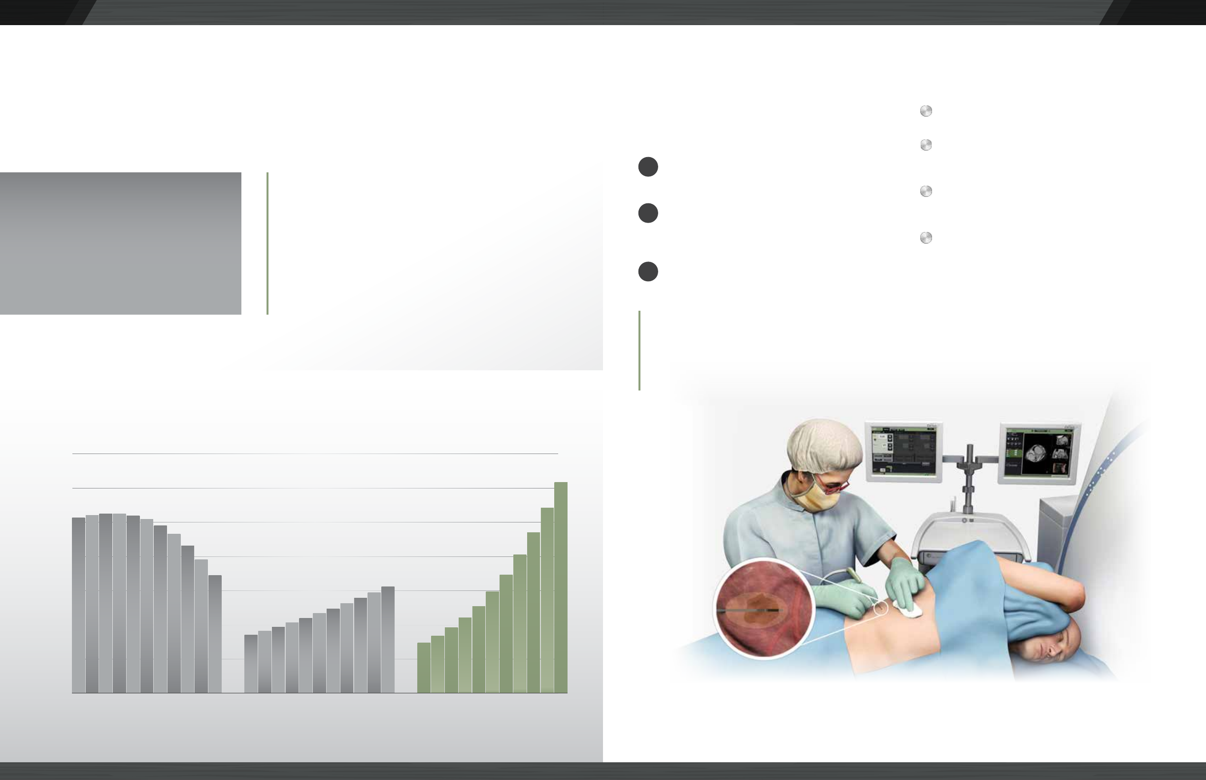
MICROWAVE LEADS ABLATION MARKET GROWTH
ABLATION PROCEDURE GROWTH 2013 - 2023
The U.S. ablaon market is growing rapidly due to the rising interest in minimally invasive opons. Microwave ablaon is
currently the fastest growing modality in the ablaon market, and will largely replace radiofrequency as the preferred ablaon
choice in the future.1Microwave ablaon is a minimally invasive outpaent
procedure that uses electromagnec waves to generate so
ssue necrosis in the lung, liver and kidney.
35000
30000
25000
20000
15000
10000
500
0
“The use of microwave ablaon will increase
substanally in the liver and lung indicaons
through 2023...Some companies, such as
NeuWave Medical, provide devices that are
specifically designed to effecvely ablate
ssue in delicate, aerated lung ssue.”
Radiofrequency Ablaon
-4.1%
CAGR 2013 - 2023
Cryoablaon
5.9%
Microwave Ablaon
14.8%
- Millennium Research Group 2015 1
MICROWAVE ABLATION
TO SURGE IN THE U.S.
The growth of microwave ablaon
connues to surge in the U.S. due to the
limitaons imposed by the alternave RF
ablaon technology.1
1. Intervenonal Oncology Devices / US/ 2015 Market Analysis Millennium Research Group, August 2014
Using CT or Ultrasound imaging, a small probe is
inserted percutaneously into lesion.
Electromagnec waves are delivered to the ssue,
producing friconal heang to generate ssue
necrosis at >60°C.
Ablaon Confirmaon™ soware, only from
Neuwave Medical®, is used to assess the technical
success of the ablaon procedure.
HOW MICROWAVE
ABLATION WORKS
BENEFITS OF
MICROWAVE ABLATION*
Supported by mulple Clinical Pracce Guidelines.
Strong efficacy and low complicaon rate versus
other common treatment modalies.1
Most procedures completed with 5-10 minutes of
ablaon me.
Many paents leave the hospital the same day2 as
the procedure with only a bandage at the probe
inseron point.
1
2
3
* All medical procedures carry potenal risks that should be discussed with paents prior to any microwave ablaon procedure. See the Certus 140 User Manual for a full list of warnings and cauons.
1. T. Ziemlewicz, et al, Percutaneous Microwave Ablaon of Hepatocellular Carcinoma with Gas-Cooled System: Inial Clinical Results with 107 Tumors. Journal of Vascular Intervenonal Radiology 2015;
26: 62–68. A. Moreland, et al, High-Powered Microwave Ablaon of T1a Renal Cell Carcinoma: Safety and Inial Clinical Evaluaon. Journal Of Endourology Sept. 2014; Volume 28, Number 9. B.T.
March, et al, Microwave ablaon for lung neoplasms: a retrospecve analysis of long term results. Journal of Vascular and Intervenonal Radiology 2014, Volume 25, Issue 3 , S97.
2. J. Horn, et al, Percutaneous Microwave Ablaon of Renal Tumors Using a Gas-Cooled 2.4-GHz Probe: Technique and Inial Results. Journal of Vascular Intervenonal Radiology 2014; 25: 448 – 453
Precision™ PR
probe inserted
into lesion
“From a paent’s perspecve, this procedure is really wonderful because it’s very
precise and the aermath impacts are negligible. I didn’t have any discomfort, I
was up the next day feeling fine and I was back in the classroom the day aer.”
- Kevin McSweeny, paent**

COMPUTER CONTROLLED SYSTEM DELIVERING HIGH
POWERED, FAST ABLATIONS
Touchscreen interface
Procedure data stored and
easily accessible
Daily call home system for
performance monitoring
Independent power and me control
on each channel during procedure
Highest power output = faster
ablaon (195W)
THE NEUWAVE MEDICAL® INTELLIGENT ABLATION SYSTEM
CO2 COOLED SYSTEM FOR THE SMALLEST GAUGE PROBE
AND TISSU-LOC™ TO REDUCE PROBE MIGRATION
17 gauge probe =
Less invasive procedures
Reduces probe migraon
Tissu-Loc™ technology “scks" the
probe in place, minimizing probe
migraon during imaging and
addional probe placement
NeuWave Medical®
17g
Compeon
13g
When it comes to ablang lesions, achieving the best outcomes for each individual paent is paramount. The totality of the
NeuWave Medical Intelligent Ablaon System allows you to tailor treatments to each paents’ clinical need and offers the only
integrated in-procedure confirmaon of your ablaon.
VERSATILE PROBE PORTFOLIO WITH MULTI-PROBE SYNCHRONY
FOR TAILORED TREATMENTS WITH 1, 2 OR 3 PROBES
PRECISION™ PR PROBE
≤ 4 cm single probe ablaons in any so ssue1
The only probe available with limited ablaon
length to limit necrosis of adjacent ssue,
including crical structures
For ≤ 4 cm, single probe ssue sparing ablaons1
1. Bovine liver ex-vivo: 65W, 5 minutes = 4.14 cm x 2.64 cm, 65W, 10 minutes = 4.65 cm x 3.06 cm, Bovine kidney ex-vivo: 65W, 5 minutes = 3.28 cm x 2.30 cm, 65W, 10 minutes = 3.82 cm x 2.60 cm,
Bovine lung ex-vivo: 65W, 5 minutes = 2.80 cm x 1.74 cm, 65W, 10 minutes = 4.08 cm x 3.22 cm
2. Bovine liver ex-vivo: 140W , 5 minutes = 6.0 cm x 3.7cm, 140W , 10 minutes = 7.0 cm x 4.0 cm, Bovine kidney ex-vivo: 140W , 5 minutes = 4.4 cm x 2.5 cm, 140W , 10 minutes = 6.4 cm x 3.4 cm, Bovine
lung ex-vivo: 140W , 5 minutes = 6.0 cm x 3.3 cm, 140W , 10 minutes = 6.0 cm x 3.9 cm
3. Laeseke et al. Mulple-Antenna Microwave Ablaon: Spaally Distribung Power Improves Thermal Profiles and Reduces Invasiveness. Journal of Intervenonal Oncology. 2009; 2(2):105-112. Brace et
al. Simultaneous Acvaon of Mulple Microwave Antennas Improves Circularity and Ablaon Zone Volume Compared to Sequenally Overlapping Ablaons. Presented at conference with
accompanying poster, WCIO 2014.
Rounder ablaons with no cleing
Mulple synchronized probes create larger and more
spherical ablaon zones than a single reposioned probe3
4
MAX™ LK AND LN PROBES
Tissue-tuned for large, single probe ablaons2
MAX LK single probe ablaons2
≤ 7 cm = liver, ≤ 6 cm = kidney
MAX LN single probe ablaon2
≤ 6 cm = lung
6
7,6
COMPETITION: 1 probe reposioned 3 mes
NEUWAVE MEDICAL®: 3 synchronized probes
10 min burn
5 min burn
2 min burn
1 cm eming point
PR PROBE BURN PATTERN: Tip-weighted probe with eming point 1 cm
from p. Ablaon encompasses p and then burns proximally (away).
2 cm eming point
10 min burn
5 min burn
2 min burn
LK & LN PROBE BURN PATTERNS: Ablaon progresses uniformly both
distally and proximally from the eming point 2 cm from p.
Increased confidence in covering target with
less reliance on precise probe placement
Consistent, predictable ablaons as waves
work in synchrony
MULTI-PROBE SYNCHRONY
ABLATION CONFIRMATION™ OFFERING THE ONLY INTEGRATED
IN-PROCEDURE CONFIRMATION TO SHOW YOU IF YOU GOT IT ALL!
Ablaon Confirmaon (AC) is a CT image processing soware that resides on a second, dedicated monitor aached to the NeuWave
Medical Intelligent Ablaon System. AC takes the uncertainty out of procedures by assisng physicians in idenfying ablaon probe
placement and confirming ablaon zones without having to leave the procedure room.
Pre-procedure and post-procedure scans registered
(overlaid) into one image to confirm technical success
of procedure.
Pre-procedure and post-procedure scans are on separate
monitors. Physician must "imagine" scans overlaid in aempt
to confirm technical success of procedure.
WITHOUT
ABLATION CONFIRMATION
WITH
ABLATION CONFIRMATION
BENEFITS OF ABLATION CONFIRMATION
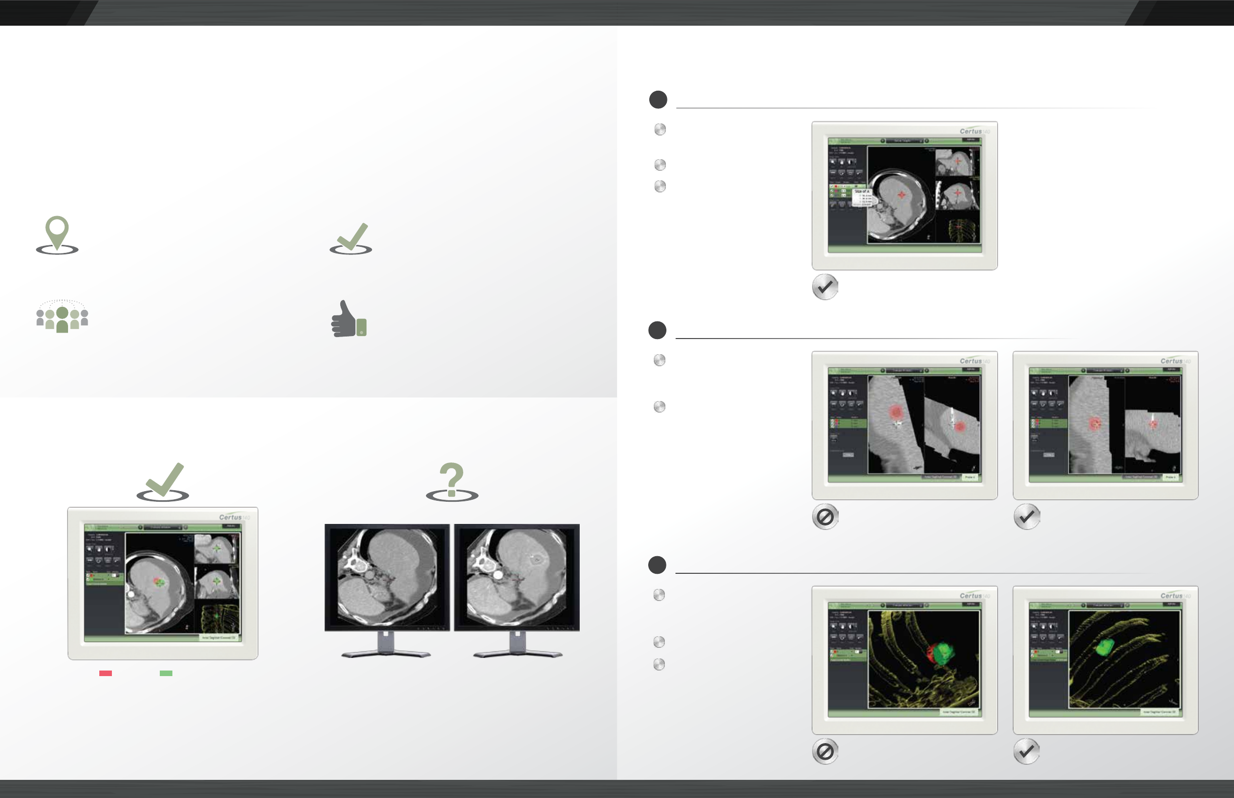
ABLATION CONFIRMATION™ IN 3 EASY STEPS
Pull set-up CT scan into
soware
Segment lesion
Define margin
DEFINE YOUR TARGET
1
Place probes, perform
CT scan and pull scan
into soware
Register scans to assess
probe placement in
relaonship to
idenfied target
Available Views:
2D, 3D, Periscope
and Needle
PLACE PROBES AND VERIFY
2
Perform ablaon, take
post-procedure CT scan
and pull scan into soware
Segment ablaon zone
Register ablaon scans
to assess and/or confirm
technical success of
procedure
Available Views:
2D, 3D
ABLATE AND CONFIRM
3
Probe missed lesion
Segmented lesion and margin defined
Probe placed in lesion
Ablaon doesn’t cover lesion and margin Ablaon covers lesion and margin
Confident Probe Placement
See the exact proximity of the probe to the
lesion so you can ablate with greater confidence
Confirm Complete Ablaon
Verify technical success of the procedure
by viewing combined target and ablaon
zone scans
Collaborate with Colleagues Remotely
Remote access allows colleagues to support
ablaon cases in real-me
Demonstrate Your Success
Clear 2D and 3D procedure images are
stored to PACS and can be shared among
paents, referrers and peers to showcase
your procedure success
Target Ablaon Zone Pre-Procedure Scan Post-Procedure Scan
2D View
Periscope and Needle View
3D View
